field manual of wildlife diseases
TRANSCRIPT
Field Manual ofWildlife DiseasesGeneral Field Procedures and Diseases of Birds
U.S. Department of the InteriorU.S. Geological Survey
I
Field Manual ofWildlife DiseasesGeneral Field Procedures and Diseases of Birds
Biological Resources DivisionInformation and Technology Report 1999–001
Milton Friend and J. Christian Franson, Technical Editors
Elizabeth A. Ciganovich, Editor
Phillip J. Redman, Design and layout
Rosemary S. Stenback, Illustrator
U.S. Department of the InteriorU.S. Geological Survey

II
Washington, D.C.
Any use of trade, product, or firm names in this publication is for descriptive purposes only and doesnot imply endorsement by the U.S. Government.
To order copies of this book telephone the Superintendent of Documents Telephone Order Desk at202-512-1800 Monday through Friday from 7:30 a.m. to 4:30 p.m. Eastern time. Or visithttp://www.gpo.gov/sales and enter the title in the Sales Product Catalog search box.
For additional information about this book:
USGSBiological Resources DivisionNational Wildlife Health Center6006 Schroeder RoadMadison, WI 53711U.S.A.World Wide Web: http://www.emtc.usgs.gov/nwhchome.html
For more information about the USGS and its products:
Telephone: 1-888-ASK-USGSWorld Wide Web: http://www.usgs.gov
Library of Congress Cataloging-in-Publication DataField manual of wildlife diseases : general field procedures and
diseases of birds / Biological Resources Division.p. cm. — (Information and technology report ; 1999–001)
Includes bibliographic referencesISBN 0-607-88096-1 (alk. paper)1. Birds–Diseases Handbooks, manuals. 2. Wildlife diseases
Handbooks, manuals, etc. I. Geological Survey (U.S.). BiologicalResources Division. II. Series.SF994.F54 1999639.9’78—dc21 99–25869
CIP
U.S. Department of the InteriorBruce Babbitt, Secretary
U.S. Geological SurveyCharles G. Groat, Director
Major funding support was provided by the U.S. Fish and Wildlife Service,Division of Federal Aid, Administrative Grant No. AP95-017.

III
Pho
to b
y M
icha
el S
amue
l
Dedication
We dedicate this Manual to the countless field biologists within the
U.S. Fish and Wildlife Service and the State wildlife agencies with whom
we have had the privilege of working for nearly a quarter-century. Their
endless assistance and devotion to the conservation of our Nation’s wild-
life resources has stimulated our own efforts to address wildlife health
issues and made those efforts more rewarding than we originally believed
was possible. We thank you for your efforts and hope that the material
provided within these pages will be useful to you in the days ahead.
Milton Friend and J. Christian Franson

IV
Acknowledgements
This publication was made possible through the generous contributions of time and effort by manyindividuals, and we offer our sincere thanks to all. The following people are particularly deserving ofrecognition. Ms. Debra Ackers endured much from us in typing draft manuscripts, making countlessadjustments to the drafts, organizing and maintaining project files, tracking the progress of the manycomponents of the Manual, arranging meetings among individuals involved with this project, andassisting in many other ways — always with a smile. Her contributions were invaluable. Dr. Louis N.Locke deserves special recognition for his technical input and laborious review of the draft manu-script. We thank Dr. James R. March and Ms. Barbara C. Scudder for contributing their time to readand comment on the final draft. Mr. Scott Hansen assembled the lists of chemicals and birds forAppendices E and G, respectively, and assisted us in locating many of the photographs used toillustrate the Manual. Ms. Kathryn Cleary, Ms. Karen Cunningham, and Mr. Harold Rihn providedtabulations of information from the National Wildlife Health Center database that were used in manyof the graphics and tables. We thank the following individuals for helpful and timely reviews of variousparts of the Manual: Dr. Donald Anderson, Mr. Tom Augspurger, Dr. Val Beasley, Dr. Rebecca Cole,Dr. Guy Connolly, Mr. Terry Creekmore, Mr. Doug Docherty, Mr. Monte Garrett, Dr. Robert Hallock,Dr. Larry Hansen, Dr. Wallace Hansen, Dr. Tuula Hollmén, Dr. David Jessup, Dr. Ken Langelier,Dr. Linda Lyon, Dr. Pierre Mineau, Dr. Patrick Redig, Dr. Milton Smith, Mr. Stanley Wiemeyer, andDr. Thierry Work. We apologize in the event that we have failed to acknowledge some individuals whohave assisted with this project over the extended period of time required for its completion.

V
Foreword
DO WILDLIFE DISEASES REALLY MATTER? The waterfowl manager who wakes up one morning to find ten thousanddead and dying birds in the marsh would think so. Yet virtually every wild bird and mammal harbors at least a few parasitesseemingly without obvious adverse consequences. Parasites, viruses, bacteria, and fungi are component parts of the ecosys-tems in which wildlife are found, but do not necessarily cause disease. Millennia of coevolution have engendered a modusvivendi that assures the survival of both host and parasite populations.
Then why the ten thousand sick and dying birds? Ecosystems are changing. Waterfowl are concentrated on shrinkingwetlands and remain there for longer periods of time, facilitating bird-to-bird spread of the bacteria that cause avian cholera.Or permitting the buildup of parasites in their hosts from a small, relatively benign number to massive numbers that causedisease and death. Water quality of wetlands changes, favoring the production of deadly botulinum toxin by bacteria and itsmobilization up the food chain to waterfowl. New, totally artificial habitats are created with unpredictable results. The ex-treme temperature, salinity, and other conditions of the Salton Sea have created an unusual ecosystem in which botulismoccurs in fish and in birds through biological cycles that are not yet understood. Wetland loss in southern California leavesfew alternative places for waterbirds to go, so they are attracted to the Salton Sea.
Behavior changes. Mallard ducks take up residence on the ponds and lakes of city parks and lose their migratory habits.They share these bodies of water with exotic species, such as Muscovy ducks that have also taken up residence there afterintroduction by people, setting the scene for outbreaks of duck plague, and creating the risk of spread to migratory waterfowlthat also use these areas. Raccoons and skunks become well adapted to urban life, bringing rabies and canine distemper withthem into the city.
The environment changes the physiology of wild animals. Human activity introduces into wildlife habitats chemicalcompounds that adversely affect physiological processes such as reproduction and immune responsiveness. These com-pounds become incorporated into the ecosystems, often becoming more concentrated as they move up food chains. Theireffects can influence wildlife populations. Some of these endocrine-disrupting chemicals, such as chlorinated hydrocarbons(DDE, PCBs), interfere with normal endocrine function by mimicking natural hormones, with resulting eggshell thinningand breakage. Effects of these chemical compounds on immune-system responses to infectious and parasitic agents are lesswell understood.
What to do? Incorporating disease-prevention measures into wildlife management practices requires more informationthan is usually available. The information-gathering process must begin in the field. Field biologists must monitor diseaseoccurrence. This Field Manual is a valuable aid in identifying the diseases that are likely to be present, and in giving guidanceon the gathering and treatment of specimens needed to establish the diagnosis in the laboratory.
But the wildlife field biologist is in a position to provide valuable information that goes beyond the collection of samplesfrom sick and dead individuals. Although diseased individuals are the basic unit of surveillance, the occurrence of diseasemust be put into ecological perspective. A careful description of the ecological setting in which the disease is occurring, andany changes that have occurred over time, are ultimately as important as a careful description of the lesions observed in theindividual, if the epidemiology of that disease is to be understood, and the disease prevented through sound wildlife-manage-ment practices.
It is my hope that the awareness of diseases affecting wildlife and the good disease-surveillance practices promoted by thismanual will spread throughout the range of the species we are trying to mange and protect. We must know more than we docurrently about disease occurrence throughout the ranges that the wildlife occupy. Many migratory species know nothing ofinternational boundaries. Neither do their diseases. Until we have a much more complete picture of the disease-environmentrelationships of the blue-winged teal from its nesting ground in Canada, its migration route through the United States andoverwintering areas in Central America or the Cienaga Grande de Santa Marta in Columbia, sound disease-prevention man-agement of that species will not be possible. Similar considerations exist for other species.
Thomas M. YuillMadison, WisconsinMay, 1999

VI
“Ingenuity, knowledge, and organization alter but cannot
cancel humanities vulnerability to invasion by parasitic forms
of life. Infectious diseases which antedated the emergence
of humankind will last as long as humanity itself, and will
surely remain, as has been hitherto, one of the fundamental
parameters and determinants of human history.”
(McNeill)
Photo by J.Christian Franson

VII
Introduction
I was first employed in the field of wildlife conservation in1956 as an assistant waterfowl biologist. Had I decided thento join some of my colleagues in preparing a manual aboutthe diseases of wild birds similar to this publication, the taskwould have been much simpler. The number of chaptersneeded would have been far less because some of the dis-eases described in this Manual were not yet known to existin free-ranging North American birds or, if they were known,they were not considered to be of much importance. This isespecially true for diseases caused by viruses; also, organo-phosphorus and carbamate pesticides had not come into wideuse. These types of differences are evident between this FieldManual of Wildlife Disease — General Field Procedures andDiseases of Birds and the Field Guide to Wildlife Diseases— General Field Procedures and Diseases of Migratory Birdsthat was published little more than a decade ago. The cur-rent Manual reflects both expanded knowledge about aviandiseases and an increase in both the occurrence of disease inwild birds and the variety of agents responsible for illnessand death of wild birds.
Landscape changes and environmental conditions that arerelated to them are a major factor associated with diseaseoccurrence in wild birds. The direct association betweenenvironment and human health has been recognized since
ancient times and was aptly stated by Louis Pasteur, “Themicrobe is nothing; the terrain everything.” Despite this welldocumented relationship, which serves as a basic founda-tion for addressing many human and domestic animal dis-eases, there has been little consideration of “the terrain” asa factor for diseases of wild birds. We must learn to “readthe terrain” in a manner similar to the teaching ofHippocrates and apply that knowledge to disease preven-tion or else the next edition of this Manual a decade fromnow will likely include another major expansion in the num-ber of diseases being addressed.
Although this Manual is much larger than the 1987 Gen-eral Field Procedures and Diseases of Migratory Birds thebasic format and “terrain” approach of the previous publi-cation were retained because of the positive comments thatwere received from its users. The format, the photographspreviously used, and most of Section 1, General Field Pro-cedures, have been basically retained, but the text for chap-ters about individual diseases (Sections 2 through 8) hasbeen extensively reworked. This Manual also has separatesections that address biotoxins and chemical toxins in addi-tion to major expansion of the number of individual dis-eases within the sections on bacterial, fungal, viral, and para-sitic diseases. The presentations in the various sections have
“When one comes into a city in which he is a stranger, he ought to consider its situation, how it lies as
to the winds and the rising of the sun; for its influence is not the same whether it lies to the north or to
the south, to the rising or to the setting sun. These things one ought to consider most attentively, and
concerning the waters which the inhabitants use, whether they be marshy and soft, or hard and
running from elevated and rocky situations, and then if saltish and unfit for cooking; and the ground,
whether it be naked and deficient in water, or wooded and well-watered, and whether it lies in a hollow,
confined situation, or is elevated and cold …From these things he must proceed to investigate
everything else. For if one knows all these things well, or at least the greater part of them, he cannot
miss knowing, when he comes into a strange city, either the diseases peculiar to the place, or the
particular nature of the common diseases, so that he will not be in doubt as to the treatment of the
diseases, or commit mistakes, as is likely to be the case provided one had not previously considered
these matters. And in particular, as the season and year advances, he can tell what epidemic disease
will attack the city, either in the summer or the winter, and what each individual will be in danger of
experiencing from the change of regimen.”
—Hippocrates, On Airs, Water, and Places, c. 400 B.C.
Facing page quote from:
McNeill, W.H., 1976, Plagues and peoples: Anchor Press/Doubleday, Garden City, N.Y., p. 291

VIII
been supplemented with introductory comments regardingthe subject area, and most sections have been highlightedwith descriptions of miscellaneous disease conditions thatmay interest users and readers.
As with the 1987 publication, the focus of this Manual ison conveying practical information and insights about thediseases in a manner that will help National Wildlife Refugemanagers and other field personnel address wildlife healthissues at the field level. The information represents a com-posite of our understanding of the scientific literature, ofour personal experiences with and investigations of the vari-ous diseases, and of information generously provided by ourcolleagues within the wildlife disease and related fields. Inpresenting this information, we have borrowed freely fromall of those sources. Because this is a synoptic field manualand not a textbook, literature citations are not provided insupport of statements. Only a small portion of the specificliterature that is the basis for the statements has been listed,and the supplementary reading lists are intended to provideentry into the scientific literature for more precise evalua-tion of specific topics.
The need to generalize and, thus, provide a practical over-view of complex biological situations often results in a lossof precision for some information. We have attempted to pro-vide detail where it is of significant importance and havebeen more general elsewhere. In all cases, we have attemptedto represent the information objectively and accurately. Forexample, Appendix E presents specific brain cholinesterasevalues that are supported by laboratory data for different birdspecies to provide a baseline against which others can makejudgements about mortality due to organophosphorus andcarbamate pesticides. In contrast, representation of the geo-graphic distribution, frequency of occurrence, and speciessusceptibility associated with specific diseases is of a gen-eral nature and is intended only for gross comparison. Thedifferences in these representations of general informationbetween the 1987 publication and this Manual are both apositive and a negative outcome of the last decade. Thesedifferences reflect enhanced information about disease inwild birds as a result of expanded study (a positive outcome),changes in disease patterns (a negative outcome due to ex-pansion of disease), and both, depending on the disease.
Current understanding about wild bird diseases is beingprovided by those with technical knowledge about disease
processes to those with technical knowledge and steward-ship and conservation of our wild bird resources. Commonlanguage has been used whenever possible to aid in this com-munication and to stimulate greater interest in wildlife dis-ease among others who may wish to read this Manual butwho may not be familiar with some of the terms. Technicalterms have been translated in a manner that we hope will beuseful for readers as they pursue additional subject matterdetail in the scientific literature. Technical terms have alsobeen inserted into the text and defined where they providevalue-added precision for the statements. It is my personalhope that a decade from now, when consideration is beinggiven to a revision of this Manual, that a great deal of thepreparation of the revision will be done by wildlife biolo-gists who have become practitioners in the art of diseaseprevention and control because of an enhanced understand-ing of disease ecology that we have all gained through ourcollective efforts. The transition hoped for is no greater thanother changes that have taken place since the 1987 publica-tion of the original Field Guide. At that time, the NationalWildlife Health Center (NWHC) was part of the Departmentof the Interior, U.S. Fish and Wildlife Service. Since then,the Center has become part of the Department of the Inte-rior, U.S. Geological Survey, Biological Resources Division.
My professional situation has also changed. Those famil-iar with the 1987 publication will note that I was Director ofthe NWHC when that publication became available. InDecember 1997, Secretary of the Interior Bruce Babbitt askedme to accept the challenge of coordinating the science ef-forts that will aid and guide decisions for managementactions to improve the health of the Salton Sea, California’slargest inland body of water. Recurring major disease eventsinvolving migratory birds at the Sea since 1994 have focusedpublic attention on it. These disease events became a cata-lyst for the expansion of efforts to improve the environmen-tal quality of the Sea, and in June 1998, a combinedNational Environmental Policy Act (NEPA)/CaliforniaEnvironmental Quality Act (CEQA) process was initiated topursue attainment of that goal. I officially became part ofthe multiagency effort to “Save the Salton Sea” with my re-assignment in April 1998 from Director of the NWHC toExecutive Director, Salton Sea Science Subcommittee.
Milton Friend

IX
Section 1 General Field Procedures
Chapter 1 Recording and Submitting Specimen History Data ...................................................................... 3
Chapter 2 Specimen Collection and Preservation ......................................................................................... 7
Chapter 3 Specimen Shipment .................................................................................................................... 13
Chapter 4 Disease Control Operations ........................................................................................................ 19
Chapter 5 Euthanasia .................................................................................................................................. 49
Chapter 6 Guidelines for Proper Care and Use of Wildlife in Field Research .............................................. 53
Section 2 Bacterial Diseases
Chapter 7 Avian Cholera .............................................................................................................................. 75
Chapter 8 Avian Tuberculosis ...................................................................................................................... 93
Chapter 9 Salmonellosis .............................................................................................................................. 99
Chapter 10 Chlamydiosis ............................................................................................................................. 111
Chapter 11 Mycoplasmosis .......................................................................................................................... 115
Chapter 12 Miscellaneous Bacterial Diseases ............................................................................................ 121
Section 3 Fungal Diseases
Chapter 13 Aspergillosis .............................................................................................................................. 129
Chapter 14 Candidiasis ................................................................................................................................ 135
Chapter 15 Miscellaneous Fungal Diseases ............................................................................................... 137
Section 4 Viral Diseases
Chapter 16 Duck Plague .............................................................................................................................. 141
Chapter 17 Inclusion Body Disease of Cranes ............................................................................................ 153
Chapter 18 Miscellaneous Herpesviruses of Birds...................................................................................... 157
Chapter 19 Avian Pox .................................................................................................................................. 163
Chapter 20 Eastern Equine Encephalomyelitis ........................................................................................... 171
Chapter 21 Newcastle Disease.................................................................................................................... 175
Table of Contents

X
Chapter 22 Avian Influenza .......................................................................................................................... 181
Chapter 23 Woodcock Reovirus .................................................................................................................. 185
Section 5 Parasitic Diseases
Chapter 24 Hemosporidiosis ....................................................................................................................... 193
Chapter 25 Trichomoniasis .......................................................................................................................... 201
Chapter 26 Intestinal Coccidiosis ................................................................................................................ 207
Chapter 27 Renal Coccidiosis ..................................................................................................................... 215
Chapter 28 Sarcocystis ................................................................................................................................ 219
Chapter 29 Eustrongylidosis ........................................................................................................................ 223
Chapter 30 Tracheal Worms ........................................................................................................................ 229
Chapter 31 Heartworm of Swans and Geese .............................................................................................. 233
Chapter 32 Gizzard Worms.......................................................................................................................... 235
Chapter 33 Acanthocephaliasis ................................................................................................................... 241
Chapter 34 Nasal Leeches .......................................................................................................................... 245
Chapter 35 Miscellaneous Parasitic Diseases ............................................................................................. 249
Section 6 Biotoxins
Chapter 36 Algal Toxins ............................................................................................................................... 263
Chapter 37 Mycotoxins ................................................................................................................................ 267
Chapter 38 Avian Botulism .......................................................................................................................... 271
Section 7 Chemical Toxins
Chapter 39 Organophosphorus and Carbamate Pesticides ........................................................................ 287
Chapter 40 Chlorinated Hydrocarbon Insecticides ...................................................................................... 295
Chapter 41 Polychlorinated Biphenyls ......................................................................................................... 303
Chapter 42 Oil .............................................................................................................................................. 309
Chapter 43 Lead .......................................................................................................................................... 317
Chapter 44 Selenium ................................................................................................................................... 335
XI
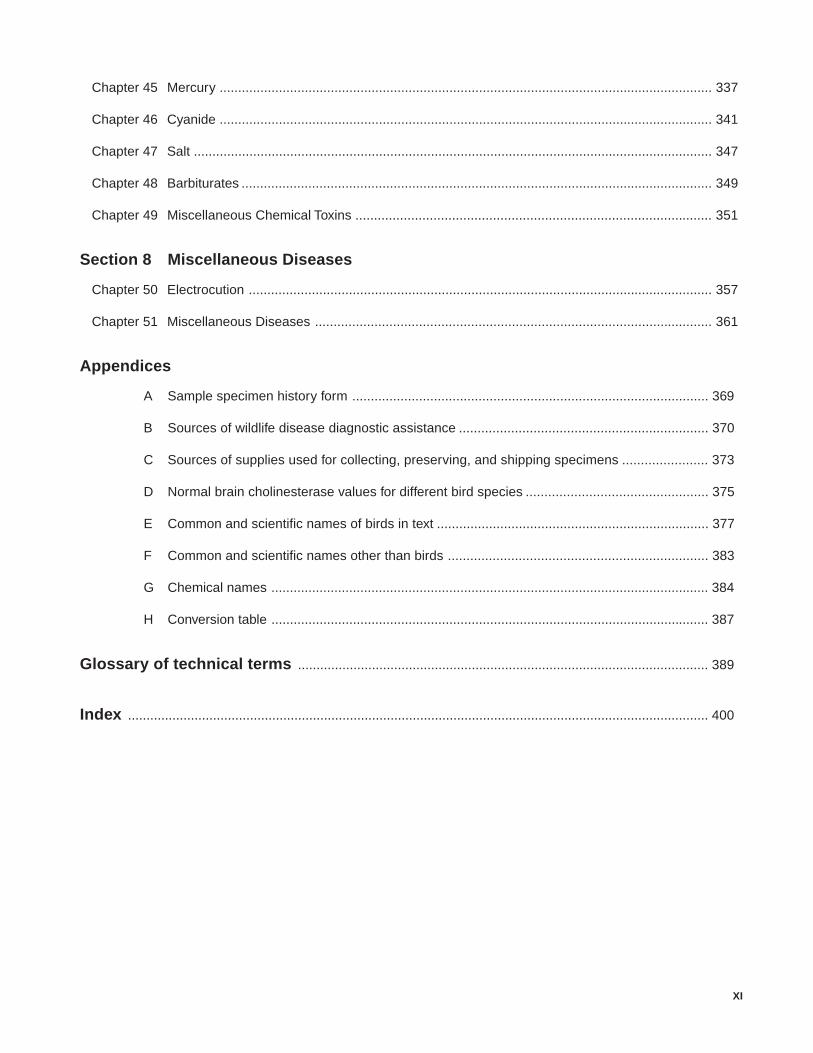
Chapter 45 Mercury ..................................................................................................................................... 337
Chapter 46 Cyanide ..................................................................................................................................... 341
Chapter 47 Salt ............................................................................................................................................ 347
Chapter 48 Barbiturates ............................................................................................................................... 349
Chapter 49 Miscellaneous Chemical Toxins ................................................................................................ 351
Section 8 Miscellaneous Diseases
Chapter 50 Electrocution ............................................................................................................................. 357
Chapter 51 Miscellaneous Diseases ........................................................................................................... 361
Appendices
A Sample specimen history form ................................................................................................ 369
B Sources of wildlife disease diagnostic assistance ................................................................... 370
C Sources of supplies used for collecting, preserving, and shipping specimens ....................... 373
D Normal brain cholinesterase values for different bird species ................................................. 375
E Common and scientific names of birds in text ......................................................................... 377
F Common and scientific names other than birds ...................................................................... 383
G Chemical names ...................................................................................................................... 384
H Conversion table ...................................................................................................................... 387
Glossary of technical terms ............................................................................................................... 389
Index ............................................................................................................................................................. 400

Introduction to General Field Procedures 1
Section 1Introduction to GeneralField Procedures
Recording and SubmittingSpecimen History Data
Specimen Collection andPreservation
Specimen Shipment
Disease Control Operations
Euthanasia
Guidelines for Proper Care andUse of Wildlife in Field Research
Dissecting a bird at the National Wildlife Health CenterPhoto by Phillip J. Redman
2 Field Manual of Wildlife Diseases: Birds
Introduction to General Field Procedures
A basic premise for the preparation of this Manual is thatdisease in free-ranging wildlife is of concern and that dis-ease prevention and control are desirable actions. However,these are not universally held perspectives. There are thosewho when confronted with disease outbreaks in free-rang-ing wildlife ask — “Why bother?” Also, the same individu-als who may reject the need for response to one situationmay demand a response to another situation. We acknowl-edge in this Manual the existence of this question by makingreference to it, but we do not offer a direct response. To doso would require this Manual to address the full spectrum ofindividually held values, perspectives, interests, and beliefswithin human society that form the basis for the underlyingissues which create the question of “why bother?” Those fac-tors would also need to be addressed within a context of thedifferent roles and responsibilities of public agencies, andwould need to include some additional considerations. Suchan undertaking is outside the scope and purpose of thisManual. Although no direct response is offered, readers willgain considerable information regarding disease occurrenceand impacts in the chapters that follow. This informationshould be of value in assisting readers to address the ques-tions of “why bother?” from their own set of values and in-terests.
Section 1 of the Manual provides basic information re-garding general field procedures for responding to wildlifedisease events. Field biologists provide a critical linkage indisease diagnostic work and greatly affect the outcome ofthe laboratory efforts by the quality of the materials and in-formation that they provide. The chapters in this section areoriented towards providing guidance that will assist field bi-ologists in gathering the quality of information and speci-mens that are needed. Readers will find information regard-ing what to record and how; guidance for specimen collec-tion, preservation, and shipment; and how to apply euthana-sia when such actions are warranted. Disease operations aremanaged at the field level and they can be aided by generalpreplanning that can be utilized when disease emergenciesarise; therefore, contingency planning is included within theDisease Control Operations chapter. Disease control tech-niques, including equipment that is used, are the main focusfor this highly illustrated chapter. Section 1 is concluded witha chapter about the proper care and use of wildlife in fieldresearch. The guidelines provided address the continual needto consider animal welfare in all aspects of wildlife manage-ment.
“Given the conspicuous role that diseases have played, and in many parts of the
world continue to play, in human demography, it is surprising that ecologists have
given so little attention to the way diseases may affect the distribution and
abundance of other animals and plants. Until recently, for example, ecology
textbooks had chapters discussing how vertebrate and invertebrate predators
may influence prey abundance, but in most cases you will search the index in
vain for mention of infectious diseases.” (May)
Quote from:
May, R.M., 1988, Conservation and disease: ConservationBiology, v. 2, no. 1, p. 28–30.
Recording and Submitting Specimen History Data 3
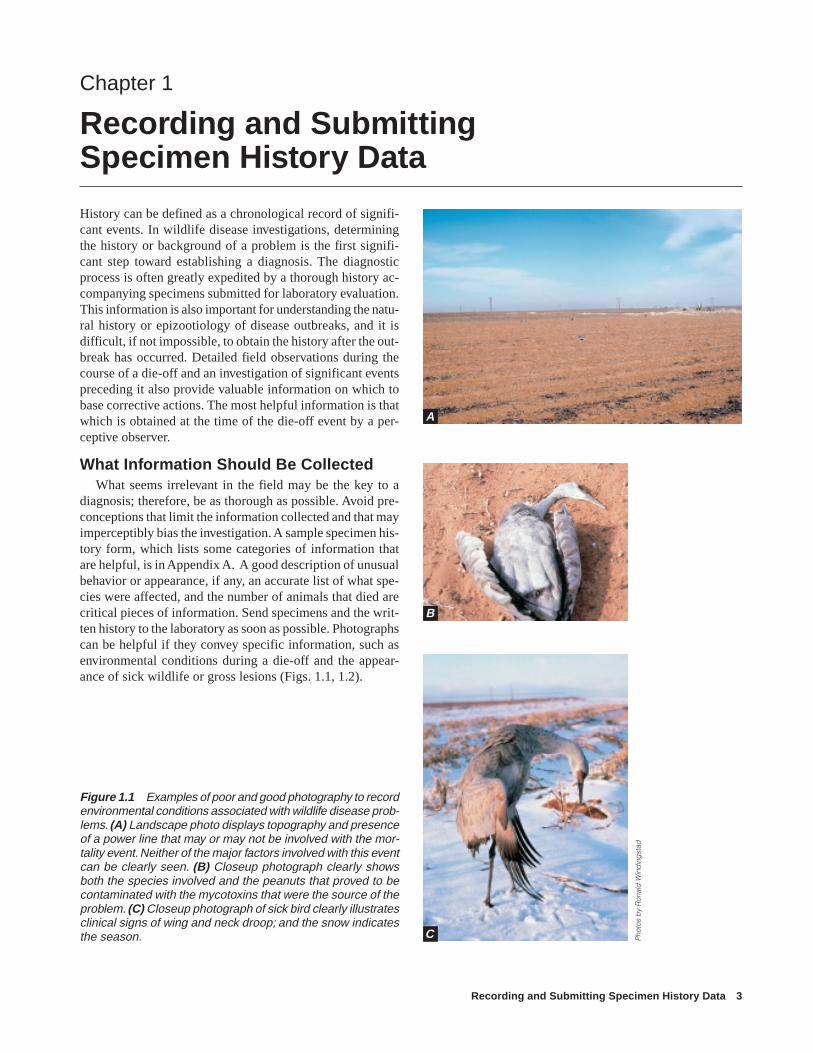
History can be defined as a chronological record of signifi-cant events. In wildlife disease investigations, determiningthe history or background of a problem is the first signifi-cant step toward establishing a diagnosis. The diagnosticprocess is often greatly expedited by a thorough history ac-companying specimens submitted for laboratory evaluation.This information is also important for understanding the natu-ral history or epizootiology of disease outbreaks, and it isdifficult, if not impossible, to obtain the history after the out-break has occurred. Detailed field observations during thecourse of a die-off and an investigation of significant eventspreceding it also provide valuable information on which tobase corrective actions. The most helpful information is thatwhich is obtained at the time of the die-off event by a per-ceptive observer.
What Information Should Be CollectedWhat seems irrelevant in the field may be the key to a
diagnosis; therefore, be as thorough as possible. Avoid pre-conceptions that limit the information collected and that mayimperceptibly bias the investigation. A sample specimen his-tory form, which lists some categories of information thatare helpful, is in Appendix A. A good description of unusualbehavior or appearance, if any, an accurate list of what spe-cies were affected, and the number of animals that died arecritical pieces of information. Send specimens and the writ-ten history to the laboratory as soon as possible. Photographscan be helpful if they convey specific information, such asenvironmental conditions during a die-off and the appear-ance of sick wildlife or gross lesions (Figs. 1.1, 1.2).
Chapter 1
Recording and SubmittingSpecimen History Data
Figure 1.1 Examples of poor and good photography to recordenvironmental conditions associated with wildlife disease prob-lems. (A) Landscape photo displays topography and presenceof a power line that may or may not be involved with the mor-tality event. Neither of the major factors involved with this eventcan be clearly seen. (B) Closeup photograph clearly showsboth the species involved and the peanuts that proved to becontaminated with the mycotoxins that were the source of theproblem. (C) Closeup photograph of sick bird clearly illustratesclinical signs of wing and neck droop; and the snow indicatesthe season. P
hoto
s b
y R
onal
d W
ind
ing
stad
B
C
A

4 Field Manual of Wildlife Diseases: Birds
The following basic information is helpful for diagnos-ing the cause and assessing the severity of a wildlife healthproblem. Waterfowl are used as an illustrative example.
Environmental FactorsDetermine if the start of mortality coincided with any
unusual event. Environmental changes such as storms, pre-cipitation, and abrupt temperature changes are potentialsources of stress that can contribute to disease outbreaks. Afood shortage may degrade the condition of birds and in-crease their susceptibility to disease. Water-level changes inan area may concentrate or disperse birds, alter the accessi-bility of toxins in food or water, or cause an invertebrate die-off that could lead to an avian botulism outbreak. Attempt todetermine whether or not biting insect populations have in-creased or if such insects are present, because some insectsare carriers of blood-borne infections in waterfowl.
The quality of the water used as a source for an impound-ment may contribute to disease or mortality; for example,poor water quality may contribute to avian botulism or maybe a primary cause of mortality if water contamination bytoxic materials and substances such as oil, which can affect
the integrity of feathers, is severe. Record recent pesticideapplications and other habitat or crop management practicesas well as previous disease problems in the area.
Estimating Disease OnsetWhen estimating the onset of disease, consider: (1) the
earliest date when on-site activities could have resulted inthe detection of sick or dead birds, if they were present, andthe actual date when diseased birds were first seen, and (2)the proportion of fresh carcasses compared with the numberof scavenged and decomposed carcasses. The abundance andtypes of scavengers and predators can be used to predict howlong carcasses remain in the area. Other useful informationabout the onset of mortality can be gained from noting anydifferences in plumage, including stage of molt, if present,between live and dead birds. Size differences between liveand dead nestlings and fledglings may also provide usefulinformation for comparison with known growth rates. Also,air, water, and soil temperatures will affect the speed of de-composition and they should be considered in assessing howlong birds have been dead. Include these observations in thehistory.
Figure 1.2 The observer may use photography to illustrate field observations associated with wildlife morbidity and mortality.(A) For example, when sick birds are left undisturbed or approached quietly, they often remain motionless along the water’sedge with their heads hanging down. When startled, these birds may attempt to escape by propelling themselves with theirwings across water (B) or land (C) but are unable to fly. (D) This bird has lost the use of its legs, a common occurrence with avianbotulism and certain toxins such as organophosphorus or carbamate compounds.
Pho
tos
by
Milt
on F
riend
BA
C D

Recording and Submitting Specimen History Data 5
Species AffectedMuch can be learned by knowing what species are dying.
Those species present but unaffected are especially impor-tant to note, because some diseases infect a narrow host rangeand others infect a wide variety of species. For example, duckplague affects only ducks, geese, and swans, but avian chol-era affects many additional species of water birds as well.Species with similar feeding habits may be dying as a resultof exposure to toxins, while birds with different food require-ments remain unaffected.
AgeSome disease agents may kill young birds but leave adults
unaffected because of age-related disease resistance; otherdiseases kill birds of all ages, although young or old birdsmay be more susceptible because of additional stress placedon these age groups. When toxins are involved, differencesin food habits may result in exposure of young birds, but notof adult birds, or vice versa.
SexSex differences in mortality may be apparent in colonial
nesters where females are incubating eggs, or in other situa-tions where the sexes are segregated.
Number Sick/Number DeadThe longer a disease takes to kill, the more likely it is that
significant numbers of sick birds will be found. For example,more sick birds will probably be observed during an avianbotulism die-off than during an outbreak of a more acutedisease such as avian cholera.
Clinical SignsWhen observing sick birds, describe the clinical signs in
as much detail as possible. Include any abnormal physicalfeatures and describe unusual behaviors, such as a sick bird’sresponse to being approached. Photographs (Fig. 1.2) of vari-ous behaviors or conditions associated with a disease can beespecially useful and should be included with the history.
Population at RiskTry to determine what species, and in what numbers, are
in the vicinity of the die-off. This information can provideclues about the transmissibility of disease, and it may be use-ful during control efforts.
Population MovementRecord recent changes in the number of birds in the area,
as well as the species present. In particular note the presenceof endangered species. If bird numbers have increased, tryto determine where they came from; if bird numbers havedecreased, attempt to determine where they have gone. Thiscan often be accomplished when population movements arebeing monitored for census, hunting forecasts, and other
purposes. State, Federal, and private refuge personnel andother natural resources managers are good primary sourcesof information.
Specific Features of Problem AreasDescribe the location of a die-off so that a relatively spe-
cific area can be identified on a road map. Also include anyavailable precise location data, such as global positioninginformation or data that will facilitate entering of specificlocations into geographical information system databases.Describe the problem area in terms that are sufficientlygraphic so that someone with no knowledge of it can visual-ize its major characteristics, such as topography, soil, veg-etation, climate, water conditions, and animal and humanuse.
Example description of die-off location
The problem area is a 10-acre freshwater pond locatedin Teno County, North Carolina, 1/2 mile east of CountyKV, 5 miles north of Highway 43. The pond has anaverage water depth of 6–12 feet and a sandy substrate.Vegetation around the pond border is bullbrush andreed canary grass. The surrounding uplands are essen-tially flat for one-half mile in all directions and lie fal-low, covered with grasses and some shrubs. The areais coastal with enough relief to prevent saltwater in-trusion into the pond even during major storms.Weather for the past 2 weeks has been pleasant andthere has been no precipitation. Daytime temperaturesare currently in the mid-80s (°F) and evening tempera-tures in the 70s. This is an isolated body of freshwaterwith good clarity, and sustains several hundred water-fowl, gulls, and small numbers of wading birds andshorebirds, and healthy warm water fish and amphib-ian populations. Cattle graze the adjacent area. Thereare no residential or industrial buildings within 1 mileof the site. Human visitation is frequent for bird watch-ing, fishing, and hiking. Companion animals such asdogs are allowed on the area.
Identify where sick and dead birds are found. Especiallynote the locations of groups of dead birds and any differ-ences of habitat where dead and sick birds are found. Birdsfound in agricultural fields may be dying of pesticide expo-sure, birds with more chronic toxicoses usually seek densecover, and birds dying of acute diseases may be found in avariety of situations. Check any relation between specificbird use of the area and the location of affected birds, suchas roost sites, loafing areas, and feeding sites.
If followup investigations are conducted after specimenshave been submitted, summarize the findings and observa-tions of those investigations in a supplemental report to theoriginal history. Maintain a copy of the new report in station

6 Field Manual of Wildlife Diseases: Birds
files, and provide a copy to the diagnostic laboratory wherethe specimens were sent. Both reports should contain thedates of the investigations, whether air or ground searcheswere performed, the number of investigators and the timespent on the investigation, the weather conditions, and thetime of day when the site was investigated.
The insight provided by good specimen history data andby field observations is invaluable to disease specialists. Thisinformation enhances understanding of the ecology of dis-ease, thereby serving as a basis for developing ways to pre-vent future die-offs or to reduce the magnitude of losses thatmight otherwise occur.
J. Christian Franson
Supplementary ReadingWobeser, G.A., 1994, Investigation and management of disease in
wild animals: New York, N.Y., Plenum Press, 265 p.

Specimen Collection and Preservation 7
Specimens are used to provide supporting information lead-ing to the diagnosis of a cause of disease or death. A speci-men may be an intact carcass, tissues removed from carcasses,parasites, ingested food, feces, or environmental samples.The specimen should be as fresh and undamaged as pos-sible.
Choosing a SpecimenAn entire, fresh carcass is the best specimen to submit to
the laboratory for diagnosis. This allows the diagnosticianto assess all of the organ systems and to use appropriate or-gans for different diagnostic tests. Obtain the best specimenspossible for necropsy; decomposed or scavenged carcassesare usually of limited diagnostic value. A combination ofsick animals, animals that were euthanized after clinical signswere observed and recorded, and some of the freshest avail-able carcasses compose an ideal specimen collection. Themethod of euthanasia should not compromise the diagnosticvalue of the specimen (see Chapter 5, Euthanasia). More thanone disease may be affecting the population simultaneously,and the chances of detecting multiple diseases will be maxi-mized if both sick and dead animals are collected. Speci-mens submitted should be representative of the species in-volved. If more than one species is affected, collect severalspecimens of each species; try to obtain a minimum of fivespecimens per species.
Tissue CollectionThe primary consideration when collecting carcasses or
tissues for diagnosis should be personal safety. Some wild-life diseases are transmissible to humans, and every carcassshould be treated as a potential health hazard. Wear dispos-able rubber or plastic gloves, coveralls, and rubber boots. Ifgloves are not available, inverted plastic bags may be used(Fig. 2.1). Before leaving an area where carcasses are beingcollected, double-bag used gloves and coveralls, and disin-fect boots and the outside of plastic bags with a commercialdisinfectant or a 5 percent solution of household chlorinebleach. Also, double-bag specimens in plastic before remov-ing them from the area. These precautions will help protectthe people in the field and minimize transmission of diseaseto unaffected wildlife populations.
If it is impossible to submit an entire carcass for diagno-sis, appropriate organs must be removed from specimens. Ifpossible, do not dissect carcasses in the field without firstconsulting disease specialists about methods of dissectingand preserving tissues or parasites or both. Assistance canbe obtained from a variety of sources (Appendix B). It is
Chapter 2
Specimen Collection and Preservation
Figure 2.1 Use a plastic bag to protect hands from directcontact with animal tissues during the collection of specimensif plastic or other waterproof gloves are not available. (A) Graspbag at the bottom and (B) with other hand pull open end downover hand holding bag (C). Repeat for the “unbagged” hand.Reversing this process when handling small specimens willautomatically place specimens in the bag, which then needonly be sealed and put into a second bag for packaging andshipment.
A
B
C
8 Field Manual of Wildlife Diseases: Birds
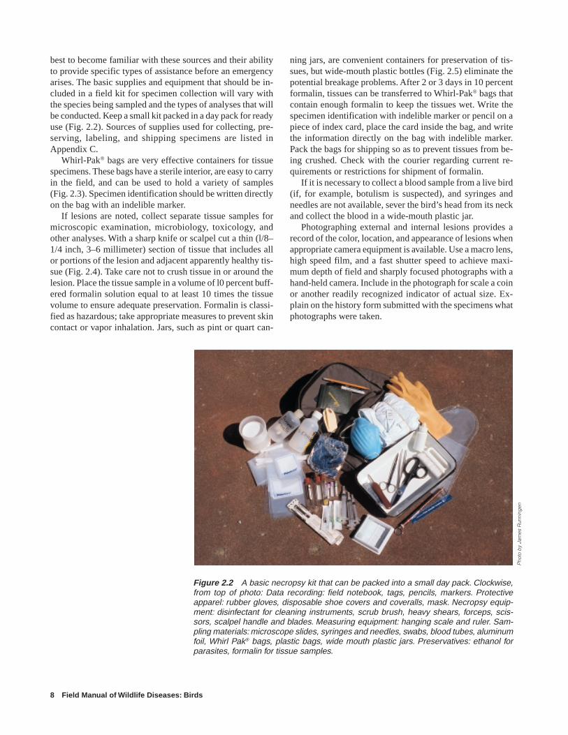
best to become familiar with these sources and their abilityto provide specific types of assistance before an emergencyarises. The basic supplies and equipment that should be in-cluded in a field kit for specimen collection will vary withthe species being sampled and the types of analyses that willbe conducted. Keep a small kit packed in a day pack for readyuse (Fig. 2.2). Sources of supplies used for collecting, pre-serving, labeling, and shipping specimens are listed inAppendix C.
Whirl-Pak® bags are very effective containers for tissuespecimens. These bags have a sterile interior, are easy to carryin the field, and can be used to hold a variety of samples(Fig. 2.3). Specimen identification should be written directlyon the bag with an indelible marker.
If lesions are noted, collect separate tissue samples formicroscopic examination, microbiology, toxicology, andother analyses. With a sharp knife or scalpel cut a thin (l/8–1/4 inch, 3–6 millimeter) section of tissue that includes allor portions of the lesion and adjacent apparently healthy tis-sue (Fig. 2.4). Take care not to crush tissue in or around thelesion. Place the tissue sample in a volume of l0 percent buff-ered formalin solution equal to at least 10 times the tissuevolume to ensure adequate preservation. Formalin is classi-fied as hazardous; take appropriate measures to prevent skincontact or vapor inhalation. Jars, such as pint or quart can-
Figure 2.2 A basic necropsy kit that can be packed into a small day pack. Clockwise,from top of photo: Data recording: field notebook, tags, pencils, markers. Protectiveapparel: rubber gloves, disposable shoe covers and coveralls, mask. Necropsy equip-ment: disinfectant for cleaning instruments, scrub brush, heavy shears, forceps, scis-sors, scalpel handle and blades. Measuring equipment: hanging scale and ruler. Sam-pling materials: microscope slides, syringes and needles, swabs, blood tubes, aluminumfoil, Whirl Pak® bags, plastic bags, wide mouth plastic jars. Preservatives: ethanol forparasites, formalin for tissue samples.
ning jars, are convenient containers for preservation of tis-sues, but wide-mouth plastic bottles (Fig. 2.5) eliminate thepotential breakage problems. After 2 or 3 days in 10 percentformalin, tissues can be transferred to Whirl-Pak® bags thatcontain enough formalin to keep the tissues wet. Write thespecimen identification with indelible marker or pencil on apiece of index card, place the card inside the bag, and writethe information directly on the bag with indelible marker.Pack the bags for shipping so as to prevent tissues from be-ing crushed. Check with the courier regarding current re-quirements or restrictions for shipment of formalin.
If it is necessary to collect a blood sample from a live bird(if, for example, botulism is suspected), and syringes andneedles are not available, sever the bird’s head from its neckand collect the blood in a wide-mouth plastic jar.
Photographing external and internal lesions provides arecord of the color, location, and appearance of lesions whenappropriate camera equipment is available. Use a macro lens,high speed film, and a fast shutter speed to achieve maxi-mum depth of field and sharply focused photographs with ahand-held camera. Include in the photograph for scale a coinor another readily recognized indicator of actual size. Ex-plain on the history form submitted with the specimens whatphotographs were taken.
Pho
to b
y Ja
mes
Run
ning
en

Specimen Collection and Preservation 9
A
Figure 2.4 Tissue sample collection for microscopic exami-nation. (A) Tissue sample should include lesion, such as spotsin liver, plus some apparently healthy tissue. The sample mustbe no thicker than 1/4 inch to ensure adequate chemical fixa-tion by preservative. Use as sharp an instrument as possible(scalpel, knife, razor) for a clean cut. (B) Place tissue sampleinto container of 10 percent buffered formalin or other suitablefixative or preservative. The volume of formalin in the con-tainer should be about 10 times the amount of tissue sample.(C) Complete the process by securing the lid and properlylabeling the container.Figure 2.5 Plastic bottles used for tissue
specimens. Regardless of size or shape,specimen bottles should have a wide mouthand threaded caps for secure closure.
Figure 2.3 Using Whirl-Pak® bag for specimen collection.(A) Remove top at perforation. (B) Open bag by simultaneouslypushing the protruding wire-reinforced tabs toward the centerto insert the specimen and any appropriate preservative.(C) Close bag by pulling on tabs and then twirling bag whileholding tabs. (D) Secure the closure by folding tabs aroundbags and label bag with type of specimen, date, and any iden-tifying numbers.
B
C
D
A B
C

10 Field Manual of Wildlife Diseases: Birds
Figure 2.6 Dissecting a duck carcass: (A) incision line; (B) reflect the skin to expose the underlying anatomy; (C) make atransverse abdominal cut below the breast muscle; (D) extend cut through the ribs and wishbone; (E) remove breast plate; (F)dissect out heart; (G) remove liver; and (H) tie off and remove the gastrointestinal tract.
CAbdominaltransverse
cut
FRemove heart
GRemove liver
ERemove
breast plate
Trachea
Syrinx
Heart
Liver
Gizzard
BReflected
skin
DVent to bill
incision
Esophagus
AIncision
line

Specimen Collection and Preservation 11
Avian Dissection
When dissecting a bird, it is always advisable towear protective clothing, particularly disposablegloves. To begin, insert a scalpel or a knife to make amidline incision through the skin of the breast (Fig2.6 A). Take care not to penetrate the body cavity,particularly in the abdominal region. Continue theskin incision to the vent and to the base of the bill.Reflect the skin away from the neck, breast, and ab-dominal areas. (B) Use the thumb and the first fin-ger of each hand to reflect the skin to expose theunderlying tissues. It is easiest to place the thumband the first finger of each hand along the incisionline in the breast area and then push and gently pullthe skin to the side. When an opening in the skin hasbeen established, work towards the bill and then thevent. (C) With a sharp blade, make a shallow trans-verse incision just below the breast muscles and ster-num. (D) Insert the thumb of one gloved hand intothe incision along the midpoint of the sternum andapply a slight pressure upwards. With a scissors inthe other gloved hand, carefully cut through the ribsextending the cut on each side of the breast throughthe area of the wishbone. (E) Gently separate thebreastplate from the carcass; use a scissors or otherinstrument to sever any connections and push asidethe air sacs. (F) Dissect out the heart without cuttinginto other tissues. (G) Gently remove the liver andcarefully cut away its area of connection with othertissues. (H) Tie off the gastrointestinal tract near thethroat area, cut the esophagus above the tied-off area,and gently remove the entire gastrointestinal area.
Avian Anatomy
Figure 2.6 illustrates organs and tissues that mayexhibit various lesions and that may be sampled for
the diagnosis of disease agents described in thisManual. Species variation may result in
some differences in the appearance andrelative size of particular organs and
tissues, but their location will besimilar among species. Notable dif-ferences between the types of spe-cies illustrated are the small flatspleen in normal ducks and thelarger oval spleen in pheasants.Also, pheasants have a crop andducks do not; instead, the areajust forward of the gizzard (theproventriculus) is more prom-inant in waterfowl.
HRemove
gastrointestinaltract
Intestinaltract
OvaryKidneyOviduct
TracheaEsophagus
CropLung
HeartLiver
Gall bladder
ProventriculusSpleen
Gizzard
PancreasSmall intestine
Large intestineCecumCloaca
Vent
Cut
Tie off topreventcontents fromleaking out,and separatefrom bodyabove thetied-offarea

12 Field Manual of Wildlife Diseases: Birds
Labeling SpecimensProper labeling, maintaining label readability, and pre-
venting label separation from specimens are as critical asproper specimen selection and preservation. The label shouldbe as close to the specimen as possible; for example, a labelshould be attached to a carcass, attached to a tube of blood,or placed within the vial of preservative with a parasite.Double labeling, or placing a label on the outside of a plasticbag holding the specimen whenever practical, is worth theeffort. The double labeling prevents confusion and potential
errors in specimen records at the diagnostic laboratory whenspecimens are received from multiple carcasses. Manila tagscan be used, but take care to prevent their exposure to largeamounts of fluids that may destroy the tag; tag destructioncan be reduced by using tags with high rag content or evenlinen tags. Use soft lead pencil or waterproof ink on thesetags; do not use ballpoint pen, nonpermanent ink, or hardlead pencil. The most durable tag is made of soft metal, suchas copper or aluminum, and can be inscribed with ballpointpen, pencil, or another instrument that leaves an impressionon the tag.
CarcassIdentify each carcass with a tag fastened with wire to a
leg (Fig. 2.7). If tags are not available, use a 3- by 5-inchcard placed inside a plastic bag within the bag holding thecarcass. Information on the tag should include the name, ad-dress, and telephone number of the submitter, collection site,species; whether the animal was found dead or was eutha-nized (indicate method); and a brief summary of any clinicalsigns. Place each tagged carcass in a separate plastic bagand seal the bag.
Tissues and OrgansWhen a specimen is in a plastic bottle, jar, or tube, wrap a
piece of adhesive or masking tape entirely around the con-tainer and use an indelible marker to write on the tape. Listthe type of animal from which the sample was taken, thekind of tissue, and the date the sample was taken. When plas-tic bags are used as the first containers for tissues, they shouldbe labeled with the same information directly on the bag. Donot insert tags inside containers with tissues and organs col-lected for microbiological or chemical analyses because thetag or the ink on it may contaminate the specimen. Whenchemically resistant tags are available, insert the tags intocontainers with preservatives such as formalin or alcohol.
Specimen PreservationChill or freeze all specimens, depending on how long it
will take to ship to a diagnostic laboratory. Freezing reducesthe diagnostic usefulness of carcasses and tissues, but if speci-mens must be held for 2 or more days, freezing the speci-mens as soon as possible after collecting them minimizestheir decomposition. Formalin-fixed tissues should not befrozen. See Chapter 3, Specimen Shipment, for detailed in-structions for packing and shipping specimens.
J. Christian Franson(All illustrations in this chapter are by Randy Stothard Kampen, with the exception of
Figure 2.6)
Supplementary ReadingRoffe, T.J., Friend, M., and Locke, L.N., 1994, Evaluation of
causes of wildlife mortality, in Bookhout, T.A., ed., Researchand Management Techniques for Wildlife and Habitats (5):Bethesda, Md., The Wildlife Society, p. 324–348.
Wobeser, G.A., 1997, Necropsy and sample preservation tech-niques, in Diseases of wild waterfowl (2nd ed): New York,N.Y., Plenum Press, p. 237–248.
Figure 2.7 Proper tagging of specimen. History of the speci-men (see text for details) should be placed on back of tag.

Specimen Shipment 13
Procedures for shipping specimens vary with different dis-ease diagnostic laboratories. Therefore, it is important tocontact the receiving laboratory and obtain specific shippinginstructions. This will facilitate processing of specimens whenthey reach the laboratory and assure that the quality of speci-mens is not compromised. Time spent on field investigation,specimen collection, and obtaining an adequate history willbe of little value if specimens become contaminated, decom-posed, or otherwise spoiled during shipping to the diagnos-tic laboratory.
There are five important consider-ations for proper specimen shipment:(l) prevent cross-contamination fromspecimen to specimen, (2) prevent de-composition of the specimen, (3) pre-vent leakage of fluids, (4) preserve in-dividual specimen identity, and (5)properly label the package. Basic sup-plies needed for specimen shipment areshown in Fig. 3.l.
Preventing Breakage andLeakage
Isolate individual specimens fromone another by enclosing them in sepa-rate packages such as plastic bags. Pro-tect specimens from direct contact withany coolant used (e.g., wet ice or dryice), and contain all materials withinthe package so that leakage to the out-side of the shipment container is pre-vented if breakage occurs (e.g., bloodtubes) or materials thaw (wet ice andfrozen carcasses) due to transit delays.
Containing SpecimensPlastic bags should be strong
enough to resist being punctured bymaterials contained within them andfrom contact with other containerswithin the package.
Styrofoam® coolers, shipped incardboard boxes, are useful for their in-sulating and shock absorbing qualities.Styrofoam® at least 1–inch thick ispreferred. When possible, selectStyrofoam® coolers that have straightsides. Coolers that are wider at the top
Chapter 3
Specimen Shipment
Figure 3.1 Basic specimen shipment supplies.
than at the bottom are more likely to break during transitthan those with straight sides. Fill the space between theoutside of the Styrofoam® cooler and the cardboard box withnewspaper or other packing material to avoid cooler break-age (Fig. 3.2). If coolers are not available, cut sheets ofStyrofoam® insulation to fit the inside of cardboard boxes.
The cardboard box protects the Styrofoam® cooler frombeing crushed during transit and serves as containment forthe entire package (Fig. 3.3). The strength of the box should

14 Field Manual of Wildlife Diseases: Birds
Figure 3.2 Proper packing to prevent Styrofoam® coolersfrom becoming crushed during transit. Place the sealedStyrofoam® cooler in a sturdy cardboard box. Use crumplednewspaper or other packing material to fill all space betweenthe cooler and the box.
Figure 3.3 This Styrofoam® coolerwas not packaged in a cardboard boxfor shipping.
Figure 3.4 Chemical coolants are available in (A) soft plas-tic, (B) hard plastic, and (C) metal containers.
be consistent with the weight of the package. Cardboardboxes are not needed when hard plastic or metal insulatedchests are used for specimen shipment, but boxes can be usedto protect those containers from damage and to provide asurface for attaching labels and addresses to the shipment.
Cooling and RefrigerationChemical ice packs (Fig. 3.4) are preferable to wet ice
because their packaging prevents them from leaking whenthey thaw. Ice cubes or block ice may be used if leakage canbe prevented. This can be accomplished most easily by fill-ing plastic jugs such as milk, juice, and soda containers withwater and freezing them. The lids of these containers shouldbe taped closed to prevent them from being jarred open dur-ing transit.
Use dry ice to keep materials frozen, but do not use it toship specimens that should remain chilled because it willfreeze them. Also, the carbon dioxide given off by dry icecan destroy some disease agents; this is of concern whentissues, rather than whole carcasses, are being shipped. Ship-ment of dry ice, formalin, and alcohol is regulated and shouldbe cleared with the carrier before shipping.
Preparing Specimens for Shipment to theNational Wildlife Health Center (NWHC)
Other disease diagnostic laboratories may require minorvariations in shipping procedures.
1. Call the NWHC (608-270-2400) to determine the op-timal type and number of specimens for diagnostic proce-dures, how these specimens are best preserved during transit(whether they should be chilled or frozen), and when theyshould be shipped. In most cases, the NWHC requests thatspecimens be shipped the same day or within 24 hours.
2. Double-bag carcasses (Fig. 3.5) and place them in aStyrofoam® cooler lined with a plastic bag. When both fro-zen and fresh whole carcasses are shipped in the same con-tainer, the frozen carcasses can be used as a refrigerant tokeep the fresh carcasses chilled. This can be accomplishedby interspersing individually bagged frozen carcasses amongthe individually bagged fresh carcasses or by placing the freshcarcasses between two layers of frozen carcasses (Fig. 3.6).Blood tubes and other breakable containers of uniform sizecan be protected by packing them in a common plastic bagthat is sealed within a metal can or a hard plastic containerwith a lid (Fig. 3.7). Pack any space around the specimencontainers within the can (side and top) with paper or someother absorbent material to prevent jarring that could causebreakage and to collect fluids if tubes do break. Seal the canwithin a plastic bag before placing it in the Styrofoam® cooler.
3. When using chemical ice packs, intersperse themamong specimens; place within the Styrofoam® containerother types of coolants in locations that will provide maxi-mum cooling for all contents or, if dry ice is used, will keepeverything frozen (Fig. 3.8). Fill all empty space within the
A B C
Pho
tos
by
Jam
es R
unni
ngen
Specimen Shipment 15
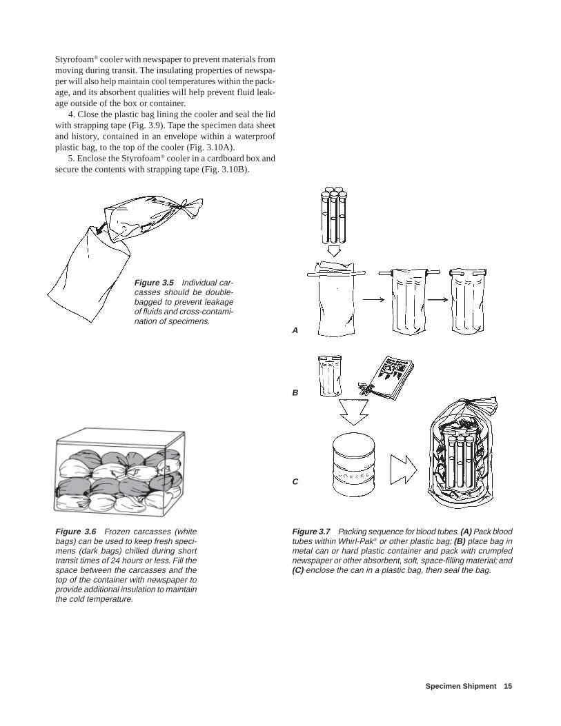
Figure 3.5 Individual car-casses should be double-bagged to prevent leakageof fluids and cross-contami-nation of specimens.
Styrofoam® cooler with newspaper to prevent materials frommoving during transit. The insulating properties of newspa-per will also help maintain cool temperatures within the pack-age, and its absorbent qualities will help prevent fluid leak-age outside of the box or container.
4. Close the plastic bag lining the cooler and seal the lidwith strapping tape (Fig. 3.9). Tape the specimen data sheetand history, contained in an envelope within a waterproofplastic bag, to the top of the cooler (Fig. 3.10A).
5. Enclose the Styrofoam® cooler in a cardboard box andsecure the contents with strapping tape (Fig. 3.10B).
Figure 3.6 Frozen carcasses (whitebags) can be used to keep fresh speci-mens (dark bags) chilled during shorttransit times of 24 hours or less. Fill thespace between the carcasses and thetop of the container with newspaper toprovide additional insulation to maintainthe cold temperature.
Figure 3.7 Packing sequence for blood tubes. (A) Pack bloodtubes within Whirl-Pak® or other plastic bag; (B) place bag inmetal can or hard plastic container and pack with crumplednewspaper or other absorbent, soft, space-filling material; and(C) enclose the can in a plastic bag, then seal the bag.
A
B
C

16 Field Manual of Wildlife Diseases: Birds
Figure 3.9 Closing a specimen container. (A) Secure thelarge plastic bag containing the specimens by tying the top;(B) close the container lid and (C) secure the container withseveral bands of strapping tape.
Figure 3.10 Completing the packaging process. (A) Tapespecimen data sheet and history, contained in an envelopewithin a waterproof plastic bag, to top of cooler. (B) Place coolerin cardboard box, secure box with several bands of strappingtape, and secure another copy of the specimen data sheet tothe outside of the box. If the specimens were placed inside aStyrofoam® cooler, then use crumpled newspaper or otherpacking material to fill all spaces between the cooler and thebox.
A
B
C
A
B
Figure 3.8 Packing specimens for shipment when (A) icepacks (B) wet ice, and (C) dry ice are used as coolants. Notethat the shipping container is always lined with a large plasticbag.
A
B
C

Specimen Shipment 17
Federal Shipping Regulations forPackaging and Labeling
Your packaging and labeling of specimens must conformto the following regulations.
The Code of Federal Regulations (CFR) states under 50CFR Part 14 of Fish and Wildlife Regulations that contain-ers with wildlife specimens must bear the name and addressof the shipper and consignee, and a list of the species andnumbers of each species must be conspicuously marked onthe outside of the container. You may instead conspicuouslymark the outside of each package or container with the word“wildlife” or the common names of the species containedwithin the package. Secure an invoice or packing list thatincludes the name and address of the consignee and shipperand that accurately states the number of each species con-tained in the shipment to the outside of one container in theshipment.
In addition to Fish and Wildlife Service regulations, theinterstate shipment of diagnostic specimens is subject to ap-plicable packaging, labeling, and shipping requirements fordisease-causing etiologic agents (42 CFR Part 72). Theseregulations do not require you to identify diagnostic speci-mens as etiologic agents when the disease agent is not knownor is only suspected. However, all specimen packages sentto the NWHC should be prominently labeled with the words“DIAGNOSTIC SPECIMENS.” You can meet packagingrequirements under 42 CFR Part 72 by following recommen-dations 2 through 5 above for enclosing specimens withintwo containers before enclosing them within the package.
Hazardous Materials Regulations of the Department ofTransportation apply whenever dry ice is contained withinthe shipping container (49 CFR Part 172, 173, 175). Alwayscall the carrier ahead of time for the current shipping andpackage labeling requirements. At the time of this writing,the following must be clearly visible on containers with dryice: DRY ICE 9, UN1845, weight of dry ice (kilograms), ahazardous materials miscellaneous 9 sticker, and the com-plete addresses of the shipper and recipient. The dry icelabeling should go on the side of the container, so it is vis-ible if something is stacked on top of it. Always include thewords “DIAGNOSTIC SPECIMENS (WILDLIFE)” on thecontainer. A properly labeled container is illustrated in Fig.3.11. Label containers with permanent markers, if possible.
Commercial CarriersSpecimens should be shipped by carriers that can guaran-
tee 24-hour delivery to the location of the diagnostic labora-
Figure 3.11 Proper package labeling.
tory. For many locations, commercial delivery services willpick up packages at the point of origin. When shipping ar-rangements have been made, contact the NWHC again andprovide the airbill number and estimated time of arrival. Thisinformation is needed to allow prompt tracing of shipmentsthat may not arrive on schedule and to schedule work at thelaboratory.
J. Christian Franson(All illustrations in this chapter are by Randy Stothard Kampen, with the exception of
Figure 3.11)
Supplementary ReadingCode of Federal Regulations. Title 42; Part 72Code of Federal Regulations. Title 49; Parts 172, 173, 175.Code of Federal Regulations. Title 50; Part 14.
From:Complete return address
To:National Wildlife Health Center6006 Schroeder RoadMadison, WI 53711
Diagnostic Specimens (Wildlife)
DRY ICE 9UN18451x __kg
Top of box
Side of box
9

18 Field Manual of Wildlife Diseases: Birds
Disease Control Operations 19
Individual disease outbreaks have killed many thousands ofanimals on numerous occasions. Tens of thousands of mi-gratory birds have died in single die-offs with as many as1,000 birds succumbing in 1 day. The ability to successfullycombat such explosive situations is highly dependent on thereadiness of field personnel to deal with them. Because manydisease agents can spread through wildlife populations veryquickly, advance preparation is essential for preventing in-fected animals from spreading disease to additional speciesand locations. Carefully thought-out disease contingencyplans should be developed as practical working documentsfor field personnel and updated as necessary. Well-designedplans can prove invaluable in minimizing wildlife losses andthe costs associated with disease control activities.
Although requirements for disease control operations varyand must be tailored to each situation, all disease contingencyplanning involves general concepts and basic biological in-formation. This chapter, which is intended to be a practicalguide, identifies the major activities and needs of diseasecontrol operations, and relates them to disease contingencyplanning.
Planning Activities
Identification of NeedsEffective planning for combating wildlife disease out-
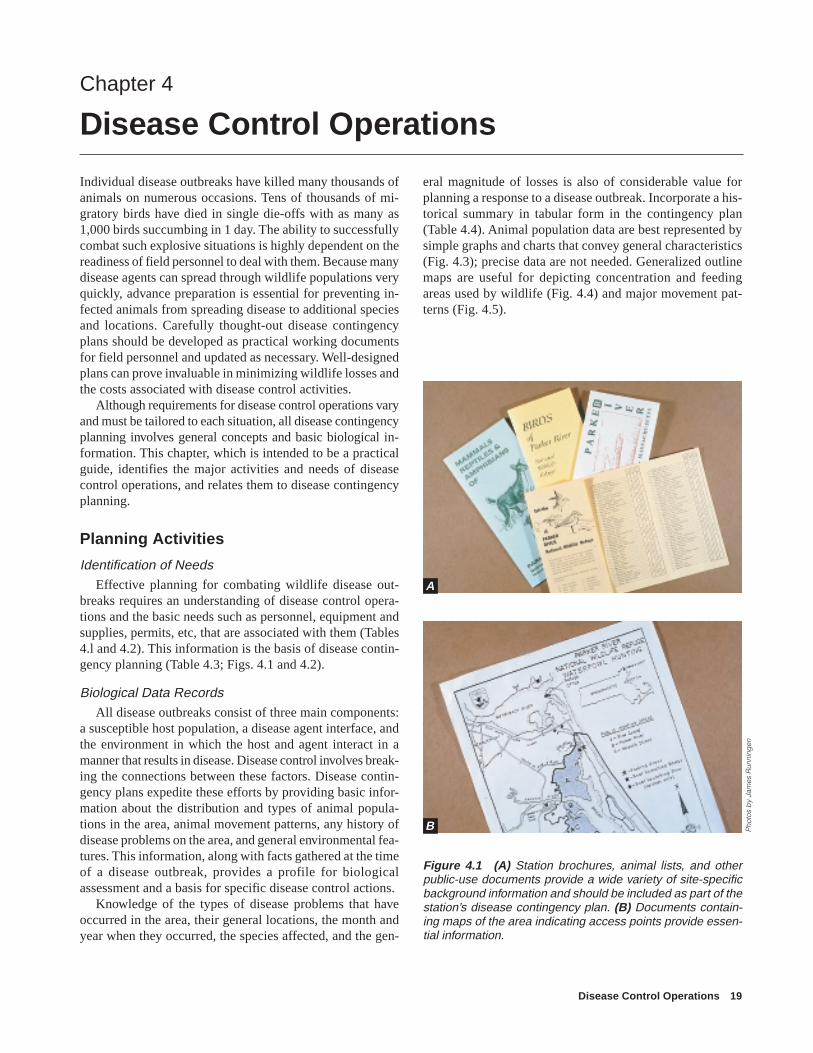
breaks requires an understanding of disease control opera-tions and the basic needs such as personnel, equipment andsupplies, permits, etc, that are associated with them (Tables4.l and 4.2). This information is the basis of disease contin-gency planning (Table 4.3; Figs. 4.1 and 4.2).
Biological Data RecordsAll disease outbreaks consist of three main components:
a susceptible host population, a disease agent interface, andthe environment in which the host and agent interact in amanner that results in disease. Disease control involves break-ing the connections between these factors. Disease contin-gency plans expedite these efforts by providing basic infor-mation about the distribution and types of animal popula-tions in the area, animal movement patterns, any history ofdisease problems on the area, and general environmental fea-tures. This information, along with facts gathered at the timeof a disease outbreak, provides a profile for biologicalassessment and a basis for specific disease control actions.
Knowledge of the types of disease problems that haveoccurred in the area, their general locations, the month andyear when they occurred, the species affected, and the gen-
Chapter 4
Disease Control Operations
Figure 4.1 (A) Station brochures, animal lists, and otherpublic-use documents provide a wide variety of site-specificbackground information and should be included as part of thestation’s disease contingency plan. (B) Documents contain-ing maps of the area indicating access points provide essen-tial information.
eral magnitude of losses is also of considerable value forplanning a response to a disease outbreak. Incorporate a his-torical summary in tabular form in the contingency plan(Table 4.4). Animal population data are best represented bysimple graphs and charts that convey general characteristics(Fig. 4.3); precise data are not needed. Generalized outlinemaps are useful for depicting concentration and feedingareas used by wildlife (Fig. 4.4) and major movement pat-terns (Fig. 4.5).
Pho
tos
by
Jam
es R
unni
ngen
B
A

20 Field Manual of Wildlife Diseases: Birds
Clean area
Contaminated area
Transition area
1
2
3
4
7
77
7
7
7
78
9
10
6
5
1
2
3
4
7
8
9
10
6
5
EXPLANATION
Command post and headquarters administrative area
Staff and press briefing room
Parking
Eating area and conference room
Staff rest area and visitors' center
Equipment and supply receipt—garage
Decontamination areas—boathouses, transition areas, parking lots
Carcass disposal site and observation hill
Animal holding—pole barn (has cement slab and electricity)
Laboratory investigations—shed (has cement slab, water, electricity)
Figure 4.2 Existing work areas used for disease control operations on a wildlife management area.

Disease Control Operations 21
Response ActivitiesResponse to wildlife die-offs will vary somewhat with
the species but will always involve a set of common factors.Waterfowl die-offs are used to illustrate specific approachesto addressing these common factors. For large mammals, theirsize and weight pose additional needs regarding carcass trans-port and disposal.
Problem IdentificationEarly detection and rapid and accurate assessment of the
causes of disease problems are essential to effective diseasecontrol operations. This is accomplished through surveillanceof animal populations to detect sick and dead wildlife, andthe prompt submission of specimens to qualified diseasediagnostic facilities. The speed with which large numbers ofanimals can become exposed to disease agents and the dif-ferences in control activities required for different types ofdisease problems place a premium on both the speed andaccuracy of diagnostic assessments. Once a disease problemhas been identified, the following basic activities are carriedout.
Carcass Removal: Protective Clothing and SuppliesWildlife that have died from disease are often a primary
source of the disease agent, and for most situations their car-casses need to be removed from the environment to preventdisease transmission to other animals through contact withor consumption of the carcass. Disease organisms releasedfrom tissues and body fluids as carcasses decompose alsocontaminate the environment. Some disease-causing virusesand bacteria can survive for several weeks or longer in pondwater, mud, and soil.
Because carcass collection concentrates diseased mate-rial in a small area, it is essential that carcasses be handledso that they do not release infectious agents into the environ-ment or jeopardize the health of personnel. Great care alsoneeds to be taken to prevent mechanical movement of thedisease agent from the problem area to other areas.
Personnel assigned to this task need to wear outer gar-ments that provide a protective barrier against direct contactwith disease organisms and that can be disinfected andremoved before personnel leave the area. Typically, theseinclude boots, coveralls or raingear, gloves, and a head cov-ering (Fig. 4.6).
Use disposable coveralls and outer gloves when possible;the durability and cost of garments are considerations in de-cisions about whether or not disposable garments will beused. Personnel should remove coveralls and outer glovesbefore they leave the area, and the garments should bedestroyed if they are disposable or they should be double-bagged before they are transported to a location where theycan be thoroughly washed before they are reused.Dishwashing gloves, work gloves, and other types of rubbergloves are readily available at hardware and other retail stores,
as are scrub brushes for cleaning (Fig. 4.7).Carcass removal requires heavy-duty plastic bags or con-
tainers. Plastic body bags used by the military are excellentfor containing wildlife carcasses. Plastic garbage cans linedwith commercially available heavy-gauge leaf and litter plas-tic bags are also excellent containers for transporting car-casses. These containers are especially useful when person-nel collect bird carcasses by boat (Fig. 4.8A), and for trans-porting carcasses in truck beds. Tie the bags shut and securegarbage can lids when transporting these containers to car-cass disposal sites (Fig. 4.8B).
Depending on conditions, a variety of watercraft (Fig. 4.9)and all-terrain vehicles (Fig. 4.10) are useful for searchingfor carcasses and for transporting carcasses to collection anddisposal sites. In some instances, the expense of helicoptersmay be warranted. Pickup trucks and other four-wheel ve-hicles are also indispensable under some field conditions.
Dogs have been used extensively in wildlife management,and they are a valuable search tool when they are appropri-ately chosen and handled. Use dogs whenever possible tolocate carcasses if there is no disease risk to them. Infectiousdiseases of wild North American birds do not pose a signifi-cant health threat to dogs. Determine disease risk on a case-by-case basis by consulting with wildlife disease specialists.Local retriever clubs or kennels may provide dogs.
The contingency plan should identify sources of variousequipment, whether equipment can be borrowed or rented,and contact persons and their telephone numbers. Commonlyused supplies and equipment needed to support disease con-trol operations are summarized in Table 4.2.
Carcass DisposalThe primary goal of carcass disposal is to prevent spread
of the disease agent to other animals through environmentalcontamination. Because personnel will handle concentratedamounts of infectious or highly toxic agents, this activityrequires proper training and supervision. Incineration, bury-ing, rendering, and composting are the four basic disposalmethods.
Incineration is generally the preferred method for dispos-ing of carcasses and contaminated materials associated withwildlife disease outbreaks. However, air-quality standardsoften preclude open burning, even for disease emergencies.Consider purchasing or constructing portable incinerators(Fig. 4.11) for areas with recurring disease problems if localregulations allow using such equipment. Portable garbageincinerators can sometimes be borrowed from State parksand other sources. If portable incinerators are not available,open burning with tires or other fuel or both can be used,depending on local air pollution standards. Carcasses maybe burned either above or below ground (Fig. 4.12). It isimportant to keep the fire contained and to get sufficient airmovement under the carcasses to maintain a hot fire and com-pletely burn the carcasses. Wood, coal, fuel oil, napalm, and

22 Field Manual of Wildlife Diseases: Birds
9,000
6,000
3,000
0
9,000
6,000
3,000
0
9,000
6,000
3,000
0
12,000
9,000
6,000
3,000
0JAN FEB MAR APR MAY JUNE JULY AUG SEPT OCT NOV DEC
NU
MB
ER
OF
AN
IMA
LS
MONTH
100
80
60
40
20
0JULY AUG SEPT OCT NOV DEC JAN FEB MAR APR MAY
MONTH
120
100
80
60
40
20
01981 1982 1983 1984 1985
YEAR
Number of geese and ducks on E. Overshoe NWR(5-year average)
Peak populations of waterfowl on E. Overshoe NWR
PO
PU
LAT
ION
, IN
TH
OU
SA
ND
SP
OP
ULA
TIO
N, I
N T
HO
US
AN
DS
Cropland
Cropland
Upland
Upland
Road
Clay Lake
MudLake
Creek
April–August: Nesting birds and broods on Mud and Clay Lakes and adjacent uplands.
September–mid-October: Fall migrants using Mud and Clay Lakes.
Mid-October–January: During hunting season, birds concentrated on Clay Lake and adjacent marsh.
February–March: After hunting season, birds distributed between Mud and Clay Lakes.
Canadagoose
Mallard
Pintail
Wigeon
A
B
C
D
Geese
Ducks
Geese
Ducks
Figure 4.3 Examples of how to present data on seasonal and annual wildlife use of a specific area. (A) General narrativeformat with map; (B) seasonal waterfowl populations by species, and total duck and goose use by (C) month and (D) year.
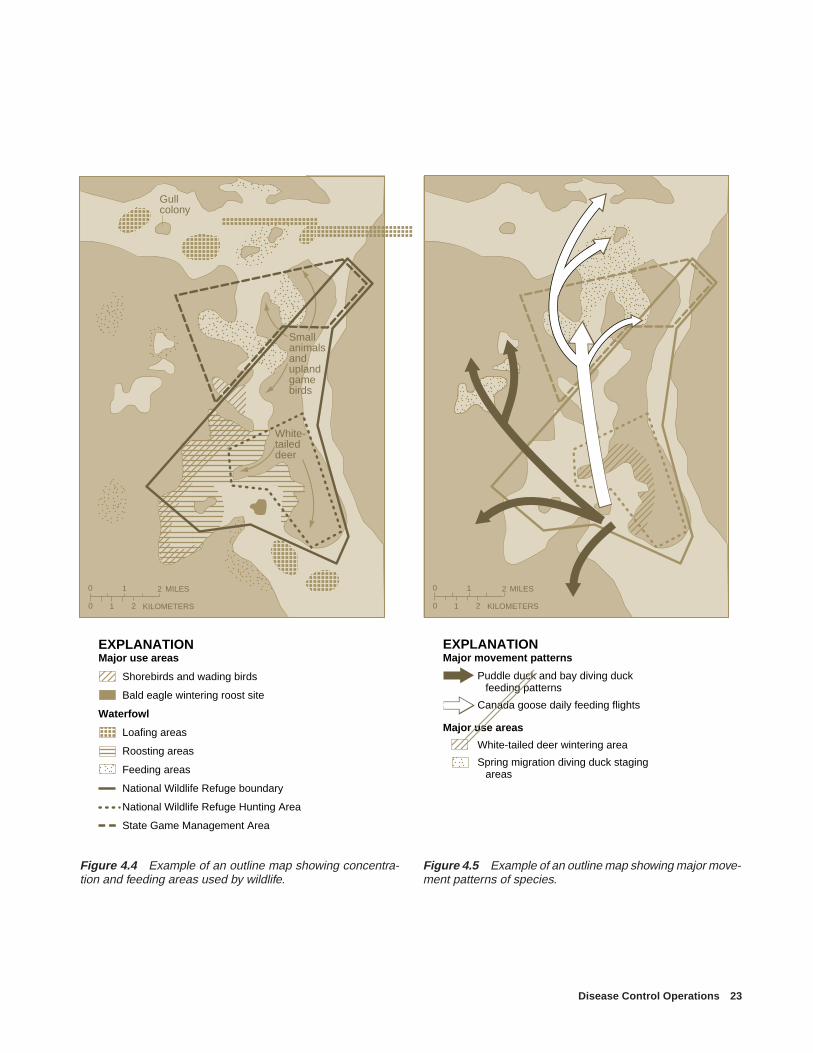
Disease Control Operations 23
Gullcolony
Small animals andupland gamebirds
White-tailed deer
0 1 MILES
0 1 KILOMETERS2
2
EXPLANATIONMajor use areas
Shorebirds and wading birds
Bald eagle wintering roost site
Waterfowl
Loafing areas
Roosting areas
Feeding areas
National Wildlife Refuge boundary
National Wildlife Refuge Hunting Area
State Game Management Area
0 1 MILES
0 1 KILOMETERS2
2
EXPLANATIONMajor movement patterns
Puddle duck and bay diving duck feeding patterns
Canada goose daily feeding flights
Major use areas
White-tailed deer wintering area
Spring migration diving duck staging areas
Figure 4.4 Example of an outline map showing concentra-tion and feeding areas used by wildlife.
Figure 4.5 Example of an outline map showing major move-ment patterns of species.

24 Field Manual of Wildlife Diseases: Birds
Figure 4.6 (A) Protective clothing suchas coveralls, boots, head coverings, andgloves should be warn during carcasscleanup activities. (B) Before leaving thearea, boots should be decontaminatedand outer clothing removed and baggedfor transportation to a location wherethey can be washed before being re-used.
Figure 4.7 (A) Examples of readily available disposable and reusable gloves for disease control operations. Dishwashinggloves, surgical gloves, rubber work gloves, and other types can be purchased at drug and hardware stores and medical andlaboratory supply houses. (B) A wide variety of scrub brushes needed for decontaminating boots, equipment, and other sur-faces are also readily available from local merchants.
Pho
to b
y W
illia
m B
air,
U.S
. Fis
h an
d W
ildlif
e S
ervi
ce
Pho
to b
y M
ilton
Frie
ndP
hoto
s b
y Ja
mes
Run
ning
en
A
A B
B

Disease Control Operations 25
Figure 4.8 (A) Plastic barrel beingused to transport carcasses from col-lection sites by airboat to disposal site.Note use of plastic bag to line barrel.The plastic bag containing carcassescan be secured, removed, and placedin a second plastic bag for further trans-portation if disposal site is not at the boatdocking location, thereby allowing im-mediate reuse of the barrel. If the barrelcontaining carcasses is to be trans-ported to some other location, the plas-tic bag should be tied closed and a coverplaced on the barrel and secured.(B) Examples of improper transportationof carcasses to disposal site. Note un-tied bags, unbagged carcasses, woodentruck bed, and lack of tailgate. Car-casses and fluids contaminated with dis-ease organisms could easily be re-leased from the bags during transit. Flu-ids could contaminate the truck bed andleak to the ground through the cracksbetween the wooden boards. Wood ab-sorbs fluids and is much more difficultto decontaminate than a nonporous sur-face. Also, carcasses could fall out of thetruck because there is no tailgate.P
hoto
s b
y M
ilton
Frie
nd
B
A

26 Field Manual of Wildlife Diseases: Birds
Figure 4.9 Different types of (A) motorized and (B) nonmotorized watercraft are useful for carcasscollection. Note the use of plastic bags for containment of carcasses and further transportation to disposalsites.
Pho
to b
y M
ilton
Frie
ndP
hoto
by
Jam
es R
unni
ngen
A
B

Disease Control Operations 27
Figure 4.10 Selection of all-terrain vehicles should bematched to local conditions. All-terrain vehicles such as thesethree-wheel machines can (A) be equipped with small bas-kets to hold carcasses or live birds and (B) be used in waterno more than 2-feet deep. Because of safety concerns, three-wheeled vehicles are not recommended. (C) Large trackedvehicles such as this equipment negotiate marshy terrain butare not amphibious. The major advantages of this equipmentare the large capacity for carrying personnel, supplies, andequipment and excellent visibility afforded by the height of thevehicle. (D) Small amphibious vehicles such as this six-wheelmachine are capable of transporting two persons and are morestable and versatile than three-wheel vehicles but are muchslower on land surfaces.
Pho
tos
by
Milt
on F
riend
Pho
to b
y R
onal
d W
ind
ing
stad
A
B
C
D
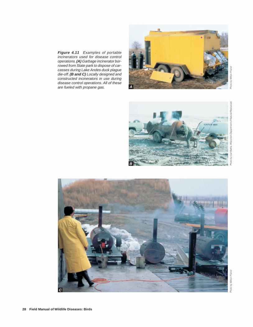
28 Field Manual of Wildlife Diseases: Birds
Pho
to b
y M
ilton
Frie
ndPh
oto
by C
arl B
atha
, Wis
cons
in D
epar
tmen
t of N
atur
al R
esou
r ces
Pho
to b
y M
ilton
Frie
nd
A
B
C
Figure 4.11 Examples of portableincinerators used for disease controloperations. (A) Garbage incinerator bor-rowed from State park to dispose of car-casses during Lake Andes duck plaguedie-off. (B and C) Locally designed andconstructed incinerators in use duringdisease control operations. All of theseare fueled with propane gas.

Disease Control Operations 29
Figure 4.12 Examples of above-groundand in-trench methods for incineration of car-casses. (A) Portable grate the width of apickup truck bed fashioned from metal pipes.(B) Simple grate suspended over pit intowhich carcass remains are placed for burial.(C) Major burning pit for large-scale opera-tion—note surrounding area cleaned of veg-etation for fire protection, the size and depthof pit, burning platform, rubber tires for fuel.—Figure 4.12 is continued on p. 30.
Pho
tos
by
Milt
on F
riend
A
B
C
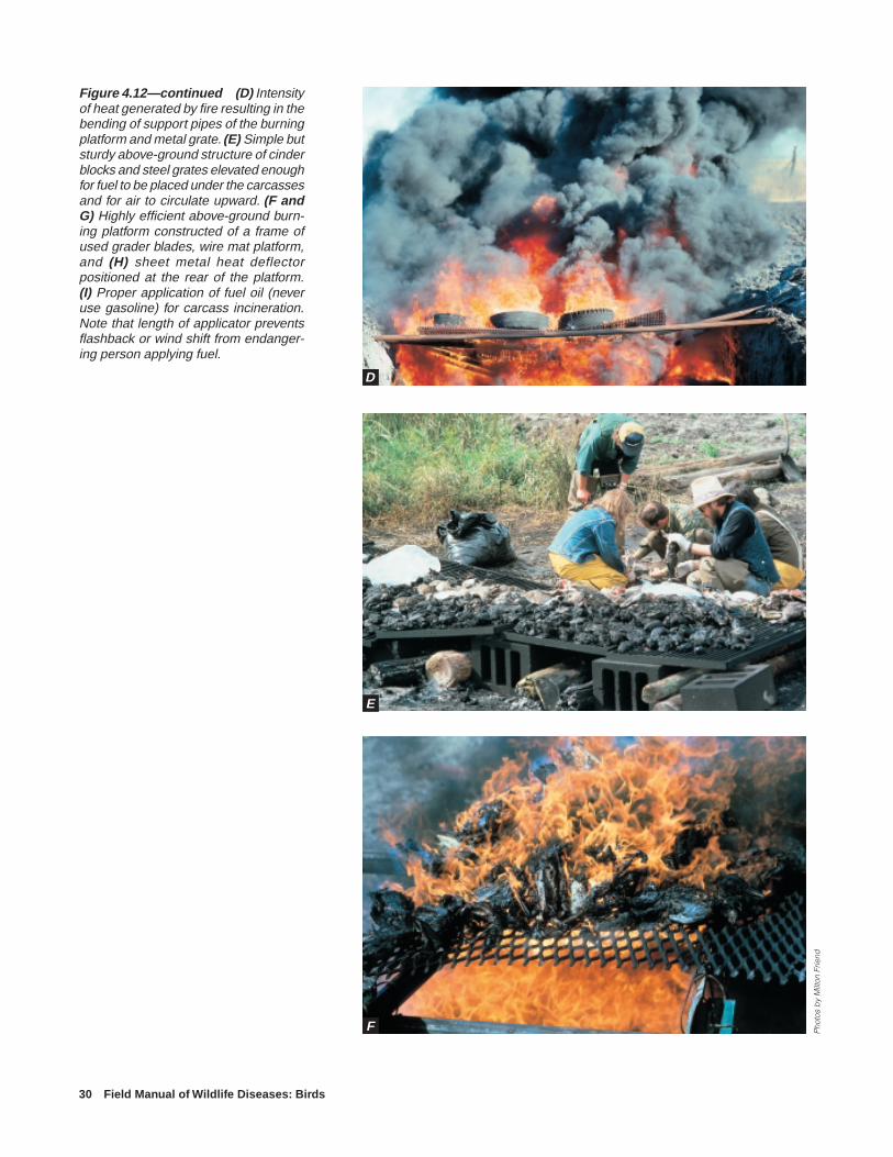
30 Field Manual of Wildlife Diseases: Birds
Figure 4.12—continued (D) Intensityof heat generated by fire resulting in thebending of support pipes of the burningplatform and metal grate. (E) Simple butsturdy above-ground structure of cinderblocks and steel grates elevated enoughfor fuel to be placed under the carcassesand for air to circulate upward. (F andG) Highly efficient above-ground burn-ing platform constructed of a frame ofused grader blades, wire mat platform,and (H) sheet metal heat deflectorpositioned at the rear of the platform.(I) Proper application of fuel oil (neveruse gasoline) for carcass incineration.Note that length of applicator preventsflashback or wind shift from endanger-ing person applying fuel.
F
E
D
Pho
tos
by
Milt
on F
riend

Disease Control Operations 31
Pho
tos
by
Milt
on F
riend
G
H
I

32 Field Manual of Wildlife Diseases: Birds
other fuels have been successfully used. Never use gasolinebecause of the hazards involved. Incineration is facilitatedby stacking or piling carcasses on the burning platform, soak-ing them with used oil or some other fuel, and waiting about10 to 15 minutes before igniting them. The heat generatedby large-scale carcass burning operations is intense enoughto cause metal pipes to bend (Fig. 4.12D). Therefore, con-struct a sturdy carcass support surface so that it does notcollapse into the fire.
During dry weather, burning carcasses in a pit surroundedby a vegetation-free area is more desirable than above-groundburning. In either situation, piling too many carcasses on thefire at once is a common mistake; burn carcasses one layerat a time (Fig. 4.13). When cinder blocks are used to supportburning platforms, the length of the platform should be ex-tended to keep the blocks out of direct heat or they will sooncrumble.
When burning is not feasible or needed, burial is often asuitable alternative. Select burial sites carefully with consid-eration given to ground-water circulation and drainage, andany potential for later carcass exposure. Sprinkle lime or fueloil on carcasses to discourage uncovering by scavengers andcover the carcasses with at least 3 to 4 feet of soil.
Composting is commonly used for the disposal of somedomestic animal carcasses, and it is a technique that can beadapted to wildlife situations. The requirements for com-posting carcasses include an impermeable surface on whichto place composting piles, a roof or other means of control-ling moisture in the piles, and raw materials to mix with car-casses to achieve the correct carbon to nitrogen ratio for op-timal decomposition of carcasses (Fig. 4.14).
When the combination of animal species, cause of mor-tality, and local situation allow, carcasses may also be dis-posed of by an animal rendering plant, and in rare instancesinfected wildlife may be killed and processed for food. Bothof these methods are sometimes used for domestic speciesand captive-reared wildlife, but conservation laws generally
prohibit the processing of free-living wildlife (with the ex-ception of fish) as a commercial food source within the UnitedStates. Judgments on the use of rendering and food process-ing as animal disposal methods should be made only by quali-fied disease control specialists.
To the extent possible, dispose of carcasses on-site to re-duce the risk associated with transporting contaminated ma-terial. Regardless of whether burning, burial, or large-scalecomposting is used, earth-moving equipment is needed. Thedisease contingency plan should identify how and wherebulldozers, backhoes, and similar equipment can be obtained.
Animal RelocationIt is often as necessary to deal with the live, apparently
healthy population during disease control activities as it is toremove and dispose of animals dying from disease. Depend-ing on individual circumstances, consider denying animaluse of specific sites by dispersing animals from the problemarea, concentrating and holding wildlife within a specific area,or trapping animals for sampling.
Scare devices such as propane exploders (Fig. 4.15A) andcracker shells (Fig. 4.15B) may be useful for keeping wild-life away from a toxin or infectious agent within a specificarea. Hazing wildlife with airplanes, helicopters, airboats,snowmobiles, and other motorized equipment has also beensuccessful for moving them away from disease problemareas. Conversely, wildlife can be concentrated in an areafor euthanasia, and they can be lured to other areas by broad-casting and dumping large amounts of grain and other feedto prevent their movement to problem areas, by knockingdown standing grain to make it more available to them, byproviding water through pumping operations and divertingwater flow, and by providing refuge by closing the area tohunting and other interactions between wildlife and humans(Fig. 4.16). Take care to assure that grain used for attractingwildlife is not moldy and does not contain dangerous con-centrations of mycotoxins.
Figure 4.13 Examples of (A) correct and (B) incorrect layering of carcasses for burning. Carcasses must be burned one layerat a time to prevent charred outer carcasses from insulating inner carcasses from incineration. (Illustration by Randy StothardKampen)
A B

Disease Control Operations 33
Figure 4.14 Example of a simple composting bin for waterfowl carcasses. Litter (beddedmanure from poultry houses is a good source), straw, and carcasses are added propor-tionally to achieve the appropriate moisture content and carbon to nitrogen ratio. (Modifiedfrom Rynk, 1992.)
Food and water are also helpful in trapping wildlife forassessing disease control activities. When birds have beenlured to a site, they may be captured by such means as drugsincorporated within feed, rocket nets, drop nets, walk-in andswim-in traps, or other means of preventing escape (Fig.4.17).
A timely response to disease outbreaks can be facilitatedif such factors as need for special permits, area closures,possible involvement of endangered species, and water pur-chase can be anticipated and addressed before an urgent situ-ation arises.
Because of the potential complexity of biological inter-actions in animal relocation, field managers should seek theadvice of disease control specialists whenever possible be-fore taking independent action. As a general rule, animal dis-persal is not recommended when infectious disease is in-volved unless it can be assured that the population being dis-persed will not infect other wildlife. Also, it is important thatwater manipulation not produce conditions favorable to de-velopment of botulism or other disease problems.
DisinfectionThe purpose of disinfection is to prevent the mechanical
transmission of disease agents from one location to anotherby people, equipment, and supplies. Some viruses, bacteria,and other infectious agents have considerable environmen-tal persistence. Disinfection of the local environment involved
in a disease outbreak may be required to prevent recurrenceof the disease when the site is used by other animals. Disin-fection of a disease outbreak site should always be doneunder the direct guidance of disease control specialists.
Wash thoroughly the clothing worn during disease con-trol (coveralls and clothes worn under protective raingear)before it is used again. Personnel should shower and sham-poo their hair before leaving the site, if possible, but alwaysbefore they go to other wildlife areas. Disinfect boots beforeentering vehicles when in contaminated areas, and disinfectall equipment to the extent possible before it is moved fromthe area (Fig. 4.18A and B). Give special attention to theunderside of vehicles (Fig. 4.18C and D). Put motor vehiclesthrough a car wash before moving them to other areas, andwash and clean boats and all-terrain vehicles before they leavethe area. Large volume tanks and pumps that can be oper-ated from mobile units such as trucks (Fig. 4.19) and boatsare especially useful for holding and dispensing disinfec-tant.
Disinfection procedures require a suitable disinfectant,containers for that disinfectant once it has been diluted toappropriate strength, and a way of applying the disinfectant.Commercial disinfectants are available from farm supplystores and veterinarians. Refuge managers and other fieldmanagers should consider keeping a supply of disinfectantfor general use. Chlorine bleach is a highly suitable disin-fectant and it is available at most grocery stores. For general
Litter
Waterfowl
Straw
Litter
Impermeablesurface

34 Field Manual of Wildlife Diseases: Birds
Figure 4.15 Wildlife can be discouraged from use of areasby (A) propane exploders that function by the ignition of pro-pane gas within the “cannon” due to the striking of a flint at atimed interval. With the exception of placing the cannon andmaintaining a fuel supply, this activity does not require thepresence of personnel. (B) Manual firing of cracker shells hasalso been used successfully to discourage wildlife use ofareas. These fireworks-like shells should only be fired througha break-open type shotgun so that the barrel can be checkedbetween shots to assure that there are no obstructions re-maining in the barrels. These shells should not be used wherethey can fall into dry vegetation because of fire hazard.
Figure 4.16 Closure of areas is often needed to assist dis-ease control operations. (A) Sign used to close Lake AndesNational Wildlife Refuge during the duck plague die-off. (B)Sign used to delineate refuge area so that bird disturbanceand movement was minimized during another South Dakotadisease control operation.
use, dilute one part chlorine bleach with 10 parts water. Usestronger concentrations of one part bleach to five parts waterfor disinfecting heavily contaminated areas.
Stiff bristle brushes, buckets, and containers that can beused for foot baths and pressure or hand sprayers that can beused to dispense the disinfectant are also needed. The sta-tion contingency disease plan should identify readily avail-able sources of these supplies and equipment.
When the disease problem involves an infectious agent,personnel handling contaminated materials should refrainfrom working with similar species or those susceptible tothe disease for at least 7 days following completion of theirdisease control activities. For example, a field manager in-volved in an intensive avian cholera disease control opera-tion on Monday should not band waterfowl in that refuge orelsewhere until Tuesday of the next week.
PersonnelLabor-intensive operations such as carcass removal and
disposal sometimes require more personnel than are usuallyemployed on an area. In some instances, specialized helpsuch as low level aircraft flights for surveillance may beneeded. The use of nonstation personnel for routine opera-tions has a potential educational value. For example, the useof local sportsmen clubs to help with carcass collectionsduring a major lead poisoning die-off has been highly effec-tive in changing negative attitudes towards nontoxic shot use.
Sportsmen clubs; retriever clubs; biology and wildlifeclasses at local universities and colleges; local chapters ofconservation organizations such as the Audubon Society; theactive military and National Guard, who also may providevaluable technical assistance; and similar groups have allprovided volunteer assistance in combating disease problems
Pho
tos
by
Milt
on F
riend
Pho
tos
by
Jam
es R
unni
ngen
B
A
B
A
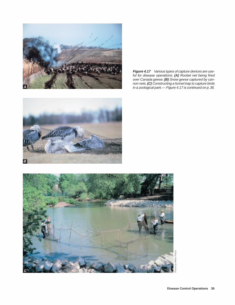
Disease Control Operations 35
Figure 4.17 Various types of capture devices are use-ful for disease operations. (A) Rocket net being firedover Canada geese. (B) Snow geese captured by can-non nets. (C) Constructing a funnel trap to capture birdsin a zoological park.— Figure 4.17 is continued on p. 36.
Pho
tos
by
Milt
on F
riend
A
B
C

36 Field Manual of Wildlife Diseases: Birds
Pho
tos
by
Milt
on F
riend
Figure 4.17—continued (D) Capturing birdswithin a funnel trap. (E) Capturing flightlessCanada geese in a drive trap. (F) Capturingwaterfowl in a large, baited funnel trap. (G)Using drugged grain to capture birds in resi-dential situations. When drugs are used,maintain close surveillance of the situationso that animals that become drugged, suchas the bird (H) lying on its back, can bepromptly collected before they are seized byother animals or drown if they venture intothe water before the drug takes effect.
D
E
F

Disease Control Operations 37
G
H Pho
tos
by
Milt
on F
riend

38 Field Manual of Wildlife Diseases: Birds
Figure 4.18 Equipment and personnel should be disinfectedto the extent possible before leaving disease operation areas.(A) Initial disinfection procedures should take place well withinthe contaminated area. (B) Boots and other items in contactwith the ground should receive a second application of disin-fectant at the point where entry is made into the “clean area,”as is being done at the location where the specimen chest isbeing transferred. (C and D) Various types of spray units canbe used to apply disinfectant to the underside of vehicles. Tiresand wheel wells are the primary areas of concern as theymay contain contaminated soil or animal fecal material fromthe disease area.
Pho
to b
y M
ilton
Frie
nd
A
B
C
D
Pho
to b
y M
ilton
Frie
ndP
hoto
by
Milt
on F
riend
Pho
to b
y J.
Chr
istia
n Fr
anso
n

Disease Control Operations 39
Figure 4.19 (A) Portable tank and pump mounted on a truck bed for dispersing disinfectant duringduck plague control operation and (B) application of that disinfectant to a structure used to house birds.The long length of hose on this unit allowed all areas of major bird use to be reached from service andperimeter roads.
Pho
to b
y M
ilton
Frie
ndP
hoto
by
Terr
y A
mun
dso
n, W
isco
nsin
Dep
artm
ent o
f Nat
ural
Res
ourc
es
A
B

40 Field Manual of Wildlife Diseases: Birds
at various times and places. Sound judgment must be exer-cised in the selection and utilization of volunteers becauseof legal liability in case of an accident. Contingency plansshould list groups and organizations and contact persons foreach group, their telephone numbers, and an approximationof the work force and times of its availability (e.g., week-ends only or Wednesday only). For technical assistance, listthe specific type of personnel needed, such as bulldozer op-erator or helicopter pilot.
In addition to preparing a station contingency plan, wild-life personnel should become familiar with the other phasesof disease control operations. Table 4.1 provides a descrip-tive outline of these phases. Especially relevant to field man-agers are the equipment and supply needs identified underthe Disease Response Section of Table 4.3.
Response ModificationsDisease control operations can be seriously undermined
without current assessment of wildlife morbidity and mor-tality and the cause of disease problems. When infectious orhighly toxic agents are involved, early detection of diseaseproblems is critical to preventing the problem from becom-ing widespread. Also, failure to accurately assess the causeof the die-off can result in control actions actually contrib-uting to the magnitude of losses and spread of the problem.Different types of disease problems require different typesof response. Do not assume that the current die-off is due tothe same cause as previous die-offs that have occurred onthe area or that only one disease agent is responsible. It isnot uncommon for two or more causes of wildlife mortalityto occur simultaneously in an area. Control of these differ-ent diseases may require opposite types of actions, thereby
requiring that a more comprehensive strategy be developedfor the disease control operation.
Refuge managers and other field biologists greatly influ-ence the effectiveness of disease control operations by theirresponsiveness, knowledge of the local situation, how wellthey are prepared, the flexibility they maintain, their resource-fulness, and when possible, their ability to obtain appropri-ate technical assistance and training for combating diseaseproblems. Timely and properly carried-out disease controlactivities can significantly reduce the magnitude of wildlifelosses that might otherwise occur. When carrying out con-trol activities, always consider the safety of the personnelinvolved.
Milton Friend and J. Christian Franson
Supplementary Reading
Friend, M., 1995, Disease considerations for waterfowl managersin Whitman, W. R., and others, eds., Waterfowl HabitatRestoration Enhancement and Management in the AtlanticFlyway (3): Dover, Del., Delaware Department of NaturalResources and Environmental Control, p. J24–J117.
Roffe, T.J., Friend, M., Locke, L.N., Evaluation of causes ofwildlife mortality, in Bookout, T. A., editor, 1994, Researchand management techniques for wildlife and habitats, Fifthed.,: Bethesda, Md., The Wildlife Society, p. 324–348
Rynk, R., ed., 1992, On-farm composting handbook: Ithaca, N.Y.,Northeast Regional Agricultural Engineering Service, 186 p.
Wobeser, G. A., 1994, Investigation and management of disease inwild animals: New York, N.Y., Plenum Press, 265 p.

Disease Control Operations 41
Table 4.1 Outline of disease control operations.
I. Planning
A. Identify needs1. Sources of additional personnel to help during disease emergencies. Potentially, these include
a. State and Federal agenciesb. Active military and National Guardc. Private conservation agenciesd. Local sporting clubse. Local universities
2. Sources and availability of equipment and supplies for disease control operations (Appendix C)
3. Special needsa. Burning permitsb. Endangered species consultationsc. Lodging and meal facilities for work crewsd. Ability to attract and hold wildlife in site-specific areas by providing food, water, refuge, or other
meanse. Ability to deny wildlife use of specific areas by scaring devices and other meansf. Ability to capture wildlife for sampling, immunization, or other needs
B. Record biological information1. Daily and seasonal wildlife movement patterns within the general area2. Migration patterns and population peaks for major and endangered species3. Past history of diseases
C. Prepare contingency plan (See Tables 4.2 and 4.3.)
II. Initial Response
A. Identify problems1. Obtain diagnosis by submitting carcasses to a qualified diagnostic laboratory as soon as mortality or
morbidity is evident. (See Chapters 3 and 4 for shipping procedures.)
2. Conduct field investigation to determine extent of problem (i.e., species, number of wildlife, and geo-graphic area involved).
3. Identify special biological, political, or physical considerations associated with problem. Beforeproceeding further with II. B and C., seek the advice of a specialist.
B. Establish control of area1. Close affected area, when warranted, to all but authorized personnel.
2. Identify special work areas for disease control activities.a. Carcass disposal sitesb. Laboratory investigations areac. Briefing area for news media and staffd. Vehicle parkinge. Assembly areas for arriving workersf. Command post
3. Initiate carcass cleanup, but do not dispose of carcasses without guidance from disease controlspecialists.

42 Field Manual of Wildlife Diseases: Birds
Table 4.1 Outline of disease control operations (continued).
C. CommunicationsNotify appropriate agency and nonagency personnel of die-off.
III. Disease Control
A. Response1. Disease control actions are dictated by the type of disease, environmental factors, species involved,
and other circumstances. Typically, actions associated with major die-offs require:a. Bringing personnel, equipment, and supplies on-siteb. Organizing workforce, briefing workers about the problem, and assigning dutiesc. Carcass pickup and disposald. Monitoring cause of mortality to detect changes in the cause of the problem (die-offs often involve
more than a single cause and different control actions may be required for these different causes)e. Decontamination of personnel and equipmentf. News media briefing sessions and “show-me” trips1
B. Management1. Disease management activities often involve:
a. Population manipulation such as removal, controlled movement including relocation and localconcentration of wildlife populations, and population dispersal
b. Habitat manipulation to prevent, attract, or maintain wildlife use of an area
2. Decontamination of the infected environment, such as:a. Chemical treatment of land, water, and structuresb. Vegetation and water removal (desiccation) to allow air and sunlight (ultraviolet) to destroy micro-
organisms
C. Controlled burning to remove vegetation and dispose of mechanical structures
IV. Surveillance
A. MonitoringAfter disease control operations have ended, the area should be kept under surveillance for 10 to 30 daysto watch for additional flareups.
B. InvestigationsThis stage is also an appropriate time to conduct followup investigations of factors that helped cause andsustain the problem, and to carry out wildlife and environmental sampling to discern diseaseexposure patterns and environmental reservoirs of disease agents.
V. AnalysesEach disease control operation provides a learning experience. It is important to the success of futureoperations to evaluate what was done, the degree of success achieved, problems encountered, and whatshould have been done differently.
1 Media briefing sessions and “show-me” trips should be conducted by personnel with comprehensive know-ledge of the situation.
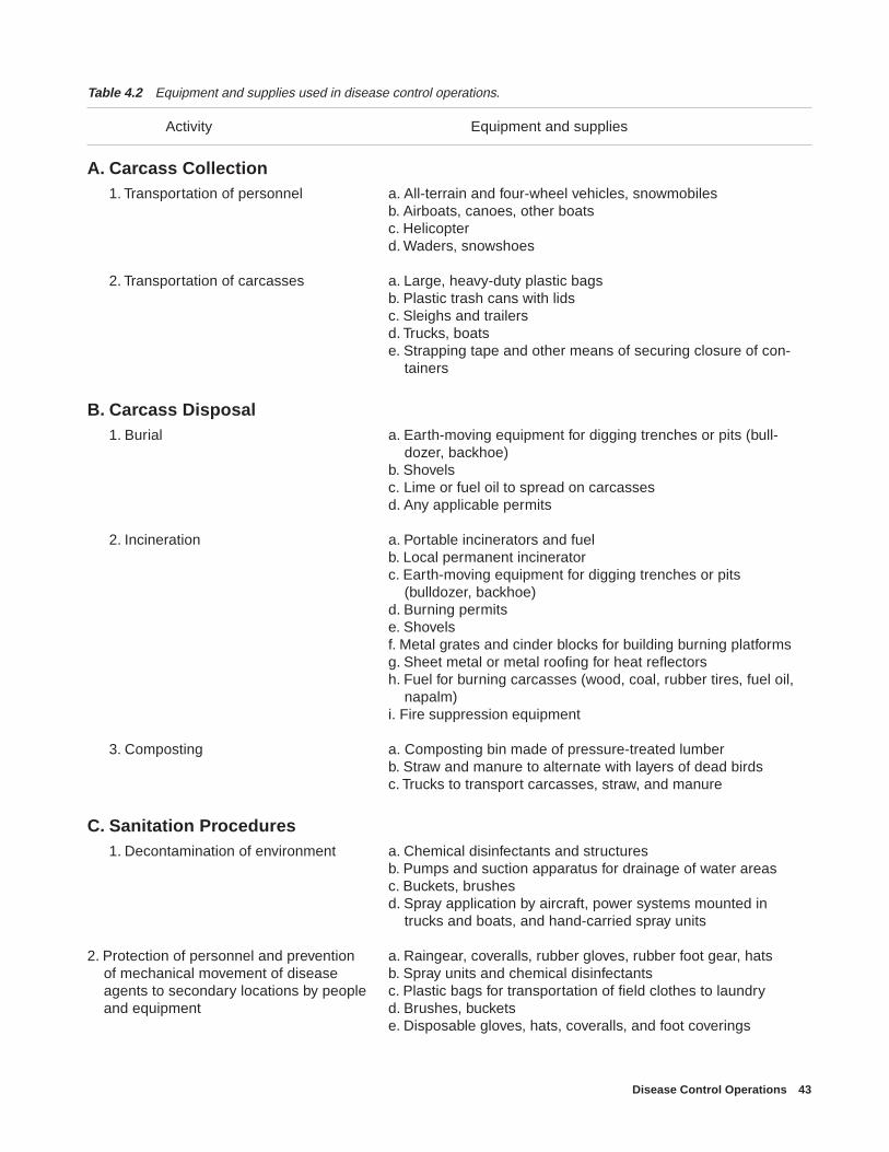
Disease Control Operations 43
Table 4.2 Equipment and supplies used in disease control operations.
Activity Equipment and supplies
A. Carcass Collection1. Transportation of personnel a. All-terrain and four-wheel vehicles, snowmobiles
b. Airboats, canoes, other boatsc. Helicopterd. Waders, snowshoes
2. Transportation of carcasses a. Large, heavy-duty plastic bagsb. Plastic trash cans with lidsc. Sleighs and trailersd. Trucks, boatse. Strapping tape and other means of securing closure of con-
tainers
B. Carcass Disposal1. Burial a. Earth-moving equipment for digging trenches or pits (bull-
dozer, backhoe)b. Shovelsc. Lime or fuel oil to spread on carcassesd. Any applicable permits
2. Incineration a. Portable incinerators and fuelb. Local permanent incineratorc. Earth-moving equipment for digging trenches or pits
(bulldozer, backhoe)d. Burning permitse. Shovelsf. Metal grates and cinder blocks for building burning platformsg. Sheet metal or metal roofing for heat reflectorsh. Fuel for burning carcasses (wood, coal, rubber tires, fuel oil,
napalm)i. Fire suppression equipment
3. Composting a. Composting bin made of pressure-treated lumberb. Straw and manure to alternate with layers of dead birdsc. Trucks to transport carcasses, straw, and manure
C. Sanitation Procedures1. Decontamination of environment a. Chemical disinfectants and structures
b. Pumps and suction apparatus for drainage of water areasc. Buckets, brushesd. Spray application by aircraft, power systems mounted in
trucks and boats, and hand-carried spray units
2. Protection of personnel and prevention a. Raingear, coveralls, rubber gloves, rubber foot gear, hats of mechanical movement of disease b. Spray units and chemical disinfectants agents to secondary locations by people c. Plastic bags for transportation of field clothes to laundry and equipment d. Brushes, buckets
e. Disposable gloves, hats, coveralls, and foot coverings

44 Field Manual of Wildlife Diseases: Birds
Table 4.2 Equipment and supplies used in disease control operations (continued).
Activity Equipment and supplies
D. Field Communications1. Field activities a. Portable radios or cellular telephones for communication
between field personnelb. Radios in vehicles for communication between field units and
between units and command post
2. Information activities a. Word processor or typewriter for preparing briefing documentsb. Maps, acetate, and other supplies for overlays depicting die-
off and control activity informationc. Telephone lines for communication with othersd. Transportation for news media “show-me” trips
E. Surveillance and Observation1. Field activities a. Aircraft and pilots certified for low-level flights (500 feet and
below) for monitoring wildlife populations andenvironmental conditions
b. Binoculars and spotting scopes
2. Office activities a. Maps, acetate, and other supplies for tracking the progress ofevents and wildlife populations associated with die-off
b. Telephone for contacting others to trace movement of migrantbird populations that might enter problem area or that havedeparted problem area
F. Wildlife Population and Habitat Manipulation1. Denying wildlife use of an area a. Aircraft, boats, snowmobiles, and other motorized means of
hazing wildlife populationsb. Propane explodersc. Cracker shells, break-open shotguns, and protective face
shieldd. Audio systems and other scare devicese. Pumps for draining water or adding water to areas
2. Concentration and maintenance of a. Grain and other sources of food wildlife in a specific area b. Pumps and water to provide habitat
c. “No Hunting” and “Area Closed” signs to provide temporaryrefuge area
G. Wildlife Sampling and Monitoring1. Wildlife capture a. Cannon nets and other capture equipment
b. Grain and other baits to lure wildlife to capture site
2. Wildlife marking a. Visible marking devices such as paint, neck collars, and otherdevices
b. Permanent marking devices such as leg bands and ear tags(see Bookhout, 1994)
c. Temporary marking devices such as radio transmitters
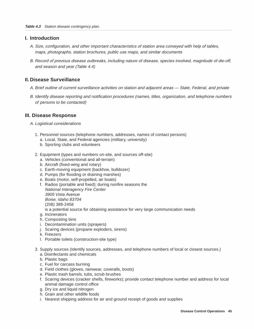
Disease Control Operations 45
Table 4.3 Station disease contingency plan.
I. Introduction
A. Size, configuration, and other important characteristics of station area conveyed with help of tables,maps, photographs, station brochures, public use maps, and similar documents
B. Record of previous disease outbreaks, including nature of disease, species involved, magnitude of die-off,and season and year (Table 4.4)
II. Disease Surveillance
A. Brief outline of current surveillance activities on station and adjacent areas — State, Federal, and private
B. Identify disease reporting and notification procedures (names, titles, organization, and telephone numbersof persons to be contacted)
III. Disease Response
A. Logistical considerations
1. Personnel sources (telephone numbers, addresses, names of contact persons)a. Local, State, and Federal agencies (military, university)b. Sporting clubs and volunteers
2. Equipment (types and numbers on-site, and sources off-site)a. Vehicles (conventional and all-terrain)b. Aircraft (fixed-wing and rotary)c. Earth-moving equipment (backhoe, bulldozer)d. Pumps (for flooding or draining marshes)e. Boats (motor, self-propelled, air boats)f. Radios (portable and fixed); during nonfire seasons the
National Interagency Fire Center3905 Vista AvenueBoise, Idaho 83704(208) 389-2458is a potential source for obtaining assistance for very large communication needs
g. Incineratorsh. Composting binsi. Decontamination units (sprayers)j. Scaring devices (propane exploders, sirens)k. Freezersl. Portable toilets (construction-site type)
3. Supply sources (Identify sources, addresses, and telephone numbers of local or closest sources.)a. Disinfectants and chemicalsb. Plastic bagsc. Fuel for carcass burningd. Field clothes (gloves, rainwear, coveralls, boots)e. Plastic trash barrels, tubs, scrub brushesf. Scaring devices (cracker shells, fireworks); provide contact telephone number and address for local
animal damage control officeg. Dry ice and liquid nitrogenh. Grain and other wildlife foodsi. Nearest shipping address for air and ground receipt of goods and supplies
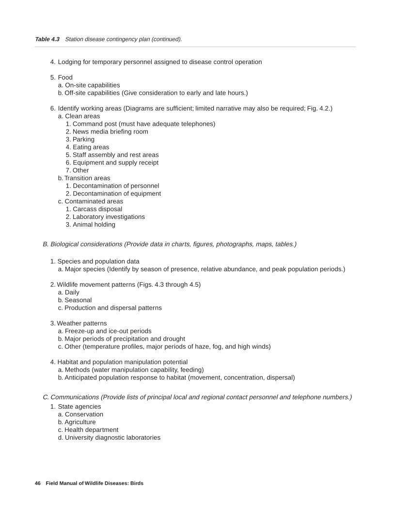
46 Field Manual of Wildlife Diseases: Birds
Table 4.3 Station disease contingency plan (continued).
4. Lodging for temporary personnel assigned to disease control operation
5. Fooda. On-site capabilitiesb. Off-site capabilities (Give consideration to early and late hours.)
6. Identify working areas (Diagrams are sufficient; limited narrative may also be required; Fig. 4.2.)a. Clean areas
1. Command post (must have adequate telephones)2. News media briefing room3. Parking4. Eating areas5. Staff assembly and rest areas6. Equipment and supply receipt7. Other
b. Transition areas1. Decontamination of personnel2. Decontamination of equipment
c. Contaminated areas1. Carcass disposal2. Laboratory investigations3. Animal holding
B. Biological considerations (Provide data in charts, figures, photographs, maps, tables.)
1. Species and population dataa. Major species (Identify by season of presence, relative abundance, and peak population periods.)
2. Wildlife movement patterns (Figs. 4.3 through 4.5)a. Dailyb. Seasonalc. Production and dispersal patterns
3. Weather patternsa. Freeze-up and ice-out periodsb. Major periods of precipitation and droughtc. Other (temperature profiles, major periods of haze, fog, and high winds)
4. Habitat and population manipulation potentiala. Methods (water manipulation capability, feeding)b. Anticipated population response to habitat (movement, concentration, dispersal)
C. Communications (Provide lists of principal local and regional contact personnel and telephone numbers.)1. State agencies
a. Conservationb. Agriculturec. Health departmentd. University diagnostic laboratories

Disease Control Operations 47
Table 4.3 Station disease contingency plan (continued).
2. Federal agenciesa. Environmental Protection Agencyb. U.S. Department of Agriculturec. U.S. Public Health Service
3. Other organizationsa. Cooperating organizations (e.g., area representatives of Audubon Society, National Wildlife
Federation, Ducks Unlimited)b. Local sporting clubsc. Private wildlife area managersd. Local game breeder organizationse. Local domestic animal husbandry and production operations
4. Mediaa. Televisionb. Radioc. Newspapers
IV. Supplemental Information
A. Location of nearby laboratories (hospitals, universities, county and State facilities)
B. Federal and State permit status for biological collections
C. Burning permits
D. Regulatory requirements
E. Background information (e.g., water sources, water-quality data, potential sources of diseasetransmission between wildlife and domestic animal concentrations)
F. Identification and location of adjacent or nearby wildlife refuges, management areas, and private reserves
G. Identification of unusual or politically sensitive aspects of area
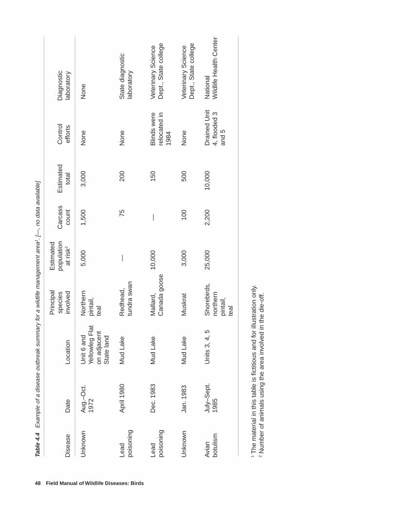
48 Field Manual of Wildlife Diseases: Birds
Tabl
e 4.
4 E
xam
ple
of a
dis
ease
out
brea
k su
mm
ary
for
a w
ildlif
e m
anag
emen
t are
a1. [
—, n
o da
ta a
vaila
ble]
Prin
cipa
lE
stim
ated
spec
ies
popu
latio
nC
arca
ssE
stim
ated
Con
trol
Dia
gnos
ticD
isea
seD
ate
Loca
tion
invo
lved
at r
isk2
coun
tto
tal
effo
rts
labo
rato
ry
Unk
now
nA
ug.–
Oct
.U
nit 6
and
Nor
ther
n5,
000
1,50
03,
000
Non
eN
one
1972
Yello
wle
g F
lat
pint
ail,
on a
djac
ent
teal
Sta
te la
nd
Lead
Apr
il 19
80M
ud L
ake
Red
head
,—
7520
0N
one
Sta
te d
iagn
ostic
pois
onin
gtu
ndra
sw
anla
bora
tory
Lead
Dec
. 198
3M
ud L
ake
Mal
lard
,10
,000
—15
0B
linds
wer
eV
eter
inar
y S
cien
cepo
ison
ing
Can
ada
goos
ere
loca
ted
inD
ept.,
Sta
te c
olle
ge19
84
Unk
now
nJa
n. 1
983
Mud
Lak
eM
uskr
at3,
000
100
500
Non
eV
eter
inar
y S
cien
ceD
ept.,
Sta
te c
olle
ge
Avi
anJu
ly–S
ept.
Uni
ts 3
, 4, 5
Sho
rebi
rds,
25,0
002,
200
10,0
00D
rain
ed U
nit
Nat
iona
lbo
tulis
m19
85no
rthe
rn4,
floo
ded
3W
ildlif
e H
ealth
Cen
ter
pint
ail,
and
5te
al
1 The
mat
eria
l in
this
tabl
e is
fict
itiou
s an
d fo
r ill
ustr
atio
n on
ly.
2 N
umbe
r of
ani
mal
s us
ing
the
area
invo
lved
in th
e di
e-of
f.

Euthanasia 49
Chapter 5
Euthanasia
BackgroundEuthanasia means to cause humane death. Some current
euthanasia techniques may become unacceptable over timeand be replaced by new techniques as more data are gath-ered and evaluated. The following information and recom-mendations are based largely on the 1993 report of the Ameri-can Veterinary Medical Association (AVMA) Panel onEuthanasia. The recommendations in the panel report wereintended to serve as guidelines, and they require the use ofprofessional judgement for specific situations. Ultimately, itis the responsibility of those persons carrying out euthanasiato assure that it is done in the most humane manner possible.
There is no perfect euthanasia technique appropriate toall situations. What is sought in each instance is immediateinsensitivity of the animal to pain as a result of depression ofthe central nervous system (brain and spinal cord). TheAVMA panel in its evaluation considered the following tobe important factors to consider when selecting a euthanasiamethod:
Considerations for selecting a euthanasia method
• Does the method cause the animal to lose conscious-ness and die without causing the animal pain, distress,anxiety, or apprehension?
• How much time does the method require to induceunconsciousness?
• Is the method reliable?
• Does the method put personnel at risk of injury orhealth problems?
• Is the method irreversible?
• Is the method compatible with the purpose of eutha-nasia?
• Will the method cause distress and anxiety amongobservers and personnel?
• Does the method interfere with or detract from thesubsequent evaluation, examination, and use of tis-sue?
• Are drugs required by the method available? Can thedrugs be abused by humans?
• Is the method appropriate for the animal age and spe-cies?
• Is the equipment required by the method in properworking order?
• Is the method cost-effective?
Methods of euthanasia are physical or chemical. Physicalmethods of euthanasia include cervical dislocation, decapi-tation, stunning and removal of blood, and gunshot. Chemi-cal methods of euthanasia involve introducing a toxic agentinto the body by injection or inhalation. After completingeuthanasia, be certain that specimens being collected areproperly identified, preserved, and packaged for transporta-tion to the diagnostic laboratory (see Chapter 2, SpecimenCollection and Preservation, and Chapter 3, Specimen Ship-ment). Be sure to indicate the euthanasia technique used.
Physical Euthanasia
Cervical DislocationCervical dislocation can be used without any special
equipment to euthanize small birds and ducks. The disloca-tion must take place at the base of the brain, or within theupper one-third of the neck (the cervical spine). Grasp thebase of the bird’s skull in one hand and its body, usually atthe base of the neck, in your other hand. Pulling rapidly andfirmly in opposite directions will separate the spinal cord(Fig. 5.1). Cervical dislocation can be used for larger birds,like geese, by separating the upper cervical spine with anemasculatome, which is available from veterinary supply
Figure 5.1 Cervical dislocation procedures. The brain canbe separated from the spine in small- to medium-sized ani-mals by grasping the animal at the base of the skull with onehand, at the base of the neck with the other, and pulling rap-idly and firmly in opposite directions with a strong snappingaction.

50 Field Manual of Wildlife Diseases: Birds
stores. As with all methods, learn how to properly use thisinstrument before applying it to a live animal.
Cervical dislocation may upset the casual observer be-cause animals, especially birds, convulse for several secondsto minutes after death. These movements are due to spinalreflexes and the animals do not feel pain. This technique iseffective, rapid, inexpensive, and only minimally affects di-agnostic testing.
DecapitationSevering the head from the neck is an effective method of
euthanasia for small mammals and any size bird, but it isoften used for larger waterfowl. Use a knife, machete, hatchet,or bolt cutters to ensure that the spinal cord, encased in thecervical spine, is severed. The same convulsions seen aftercervical dislocation will follow decapitation. This techniquehas similar attributes as cervical dislocation. However, takecare to prevent injuries to personnel resulting from the useof the sharp implements, and to prevent exposing personnelto toxic or infectious agents that may be in the blood.
Stunning and Exsanguination (Removal of Blood)This method requires striking the center of the skull to
render the animal unconscious, followed by severing the ma-jor blood vessels in the neck, and allowing the animal tobleed out. Do not use this technique if the brain is requiredfor diagnostic tests.
GunshotShooting animals in the head, or the neck if the brain is
needed for diagnostic purposes, with a small caliber rifle canbe used as a method of euthanasia. Training and experienceare required to assure a humane death, and also to reduce thehuman safety hazards.
Chemical EuthanasiaExtreme caution is required for the use of chemical eu-
thanasia, because of the potential hazards for humans. Theseprocedures should be carried out only by trained individualswho are properly authorized to use the appropriate chemi-cals.
Inhalant AnestheticsSeveral inhalant anesthetics have been used for wildlife
euthanasia. Halothane is often the inhalant selected becauseit rapidly induces unconsciousness. Enflurane also rapidlyinduces unconsciousness, but seizures under deep anesthe-sia from enflurane are more common than from halothane.Methoxyflurane vaporizes slowly and, therefore, has a longeranesthetic induction time, which can cause the bird to be-come agitated. Isoflurane has a rapid induction time, but itsodor can cause the animal to hold its breath, thereby delay-ing unconsciousness. Nitrous oxide has a low potency and isavailable only in gas form; other anesthetics are purchasedas a liquid, and they vaporize at room temperature and nor-
mal air pressure. Nitrous oxide can be used in combinationwith other inhalants to speed anesthesia, but it should not beused alone because animals often become agitated and dis-tressed before they lose unconsciousness.
To administer an inhalant anesthetic for euthanasia of anindividual bird, prepare a cone (from a syringe case or otherplastic material) that will fit snugly when it is placed overthe beak and nares (Fig. 5.2). Pour a small amount of theanesthetic agent on a piece of cotton, tissue, or cloth, andplace it in the narrow part of the cone. Restrain the bird; putthe open end of the cone over the beak and nares, and con-tinue restraining the bird until it becomes unconscious. Re-straint can then be discontinued, but keep the cone in placefor several minutes before checking to assure that the bird is
dead. Alternatively, place an individual bird, or several smallbirds, in a cage or crate; cover it with plastic or place thecage in a covered plastic barrel. Place the cotton, tissue, orcloth soaked with anesthetic agent inside the chamber withthe birds and tie or otherwise seal the plastic to prevent thevaporized agent from escaping (Fig. 5.3). Cold temperatureswill decrease the rate at which the liquid becomes gas. Smallmammals can be euthanized by similar procedures.
A animal exposed to anesthetic gas may pass through an“excitation phase” before it becomes unconscious; it mayvocalize and appear to struggle for a short time. This behav-ior may be distressing to the casual observer and it can bedangerous for the handler, depending on the species. It isimportant to assure that the animal is dead, and not just un-conscious, before shipment, necropsy, or disposition. Afterremoving the animal from the gas environment, it may wakeup quickly, with little warning. Remember this when work-ing with raptors, carnivores, and other biting animals.
Because all of these gases constitute a human health haz-ard, including the potential to cause spontaneous abortionand congenital abnormalities, the workplace must be well-ventilated.
Figure 5.2 A cone prepared from an empty syringe casecan be used for euthanasia. Tape a piece of latex glove overthe open end of the cone, and cut a slit in the latex so that thebill and nares fit through it. Place the anesthetic agent on apiece of cotton in the end of the cone.
Bill and naresof bird
Latex glove
Cotton withanesthetic agent
Syringecase

Euthanasia 51
Toxic GasToxic gases such as carbon monoxide (CO) or carbon di-
oxide (CO2) may be useful when many small birds or ani-
mals must be killed. Keep in mind that, even at concentra-tions of less than l percent, carbon monoxide is lethal andrepresents a substantial human safety hazard because it ishighly toxic and difficult to detect. In concentrations exceed-ing 10 percent, carbon dioxide can be flammable and explo-sive. Work with this gas, as with anesthetic gases, must beconducted in an open area away from electrical equipment.
Carbon monoxide and carbon dioxide may be purchasedas compressed gases in cylinders. Dry ice can also be usedas a source of carbon dioxide. If dry ice is used, protect ani-mals from contact with it. Cages covered with plastic bags(Fig. 5.3) or plastic garbage cans can be used as killing cham-bers, but the cages must be vented to allow displacement ofair within the chamber by the toxic gas. Leave the animals inthe chamber until breathing and heartbeat have ceased.
Lethal InjectionTo administer lethal injections, personnel must be trained
in injection techniques and proper doses as well as in thesafe handling and disposal of needles, syringes, and drugs.Federal drug regulations make the use of these agents, ex-cept by licensed veterinarians, largely impractical. Lethalinjections can be used for any animal that can be given anintravenous injection, but they are probably most useful formammals and large birds, such as geese.
Sleepaway® (made by Ft. Dodge Laboratories, Inc., Ft.Dodge, Iowa) and Beuthanasia – D Special® (made by Burns-
Figure 5.3 Use of cage enclosed with plastic for euthanasia of birds. (A) Anesthetic agent placed on a piece of cloth under thecage. (B) Evaporation of dry ice. (C) Direct application of carbon dioxide gas. Because this gas is heavier than air, a deep layerof gas must be built up so that the animals being euthanized cannot get above the gas. The chamber containing the animalsmust not be airtight or gas buildup may result in an explosion. Openings should be at the top of the chamber.
A B
Biotic Laboratories, Inc. Omaha, Neb.) are concentrated bar-biturate solutions plus additives. The solutions are inexpen-sive, but, due to the potential for human abuse, requirelicensing by the Federal Drug Enforcement Administration(DEA) for purchase, use, and storage. Considerable record-keeping of use of the drug is required by the DEA.
Lethal injections may not be appropriate in certain in-stances because drug residues interfere with some tests.Check first with the diagnostic laboratory to see if the pro-posed euthanasia technique is compatible with the testing tobe performed.
The need for individual handling and injection of eachanimal generally precludes using this technique for euthana-sia of more than a few birds or animals per event. Properdisposal of carcasses is needed to prevent secondary poison-ing of scavenger species in situations where more birds oranimals are euthanized than are needed for diagnostic test-ing.
J. Christian Franson(Modified from an earlier chapter by Patricia A. Gullet)
Supplementary ReadingAndrews, E.J., Chairman, 1993 Report of the American Veterinary
Medical Association panel on euthanasia, 15 January 1993:Journal of American Veterinary Medical Association 202:229–249.
C

52 Field Manual of Wildlife Diseases: Birds

Guidelines for Proper Care and Use of Wildlife in Field Research 53
ProloguePublic attitudes towards animals continue to change over
time. These changes apply to wildlife along with other spe-cies, and in recent years, attitudes have been increasinglyoriented toward assuring that all species receive proper carewhenever human interactions are involved. Guidance regard-ing the application of euthanasia is provided in the previouschapter. This chapter provides basic guidelines for the properuse of wildlife in field investigations. We believe this previ-ously published information from The Wildlife Society issufficiently important to include in this field manual. TheWildlife Society has been kind enough to grant permissionfor this reproduction. The scope of this chapter extends toall wildlife, and the application of this material extends be-yond research to all wildlife investigations. This chapter isreproduced, with the addition of illustrations and minor modi-fications, as it appeared in Research and Management Tech-niques for Wildlife and Habitats (Bookhout, 1994), and, thus,it deviates from the format for the rest of Volume I.
Introduction
PhilosophyScientists do not operate in a vacuum, but rather in an
arena with responsibilities to the organisms they study andto society. Professional scientists must consider the effectsof their activities on the organisms under study, on the valid-ity of study results, and on the use of these organisms byother segments of society. The Wildlife Society recognizesthese relationships and supports the sound application of re-sponsible methods for the conduct of animal research in allfield and laboratory investigations. This position reflects ourethical and moral concerns regarding human interactions witheach other and with other species, and recognizes the scien-tific benefits of investigations that are not compromised bythe manner in which animals are handled or maintained.These concerns are the foundation for our philosophy thatresponsible methods of animal investigations must includeall animal species. Wildlife professionals are urged to applyhigh standards of animal care and maintenance, and respon-sible methods of experimental procedures, in conducting eachanimal investigation.
Chapter 6
Guidelines for Proper Care and Use ofWildlife in Field Research
PurposeThese guidelines are intended for field research involv-
ing wild animals. The variety of wild vertebrates investigatedand of conditions encountered precludes provision of spe-cific information applicable to each situation. Lists of usefulreferences for those seeking more specific information areprovided in the Appendices.
BackgroundThe Animal Welfare Act (7 U.S.C. 2131, and following)
was enacted on 23 December 1985, with amendments in-cluding Parts l, 2, and 3 (9CFR); Fed. Register 4(168)3611236163, effective 30 October 1989. The Act establisheddefinitions of terms (Part l) used in the regulations (Part 2)and standards (Part 3) for the humane handling, care, treat-ment, and transportation of regulated animals used for re-search or exhibition purposes, sold as pets, or transported incommerce. Excluded from the provisions of the Act are cold-blooded vertebrates, birds, rats (Rattus) and mice (Mus) bredfor use in research, horses and other farm animals used orintended for use as food and fiber, and livestock and poultryused or intended for use in improving animal nutrition, breed-ing, management, or production efficiency, or for improvingthe quality of food or fiber. Also excluded are field studiesas defined by the Act, i.e., “any study conducted on free-living wild animals in their natural habitat, which does notinvolve an invasive procedure, and which does not harm ormaterially alter the behavior of the animals under study.”Collection of blood samples, ear-notching, branding, andcollection of routine weight and measurement data are ex-amples of exempted activities.
Exclusion of animal species under the Act removes re-porting requirements and reduces oversight by the U.S. De-partment of Agriculture, but does not negate coverage of thesespecies under guidelines established by other agencies. Thus,fish, amphibians, reptiles, birds, and mammals are coveredby the National Science Foundation (NSF) and the NationalInstitutes of Health (NIH) guidelines. This coverage is ex-tended to research grants funded by these agencies and toFederal agencies, such as the U.S. Fish and Wildlife Ser-vice, that function under the guidelines of the InteragencyResearch Animal Care Committee.

54 Field Manual of Wildlife Diseases: Birds
Role of Institutional Animal Care and UseCommittees
A major requirement of the Animal Welfare Act and NIH/NSF guidelines is establishment of institutional facility Ani-mal Care and Use Committees (ACUCs). The function ofACUCs is critical to the conduct of scientific investigations.Each ACUC must consist of at least three members, one ofwhom is the attending veterinarian of the research facility(or another veterinarian with delegated program responsi-bility) and one of whom is not affiliated in any way with thefacility other than as a committee member. The purpose ofthe ACUC is to evaluate the care, treatment, housing, anduse of animals and to certify compliance with the Act. Thisprocess involves evaluation of experimental protocols to en-sure that animal pain and distress are minimized. ACUC over-sight includes laboratory and field studies. Consensus rec-ommendations on effective ACUCs for laboratory animalswere provided by Orlans and others (1987). Differencesbetween laboratory and field studies (Orlans, 1988) do notnegate the need for application of responsible methods forcare and use of animals during field research activities.ACUCs and field investigators must work together in reach-ing agreement on appropriate protocols and methods for spe-cific circumstances of the field research to be undertaken.“Standards for humane treatment of wild vertebrates mustcontinue to be constantly developed, applied, and re-exam-ined. Practices that are acceptable today may well prove un-acceptable to tomorrow’s scientific community, and/or tosociety in general” (Canadian Council on Animal Care, 1984,p. 192). Wildlife professionals are strongly encouraged toserve on ACUCs and contribute their specific knowledgeabout the needs of free-living wildlife to help guide Com-mittee actions involving protocol reviews for field investiga-tions. Wildlife professionals also are encouraged to publishmanuscripts that document the proper care and maintenanceof free-living wildlife species during field investigations.Development of this information by knowledgeable field bi-ologists provides specific species information for guidingACUC decisions involving protocol reviews.
Field research study conditions for wildlife
Irrespective of the species or circumstances involved,wildlife professionals should satisfy the following condi-tions for all field research studies. Written assurance thatthese conditions will be met is a prerequisite for projectconsideration and funding by many granting agencies.These conditions also are principal points for evaluationby the ACUC.
1. Procedures employed should avoid or minimize dis-tress to animals consistent with sound research design.
2. Procedures that may cause more than momentary orslight distress to animals should be performed with ap-propriate sedation, analgesia, or anesthesia, except whenjustified for scientific reasons in writing by the investiga-tor in advance.
3. Animals that otherwise would experience severe orchronic distress that cannot be relieved will be euthanizedat the end of the procedure or, if appropriate, during theprocedure.
4. Methods of euthanasia will be consistent with recom-mendations of the American Veterinary Medical Associa-tion (AVMA) Panel on Euthanasia (Andrews and others,1993) unless deviation is justified for scientific reasonsin writing by the investigator. However, species differ-ences must be considered. As noted elsewhere, “TheAVMA recommendations cannot be taken rigidly for ecto-therms; the methods suggested for endotherms are oftennot applicable to ectotherms with significant anaerobiccapacities” [American Society of Ichthyologists and Her-petologists (ASIH), the Herpetologists’ League (HL),and the Society for the Study of Amphibians and Reptiles(SSAR), 1987, p. 2].
5. Living conditions of animals held in captivity at fieldsites should be appropriate for that species and contributeto their health and well-being (Fig. 6.1). Specific consid-erations include appropriate standards of hygiene, nutri-tion, group composition and numbers, provisions for ref-uge and seclusion, and protection from weather and otherforms of environmental stress. The housing, feeding, andnonmedical care of these animals must be directed by ascientist trained and experienced in the proper care, han-dling, and use of the species being maintained or studied.Some experiments (e.g., competition studies) will requirethe housing of mixed species, possibly in the same enclo-sure. Mixed housing also is appropriate for holding ordisplaying certain species.

Guidelines for Proper Care and Use of Wildlife in Field Research 55
Figure 6.1 (A) Temporary “field hospital” forrecovery of waterfowl with avian botulism and(B) a more permanent structure used for thesame purpose. The permanent structure pro-vides shade and has a cement floor for easycleaning and disinfection and has a water troughthe birds can swim in. For both situations, peri-odic inspection of the pens during each day isneeded for the detection and prompt removal ofdead birds. Prolonged use of the temporary hos-pital should be avoided because of fecal con-tamination that cannot be readily neutralized. Bysegmenting the temporary facility into separatepens, “pasture rotation” followed by treatment ofvacated areas can help provide reasonably cleanholding areas. An alternative would be to con-struct pens that can be easily moved. A tarpau-lin or other covering placed over the top of thetemporary structure or placement of such struc-tures under the shade of trees will enhance birdsurvival by minimizing heat stress.
Pho
to b
y J.
Chr
istia
n Fr
anso
n
Pho
to b
y Jo
shua
Dei
n
A
B

56 Field Manual of Wildlife Diseases: Birds
Wildlife Observations and Collections
GeneralBefore initiating field research, investigators must be fa-
miliar with the target species and its response to disturbance,sensitivity to capture and restraint, and, if necessary, require-ments for captive maintenance to the extent that these fac-tors are known and applicable.
To the extent feasible, animals with dependent youngshould not be removed from the wild unless the young alsoare collected or removed alive and provided for in a mannerthat facilitates their survival beyond the period of dependency.Whenever possible, voucher specimens of animals, their tis-sues, and parasitic and microbial fauna collected during fieldinvestigations should be deposited in catalogued scientificcollections available to others within the scientific commu-nity, to provide for maximum use of animals collected.
The number of animals required for investigations dependson questions being investigated, but provision of adequatesample size is essential to assure scientific validity of resultsand avoid unnecessary repetition of studies. Removal of ani-mals from a population (either for translocation or by lethalmeans) should be restricted to the fewest animals necessaryto achieve established goals, but should never jeopardize thepopulation’s well-being.
Investigator Disturbance and ImpactsPotential gains in knowledge from field investigations
must be balanced against the potential adverse consequencesassociated with the conduct of the study (Animal BehaviorSociety/Animal Society for Animal Behavior, 1986). A highlevel of sensitivity to the potential, indirect effects of inves-tigator presence and study procedures must be maintained,and appropriate steps must be taken to minimize these ef-fects. Examples of secondary impacts associated with fieldinvestigations may include nest desertion, abandonment ofyoung, increased vulnerability to predation, traumatic inju-ries and mortality resulting from panic escape response,cessation of breeding activities, increased energy use by dis-rupted species, altered feeding behavior, habitat abandon-ment, long-term marring of fragile habitats, increased vul-nerability to hunting, introduction of disease, and spread ofdisease. These effects may impact either research (target) orother (nontarget) species. Investigators should use availableinformation on secondary impacts as a basis for taking ap-propriate precautions to minimize known potential impacts.
Such factors as frequency and timing of investigator pres-ence can influence greatly research effects on target and non-target species. When applicable, remote methods of datacollection can be used to minimize disturbance. Also, habi-tat conservation should be practiced rigorously during all fieldinvestigations, and every reasonable effort should be madeto leave the study area and access to it as undisturbed aspossible.
Museum Collections and Other Killed Specimens
Collection of animals often is an essential component offield investigations. These collections may involve system-atic zoology, comparative anatomy, disease assessments, foodpreference studies, environmental contaminant evaluations,and numerous other justifiable causes and scientific needs.
Assessment of the need should involve appropriate evalu-ations to determine that the proposed collections will pro-vide scientific data that are not duplicative of informationalready available in the scientific literature (unless confir-mation of these data is needed), or that are presently avail-able in accessible scientific collections and repositories.These evaluations also should assess whether suitable infor-mation can be obtained from alternative methods that do notrequire taking live animals. Methods of collection must beresponsible, minimize the potential for the taking of non-target species, and not compromise the purpose of the study.In some instances it is possible and practical to capture ani-mals and then apply approved euthanasia methods (seeAndrews and others, 1993). However, for many field studiesthe only practical means of animal collection are those in-volving direct killing as the initial step in the collectionprocess. Under these conditions, methods of vertebrate col-lection must be as species or age-class specific as possible.Methods must not be employed that compromise data evalua-tion. Appropriate provisions also must be made for proper col-lection and preservation of biological materials associated withthe purpose of the study. Improperly collected or preservedspecimens that fail as useful and valid sources of scientificinformation negate the purpose of collecting the animals.
When shooting is the collection method, the firearm andammunition should be appropriate for the species and pur-pose of the study. The shooter should be sufficiently skilledto be able to kill the animal cleanly. If an animal is wounded,immediate attention must be given to appropriate follow-upactions to kill it quickly. Attention also must be given to theanimal’s location to assure it can be killed cleanly and that itwill be readily accessible for retrieval and data collection.
Kill traps, with attendant baits and attractants, are accept-able and effective for animal collection when used in a man-ner that minimizes the potential for collecting nontarget spe-cies. All traps should be checked regularly, at least daily, toprevent specimen loss from scavengers and predators andshould be rendered nonfunctional when not in use.
Live traps for nocturnal species should be set before dusk,checked as soon as possible after dawn, and closed duringthe day to prevent capture of nontarget species. Live trapsfor diurnal species should be shaded or positioned to avoidfull exposure to the sun. Live traps for nonfossorial mam-mals should enclose a volume of space adequate for move-ment within the trap; for fossorial mammals, trap diametershould approximate that of the burrow. The live-trap mecha-nism should not cause serious injury to the animal, and trap
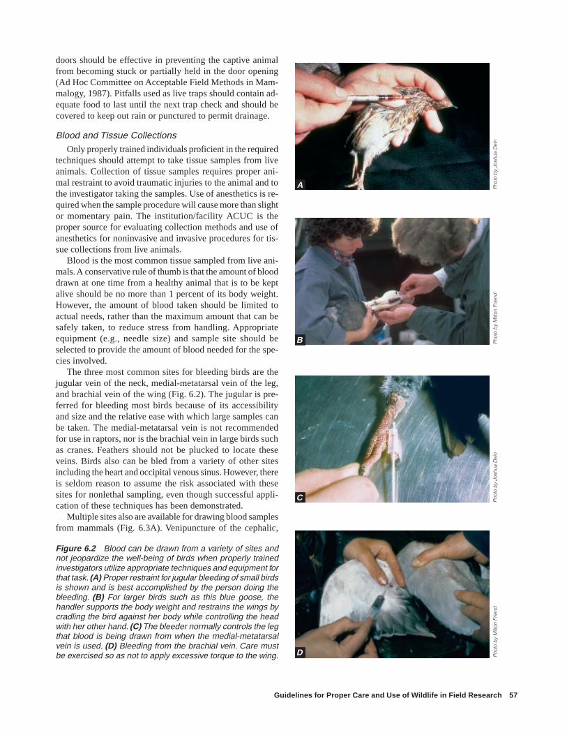
Guidelines for Proper Care and Use of Wildlife in Field Research 57
doors should be effective in preventing the captive animalfrom becoming stuck or partially held in the door opening(Ad Hoc Committee on Acceptable Field Methods in Mam-malogy, 1987). Pitfalls used as live traps should contain ad-equate food to last until the next trap check and should becovered to keep out rain or punctured to permit drainage.
Blood and Tissue CollectionsOnly properly trained individuals proficient in the required
techniques should attempt to take tissue samples from liveanimals. Collection of tissue samples requires proper ani-mal restraint to avoid traumatic injuries to the animal and tothe investigator taking the samples. Use of anesthetics is re-quired when the sample procedure will cause more than slightor momentary pain. The institution/facility ACUC is theproper source for evaluating collection methods and use ofanesthetics for noninvasive and invasive procedures for tis-sue collections from live animals.
Blood is the most common tissue sampled from live ani-mals. A conservative rule of thumb is that the amount of blooddrawn at one time from a healthy animal that is to be keptalive should be no more than 1 percent of its body weight.However, the amount of blood taken should be limited toactual needs, rather than the maximum amount that can besafely taken, to reduce stress from handling. Appropriateequipment (e.g., needle size) and sample site should beselected to provide the amount of blood needed for the spe-cies involved.
The three most common sites for bleeding birds are thejugular vein of the neck, medial-metatarsal vein of the leg,and brachial vein of the wing (Fig. 6.2). The jugular is pre-ferred for bleeding most birds because of its accessibilityand size and the relative ease with which large samples canbe taken. The medial-metatarsal vein is not recommendedfor use in raptors, nor is the brachial vein in large birds suchas cranes. Feathers should not be plucked to locate theseveins. Birds also can be bled from a variety of other sitesincluding the heart and occipital venous sinus. However, thereis seldom reason to assume the risk associated with thesesites for nonlethal sampling, even though successful appli-cation of these techniques has been demonstrated.
Multiple sites also are available for drawing blood samplesfrom mammals (Fig. 6.3A). Venipuncture of the cephalic,
Figure 6.2 Blood can be drawn from a variety of sites andnot jeopardize the well-being of birds when properly trainedinvestigators utilize appropriate techniques and equipment forthat task. (A) Proper restraint for jugular bleeding of small birdsis shown and is best accomplished by the person doing thebleeding. (B) For larger birds such as this blue goose, thehandler supports the body weight and restrains the wings bycradling the bird against her body while controlling the headwith her other hand. (C) The bleeder normally controls the legthat blood is being drawn from when the medial-metatarsalvein is used. (D) Bleeding from the brachial vein. Care mustbe exercised so as not to apply excessive torque to the wing.
Pho
to b
y M
ilton
Frie
ndP
hoto
by
Josh
ua D
ein
Pho
to b
y M
ilton
Frie
ndP
hoto
by
Josh
ua D
ein
A
B
C
D

58 Field Manual of Wildlife Diseases: Birds
femoral, tarsal, or jugular vein, the orbital sinus, or variousvenous plexuses are common procedures. In some instancescardiac bleeding also is acceptable. Need for anesthesia withany of these procedures depends upon methods of restraint,species being bled, physical condition of the animal, andvolume of blood needed. In reptiles, such as turtles, sites forblood collection are more limited (Fig. 6.3B).
Restraint and Handling
GeneralSafety of both wild animals and scientists who are study-
ing them should be the primary consideration when physicalcontact between them is judged to be necessary and unavoid-able. Nondomesticated animals almost without exception willtry to elude capture, handling, and restraint. The means bywhich a particular animal may try to prevent capture willvary with the species, sex, physiologic condition, and tem-perment of the individual. In attempts to elude capture, wildanimals are capable of inflicting severe damage to themselvesand their potential captors.
Behavioral characteristics of wild animals often may beused to assist the potential captor. For instance, animals in asmall pen or cage often voluntarily will enter a smaller con-tainer to hide and evade capture. If that container providesadequate restraint, the potentially dangerous work of secur-ing the animal can be accomplished more easily. Every ef-fort involving contact between wild animals and humansshould be carefully conceived and skillfully executed. Per-sonnel involved must know the habits and behaviors of theanimal to be handled; the plan must have suitable alterna-tives; and a genuine regard for the physical, physiological,and psychological welfare of the animal must be of deepconcern to those actually handling the animals. If the plannedand alternate procedures do not appear to be satisfactory, theresponsible thing to do is cease immediately and return tothe planning stage. Trying to enforce unworkable proceduresin a particular situation is a virtual guarantee of injury toeither the animals or the humans involved.
Physical RestraintFor many situations physical restraint is the most appro-
priate method of animal handling, because of risks fromchemical immobilization to the animal and humans whenpotentially toxic drugs are used. When physical restraint isselected, an adequate number of sufficiently trained andequipped personnel must be available to complete the tasksafely. Location and type of capture, as well as proceduresto be performed and time required to accomplish them, willinfluence the particular type of physical restraint. Gloves,catch poles, ropes, nets, body bags, holding boxes, corrals,squeeze chutes, or more sophisticated mechanical holdingdevices may be required for specific situations (Fig. 6.4).
For some highly excitable or anatomically fragile species,prolonged physical restraint without some chemical tran-
quilization may result in self-inflicted trauma, physiologicaldisturbances, or, occasionally, death. Investigators have anobligation to make every effort to avoid physical restraintprocedures that result in cardiogenic shock, capture myopa-thy, and other stress-induced causes of mortality in their ani-mal subjects (Fig. 6.5). Stress-related damage may not beimmediately apparent but may lead to debility or death afterrelease.
Chemical RestraintUse of chemicals or drugs to render a wild and poten-
tially dangerous animal safe to handle has many applicationsin wildlife research and management (Pond and O’Gara,1994). Use of anesthetics, analgesics, and sedatives is man-datory for the control of pain and distress before potentiallypainful procedures such as surgery are performed on ani-mals. Use of drugs and “tranquilizer guns,” however, is notthe panacea to wild-animal restraint. Chemicals used fortranquilization and immobilization, if not correctly handledand delivered, may be dangerous to the target animals andhumans (Fig. 6.6). In addition, during the drug inductionphase or during recovery, an unrestrained animal may besubject to increased potential for accidental injury or deathincluding predation. While under the effects of the drug theanimal may become hyper- or hypothermic, depending onchemicals used and ambient temperature, it may vomit andaspirate the vomitus, or pregnant females may abort. A dartedanimal may be able to elude its captors and hide beforebeing completely anesthetized, a particularly acute hazardwhen chemicals are employed that require administration ofan antidote. All of these circumstances and possibilities mustbe understood and evaluated by the researcher before a chemi-cal is selected as the best method of restraint in a given in-stance.
If chemical restraint is selected, it is imperative for allmembers of the capture team to have a working knowledgeof the chemical or drugs being used, even if they are to behandled and delivered by a veterinarian. It also is the re-sponsibility of researchers to know the effects, side effects,advantages, and disadvantages of the drugs being used, andto have knowledge of such factors as the minimum and maxi-mum induction times and potential for adverse drug reac-tions. This type of information is necessary to evaluate thedanger to target animals, and to humans that might be ex-posed to the drugs. Searchers should be capable of monitor-ing the condition of anesthetized animals and be able to ap-ply resuscitative routines in a life-threatening emergency.Specific recommendations for drug use and their dosage, drugdelivery systems, and physical restraint techniques applicableto the specific species are available in the published litera-ture (Pond and O’Gara, 1994). Information on use of thesemethods exists in guidelines on acceptable field techniquesby various professional societies (See “Professional societyguidelines” at the end of this chapter).
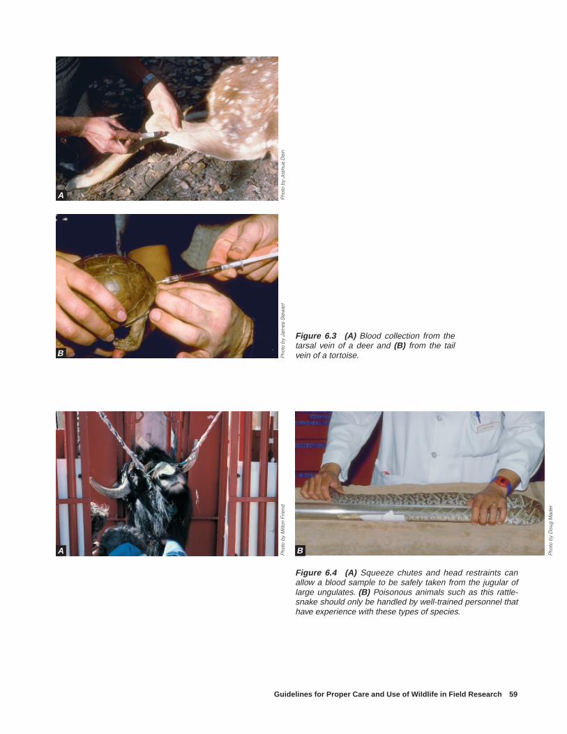
Guidelines for Proper Care and Use of Wildlife in Field Research 59
Figure 6.3 (A) Blood collection from thetarsal vein of a deer and (B) from the tailvein of a tortoise.
Figure 6.4 (A) Squeeze chutes and head restraints canallow a blood sample to be safely taken from the jugular oflarge ungulates. (B) Poisonous animals such as this rattle-snake should only be handled by well-trained personnel thathave experience with these types of species.
Pho
to b
y M
ilton
Frie
ndP
hoto
by
Josh
ua D
ein
Pho
to b
y Ja
mes
Ste
war
t
Pho
to b
y D
oug
Mad
er
A
B
A B

60 Field Manual of Wildlife Diseases: Birds
Figure 6.5 (A) The pale coloration of the muscle tissue of the right leg and discolored areas of muscletissue in the left leg of this whooping crane are lesions of capture myopathy due to stress associated withimproper/extended restraint. (B) The light area in this piece of leg muscle from an antelope is also due tocapture myopathy.
Pho
tos
by
Julie
Lan
gen
ber
g, I
nter
natio
nal C
rane
Fou
ndat
ion
A
B

Guidelines for Proper Care and Use of Wildlife in Field Research 61
Figure 6.6 Extensive tissue damage andhemorrhage, such as seen in the tissues ofthis black bear, can occur from immobilizationwith a CO2 projected dart.
Pho
to b
y M
ilton
Frie
nd
If the marking process causes pain or distress, as definedby the Animal Welfare Act, appropriate analgesics or anes-thetics should be used.
Criteria for MarkingWhen answers to the four initial questions lead to a deci-
sion to initiate an animal-marking program, researchers mustsearch among a wide array of potential techniques with vary-ing strengths and weaknesses to select the method(s) mostsuited to their particular project (Nietfeld and others, 1994).Technological and methodological constraints and availableresources can vary widely from project to project and willrequire each researcher to examine each potential markingtechnique in terms of a standard set of criteria. Specific cri-teria relate to impacts of marking on the organism, validityof the study, and other constraints such as legal requirements.
Evaluation criteria for marking techniques
The following are essential criteria for evaluation ofmarking techniques:
1. Marks should have minimal effect on the anatomyand physiology of the organism, i.e., no immediate orlong-term physical hindrance.
2. Marks should not influence the organism’s behav-ior, i.e., they should not reduce an organism’s abilityto secure food or inhibit breeding activity (unless themarks are intended as a reproductive inhibitor).
3. Marks that make an organism more conspicuousmust be evaluated carefully to ensure that they neithercause others of the same species to react differently toit than to other conspecifics nor subject it to increasedselection by potential predators (unless this is a pur-pose of the study) (Fig. 6.7).
4. Marks should be retained for the minimal periodrequired to achieve project goals.
5. Unambiguous marks that are quick and easy to ap-ply should be selected to avoid extensive handling orerror potential.
6. Marks must comply with Federal, State, and otheragency rules and regulations.
The first three criteria focus on the well-being of the or-ganism being studied and the potential for marks to influ-ence research results by affecting the fitness or behavior ofthe organisms. Criteria 4 and 5 may affect the validity of theresearch design, and criterion 6 reflects other constraintsplaced upon the researcher. Violation of any of the first fivecriteria may result in biased research results, so researchersshould specifically address these criteria in any evaluationof research resulting from a sample of marked organisms.
Although marks that may be applied to organisms arecommonly perceived as passive and visual, markers also
Animal MarkingDeveloping means of reliably identifying individual ani-
mals to achieve field research objectives often is necessary.In addition to requiring individual identification, research-ers may need information on nonconspicuous aspects ofphysiology or movements, or other aspects of animal ecol-ogy that can be determined directly or indirectly throughspecially designed markers.
Consideration for animal marking
Before initiating any marking procedure for wild ani-mals, researchers must resolve the following questionsto determine whether marking is required and appro-priate for the particular situation.
1. Do naturally occurring differences in the morphol-ogy of the animals under consideration provide suffi-cient identification to achieve research objectives?
2. How may animals must be individually identifiable?
3. If animals must be physically marked, can a suffi-cient number of animals be marked in the time avail-able?
4. Are the risks (to both the animal and researcher)associated with capture, handling, and marking, andsubsequent well-being, minimal and acceptable in bothresponsible and scientific contexts?

62 Field Manual of Wildlife Diseases: Birds
Figure 6.7 (A) Color marking waterfowl should be done with rapidly drying paints and (B and C)the painted feathers held separated until the paint dries to prevent the feathers from sticking to oneanother and hindering normal flight.
Pho
tos
by
Milt
on F
riend
exist that are active and visual (lights), that are auditory, thatfeature radiotelemetry, or that rely on chemical detection.A vast literature exists of techniques and potential concernsregarding the marking of organisms from insects to whales,and it has been summarized in detail elsewhere (see “Profes-sional society guidelines”; Day and others, 1980; Orlans,1988).
Other Professional and Ethical ConsiderationsMany organisms of interest to wildlife professionals are
free-ranging and may be enjoyed by other segments of soci-ety in many ways, from observation or photography to har-
vest as meat or trophies. Professional ethics dictate that thoseother potential uses of organisms be considered and accom-modated insofar as possible. Wild animals and birds are val-ued in part because they are wild, and the presence of human-caused marks may detract from that value. Accordingly, short-lived and inconspicuous marks should be selected wheneverthey can meet the objectives of proposed research. Scientistshave an ethical responsibility to attempt to remove collars orother external markers at the conclusion of their research ifpossible and feasible. Furthermore, professional and ethicalconsiderations dictate that permanent markers that injure orchange the appearance of an animal (e.g., toe-clipping, brand-
A B
C

Guidelines for Proper Care and Use of Wildlife in Field Research 63
ing, and tattooing) be employed only under the most humaneconditions and when alternate methods are not available toachieve desired research objectives.
Housing and Maintenance of Field Sites
GeneralProper care and responsible treatment of incarcerated ani-
mals must depend on scientific and professional judgement,on concern for the animal, on knowledge of animal behaviorand animal husbandry, and on familiarity with the species.Investigators working with species unfamiliar to them shouldobtain all pertinent information before confining those ani-mals. It also may be necessary to test and compare severalmethods of housing to determine the most appropriate onefor the well-being of the animal and the purpose of the study.Findings should be part of a permanent record system andanimal logbook associated with the study and the mainte-nance facility.
HousingHousing for wild vertebrates should approximate natural
conditions as closely as possible. Housing should providesafety and comfort for the animal as well as meet the studyobjectives. Methods of housing should provide for behav-ioral needs, safety, adequate exercise and rest, and condi-tions for the general well-being of the animal. Considerationsdepend on the animal involved and include isolation or ref-uge areas, natural materials, dust and water baths, naturalfoods, sunlight, and fresh air. Housing should incorporate asmany aspects of natural living as possible, such as brushyareas for escape, resting cover, shade and protection fromenvironmental elements, a natural stream traversing the pen,rocky areas for hoofed animals that need to wear down theirhooves, and social groups of animals kept together. Housingof compatible species in a common pen also will provide forsocial interaction. Frequency of cleaning should be a com-promise between level of cleanliness necessary to preventdisease and amount of stress imposed by cleaning (Fig. 6.8).
In general, housing must be of adequate size to allow forthe physical and behavioral needs of the animals, whileallowing scientists to collect appropriate data. For many hous-ing situations, the pen can be large and natural, with a smallerinternal or attached catch pen to restrain animals for experi-mental techniques. Pen construction materials must providefor the safety of the animals, as well as prevent the animalsfrom escaping. Materials should be of sufficient durabilityto last for the intended period of confinement. When long-term confinement (weeks or longer) is necessary, or pens areto be reused, materials with impervious surfaces should beused to facilitate sanitation and minimize the potential forsurvival of animal pathogens. All animals that are inherentlydangerous, are environmentally injurious, or have a propen-sity for escape require special attention. Double walls or
double enclosures, covered tops of enclosures, and construc-tion with metal bars or chain link may be required, depend-ing on the species. Mesh size and spacing between fencingmaterials must be small enough to prevent the head of ananimal from extending through the fence. Smaller fencingmesh also is more visible to animals. Colored flagging ma-terial may be necessary for animals to visualize fencing un-til they become accustomed to it. Animals should be releasedinto the housing in a calm and unstressed manner so thatinitial mortality and morbidity from fence encounters areminimal. A small dose of tranquilizer often will reduce theimmediate flight response when an animal is released intothe housing and may help prevent initial injuries. Once ani-mals have investigated the limits of the housing, injury oc-currence is minimized if investigators do not cause undo flightreactions.
Adequacy of housing often can be judged on normal be-havior patterns, weight gains and growth, survival rates, re-productive success, and physical appearance of the animalsinvolved in the research project. Established guidelines forhousing laboratory and farm animals were provided by theCanadian Council on Animal Care (1980, 1984). Additionalguidelines for housing requirements of fish, amphibians rep-tiles, wild birds, and small mammals were reported by theappropriate professional societies and appear in the AnimalWelfare Act (see also “Professional society guidelines” atthe end of this chapter).
NutritionNutrition must meet the needs of the animal unless devia-
tions are an approved purpose of the investigation. Research-ers are responsible for determining the appropriate nutritionalneeds of study animals prior to placing them in confinementand for obtaining adequate food supplies to sustain the ani-mals during the period of confinement. Feeding and water-ing should be under the direct supervision of an individual
Figure 6.8 A high quality enclosure for New Zealand blackstilt that approximates several aspects of the natural habitatand provides safety and comfort for the birds.
Pho
to b
y M
ilton
Frie
nd

64 Field Manual of Wildlife Diseases: Birds
trained and experienced in animal care for the species beingmaintained. Animal care personnel must be familiar with theanimals being studied so abnormalities in appearance andbehavior that may be indicative of nutritional deficienciescan be recognized quickly.
Transportation
General ConsiderationsA variety of vehicles such as conventional motor vehicles,
all-terrain vehicles, snow machines, rotary and fixed-wingaircraft, and boats are used to transport wild animals. Thespecies involved, method of transportation selected, andlength of time an animal is to be transported are importantfactors regarding the type of care and conditions of contain-ment required to maintain the animal in a state of well-
being (Fig. 6.9). To the extent possible, selection of trans-portation vehicles should take into account maintenance ofthe animal in a comfortable environment. Veterinary assis-tance may be required to prescribe and administer appropriatetranquilizers or other drugs when conditions of transporta-tion are likely to result in a high level of stress to the animaldue to its behavioral and physiological characteristics, re-strictions of confinement, engine noise, and rigors of the trip.The transportation process should be as brief as possible.This can be expedited by proper and adequate planning toassure that transportation vehicles and housing units in ap-propriate numbers and size are available and ready for useas needed; that food, water, bedding, and other needs to pro-vide for the animals also are available; that individuals in-volved in the transportation process are trained in the proce-dures to be used in containment and transportation of the
Figure 6.9 (A) Restraint of big-horn sheep being translocatedvia helicopter. The legs havebeen immobilized to prevent in-jury to the animal and holders.(B) Blinders on this caribou re-duces stress from the presenceof humans. Legs are restrainedsimilar to the procedure shownfor the bighorn sheep.
Pho
tos
by
Julie
Lan
gen
ber
g, I
nter
natio
nal C
rane
Fou
ndat
ion
A
B

Guidelines for Proper Care and Use of Wildlife in Field Research 65
animals; and that all permits, health certificates, and otherpaperwork have been completed to the extent possible.
When interstate movement of animals or shipment bycommercial carriers is involved, scheduling of transporta-tion segments to minimize the number of transfers and de-lays between transfers, having someone involved with theproject meet the shipment at each transfer point, and, whenappropriate, arranging for prompt clearance of animals byveterinary and customs inspectors can result in major reduc-tions in transit time. The receiving party should be on-sitewhen the animals reach their destination.
For some species, periodic rest periods are required toallow the animals to feed undisturbed. Other species are besttransported when they are normally inactive and do not feed.Ventilation within the housing unit and transportation ve-hicle should provide for adequate air movement to keep ani-mals comfortable and avoid buildup of exhaust gases. Sub-dued lighting and visual barriers between animals and hu-mans and between animals and their transportation environ-ment should be provided to help keep the animals calm. TheU.S. Fish and Wildlife Service has published rules for theHumane and Healthful Transport of Wild Animals and Birdsto the United States (see Fed Reg. 50 CFR Part 14).
Confinement During ShippingAnimal containers should be inspected to assure they have
no sharp edges, protrusions, or rough surfaces that couldcause injury during transport. When appropriate, containersalso should be padded to help prevent injury. The floor ofshipping containers should allow reasonable footing to pre-vent falling due to a slippery surface. Also, containers shouldnot have coatings or be constructed of materials that are toxicand could be consumed by the animal through licking orchewing during transportation. In general, housing units ofporous materials, such as cardboard boxes, should not bereused; all other containers used to house animals should besuitably disinfected between uses (Fig. 6.10). That portionof the transportation vehicle used to contain the housing unitsalso should be disinfected.
Grouping or separation of animals being transported atthe same time should take into consideration the species, age,and other appropriate factors. Direct contact generally shouldbe maintained between females and their dependent young,particularly if abandonment may result (unless the youngare to be maintained by some other means). Birds should beisolated in separate cells within the shipping container; ifthis cannot be done, each individual should have sufficientspace to assume normal postures and engage in comfort andmaintenance activities unimpeded by other birds (Ad HocCommittee on the Use of Wild Birds in Research, 1988).
Health AspectsFor short-term transportation (less than 30 min), basic con-
siderations are to prevent pain, injury, and undue stress. Ther-
moregulation capabilities of the species must be consideredwhen an animal is removed from its existing environmentand placed in the transportation environment. Transportedanimals should be protected from exposure to inclementweather, harsh environmental conditions, and major tempera-ture fluctuations and extremes.
Bedding, feed, and water should be provided, as appro-priate, and the animals should be observed periodically todetermine their state of well-being during transportation. On-site veterinary assistance may be warranted to monitor ani-mals and to provide life-support assistance should a medicalemergency occur during transportation or at the release orfield study site. Selection of veterinary assistance shouldfocus on the individual’s knowledge and experience with thewildlife species involved. Any animals that die during tran-sit should be removed as soon as practical from the sight andolfactory detection of other animals being transported. Thesecarcasses should be retained for pathological examinationsregarding cause of death. Similarly, animals that becomeseverely injured or clinically ill should be removed and re-sponsibly euthanized. Euthanasia should not take place inthe presence of other live animals. Sick animals disposed ofin this manner also should be retained for pathological as-sessments. Determinations of cause of death are needed toassess whether the remaining animals are at risk from patho-gens associated with the dead animals.
Surgical and Medical Procedures
Guidelines for wildlife medical procedures
Wildlife field research can involve surgical and medi-cal procedures such as implanting radio transmittersand surgical sex determination in birds. Incorporationof such techniques into a research protocol should fol-low these guidelines:
1. Surgical and medical techniques used should bebased on accepted protocols for the studied species orfor the most closely related domesticated species. TheCanadian Council on Animal Care’s (1984) Guide tothe Care and Use of Experimental Animals, Volume 2,is a good source of such information.
2. Protocols should be developed and, if possible,implemented in collaboration with a qualified veteri-narian. Only properly trained personnel, conversant inall techniques necessary, should conduct the proce-dures.
3. Protocols must be reviewed carefully by the ACUCwith special attention paid to limiting pain during theactual procedure and post-procedure period.
4. Adequate anesthesia and/or analgesia must be pro-vided.
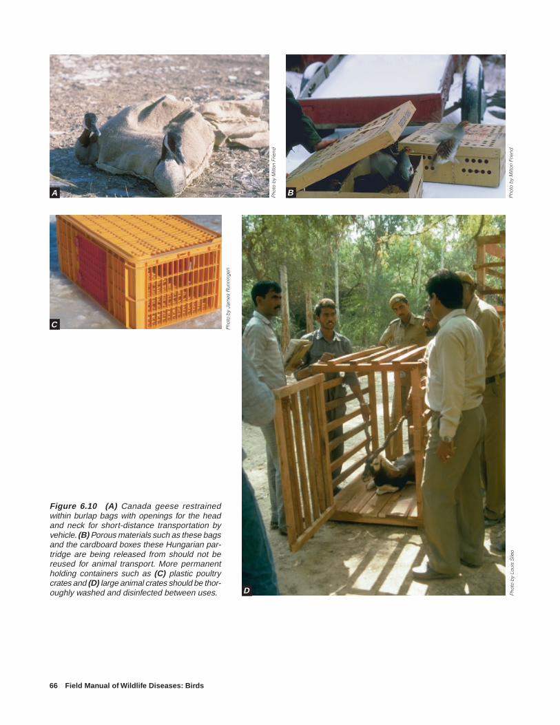
66 Field Manual of Wildlife Diseases: Birds
Figure 6.10 (A) Canada geese restrainedwithin burlap bags with openings for the headand neck for short-distance transportation byvehicle. (B) Porous materials such as these bagsand the cardboard boxes these Hungarian par-tridge are being released from should not bereused for animal transport. More permanentholding containers such as (C) plastic poultrycrates and (D) large animal crates should be thor-oughly washed and disinfected between uses. P
hoto
by
Loui
s S
ileo
Pho
to b
y M
ilton
Frie
nd
Pho
to b
y M
ilton
Frie
nd
Pho
to b
y Ja
mes
Run
ning
en
A B
C
D

Guidelines for Proper Care and Use of Wildlife in Field Research 67
Minor ProceduresMinor medical procedures such as collection of blood,
administration of drugs intravenously or intramuscularly,biopsies of superficial structures such as skin, and suturedattachment of radio transmitters usually can be performedsafely and responsibly in the field without complicated equip-ment. However, it is the researcher’s responsibility to choosethe least invasive and least painful technique, minimize theduration of the procedure, use the most appropriate equip-ment and aseptic technique, and provide analgesia or seda-tion when indicated.
Major ProceduresAs defined by the Animal Welfare Act, major operative
procedures are (p. 36,121) “any surgical intervention thatpenetrates and exposes a body cavity or any procedure whichproduces permanent impairment of physical or physiologi-cal functions.” Major surgical procedures, when survival ofthe animal is intended, should be performed only under properanesthesia and with sterile technique. Examples of majorprocedures used in wildlife research include laparotomy,surgical flight restraint, and sterilization. These proceduresshould be performed only in a clean space set aside for ster-ile surgery, with surgical instruments and drapes of the propertype, and with anesthesia protocols judged to be safe andresponsible for the species involved. Necessary equipmentand trained personnel to deal with surgery or anesthesia-related emergencies (i.e., severe blood loss, cessation ofbreathing or cardiac function, severe hypo- or hyperthermia,acid-base imbalances) should be available at all times. Thiswill maximize the success and subsequent scientific returnfrom those often costly procedures and, therefore, minimizethe number of animals needed and amount of animal dis-tress (Fig. 6.11).
Medical ConsiderationsWildlife field researchers should have access to veteri-
nary consultation and take responsibility to prepare them-selves to deal with any health problems that might arise intheir study population. Sometimes intervention and controlof a natural disease process may not be advisable and mayinterfere with the study’s goals. However, if the health prob-lem arises due to the researcher’s work, or if it will interferewith the study, the researcher must be ready to respond. Prepa-rations should include gaining familiarity with the commondiseases and health problems of the species under study, es-tablishing a contact with a veterinary consultant, and havingappropriate treatment or control equipment and drugs on handor easily accessible. The researcher also is responsible forevaluating the possible impact of disease in the study ani-mals on the larger population or ecosystem as a whole, andfor making the maintenance of their welfare a priority asdecisions are made. This is especially true when release ortranslocation of animals is part of a study; disease must beconsidered in evaluating the advisability of the program.
EuthanasiaEuthanasia is defined under the Animal Welfare Act as
(p. 36,121) “the humane destruction of an animal accom-plished by a method that produces rapid unconsciousnessand subsequent death without evidence of pain or distress,or a method that utilizes anesthesia produced by an agentthat causes painless loss of consciousness and subsequentdeath.” Euthanasia may not be an approved component of afield study, but it may become a necessary health care optionin a study involving capture, restraint, or surgical procedures.Therefore, all wildlife researchers involved in invasive stud-ies must be familiar with the approved euthanasia methodsfor their study species (Andrews and others, 1993) and havethe appropriate equipment/drugs on hand so euthanasia canbe performed quickly.
Disease ConsiderationsField investigators need to be fully aware of disease con-
cepts so they may avoid introduction of new disease prob-lems into animal populations or the spread of disease to otherpopulations and locations as a result of their studies. Dis-ease introductions and spread occur as a result of animalsbrought to the field research site to serve as biological senti-nels, as decoys to lure and capture other animals, for speciesintroductions or releases to supplement existing populations,for behavioral studies, for assistance in tracking or retriev-ing animals, and for other purposes. All of these uses of ani-mals involve acceptable methods for scientific research andwildlife management. However, under no circumstancesshould the well-being of free-ranging wildlife populationsbe unduly jeopardized by disease risks associated with ani-mal use in field research. Field investigators have ethical and
Figure 6.11 Invasive surgical procedures should be doneonly by properly trained personnel knowledgeable of tech-niques necessary to successfully carry out the procedure andappropriately respond to medical emergencies that mightarise.
Pho
to b
y J.
Chr
istia
n Fr
anso
n

68 Field Manual of Wildlife Diseases: Birds
professional obligations to take appropriate actions for mini-mizing the introduction of the following: (a) new diseaseagents, (b) vectors (e.g., ticks and internal parasites) capableof efficiently transmitting indigenous, dormant diseases orthose not currently being effectively transmitted, and (c) spe-cies that can serve as amplification hosts for transmittingindigenous diseases to other species (Fig. 6.12).
In addition, animals that are highly susceptible to diseasesindigenous to the study location should not be released intothe wild without using applicable prophylactic measures,unless these animals are to serve as biological sentinels fordisease investigations. Biological sentinels should be moni-tored closely and euthanized by approved, responsible meth-ods as soon as is practical after study objectives have beenmet.
Disease introduction and spread can result from mechani-cal means such as contaminated personnel, supplies, andequipment in addition to the biological processes identifiedabove. Steps taken to address disease prevention are far morecost effective than disease control activities initiated after aproblem has developed.
Figure 6.12 Wildlife are often referred to as a “biological package” as the relocation of animals mayinvolve life forms other than the animals themselves. The ticks feeding on this velvet covered antler couldbe disease carriers. Once introduced into a new area, the ticks may become an important vector fortransmission of an indigenous disease. Disease potential is an important consideration that should beadequately addressed when translocating wildlife.
Pho
to b
y M
ilton
Frie
nd

Guidelines for Proper Care and Use of Wildlife in Field Research 69
Wildlife disease prevention during field research
Protection of free-ranging wildlife from disease is aided by the following actions:
1. Appropriate health certification should be required for all animals being brought to the site offield investigations. State veterinary officials should be contacted to determine what specific test-ing must be done when animals are moved into their jurisdiction.
2. Appropriate disinfection procedures should be used for investigators and their equipment whendisease risks are present.
3. Prior knowledge of disease activity at the study site should be obtained to guide actions involv-ing the research study.
4. Source for any animals being brought to a field investigation site (captive-reared and relocatedwild stock) should be evaluated for inherent disease problems, and appropriate steps should betaken to avoid disease introductions.
5. To the extent possible, animals should be held under surveillance for 15–30 days prior to theirrelease into the wild, and only healthy animals should be released. These animals should not bemixed with other species during transportation and should be isolated from other animals duringthe surveillance period.
6. Any animals that die should be examined by a disease diagnostic laboratory having compe-tency for determining cause of death in the species involved; these findings should be used toguide appropriate actions (Fig. 6.13).
7. Animals that become clinically ill should be examined by disease specialists, and their counselshould be used to protect the well-being of other animals within the study area.
Figure 6.13 Timely diagnosis of causes of wildlife morbidity and mortality is in-valuable for the detection of emerging hazards that can jeopardize the well-being ofthe population being studied and may be of great potential consequences. Submis-sion of animals that die to competent laboratories provides information useful forintervention.
Pho
to b
y Ja
mes
Run
ning
en

70 Field Manual of Wildlife Diseases: Birds
Animal Disposition at Completionof Study
When live animals are in the possession of investigatorsor under their control at the time of study completion, anevaluation must be made as to whether these animals can bereleased to a free-ranging existence, should be maintainedunder controlled conditions, or should be euthanized.
Animal release guidelines
As a general rule, field-captured animals should be re-leased only:
1. At the site of the original capture, unless conserva-tion efforts or safety considerations dictate otherwise.Prior approval for releases at noncapture sites shouldbe obtained from appropriate State/Federal agencies.Relocation release sites should be within the nativerange of the species, or established range for introducedspecies, and be in habitat suitable for species survival;
2. When the released animal can be reasonably ex-pected to function normally within the population;
3. When local and seasonal conditions are conduciveto survival;
4. When the ability to survive in nature has not beenirreversibly impaired; and
5. When release is not likely to spread pathogens orcontribute to disease processes in other ways.
The decision of whether to release captive-reared animalsinto the wild after completion of a field research project de-mands more rigorous evaluation than for field-captured ani-mals. In addition to evaluating the future well-being of theanimal being released, impacts on other animals of the samespecies and competition and risks for other species sharingthat environment also must be considered. Rarely, if ever,will releases of captive-reared animals at the completion ofresearch studies be justified on the basis of animal welfareconsiderations.
When animals are to be released, efforts should be madeto enhance their chances of survival. Animals should be ingood physical condition and released when weather condi-tions are favorable, at a time of day when they are able tolocate food and cover that meet survival needs.
Animals that cannot be released should be considered fordistribution to other scientists for further study. However, ifthe animal was subject to a major invasive procedure, it maynot be appropriate for additional experimentation. Animalsnot suitable for research may be suitable display animals thatcan be donated to a zoo or other type of educational institu-tion.
When animals must be euthanized, responsible methodsappropriate for the species and circumstances must be used.
Care must be taken to assure that the animal is dead beforedisposal of the carcass. Also, disposal procedures must pre-vent carcasses containing toxic substances or drugs from theresearch investigations or euthanasia procedures to enter thefood web of other animals. To the extent feasible, euthanizedanimals should be properly preserved and used as voucherspecimens or for teaching purposes.
Safety ConsiderationsResearchers working with free-ranging wildlife are sub-
ject to enhanced levels of exposure to wildlife diseases trans-missible to humans. Disease transmission may involvedirect contact with infected animals such as those withrabies, contact with disease vectors such as ticks transmit-ting Lyme disease, or contact with contaminated environ-ments such as bird roosts harboring histoplasmosis. Fieldinvestigators should become familiar with the common dis-eases of wildlife species they are working with and the rela-tive prevalence of those diseases in the populations they arestudying. Consultation with a physician regarding immuni-zation or other preventative treatment is advised when seri-ous diseases for humans commonly occur in the populationsbeing studied. Investigators who become ill should seekmedical assistance and advise their physicians of their expo-sure to potentially hazardous animals, diseases, and envi-ronmental conditions.
AcknowledgmentsThese guidelines were prepared by a committee of The
Wildlife Society appointed by J. G. Teer during his Presi-dency. The committee acknowledges the contributions of F.J. Dein for his review of these guidelines and valuable inputprovided in enhancing the final content.
Milton Friend, Dale E. Toweill, Robert L. Brownell, Jr.,Victor F. Nettles, Donald S. Davis, and William J. Foreyt
Literature CitedAd Hoc Committee on Acceptable Field Methods in Mammalogy,
1987, Acceptable field methods in mammalogy: Preliminaryguidelines approved by the American Society of Mammalo-gists: Journal of Mammalogy, v. 68, (supplement 4), 18 p.
Ad Hoc Committee on the Use of Wild Birds in Research, 1988,Guidelines for use of wild birds in research: Auk, v. 105,(supplement 1), 41 p.
American Society of Ichthyologists and Herpetologists (ASIH),The Herpetologists League (HL), and The Society for theStudy of Amphibians and Reptiles (SSAR), 1987, Guidelinesfor the use of live amphibians and reptiles in field research:Journal of Herpetology, v. 4, p. l–14.
Andrews, E.J., and others, 1993, Report of the AVMA panel oneuthanasia: Journal of American Veterinary Medical Associa-tion, v. 202, p. 229–249.

Guidelines for Proper Care and Use of Wildlife in Field Research 71
Animal Behavior Society/Animal Society for Animal Behavior,1986, ABS/ASAB guidelines for the use of animals inresearch: Animal Behavior Society Newsletter, v. 31, p. 7–8.
Bookhout, T.A., editor, 1994, Research and managementtechniques for wildlife and habitats, Fifth ed.,: The WildlifeSociety, Bethesda, Md., 740 p.
Canadian Council on Animal Care, 1980, Guide to the care anduse of experimental animals, v. 1: Canadian Council on AnimalCare, Ottawa, Ont., 120 p.
_____ 1984, Guide to the care and use of experimental animals.vol. 2: Canadian Council on Animal Care, Ottawa, Ont., 208 p.
Day, G.I., Schemnitz, S.D., and Taber, R.D., 1980, Capturing andmarking wild animals, in S. D. Schemnitz, ed. Wildlifemanagement techniques manual, Fourth ed., rev: The WildlifeSociety, Washington, D.C., p. 61–88.
Nietfeld, M.T., Barrett, M.W., and Silvy, N., 1994, Wildlifemarking techniques, in T. A. Bookhout, ed., Research andmanagement techniques for wildlife and habitats, Fifth ed.:The Wildlife Society, Bethesda, Md., p. 140–168.
Orlans, F.B., editor, 1988, Field research guidelines: impact onanimal care and use committees: Scientists Center for AnimalWelfare, Bethesda, Md., 23 p.
Orlans, F.B., Simmonds, R.C., and Dodds, W.J., eds., 1987,Effective animal care and use committees: Scientists Center forAnimal Welfare, Bethesda, Md., 178 p.
Pond, D.B. and O’Gara, B.W., 1994, Chemical immobilization oflarge mammals, in T. A. Bookhout, ed., Research and manage-ment techniques for wildlife and habitats, Fifth ed.: TheWildlife Society, Bethesda, Md., p. 140–168.
Professional society guidelines for use of live animalsin field research
Ad Hoc Committee on Acceptable Field Methods inMammalogy, 1987, Acceptable field methods inmammalogy: preliminary guidelines approved by theAmerican Society of Mammalogists: Journal of Mam-malogy, vol. 68, (supplement 4), 18 p.
Ad Hoc Committee on the Use of Wild Birds in Research,1988, Guidelines for use of wild birds in research: Auk,vol. 105, (supplement 1), 41 p.
American Society of Ichthyologists and Herpetologists(ASIH), American Fisheries Society (AFS), and theAmerican Institute of Fisheries Research Biologists(AIFRB), 1987, Guidelines for use of fishes in fieldresearch: Copeia 1987 (supplement), 12 p.
American Society of Ichthyologists and Herpetologists(ASIH), The Herpetologists League (HL), and TheSociety for the Study of Amphibians and Reptiles(SSAR), 1987, Guidelines for the use of live amphibiansand reptiles in field research: Journal of Herpetology,vol. 4, p. l–14.
Sources of assistance for technical information,implementation, and interpretation of the AnimalWelfare Act
Animal Welfare Information CenterNational Agricultural Library10301 Baltimore Ave., 5th FloorBeltsville, MD 20705–2351(301) 504-6212fax (301) 504-7125
National Library of Medicine8600 Rockville PikeBethesda, MD 20894(301) 594-5983
Scientists Center for Animal Welfare7833 Walker Dr., Suite 340Greenbelt, MD 20770–3229(301) 345-3500
U.S. Department of AgricultureAnimal and Plant Health Inspection Service4700 River Rd.Riverdale, MD 20737(301) 734-7833

72 Field Manual of Wildlife Diseases: Birds

Introduction to Bacterial Diseases 73
Section 2Bacterial Diseases
Avian Cholera
Tuberculosis
Salmonellosis
Chlamydiosis
Mycoplasmosis
Miscellaneous Bacterial Diseases
Inoculating media for culture of bacteriaPhoto by Phillip J. Redman

74 Field Manual of Wildlife Diseases: Birds
Introduction to Bacterial Diseases
Diseases caused by bacteria are a more common cause ofmortality in wild birds than are those caused by viruses. Inaddition to infection, some bacteria cause disease as a resultof potent toxins that they produce. Bacteria of the genusClostridium are responsible for more wild bird deaths thanare other disease agents. Clostridium botulinum, which causesavian botulism, is primarily a form of food poisoning and itis included within the section on biotoxins (see Chapter 38).Other Clostridium sp. that colonize intestinal tissues producetoxins that cause severe hemorrhaging of the intestine, thusleading to tissue death or necrosis and intoxication of thebird due to the exotoxins produced by the bacterial cell. Thedescriptive pathology is referred to as a necrotizing gastro-enteritis or necrotic enteritis and the disease as clostridialenterotoxemia. The classic example in gallinaceous birds suchas quail, turkey, pheasant, grouse, and partridge, is ulcer-ative enteritis or quail disease, which is caused by Clostridiumcolinium; quail are the species most susceptible to that dis-ease. Necrotic enteritis of wild waterbirds, especially geese,has been reported with increasing frequency during recentyears. Clostridium perfringens has been associated with thesedeaths.
The frequency of wild bird mortality events and the vari-ety of infectious bacterial diseases causing that mortality hasincreased greatly during recent decades. Avian cholera hasbecome the most important infectious disease of waterbirds,but it did not appear in North American waterfowl or otherwaterbirds until 1944. Most of the geographic expansion andincreased frequency of outbreaks of avian cholera has oc-curred since 1970. Avian tuberculosis is a historic disease ofcaptive birds, but it is relatively rare in North American wildbirds. The high prevalence of avian tuberculosis infectionthat has occurred since 1982 in a free-living foster-parented
whooping crane population has challenged the survival ofthat subpopulation of cranes. Salmonellosis has become amajor source of mortality at birdfeeders throughout theNation, and mycoplasmosis in house finches has become themost rapidly spreading infectious disease ever seen in wildbirds. This disease reached the Mississippi River and beyondwithin 2 years of the 1994 index cases in the Washington,D.C. area.
Avian botulism has also expanded in geographic distri-bution and has gained increased prominence as a disease ofwaterbirds. It is undoubtedly the most important disease ofwaterbirds worldwide. Much of the geographic expansionof avian botulism has occurred during the past quarter-century.
As a group, bacterial diseases pose greater human healthrisks than viral diseases of wild birds. Of the diseases ad-dressed in this section, chlamydiosis, or ornithosis, poses thegreatest risk to humans. Avian tuberculosis can be a signifi-cant risk for humans who are immunocompromised. Salmo-nellosis is a common, but seldom fatal, human infection thatcan be acquired from infected wild birds. This section pro-vides individual chapters about only the more common andsignificant bacterial diseases of wild birds. Numerous otherdiseases afflict wild birds, some of which are identified inthe chapter on Miscellaneous Bacterial Diseases included atthe end of this section.
Timely and accurate identification of causes of mortalityis needed to properly guide disease control operations. Themagnitude of losses and the rapidity with which those lossescan occur, as reflected in the chapters of this section, shouldbe a strong incentive for those who are interested in the con-servation of wild species to seek disease diagnostic evalua-tions when sick and dead birds are encountered. In order toaccurately determine what diseases are present, specimensneed to be sent to diagnostic laboratories that are familiarwith the wide variety of possible diseases that may afflictwild birds. Those laboratories must also have the capabilityto isolate and identify the causative agents involved. Severalsources of wildlife disease expertise that might be called uponwhen wildlife mortality occurs are identified within Appen-dix B .
“Consider the difference in size between some of the very tiniest and the very largest creatures onEarth. A small bacterium weighs as little as 0.00000000001 gram. A blue whale weighs about100,000,000 grams. Yet a bacterium can kill a whale…Such is the adaptability and versatility ofmicroorganisms as compared with humans and other so-called ‘higher’ organisms, that they willdoubtless continue to colonize and alter the face of the Earth long after we and the rest of ourcohabitants have left the stage forever. Microbes, not macrobes, rule the world.”(Bernard Dixon)
Quote from:
Garrett, L., 1994, The coming plague—Newly emerging diseasesin a world out of balance: Farrar, Straus, and Giroux, NewYork, N.Y., p. 411.
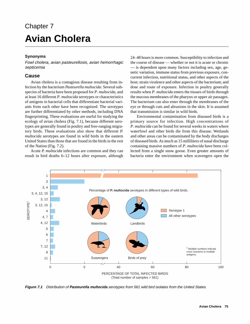
Avian Cholera 75
Chapter 7
Avian Cholera
SynonymsFowl cholera, avian pasteurellosis, avian hemorrhagicsepticemia
Cause Avian cholera is a contagious disease resulting from in-
fection by the bacterium Pasteurella multocida. Several sub-species of bacteria have been proposed for P. multocida, andat least 16 different P. multocida serotypes or characteristicsof antigens in bacterial cells that differentiate bacterial vari-ants from each other have been recognized. The serotypesare further differentiated by other methods, including DNAfingerprinting. These evaluations are useful for studying theecology of avian cholera (Fig. 7.1), because different sero-types are generally found in poultry and free-ranging migra-tory birds. These evaluations also show that different P.multocida serotypes are found in wild birds in the easternUnited States than those that are found in the birds in the restof the Nation (Fig. 7.2).
Acute P. multocida infections are common and they canresult in bird deaths 6–12 hours after exposure, although
24–48 hours is more common. Susceptibility to infection andthe course of disease — whether or not it is acute or chronic— is dependent upon many factors including sex, age, ge-netic variation, immune status from previous exposure, con-current infection, nutritional status, and other aspects of thehost; strain virulence and other aspects of the bacterium; anddose and route of exposure. Infection in poultry generallyresults when P. multocida enters the tissues of birds throughthe mucous membranes of the pharynx or upper air passages.The bacterium can also enter through the membranes of theeye or through cuts and abrasions in the skin. It is assumedthat transmission is similar in wild birds.
Environmental contamination from diseased birds is aprimary source for infection. High concentrations ofP. multocida can be found for several weeks in waters wherewaterfowl and other birds die from this disease. Wetlandsand other areas can be contaminated by the body dischargesof diseased birds. As much as 15 milliliters of nasal dischargecontaining massive numbers of P. multocida have been col-lected from a single snow goose. Even greater amounts ofbacteria enter the environment when scavengers open the
Figure 7.1 Distribution of Pasteurella multocida serotypes from 561 wild bird isolates from the United States.
0 5 40 60 80 100
PERCENTAGE OF TOTAL INFECTED BIRDS(Total number of samples = 561)
1
3
3, 4
3, 4, 12, 15
3, 12
3, 12, 15
4
4, 7
4, 12
5
6
7
7, 12
8
11
Percentage of P. multocida serotypes in different types of wild birds.
Waterbirds Landbirds
Scavengers Birds of prey
Serotype 1
All other serotypes
SE
RO
TY
PE
1
1 Multiple numbers indicate cross reactions to multiple antigens.
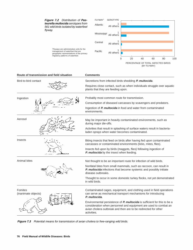
76 Field Manual of Wildlife Diseases: Birds
Figure 7.2 Distribution of Pas-teurella multocida serotypes from561 wild birds isolated by waterfowlflyway.
Figure 7.3 Potential means for transmission of avian cholera to free-ranging wild birds.
Route of transmission and field situation Comments
Bird-to-bird contact
Ingestion
Aerosol
Insects
Animal bites
Fomites(inanimate objects)
Secretions from infected birds shedding P. multocida.
Requires close contact, such as when individuals struggle over aquatic plants that they are feeding upon.
Probably most common route for transmission.
Consumption of diseased carcasses by scavengers and predators.
Ingestion of P. multocida in food and water from contaminated environments.
May be important in heavily contaminated environments, such as during major die-offs.
Activities that result in splashing of surface waters result in bacteria-laden sprays when water becomes contaminated.
Biting insects that feed on birds after having fed upon contaminated carcasses or contaminated environments (ticks, mites, flies).
Insects fed upon by birds (maggots, flies) following ingestion of P. multocida by the insect when feeding.
Not thought to be an important route for infection of wild birds.
Nonfatal bites from small mammals, such as raccoon, can result in P. multocida infections that become systemic and possibly initiate disease outbreaks.
Thought to occur in some domestic turkey flocks, not yet demonstrated in wild birds.
Contaminated cages, equipment, and clothing used in field operations can serve as mechanical transport mechanisms for introducing P. multocida.
Environmental persistence of P. multocida is sufficient for this to be a consideration when personnel and equipment are used to combat an avian cholera outbreak and then are to be redirected for other activities.
FLYWAY1 SEROTYPE
Atlantic 1All others
Mississippi 1All others
Central 1All others
Pacific 1All others
0 20 40 60 80 100
PERCENTAGE OF TOTAL INFECTED BIRDS (BY FLYWAY)
1Flyways are administrative units for the management of waterfowl that are geographic representations of the primary migratory patterns of waterfowl.
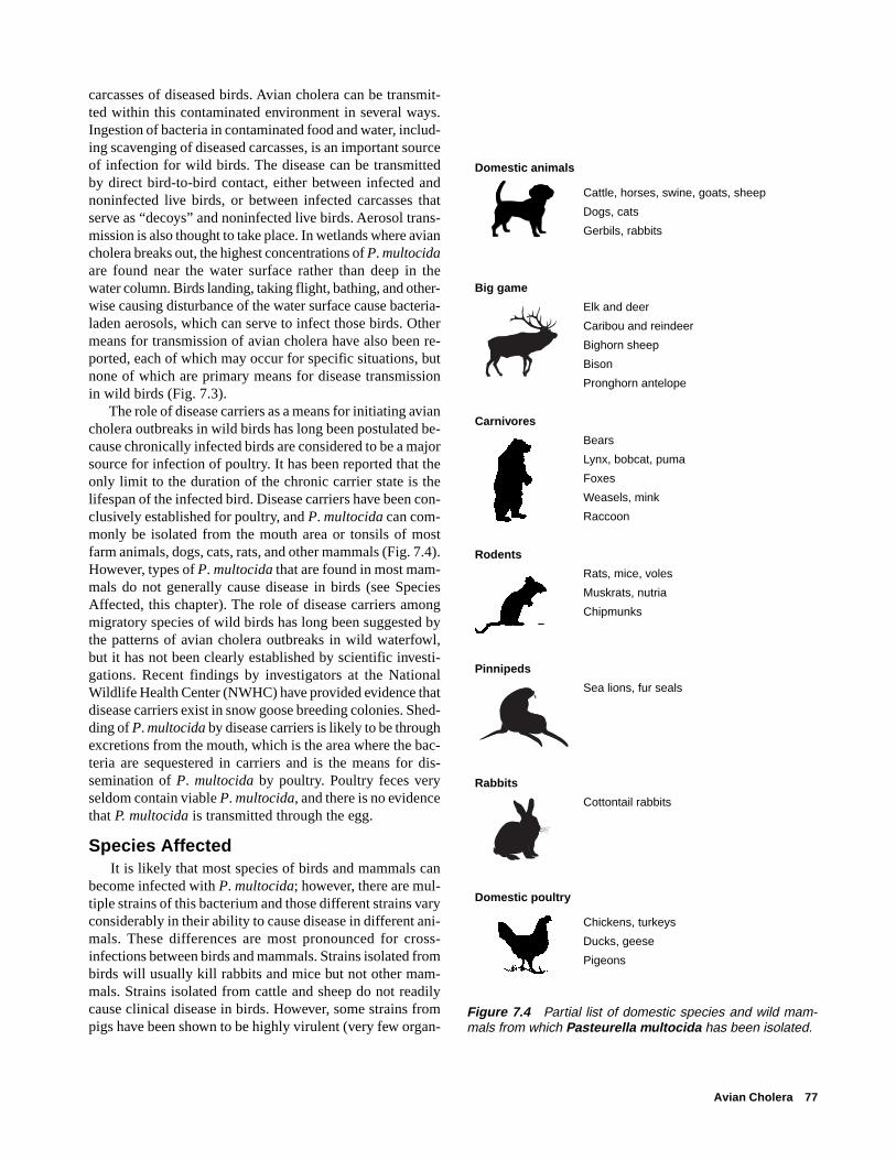
Avian Cholera 77
carcasses of diseased birds. Avian cholera can be transmit-ted within this contaminated environment in several ways.Ingestion of bacteria in contaminated food and water, includ-ing scavenging of diseased carcasses, is an important sourceof infection for wild birds. The disease can be transmittedby direct bird-to-bird contact, either between infected andnoninfected live birds, or between infected carcasses thatserve as “decoys” and noninfected live birds. Aerosol trans-mission is also thought to take place. In wetlands where aviancholera breaks out, the highest concentrations of P. multocidaare found near the water surface rather than deep in thewater column. Birds landing, taking flight, bathing, and other-wise causing disturbance of the water surface cause bacteria-laden aerosols, which can serve to infect those birds. Othermeans for transmission of avian cholera have also been re-ported, each of which may occur for specific situations, butnone of which are primary means for disease transmissionin wild birds (Fig. 7.3).
The role of disease carriers as a means for initiating aviancholera outbreaks in wild birds has long been postulated be-cause chronically infected birds are considered to be a majorsource for infection of poultry. It has been reported that theonly limit to the duration of the chronic carrier state is thelifespan of the infected bird. Disease carriers have been con-clusively established for poultry, and P. multocida can com-monly be isolated from the mouth area or tonsils of mostfarm animals, dogs, cats, rats, and other mammals (Fig. 7.4).However, types of P. multocida that are found in most mam-mals do not generally cause disease in birds (see SpeciesAffected, this chapter). The role of disease carriers amongmigratory species of wild birds has long been suggested bythe patterns of avian cholera outbreaks in wild waterfowl,but it has not been clearly established by scientific investi-gations. Recent findings by investigators at the NationalWildlife Health Center (NWHC) have provided evidence thatdisease carriers exist in snow goose breeding colonies. Shed-ding of P. multocida by disease carriers is likely to be throughexcretions from the mouth, which is the area where the bac-teria are sequestered in carriers and is the means for dis-semination of P. multocida by poultry. Poultry feces veryseldom contain viable P. multocida, and there is no evidencethat P. multocida is transmitted through the egg.
Species Affected It is likely that most species of birds and mammals can
become infected with P. multocida; however, there are mul-tiple strains of this bacterium and those different strains varyconsiderably in their ability to cause disease in different ani-mals. These differences are most pronounced for cross-infections between birds and mammals. Strains isolated frombirds will usually kill rabbits and mice but not other mam-mals. Strains isolated from cattle and sheep do not readilycause clinical disease in birds. However, some strains frompigs have been shown to be highly virulent (very few organ-
Figure 7.4 Partial list of domestic species and wild mam-mals from which Pasteurella multocida has been isolated.
Domestic animals
Cattle, horses, swine, goats, sheep
Dogs, cats
Gerbils, rabbits
Big game
Elk and deer
Caribou and reindeer
Bighorn sheep
Bison
Pronghorn antelope
Carnivores
Bears
Lynx, bobcat, puma
Foxes
Weasels, mink
Raccoon
Rodents
Rats, mice, voles
Muskrats, nutria
Chipmunks
Pinnipeds
Sea lions, fur seals
Rabbits
Cottontail rabbits
Domestic poultry
Chickens, turkeys
Ducks, geese
Pigeons
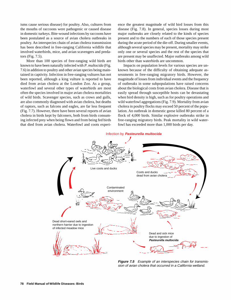
78 Field Manual of Wildlife Diseases: Birds
isms cause serious disease) for poultry. Also, cultures fromthe mouths of raccoons were pathogenic or caused diseasein domestic turkeys. Bite-wound infections by raccoons havebeen postulated as a source of avian cholera outbreaks inpoultry. An interspecies chain of avian cholera transmissionhas been described in free-ranging California wildlife thatinvolved waterbirds, mice, and avian scavengers and preda-tors (Fig. 7.5).
More than 100 species of free-ranging wild birds areknown to have been naturally infected with P. multocida (Fig.7.6) in addition to poultry and other avian species being main-tained in captivity. Infection in free-ranging vultures has notbeen reported, although a king vulture is reported to havedied from avian cholera at the London Zoo. As a group,waterfowl and several other types of waterbirds are mostoften the species involved in major avian cholera mortalitiesof wild birds. Scavenger species, such as crows and gulls,are also commonly diagnosed with avian cholera, but deathsof raptors, such as falcons and eagles, are far less frequent(Fig. 7.7). However, there have been several reports of aviancholera in birds kept by falconers, both from birds consum-ing infected prey when being flown and from being fed birdsthat died from avian cholera. Waterfowl and coots experi-
ence the greatest magnitude of wild bird losses from thisdisease (Fig. 7.8). In general, species losses during mostmajor outbreaks are closely related to the kinds of speciespresent and to the numbers of each of those species presentduring the acute period of the die-off. During smaller events,although several species may be present, mortality may strikeonly one or several species and the rest of the species thatare present may be unaffected. Major outbreaks among wildbirds other than waterbirds are uncommon.
Impacts on population levels for various species are un-known because of the difficulty of obtaining adequate as-sessments in free-ranging migratory birds. However, themagnitude of losses from individual events and the frequencyof outbreaks in some subpopulations have raised concernsabout the biological costs from avian cholera. Disease that iseasily spread through susceptible hosts can be devastatingwhen bird density is high, such as for poultry operations andwild waterfowl aggregations (Fig. 7.9). Mortality from aviancholera in poultry flocks may exceed 50 percent of the popu-lation. An outbreak in domestic geese killed 80 percent of aflock of 4,000 birds. Similar explosive outbreaks strike infree-ranging migratory birds. Peak mortality in wild water-fowl has exceeded more than 1,000 birds per day.
Figure 7.5 Example of an interspecies chain for transmis-sion of avian cholera that occurred in a California wetland.
Live coots and ducks
Contaminatedenvironment
Coots and ducksdead from avian cholera
Dead and sick mice due to ingestion of Pasteurella multocida
Infection by Pasteurella multocida
Dead short-eared owls and northern harrier due to ingestion of infected meadow mice
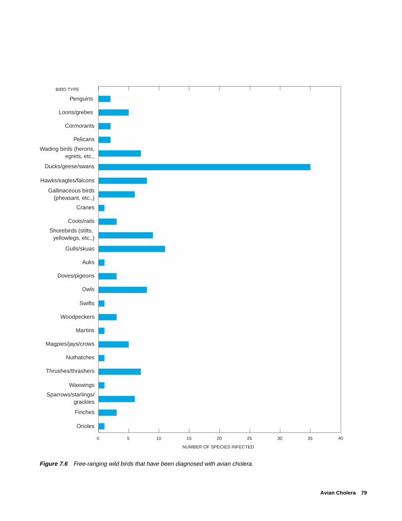
Avian Cholera 79
Figure 7.6 Free-ranging wild birds that have been diagnosed with avian cholera.
Penguins
Loons/grebes
Cormorants
Pelicans
Wading birds (herons,egrets, etc.,
Ducks/geese/swans
Hawks/eagles/falcons
Gallinaceous birds(pheasant, etc.,)
Cranes
Coots/rails
Shorebirds (stilts, yellowlegs, etc.,)
Gulls/skuas
Auks
Doves/pigeons
Owls
Swifts
Woodpeckers
Martins
Magpies/jays/crows
Nuthatches
Thrushes/thrashers
Waxwings
Sparrows/starlings/grackles
Finches
Orioles
0 5 10 15 20 25 30 35
NUMBER OF SPECIES INFECTED
BIRD TYPE
40
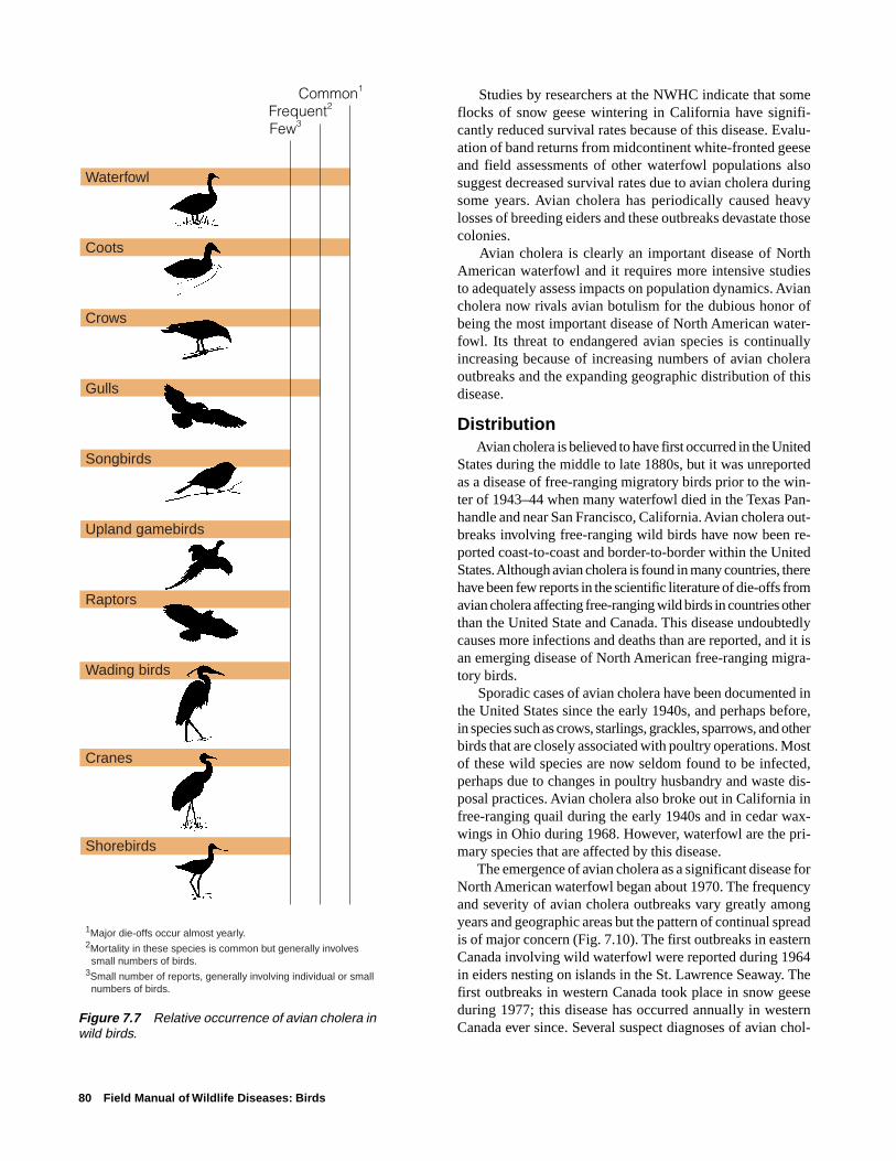
80 Field Manual of Wildlife Diseases: Birds
Figure 7.7 Relative occurrence of avian cholera inwild birds.
Waterfowl
Coots
Crows
Gulls
Songbirds
Upland gamebirds
Raptors
Wading birds
Cranes
Shorebirds
1Major die-offs occur almost yearly.2Mortality in these species is common but generally involves small numbers of birds.3Small number of reports, generally involving individual or small numbers of birds.
Common1
Frequent2
Few3
Studies by researchers at the NWHC indicate that someflocks of snow geese wintering in California have signifi-cantly reduced survival rates because of this disease. Evalu-ation of band returns from midcontinent white-fronted geeseand field assessments of other waterfowl populations alsosuggest decreased survival rates due to avian cholera duringsome years. Avian cholera has periodically caused heavylosses of breeding eiders and these outbreaks devastate thosecolonies.
Avian cholera is clearly an important disease of NorthAmerican waterfowl and it requires more intensive studiesto adequately assess impacts on population dynamics. Aviancholera now rivals avian botulism for the dubious honor ofbeing the most important disease of North American water-fowl. Its threat to endangered avian species is continuallyincreasing because of increasing numbers of avian choleraoutbreaks and the expanding geographic distribution of thisdisease.
Distribution Avian cholera is believed to have first occurred in the United
States during the middle to late 1880s, but it was unreportedas a disease of free-ranging migratory birds prior to the win-ter of 1943–44 when many waterfowl died in the Texas Pan-handle and near San Francisco, California. Avian cholera out-breaks involving free-ranging wild birds have now been re-ported coast-to-coast and border-to-border within the UnitedStates. Although avian cholera is found in many countries, therehave been few reports in the scientific literature of die-offs fromavian cholera affecting free-ranging wild birds in countries otherthan the United State and Canada. This disease undoubtedlycauses more infections and deaths than are reported, and it isan emerging disease of North American free-ranging migra-tory birds.
Sporadic cases of avian cholera have been documented inthe United States since the early 1940s, and perhaps before,in species such as crows, starlings, grackles, sparrows, and otherbirds that are closely associated with poultry operations. Mostof these wild species are now seldom found to be infected,perhaps due to changes in poultry husbandry and waste dis-posal practices. Avian cholera also broke out in California infree-ranging quail during the early 1940s and in cedar wax-wings in Ohio during 1968. However, waterfowl are the pri-mary species that are affected by this disease.
The emergence of avian cholera as a significant disease forNorth American waterfowl began about 1970. The frequencyand severity of avian cholera outbreaks vary greatly amongyears and geographic areas but the pattern of continual spreadis of major concern (Fig. 7.10). The first outbreaks in easternCanada involving wild waterfowl were reported during 1964in eiders nesting on islands in the St. Lawrence Seaway. Thefirst outbreaks in western Canada took place in snow geeseduring 1977; this disease has occurred annually in westernCanada ever since. Several suspect diagnoses of avian chol-

Avian Cholera 81
Figure 7.9 (A) Dense aggregations of waterfowl facilitate the rapid spread of avian cholera because of the highly infectiousnature of this disease. (B) Large-scale mortality has occurred in such situations.
Figure 7.8 Examples of major avian cholera outbreaks in wild birds. (Broken bars indicatevery high but indeterminate mortality.)
Pho
to b
y M
ilton
Frie
nd
25 50 75 1000
Puddle ducks
Coots
Coots
Oldsquaw duck, scoters
San Francisco BayWinter 1948–49
Texas PanhandleWinter 1956–57
Florida EvergladesWinter 1967–68
Back Bay, VirginiaFeb.–Mar. 1975
Chesapeake BayMar.–April 1978
Rainwater Basin, NebraskaMar.–April 1980
Hudson Bay, NWTJuly–Aug. 1979
MORTALITY, IN THOUSANDS
Chesapeake BayFeb.–April 1994
Banks Island, NWTSummer 1995
SaskatchewanSummer 1988
South AfricaSummer 1991
Snow goose
Redhead duck
Cape cormorant
Oldsquaw duck, scoters
Snow goose
Puddle ducks
Canada goose
A B Pho
to b
y Ja
mes
Hur
t, N
ebra
ska
Gam
e an
dP
arks
Com
mis
sion

82 Field Manual of Wildlife Diseases: Birds
Figure 7.10 (A) Reported frequency of avian cholera in free-ranging waterfowl in the United States. (B) Reported occurrenceof avian cholera in free-ranging migratory birds in the United States.
era have been reported for waterfowl mortality events in Mexicoduring recent years, but these events lack laboratory confir-mation. The absence of confirmed reports of this disease inwild waterfowl in Mexico is likely due to lack of surveillanceand reporting rather than to the absence of avian cholera.
In the United States, there are four major focal points foravian cholera in waterfowl: the Central Valley of California;the Tule Lake and Klamath Basins of northern California andsouthern Oregon; the Texas Panhandle; and Nebraska’s Rain-water Basin below the Platte River in the south-central part ofthe State. The movement of avian cholera from these areasfollows the well-defined pathways of waterfowl movement.The spread of this disease along the Missouri and MississippiRiver drainages is also consistent with waterfowl movement.No consistent patterns of avian cholera outbreaks exist withinthe Atlantic Flyway. There are periodic outbreaks in eider ducksnesting off of the coast of Maine and occasional major die-offs of sea ducks, including eiders, within the Chesapeake Bayof Maryland and Virginia (Fig. 7.11).
Seasonality Losses can occur at any time of the year. For poultry,
outbreaks of avian cholera are more prevalent in late sum-mer, fall, and winter. Those time periods have no special bio-logical associations, except, possibly, with production sched-ules in response to holiday market demands that influencepoultry age-classes within production facilities. Chickensbecome more susceptible as they reach maturity. Turkeys aremuch more susceptible than chickens, and turkeys die at allages, but the disease usually occurs in young mature tur-keys. Losses in domestic ducks are usually in birds olderthan 4 weeks of age.
For wild waterfowl, a predictably seasonal pattern existsin areas where avian cholera has become well established.This pattern is closely associated with seasonal migrationpatterns and it has resulted in avian cholera becoming a “dis-ease for all seasons,” killing waterfowl during all stages oftheir life cycle (Fig. 7.12). Some areas experience prolongedperiods of avian cholera mortality. Outbreaks in California
EXPLANATION
Frequency of occurrence of avian cholera, by State (Map A)
Annual to nearly annual occurrence, often resulting in deaths of thousands of birds during individual events
Frequent occurrences, most resulting in death of moderate to small numbers of birds
Occasional occurrences, many of which result in large-scale mortality
Occasional occurrences, most resulting in death of moderate to small numbers of birds
Not reported
Time period of first reported occurrence of avian cholera, by State (Map B)
1944 — 53
1954 — 63
1964 — 73
1974 — 83
1984 — 93
1994 — 97
Not reported
A
B

Avian Cholera 83
Figure 7.11 The occurrence of aviancholera in waterfowl seems to be closelyrelated to bird movements west of theMississippi River. There is no apparentpattern for outbreaks along the Atlanticseaboard.
normally start during fall and continue into spring. Otherareas have seasonal avian cholera outbreaks in the same geo-graphic location. For example, Nebraska, which has had out-breaks most springs since 1975, now frequently also hasoutbreaks in the fall.
Field Signs Few sick birds are seen during avian cholera outbreaks
because of the acute nature of this disease. However, thenumber of sick birds increases when a die-off is prolongedover several weeks. Sick birds often appear lethargic ordrowsy (Fig. 7.13), and they can be approached quite closelybefore they attempt to escape. When captured, these birdsoften die quickly, sometimes within a few seconds or min-utes after being handled. Other birds have convulsions (Fig.7.14), swim in circles, or throw their heads back betweentheir wings and die (Fig. 7.15). These signs are similar tothose seen in duck plague and in some types of pesticidepoisoning. Other signs include erratic flight, such as flyingupside down before plunging into the water or onto theground, and attempting to land a foot or more above the sur-face of the water; mucous discharge from the mouth; soilingand matting of the feathers around the vent, eyes, and bill;pasty, fawn-colored, or yellow droppings; and blood-staineddroppings or nasal discharges, which also are signs of duckplague (duck virus enteritis or DVE).
Always suspect avian cholera when large numbers of deadwaterfowl are found in a short time, when few sick birds areseen, and when the dead birds appear to be in good flesh.Death can be so rapid that birds may literally fall out of thesky or die while feeding, with no signs of illness. When sickbirds are captured and die within a few minutes, avian chol-era should also be suspected. None of the signs described
above are unique to this disease; these signs should be re-corded as part of any history being submitted with speci-mens and are considered along with lesions seen at necropsy.
Gross Lesions Under most conditions, birds that have died of avian chol-
era will have substantial amounts of subcutaneous and vis-ceral fat, except for seasonal losses of fat. The most promi-nent lesions seen at necropsy are in the heart and liver and,sometimes, the gizzard. Hemorrhages of various sizes arefrequently found on the surface of the heart muscle or thecoronary band or both (Fig. 7.16). Hemorrhages are alsosometimes visible on the surface of the gizzard. Areas oftissue death that appear as small white-to-yellow spots arecommonly seen within the liver. Where the area of tissuedeath is greater, the spots are larger and, in some instances,the area of tissue death is quite extensive (Fig. 7.17).
The occurrence of the abnormalities described for theheart, liver, and gizzard are dependent upon how long thebird lived after it became infected. The longer the survivaltime, the more abundant and dramatic the lesions. In addi-tion, there may be changes in the color, size, and texture ofthe liver. There is darkening or a copper tone to the liver, andit may appear swollen and rupture upon handling. Becausebirds infected with avian cholera often die so quickly, theupper portions of the digestive tract may contain recentlyingested food. All of these findings are similar to what mightalso be seen with duck plague; therefore, laboratory diagno-sis is needed.
Freshly dead ducks and geese that have succumbed toavian cholera may have a thick, mucous-like, ropy nasal dis-charge. The lower portions of the digestive tract (below thegizzard) commonly contain thickened yellowish fluid (Fig.
EXPLANATIONAvian cholera in waterfowl outbreak sites, 1944–97
Outbreak site
Migratory movements of waterfowl

84 Field Manual of Wildlife Diseases: Birds
Figure 7.12 Relative monthly probability for the occurrence of avian cholera in migratory waterfowl, expressed as a percent-age of outbreaks throughout the year. Information from the National Wildlife Health Center database.
Atlantic Flyway
Mississippi Flyway
Central Flyway
Pacific Flyway
MONTHJuly Aug. Sept. Oct. Nov. Dec. Jan. Feb. March April May June
Maine
Maryland
Virginia
North Carolina
Florida
Minnesota
Wisconsin
Iowa
Missouri
Illinois
Louisiana
Kentucky
Wyoming
Colorado
New Mexico
North Dakota
South Dakota
Nebraska
Kansas
Oklahoma
Texas
Washington
Oregon
California
Idaho
Nevada
Montana (western)
Utah
Canada
1Mexico
PERCENTAGE OF AVIAN CHOLERA OUTBREAKS, BY MONTH
1007550250
July Aug. Sept. Oct. Nov. Dec. Jan. Feb. March April May June
STATE OR COUNTRY
1Suspected occurrences — not laboratory confirmed
1007550250

Avian Cholera 85
Figure 7.15 Avian cholera-infected drake mallard. (A) Note tossing of head toward back and circular swimming as evidencedby ripples in water. (B) Bird at death with head resting on back.
Figure 7.13 Lethargic appearance of drake northern pintailwith avian cholera.
Pho
to b
y M
ilton
Frie
nd
Figure 7.14 Avian cholera-infected crow in convulsions.
Pho
to b
y M
ilton
Frie
ndP
hoto
s b
y M
ilton
Frie
nd
BA

86 Field Manual of Wildlife Diseases: Birds
Figure 7.16 Hemorrhages of varying degrees of severity are often seen on the hearts of avian cholera-infected birds. (A) Pinhead-sized hemorrhages along fatty areas of the heart are readily evident in thisbird. (B) Broad areas of hemorrhage also occur.
Pho
tos
by
Milt
on F
riend
➡
➡
➡
B
A

Avian Cholera 87
Figure 7.17 Lesions in the livers ofavian cholera-infected birds generallyappear as small, discrete, yellowishspots, which are dead tissue. Note thevariation in size and appearance ofthese lesions. (A) Note also the absenceof any apparent heart lesions in one bird,(B) only a few minor hemorrhages onthe coronary band of another bird, (C)and more extensive hemorrhages on theheart muscle of the third bird. Also notethe abundance of fat covering the giz-zards of all these birds. This fat atteststo the excellent condition these birdswere in before exposure to the bacte-rium and to the rapidity with which eachbird died.
Pho
tos
by
Milt
on F
riend
A
B
C

88 Field Manual of Wildlife Diseases: Birds
Figure 7.18 The thickened, yellowish fluid present in the intestines of this avian cholera-infected bird con-tains millions of bacteria. These bacteria contaminate the environment when the carcass decomposes or isscavenged, serving as a source of infection for other wildlife.
Pho
to b
y M
ilton
Frie
nd
7.18). Both of these fluids are heavily laden with P. multocidaand care must be taken to not contaminate the environment,field equipment, or oneself with these fluids.
Diagnosis As with all diseases, isolation of the causative agent is
required for a definitive diagnosis. A whole carcass providesthe diagnostician with the opportunity to evaluate gross le-sions seen at necropsy and also provides all appropriate tis-sues for isolation of P. multocida.
When it is not possible to send whole carcasses, sendtissues that can be collected in as sterile a manner as pos-sible in the field. The most suitable tissues for culturing areheart blood, liver, and bone marrow. Remove the entire heartand place it in a Whirl-Pak® bag for shipment as identified inChapter 2, Specimen Collection and Preservation; do notattempt to remove the blood from the heart. The liver shouldalso be removed and placed in a separate bag. A major por-tion of this organ (at least half) should be submitted if itcannot be removed intact. These samples must be refriger-ated as soon as possible after collection and kept cool duringshipment. When shipment is to be delayed for more than 1day or when transit time is expected to exceed 24 hours, freezethese specimens.
P. multocida persists several weeks to several months inbone marrow. The wings of badly scavenged or decomposedcarcasses should be submitted whenever avian cholera issuspected as the cause of death, and when more suitable tis-sue samples are not available.
Control Numerous factors must be considered in combating avian
cholera (Fig. 7.19). Avian cholera is highly infectious and itspreads rapidly through waterfowl and other bird popula-tions. This process is enhanced by the gregarious nature ofmost waterfowl species and by dense concentrations of mi-gratory waterbirds resulting from habitat limitations. Theprolonged environmental persistence of this bacterium fur-ther promotes new outbreaks (Table 7.1). Pond water re-mained infective for 3 weeks after dead birds were removedfrom one area in California; bacterial survival in soil for upto 4 months was reported in another study; and the organismcan persist in decaying bird carcasses for at least 3 months.
Early detection of avian cholera outbreaks is a first lineof defense for controlling this disease. Frequent surveillanceof areas where migratory birds are concentrated and thetimely submission of carcasses to disease diagnostic labora-tories allows disease control activities to be initiated before
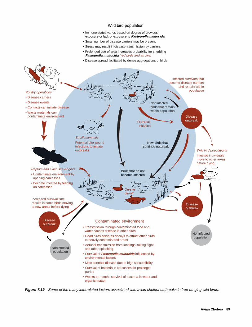
Avian Cholera 89
Figure 7.19 Some of the many interrelated factors associated with avian cholera outbreaks in free-ranging wild birds.
Noninfected population
Noninfected population
Wild bird population
Diseaseoutbreak
On-site die-off
Birds that do not become infected
Diseaseoutbreak
Outbreakinitiation
New birds that continue outbreak
Contaminated environment• Transmission through contaminated food and
water causes disease in other birds
• Dead birds serve as decoys to attract other birds to heavily contaminated areas
• Aerosol transmission from landings, taking flight, and other splashing
• Survival of Pasteurella multocida influenced by environmental factors
• Mice contract disease due to high susceptibility
• Survival of bacteria in carcasses for prolonged period
• Weeks-to-months survival of bacteria in water and organic matter
Infected survivors that become disease carriers
and remain within population
Wild bird populations
Infected individuals move to other areas before dying
Raptors and avian scavengers
• Contaminate environment by opening carcasses
• Become infected by feeding on carcasses
Increased survival time results in some birds moving to new areas before dying
Poultry operations
• Disease carriers
• Disease events
• Contacts can initiate disease
• Waste materials can contaminate environment
• Immune status varies based on degree of previous exposure or lack of exposure to Pasteurella multocida
• Small number of disease carriers may be present
• Stress may result in disease transmission by carriers
• Prolonged use of area increases probability for shedding Pasteurella multocida (red birds and arrows)
• Disease spread facilitated by dense aggregations of birds
Small mammals
Potential bite wound infections to initiate outbreaks
Diseaseoutbreak
Noninfectedbirds that remain within population

90 Field Manual of Wildlife Diseases: Birds
Table 7.1 Examples of reported environmental persistence for Pasteurella multocida.
Substrate Survival time Comments
General Highly variable Amount of moisture, temperature, and pH affect survival ofP. multocida.
Survival in soils enhanced when moisture content is 50percent or greater.
Survival in water is enhanced by high organic content andturbidity.
Survival in wetland waters enhanced by presence ofmagnesium and chloride ions.
Garden soil 3 months
Unspecified soil 113 days at 3 °C; Soil chemistry information needed to properly evaluate15–100 days at 20 °C; data.21 days at 26 °C
Poultry yard 2 weeks Infectious for birds after last death and removal of all birds.
Water 3 weeks Following removal of 100 dead snow geese; no otherwaterfowl use.
99 days Water contaminated with turkey litter.
30 days In marsh near carcass that had been opened.
Infected tissues 120 days but American coot hearts buried in marsh after birds died fromnot 240 days avian cholera.
Fomites 8 days but not 30 days Dried turkey blood on glass at room temperature.(Inanimateobjects)
the outbreaks reach advanced stages. The opportunity to pre-vent substantial losses is greatest during the early stages ofoutbreaks, and costs are minimal in comparison with handlinga large-scale die-off. Control actions need to be focused onminimizing the exposure of migratory and scavenger birdspecies to P. multocida and minimizing environmental con-tamination by this organism.
The NWHC recommends carcass collection and incin-eration as standard procedures. Carcass collection contrib-utes to avian cholera control in several ways. Several millili-ters of fluids containing large concentrations of P. multocidaare often discharged from the mouths of birds dying fromthis disease, resulting in heavy contamination of the surround-ing area. Carcass decomposition results in additional con-tamination. These carcasses attract (decoy) other birds,
thereby increasing the probability for infection. Scavengingof carcasses also transmits the disease through the directconsumption of diseased tissue (oral exposure).
Care must be exercised during carcass collection to mini-mize the amount of fluid discharged from the mouths of birdsinto the environment. Birds should be picked up head first,preferably by the bill, and immediately placed in plastic bags.Care must also be taken to avoid contaminating new areaswhile carcasses are transported to the disposal site. Double-bagging is recommended to prevent fluids leaking from punc-tures to the inner bag. Bags of carcasses should always besecurely closed before they are removed from the area.
Prompt carcass removal also prevents scavenging by avianspecies that can mechanically transport infected material toother sites or by feeding or drinking at other locations fol-

Avian Cholera 91
lowing consumption of infected tissue. This situation is ag-gravated by apparently longer disease-incubation times ingulls, crows, and some other avian scavengers. Instead ofdying within hours or 1–2 days after exposure to virulentstrains of P. multocida, avian scavengers more typically dieafter several days to 1–2 weeks, and they may die far fromthe site of exposure. When these birds die, they may serve asnew potential focal points for contamination.
In some instances, population reduction of gulls andcrows has been used to limit the role of these species inspreading and transmitting avian cholera. This technique haslimited application and it is not recommended as normal op-erating procedure. To be most effective, population reduc-tion must be undertaken before there is a major influx ofscavengers in response to carcass availability. Also, the tech-niques used must not result in dispersal of infected birds outof the area.
Population reduction of infected American coots, gulls,terns, and eiders has also been used to directly combatavian cholera. Destruction of migratory birds infectedwith this disease can be justified only under specialcircumstances and conditions:
1. The outbreak must be discreet and localized ratherthan generalized and widespread.
2. Techniques must be available that will allow com-plete eradication without causing widespread dispersalof potentially infected birds.
3. The methods used must be specific for target spe-cies and pose no significant risk for nontarget species.
4. Eradication must be justified on the basis of risk toother populations if the outbreak is allowed to con-tinue.
5. The outbreak represents a new geographic exten-sion of avian cholera into an important migratory birdpopulation.
Habitat management is another useful tool for combat-ing avian cholera outbreaks. In some instances, it may benecessary to prevent further bird use of a specific wetland orimpoundment because it is a focal point for infection of water-fowl migrating into the area. Drainage of the problem areain conjunction with creation or enhancement of other habi-tat within the area through water diversion from other sourcesor pumping operations denies waterfowl the use of the prob-lem area and redistributes them into more desirable habitat.The addition of a large volume of water to a problem areacan also help to dilute concentrations of P. multocida to lessdangerous levels. These actions require careful evaluationof bird movement patterns and of the avian cholera diseasecycle. Movement of birds infected with avian cholera from
one geographic location to another site is seldom desirable. Under extreme conditions, disinfection procedures to kill
P. multocida may be warranted in wetlands where large num-bers of birds have died during a short time period. The envi-ronmental impact of such measures must be evaluated andappropriate approvals must be obtained before these actionsare undertaken. A more useful approach may be to enhancethe quality of the wetland in a way that reduces the survivalof P. multocida; the best means of accomplishing this is stillbeing investigated.
Hazing with aircraft has been successfully used to movewhooping cranes away from a major outbreak of avian chol-era. This type of disease prevention action can also be ac-complished by other methods for other species. Eagles canbe attracted to other feeding sites using road-killed animalsas a food source, and waterfowl can be held at sites duringcertain times of the year by providing them with refuge andfood. During an avian cholera outbreak in South Dakota, alarge refuge area was temporarily created to hold infectedsnow geese in an area by closing it to hunting. At the sametime, a much larger population of snow geese about 10 milesaway was moved out of the area to prevent transmission ofthe disease into that population. The area closed to huntingwas reopened after the desired bird movement had occurred.
Vaccination and postexposure treatment of waterfowlhave both been successfully used to combat avian cholera inCanada goose propagation flocks. The NWHC has devel-oped and tested a bacterin or a killed vaccine that totallyprotected Canada geese from avian cholera for the entire 12months of a laboratory study. This product has been used forseveral years with good results in a giant Canada goose propa-gation flock that has a great deal of contact with free-flyingwild waterfowl and field outbreaks of avian cholera. Beforeuse of the bacterin, this flock of Canada geese suffered anoutbreak of avian cholera and was successfully treated withintramuscular infections of 50 milligrams of oxytetracyclinefollowed by a 30-day regimen of 500 grams of tetracyclineper ton of feed. A NWHC avian cholera bacterin has alsobeen used to successfully vaccinate snow geese on WrangleIsland, Russia, and Banks Island, Canada. Vaccine use in theseinstances was in association with studies to evaluate aviancholera impacts on survival rates rather than to control dis-ease in those subpopulations.
As yet, there is no practical method for immunizing largenumbers of free-living migratory birds against avian chol-era. However, captive propagation flocks can be protectedby this method. Endangered species can be trapped and im-munized if the degree of risk warrants this action. Live vac-cines should not be used for migratory birds without adequatesafety testing.
Human Health Considerations Avian cholera is not considered a high risk disease for
humans because of differences in species susceptibility to

92 Field Manual of Wildlife Diseases: Birds
different strains of P. multocida. However, P. multocida in-fections in humans are not uncommon. Most of these infec-tions result from an animal bite or scratch, primarily fromdogs and cats. Regardless, the wisdom of wearing glovesand thoroughly washing skin surfaces is obvious when han-dling birds that have died from avian cholera.
Infections unrelated to wounds are also common, and inthe majority of human cases, these involve respiratory tractexposure. This is most apt to happen in confined areas of airmovement where a large amount of infected material ispresent. Processing of carcasses associated with avian chol-era die-offs should be done outdoors or in other areas withadequate ventilation. When disposing of carcasses by openburning, personnel should avoid direct exposure to smokefrom the fire.
Milton Friend
Supplementary ReadingBotzler, R.G., 1991, Epizootiology of avian cholera in wildfowl:
Journal of Wildlife Diseases, 27 p. 367–395.Brand, C.J., 1984, Avian cholera in the Central Mississippi
Flyways during 1979–80: Journal of Wildlife Management, 48,p. 399–406.
Rimler, R.B., and Glisson, J.R. 1997, Fowl cholera, in Calnek,B.W., and others, eds., Diseases of poultry (10th ed.): Ames,Iowa, Iowa State University Press, p. 143–159.
Wobeser, G.A., 1997, Avian cholera, in Diseases of wild water-fowl (2nd ed): New York, N.Y., Plenum Press, p. 57–69.

Tuberculosis 93
Chapter 8
Tuberculosis
SynonymsMycobacteriosis, tuberculosis, TB
CauseAvian tuberculosis is usually caused by the bacterium
Mycobacterium avium. At least 20 different types of M. aviumhave been identified, only three of which are known to causedisease in birds. Other types of Mycobacterium rarely causetuberculosis in most avian species; however, parrots, macaws,and other large perching birds are susceptible to human andbovine types of tuberculosis bacilli. Avian tuberculosis gen-erally is transmitted by direct contact with infected birds,ingestion of contaminated feed and water, or contact with acontaminated environment. Inhalation of the bacterium cancause respiratory tract infections. Wild bird studies in theNetherlands disclosed tuberculosis-infected puncture-typeinjuries in birds of prey that fight at the nest site (kestrels) oron the ground (buteo-type buzzards), but tuberculosis-infected injuries were not found in accipiters (falcons), whichfight in the air and seldom inflict such wounds.
Species AffectedAll avian species are susceptible to infection by M. avium.
Humans, most livestock species, and other mammals can alsobecome infected. Recent molecular studies with a limitednumber of isolates from birds, humans, and other mammalsclearly indicated that M. avium can be transmitted betweenbirds and pigs, but the studies did not disclose a similar crosstransmission between birds and humans for the isolates tested.It is generally accepted that pigs, rabbits, and mink are highlysusceptible to M. avium; deer can also become infected. Dogsappear to be quite resistant to the avian type of tuberculosis(Fig. 8.1).
In captivity, turkeys, pheasants, quail, cranes, and certainbirds of prey are more commonly infected than waterfowl.However, when avian tuberculosis becomes established, itcan be a common and lethal disease in captive waterfowlflocks. Chronic infections exist in some captive nene gooseflocks, making these flocks unsuitable donors to supplementthe wild population of this endangered species. Pheasantsare unusually susceptible to avian tuberculosis.
In free-ranging wild birds, avian tuberculosis is found mostoften in species that live in close association with domesticstock (sparrows and starlings) and in scavengers (crows andgulls). The prevalence of tuberculosis in free-ranging NorthAmerican birds has not been determined, although generallyless than 1 percent of birds examined at postmortem are af-fected. Sampling biases due to the limited numbers of speci-
Figure 8.1 Relative susceptibility of various animal groupsto M. avium.
Highly susceptibleModerately susceptible
SusceptibleModerately resistantHighly resistant
Birds
Pigs
Mink
Rabbits
Sheep
Deer
Cattle
Horses
Goats
Dogs
Cats
Human

94 Field Manual of Wildlife Diseases: Birds
mens examined preclude extending findings to reflect actualprevalence (Fig. 8.2). A decade-long study of nearly 12,000wild birds necropsied in the Netherlands disclosed that 0.7percent of the birds had tuberculosis. The sample includedwaterbirds, birds of prey, songbirds, and pheasants. Studiesin the United States disclosed that 0.3 percent of 3,000waterfowl necropsied were infected with tuberculosis, and astudy in British Columbia found tuberculosis in 0.6 percentof more than 600 wild birds. Tuberculosis in whooping cranesstands in marked contrast to other wild birds; approximately39 percent of the western population’s free-ranging whoop-ing cranes necropsied at the National Wildlife Health Centerhave been infected with avian tuberculosis.
DistributionAvian tuberculosis is a ubiquitous and cosmopolitan dis-
ease of free-ranging, captive, and domestic birds. The dis-ease is most commonly found in the North Temperate Zone,and, within the United States, the highest infection rates inpoultry are in the North Central States. Distribution of thisdisease in free-ranging wild birds is inferred from birds sub-mitted for necropsy; however, the sampling underrepresentsboth the geographic distribution and the frequency of infec-tion for individual species. Avian tuberculosis likely existsin small numbers of free-ranging wild birds wherever thereare major bird concentrations.
SeasonalitySeasonal trends of tuberculosis in wild birds have not been
documented. The chronic nature of this disease guaranteesits presence yearround for both wild and captive birds.
Factors that may influence seasonal exposure to tubercu-losis in migratory birds are changes in habitat used, foodbase during the year, and interspecies contacts. Contaminatedsewage and wastewater environments containing tuberclebacilli are more likely to be used by waterfowl during falland winter than during warmer months. Wastewater sites areoften closed to hunting, thereby serving as refuge areas, andwarm water discharges to these sites maintain open water insubfreezing temperatures, thus inviting ready use by water-fowl. Predatory and scavenger species such as raptors andcrows often ingest many different food items during differ-ent periods of the year; scavengers, therefore, may be ex-posed to tuberculosis through contaminated food yearround.Contact between wild birds and poultry and livestock isoften restricted to specific periods of the year owing to hus-bandry practices. Wild birds may be exposed to M. avium inmanure that is spread on fields during early spring.
Environmental conditions can greatly affect the suscepti-bility of birds to tuberculosis and the prevalence of tubercu-losis in captive birds. Captive birds that are on an inadequate
Figure 8.2 Relative occurrence of avian tuberculosis in birds.
Chickens
Captive reared
Free-ranging Partridge/quail
Captive reared
Free-ranging
Cranes
Captive reared
Free-ranging
Pigeons
Captive reared
Free-ranging
Hawks/owls
Captive reared
Free-ranging
Waterfowl
Captive reared
Free-ranging
Pheasants
Captive reared
Free-ranging
Gulls
Captive reared
Free-ranging
Crows
Captive reared
Free-ranging
Turkeys
Captive reared
Free-ranging
Songbirds
Captive reared
Free-rangingVultures/buzzards
Captive reared
Free-ranging
FrequentCommon
Occasional
Not applicable
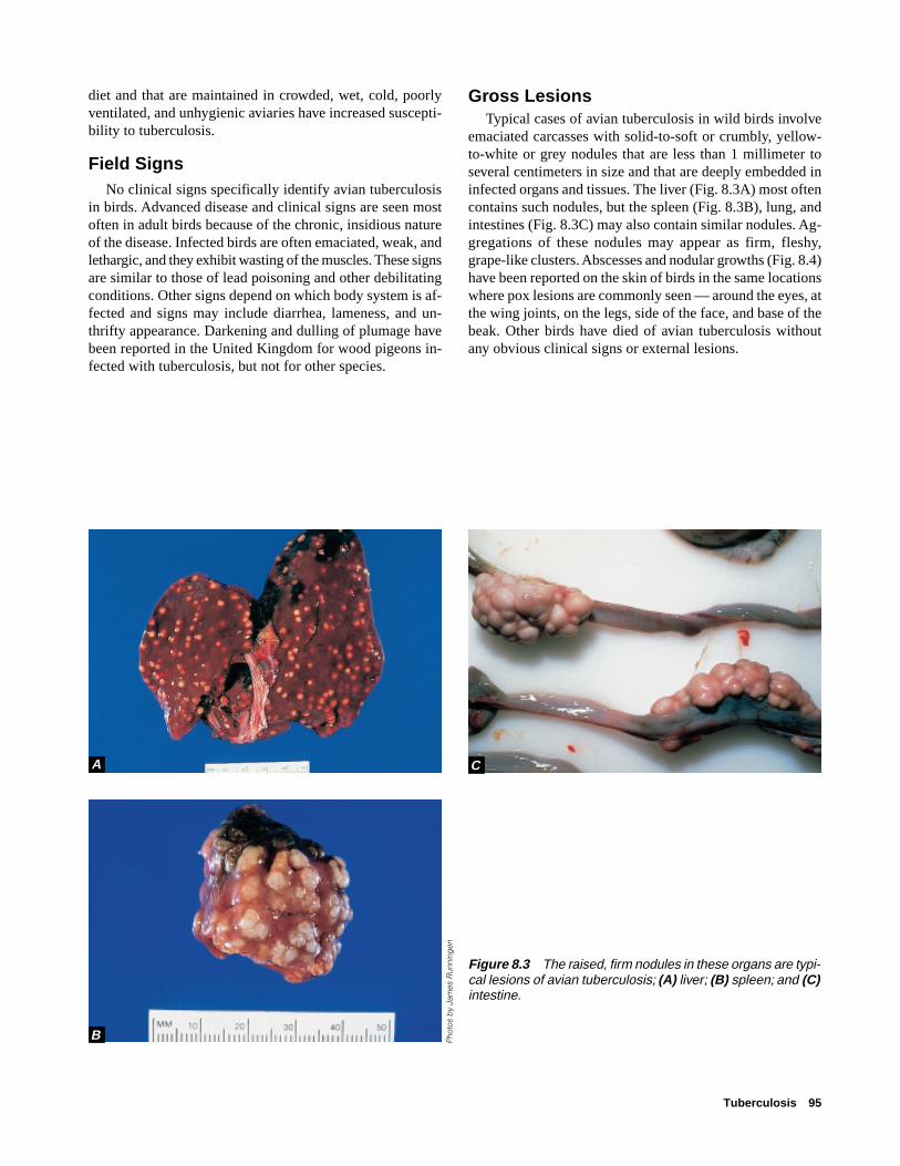
Tuberculosis 95
diet and that are maintained in crowded, wet, cold, poorlyventilated, and unhygienic aviaries have increased suscepti-bility to tuberculosis.
Field SignsNo clinical signs specifically identify avian tuberculosis
in birds. Advanced disease and clinical signs are seen mostoften in adult birds because of the chronic, insidious natureof the disease. Infected birds are often emaciated, weak, andlethargic, and they exhibit wasting of the muscles. These signsare similar to those of lead poisoning and other debilitatingconditions. Other signs depend on which body system is af-fected and signs may include diarrhea, lameness, and un-thrifty appearance. Darkening and dulling of plumage havebeen reported in the United Kingdom for wood pigeons in-fected with tuberculosis, but not for other species.
Gross LesionsTypical cases of avian tuberculosis in wild birds involve
emaciated carcasses with solid-to-soft or crumbly, yellow-to-white or grey nodules that are less than 1 millimeter toseveral centimeters in size and that are deeply embedded ininfected organs and tissues. The liver (Fig. 8.3A) most oftencontains such nodules, but the spleen (Fig. 8.3B), lung, andintestines (Fig. 8.3C) may also contain similar nodules. Ag-gregations of these nodules may appear as firm, fleshy,grape-like clusters. Abscesses and nodular growths (Fig. 8.4)have been reported on the skin of birds in the same locationswhere pox lesions are commonly seen — around the eyes, atthe wing joints, on the legs, side of the face, and base of thebeak. Other birds have died of avian tuberculosis withoutany obvious clinical signs or external lesions.
Figure 8.3 The raised, firm nodules in these organs are typi-cal lesions of avian tuberculosis; (A) liver; (B) spleen; and (C)intestine.
Pho
tos
by
Jam
es R
unni
ngen
A
B
C
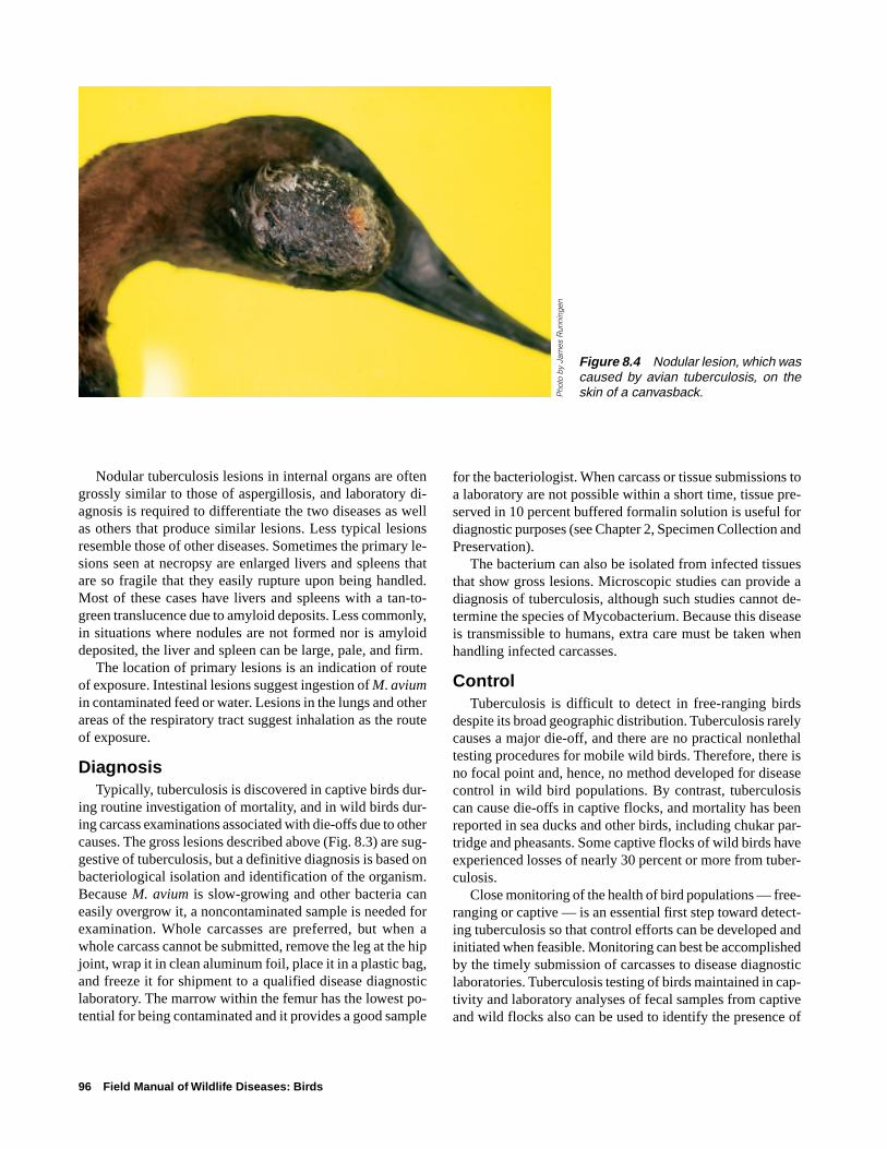
96 Field Manual of Wildlife Diseases: Birds
Nodular tuberculosis lesions in internal organs are oftengrossly similar to those of aspergillosis, and laboratory di-agnosis is required to differentiate the two diseases as wellas others that produce similar lesions. Less typical lesionsresemble those of other diseases. Sometimes the primary le-sions seen at necropsy are enlarged livers and spleens thatare so fragile that they easily rupture upon being handled.Most of these cases have livers and spleens with a tan-to-green translucence due to amyloid deposits. Less commonly,in situations where nodules are not formed nor is amyloiddeposited, the liver and spleen can be large, pale, and firm.
The location of primary lesions is an indication of routeof exposure. Intestinal lesions suggest ingestion of M. aviumin contaminated feed or water. Lesions in the lungs and otherareas of the respiratory tract suggest inhalation as the routeof exposure.
DiagnosisTypically, tuberculosis is discovered in captive birds dur-
ing routine investigation of mortality, and in wild birds dur-ing carcass examinations associated with die-offs due to othercauses. The gross lesions described above (Fig. 8.3) are sug-gestive of tuberculosis, but a definitive diagnosis is based onbacteriological isolation and identification of the organism.Because M. avium is slow-growing and other bacteria caneasily overgrow it, a noncontaminated sample is needed forexamination. Whole carcasses are preferred, but when awhole carcass cannot be submitted, remove the leg at the hipjoint, wrap it in clean aluminum foil, place it in a plastic bag,and freeze it for shipment to a qualified disease diagnosticlaboratory. The marrow within the femur has the lowest po-tential for being contaminated and it provides a good sample
for the bacteriologist. When carcass or tissue submissions toa laboratory are not possible within a short time, tissue pre-served in 10 percent buffered formalin solution is useful fordiagnostic purposes (see Chapter 2, Specimen Collection andPreservation).
The bacterium can also be isolated from infected tissuesthat show gross lesions. Microscopic studies can provide adiagnosis of tuberculosis, although such studies cannot de-termine the species of Mycobacterium. Because this diseaseis transmissible to humans, extra care must be taken whenhandling infected carcasses.
ControlTuberculosis is difficult to detect in free-ranging birds
despite its broad geographic distribution. Tuberculosis rarelycauses a major die-off, and there are no practical nonlethaltesting procedures for mobile wild birds. Therefore, there isno focal point and, hence, no method developed for diseasecontrol in wild bird populations. By contrast, tuberculosiscan cause die-offs in captive flocks, and mortality has beenreported in sea ducks and other birds, including chukar par-tridge and pheasants. Some captive flocks of wild birds haveexperienced losses of nearly 30 percent or more from tuber-culosis.
Close monitoring of the health of bird populations — free-ranging or captive — is an essential first step toward detect-ing tuberculosis so that control efforts can be developed andinitiated when feasible. Monitoring can best be accomplishedby the timely submission of carcasses to disease diagnosticlaboratories. Tuberculosis testing of birds maintained in cap-tivity and laboratory analyses of fecal samples from captiveand wild flocks also can be used to identify the presence of
Figure 8.4 Nodular lesion, which wascaused by avian tuberculosis, on theskin of a canvasback.P
hoto
by
Jam
es R
unni
ngen

Tuberculosis 97
this disease. These tests do not detect all infected birds, butthe tests are useful for identifying infected flocks.
Fecal contamination of the environment is the major meansof tuberculosis dissemination; ingestion of the bacterium incontaminated feed and water is the most common means ofdisease transmission. Because this bacterium can surviveoutside of the vertebrate host for long time periods in anorganic substrate (Fig. 8.5), a few infected animals can con-taminate an area that has prolonged bird-use patterns. Thelong-term environmental survival of M. avium that is shedby disease carriers when combined with repeated site useand, possibly, a high degree of susceptibility to avian tuber-culosis may be the factors contributing to the high preva-lence of this disease observed in whooping cranes. A sitecan also be contaminated by wastewater discharges contain-ing M. avium and by the application of contaminated ma-nure for fertilizer. Tuberculosis outbreaks in birds have beenassociated with sewage effluents and discharges from slaugh-ter houses, meat processing plants, and dairies. In one in-stance, an outbreak occurred in a captive waterfowl flockwhen contaminated water was sprayed into the enclosure.These events illustrate the importance of disease preventionfor addressing tuberculosis in free-ranging and captive wildbirds.
The use of wastewater for maintaining captive waterfowland other wild birds is questionable without adequate test-ing or treatment or both to assure that the wastewater doesnot contain tubercle bacilli. Also, the use of wetlands forwastewater discharges and the use of wastewater to createwetlands for migratory bird habitat should be carefully con-sidered because of the possible presence of M. avium in thewastewater. Other actions that should be considered include
preventing land use that could place tuberculosis-infectedswine in close proximity to major wild bird concentrationsand not using unexamined chicken and pigeon carcasses asfood for raptors being reared in captivity for release into thewild.
Infected flocks of captive birds should be destroyed be-cause treatment is ineffective and because not all infectedbirds will be detected by current testing procedures. Becauseof the long-term environmental persistence of the tuberclebacilli, additional bird use of the site should be avoided forapproximately 2 years. Vegetation removal and turning ofthe soil several times during this period will facilitate sun-light-induced environmental decay of the bacilli. Eradica-tion of free-ranging migratory flocks is rarely feasible. How-ever, when a major outbreak of tuberculosis occurs in wildbirds, the circumstances should be assessed, and limitedpopulation reduction should be considered if the remainingpopulation-at-risk is well defined, limited in immediate dis-tribution, and involves species that can withstand this ac-tion. Habitat manipulation, such as drainage, and scaringdevices, such as propane exploders, can sometimes be usedto deny birds use of areas where tuberculosis outbreaksoccur.
The insidious nature of avian tuberculosis combined withthe long environmental persistence of the causative bacte-rium strongly indicate a need to prevent the establishment ofthis disease in wild bird populations. When the disease be-comes established in free-ranging populations, interspeciestransmission and the mobility of free-ranging birds couldserve to spread it widely. The continued persistence of aviantuberculosis as a major cause of avian mortality in zoologi-cal collections attests to the difficulty of disease control.
Figure 8.5 Examples of environmental persistence of M. avium.
0 10 20 30 40 50
ENVIRONMENTAL PERSISTENCE, IN MONTHS
Poultry litter
Poultry carcasses buried 3 feet deep
Sawdust at 20 °C
Sawdust at 37 °C
Body exudates shielded from the sun
Body exudates exposed to sun
Organisms 5 cm deep in soil
Lighter color indicates variable range
98 Field Manual of Wildlife Diseases: Birds
Human Health ConsiderationsThere are many authenticated cases of M. avium infec-
tion in people, although humans are considered highly resis-tant to this organism. Avian tuberculosis is generally consid-ered noncontagious from an infected person to an uninfectedperson. Infection is more likely to occur in persons with pre-existent diseases, especially those involving the lungs, andin persons whose immune systems are impaired by an ill-ness, such as AIDS or steroid therapy.
Milton Friend(Modified from an earlier chapter by Thomas J. Roffe)
Supplementary ReadingKarlson, A.G., 1978, Avian tuberculosis, in Montali, R.J., ed.,
Mycobacterial infections of zoo animals: Washington, D.C.,Smithsonian Institution Press, p. 21–24.
Smit, T., Eger, A., Haagsma, J., and Bakhuizen, T., 1987, Aviantuberculosis in wild birds in the Netherlands: Journal ofWildlife Diseases 23, p. 485–487.
Thoen, C.O., 1997, Tuberculosis, in Calnek, B.W., and others,eds., Diseases of poultry (10th ed.): Ames, Iowa, Iowa StateUniversity Press, p. 167–178.
Wobeser, G.A., 1997, Tuberculosis, in Diseases of wild waterfowl(2nd ed): New York, N.Y., Plenum Press, p. 71–75.
Salmonellosis 99
Chapter 9
Salmonellosis
SynonymsSalmonellosis; paratyphoid; bacillary white diarrhea(a synonym for pullorum disease); pullorum disease1,fowl typhoid1
CauseAvian salmonellosis is caused by a group of bacteria of
the genus salmonella. Approximately 2,300 different strainsof salmonellae have been identified, and these are placedinto groupings called “serovars” on the basis of their anti-gens or substances that induce immune response by the host,such as the production of specific antibody to the antigen.Current taxonomic nomenclature considers the 2,300 differ-ent serovars to be variants of two species, Salmonella entericaand S. bongori. S. enterica is further subdivided into six sub-species on the basis of biochemical characteristics. This re-sults in complex nomenclature for each serovar, such as, S.enterica subsp. enterica serovar typhimurium. Readers shouldbe aware of this convention for naming salmonellae becausethey will find this nomenclature in the current scientific lit-erature. In this chapter, different serovars of salmonellae willbe referred to by their previous, less complex nomenclature,such as S. typhimurium.
Pullorum disease, (S. pullorum) and fowl typhoid (S. gal-linarum) are two classic and distinctive diseases of poultrythat have received considerable attention because of theireconomic impacts. Wild birds have been infected withpullorum disease and fowl typhoid, but wild birds are morecommonly infected by the variants of salmonellae that arecollectively referred to as paratyphoid forms, of which S. ty-phimurium is a prominent representative. The paratyphoidforms constitute the great majority of salmonellae, and theyare becoming increasingly important as causes of illness anddeath in wild birds (Table 9.1).
Salmonella infections can be transmitted in many ways(Table 9.2), and the importance of different modes for trans-mission varies with the strain of salmonellae, behavioral andfeeding patterns of the bird species, and husbandry practiceswhen human intervention becomes part of the hatching andrearing processes. For example, ovarian transmission ofS. typhimurium occasionally occurs in turkeys, but it is un-common in chickens. Egg transmission and environmentalcontamination of rearing facilities are of more importancefor infecting poultry than are contaminated feeds. For wild
birds and humans, contaminated foods are the primary sourcefor infection; food and water become contaminated by fecaldischarges from various sources. Rats, mice, and other spe-cies, including reptiles and turtles, in addition to birds, aresources of fecal discharges of paratyphoid forms of salmo-nellae. Inhalation of the bacterium during close confinementin high humidity environments such as hatching and brooderoperations, direct contact with infected birds and animals,and insects are other demonstrated transmission routes forsalmonellosis.
Intestinal microflora are an important factor influencinginfection and disease by salmonellae in poultry. Very smallnumbers of salmonellae can cause infection of poultry dur-ing the first few weeks of life. Thereafter, the infectious dosebecomes progressively higher, apparently because poultryacquire intestinal microflora that protect them against infec-tion even in the presence of a highly salmonella-contami-nated environment. This may explain the high prevalence ofsalmonellosis occasionally found in chicks of some colonialnesting species, such as gulls and terns, and in heron andegret rookeries, but the lower-than-expected infection ratesin adult birds from those same colonies. Experimental stud-ies with full-grown herring gulls disclosed a rapid elimina-tion of salmonella bacteria from the intestines of these birds,which suggests that adult herring gulls may be passively,rather than actively, infected and may simply serve as a me-chanical transport mechanism for the movement of salmo-nellae ingested from contaminated environments.
Individual infected birds can excrete salmonella bacteriafor prolonged periods of time ranging from weeks to months.Prolonged use of sites by birds and high density of individu-als at those sites can result in cycles of salmonellosis withinthose populations. Persistently contaminated environmentsresult from a small percentage of birds which remain as life-long carriers that intermittently excrete salmonellae into theenvironment. The environmental persistence of these bacte-ria is another factor influencing the probability for infectionsof birds using that site (Table 9.3). The common practices ofusing sewage sludge and livestock feces and slurry as fertil-izer provide another means for infecting wild birds. Tests ofsewage sludge often disclose contamination with salmonel-lae. Survival periods for salmonellae in cattle slurry sampleshave been reported to range from 11 to 12 weeks and formonths in fields where the slurry has been applied as fertil-izer. There are numerous reports of the isolation of salmo-nellae from rivers and streams as a result of pollution bysewage effluent and slurry runoff from fields.1 Distinct forms of salmonellosis caused by specific vari-
ants of salmonellae.

100 Field Manual of Wildlife Diseases: Birds
Table 9.1 Characteristics of important salmonellae-causing disease in birds.
Salmonella Salmonella SalmonellaCharacteristic pullorum gallinarum typhimurium
Common name Pullorum disease Fowl typhoid Salmonellosis
Natural hosts Chickens (primary), Chickens, turkeys Wide range of vertebrates; notturkeys restricted to birds.
Age Mortality usually Generally infects growing All ages affected; moresusceptibility confined to the first and adult birds; disease common in young and often
2–3 weeks of age. also infects young due to in association with con-egg transmission. current disease agents.
Transmission Infected hatching eggs Infected carrier birds most Contaminated environmentfollowed by spread from important; egg transmission resulting in ingestion throughinfected chicks to un- of secondary importance. food and water; egg transmis-infected chicks that hatch. sion can also occur.
Relative Rare in free-ranging Uncommon in free-ranging Prevalence varies with species;occurrence species; not maintained species; not maintained most common in thosein wild birds within wild populations. within wild populations. species associated with
landfills, sewage lagoons,and other waste-disposal sitesand those with close associa-tions with livestock and poultryoperations.
Other naturally Ducks, coots, pheasants, Ducks, swans, curlews, Wide range of species;infected avian partridges, guinea fowl, pheasants, quail, partridge commonly found in gullsspecies sparrows, European bull- grouse, guinea fowl, peafowl, and terns and passerine
finch, magpies, canaries, wood pigeon, ring dove, rock birds using birdfeedinghawk-headed parrot. dove, owls, rooks, jackdaws, stations. Also reported in
sparrows, blackbirds, gold- herons, egrets, ducks,finches,ostrich, parrots. geese, cormorants, cranes,
owls, eagles, falcons,hawks, and other species.
Current Rare in most advanced Essentially eliminated from Worldwide due to widegeographic poultry-producing areas. commercial poultry within the range of species infected.occurrence United States. Low incidence
in Canada, USA, and severalEuropean countries; significantdisease in Mexico, Centraland South America, Africa,and Middle East.
Relative Occasional infections Rare and of little public One of the most commonhuman health following massive exposure health significance. causes of food-bornesignificance (contaminated food); disease in humans.
prompt recovery withouttreatment.

Salmonellosis 101
Table 9.2 Pathways for transmission of Salmonella sp. in birds.
Type of transmission Means Consequences/processes
Vertical Through contaminanted eggs from Infection of hatchlings at age of greatest(from parent to offspring) infected female; embryo may be susceptibility. Infected hatchlings become
infected or surface of egg becomes source of infection for other hatchlings.infected as it passes down oviduct.
Horizontal Bird-to-bird contact Infected birds shed organism in feces. Birdsin close contact inhale salmonellae thatbecome airborne or ingest salmonellaewhen pecking at contaminated surfaces ofinfected birds.
Contaminated environments Multiple sources of fecal contaminationfrom a wide variety of warm- and cold-blooded species results in ingestion of sal-monellae when pecking at contaminatedfeathers, litter, and other materials. Infectedbirds and other animals that are fed uponby birds with predatory and scavengingfood habits become exposed to salmonel-losis. Birds that feed in landfills, dung piles,wastewater discharge areas, and sewagelagoons are at highest risk to acquireinfections.
Contaminated feeds Salmonella-contaminated feed has beenthe source of salmonella outbreaks in poul-try. Little is known about levels of salmo-nella contamination in commercial feedused at birdfeeding stations.
Inapparent infections Stress of translocation or conditions caus-ing birds to be brought into rehabilitationcan result in shedding of salmonellae bycarrier birds or result in clinical disease inbirds with subclinical infections. Diseasecan be transmitted to other birds in closeproximity; contamination of the environmentcan result in further transmission, and re-lease of actively shedding birds can serveto spread the disease and contaminateother environments.

102 Field Manual of Wildlife Diseases: Birds
Table 9.3 Examples of reported environmental persistence for Salmonella sp. in different substrates. [—, no data available.]
Temperature
Substrate 11 °C 25 °C 38 °C Ambient Serovar
Poultry feed 18 months 16 months 40 days — S. typhimurium
Poultry litter 18 months 18 months 13 days — S. typhimurium
Soil from vacated — — — 6–7 months Unspecified paratyphoid formturkey pens
Urban garden soil — — — 280 days S. typhimurium
Hatchery fluff — — — 5 years Unspecified paratyphoid form
Avian feces — — — 28 months Unspecified paratyphoid form
Reptilian feces — — — 30 months Unspecified paratyphoid form
Manure — — — 36 months Unspecified paratyphoid form

Salmonellosis 103
Figure 9.1 Relative rates of isolation of Salmonella sp. infree-ranging wild birds.
Species AffectedAll species of birds should be considered susceptible to
infection by salmonellae. The outcome of salmonella infec-tions is reported to be highly dependent upon the age of thebirds, concurrent stress, serovar and strain virulence, andsusceptibility of the host species.
Salmonellosis has been studied as a disease of poultrysince at least 1899. Wild bird surveys have often been con-current with studies of this disease in poultry and as sourcesfor human infections. These and other investigations haveresulted in numerous strains of Salmonella sp. being isolatedfrom free-ranging (Fig. 9.1) and captive wild birds. How-ever, findings from these studies have also disclosed a muchlower infection rate than anticipated and have caused nu-merous investigators to conclude that in general, salmonel-losis is not an important disease of free-ranging wild birds.
The historic patterns of salmonellosis in wild birds are ofisolated mortality events involving individual or very smallnumbers of birds and incidental findings associated withconcurrent infections involving other disease agents. Beforethe 1980s, major mortality events from this disease were rarein free-ranging wild birds.
Prior to the 1980s most isolations of Salmonella sp. fromfree-ranging wild birds were made from apparently healthybirds, were incidental findings from birds with other diseaseconditions, or were from lethal cases of salmonellosis in-volving small number of birds. This is no longer the situa-tion. Large-scale mortalities of birds using feeding stationshave become common in the United States (Fig. 9.2), andsuch mortalities are also reported from Canada and Europe,including Scandinavia. Typically, these events are caused byS. typhimurium and usually involve passerine birds (Fig. 9.3).European starling, blackbirds, common grackle, and mourn-ing dove are also among the species that have been founddead from S. typhimurium at birdfeeding stations.
Salmonellosis has also been the cause of die-offs of aquaticbirds including several species of ducks, mute swan, variousspecies of gulls and terns, American coot, double-crestedcormorant, eared grebe, and several species of egrets andherons. However, large-scale mortality events in free-ranging populations, except for songbirds and colonial nest-ing birds, have rarely been reported.
Many species of captive-reared birds commonly becomeinfected with salmonellae and die from salmonellosis.Aquatic species have died from salmonellosis in zoologicalgardens and other captive collections. Gamebirds, such asgrouse and pheasants, being reared in captivity for sportingpurposes and cranes being reared for species conservationefforts are often victims of salmonellosis. Mortality is gen-erally confined to chicks.
Gulls/terns
Songbirds
Ducks/geese/swans
Herons/egrets
Doves/pigeons
Pheasants/quail/grouse/partridges
Starlings/blackbirds/cowbirds
Coots
Cranes
Cormorants/gannets
Guillemots/razorbills
Penguins
Falcons/hawks/owls
Crows/rooks/magpies
FrequentCommon
OccasionalInfrequent

104 Field Manual of Wildlife Diseases: Birds
Pine siskin
Evening grosbeak
House sparrow
Brown-headed cowbird
Northern cardinal
Goldfinch
English sparrow
Cassin's finch
Purple finch
Robin
Chestnut-backed chickadee
House finch
Chipping sparrow
Dusky song sparrow
Tree sparrow
Rufous-sided towhee
Northern mockingbird
Cedar waxwing
FrequentCommon
OccasionalInfrequent
DistributionExtensive and prolonged control programs have essen-
tially eliminated pullorum disease as a disease confrontingcommercial poultry production in most of the world and fowltyphoid from most Western countries. In contrast, salmonel-losis due to paratyphoid infections occurs worldwide(Table 9.1) and is increasingly prevalent among wild birdsin a wide variety of habitats. Salmonellosis in songbirds isclearly an emerging disease of urban and suburban environ-ments and it has also been introduced into remote bird popu-lations, such as Antarctic penguins and skua. The geographicdistribution of salmonellosis in free-ranging wild birds isclosely associated with sources of environmental contami-nation that enters the food web of birds and is passed to otherspecies when infected individuals are fed upon by predatorsand scavengers.
SeasonalitySalmonellosis can present itself at any time of year. Out-
breaks at birdfeeding stations are closely associated with theperiods of greatest use of those stations (Fig. 9.4); fall andspring die-offs of songbirds from salmonellosis are commonin England. Other outbreaks occur among the young of co-lonial nesting species, such as gulls and terns, shortly afterthe young are hatched during the summer (Fig. 9.5).
Field SignsThere are no distinctive signs associated with salmonel-
losis in wild birds. Different species and ages of birds mayhave different signs even if they are infected with the sameserovar; young birds typically exhibit more pronounced signsof disease. Infection may result in acute disease with suddenonset of death, or it may result in a more prolonged course
Figure 9.2 Locations of reported out-breaks of salmonellosis at birdfeedingstations within the United States. (FromNational Wildlife Health Center data-base.)
Figure 9.3 Relative occurrence of species found dead fromsalmonellosis outbreaks at birdfeeding stations within theUnited States.
EXPLANATION
Location of salmonellosis outbreaks, 1983–1997
�
�
�
���
�
��
��
�
�
�
�
�
�
�
�
�
�
��
��
��
�
�
�
�
�
�
�
� �
�
�
�
��
�
�
��
�
�
�
��
�
�
�
�
�
���

Salmonellosis 105
of infection that may become septicemic or be characterizedby the presence and persistence of bacteria in the blood, orresult in localized infection within the body. The disease inpoultry has been described to result in gradual onset of de-pression over a few days and by unthrifty appearance. Thesebirds huddle, are unsteady, shiver, and breathe more rapidlythan normal; their eyes begin to close shortly before death;and they exhibit nervous signs including incoordination, stag-gering, tremors, and convulsions. Blindness has also beenreported in some birds.
The rapid death of songbirds at feeding stations has oftencaused observers to believe the birds had been poisoned.Neurological signs, such as those described above for poul-try, have also be reported in infected songbirds. In contrast,young domestic ducklings are reported to die slowly, exhib-iting tremors and gasping for air. Their wings often droopand they sometimes stagger and fall over just before death.Like infected chickens, these birds often have pasted ventsand eyelids that are swollen and stuck together by a fluiddischarge. Commonly reported signs among all species in-clude ruffled feathers, droopiness, diarrhea, and severe leth-argy. Chronically infected birds often appear severely ema-ciated.
Figure 9.4 Seasonal occurrence of salmo-nellosis outbreaks at birdfeeding stationswithin the United States.
Figure 9.5 (A) Salmonellosiscan cause large-scale losses ofcolonial nesting birds. (B) Youngbirds are especially vulnerable.
Pho
tos
by
Milt
on F
riend
0
5
10
15
20
25
30
35
NU
MB
ER
OF
OU
TB
RE
AK
S
SUMMER FALL WINTER SPRING
B
A

106 Field Manual of Wildlife Diseases: Birds
Gross LesionsThe occurrence and types of gross lesions are highly vari-
able depending on the course of the infection, the virulenceof the organism, and the resistance of the host. In acute cases,obvious lesions can be completely absent. Livers often be-come swollen and crumbly with small reddened or pale spotsif the course of the disease has been prolonged. In other in-fections, so-called paratyphoid nodules develop in the liverand extend into the body cavity. These are small tan-to-whitegranular nodules that are best seen under a microscope. Insome birds, these nodules are more visible and appear asplaques or granular-abscess-like lesions seen within breast
Figure 9.6 Lesions of salmonellosis in the esophagus of(A) an English sparrow and (B) and (C) an evening grosbeak.(A) From the surface, these lesions appear as a yellow, cheesynodule that could be mistaken for a seed taken in as a fooditem. (B) When the esophagus is opened, lesions may be seenthat appear as large, diffuse, plaque-like areas (C) or as aseries of discrete, nodular plaques.
muscle and other tissues and organs. Infected songbirds of-ten have yellow, cheesy nodules visible on the surface of theesophagus. When the esophagus is cut open, the nodules maybe seen as large, diffuse plaque-like lesions or as discrete,nodular areas within the esophagus (Fig. 9.6).
An acute intestinal infection can be recognized by the red-dening of the internal lining of the posterior two-thirds toone-half of the small intestine, the ceca, which are the blindpouches that extend from both sides of the beginning of thelarge intestine, and the colon. As the disease progresses, theintestinal lining becomes coated with a pale, tightly adher-
Figure 9.7 Necrotic, crumbly coresthat appear as thick, cheesy areas areoften found in the intestines of birdsdying from salmonellosis.
Pho
tos
by
J. C
hris
tian
Fran
son
Pho
to b
y Ja
mes
Run
ning
en
A B
C

Salmonellosis 107
ing, fibrinous material. In some infected birds, the intestinalceca contain thick, crumbly necrotic cores (Fig. 9.7). En-largement and impaction of the rectum are commonly re-ported in domestic ducklings.
Arthritis in the wings of pigeons is common. Domesticducks with paratyphoid infections often have arthritis of thehips and knee joints. Small external abscesses about 1 milli-meter in diameter have been described for infected pigeonsand house sparrows. These abscesses appear in small bunchesalong the underside of the bird along the mid-to-posteriorareas of the body.
DiagnosisGross lesions of salmonellosis can be similar to several
other diseases, including avian cholera and colibacillosis.Diagnosis requires laboratory isolation and identification ofSalmonella sp. from infected tissues in conjunction withpathological findings. Therefore, whole carcasses should besubmitted for examination. Birds with markedly abnormalbehavior patterns, such as convulsions and tumbling, oftenhave lesions observable by microscopic examination of thebrain. Isolation of salmonellae from the intestine without sig-nificant lesions and accompanying isolation of the bacteriafrom other tissues generally indicates that the bird was a car-rier, rather than a victim, of salmonellosis.
Salmonellae are often confined to the gut. The ceca offerthe greatest potential for obtaining positive cultures for moststrains of salmonellae. Therefore, when whole carcasses can-not be submitted, submit the intestine as a minimum sample.The liver and heart should also be removed and submitted, ifpossible. Wrap each different tissue in a separate piece ofaluminum foil. Place the foil-wrapped specimens in tightlysealed plastic bags, and ship them frozen to the diagnosticlaboratory (Chapter 2, Specimen Collection and Preserva-tion and Chapter 3, Specimen Shipment).
Fecal droppings can be checked for Salmonella sp., butthese need special handling and they should not be submit-ted as diagnostic specimens without prior discussions withthe diagnostic laboratory. Submission of whole eggs shouldbe considered when low hatchability is encountered. Eggshells and shell membranes can also be cultured for salmo-nellae; this is an effective means of detecting salmonellae ineggs that have hatched, provided that the egg fragments havenot been subjected to environmental conditions that woulddestroy the bacteria. Eggs, too, should only be submitted fol-lowing consultation with disease specialists.
ControlPrevention of infection by pathogenic forms of Salmo-
nella sp. and control of salmonellosis is warranted for wildbird populations despite the fact that Salmonella sp. havebeen isolated from a wide variety of wild bird species frommany different types of habitats. Surveys have disclosed thatthe prevalence of salmonellae in most wild bird populations
is generally low. Other studies have indicated a rapid elimi-nation of salmonellae from the intestines of their avian host,suggesting passive, rather than active, infection in some in-stances. The relatively recent increase in the frequency ofoccurrence of large-scale salmonella outbreaks in wild birds,especially songbirds, is without precedent and it suggeststhat environmental contamination is an important source forinfection of birds.
Landfills and waters where sewage effluent is dischargedare common feeding areas for gulls, the wild bird speciesgroup with the highest prevalence of salmonella infections.Ducks and other waterbirds also feed heavily in areas of sew-age effluent, and they generally have a higher prevalence ofsalmonellae than most land birds except for pigeons and spar-rows, two species that feed in manure piles. Raptors arethought to become infected from the prey they feed upon(often small rodents such as mice).
Eliminating point sources of infection should be the fo-cus for combating salmonellosis in wild bird populations (Fig.9.8). Disease prevention should be practical at birdfeedingstations; the public should be educated to maintain clean feed-ers and to remove spilled and soiled feed from the area un-der the feeder. Feeders occasionally should be disinfectedwith a 1:10 ratio of household bleach and water as part ofthe disease-prevention program. In the event of a die-off fromsalmonellosis, more rigorous disinfection of feeding stationsis necessary and station use should be discontinued tempo-rarily.
Other potential point sources of infection include garbage,sewage wastewater, and wastewater discharges from livestockand poultry operations. The potential for contaminating mi-gratory bird habitat with Salmonella sp. should be consid-ered when wastewater is intentionally used to create wet-land habitat; when existing wetlands are used to receivewastewater discharges; when agricultural fields on wildlifeareas are to receive manure and slurries as fertilizer; and whendevelopment of landfill, livestock, and poultry operations areproposed in areas where contamination of environments usedby migratory birds is likely. A 1995 outbreak of S. enteriditisin California poultry was traced to sewage treatment plantwastewater which entered a stream that bordered the poultryfarm. Contamination of feral cats and wildlife by the watersof the stream was thought to be the source of entry of S.enteriditis in the poultry.
Control of salmonellosis in captive flocks of migratorybirds is necessary to prevent major losses, especially in youngbirds. Control of this disease should be of continual concernwhenever migratory birds and other wild birds are beingpropagated for release programs or are being maintained incaptivity during rehabilitation. The conditions causing birdsto be brought to rehabilitation and the stresses of confine-ment may result in inapparent infections developing intosystemic clinical salmonellosis that may jeopardize the well-being of the infected bird and of other birds within the facil-

108 Field Manual of Wildlife Diseases: Birds
Figure 9.8 (A) Sources and (B) consequences of salmonellosis in wild birds.
Inapparent carrier"Salmonella Jane"
Stress Concurrent disease
Acute mortality Infertility
Chick mortality
Reduced reproduction
Livestock feedlot
Dairy cattle
Poultry houses
Poultry processing plant
Sewage treatmentplant
Refuge dump site
Manure spreading
River
Wetland
River
Agricultural fields
Runoff
A
B
Salmonellosis 109
ity. Strict sanitation measures need to be instituted and judi-ciously followed. Salmonella carriers can be identified byfecal culturing and should be destroyed. Multiple periodicfecal cultures are required to identify carrier birds becausesalmonellae are intermittently shed from the intestine. Allbirds that die should undergo necropsy and appropriate labo-ratory testing to determine the cause of mortality and anyactions required to prevent further losses.
Infected adults should never be used for breeding. Anti-biotic therapy may aid in overcoming an outbreak of salmo-nellosis, but antibiotic therapy will not eliminate carriers andvertical transmission via eggs could result in new outbreaksand disease spread. Storage of food in rodent- and insect-proof containers should be part of a disease prevention pro-gram. Many outbreaks in domestic poultry operations havebeen traced to food contaminated by rodent feces becauserats and mice are common sources of salmonellae.
Human Health ConsiderationsBacteria of the genus Salmonella are well-documented
human pathogens. “Food poisoning” characterized by acuteintestinal pain and diarrhea is the most common form of hu-man infection. However, more serious forms of salmonello-sis also affect humans. The general level of Salmonella sp.in most species of wild birds is low, but extra care with per-sonal hygiene is warranted by people who handle these birdsor materials soiled by bird feces. This consideration is notlimited to situations where disease is apparent, and it ex-tends to routine maintenance of birdfeeders, cleaning trans-port cages, and handling birds during banding and other fieldactivities.
Milton Friend(Modified from an earlier chapter by Richard K. Stroud and Milton Friend)
Supplementary ReadingGast, R.K., 1997, Paratyphoid infections, in Calnek, B.W., and
others., eds., Diseases of poultry (10th ed.): Ames, Iowa, IowaState University Press, p. 97–121.
Snoeyenbos, G.H., 1994, Avian salmonellosis, in Beran, G.W.,and Steele, J.H., eds., Handbook of zoonoses (2d ed., SectionA): Bacterial, rickettsial, chlamydial, and mycotic: BocaRaton, Fla., CRC Press, p. 303–310.
Steele, J.H., and M.M. Galton, 1971, Salmonellosis, in Davis,J.W., and others, eds., Infectious and parasitic diseases of wildbirds: Ames, Iowa, Iowa State University Press, p. 51–58.

110 Field Manual of Wildlife Diseases: Birds

Chlamydiosis 111
Chapter 10
Chlamydiosis
SynonymsParrot fever, psittacosis, ornithosis, parrot disease,Louisiana pneumonitis
CauseChlamydiosis refers to an infection with organisms of the
genus Chlamydia sp., which are bacteria that live within ani-mal cells. Chlamydia psittaci is the species generally associ-ated with this disease in birds. The severity of the diseasediffers by the strain of C. psittaci and the susceptibility ofdifferent species of birds. As a result, chlamydiosis may rangefrom an inapparent infection to a severe disease with highmortality. The organism is excreted in the feces and nasaldischarges of infected birds and can remain infective for sev-eral months. Infection commonly occurs from inhaling thebacteria in airborne particles from feces or respiratory exu-dates. Because of the organism’s resistance to drying, in-fected bird feces at roosts are especially hazardous.
Species AffectedChlamydiosis was first recognized as an infectious dis-
ease affecting parrots, parakeets, and humans involved in theinternational parrot trade in the late l920s to l930s. Chlamy-diosis has since become known as a serious disease of do-mestic turkeys in the United States, of domestic ducks andgeese in central Europe, and as a common infection of do-mestic and feral pigeons worldwide. The feral city pigeon isthe most common carrier of Chlamydia sp. within the UnitedStates.
Chlamydial infections have been reported from at least159 species of wild birds in 20 orders, but most isolationshave been made from six groups of birds (Figure 10.1). Psit-tacine birds such as parakeets, parrots, macaws, and cock-atiels are most commonly identified with this disease, whileamong other caged birds Chlamydia sp. occurs most fre-quently in pigeons, doves, and mynahs. Waterfowl, herons,and pigeons are the most commonly infected wild birds inNorth America (Figure 10.2). Chlamydiosis also occasion-ally infects gulls and terns, shorebirds, songbirds, and up-land gamebirds.
DistributionAmong free-living birds, avian chlamydiosis has been
found worldwide in the feral pigeon, in gulls and fulmars onislands of coastal Great Britain, in waterfowl and shorebirdsin the Caspian Sea, and in herons, waterfowl, gulls, and dovesin the United States. Infected parrots and parakeets have beenfound throughout the tropics and Australia.
Figure 10.1 Relative occurrence of reported chlamydiosisin the most frequently infected groups of birds. (Adapted fromBurkhart and Page, 1971).
ParrotsFree-ranging birds
Captive birds
Shorebirds, gulls, alcidsFree-ranging birds
Captive birds
SongbirdsFree-ranging birds
Captive birds
WaterfowlFree-ranging birds
Captive birds
Herons and alliesFree-ranging birds
Captive birds
Pigeons and dovesFree-ranging birds
Captive birds
0 5 10 15 20 25 30 35
NUMBER OF SPECIES INFECTEDWITH CHLAMYDIOSIS
none
none
none

112 Field Manual of Wildlife Diseases: Birds
SeasonalityIndividual cases may occur at any time because of healthy
carriers and latent infections within bird populations. Ship-ping, crowding, chilling, breeding, and other stressors havebeen attributed to active shedding of the infectious agentamong captive birds with latent infections. Groupings of wildbirds together in flocks, such as during spring and fall mi-grations, may facilitate the transmission of chlamydiosis. Incaged birds, the onset of disease following exposure toC. psittaci occurs across a broad range of time from as quicklyas 3 days to as long as several weeks. Young birds are moresusceptible than adults, and the disease can spread rapidlyamong colonial nesting birds.
Field SignsSigns of infection depend on the species of bird, viru-
lence of the strain of Chlamydia sp., the physiological con-dition of the bird as influenced by stressors, and route ofexposure to the organism. Chlamydiosis in wild birds is of-ten inapparent and infected birds can serve as asymptomaticcarriers. Infection may also result in an acute, subacute, orchronic form of disease. C. psittaci can cause severe, acutedisease that may be rapidly fatal in highly susceptible spe-cies. Birds often become weak, stop eating, and develop pu-rulent (fluid containing pus) discharges of the eyes and nares.Birds tend to become motionless, remain in a fixed position,huddled up with ruffled feathers (Fig 10.3). Birds may havediarrhea, sometimes rust-colored because of the presence ofblood, and respiratory distress is common. Feces from birdsthat stop eating are often dark green. In an outbreak ofchlamydiosis in wild gulls, primarily fledglings died and thebirds that were found dead were typically thin. Captive snowyand American egrets with chlamydiosis exhibited weakness,abnormal gait, ruffled feathers, diarrhea, and rapid weightloss; the birds generality died 1–2 days after the onset ofsigns. In other species of egrets, the infection may be inap-parent even though the organism can be isolated from swabsof the cloaca or respiratory tract.
Feral pigeons exhibit many of the same signs; however,their diarrhea is likely to be more frequently tinged withblood. Mortality rates in young pigeons are often very high.Purulent discharges from the eyes of a very sick pigeon shouldcause the observer to think first of chlamydiosis. Suddendeath without any signs of illness has been reported amongcaptive cage birds (Java finch, parrots) and among wild par-rots in Australia where king parrots were reported to havefallen out of trees and died within minutes.
Gross LesionsThe most common anatomical change in infected birds is
an enlargement of the spleen or splenomegaly or of the liveror hepatomegaly or both, up to three-or-four times normalsize (Figure 10.4). During an outbreak of chlamydiosis ingulls, splenomegaly was noted in each of nine birds exam-
Figure 10.2 Relative occurrence of chlamydial infections inwild birds in North America.
Feral pigeon
Waterfowl
Herons, egrets, ibises
Gulls, terns
Songbirds
Upland gamebirds
Shorebirds
Cranes
Raptors
CommonFrequent
OccasionalInfrequent
Rare or not reported

Chlamydiosis 113
ined and hepatomegaly was noted in four of the nine. Peri-carditis, which is an inflammation and thickening of thepericardial sac that surrounds the heart (Figure 10.5), is astriking lesion sometimes seen with acute or subacutechlamydiosis. The air sacs may be thickened and the lungsare often congested, appearing darker than normal.
DiagnosisDiagnosis is based upon the isolation of Chlamydia sp.
from tissues of infected birds. Whole birds should be sub-mitted. When this is not possible, selected tissues should becollected (Chapter 2, Specimen Collection and Preservationand Chapter 3, Specimen Shipment). The lungs, spleen, liver,and affected air sacs are the preferred tissues for microbialexamination. Because C. psittaci is also a human pathogen,care must be taken in handling carcasses and tissues.
Diagnosis cannot be based on gross lesions alone becausethe lesions of some other diseases are similar. Chronic aviancholera infection can produce similar gross lesions in gulls,avian malaria can cause enlarged spleens, and early stagesof aspergillosis can produce somewhat similar changes inthe lungs and air sacs.
ControlChlamydia sp. are present in the tissues, feces, discharges
from the eyes and nares, and may also be present on plum-age of infected birds. When the excreta and discharges dry,the resulting material can become airborne. Infection maybe transmitted by direct contact with affected birds, or byinhaling dried bird fecal material or respiratory exudates thatcontain Chlamydia sp. organisms. Sick birds should be col-lected and euthanized and carcasses should be picked up.The removal and incineration of carcasses will help reducethe amount of infective material in the area. However, thelevel of human activity in the area should be carefully con-sidered because it may cause redistribution of birds that couldresult in the spread of infection to new areas.
Human Health ConsiderationsChlamydiosis can be a serious human health problem,
infecting more frequently those who work with birds. Theclose association between parrots and this disease in humansprompted the United States and most nations of WesternEurope to outlaw the importation of parrots and parakeetsfrom l930 to 1960. Individuals who work in areas in whichthere is a strong possibility of inhaling airborne avian fecalmaterial should consider wearing a mask or respirator. Dry,dusty areas with bird droppings can be wetted down with a 5percent solution of household bleach, or a commercial dis-infectant. Working with large numbers of birds in dusty,closely confined areas should be avoided as much as pos-sible.
Figure 10.5 Pericarditis in a ring-billed gull that died ofchlamydiosis (From Franson and Pearson, 1995. Reprintedwith permission from the Journal of Wildlife Diseases.)
Figure 10.3 Classic appearance of an immature little blueheron with severe chlamydia infection.
Pho
to b
y M
ilton
Frie
nd
Figure 10.4 Enlarged spleen (top) and liver (bottom) of aring-billed gull affected with chlamydiosis. (From Franson andPearson, 1995. Reprinted with permission from the Journalof Wildlife Diseases).
Pho
to b
y Ja
mes
Run
ning
enP
hoto
by
Mic
hael
Cof
fey
114 Field Manual of Wildlife Diseases: Birds
Outbreaks have occurred among poultry slaughterhouseworkers and there have also been several severe cases amongwildlife biologists. These biologists were thought to havebecome infected from handling snow geese, common egrets,snowy egrets, white-winged doves, and ducks.
Before the availability of antibiotics, chlamydiosis wasfatal in about 20 percent of the human cases. Today, suchfatalities are rare. However, persons working with birdsshould inform their physicians of that fact to help avoid po-tential situations where early signs of chlamydiosis could beoverlooked or dismissed.
J. Christian Franson(Modified from an earlier chapter by Louis N. Locke)
Supplementary ReadingBrand, C.J., 1989, Chlamydial infections in free-flying birds:
Journal of the American Veterinary Medical Association,v. 195, no. 11, p. 1,531–1,535.
Burkhart, R. L., and Page, L.A., 1971, Chlamydiosis (ornithosis-psittacosis), in Davis, J.W., and others, eds., Infectious andparasitic diseases of wild birds: Ames, Iowa, Iowa StateUniversity Press, p. 118–140
Franson, J.C., and Pearson, J.E., 1995, Probable epizooticchlamydiosis in wild California (Larus californicus) and ring-billed (Larus delawarensis) gulls in North Dakota: Journal ofWildlife Diseases, v. 31, no. 3, p. 424–427.
Grimes, J.E., 1994, Avian chlamydiosis, in Beran, G. W., andothers, eds., Handbook of zoonoses (2nd ed.): Boca Raton,Fla., CRC Press, p. 389–402.
Wobeser, G.A., 1997, Chlamydiosis, in Diseases of wild water-fowl (2nd ed): New York, N.Y., Plenum Press, p. 88–91.
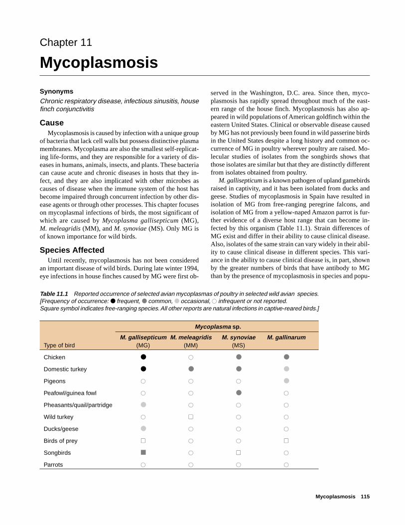
Mycoplasmosis 115
Chapter 11
Mycoplasmosis
SynonymsChronic respiratory disease, infectious sinusitis, housefinch conjunctivitis
CauseMycoplasmosis is caused by infection with a unique group
of bacteria that lack cell walls but possess distinctive plasmamembranes. Mycoplasma are also the smallest self-replicat-ing life-forms, and they are responsible for a variety of dis-eases in humans, animals, insects, and plants. These bacteriacan cause acute and chronic diseases in hosts that they in-fect, and they are also implicated with other microbes ascauses of disease when the immune system of the host hasbecome impaired through concurrent infection by other dis-ease agents or through other processes. This chapter focuseson mycoplasmal infections of birds, the most significant ofwhich are caused by Mycoplasma gallisepticum (MG),M. meleagridis (MM), and M. synoviae (MS). Only MG isof known importance for wild birds.
Species AffectedUntil recently, mycoplasmosis has not been considered
an important disease of wild birds. During late winter 1994,eye infections in house finches caused by MG were first ob-
served in the Washington, D.C. area. Since then, myco-plasmosis has rapidly spread throughout much of the east-ern range of the house finch. Mycoplasmosis has also ap-peared in wild populations of American goldfinch within theeastern United States. Clinical or observable disease causedby MG has not previously been found in wild passerine birdsin the United States despite a long history and common oc-currence of MG in poultry wherever poultry are raised. Mo-lecular studies of isolates from the songbirds shows thatthose isolates are similar but that they are distinctly differentfrom isolates obtained from poultry.
M. gallisepticum is a known pathogen of upland gamebirdsraised in captivity, and it has been isolated from ducks andgeese. Studies of mycoplasmosis in Spain have resulted inisolation of MG from free-ranging peregrine falcons, andisolation of MG from a yellow-naped Amazon parrot is fur-ther evidence of a diverse host range that can become in-fected by this organism (Table 11.1). Strain differences ofMG exist and differ in their ability to cause clinical disease.Also, isolates of the same strain can vary widely in their abil-ity to cause clinical disease in different species. This vari-ance in the ability to cause clinical disease is, in part, shownby the greater numbers of birds that have antibody to MGthan by the presence of mycoplasmosis in species and popu-
Table 11.1 Reported occurrence of selected avian mycoplasmas of poultry in selected wild avian species.[Frequency of occurrence: ● frequent, ● common, ● occasional, � infrequent or not reported.Square symbol indicates free-ranging species. All other reports are natural infections in captive-reared birds.]
Mycoplasma sp.
M. gallisepticum M. meleagridis M. synoviae M. gallinarumType of bird (MG) (MM) (MS)
Chicken ● � ● ●
Domestic turkey ● ● ● ●
Pigeons � � � ●
Peafowl/guinea fowl � � ● �
Pheasants/quail/partridge ● � � �
Wild turkey � � � �
Ducks/geese ● � � �
Birds of prey � � � �
Songbirds ■ � � �
Parrots � � � �

116 Field Manual of Wildlife Diseases: Birds
lations tested. The isolates of MG from wild songbirds donot cause significant disease in chickens.
Chickens and turkeys are commonly infected with MG,and direct contact of susceptible birds with infected carrierbirds causes outbreaks in poultry flocks. Aerosol transmis-sion via dust or droplets facilitates spread of MG throughoutthe flock. Transmission through the egg is also important forpoultry, and MG is thought to spread by contact with con-taminated equipment. The highly gregarious behavior ofhouse finches and their use of birdfeeders likely facilitatescontact between infected birds or with surfaces contaminatedwith the bacteria. Infected finches are thought to be respon-sible for spreading this disease because they move betweenlocal birdfeeders and to distant locations during migration.
M. meleagridis causes an egg-transmitted disease of do-mestic turkeys, and it appears to be restricted to turkeys. Clini-cal disease has not been documented in wild turkeys, andreports of infection in other upland gamebirds have not been
confirmed. Airborne transmission and indirect transmissionby contact with contaminated surfaces also happen.M. synoviae has a broader host range than MM. Chickens,turkeys, and guinea fowl are the natural hosts. Several otherspecies have been naturally infected, and others have beeninfected by artificial inoculation. Transmission is similar tothat for MG, except that MS spreads more rapidly.
Many other avian mycoplasmas have been designated dis-tinct species, some of which are identified in Table 11.2. Thenumber of mycoplasma species identified from birds has in-creased rapidly during recent years and it will continue togrow. For example, M. sturni was recently isolated from theinner eyelids (conjunctiva) of both eyes of a European star-ling that had the clinical appearance of MG infection in housefinches. Enhanced technology is providing greater capabili-ties for studying and understanding the biological signifi-cance of this important group of microorganisms. Too littleis known about mycoplasma infections in wild birds to cur-
Table 11.2 Primary hosts of some mycoplasma species isolated from birds. [—, no data available.]
Primary host
species Chicken Turkey Pigeons Waterfowl Partridge Birds of prey Songbirds
M. gallisepticum ● ● — — — — —
M. synoviae ● ● — — — — —
M. iowae ● ● — — — — —
M. gallopavonis — ● — — — — —
M. cloacale — ● — — — — —
M. gallinarum ● — — — — — —
M. gallinaceum — ● — — — — —
M. pullorum ● — — — — — —
M. iners ● — — — — — —
M. lipofaciens ● — — — — — —
M. glycophilum ● — — — — — —
M. columbinasale — — ● — — — —
M. columbinum — — ● — — — —
M. columborale — — ● — — — —
M. anatis — — — ● — — —
M. anseris — — — ● — — —
M. imitavis — — — ● ● — —
M. sturni — — — — — — ●
M. buteonis — — — — — ● —
M. falconis — — — — — ● —
M. gypis — — — — — ● —
Mycoplasma
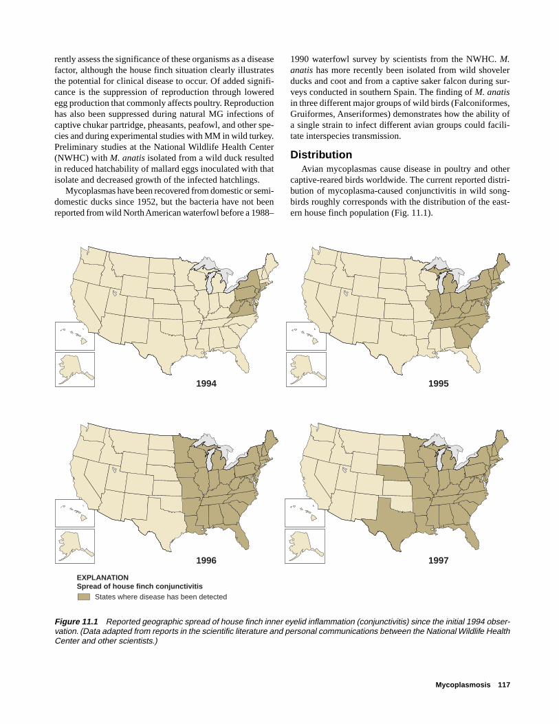
Mycoplasmosis 117
rently assess the significance of these organisms as a diseasefactor, although the house finch situation clearly illustratesthe potential for clinical disease to occur. Of added signifi-cance is the suppression of reproduction through loweredegg production that commonly affects poultry. Reproductionhas also been suppressed during natural MG infections ofcaptive chukar partridge, pheasants, peafowl, and other spe-cies and during experimental studies with MM in wild turkey.Preliminary studies at the National Wildlife Health Center(NWHC) with M. anatis isolated from a wild duck resultedin reduced hatchability of mallard eggs inoculated with thatisolate and decreased growth of the infected hatchlings.
Mycoplasmas have been recovered from domestic or semi-domestic ducks since 1952, but the bacteria have not beenreported from wild North American waterfowl before a 1988–
1990 waterfowl survey by scientists from the NWHC. M.anatis has more recently been isolated from wild shovelerducks and coot and from a captive saker falcon during sur-veys conducted in southern Spain. The finding of M. anatisin three different major groups of wild birds (Falconiformes,Gruiformes, Anseriformes) demonstrates how the ability ofa single strain to infect different avian groups could facili-tate interspecies transmission.
DistributionAvian mycoplasmas cause disease in poultry and other
captive-reared birds worldwide. The current reported distri-bution of mycoplasma-caused conjunctivitis in wild song-birds roughly corresponds with the distribution of the east-ern house finch population (Fig. 11.1).
Figure 11.1 Reported geographic spread of house finch inner eyelid inflammation (conjunctivitis) since the initial 1994 obser-vation. (Data adapted from reports in the scientific literature and personal communications between the National Wildlife HealthCenter and other scientists.)
1994 1995
1996 1997
EXPLANATIONSpread of house finch conjunctivitis
States where disease has been detected
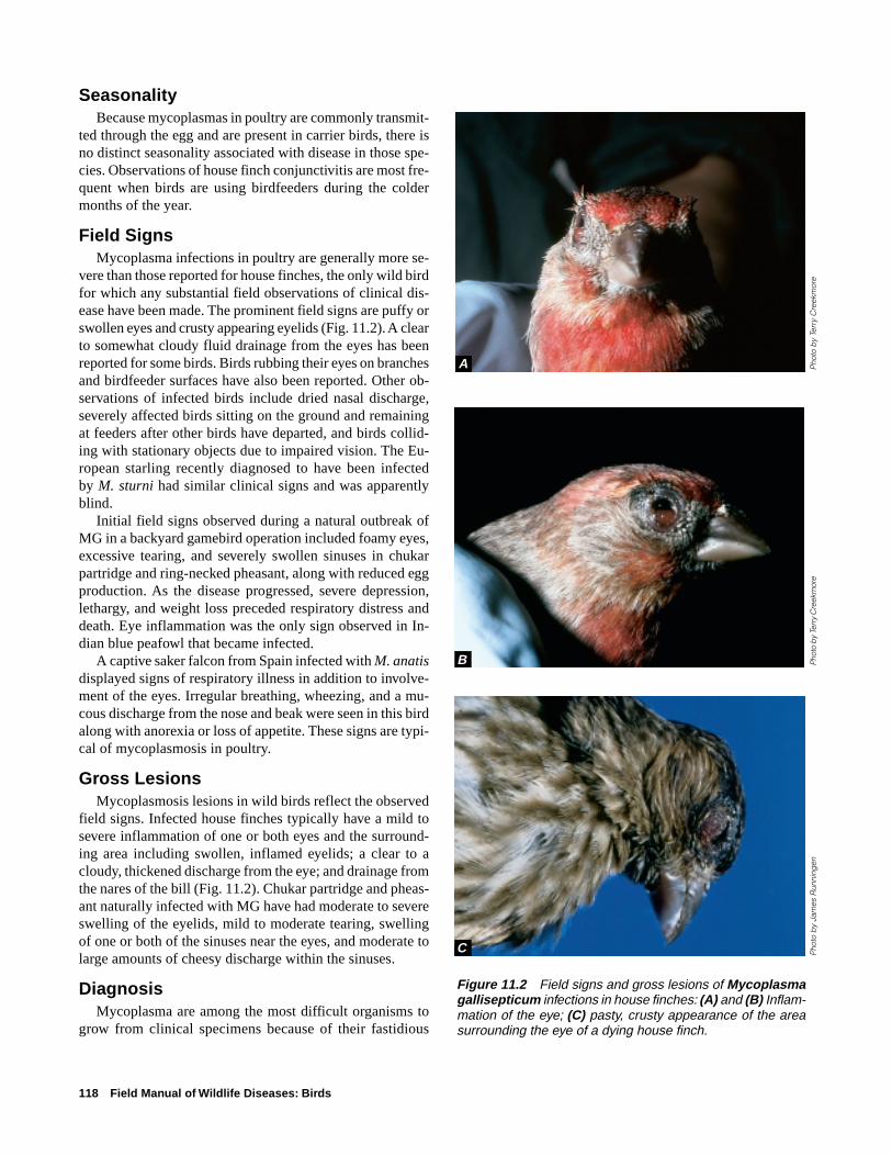
118 Field Manual of Wildlife Diseases: Birds
SeasonalityBecause mycoplasmas in poultry are commonly transmit-
ted through the egg and are present in carrier birds, there isno distinct seasonality associated with disease in those spe-cies. Observations of house finch conjunctivitis are most fre-quent when birds are using birdfeeders during the coldermonths of the year.
Field SignsMycoplasma infections in poultry are generally more se-
vere than those reported for house finches, the only wild birdfor which any substantial field observations of clinical dis-ease have been made. The prominent field signs are puffy orswollen eyes and crusty appearing eyelids (Fig. 11.2). A clearto somewhat cloudy fluid drainage from the eyes has beenreported for some birds. Birds rubbing their eyes on branchesand birdfeeder surfaces have also been reported. Other ob-servations of infected birds include dried nasal discharge,severely affected birds sitting on the ground and remainingat feeders after other birds have departed, and birds collid-ing with stationary objects due to impaired vision. The Eu-ropean starling recently diagnosed to have been infectedby M. sturni had similar clinical signs and was apparentlyblind.
Initial field signs observed during a natural outbreak ofMG in a backyard gamebird operation included foamy eyes,excessive tearing, and severely swollen sinuses in chukarpartridge and ring-necked pheasant, along with reduced eggproduction. As the disease progressed, severe depression,lethargy, and weight loss preceded respiratory distress anddeath. Eye inflammation was the only sign observed in In-dian blue peafowl that became infected.
A captive saker falcon from Spain infected with M. anatisdisplayed signs of respiratory illness in addition to involve-ment of the eyes. Irregular breathing, wheezing, and a mu-cous discharge from the nose and beak were seen in this birdalong with anorexia or loss of appetite. These signs are typi-cal of mycoplasmosis in poultry.
Gross LesionsMycoplasmosis lesions in wild birds reflect the observed
field signs. Infected house finches typically have a mild tosevere inflammation of one or both eyes and the surround-ing area including swollen, inflamed eyelids; a clear to acloudy, thickened discharge from the eye; and drainage fromthe nares of the bill (Fig. 11.2). Chukar partridge and pheas-ant naturally infected with MG have had moderate to severeswelling of the eyelids, mild to moderate tearing, swellingof one or both of the sinuses near the eyes, and moderate tolarge amounts of cheesy discharge within the sinuses.
DiagnosisMycoplasma are among the most difficult organisms to
grow from clinical specimens because of their fastidious
Figure 11.2 Field signs and gross lesions of Mycoplasmagallisepticum infections in house finches: (A) and (B) Inflam-mation of the eye; (C) pasty, crusty appearance of the areasurrounding the eye of a dying house finch.
Pho
to b
y Te
rry
Cre
ekm
ore
Phot
o by
Ter
ry C
reek
mor
eP
hoto
by
Jam
es R
unni
ngen
B
C
A

Mycoplasmosis 119
nature, intimate dependence upon the host species they colo-nize, and slow growth on artificial media. The greatest suc-cess in isolating MG from house finches has been when tis-sue swabs were obtained from live trapped, freshly killed, orfresh dead birds. There has been limited success from frozencarcasses. When mycoplasma is suspected, contact with adisease diagnostic laboratory is recommended to obtain guid-ance on how to handle specimens. If field conditions permit,selective media provided by a diagnostic laboratory shouldbe inoculated with swabs from the inner eyelids, sinus, thefunnel-shaped area at the back of the sinuses where they splitright and left (choanal cleft), and trachea of suspect birdsand shipped to the laboratory with the freshly killed or deadbirds from which those swabs were made. If birds can besubmitted, they should be chilled, rather than frozen, andimmediately transported to a qualified disease diagnosticlaboratory.
ControlRoutine cleaning and disinfection of birdfeeders with
household bleach is recommended to prevent mycoplasmosisand other diseases that can be transmitted at birdfeeders. A10 percent solution of household bleach applied weekly forfeeders with high bird use will reduce the potential for con-taminated surfaces to transmit disease. Close observation ofbirds using feeders and the prompt reporting of suspect casesof mycoplasmosis to authorities will provide the opportu-nity for early intervention based on timely diagnosis and forinitiating an appropriate disease-control strategy specific tothe location and population involved. Special considerationneeds to be given to the fact that house finch conjunctivitis isa new and emerging disease problem that has been docu-mented in two additional species of songbirds. One of theseincluded a case where a blue jay being rehabilitated in a cagepreviously occupied by an infected house finch became in-fected. That case demonstrates the need for adequate clean-ing and disinfection of cages used in wildlife rehabilitation.Birds that survive infection can become disease carriers thatserve as a source for initiating new outbreaks. Also, aerosoland egg transmission of mycoplasmosis is common for poul-try. Similar transmission is likely for wild birds and must betaken into consideration during the rehabilitation of wild birdsinfected with mycoplasmosis.
The potential for interspecies transmission of MG frompoultry to upland gamebirds being reared in captivity forsporting purposes must also be considered. This same con-sideration exists for raptors that may be fed poultry carcassesand waste.
Human Health ConsiderationsNone. Mycoplasmas that infect birds are not known to be
hazards for humans.
Milton Friend
Supplementary ReadingCookson, K.C., and Shivaprasad, H.L., 1994, Mycoplasma
gallisepticum infection in chukar partridges, pheasants, andpeafowl: Avian Diseases, v. 38, p. 914–921.
Dhondt, A.A., Tessaglia, D.L., and Slothower, R.L., 1998,Epidemic mycoplasmal conjunctivitis in house finches fromeastern North America: Journal of Wildlife Diseases, v. 34,p. 265–280.
Fischer, J.R., Stallknecht, D.C., Luttrell, P., Dhondt, A.A., andConverse, K.A., 1997, Mycoplasmal conjunctivitis in wildsongbirds: The spread of a new contagious disease in a mobilehost population: Emerging Infectious Diseases, v. 3, p. 69–72.
Poveda, J.B., Carranza, J., Miranda, A., Garrido, A., Hermoso, M.,Fernandez, A., and Comenech, J., 1990, An epizootiologicalstudy of avian mycoplasmas in southern Spain: AvianPathology , v. 19, p. 627– 633.

120 Field Manual of Wildlife Diseases: Birds

Miscellaneous Bacterial Diseases 121
Chapter 12
Miscellaneous Bacterial Diseases
Disease in free-ranging birds is caused by many other patho-genic bacteria in addition to those illustrated within this sec-tion. These other diseases are currently considered less im-portant because of their infrequent occurrence, the smallnumbers of birds generally lost annually, or because theyprimarily result from infection by opportunistic pathogensand they require concurrent disease processes for them tobecome apparent. The following brief highlights about themore important of these diseases are included to acquaintreaders with their existence and provide some basic infor-mation about their ecology.
ErysipelasErysipelas is caused by infection with the bacterium Ery-
sipelothrix rhusiopathiae. This disease is primarily associ-ated with swine and domestic turkeys, but it has been diag-nosed in many groups of birds (Fig. 12.1) and in mammals.The causative agent has also been isolated from the slimelayer of marine and freshwater fish and from crocodiles. Ery-sipelas is found worldwide. Little is known of the ecology ofthis disease in birds. Most reports of erysipelas in free-ranging birds involve individuals or small numbers of birds,but major die-offs can occur. The largest recorded die-offkilled an estimated 5,000 birds, primarily eared grebes, dur-ing 1975 on the Great Salt Lake, Utah. Small numbers ofwaterfowl (green-winged teal, northern shoveler, and com-mon mergansers) and a few herring gulls also died. Erysi-pelas has also been diagnosed as the cause of a die-off ofbrown pelicans in southern California during the late 1980s.Other free-ranging birds diagnosed with erysipelas includehawks, crows, raven, wood pigeon, starling, doves, finches,and European blackbird. The causative bacterium is able tosurvive in the environment for prolonged periods of time,and it was isolated from grebe carcasses approximately 18weeks after their death during the Great Salt Lake mortalityevent. The bacteria probably are transmitted through inges-tion, such as when gulls feed on carcasses, or entry of theorganism through cuts and abrasions. Humans are suscep-tible to infection. Most human cases involve localized infec-tions resulting from entry through a cut in the skin. Humancases have been fatal when the disease progressed to an in-fection of the blood and spreads throughout the body (a sep-ticemic infection).
Figure 12.1 Reported occurrences of erysipelas in birds.
FrequentCommon
OccasionalRare
POULTRY
Turkeys
Chickens
Ducks
WILD BIRDS
Songbirds/blackbirds/starling
Pheasant/quail
Grebes/cormorants
Gulls/terns
Parrots/parakeets/canaries
Doves/pigeons
Peacock/guinea fowl
Ducks/geese/swans
Cranes/storks
Pelicans
Eagles/hawks
Crows/raven
Penguins

122 Field Manual of Wildlife Diseases: Birds
New Duck DiseasePasteurella anatipestifer causes an important disease of
domestic ducks that has infrequently caused the deaths ofwild birds. This disease has also killed domestic turkeys andchickens and captive-reared pheasants, quail, and waterfowl.Major mortality events from infection with P. anatipestiferhave occurred in free-ranging black swans in Tasmania andin tundra swans in Canada. New duck disease has also beendiagnosed as the cause of mortality in small numbers of otherfree-ranging birds, including lesser snow geese. In the do-mestic duck industry, mortality primarily involves birds 2–3months old. The swans that died in Tasmania and Canadawere primarily young-of-the-year, which is consistent withmortalities of captive wild waterfowl. Birds can die within24–48 hours after the onset of clinical signs of listlessness, adroopy appearance, fluid discharges from the eyes and bill,greenish diarrhea, and variety of nervous system disorders.The most prominent lesion seen during postmortem exami-nation is a fibrinous covering on the surface of various or-gans such as the liver and heart (Fig. 12.2).
Necrotic EnteritisNecrotizing enteritis is caused by an enterotoxemia or
toxins in the blood produced in the intestine resulting frominfections with Clostridium perfringens. This disease is foundthroughout much of the world where poultry are produced,and it is often an important cause of mortality for adult do-mestic breeder ducks. Sporadic cases have been diagnosedin waterfowl collections and in wild mallards, black ducks,and Canada geese. A die-off in Florida involved mallardsand other wild ducks along with several species of shore-birds and wading birds. Wild ducks are also reported to havedied from this disease in Germany.
During recent years, increasing numbers of small die-offshave been detected in snow geese, Canada geese, and white-fronted geese in Canada and the United States. An abruptchange in diet associated with seasons and bird migrationsare thought to disrupt the intestinal microflora and allowC. perfringens to proliferate in the intestine. The toxins pro-duced by these bacteria are the cause of death. The onset ofdeath is generally rapid and without obvious clinical signs.Severe depression is sometimes observed in chickens alongwith reluctance to move, diarrhea, and ruffled feathers. Le-sions generally appear as a mixture of dead cellular materi-als and plasma debris, tan-yellow in color, that covers muchof the lower region of the intestine of affected waterfowl (Fig.12.3).
Ulcerative EnteritisQuail are highly susceptible to infection by Clostridium
colinum, the cause of ulcerative enteritis or “quail disease.”Outbreaks of this disease in free-ranging wild birds are rare,but outbreaks have been reported for California quail inWashington State. This acute bacterial infection is charac-
Figure 12.2 Fibrinous covering on the heart and liver of abird with Pasteurella anatipestifer.
Pho
to b
y Ja
mes
Run
ning
en
Figure 12.3 Lesions of necrotic enteritis in the intestine of agoose.
Pho
to b
y Ja
mes
Run
ning
en
Figure 12.4 Advanced lesions of ulcerative enteritis in theintestine of a chukar partridge.
Pho
to b
y M
ilton
Frie
nd

Miscellaneous Bacterial Diseases 123
terized by sudden onset followed by rapid spread throughthe flock. Outbreaks have been reported worldwide where-ever game birds are raised in captivity under crowded condi-tions. In addition to upland game species such as grouse,quail, pheasant, and partridges, outbreaks have been reportedin chickens, pigeons and robins. Mortality in young quailcan reach 100 percent of the flock. Gross lesions vary anddepend upon how long the bird lives following infection. Ul-cers within the intestine originate as small yellow spots orinfected areas with hemorrhagic borders and progress to cir-cular forms that may join together as large areas of deadtissue that resemble thickened mucous membranes withraised edges (Fig. 12.4). Liver lesions include yellow areasof tissue death or necrosis along the edges of the liver andscattered grey spots or small yellow circumscribed spotswithin the liver itself that sometimes are surrounded by alight yellow halo effect.
StaphylococcosisAll avian species are susceptible to staphylococcal infec-
tions, and Staphylococcus aureus is the most common causeof disease. An often observed form of infection is a lesionthat appears as an inflammation of the skin of the foot orpododermatitis, that is commonly referred to as “bumblefoot”(Fig. 12.5). Staphylococcal bacteria are ubiquitous, normalinhabitants of the skin and mucous membranes, and the bac-teria require a break in those protective layers for infectionto occur. Captive birds are more commonly found infectedthan free-ranging birds. Abrasions from rough surfaces wherebirds perch or stand may contribute to the occurrence of thisdisease. Studies in Spain with free-ranging imperial eaglesdemonstrated that staphylococcal infection can be transferredfrom humans to chicks being handled for banding. Infectionwas common in nestlings handled without latex gloves,whereas infection was rare in those birds handled with gloves.Mallard and redhead duck, bald and golden eagle, and fer-ruginous hawk have been among the species submitted tothe National Wildlife Health Center (NWHC) that have beendiagnosed with this condition.
Septicemic staphylococosis or staphylococcal blood poi-soning can also occur, generally in birds that are immuno-compromised or whose immune systems are not fully func-tioning. These types of infection can result in sudden death.Lesions associated with this form of infection generally con-sist of congestion of internal organs, including the liver,spleen, kidneys, and lungs, accompanied by areas of tissuedeath (Fig. 12.6). Bald eagles, American kestrels, red-tailedhawks, a duck, a mute swan, and herring and ring-billed gullsare among the species submitted to the NWHC for whichsepticemic staphylococcal infections have been diagnosed.
S. aureus can also cause serious disease in humans bothas a wound infection and as a source of food poisoning. Goodsanitation procedures should always be followed when han-
Figure 12.5 Bumblefoot in a domestic duck.
Pho
to b
y C
arol
Met
eyer
Pho
tos
by
Jam
es R
unni
ngen
Figure 12.6 Congested liver (A) and spleen (B) from birdswith staphylococosis.
A
B

124 Field Manual of Wildlife Diseases: Birds
dling animals, and protective gloves should be worn whenhandling wildlife found dead.
TularemiaTularemia is primarily a disease of mammals, but natural
infections by Francisella tularensis have caused die-offs ofruffed grouse and other grouse species. A variety of avianspecies have been found to be susceptible to infection as aresult of serological surveys that have detected antibodyagainst tularemia, experimental studies to determine suscep-tibility, and by cause-of-death assessments for birds submit-ted for necropsy (Table 12.1). The strains of F. tularensisthat caused natural infection of ruffed grouse are of low viru-lence for humans despite ruffed grouse becoming infectedby the same tick (Haemaphysalis leporispaulstris) that causeshighly virulent tularemia in snowshoe hare.
Ticks are the primary source for disease transmission innatural cases of tularemia in upland gamebirds such as grouseand pheasants; ingestion of diseased birds and rodents is theprimary source of disease transmission to raptors, gulls, andother scavenger species. Tularemia is infrequently reportedas a cause of disease in wild birds. Ruffed grouse in northernclimates have been the primary focus for reports in the sci-entific literature. The primary lesion seen is multiple, dis-crete spots scattered throughout the liver tissue (Fig. 12.7).
Table 12.1 Avian species reported to be susceptible toinfection by Francisella tularensis.
Upland game species Other birds
Ruffed grouse Gulls and ternsSharp-tailed grouse Raptors (such asSage grouse hawks and eagles)Ptarmigan Scavengers (such asBlue grouse shrikes)Bobwhite quail Ducks and geesePheasant
Figure 12.7 The numerous, small, yellow and white spots on the liver of this beaver that died of tulare-mia are similar to the appearance of liver lesions in ruffed grouse.
Pho
to b
y Ja
mes
Run
ning
en
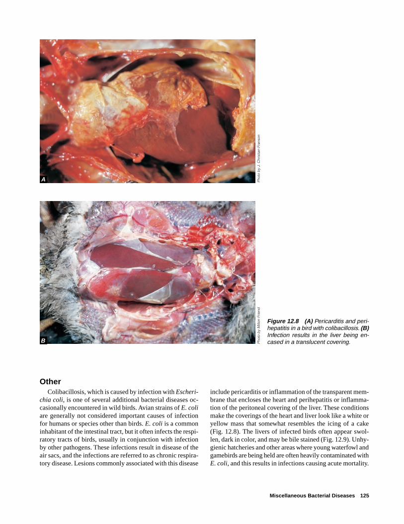
Miscellaneous Bacterial Diseases 125
Pho
to b
y J.
Chr
istia
n Fr
anso
nP
hoto
by
Milt
on F
riend
Figure 12.8 (A) Pericarditis and peri-hepatitis in a bird with colibacillosis. (B)Infection results in the liver being en-cased in a translucent covering.
OtherColibacillosis, which is caused by infection with Escheri-
chia coli, is one of several additional bacterial diseases oc-casionally encountered in wild birds. Avian strains of E. coliare generally not considered important causes of infectionfor humans or species other than birds. E. coli is a commoninhabitant of the intestinal tract, but it often infects the respi-ratory tracts of birds, usually in conjunction with infectionby other pathogens. These infections result in disease of theair sacs, and the infections are referred to as chronic respira-tory disease. Lesions commonly associated with this disease
include pericarditis or inflammation of the transparent mem-brane that encloses the heart and perihepatitis or inflamma-tion of the peritoneal covering of the liver. These conditionsmake the coverings of the heart and liver look like a white oryellow mass that somewhat resembles the icing of a cake(Fig. 12.8). The livers of infected birds often appear swol-len, dark in color, and may be bile stained (Fig. 12.9). Unhy-gienic hatcheries and other areas where young waterfowl andgamebirds are being held are often heavily contaminated withE. coli, and this results in infections causing acute mortality.
B
A

126 Field Manual of Wildlife Diseases: Birds
Figure 12.9 Swollen bile-stained liver in a bird with coliba-cillosis.
Pho
to b
y Ja
mes
Run
ning
en
Similar to the other sections of this Manual, the bacterialdiseases discussed are not comprehensive of diseases of wildbirds. The similarities in clinical signs and gross lesions dis-played in illustrations in this section emphasize the need forcause-of-death evaluations by qualified animal disease labo-ratories. Also, the environmental persistence and humanhealth impacts noted for some of these pathogens emphasizethe need to consider personal and environmental protectionwhen handling dead birds. Assumptions that the cause ofdeath is due to a pathogen of minor importance could haveserious consequences if highly virulent infections are in-volved.
Milton Friend
Supplementary Reading
Calnek, B.W., and others, eds., 1997, Diseases of poultry(10th ed.): Ames, Iowa, Iowa State University Press, 929 p.
Davis, J.W., Anderson, R.C., Karstad, L., and Trainer, D.O. eds.,1971, Infectious and parasitic diseases of wild birds: Ames,Iowa, Iowa State University Press, 344 p.
Jellison, W.L., 1974, Tularemia in North America, 1930–1974:University of Montana, Missoula, p. 1–276.
Jensen, W.I., and Cotter, S.E., 1976, An outbreak of erysipelas ineared grebes (Podiceps nigricollis)., Journal of WildlifeDiseases, v. 12: 583–586
Wobeser, G., and Rainnie, D.J., 1987, Epizootic necrotic enteritisin wild geese: Journal of Wildlife Diseases, v. 23: 376–386

Introduction to Fungal Diseases 127
Section 3
Fungal Diseases
Aspergillosis
Candidiasis
Miscellaneous Fungal Diseases
Moldy grain may be the source of aspergillosis in wild waterfowlPhoto from National Wildlife Health Center
128 Field Manual of Wildlife Diseases: Birds
Introduction to Fungal Diseases
Fungi are important causes of disease in wild birds and otherspecies. Three basic types of disease are caused by theseagents: mycosis, or the direct invasion of tissues by fungalcells, such as aspergillosis; allergic disease involving thedevelopment of a hypersensitivity of the host to fungal anti-gens; and mycotoxicosis, which results from ingestion oftoxic fungal metabolites. Mycosis and allergic disease mayoccur together, especially when the lung is infected. Thissection will address only mycosis. Mycotoxicosis is ad-dressed in Section 6, Biotoxins. Allergic disease is not wellstudied in wild birds and it is beyond the scope of this Manual.
Most disease-causing fungi are commonly found withinthe normal environment of hosts that may become diseased.Host resistance is the main determinant of whether or notdisease will occur. Opportunistic infections often result when
birds and other species are immunosuppressed, when theirmechanisms for inflammatory response are inhibited, or whenthey experience physical, nutritional, or other stress for pro-longed periods of time. Newborn do not have fully function-ing immune systems and are, therefore, especially vulner-able to mycosis as are very old animals that are likely tohave impaired immune systems. Inhalation is the primaryroute for exposure to most fungi-causing mycosis.
Aspergillosis is the primary mycosis affecting wild birds.Candidiasis is a less common mycosis of wild birds and otherspecies, but it differs greatly from aspergillosis by being trans-mitted by ingestion. These two diseases are the primarymycoses of wild birds and are the main subjects of this sec-tion.
“Fungi are of an ancient lineage and have a fossil record that extends back to the
Devonian and Pre-Cambrian eras…the earliest written record of fungi are not of the
fungi themselves, but of their depredations… To the physician and poet Nicander
[ca. 185 B.C.], fungi were ‘the evil ferment of the earth; poisonous kinds originating
from the breath of vipers,’…”
(Ainsworth)
Quote from:
Ainsworth, G.C., and Sussman, A.S., 1965, The fungi: Anadvanced treatise, v. 1 of The Fungal Cell: Academic Press,New York, p. 4, 8.

Aspergillosis 129
Chapter 13
Aspergillosis
SynonymsBrooder pneumonia, pseudotuberculosis, “asper”mycosis, mycotic pneumonia
CauseAspergillosis is a respiratory tract infection caused by
fungi of the genus Aspergillus, of which A. fumigatus is theprimary species responsible for infections in wild birds (Fig.13.1). Aspergillosis is not contagious (it will not spread frombird to bird), and it may be an acute, rapidly fatal disease ora more chronic disease. Both forms of the disease are com-monly seen in free-ranging birds, but the acute form is gen-erally responsible for large-scale mortality events in adultbirds and for brooder pneumonia in hatching birds. Aspergil-lus sp. also produce aflatoxins (see Chapter 37, Mycotox-ins), but the significance of those toxins in the ability of thefungus to cause disease in birds is unknown.
Aspergilli are saprophytic (live upon dead or decayingorganic matter) molds that are closely associated with agri-culture and other human activities that make nutrients avail-able to fungi. A. fumigatus commonly grows in damp soils,decaying vegetation, organic debris, and feed grains. Highnumbers of spores (called conidia) are released into the at-mosphere and are inhaled by humans, birds, and other ani-mals. These spores travel through the upper respiratory tractto the lungs. If the spores colonize the lungs, then the fungimay be disseminated to other parts of the body and disease,often leading to death, occurs.
Acute aspergillosis has caused devastating loss of birdsin hatcheries. The source of infection in some instances hasbeen contaminated litter. Also, infection of broken eggs priorto hatching provides an ideal growth medium for the fungusand the subsequent production of massive numbers of sporesfor infection of newly hatched birds. Inhaled spores initiatea cellular response in the lungs that results in the air pas-sages soon becoming obliterated by cellular material andbranching fungal filaments. Asphyxiation quickly follows andcauses death. Acute aspergillosis has also been found in free-ranging waterfowl. The circumstances of these events areuniformly associated with birds feeding in waste grain andin silage pits during inclement weather. The mallard duckhas been the primary species involved, and the events haveonly lasted a few days, terminating when the weather im-proved and allowed the ducks to resume normal feeding. Fieldinvestigations of several events disclosed heavily contami-nated feed that resulted in overwhelming exposure toA. fumigatus (Fig. 13.2).
Chronic forms of aspergillosis have been described in wildbirds since at least 1813. Typically, the lungs and air sacs arechronically infected, resulting in a gradual reduction in res-piratory function. Eventual dissemination of the fungusto the liver, gut wall, and viscera is facilitated by infection ofthe extensive system of air sacs that are part of the avianrespiratory system.
Figure 13.1 Primary causes of aspergillosis in birds.
Figure 13.2 Moldy grain pile that was the source of acuteaspergillosis in wild waterfowl.
CommonOccasional
Infrequent
Aspergillus fumigatus
A. flavus
A. terrus, A. glacus
A. nidulans, A. niger
A. amstelodami, A. nigrescens
Nat
iona
l Wild
life
Hea
lth C
ente
r fil
e p
hoto

130 Field Manual of Wildlife Diseases: Birds
Species AffectedA wide variety of birds have died of aspergillosis and prob-
ably all birds are susceptible to it. Aspergillosis was one ofthe first diseases described for wild birds; it was noted in ascaup in 1813 and in a European jay in 1815. Loons andmarine birds that are brought into rehabilitation, captive rap-tors, and penguins being maintained in zoological parks andother facilities commonly die from aspergillosis. This dis-ease also develops at birdfeeding stations and it causeswaterfowl die-offs. Young birds appear to be much more sus-ceptible than adults. Most reported mortalities of free-rang-ing wild birds involve isolated mortalities found during post-mortem evaluations rather than mortalities found duringmajor die-offs (Fig. 13.3).
DistributionAspergillosis in birds is reported nearly worldwide.
SeasonalityMost aspergillosis outbreaks in waterfowl happen in fall
to early winter; individual cases can occur at any time, par-ticularly among birds stressed by crippling, oiling, mal-nutrition, recent capture, and concurrent disease conditions.This disease can cause serious losses among seabirds in re-habilitation programs after oil spills. Aspergillosis is a fre-quent complication in hunter-crippled waterfowl, amongbirds on nutritionally deficient diets, and in Canada geesewhose immune systems have been compromised by expo-sure to environmental contaminants such as lead.
Environmental factors also contribute to the time of yearwhen aspergillosis is seen. Scattered outbreaks of this dis-ease occurred among American coot, diving ducks, tundraswan, and passerine birds throughout California one winterat the end of a 3-year drought. Severe dust conditions asso-ciated with this weather pattern are thought to have inter-fered with respiratory clearance mechanisms by reducing theamount of mucous and other body secretions that coat thecellular lining of the throat and air passages to the lungs,thereby increasing bird susceptibility to aspergillosis. A falloutbreak in Steller’s jays in British Columbia was associ-ated with a particularly dry and warm summer.
Brooder pneumonia, a specialized springtime form of as-pergillosis, infects chicks or ducklings that are placed in As-pergillus-contaminated brooders. Catastrophic losses haveoccurred on game farms under these circumstances. Chickshave also been lost during captive-rearing of endangeredspecies. Aspergillosis is also an important cause of mortalityin winter roosts of blackbirds in Maryland and Pennsylva-nia.
Figure 13.3 Relative occurrence of aspergillosis in free-ranging wild birds.
Waterfowl
Gulls
Crows, raven
Raptors (free-living)
(captive)
Songbirds
Upland gamebirds
Blackbirds, cowbirds, grackles
Herons
Shorebirds
CommonOccasional
InfrequentRare or not reported

Aspergillosis 131
Field SignsThe typical aspergillosis-affected bird is emaciated, and
it frequently exhibits severe and progressive difficulty inbreathing by gaping or rapid opening and closing of the bill(Fig. 13.4A). Birds often appear to be unthrifty, and theirwings may droop (Fig. 13.4B). Infected birds are usuallyweak and may fail to try to escape. With the exception ofvisible evidence of breathing difficulties, these signs are simi-lar to those for lead poisoning. Infection that reaches the braincan result in obvious loss of muscular coordination and twist-ing of the head and neck so that the head is held in unnaturalpositions. Inflammation of the covering of the brain or men-ingoencephalitis with associated areas of brain tissue deathhas been reported for eider ducklings dying from aspergillo-sis.
Epizootic aspergillosis and brooder pneumonia outbreaksare often characterized by sudden deaths of previously healthybirds. Sick birds show acute respiratory distress and failure.
Gross LesionsBirds infected with the more typical chronic form of as-
pergillosis usually have variously sized lesions in their lungsand air sacs. Typically, these lesions appear as flattened, yel-low plaques with a cheesy appearance and consistency (Fig.13.5). Continuous masses of these lesions may completelyline the air sac. There may also be an extensive fungus growthon tissue and air sac surfaces that appears similar to breadmold. This velvety, blue-green or grey fungal mat is strikingin appearance (Fig. 13.6).
In cases of acute aspergillosis, the birds are usually ingood flesh and have good-to-moderate deposits of fat. Airsacs are usually thickened, but the most striking lesion is adark red, firm lung that is often studded or peppered withsmall, 1–2 millimeter, yellow nodules (Fig. 13.7).
Other, less common lesions that have been described in-clude necrotic skin granulomas or semifirm growths of granu-lar consistency in chickens and pigeons. Cheesy plaques thatform in the eye beneath the nictitating membrane, which isthe transparent membrane that forms a rapidly moving thirdeyelid that keeps the eye clean and moist, or on the surfaceof the eye have also been observed.
DiagnosisWhole carcasses should be submitted for necropsy by
qualified diagnosticians. Diagnosis is based on finding thetypical lesions and on isolating the fungus from the tissues.Aspergillus sp. can be identified by microscopically exam-ining material from fungal mats and from tissue sections thathave been specially stained. However, the specific speciesof Aspergillus cannot be identified by these means.
ControlThe spores of the mold A. fumigatus are widely distrib-
uted and are often present in moldy feeds, unclean broodersand incubators, moldy straw, and rotting agricultural waste.Aspergillus grows best on decomposing organic matter leftin a warm, dark, moist environment. Failure to maintain aclean environment often leads to severe outbreaks.
Aspergillosis has broken out in mallards feeding in fieldsthat were previously covered by discarded moldy corn andsilage. Dusty straw hay placed as litter in the bottoms of woodduck nest boxes has resulted in losses of wood duck duck-lings. Avoid using moldy or dusty straw, silage, or feed, anddumping moldy waste grain in areas where waterfowl andother birds feed. Birds should be denied the use of fieldswhere moldy agricultural waste products such as waste corn,peanuts, straw, or hay have accumulated. Monitoring for suchsituations in waterfowl concentration areas and establishingcontingency plans that can be implemented at the onset ofinclement weather can minimize the potential for waterfowldeaths if the concentrated sources of Aspergillus spores cannot be dealt with in other ways. People who feed birds shouldbe educated to periodically clean their feeding stations.
Figure 13.4 (A) Respiratory distress and gaping (note theopen bill) in a herring gull suffering from aspergillosis. (B) Wingdroop also occurs. Note that the wing on the near side of thisbird is drooping well below the body.
A
B Pho
tos
by
Milt
on F
riend

132 Field Manual of Wildlife Diseases: Birds
Figure 13.5 “Cheesy” plaques in thelungs and air sacs of a bird with as-pergillosis.
Figure 13.6 Lung of a bird withchronic aspergillosis showing(A) “cheesy” fungal plaques, and(B) “bread mold” fungal mat totallyinvolving the air sac adjacent to theheart of this bird.
Pho
to b
y M
ilton
Frie
nd
A
B Pho
tos
by
Milt
on F
riend
➡
➡➡
➡
➡
➡
➡
➡

Aspergillosis 133
Human Health ConsiderationsAspergillosis is not contagious. However, when human
resistance to infection is impaired, aspergilli can cause rap-idly developing acute infection following environmental ex-posure. Invasive aspergillosis in humans involving dissemi-nation of fungi to organs other than the lungs is often associ-ated with the person being immunocompromised and, if thedisease is not properly diagnosed, it may be life threatening.A few individuals who have worked with A. fumigatus havebecome allergic to it. Allergic response can result in an acute,life-threatening reaction to this fungus. It is unlikely that in-fected bird carcasses would provide sufficient exposure toresult in either of these outcomes.
Milton Friend
Figure 13.7 Acute aspergillosis or “brooder pneumonia” ina lung of a wood duck duckling. Note dark red, “studded”(granular) appearance of lung.
Pho
to b
y Ja
mes
Run
ning
en
Supplementary ReadingAdrian, W.J., Spraker, T.R., and Davies, R.B., 1978, Epornitics of
aspergillosis in mallards (Anas platyrhynchos) in north centralColorado: Journal of Wildlife Diseases, v.14, p. 212–217.
Barden, E.S., Chute, H.L., O’Meara, D.C., and Wheelwright, H.T.,1971, A bibliography of avian mycosis (partially annotated):Orono, Me., College of Life Sciences and Agriculture. Univ. ofMaine, 193 p.
O’Meara, D.C., and Witter, J.F., 1971, Aspergillosis, in Davis,J.W., and others, eds., Infectious and parasitic diseases of wildbirds: Ames, Iowa, Iowa State University Press, p. 153–162.
Powell, K.A., Renwick, A. and Peberdy, J.F., 1994, The genusAspergillus from taxonomy and genetics to industrial applica-tion: Plenum Press, New York, N.Y., 380 p.
Wobeser, G.A., 1997, Aspergillosis, in Diseases of wild waterfowl(2nd ed): New York, N.Y., Plenum Press, p. 95–101.

134 Field Manual of Wildlife Diseases: Birds

Candidiasis 135
Chapter 14
Candidiasis
SynonymsMoniliasis, candidiasis, thrush, sour crop
CauseCandida albicans, a yeast-like fungi, is the primary cause
of candidiasis or candidiosis. C. albicans is a normal inhab-itant of the human alimentary canal, as well as that of manyspecies of lower animals. Ingestion in food or in water is theusual means for its transmission. Contaminated environ-ments, such as litter from poultry and gamebird rearingfacilities, refuse disposal areas, discharge sites for poultryoperations, and areas contaminated with human waste haveall been suggested as sources for Candidia exposure for birds.
Species AffectedThere have been few reports of candidiasis causing dis-
ease in free-ranging wild birds and few investigations of itsprevalence. Therefore, little can currently be said about itsoccurrence in wild species. Candidiasis is an occasional dis-ease of importance within some poultry flocks, and it hasbeen reported as a disease or an intestinal infection in nu-merous species of wild birds being raised in captivity. It hasalso been an occasional cause of disease in wild speciesbeing transported within the pet bird industry (Fig. 14.1).
DistributionCandidiasis is found worldwide.
SeasonalityThere is no known seasonal occurrence. Life-cycle pat-
terns for bird populations are likely to influence any tempo-ral occurrence for this disease because young birds are gen-erally more susceptible to infection.
Field SignsThere are no unique signs of disease. Affected poultry
have retarded growth, stunted appearance, are listless, andhave ruffled feathers.
Gross LesionsLesions are generally confined to the upper areas of the
digestive tract. The mouth, esophagus, and, primarily, thecrop, may have grayish-white, loosely attached, plaque-likeareas on their internal surfaces. Circular, raised, ulcerative
Figure 14.1 Avian groups reported to have been infectedwith candidiasis.
Chickens/turkeys
Guinea fowl
Quail
Pigeons
Ducks/geese
Gulls/terns
Ducks
Shorebirds
Pheasants/ partridges/ grouse/quail
Wood pigeon
Peacock
Crows/jackdaw
Parrots/parakeets/rosella
Lovebirds/budgerigar/lorikeets/lories
Sparrows/starlings
Kites
Songbirds
Poultry Wild species

136 Field Manual of Wildlife Diseases: Birds
nodules that appear as rose-like clusters may be within thecrop, and the crop surface is often so unevenly thickenedthat it appears to have the texture of a Turkish bath towel orcurds. Other areas of the upper digestive tract develop falsemembranes that resemble those which develop duringdiptheria, areas of dead tissue, and contain considerable tis-sue debris.
ControlThe infrequent reports of this disease in free-ranging wild
birds do not warrant the need for disease control. This dis-ease is more likely to be encountered in captive-rearing situ-ations. Disease prevention should be practiced to preventinfections. Cages, equipment, and other materials in contactwith infected birds should be disinfected because of the broadhost range of species that can become infected.
Human Health ConsiderationsHumans can be infected, and infections can result in acute
or chronic disease that can involve the mucous membranes(oral thrush), skin, nails, and internal organs.
Milton Friend
Supplementary ReadingChute, H.L., 1997, Thrush (mycosis of the digestive tract), in
Calnek, B.W., and others, eds., Diseases of Poultry (10th ed.):Ames, Iowa, Iowa State University, p. 361–365.
Odds, F.C., 1988, Candida and candidosis: London, BaillièreTindall, 468 p.
O’Meara, D.C., and Witter, J.F., 1971, Candidiasis, in Davis, J.W.,and others, eds., Infectious and parasitic diseases of wild birds:Ames, Iowa, Iowa State University, p. 163–169.

Miscellaneous Fungal Diseases 137
Chapter 15
Miscellaneous Fungal Diseases
As for other types of disease, fungal infections probably aremore common causes of disease in wild birds than is cur-rently recognized. Also, the similarity in gross lesions pro-duced by some fungi mask the detection of less commonfungi as disease agents. Numerous types of disease-causingfungi in addition to Aspergillus fumigatus and Candidaalbicans have been isolated from birds; most isolations havebeen from poultry and wild birds being maintained in cap-tivity. Enhanced disease surveillance that is often associatedwith privately owned birds and greater opportunity to detectdisease in confined birds are reasons for these findings ratherthan any known differences in the occurrence of fungal dis-eases in free-ranging and captive birds. Many of the reportedinfections appear to have been opportunistic invasions bythe fungi involved. The important points are that many fungiare capable of causing disease in birds but their collectiveimpacts do not rival A. fumigatus as a single cause of diseasein wild birds. Nevertheless, it is important to be aware of thediversity of pathogenic or disease causing fungi.
Infectious diseases caused by fungi have been groupedinto categories that represent their involvement within thehost.
Trichophyton gallinae is the primary cause of ringworm,or fowl favus, in birds, and has been reported in poultry andseveral species of wild birds in addition to companion ani-mals, humans, and other mammalian species. T. gallinae iswidely distributed geographically, and infection by this fun-gus is a striking example of a cutaneous mycosis (Fig. 15.1).Ringworm in birds is highly contagious, and it is transmittedby direct bird-to-bird contact or by contact with a contami-nated environment. The fungus can remain viable at roomtemperature in infected scales or skin lesions that slough fromthe body for up to 1 year. Microsporum gallinae is anotherwidely distributed fungus that is a significant cause of ring-worm in birds and mammals.
Dactylaria gallopova causes a subcutaneous mycosis re-ported for poultry. This fungus is found in warm habitatssuch as hot springs and thermal soils. The fungi generallyenter the body at a traumatized or injured site and may theninvade other sites following fungal establishment and growth.D. gallopova is not contagious, but it can invade the brainfollowing its spread from the site of infection. Death is theoutcome when the brain is invaded.
Aspergillus niger is another fungus within the genus As-pergillus that has caused bird deaths.
As noted in the Introduction of this Section, disease dueto infection of tissues is only one aspect of the potential im-pacts of fungi. The added issues of mycotoxins (see Section6, Biotoxins), allergic responses, and other aspects of fungaldiseases make fungi an important area for consideration inthe management and stewardship of free-ranging bird popu-lations.
Types of mycosis(direct invasion of tissue by fungal cells)
Category Area of the body affected
Superficial Found on the outermost layers of thebody covering; are generally ofcosmetic impact rather than causesof illness or death; have not beenreported in birds.
Cutaneous Found on the skin and appendages.(dermato-phytosis)
Subcutaneous Usually found in the fat-containingtissues underneath the skin and in theskin.
Systemic Result in infection of internal organsas well as other tissues.
Aspergillosis and candidiasis are diseases characteristicof systemic mycosis. Candidiasis can also be a cutaneousmycosis.
Figure 15.1 Extensive loss of feathers of the head of a loonbelieved to have been caused by ringworm resulting from in-fection by Trichophyton sp.
Pho
to b
y U
.S. F
ish
and
Wild
life
Ser
vice

138 Field Manual of Wildlife Diseases: Birds
Milton Friend
Supplementary ReadingHubbard, G.B., Schmidt, R.E., Eisenbrandt, D.L., Witt, W.M., and
Fletcher, K.C., 1985, Fungal infections of ventriculi in captivebirds: Journal of Wildlife Diseases, v. 21, p. 25–28

Viral Diseases 139
Section 4Viral Diseases
Duck Plague
Inclusion Body Disease of Cranes
Miscellaneous Herpesvirusesof Birds
Avian Pox
Eastern Equine Encephalomyelitis
Newcastle Disease
Avian Influenza
Woodcock Reovirus
Inclusion bodies in the liver of a bird that died of herpesvirus infectionPhoto by Lou Sileo

140 Field Manual of Wildlife Diseases: Birds
Introduction to Viral Diseases
Historically, viral diseases have not been recognized asmajor causes of illness and death in North American wildbirds. Until relatively recently, this may have been due toinadequate technology to culture and identify these organ-isms. Unlike bacteria, viruses are too small to be seen underthe light microscope and they cannot be grown on artificialmedia. Nevertheless, studies of infectious diseases causedby viruses have often predated discovery of the causativeagents by many years as evidenced by smallpox immuniza-tions being used centuries before that virus was identified.The isolations of a tobacco mosaic virus in 1892 and footand mouth disease viruses in 1898 mark the development ofvirology as a distinct biological science. The era of modernvirology began in the post-World War II years of 1945–50with the application of cell culture techniques to the study ofanimal viruses.
For centuries, gross and microscopic pathology associ-ated with tissue alterations caused by viral infections havebeen recorded for species of domestic birds, captive wildbirds, and, occasionally, for free-living wild birds. However,significant concern about viral diseases in wild birds hasprimarily occurred since the 1970s. This timeframe isconsistent with an apparent increase of emerging infectiousdiseases and emerging viruses in other species, includinghumans. It is noteworthy that this pattern exists for the dis-eases included in this section. Duck plague first appeared in
the United States in 1967 and the first major loss of wildwaterfowl from duck plague occurred in 1973. Eastern equineencephalitis erupted in a captive breeding flock of whoop-ing cranes in 1984; a highly virulent form of Newcastle dis-ease virus has appeared several times among double-crestedcormorants in Canada since 1990 and in the United Statessince 1992; and a previously undescribed reovirus was thecause of death for woodcock in 1989 and again in 1993. In1978, inclusion body disease of cranes appeared in a captivecrane breeding colony in the Midwestern United States; thatoutbreak was the first identification of this herpesvirus in-fection. In 1978 also, avian pox viruses were first isolatedfrom free-living waterfowl and from bald eagles the follow-ing year.
Avian influenza has been included in this section to givewildlife resource managers basic information about this groupof generally avirulent viruses that exchange genetic materialto create new forms of the virus, some of which are capableof causing disease. Interest in influenza is primarily focusedon the role of migratory birds as a source of viruses thatinfect domestic poultry and humans.
It seems likely that viral diseases will assume even greaterfuture importance as causes of disease in wild birds. Greaterattention needs to be given to the study of this source of dis-ease, especially in captive-propagation programs intendedfor supplementing and enhancing wild stocks of birds.
“The viruses almost surely antedate our species.” (Johnson)
“…viral emergence is essentially a two-step process: (1) introduction of the virus (what-
ever its origin) into a new host, followed by (2) dissemination within that new host
population…That second step might not occur at all…However, changing conditions
might increase the chances of this second step occurring.” (Morse)
Quotes from:
Johnson, K.M., 1993, Emerging viruses in context: an overview ofviral hemmorhegic fevers, in Morse, S.S. [editor], EmergingViruses: Oxford University Press, New York, p. 46.
Morse, S.S., 1993 Examining the origins of emerging viruses, inMorse, S.S. [editor], Emerging Viruses: Oxford UniversityPress, New York, p. 16–17.

Duck Plague 141
Chapter 16
Duck Plague
Synonyms
Duck virus enteritis, DVE
CauseDuck plague is caused by a herpesvirus. Infection often
results in an acute, contagious, and fatal disease. As withmany other herpesviruses, duck plague virus can establishinapparent infections in birds that survive exposure to it, astate referred to as latency. During latency, the virus cannotbe detected by standard methods for virus isolation. Studiesof domestic species of waterfowl have detected multiplestrains of the virus that vary in their ability to cause diseaseand death. Little is known about the response of wild water-fowl to strain differences.
Duck plague outbreaks are thought to be caused whenbirds that carry the virus shed it through fecal or oral dis-charge, thus releasing the virus into food and water withwhich susceptible birds may have contact. Experimentalstudies have demonstrated spontaneous virus shedding byduck plague carriers during spring. Changes in the durationof daylight and onset of breeding are thought to be physi-ological stresses that stimulate virus shedding at this time ofyear. The carriers are immune to the disease, but the virusshed by them causes infection and disease among suscep-tible waterfowl. Bird-to-bird contact and contact with virusthat has contaminated the environment perpetuate an out-break. Scavenging and decomposition of carcasses of infectedbirds also contaminate the environment by releasing virusesfrom tissues and body fluids. Virus transmission through theegg has been reported, but the role of the egg in the diseasecycle remains to be resolved.
Species AffectedOnly ducks, geese, and swans are susceptible to duck
plague. Other aquatic birds do not become infected, and theabsence of mortality of American coot, shorebirds, and otherwaterbirds that may be present during a waterfowl die-offcan be an important indication that duck plague may be in-volved. Susceptibility varies greatly among waterfowl spe-cies (Fig. 16.l). In one study with a highly virulent virus, ittook 300,000 times more virus material to infect northernpintail than to infect blue-winged teal.
DistributionThe first reported duck plague outbreak in North America
struck the white Pekin duck industry of Long Island, NewFigure 16.1 Comparative susceptibility of eightwaterfowl species to duck plague virus.
Blue-winged teal
Redhead duck
Wood duck
Canada goose
Gadwall
Mallard
Muscovy
Pintail
Highly susceptibleModerately susceptible
Slightly susceptible

142 Field Manual of Wildlife Diseases: Birds
Figure 16.2 Frequency of duck plague since year of first outbreak (1967–1996).
Figure 16.3 Reported North American distribution of duck plague by period of first occurrence.
EXPLANATIONDuck plague activity index*
1.0 and over
0.50–0.99
0.10–0.49
0.01–0.09
No events recorded
Not calculated. First year of occurrence is current year
Year of initial outbreak
Suspected diagnosis, not confirmed by virus isolation
1980 *The activity index is an expression of the frequency of duck plague outbreaks in relation to time.
Total number of outbreaks x number of years with outbreaks
Current year – year of first outbreak within that state (x10)Index =
1988
1973
1972
1992
1980 1975
1988
1973
1973
1973
1981
1980
19861982
1986
1968
1967
1990
1968
(D.C.) 19751973
19931999
EXPLANATIONYear of first reported occurrence of duck plague
1967–69
1970–74
1975–79
1980–84
1985–89
1990–94
1995–99
No duck plague reported

Duck Plague 143
York in 1967. Since then, duck plague has broken out fromcoast to coast and from Canada to Texas. The frequency ofduck plague outbreaks has varied considerably geographi-cally. The greatest frequency of duck plague activity has beenreported in Maryland, followed by California, Virginia, andNew York (Fig. 16.2). The disease has also been reported inseveral Canadian Provinces since it first was observed in theUnited States (Fig. 16.3). First reported in the Netherlandsin 1923, duck plague has also been reported in several othercountries in Europe and in Asia since 1958. The frequencyof duck plague varies within different types of waterfowl,and failure to respond to these differences complicates dis-ease prevention and control efforts. The different types ofwaterfowl aggregations involved and the relative frequencyof duck plague activity within these different populationsare highlighted in Tables 16.1 and 16.2.
Despite the cumulative widespread geographic distribu-tion and frequent occurrence of duck plague in captive and
feral waterfowl in North America, wild waterfowl have beenaffected only infrequently. The only major outbreaks in mi-gratory waterfowl have happened in South Dakota and NewYork. In January 1973, more than 40,000 of 100,000 mal-lards and a smaller number of Canada geese and other spe-cies died at Lake Andes National Wildlife Refuge in SouthDakota while they were wintering there (Fig. 16.4). The onlyother duck plague event that caused substantial loss of wildwaterfowl occurred during February 1994 in the Finger Lakesregion of western New York State. Approximately 1,200 car-casses were recovered, primarily American black duck andmallard, with nearly three times as many black duck as mal-lard carcasses. The carcasses that were recovered were ap-proximately 24 percent of the black duck and 3 percent ofthe mallard populations present at the outbreak location.During the initial 1967 outbreak in white Pekin ducks onLong Island, several hundred wild waterfowl carcasses (pri-marily mallard and American black duck) were recovered
Table 16.2 Relative frequency of duck plague in different types of waterfowl within the United States.
Occurrence of disease
Waterfowl classification Mortality events Trends, 1967–1996
Commercial Rare Was the primary virussource, but is currently rare
Captive collections Occasional None; sporadic outbreaks
Game farm Occasional None; sporadic outbreaks
Feral Common Increasing outbreaks, andcurrently prime virus source
Nonmigratory Occasional None; sporadic outbreaks
Migratory Rare None; rare
Table 16.1 Types of waterfowl involved in outbreaks of duck plague in the United States.
Waterfowl classification Population composition
Commercial Birds raised for consumptive markets; for example, white Pekin ducks.
Captive collections Zoological and other collections of birds for display and research.
Game farm Birds raised for release for sporting programs; for example, mallard ducks.
Feral Nonmigratory, nonconfined waterfowl of various species.
Nonmigratory Resident populations of native wild species; for example, mallard ducks andCanada geese.
Migratory North American waterfowl that breed in one geographic area and winter inanother before returning to their Northern breeding grounds.

144 Field Manual of Wildlife Diseases: Birds
Figure 16.4 During the 1973 outbreakof duck plague at Lake Andes NationalWildlife Refuge in South Dakota, morethan 40,000 mallards died.
Pho
to b
y M
ilton
Frie
nd
from adjacent Flanders Bay, apparently as a result of diseasetransmission from white Pekin ducks. Those carcasses rep-resented approximately 5 percent of the wild mallard andblack duck populations on Flanders Bay during the duckplague outbreak. Mortality in the white Pekin duck flockswas much greater, averaging 45 percent in mature ducks(2-year olds) and 17 percent in immature ducks (youngerthan 5 months of age). Equally important was the 25–40 per-cent decrease in egg production by mature breeder ducksthat were present during the outbreak. With the exception ofthe Lake Andes, Finger Lakes, and Flanders Bay outbreaks,duck plague in migratory waterfowl has been limited to asmall number of birds. All confirmed outbreaks have alsoinvolved commercial, avicultural, captive-raised, or feralwaterfowl.
The pattern of duck plague within North America is thatof an emerging disease. The number of outbreaks being di-agnosed is increasing each decade (Fig. 16.5). The greatmajority of outbreaks occur within the Atlantic Flyway (Fig.16.6) and nearly all of those events are within Maryland andVirginia (Fig. 16.7). The factors responsible for the contin-ued emergence and geographic spread of duck plague withinNorth America are unknown, as is the distribution of duckplague among free-living North American waterfowl popu-lations.
Some individuals believe that a large number of surviv-ing wild waterfowl exposed to this disease at Lake Andesbecame disease carriers, that these disease carriers have per-petuated infections in other wild waterfowl, and that duckplague is now widespread among migratory waterfowl. How-ever, surveys of wild waterfowl conducted by the U.S.
Department of Agriculture in 1967 and by the National Wild-life Health Center (NWHC) from 1978 to 1986 and in 1982–1983 did not detect any evidence of duck plague carriers. Inthe latter NWHC survey, more than 4,500 waterfowl acrossthe United States were sampled (Fig. 16.8). Sampling sitesincluded major waterfowl concentration areas and areaswhere duck plague has been a recurrent disease problem incaptive and feral waterfowl. Although none of the birdssampled during either NWHC survey were shedding detect-able duck plague virus, the previously described problem ofinapparent carriers complicates interpretation of these results.New technology that was not yet developed at the time ofthat survey provides increased ability to detect duck plaguecarriers and resolve the question of sources for infection.
The absence of duck plague as a cause of mortality in thethousands of wild waterfowl necropsied by the NWHC pro-vides additional evidence that duck plague is not an estab-lished disease in wild North American waterfowl. These ex-aminations, performed since 1975, were of waterfowl founddead on National Wildlife Refuges and other major water-fowl concentration areas.
SeasonalityDuck plague outbreaks have been reported during every
month except August and September. Approximately 86 per-cent of these outbreaks occurred from March through June(Fig. 16.9). This pattern of spring outbreaks has also beenreported for captive waterfowl collections in England, and itmay be associated with the physiological changes referredto above.

Duck Plague 145
0
5
10
15
20
25
30
1970s 1980s 1990s
TIME PERIOD
NU
MB
ER
OF
OU
TB
RE
AK
EV
EN
TS
Confirmed
Suspected
AtlanticMississippiCentralPacific
(through May, 1999)
Flyway
35
0
5
10
15
20
25
30
1970s 1980s 1990s
TIME PERIOD
NU
MB
ER
OF
OU
TB
RE
AK
EV
EN
TS
Confirmed
Suspected
Entire flywayMaryland and Virginia
Flyway
35
(through May, 1999)
Figure 16.5 Duck plague outbreaks in the UnitedStates, 1970s to 1999.
Figure 16.6 Duck plague outbreaks in the UnitedStates by flyway, 1970s to 1999.
Figure 16.7 Duck plague outbreaks in the AtlanticFlyway, 1970s to 1999.
�
EXPLANATION
Duck plague survey sampling locations
�
��
� �
���
�
��
�
�
� �
� �
�
�
�
��
�
�
�
���
�
Figure 16.8 Sampling locations for 1982–1983 duck plague survey.
Figure 16.9 Month of onset of duck plague out-breaks, 1967–1996.
0
10
20
30
40
50
1970s 1980s 1990s
TIME PERIOD
NU
MB
ER
OF
O
UT
BR
EA
K E
VE
NT
SSuspected
Confirmed
60
(through May, 1999)
0
5
10
15
20
25
30
35
40
JAN FEB MAR APR MAY JUNE JULY AUG SEPT OCT NOV MONTH
PE
RC
EN
T O
F O
UT
BR
EA
KS

146 Field Manual of Wildlife Diseases: Birds
Field SignsThere is no prolonged illness associated with duck plague;
therefore, sick birds are seldom seen in the field, and birdsthat are healthy one day may be found dead the next. Theincubation period between virus exposure and death is gen-erally 3–7 days in domestic ducks, and experimental studieshave found that it is as long as 14 days in wild waterfowl.Wing-clipped mallards released to monitor the Lake Andesduck plague outbreak died 4–11 days after their release.
Sick birds may be hypersensitive to light, causing themto seek dense cover or other darkened areas. They may ex-hibit extreme thirst, droopiness, and bloody discharge fromthe vent (Fig. 16.10A) or bill (Fig. 16.10B). The ground maybe blood-stained where sick birds have rested (Fig. 16.10C).Therefore, duck plague should be suspected when blood-soiled areas are seen following the flushing of birds, whereblood splotches that do not appear to be related to predationor other plausible explanations are seen in the environment,or where bloody discharges are seen where dead birds arelying (Fig. 16.10D). In males, the penis may be prolapsed(Fig. 16.10E).
An ulcerative “cold sore” lesion under the tongue fromwhich virus can be shed has been seen in some infected water-fowl (Fig. 16.11). Routine examination of apparently healthywaterfowl for this lesion during banding operations may behelpful in identifying inapparent carriers. Birds with theselesions should be euthanized (see Chapter 5, Euthanasia) andsubmitted to a qualified disease diagnostic laboratory for ex-amination.
Death may be preceded by loss of wariness, inability tofly, and finally by a series of convulsions that could be mis-interpreted as pesticide poisoning or other diseases such asavian cholera (Fig. 16.12).
Gross LesionsDuck plague virus attacks the vascular system, and can
result in hemorrhaging and free blood throughout the gas-trointestinal tract (Fig. 16.13A). At the Lake Andes outbreak,the most prominent lesions were hemorrhagic or necroticbands circumscribing the intestine in mallards (Figs. 16.13B,C, and D) and disk-shaped ulcers in Canada geese (Figs.16.13E and F). Sometimes there were “cheesy,” raisedplaques along the longitudinal folds of the esophagus andproventriculus (Fig. 16.14A) and on the mucosal surface ofthe lower intestine (Fig. 16.14B). Areas of tissue death (spots)were also evident in the liver (Fig. 16.14C), as was hemor-rhaging on the heart surface of some birds (Fig. 16.14D).
It is important to recognize that the appearance of lesionsmay differ somewhat from species to species and that not alllesions are present in all birds at all times. Outbreaks of duckplague in captive and nonmigratory waterfowl have oftenresulted in infected birds with less distinct lesions. Of all thelesions illustrated, those of greatest value in diagnosing duck
Figure 16.10 Field signs associated with duck plague in-clude: (A) blood staining of the vent area; (B) blood drippingfrom the bill or a blood-stained bill; (C) blood-stained environ-ment from which a resting mallard has just taken flight; (D)blood-stained ice from the nasal discharge of a mallard dyingfrom duck plague; and (E) prolapse of the penis.
A
B

Duck Plague 147
Pho
tos
by
Milt
on F
riend
C D
E

148 Field Manual of Wildlife Diseases: Birds
Figure 16.11 A “cold sore” under thetongue. P
hoto
by
Jam
es R
unni
ngen
Figure 16.12 Death sequence observed during terminal stages of duck plague infection at Lake Andes National WildlifeRefuge began with (A) the head of the bird dropping forward, wings becoming partially extended from the sides, and tailbecoming fanned and rigid. This was followed by (B) the bird swimming in a tight circle while rapidly beating the water with itswings and with the head pulled back and twisted to the side. (C) At times, birds would fall over on their side, be unable to regaina normal body position, and drown. (D) Other birds would simply stop swimming, relax, and quietly die. This entire sequencegenerally lasted only a few minutes.
Pho
tos
by
Milt
on F
riend
A B
C D

Duck Plague 149
Figure 16.13 Appearance of major lesions of duck plague;(A) hemorrhage and free blood in the lumen of the gastrointes-tinal tract; (B and C) external appearance of hemorrhagicbands in mallard intestine; and (D) appearance of bands whenintestine is opened; (E) external appearance of similar lesionsin intestine of a Canada goose; and (F) buttonlike rather thanbandlike appearance of lesions when intestine is opened.
Pho
to b
y Ja
mes
Run
ning
en
A
B C
D
F
➡
➡ ➡
➡➡
➡
➡
➡
Pho
tos
by
Milt
on F
riend
(ex
cep
t pho
to D
)
E

150 Field Manual of Wildlife Diseases: Birds
Figure 16.14 Other internal lesions of duckplague include: (A) cheesy, raised plaquesalong the longitudinal folds of the esopha-gus, proventriculus, and (B) inside (mucosal)surface of the lower intestine. (C) Necroticspots may occur in the liver, and (D) varyingdegrees of hemorrhage on the heart surface.
Pho
to b
y M
ilton
Frie
nd
A
C
D
➡➡
➡
➡
B
Pho
to b
y S
teve
Sch
mid
tP
hoto
by
Milt
on F
riend
Pho
to b
y M
ilton
Frie
nd

Duck Plague 151
plague are hemorrhagic or necrotic bands or disks within theintestine, large amounts of free blood in the digestive tract,and cheesy plaques in the esophagus and cloaca. Liver andheart lesions of duck plague are grossly similar to those ofavian cholera, and they cannot be used to distinguish be-tween these two diseases.
DiagnosisAlthough a presumptive diagnosis of duck plague may be
made on the basis of characteristic internal lesions, final di-agnosis can only be made by virus isolation and identifica-tion. Ducks, geese, and swans that have characteristic signsor lesions should be euthanized and shipped to a qualifieddiagnostic laboratory as quickly as possible. Submit wholebirds rather than tissues. When this is not possible, the livershould be removed, wrapped in clean aluminum foil, andthen placed in a plastic bag and frozen for shipment. Theremainder of the carcass should be incinerated if possibleand the area and instruments used to process the carcass dis-infected. Take particular care in preserving and packagingspecimens to avoid their decomposition during transit andcontamination of the shipping containers (see Chapter 2,Specimen Collection and Preservation, and Chapter 3, Speci-men Shipment).
ControlThe primary objectives for duck plague control activities
are to minimize exposure of the population-at-risk at theoutbreak site and to minimize the amount of virus present inthe environment as a source for potential exposure of water-fowl that may use the site in the near future. Control of duckplague outbreaks requires rapid response and aggressive ac-tions to prevent disease spread and establishment.
Birds with inapparent duck plague infections are prob-ably the major reservoir of this disease and they pose thegreatest problem for disease prevention and control. Clini-cally ill birds actively shed the virus and are recognized assick birds. However, asymptomatic healthy duck plague car-riers can shed the virus periodically, but they are not overtlyidentifiable. Therefore, destruction of infected flocks, includ-ing eggs, is recommended whenever possible because in-fected birds that survive are likely to become carriers andcan initiate subsequent outbreaks. New technology providespromise for determining whether or not there are carriers ina flock. The success of new technology for detecting carri-ers will allow selective euthanization of those birds and notthe remainder of the flock.
Duck plague virus is hardy, and it can remain viable forweeks under certain environmental conditions; for example,the virus could be recovered from Lake Andes water held at4 °C for 60 days under laboratory conditions. Duck plaguevirus is instantly inactivated at pH 3 and below and at pH 11and above. Therefore, rigorous decontamination of infectedwaters (for example, by chlorination) and grounds (that is,
by raising pH) and burning or decontamination of physicalstructures, litter, and other materials at outbreak sites shouldbe carried out to the extent practical. Carcass collectionshould be thorough and incineration used for disposal. Per-sonnel and equipment used at outbreak sites should be de-contaminated before leaving the site to prevent mechanicalspread of the virus to other waterfowl areas; chlorine bleachand phenol base disinfectants are suitable for this (see Chap-ter 4, Disease Control Operations).
A low virulence live-virus vaccine has been developedfor combating duck plague in the domestic white Pekin, butthis vaccine has not been proven entirely reliable in protect-ing other species of ducks and geese. It should not be con-sidered as a means of controlling or preventing outbreaks inmigratory birds.
The close association between duck plague outbreaks andcaptive waterfowl, especially muscovy and mallard, needsto be considered. Waterfowl release programs should not usebirds or eggs from flocks with a history of this disease un-less the flock has subsequently been shown by adequate test-ing and other technical assessments to be free of duck plague.Birds scheduled for release should be confined for at least 2weeks before release. Birds that die during this period shouldbe submitted to a qualified disease diagnostic laboratory. Ifduck plague is found to be the cause of death in any of thesebirds, none of the remaining birds should be released. Also,managers of areas for wild waterfowl should not permit themaintenance of domestic waterfowl, especially muscovyducks, on the area or waterfowl display flocks that have notbeen certified free of duck plague.
Human Health ConsiderationsNone.
Milton Friend(Modified from an earlier chapter by Christopher J. Brand)
Supplementary ReadingBrand, C.J., and Docherty, D.E., 1984, A survey of North
American migratory waterfowl for duck plague (duck virusenteritis) virus: Journal of Wildlife Disease, v. 20, p. 261–266.
Hansen, W.R., Brown, S.E., Nashold, S.W., and Knudson, D.L.,1999, Identification of duck plague virus by polymerase chainreaction: Avain Diseases v. 43, p. 106–115.
Leibovitz, L., 1971, Duck plague, in Davis, J. W., and others, eds.,Infectious and parasitic diseases of wild birds: Ames, Iowa,Iowa State University Press, p. 22–33.
Wobeser, G.A., 1997, Duck plague, in Diseases of wild waterfowl(2nd ed): New York, N.Y., Plenum Press, p. 15–27.

152 Field Manual of Wildlife Diseases: Birds
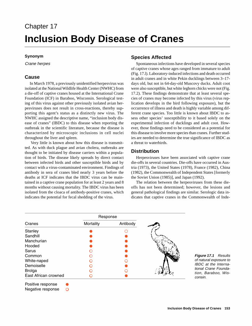
Inclusion Body Disease of Cranes 153
Chapter 17
Inclusion Body Disease of Cranes
Synonym
Crane herpes
CauseIn March 1978, a previously unidentified herpesvirus was
isolated at the National Wildlife Health Center (NWHC) froma die-off of captive cranes housed at the International CraneFoundation (ICF) in Baraboo, Wisconsin. Serological test-ing of this virus against other previously isolated avian her-pesviruses does not result in cross-reactions, thereby sup-porting this agent’s status as a distinctly new virus. TheNWHC assigned the descriptive name, “inclusion body dis-ease of cranes” (IBDC) to this disease when reporting theoutbreak in the scientific literature, because the disease ischaracterized by microscopic inclusions in cell nucleithroughout the liver and spleen.
Very little is known about how this disease is transmit-ted. As with duck plague and avian cholera, outbreaks arethought to be initiated by disease carriers within a popula-tion of birds. The disease likely spreads by direct contactbetween infected birds and other susceptible birds and bycontact with a virus-contaminated environment. Findings ofantibody in sera of cranes bled nearly 3 years before thedeaths at ICF indicates that the IBDC virus can be main-tained in a captive crane population for at least 2 years and 8months without causing mortality. The IBDC virus has beenisolated from the cloaca of antibody-positive cranes, whichindicates the potential for fecal shedding of the virus.
Species AffectedSpontaneous infections have developed in several species
of captive cranes whose ages ranged from immature to adult(Fig. 17.l). Laboratory-induced infections and death occurredin adult cranes and in white Pekin ducklings between 3–17-days old, but not in 64-day-old Muscovy ducks. Adult cootwere also susceptible, but white leghorn chicks were not (Fig.17.2). These findings demonstrate that at least several spe-cies of cranes may become infected by this virus (virus rep-lication develops in the bird following exposure), but theoccurrence of illness and death is highly variable among dif-ferent crane species. Too little is known about IBDC to as-sess other species’ susceptibility to it based solely on theexperimental infection of ducklings and adult coot. How-ever, those findings need to be considered as a potential forthis disease to involve more species than cranes. Further stud-ies are needed to determine the true significance of IBDC asa threat to waterbirds.
DistributionHerpesviruses have been associated with captive crane
die-offs in several countries. Die-offs have occurred in Aus-tria (1973), the United States (1978), France (1982), China(1982), the Commonwealth of Independent States [formerlythe Soviet Union (1985)], and Japan (1992).
The relation between the herpesviruses from these die-offs has not been determined; however, the lesions andgeneral pathological findings are similar. Serologic data in-dicates that captive cranes in the Commonwealth of Inde-
Figure 17.1 Resultsof natural exposure toIBDC at the Interna-tional Crane Founda-tion, Baraboo, Wis-consin.

154 Field Manual of Wildlife Diseases: Birds
pendent States and Japan have been exposed to the IBDCvirus or to a very closely related herpesvirus.
Since the ICF die-off, many zoological collections havesubmitted crane sera for testing by the NWHC. Nine collec-tions in the United States contained cranes that were foundto have been exposed to the virus because they tested posi-tive for antibodies to it. Testing of endangered species ofcranes that were imported into the United States detectedfour additional exposed cranes. All of the antibody positivecranes came from Asia. Serological testing by the NWHChas found the antibody to the IBDC virus in 11.3 percent of452 samples from 14 species of captive cranes in the UnitedStates. Results from other laboratories are not available; how-ever, it is known that some antibody-positive cranes havebeen detected in United States zoological collections in ad-dition to samples tested by the NWHC.
There is no evidence that wild North American cranepopulations have been exposed to IBDC. None of 95 sand-hill crane sera collected in Wisconsin and Indiana during l976and l977 had antibody to this virus. Additional testing wouldprovide more information about the status of IBDC in wildcranes.
SeasonalityThere have not been enough known outbreaks of IBDC
to indicate whether or not the disease has seasonal trends.The outbreak of IBDC in Wisconsin happened in March. Theother herpesvirus-associated die-offs in Austria, the Com-monwealth of Independent States, and Japan happened inDecember. There is not enough information currently avail-able to determine the season of the die-off in China.
Field SignsDuring the ICF die-off, signs such as lethargy and loss of
appetite persisted for 48 hours, with occasionally bloody di-arrhea just before death. Critically ill cranes often died whenthey were handled.
Gross LesionsCranes that died from IBDC at the ICF had swollen livers
and spleens. These organs contained many pinpoint-to-pin-head-size lesions that appeared as yellow-white spotsthroughout the tissue (Fig. 17.3). Other notable gross lesionsincluded hemorrhages in the thymus gland and intestines.The acute nature of the disease was evident by abundant sub-cutaneous fat in the carcasses that were examined.
DiagnosisA presumptive diagnosis can be made on the basis of gross
lesions in the liver and spleen (Fig. 17.3). However, labora-tory confirmation of this diagnosis is essential and it requiresvirus isolation from affected tissues. Submit whole carcassesto a disease diagnostic laboratory (see Chapter 3, SpecimenShipment). When this is not possible, remove the liver andspleen (see Chapter 2, Specimen Collection and Preserva-tion), place them in separate plastic bags, and ship them fro-zen. Because this disease causes characteristic intranuclearinclusion bodies in the liver and spleen, it is also useful toplace a piece of the liver and spleen in l0 percent bufferedformalin when whole carcasses cannot be submitted. Caremust be taken not to contaminate tissue samples being takenfor virus isolation when taking a portion of these tissues forformalin fixation.
ControlAny outbreak of IBDC in North America should be con-
sidered a serious event requiring the immediate involvementof disease control specialists; destroying the infected flockand decontaminating the site of the outbreak currently arethe only means of controlling the disease. This extreme re-sponse is complicated because endangered species of cranesmay be involved and it may be difficult to sacrifice them forthe benefit of other species. Nevertheless, failure to take ag-gressive action could result in IBDC being established as asignificant cause of mortality in free-living North Americancranes, jeopardize captive breeding programs for endangered
-
-
-
Figure 17.2 Known susceptibility of avian species to experi-mental infection of IBDC.

Inclusion Body Disease of Cranes 155
Figure 17.3 Gross lesions of IBDC: (A) small, yellow-white spots throughout the cut surface of the liver; (B) abundance ofspots create mottled appearance of the liver surface; (C) external surface of the spleen; (D) cut surface of the spleen.
Pho
tos
by
Jam
es R
unni
ngen
A
B
C
D
➡
➡
➡
species of cranes, and result in this disease becoming a seri-ous mortality factor among zoological collections.
When captive infected flocks cannot be destroyed, it isimportant to make every effort to permanently isolate thesurvivors from other birds. Birds that survive infection canbecome carriers of the virus and infect other birds by inter-mittently discharging virus into the environment. Care mustalso be taken to prevent spread of the virus to susceptiblebirds by contact with potentially contaminated materials suchas litter, water, feed, and feces from the confinement area.Clothes and body surfaces of personnel who were in contactwith diseased birds are other potential sources of contami-nation.
There is no evidence that the IBDC virus can be transmit-ted through the egg. However, until more is known aboutthis disease, eggs from birds surviving infection should bedisinfected and hatched elsewhere. Young from these eggs
should be reared at a facility free of IBDC, tested, and foundfree of exposure to IBDC before they are allowed to havecontact with other birds.
Infection with the IBDC virus elicits an antibody responsethat persists for several years. This is a useful indicator ofexposure to this virus. All captive cranes that are being trans-ferred to other facilities or released into the wild should betested for exposure to the IBDC virus. Birds found to haveantibodies to IBDC should be considered potential carriersof this virus and either be destroyed or confined under theconditions specified above.
Good husbandry practices are important for reducing thepotential for transmitting IBDC and for minimizing condi-tions favorable to virus shedding. Crowding, inclementweather, interspecies interactions, and poor sanitation wereall possible contributing factors to the die-off at the ICF.IBDC has not reappeared at the ICF since corrective actions
156 Field Manual of Wildlife Diseases: Birds
were taken, which include isolating the survivors of the die-off and initiating and maintaining an aggressive flock health-surveillance program.
Human Health ConsiderationsNone known.
Douglas E. Docherty
Supplementary ReadingDocherty, D.E., and Henning, D.J., l980, The isolation of a
herpesvirus from captive cranes with an inclusion bodydisease: Avian Diseases, v. 24, p. 278–283.
Docherty, D.E., and Romaine, R.I., l983, Inclusion body diseaseof cranes: a serological follow-up to the l978 die-off: AvianDiseases, v. 27, p. 830–835.
Schuh, J.C.L., and Yuill, T.M., l985, Persistence of inclusion bodydisease of cranes virus: Journal of Wildlife Disease,v. 21, p. lll–ll9.
Schuh, J.C.L., Sileo, L., Siegfried, L.M., and Yuill, T.M., 1986,Inclusion body disease of cranes: Comparison of pathologicfindings in cranes with acquired versus experimentally induceddisease: Journal of American Veterinary Medical Association,v. 189, p. 993–996.

Miscellaneous Herpesviruses of Birds 157
Chapter 18
Miscellaneous Herpesviruses of Birds
SynonymsInclusion body disease of falcons, owl herpesvirus,pigeon herpes encephalomyelitis virus, psittacine her-pesvirus
CauseHerpesviruses other than duck plague and inclusion body
disease of cranes (see Chapters 16 and 17 in this Section)have been isolated from many groups of wild birds. The dis-eases that these viruses cause have been described, but theircomparative taxonomy and host ranges require additionalstudy. All of these DNA viruses are classified in the familyHerpesviridae, but they belong to various taxonomic sub-families. The mechanisms for transmitting avian herpesvi-ruses appear to be direct bird-to-bird contact and exposureto a virus-contaminated environment. The virus is transmit-ted to raptors and owls when they feed on infected prey thatserve as a source of virus exposure. The development of dis-ease carriers among birds that survive infection is typical ofherpesvirus. Stress induced by many different factors is oftenassociated with the onset of virus shedding by carrier birdsresulting in the occurrence and spread of clinical disease.
Species AffectedHerpesviruses infect a wide variety of avian species
(Fig. 18.1). Many virus strains appear to be group-specificin the bird species they infect and sometimes only infect alimited range of species within a group. A few of these vi-ruses infect a wide species range. For example, although duckplague only affects ducks, geese, and swans, it affects mostspecies within this taxonomic grouping (see Chapter 16).However, inclusion body disease of cranes has been shownunder experimental conditions to infect birds of several fami-lies (see Chapter 17). Viruses included in the falcon-owl-pigeon complex resulted from experiments to cross-infectbirds in these different groups. Herpesviruses as a group havebeen isolated from almost every animal species in which theyhave been sought and the viruses also cause disease in hu-mans. In nature, the ability of these viruses to transmit tonew hosts is governed by species behavior and host suscep-tibility to specific types of herpesviruses.
DistributionTo date, avian herpesviruses have been reported from
North America, Europe, the Middle East (Iraq), Asia, Rus-sia, Africa, and Australia and they are probably distributedworldwide (Table 18.1). Knowledge of their distribution inwild bird populations is limited to occasional isolated dis-
ease events in the wild, isolation of the viruses in associa-tion with other disease events, and from surveys of healthybirds. Unfortunately, there are few followup laboratory orfield studies to expand information on those viruses that havebeen isolated. Most of the information on avian herpesviruscomes from disease events that affect or are found in captiveflocks. The presence of this group of viruses in wild birdpopulations is probably more extensive than current datawould indicate.
SeasonalityLittle is known about the seasonality of disease caused
by avian herpesviruses. Late spring appears to be the peakseason for duck plague outbreaks (see Chapter 16), but lessinformation about other herpesvirus infections of wild birdsis available. The ability of this virus group to establish latentor persistently infected birds reduces the requirement forcontinual virus transmission to survive in an animal popula-tion (see Chapter 16, Duck Plague, and Chapter 17, Inclu-sion Body Disease of Cranes). Breeding season probably pro-vides the best time of the year for bird-to-bird virus trans-mission in solitary species. Transmission of herpesvirusesvia the egg has been shown for some species, but more re-search is required to determine the importance of egg trans-mission for virus perpetuation. Seasonality probably plays amore important role for virus transmission in and among birdspecies that assemble for migration between summer breed-ing and wintering grounds.
Field SignsThe general signs of disease include depression of nor-
mal activity and sudden mortality in a group of birds. Respi-ratory distress may also be seen. Captive pigeons may showpronounced neurological signs such as extremity paralysis,head-shaking, and twisting of the neck.
Gross LesionsBirds dying from infection with this group of viruses can
have tumors (chicken and pigeon), hemorrhagic lesions(chicken, pheasants, ducks, cranes, peafowl, and guinea fowl),or, more commonly, hepatitis, and disseminated focal ne-crosis or visual areas of localized tissue death that appear asspots within the normal tissue in the liver, spleen, (Fig. 18 .2)and bone marrow along with occasional intestinal necrosis.This broad array of lesions complements and extends thoseseen for duck plague and inclusion body disease of cranes(Chapters 16 and 17).

158 Field Manual of Wildlife Diseases: Birds
Species group Disease
Waterfowl Duck plague
Psittacines Hepatitis
Raptors (owls, eagles, Hepatitisfalcons)
Pigeons and Encephalomyelitisdoves
Domestic poultry Marek’s Disease(chicken Infectiousand turkey) laryngotracheitis
Cranes Hepatitisand storks
Quail and Hepatitispheasants
Songbirds Conjunctivitis
Marine birds Unknown(cormorants)
CommonOccasional
InfrequentRare or unknown
Figure 18.1 Relative frequency ofdisease from herpesvirus infections inbirds of North America.

Miscellaneous Herpesviruses of Birds 159
Table 18.1 Geographical distribution of avian herpesvirus infections.
ContinentsNorth America Europe Africa Australia Other
Raptors
Booted eagle •Bald and golden eagles •Common buzzard (Old World) •Falcons
Prairie •Red-headed •Peregrine •Gyrfalcon •Kestrels •
Owls
Eagle owl •Long-eared owl •Great horned owl •Snowy owl • •
Pigeon • • • • Egypt
Ringed turtle dove •Storks •Cranes • • China
Japan
Russia
Wild turkey •Psittacines (several species) • • Japan
Bobwhite quail •Waterfowl (non-duck plague) • • •Black-footed penguin •Passeriforms
Exotic finches •Weavers •Finches, including canary •
Cormorants •
Gallinaceous birds such as chicken, pheasants, peafowl, and guinea fowl raised in captivityhave also been infected.

160 Field Manual of Wildlife Diseases: Birds
DiagnosisThe primary methods for diagnosing herpesvirus as a
cause of disease are virus isolation from infected tissues andfinding, during microscopic examination of infected cells,the characteristic accumulations of cellular debris referredto as intranuclear (Cowdry type A) inclusion bodies (Fig.18.3). These lesions are most often seen in the liver, spleen,and bone marrow. The virus can usually be isolated in chickenor duck embryo fibroblast tissue culture or in embryonatedchicken eggs.
ControlControl actions warranted for outbreaks of herpesvirus
infections are dependent upon the type of herpesvirus infec-tion and the prevalence of disease in the species or popula-tions involved (see Chapter 4, Disease Control Operations).Euthanasia of infected flocks should be considered for ex-otic viruses and viruses that are likely to cause high mortal-ity within the population at risk. When depopulation is notappropriate because of the ubiquitous nature of the disease,or for other reasons, disease-control steps should still betaken. Sick birds, as well as those in the preclinical stages ofillness, will be shedding virus into their environment; there-fore, birds that are suspected of being infected should besegregated from other birds and quarantined for 30 days. Anybirds noticeably ill should be isolated from the rest of thecontact group. A high level of sanitation should be imposedand maintained for the full quarantine period where birdsare housed. Decontamination procedures are needed to mini-mize disease transmission via virus that is shed in feces andby other means.
Dead birds should be removed immediately and submit-ted for disease evaluations. Standard bagging and decontami-nation procedures should be used to avoid off-site transferof the virus. Personnel should follow good hygiene methods
and should not have any contact with other birds for 7 daysto prevent mechanically carrying contamination from thequarantine site.
Surviving birds should be tested for virus and virus spe-cific antibody. All birds with antibody are probably viruscarriers and they pose a risk as a source for future virus in-fection. Future use of these birds should take this into con-sideration. This is especially important when endangeredspecies are involved and for wildlife rehabilitation activitiesbecause survivors of herpesvirus infections are potentialsources for the initiation of new outbreaks and further spreadof the disease.
Human Health ConsiderationsAvian herpesviruses have not been associated with any
disease of humans.
Figure 18.2 Herpesviruses can produce areas of tissuenecrosis, appearing as white spots, such as in this peregrinefalcon liver (A) and this great horned owl spleen (B).
Pho
to b
y Ja
mes
Run
ning
en
Pho
to b
y C
arol
Met
eyer
A B
Figure 18.3 Inclusion bodies (arrows) in liver cell nuclei of agreat horned owl that died of herpesvirus infection.
Pho
to b
y Lo
u S
ileo
Wallace Hansen
➡➡

Miscellaneous Herpesviruses of Birds 161
Supplementary ReadingBurtscher, H., and Sibalin, M., 1975, Herpesvirus strigis: Host
spectrum and distribution in infected owls: Journal of WildlifeDiseases, v. 11, p. 164–169.
Graham, D.L., Mare, C.J., Ward, F.P., and Peckham, M.C., 1975,Inclusion body disease (herpesvirus infection) of falcons(IBDF): Journal of Wildlife Diseases, v. 11, p. 83–91.
Kaleta, E.F., 1990, Chapter 22; Herpesviruses of free-living andpet birds, in A laboratory manual for the isolation andidentification of avian pathogens, American Association ofAvian Pathologists. p. 97–102.

162 Field Manual of Wildlife Diseases: Birds

Avian Pox 163
Chapter 19
Avian Pox
Synonyms
Fowl pox, avian diphtheria, contagious epithelioma, andpoxvirus infection
CauseAvian pox is the common name for a mild-to-severe, slow-
developing disease of birds that is caused by a large virusbelonging to the avipoxvirus group, a subgroup of pox-viruses. This group contains several similar virus strains;some strains have the ability to infect several groups or spe-cies of birds but others appear to be species-specific. Mos-quitoes are common mechanical vectors or transmitters ofthis disease. Avian pox is transmitted when a mosquito feedson an infected bird that has viremia or pox virus circulatingin its blood, or when a mosquito feeds on virus-laden secre-tions seeping from a pox lesion and then feeds on anotherbird that is susceptible to that strain of virus. Contact withsurfaces or exposure to air-borne particles contaminated withpoxvirus can also result in infections when virus enters thebody through abraded skin or the conjunctiva or the mucousmembrane lining that covers the front part of the eyeball andinner surfaces of the eyelids of the eye.
Species AffectedThe highly visible, wart-like lesions associated with the
featherless areas of birds have facilitated recognition of avianpox since ancient times. Approximately 60 free-living birdspecies representing about 20 families have been reportedwith avian pox. However, the frequency of reports of thisdisease varies greatly among different species (Fig. 19.1).Avian pox has rarely been reported in wild waterfowl, andall North American cases have been relatively recent (Table.19.1). The first case was in a free-living green-winged tealin Alaska. Single occurrences have also been documented ina Canada goose in Ontario, Canada, a mallard duck in Wis-consin, a feral mute swan cygnet in New York, and a tundraswan in Maryland. Three cases in American goldeneye havebeen reported in Saskatchewan, Canada, and New York. Avianpox also appeared in Wisconsin among captive-reared trum-peter swans that were part of a reintroduction program. Zoo-logical garden cases include common scoter in the Philadel-phia Zoo and a Hawaiian goose in the Honolulu Zoo.
Avian pox in a bald eagle was first diagnosed in 1979 inAlaska and it was a lethal infection. Since then, additionalbald eagles in Alaska and at other locations have been diag-nosed with this disease (Fig. 19.2). The severity of infectionresulted in several of these cases being lethal. Poxvirus in-fections have been reported in other raptors, most recently
Figure 19.1 Reported avian pox occurrence in wild birds inNorth America.
Songbirds
Upland gamebirds
Marine birds
Raptors
Waterfowl
Wading birds
Shorebirds
1Reports often involve a number of birds in a single event.2Reports tend to involve individuals rather than groups of birds.3Small number of reports, generally involving individual birds.
Multiple1
Occasional2
Few3
Rare or not reported

164 Field Manual of Wildlife Diseases: Birds
Table 19.1 Waterfowl in North America reported to haveavian pox.
Year of firstreport
Species Locations in species
Harlequin duck Alaska 1994Blue-winged teal Wisconsin 1991Wood duck Wisconsin 1991Redhead duck Wisconsin 1991Trumpeter swan Wisconsin 1989Common goldeneye New York 1994
Saskatchewan 1981Tundra swan Maryland 1978Green-winged teal Alaska 1978Mallard Wisconsin 1978Canada goose Ontario 1975Common scoter Pennsylvania 1967Mute swan New York 1964
(Fig. 19.3) frequently have been reported to have been struckby avian pox epizootics. Avian pox is suspected as a factorin the decline of forest bird populations in Hawaii and north-ern bobwhite quail in the southeastern United States, whereit is also an important disease of wild turkey.
Distribution Avian pox occurs worldwide, but little is known about its
prevalence in wild bird populations. The increased frequencyof reported cases of this highly visible disease and the in-volvement of new bird species during recent years suggeststhat avian pox is an emerging viral disease. Birds can be-come disease carriers and spread avian pox among local popu-lations, such as between birdfeeding stations, and along mi-gratory routes used by various bird species. Mosquitoes thatfeed on birds play the most important role for both diseasetransmission and long term disease maintenance. However,contamination of perches and other surfaces used by captivebirds can perpetuate disease in captivity. Pox outbreaks arecommonly reported at aviaries, rehabilitation centers, andother places where confinement provides close contact amongbirds. The disease can spread rapidly when avian pox is in-troduced into such facilities. Species that would not ordi-narily have contact with avian pox virus in the wild oftenbecome infected in captivity if the strain of virus present iscapable of infecting a broad spectrum of species. Commonmurres rescued from an oil spill in California developed pox-virus lesions while they were in a rehabilitation center. En-dangered avian species also have been infected during cap-tive rearing.
SeasonalityAlthough wild birds can be infected by pox virus year-
round (Fig. 19.4), disease outbreaks have been associatedwith the environmental conditions, the emergence of vectorpopulations, and the habits of the species affected. Environ-mental factors such as temperature, humidity, moisture, andprotective cover all play a role in the occurrence of this dis-ease by affecting virus survival outside of the bird host. Avianpox virus can withstand considerable dryness, thereby re-maining infectious on surfaces or dust particles. Mosquitoesthat feed on birds are the most consistent and efficient trans-mitters of this disease. Mosquito populations are controlledby breeding habitat and annual moisture.
The time of appearance and magnitude of vector popula-tions varies from year to year, depending on annual weatherconditions. This influences the appearance and severity ofthe disease in any given year. Only limited studies have beencarried out to assess the relations between avian pox andinsect vector populations. Studies on the Island of Hawaiidisclose a close relation between the prevalence of poxvirusinfections in forest birds and seasonal mosquito cycles. Thelowest prevalence of pox virus infection in California quailin Oregon was reported in the dry summer months and the
8
1
2
1 1
12
1
4
2
2
2
9
1
1
2
1
1
Figure 19.2 Number of bald eagles with cutaneous pox byState, 1979–97. (From National Wildlife Health Center records.)
in the eastern screech owl and barred owl in Florida (Table19.2).
On Midway Atoll, large numbers of colonial nesting birds,such as the Laysan albatross, have become infected with avianpox. Red-tailed tropicbirds on Midway Atoll previously hadbeen affected by avian pox. The shift in predominant speciesinfected relates to the dramatic shift in population densitiesfor the two species over time (1963–1978). Mourning dove,finches, and other perching birds using backyard feeders

Avian Pox 165
Table 19.2 Birds of prey from North America reported tohave contracted avian pox.
Year of firstreport
Species Locations in species
Barred owl Florida 1995
Bald eagle Maine 1995Ohio 1995Rhode Island 1993Michigan 1992Minnesota 1989California 1987Nebraska 1987Maryland 1986Massachusetts 1986South Dakota 1986Wisconsin 1986Pennsylvania 1985Arkansas 1984New York 1983Florida 1982Virginia 1981Washington 1981Alaska 1978
Eastern screech owl Florida 1994
Peregrine falcon New York 1994
Ferruginous hawk Texas 1993
Golden eagle Missouri 1989Kansas 1986California 1976British Columbia 1970
Red-tailed hawk Nebraska 1988Wisconsin 1985Washington 1981Missouri 1970
Rough-legged hawk North Dakota 1971
highest was reported during the wetter fall and winter months.In Florida, reports of avian pox in wild turkey correspond tothe late summer and early fall mosquito season. On SandIsland of the Midway Atoll, avian pox was first reported inSeptember l963 in the nestlings of the red-tailed tropicbird.In March and April of the late l970s, this disease was foundin nestling Laysan albatross on Sand Island. This is an ex-ample of disease seasonality influenced by dramatic shiftsin predominant species populations.
Birdfeeding stations have been the source of numerouspoxvirus outbreaks in the continental United States(Fig. 19.3). Contact transmission of the virus through infectedsurfaces and close association of birds using those feeders isthe likely means of transmission during cooler periods ofthe year when mosquitoes are not a factor, and birdfeedersprovide additional sources of infection when mosquitoes arepresent.
Field SignsBirds with wart-like nodules on one or more of the feath-
erless areas of the body, including the feet, legs, base of thebeak, and eye margin should be considered suspect cases ofavian pox (Fig. 19.5). The birds may appear weak and ema-ciated if the lesions are extensive enough to interfere withtheir feeding. Some birds may show signs of labored breath-ing if their air passages are partially blocked. Although thecourse of this disease can be prolonged, birds with extensivelesions are known to completely recover if they are able tofeed.
Gross LesionsAvian pox has two disease forms. The most common form
is cutaneous and it consists of warty nodules that develop onthe featherless parts of the bird. This form of the disease isusually self-limiting; the lesions regress and leave minorscars. However, these nodules can become enlarged and clus-tered, thus causing sight and breathing impairment and feed-ing difficulty (Figs. 19.6A and B). Secondary bacterial andother infections are common with this form of the disease,and these infections can contribute to bird mortality. In somebirds, feeding habits result in the large warty nodules be-coming abraded and then infected by bacterial and fungalinfections (Figs. 19.6C and D).
The internal form of disease is referred to as wet pox andit is primarily a problem of young chickens and turkeys. Thisdiphtheritic form appears as moist, necrotic lesions on themucus membranes of the mouth and upper digestive and res-piratory tracts (Fig. 19.7), and it has occasionally been re-ported in wild birds (Fig. 19.8). This form of avian pox prob-ably occurs more frequently in wild birds than it is reportedbecause it is less observable than the cutaneous form. Also,the more severe consequences of wet pox undoubtedly causesgreater morbidity and mortality, thereby leading to removalof infected birds by predators and scavengers.
DiagnosisA presumptive diagnosis of avian pox can be made from
the gross appearance of the wart-like growths that appear onbody surfaces. However, these observations must be con-firmed by examining lesions microscopically for character-
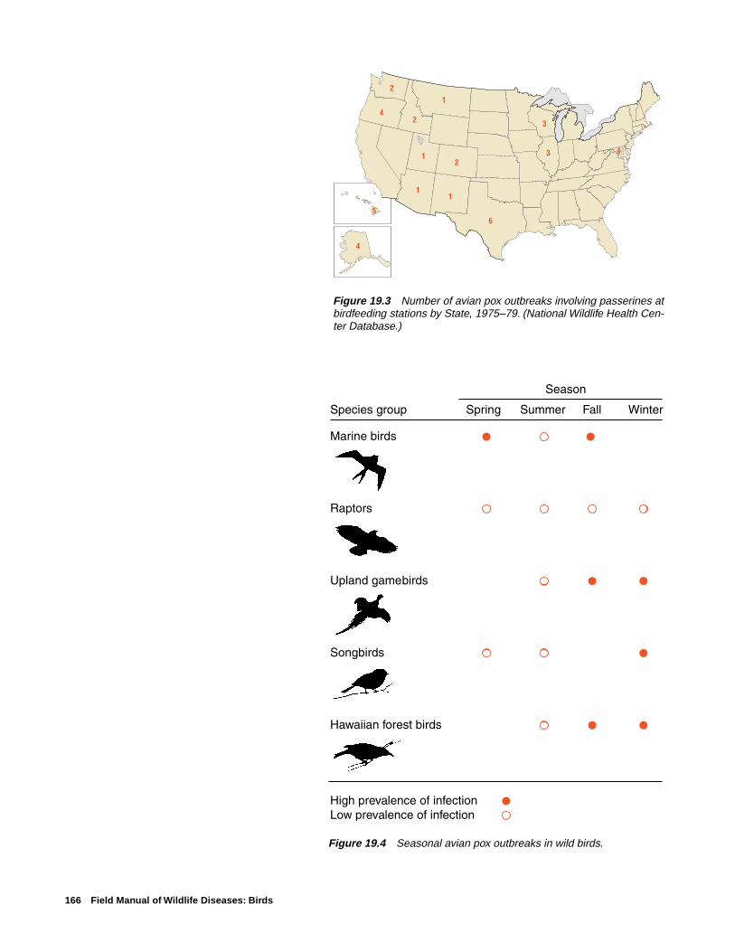
166 Field Manual of Wildlife Diseases: Birds
Figure 19.4 Seasonal avian pox outbreaks in wild birds.
Figure 19.3 Number of avian pox outbreaks involving passerines atbirdfeeding stations by State, 1975–79. (National Wildlife Health Cen-ter Database.)
4
1
2
1 3
1
6
1
2
2
3
4
4
5

Avian Pox 167
Figure 19.5 (A) Avian pox lesions typi-cally are found on featherless parts ofthe body. This Laysan albatross chickhas small pox nodules on the face andeyelid. (B) As the disease progresses,these lesions become more extensive.(C) Lesions also are commonly seen onthe legs and (D) feet.
Pho
to b
y W
alla
ce H
anse
nP
hoto
by
Milt
on F
riend
Pho
to b
y M
ilton
Frie
nd
Pho
to b
y W
alla
ce H
anse
n
A
B
C D

168 Field Manual of Wildlife Diseases: Birds
Pho
tos
by
Jam
es R
unni
ngen
Figure 19.6 Pox lesions can be so extensive that they im-pair breathing, sight, and feeding as seen in these bald eagles:(A) extensive infection of both sides of the face, (B) obstruc-tion to feeding due to the size and location of these lesions atthe base of the bill, and (C) obstruction of sight due to com-plete occlusion of the eye. (D) Massive facial lesions oftenbecome abraded and subject to secondary infections.
Figure 19.7 Diphtheritic form of avian pox (arrow) in a Laysanalbatross at Midway Atoll. P
hoto
by
Lou
Sile
o
A
B
D
C
➡
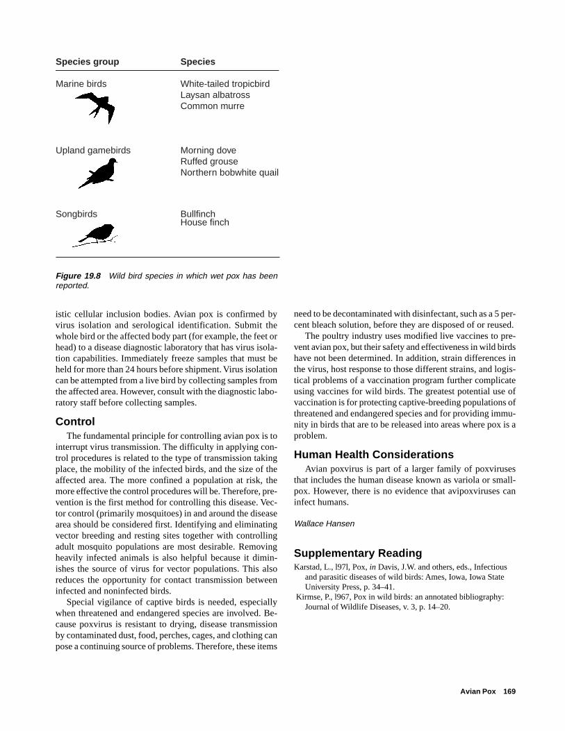
Avian Pox 169
istic cellular inclusion bodies. Avian pox is confirmed byvirus isolation and serological identification. Submit thewhole bird or the affected body part (for example, the feet orhead) to a disease diagnostic laboratory that has virus isola-tion capabilities. Immediately freeze samples that must beheld for more than 24 hours before shipment. Virus isolationcan be attempted from a live bird by collecting samples fromthe affected area. However, consult with the diagnostic labo-ratory staff before collecting samples.
ControlThe fundamental principle for controlling avian pox is to
interrupt virus transmission. The difficulty in applying con-trol procedures is related to the type of transmission takingplace, the mobility of the infected birds, and the size of theaffected area. The more confined a population at risk, themore effective the control procedures will be. Therefore, pre-vention is the first method for controlling this disease. Vec-tor control (primarily mosquitoes) in and around the diseasearea should be considered first. Identifying and eliminatingvector breeding and resting sites together with controllingadult mosquito populations are most desirable. Removingheavily infected animals is also helpful because it dimin-ishes the source of virus for vector populations. This alsoreduces the opportunity for contact transmission betweeninfected and noninfected birds.
Special vigilance of captive birds is needed, especiallywhen threatened and endangered species are involved. Be-cause poxvirus is resistant to drying, disease transmissionby contaminated dust, food, perches, cages, and clothing canpose a continuing source of problems. Therefore, these items
Species group Species
Marine birds White-tailed tropicbirdLaysan albatrossCommon murre
Upland gamebirds Morning doveRuffed grouseNorthern bobwhite quail
Songbirds BullfinchHouse finch
need to be decontaminated with disinfectant, such as a 5 per-cent bleach solution, before they are disposed of or reused.
The poultry industry uses modified live vaccines to pre-vent avian pox, but their safety and effectiveness in wild birdshave not been determined. In addition, strain differences inthe virus, host response to those different strains, and logis-tical problems of a vaccination program further complicateusing vaccines for wild birds. The greatest potential use ofvaccination is for protecting captive-breeding populations ofthreatened and endangered species and for providing immu-nity in birds that are to be released into areas where pox is aproblem.
Human Health ConsiderationsAvian poxvirus is part of a larger family of poxviruses
that includes the human disease known as variola or small-pox. However, there is no evidence that avipoxviruses caninfect humans.
Wallace Hansen
Supplementary ReadingKarstad, L., l97l, Pox, in Davis, J.W. and others, eds., Infectious
and parasitic diseases of wild birds: Ames, Iowa, Iowa StateUniversity Press, p. 34–41.
Kirmse, P., l967, Pox in wild birds: an annotated bibliography:Journal of Wildlife Diseases, v. 3, p. 14–20.
Figure 19.8 Wild bird species in which wet pox has beenreported.

170 Field Manual of Wildlife Diseases: Birds

Eastern Equine Encephalomyelitis 171
Chapter 20
Eastern Equine Encephalomyelitis
Synonyms
EEE, eastern encephalitis, EE, eastern sleeping sick-ness of horses
CauseEastern equine encephalitis (EEE) is caused by infection
with an RNA virus classified in the family Togaviridae. Thevirus is also referred to as an “arbovirus” because virus rep-lication takes place within mosquitoes that then transmit thedisease agent to vertebrate hosts such as birds and mammals,including humans. The term arbovirus is shortened nomen-clature for arthropod (insect) borne (transmitted) viruses.Culiseta melanura is the most important mosquito vector; itsilently (no disease) transmits and maintains the virus amongbirds. However, several other mosquito species can transmitthis virus, including the introduced Asian tiger mosquito. Newhosts become infected when they enter this endemic naturalcycle and are fed upon by an infected mosquito. Therefore,the presence of mosquito habitat, the feeding habits of dif-ferent mosquito species, and the activity patterns of verte-brate hosts are among the important factors for disease trans-mission.
Distribution This disease is primarily found in eastern North America
especially along the Atlantic and Gulf Coasts, and the dis-ease range extends into Central and South America. The caus-ative virus has been isolated from eastern Canada to Argen-tina and Peru, and it is maintained in a mosquito-wild birdcycle as an endemic (enzootic) focus of infection in naturethat is usually associated with freshwater marshes. Wild birddie-offs from EEE have been limited to captive-rearing situ-ations. Die-offs have occurred in pheasants in coastal Statesfrom New Hampshire to Texas, where they have been raised,in chukar partridge and whooping cranes in Maryland, andin emus and ostriches in Louisiana, Georgia, Florida, andTexas.
Species AffectedEEE virus produces inapparent or subclinical infections
in a wide range of wild birds (Fig. 20.1). However, EEEvirus has caused mortality in glossy ibis and in several birdspecies that are exotic to the United States, including pigeon,house sparrow, pheasants, chukar partridge, white Pekingducklings, and emu. The infection rate in penned emus inthe United States has reached 65 percent with a case mortal-ity rate of 80 percent. In the past, extensive losses have
Figure 20.1 Relative frequency of EEE virus isolationor presence of antibodies in birds.
Songbirds
Upland gamebirds
Shorebirds
Gulls
Cranes
Waterfowl
Marine birds
Raptors
FrequentCommon
OccasionalRare
Unknown

172 Field Manual of Wildlife Diseases: Birds
In September and November of 1984, EEE virus was as-sociated with the deaths of 7 of 39 captive whooping cranesat the Patuxent Wildlife Research Center in Laurel, Mary-land. Sandhill cranes coexisting with the whooping cranesdid not become clinically ill or die.
Passerines (perching songbirds), some small rodents, andbats are highly susceptible to infection and they often diefrom experimental infections. Horses are highly susceptibleand they often die from natural infections.
SeasonalityEEE is associated with the early summer appearance of
C. melanura mosquito populations (Fig. 20.2). Nestling birds,such as passerines and other perching birds, are the amplifi-cation hosts for the virus, producing high concentrations ofvirus in their blood or viremia following mosquito infec-tion. New populations of emerging mosquitoes become in-fected when they feed on the viremic birds. C. melanuraand other species of infected mosquitoes can transmit thevirus to other species of birds susceptible to disease (Fig.20.3).
The summer-fall transmission cycle is followed by littlevirus transmission during the winter and spring months. Theoverwintering mechanism for virus survival is not known.Infected mosquitoes, other insects, cold-blooded vertebratespecies, or low levels of virus transmission by mosquitoesare among current theories for virus cycle maintenance inmilder climates. It is also believed that bird migration spreadsthe virus to higher latitudes in the spring.
Field SignsClinical signs do not develop in most native species of
wild birds infected with EEE virus. Clinical signs for non-indigenous birds (including pheasants) include depression,tremors, paralysis of the legs, unnatural drowsiness, profusediarrhea, voice changes, ataxia or loss of muscle coordina-tion, and involuntary circular movements (Fig. 20.4). Someof the EEE-infected whooping cranes became lethargic andincoordinated or ataxic, with partial paralysis or paresis ofthe legs and neck 3–8 hours prior to death; other cranes didnot develop clinical signs before they died.
Gross LesionsGross lesions in whooping cranes included fluid accu-
mulation in the abdominal cavity or ascites, intestinal mu-cosal discoloration, fat depletion, enlarged liver or hepatome-galy, enlarged spleen or splenomegaly, and visceral gout(Fig. 20.5).
DiagnosisBecause of human health hazards, field personnel should
not dissect birds suspected of having died from EEE. Wholecarcasses should be submitted to diagnostic laboratories ca-pable of safely handling such specimens. EEE can be diag-
FrequentCommon
OccasionalRare
Species
Songbirds
Spring
Summer
Fall
Winter
Upland gamebirds
Spring
Summer
Fall
Winter
Cranes
Spring
Summer
Fall
Winter
Waterfowl
Spring
Summer
Fall
Winter
Shorebirds
Spring
Summer
Fall
Winter
Gulls
Spring
Summer
Fall
Winter
Mosquitoes
Spring
Summer
Fall
Winter
Figure 20.2 Seasonality of virus isolation from birds and mos-quitoes.
occurred in ring-necked pheasant being reared in captivityfor sporting purposes, including one outbreak in a SouthDakota pheasant farm. Large-scale mortalities in captivepheasants are perpetuated by bird-to-bird disease spreadthrough pecking and cannibalism after EEE has been intro-duced by mosquitoes, usually of the genus Culiseta. Out-breaks have not been reported during recent years.

Eastern Equine Encephalomyelitis 173
Figure 20.3 Transmission of eastern equine encephalomyelitis. (A) Virus circulates in songbird populations by being transmit-ted by mosquitoes. Those birds are susceptible to infections, but they do not become clinically ill or die. (B) The outbreak cycleis started either when an infected mosquito from the enzootic cycle feeds on highly susceptible birds such as pheasants orcranes, or when another species of mosquito, that primarily feeds on these same birds, becomes infected after feeding onsongbirds in the enzootic cycle and transmits the virus. The epizootic cycle is maintained by the second mosquito species. (C)The broader host feeding range of the second mosquito results in exposure of horses and humans. No disease cycle is main-tained between these species by mosquitoes.
Figure 20.4 A hen pheasant with EEEexhibiting neurologic signs.
Pho
to b
y M
ilton
Frie
nd
Figure 20.5 The white, grainy material on the liver of this whooping crane is evi-dence of visceral gout.
Pho
to b
y Ja
mes
Run
ning
en
Enzooticmosquito vector
(Culiseta sp.)
A Virus maintenance and amplification cycle (enzootic silent cycle — no disease)
B Outbreak cycle (epizootic disease cycle)
C Accidental (dead-end) hosts
Epizootic mosquito vector
(Aedes sp.)
Songbirds
Pheasants
Humans
Horses
Epizootic mosquito vector
(Aedes sp.)
Enzooticmosquito vector
(Culiseta sp.)
Cranes
174 Field Manual of Wildlife Diseases: Birds
nosed by virus isolation from infected whole blood or brainand other tissues from dead birds. Diagnosis of virus activ-ity can be made from surviving birds because they will havevirus neutralizing antibodies in their blood serum. The riseand fall in serum antibodies that occurs after virus exposurecan be used to assess infection rates and the relative timingof exposure before antibody levels reach nondetectible lev-els. Most native birds do not suffer clinical infections.
ControlThere are two approaches to protecting susceptible ani-
mals from infection from vector-borne diseases. The firstapproach includes separating mosquitoes from animals atrisk. This requires eliminating mosquito breeding and rest-ing sites in an endemic area or protecting animals from mos-quito contact by maintaining them in an insect-proof enclo-sure. In the second approach, vaccination is used to renderthe animal immune. A killed-virus vaccine was used in cap-tive whooping cranes to protect the rest of the breeding flockfollowing the 1984 outbreak. Vaccination has also been usedto protect whooping cranes released into an area where EEEis prevalent in mosquito populations. Field data suggest thatimmunity in those cranes is being boosted by natural infec-tions after their release.
Human Health ConsiderationsHumans are susceptible to EEE and human cases typi-
cally arise after the disease has appeared in horses. EEE is asignificant disease in humans with a case fatality rate of be-tween 30–70 percent and it often causes severe permanentneurological disorders among survivors. Aerosol infectionis possible but rare. Laboratory personnel have been infectedwith this virus. Human pre-exposure vaccination is recom-mended for people who may handle infected tissues.
Wallace Hansen and Douglas E. Docherty
Supplementary ReadingDein, J.F., Carpenter, J.W., Clark, G.G., Montali, R.J., Crabbs,
C.L., Tsai, T. F., and Docherty, D.E., 1986, Mortality ofcaptive whooping cranes caused by eastern equine encephalitisvirus: The Journal of American Veterinary Medical Associa-tion, v. 189, p. 1,006–1,010.
Gibbs, E.P.J., and Tsai, T.F. 1994, Eastern encephalitis, in Beran,G.W., and Steele, J.H., eds., Handbook of zoonoses (2d ed.),Section B: Viral: Boca Raton, Fla., CRC Press, p. 11–24.
Kissling, R.E., Chamberlain, R.W., Sikes, R.K., and Eidson, M.E.,1954, Studies on the North American arthropod-borneencephalitides III. eastern equine encephalitis in wild birds:American Journal of Hygiene, v. 60, p. 251–265.

Newcastle Disease 175
Chapter 21
Newcastle Disease
Synonyms
ND, paramyxovirus-1, NDV, VVND, NVND
Newcastle Disease (ND) in domestic poultry is a focus forconcern throughout much of the world’s agricultural com-munity because of severe economic losses that have occurredfrom illness, death, and reduced egg production followinginfection with pathogenic or disease causing strains. Prior to1990, this disease had rarely been reported as a cause ofmortality in the free-living native birds of the United Statesor Canada. Repeated large-scale losses of double-crestedcormorants from ND in both countries has resulted in a needfor enhanced awareness of ND as a disease of wild birdsand, therefore, its inclusion within this Manual. Backgroundinformation about ND in poultry is needed to provide a per-spective for understanding the complexity of the diseaseagent, Newcastle disease virus (NDV). Some general infor-mation about ND in other avian species is also provided, butthe primary focus for this chapter is the effect of NDV ondouble-crested cormorants.
CauseNewcastle disease is caused by infection with an RNA
virus within the avian paramyxovirus-1 group. NDV is highlycontagious and there is great variation in the severity of dis-ease caused by different strains of this virus. A classificationsystem for the severity of disease has been established toguide disease control efforts in poultry because of the eco-nomic damage of ND.
The most virulent ND form causes an acute, lethal infec-tion of chickens of all ages with mortality in affected flocksoften reaching 100 percent. These strains produce hemor-rhagic lesions of the digestive tract, thus resulting in the dis-ease being referred to as viscerotropic or having an affinityfor abdominal tissue, and velogenic or highly virulent New-castle disease or VVND. This form of ND is rare in the UnitedStates, and it is primarily introduced when exotic species ofbirds are trafficked in the pet bird industry. Another acute,generally lethal infection of chickens of all ages affects res-piratory and neurologic tissues and is referred to as neuro-tropic velogenic Newcastle disease or NVND. Morbidity orillness from NVND may affect 100 percent of a flock, butmortality is generally far less with extremes of 50 percent inadult birds and 90 percent in young chickens. The NVNDform of ND was essentially eradicated from the United Statesin about 1970, but it has occasionally been reintroduced viapet birds and by other means. A less pathogenic form of ND
causes neurologic signs, but usually only young birds dieand, except for very young susceptible chicks, mortality islow. These strains are classified as mesogenic or moderatelyvirulent. NDV strains that cause mild or inapparent respira-tory infections in chickens are classified as lentogenic or lowvirulence. Lentogenic strains do not usually cause disease inadult chickens, but these forms can cause serious respiratorydisease in young birds. Some strains of lentogenic NDV causeasymptomatic-enteric infections without visible disease(Table 21.1).
The virus classification standard applies to ND in poultryand the standard is not directly transferrable to wild birds.Experimental studies have demonstrated differences in birdresponse to the same strain of NDV. Thus, a highly patho-genic strain isolated from wild birds may be less hazardousfor poultry and vice-versa. ND may be transmitted amongbirds by either inhalation of contaminated particulate matteror ingestion of contaminated material.
Species AffectedNDV is capable of infecting a wide variety of avian spe-
cies. In addition to poultry, more than 230 species from morethan one-half of the 50 orders of birds have been found to besusceptible to natural or experimental infections with avianparamyxoviruses. Experimental infections in mallard ducksexposed to large amounts of a highly virulent form of NDVfor chickens disclosed that ducklings were more susceptiblethan adults, and that mortality of 6-day-old ducklings washigher than in 1-day-old and 3-day-old ducklings. Captive-reared gamebirds, such as pheasants and Hungarian partridge,have died of ND. However, large-scale illness and death fromNDV in free-ranging wild birds has only occurred in double-crested cormorants in Canada and the United States. Whitepelicans, ring-billed gulls, and California gulls were alsoreported to have died from NDV in association with cormo-rant mortalities in Canada.
The 1990 epizootic of ND in Canada killed more than10,000 birds, mostly double-crested cormorants. Mostly sub-adult cormorants died in these cormorant colonies. Lossesin the United States have been primarily in nestlings andother young of the year. The total mortality attributed to NDduring 1992 exceeded 20,000 birds. Mortality in Great Lakescormorant colonies ranged from 2 to 30 percent, while thatin Midwestern colonies was estimated to be 80 to 90 per-cent. In 1997, nesting failure of a cormorant colony at theSalton Sea in California was attributed to NDV. The total
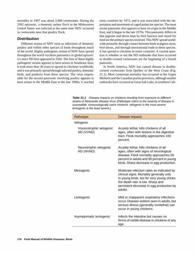
176 Field Manual of Wildlife Diseases: Birds
mortality in 1997 was about 2,000 cormorants. During the1992 epizootic, a domestic turkey flock in the MidwesternUnited States was infected at the same time NDV occurredin cormorants near that poultry flock.
DistributionDifferent strains of NDV exist as infections of domestic
poultry and within other species of birds throughout muchof the world. Highly pathogenic strains of NDV have spreadthroughout the world via three panzootics or global epizoot-ics since ND first appeared in 1926. The first of these highlypathogenic strains appears to have arisen in Southeast Asia;it took more than 30 years to spread to chickens worldwide,and it was primarily spread through infected poultry, domesticbirds, and products from these species. The virus respon-sible for the second panzootic involving poultry appears tohave arisen in the Middle East in the late 1960s; it reached
most countries by 1973, and it was associated with the im-portation and movement of caged psittacine species. The mostrecent panzootic also appears to have its origin in the MiddleEast, and it began in the late 1970s. This panzootic differs inthat pigeons and doves kept by bird fanciers and raised forfood are the primary species involved. This NDV spread world-wide primarily through contact between birds at pigeon races,bird shows, and through international trade in these species.It has spread to chickens in some countries. A current ques-tion is whether or not the ND outbreaks that have occurredin double-crested cormorants are the beginning of a fourthpanzootic.
In North America, NDV has caused disease in double-crested cormorants from Quebec to the West Coast (Fig.21.1). Most cormorant mortality has occurred in the UpperMidwest and the Canadian prairie provinces, although smalleroutbreaks have occurred at Great Salt Lake, in southern Cali-
Table 21.1 Disease impacts on chickens resulting from exposure to differentstrains of Newcastle disease virus. [Pathotype refers to the severity of disease insusceptible, immunologically naive chickens. Velogenic is the most severe;lentogenic is the least severe.]
Pathotype Disease impacts
Velogenic
Viscerotrophic velogenic Acutely lethal, kills chickens of allND (VVND) ages, often with lesions in the digestive
tract. Flock mortality approaches 100percent.
Neurotrophic velogenic Acutely lethal, kills chickens of allND (NVND) ages, often with signs of neurological
disease. Flock mortality approaches 50percent in adults and 90 percent in youngbirds. Sharp decrease in egg production.
Mesogenic Moderate infection rates as indicated byclinical signs. Mortality generally onlyin young birds, but for very young chicksthe death rate is low. Sharp andpersistent decrease in egg production byadults.
Lentogenic Mild or inapparent respiratory infectionsoccur. Disease seldom seen in adults, butserious illness (generally nonlethal) canoccur in young chickens.
Asymptomatic lentogenic Infects the intestine but causes noforms of visible disease in chickens of anyage.

Newcastle Disease 177
fornia, and on the Columbia River between Washington andOregon. Cormorants, the closely related shag, and gannets,which are another species of marine bird that has close asso-ciations with cormorants, were believed to be an importantsource of NDV for the poultry outbreaks along the coast ofBritain during the 1949–51 epizootic in that country.
SeasonalityAll of the North American cormorant die-offs from ND
have occurred in breeding colonies. Mortality has occurredduring the months of March through September.
Field SignsClinical signs, observed only in sick juvenile double-
crested cormorants, include torticollis or twisting of the headand neck, ataxia or lack of muscular coordination, tremors,paresis or incomplete paralysis including unilateral or bilat-
eral weakness of the legs and wings, and clenched toes (Fig.21.2). Paralysis of one wing is commonly observed in birdssurviving NVD infection at the Salton Sea in southern Cali-fornia (Fig. 21.3).
Experimental inoculations in adult mallard ducks with ahighly virulent form of NDV from chickens resulted in onsetof clinical signs 2 days after inoculation. Initially, mallardswould lie on their sternum with their legs slightly extendedto the side. As the disease progressed, they were unable torise when approached and they laid on their sides and exhib-ited a swimming motion with both legs in vain attempts toescape. Breathing in these birds was both rapid and deep.Other mallards were unable to hold their heads erect. By day4, torticollis and wing droop began to appear, followed byparalysis of one or both legs (Fig. 21.4). Muscular tremorsalso became increasingly noticeable at this time.
Figure 21.1 Locations in North America where Newcastle disease has causedmortality in double-crested cormorants.
��
�
�
�
����
�
�
���
�� �
�� �
�� �
�
��
�
��
�
�
�
���
��
�
�
�
�
�
� �
�
�
�
��
�
��
�
�
�
�
EXPLANATION
Sites of mortality in double-crested cormorants caused by Newcastle disease in North America
�

178 Field Manual of Wildlife Diseases: Birds
Figure 21.2 Clinical signs of New-castle disease in cormorants include (A)torticollis or twisting of head and neckin these two nestlings, and (B) wingdroop and abnormal posture in thissubadult.P
hoto
by
Lind
a G
lase
r
Pho
to b
y G
reg
Kid
d
A B
Gross LesionsDead cormorants examined at necropsy have had only
nonspecific lesions. Mildly enlarged livers and spleens andmottled spleens have been noted, but these may be the resultof other concurrent diseases, such as salmonellosis.
DiagnosisVirus isolation and identification, supported by charac-
teristic microscopic lesions in tissues, is necessary to diag-nose ND as the cause of illness or death. Whole carcassesshould be submitted, and the samples should be representa-tive of all species and age-classes affected. Clinically ill birdsshould be collected, euthanized by acceptable methods (seeChapter 5, Euthanasia), and, if possible, a blood sampleshould be collected from euthanized birds and the sera sub-mitted with the specimens. Contact with the diagnostic labo-ratory is recommended to obtain specific instructions onspecimen collection, handling, and shipment. A good fieldhistory describing field observations is of great value (seeChapter 1) and should be included with the submission.
ControlAn outbreak of ND is a serious event requiring immedi-
ate involvement of disease control specialists. NDV infec-tions can be devastating for the domestic poultry industryand an immediate objective in the diagnosis is to determineif the strain of virus involved poses a high risk for poultry.As soon as ND is suspected, strict biosecurity proceduresshould be followed to contain the outbreak as much as pos-sible and to prevent disease from spreading to other sites.
Figure 21.3 A double-crested cormorant fleeing from ob-servers during the Newcastle disease outbreak at the SaltonSea, California. Note that only the right wing is functional. Thisbird is typical of juvenile birds surviving infection. The samecondition was also observed in adults prior to the breedingseason; these birds were presumably survivors from a previ-ous Newcastle disease outbreak.
Pho
to b
y M
ilton
Frie
nd

Newcastle Disease 179
Large amounts of virus are often shed in the excrement ofinfected birds and these can contaminate the surroundingenvironment. Also, NDV is relatively heat-stable and, underthe right conditions, it can remain infectious in a carcass forweeks.
The spread of ND in poultry epizootics has occurred viaseveral means including human movement of live birds suchas pet or exotic species or both, gamebirds, poultry and othertypes of birds; other animals; movement of people and equip-ment; movement of poultry products; airborne spread; con-taminated poultry feed; water; and vaccines. Humans andtheir equipment have had the greatest role because contami-nated surfaces provide mechanical transportation for the vi-rus to new locations and to susceptible bird populations.
The critical points are to recognize the outbreak site as acontaminated area, regardless of whether or not poultry orwild birds are involved, to be sensitive to the wide variety ofways that NDV can be moved from that site, and to take allreasonable steps to combat the disease and minimize itsspread to other sites and to additional birds at that site.
Control efforts can become complicated by wildlife reha-bilitation interests, the presence of strains of NDV that arehighly virulent for domestic poultry, and the proximity ofthe wildlife involved to domestic poultry operations. Col-laboration involving all concerned parties is essential in thesesituations.
Human Health ConsiderationsNDV is capable of causing a self-limiting conjunctivitis
or inflammation of the membrane covering the eyeball and amild flu-like disease in humans. Most reported cases in hu-mans have occurred among poultry slaughterhouse workers,laboratory personnel, and vaccinators applying live virusvaccines. Aerosols, rather than direct contact, are most ofteninvolved as the route for transmission to humans.
Douglas E. Docherty and Milton Friend
Supplementary ReadingAlexander, D.J., 1997, Newcastle disease and other paramyxo-
virus infections in Calnek, B.W., and others, eds., Diseases ofPoultry, (10th ed.): Ames, Iowa, Iowa State University Press,p. 541–569.
Friend, M., and Trainer, D.O., 1972, Experimental Newcastledisease studies in the mallard: Avian Diseases, v. 16, no. 4,p. 700–713.
Meteyer, C.U., Docherty, D.E., Glaser, L.C., Franson, J.C.F.,Senne, D.A., and Duncan, R., 1997, Diagnostic findings in the1992 epornitic of neurotropic velogenic Newcastle disease indouble-crested cormorants from the upper midwestern UnitedStates: Avian Diseases, v. 41, p. 171–180.
Wobeser, G.F., Leighton, F.A., Norman, R., Meyers, D.J.,Onderka, D., Pybus, M.J., Neufeld, J.L., Fox, G.A., andAlexander, D.J., 1993, Newcastle disease in wild water birds inwestern Canada, 1990: Canada Veterinary Journal, v. 34,p. 353–359.
Figure 21.4 Clinical signs of Newcastle disease in adult mal-lards that were experimentally infected with a velogenic formof NDV: (A) leg paralysis and inability of two of the birds tohold their heads erect, (B) torticollis, and (C) wing droop.
A
B
Pho
tos
by
Milt
on F
riend
C

180 Field Manual of Wildlife Diseases: Birds
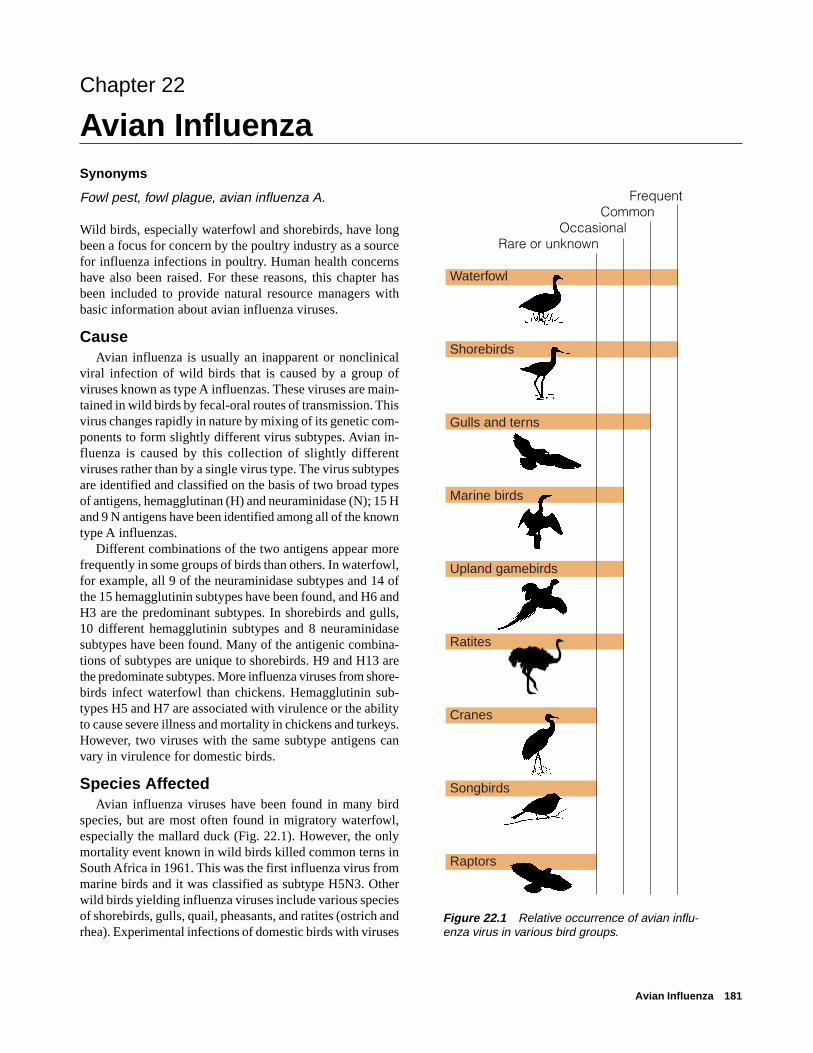
Avian Influenza 181
FrequentCommon
OccasionalRare or unknown
Waterfowl
Shorebirds
Gulls and terns
Marine birds
Upland gamebirds
Ratites
Cranes
Songbirds
Raptors
Chapter 22
Avian InfluenzaSynonyms
Fowl pest, fowl plague, avian influenza A.
Wild birds, especially waterfowl and shorebirds, have longbeen a focus for concern by the poultry industry as a sourcefor influenza infections in poultry. Human health concernshave also been raised. For these reasons, this chapter hasbeen included to provide natural resource managers withbasic information about avian influenza viruses.
CauseAvian influenza is usually an inapparent or nonclinical
viral infection of wild birds that is caused by a group ofviruses known as type A influenzas. These viruses are main-tained in wild birds by fecal-oral routes of transmission. Thisvirus changes rapidly in nature by mixing of its genetic com-ponents to form slightly different virus subtypes. Avian in-fluenza is caused by this collection of slightly differentviruses rather than by a single virus type. The virus subtypesare identified and classified on the basis of two broad typesof antigens, hemagglutinan (H) and neuraminidase (N); 15 Hand 9 N antigens have been identified among all of the knowntype A influenzas.
Different combinations of the two antigens appear morefrequently in some groups of birds than others. In waterfowl,for example, all 9 of the neuraminidase subtypes and 14 ofthe 15 hemagglutinin subtypes have been found, and H6 andH3 are the predominant subtypes. In shorebirds and gulls,10 different hemagglutinin subtypes and 8 neuraminidasesubtypes have been found. Many of the antigenic combina-tions of subtypes are unique to shorebirds. H9 and H13 arethe predominate subtypes. More influenza viruses from shore-birds infect waterfowl than chickens. Hemagglutinin sub-types H5 and H7 are associated with virulence or the abilityto cause severe illness and mortality in chickens and turkeys.However, two viruses with the same subtype antigens canvary in virulence for domestic birds.
Species AffectedAvian influenza viruses have been found in many bird
species, but are most often found in migratory waterfowl,especially the mallard duck (Fig. 22.1). However, the onlymortality event known in wild birds killed common terns inSouth Africa in 1961. This was the first influenza virus frommarine birds and it was classified as subtype H5N3. Otherwild birds yielding influenza viruses include various speciesof shorebirds, gulls, quail, pheasants, and ratites (ostrich andrhea). Experimental infections of domestic birds with viruses
Figure 22.1 Relative occurrence of avian influ-enza virus in various bird groups.

182 Field Manual of Wildlife Diseases: Birds
Pacific Flyway
Pintail
Pintail
Snow goose
Canada goose
Black duck
Mallard
Mallard
Central Flyway
Mississippi Flyway Atlantic Flyway
C A N A D A
PacificOcean
Arctic Ocean
AtlanticOcean
Gulf of Mexico
HudsonBay
C A N A D A
MEXICOCanvasback
PacificOcean
Arctic Ocean
AtlanticOcean
Gulf of Mexico
HudsonBay
C A N A D A
MEXICO
PacificOcean
Arctic Ocean
AtlanticOcean
Gulf of Mexico
HudsonBay
MEXICO
PacificOcean
Arctic Ocean
AtlanticOcean
Gulf of Mexico
HudsonBay
C A N A D A
MEXICO
Figure 22.2 General migratory pathways followed by North American waterfowl. Species shown are typical of these flyways(Modified from Hawkins and others, 1984).

Avian Influenza 183
from wildlife do not cause mortality. Likewise, virulentviruses or viruses that cause disease in domestic fowl do notcause mortality in wild waterfowl.
DistributionAlthough influenza tends to be most commonly detected
in birds that use the major waterfowl flyways, these virusesare found throughout North America and around the world.The majority of North American waterfowl migration takesplace within four broad geographic areas (Fig 22.2). Manyspecies other than waterfowl follow these same migratorypathways from their breeding grounds to the winteringgrounds and return to the breeding grounds. The virus sub-type that are found in birds in adjacent flyways will differ,especially if the birds from each flyway do not mix duringmigration. In any given year the percentage of waterfowl andshorebirds carrying influenza viruses will vary by flyway.Likewise, the percentage of birds carrying virus in an fly-way will vary in consecutive years. The virus subtype foundin birds that use a flyway are rarely the same in consecutiveyears.
SeasonalityInfluenza virus has been found in wild birds throughout
the year, but waterfowl are the only group in which theseviruses are found year round (Fig. 22.3). The highest occur-rence of infection is in the late summer months in juvenilewaterfowl when they assemble for their first southward mi-gration. The number of infected waterfowl decreases in thefall as birds migrate toward their southern wintering groundsand is lowest in the spring, when only one bird in 400 isinfected during the return migration to the north. In contrast,the number of birds infected is highest in shorebirds (prima-rily ruddy turnstone) and gulls (herring) during spring (Mayand June). Infection in shorebirds is also high in Septemberand October. Influenza viruses have not been found in shore-bird and gull populations during other months of the year.Influenza viruses have been found in marine birds such asmurres, kittiwakes, and puffins while they have been nest-ing, but the pelagic habits of these species preclude sam-pling during other periods of the year.
Field SignsIn domestic birds, the signs of disease are not diagnostic
because they are highly variable and they depend on the strainof virus, bird species involved, and a variety of other factorsincluding age and sex. Signs of disease may appear as respi-ratory, enteric, or reproductive abnormalities. Included aresuch nonspecific manifestations as decreased activity, foodconsumption, and egg production; ruffled feathers; cough-ing and sneezing; diarrhea; and even nervous disorders, suchas tremors. Observable signs of illness have not been de-scribed for wild birds. In domestic chickens and turkeys,certain virus subtypes like H5N2 and H7N7, respectively,
are usually highly virulent and may cause up to 100 percentmortality in infected flocks. Another major impact of influ-enza viruses in domestic birds is decreased egg production.Too little is known about the impact of influenza viruses onthe reproductive performance of wild birds to assess whetheror not they are affected in the same manner as poultry.
Gross LesionsAvian influenza virus infection in wild birds is not indi-
cated by gross lesions. Common terns that died in SouthAfrica did not have gross lesions, but a few birds had micro-scopic evidence of meningoencephalitis or inflammation ofthe membrane that covers the brain. These lesions were notreproducible during experiments. Mallards experimentallyinfected with a virulent influenza virus developed discretepurple areas of lung firmness and cloudy lung coverings.However, virulent viruses are rarely found in wild birds, andthese lesions may not appear in natural infections.
Figure 22.3 Relative seasonal occurrenceof influenza A in birds.
FrequentCommon
OccasionalRare or unknown
Primary Species
WATERFOWL (Mallard duck)
Spring
Summer
Fall
Winter
SHOREBIRDS (Ruddy turnstone)
Spring
Summer
Fall
Winter
MARINE BIRDS (Common tern or murre)
Spring
Summer
Fall
Winter
UPLAND GAMEBIRDS (Pheasant)
Spring
Summer
Fall
Winter

184 Field Manual of Wildlife Diseases: Birds
Figure 22.4 Global cycleof avian influenza viruses inanimals.
DiagnosisInfected birds are detected by virus isolation from cloa-
cal swabs in embryonated chicken eggs, and by serologicaltesting of blood for antibody. The last test indicates that abird was exposed to these viruses rather than if it is infectedor carries the disease. Reference antisera to all of the sub-type antigen combinations are used to determine the identityof the virus; however, the virulence of a virus cannot be de-termined by the antigenic subtype. Virulent and avirulentstrains of the same subtype can circulate in nature. Labora-tory and animal inoculation tests are required to establishthe virulence of strains based on an index established fordomestic birds.
ControlAvian influenza viruses in wild birds cannot be effectively
controlled because of the large number of virus subtypes andthe high frequency of virus genetic mixing resulting in newvirus subtypes. Also, virus has been recovered from waterand fecal material in areas of high waterfowl use. Duringexperiments, influenza virus was recovered from infectedwaterfowl fecal material for 8 days and from fecal contami-nated river water for 4 days when both were held at 22 °C.Poultry manure is a primary residual source of virus for do-mestic flocks. The virus has been recovered from poultryhouses more than 100 days after flock depopulation for mar-kets.
In the domestic bird industry, preventing the entry of thevirus into poultry flocks is the first line of defense. Killedvaccines are selectively used to combat less virulent formsof this disease. Antibody present in the blood of recoveredand vaccinated birds prevents virus transmission. Therefore,these birds pose little risk to other birds. Flocks are gener-ally killed when they are infected with highly virulentviruses.
In the past, the poultry industry and the wildlife conser-vation community have been in conflict regarding wildliferefuge development and other waterbird habitat projects. The
fear that waterbirds are a source of influenza viruses for in-fection of poultry has resulted in strong industry oppositionthat has negatively impacted some projects. This issue shouldbe considered when land use near wetlands is planned andwhen wildlife managers plan for development for wildlifeareas. Open communication during project development andsound plans that are developed in a collaborative manner mayhelp industry and conservation groups avoid confrontationand support each others’ interests.
Human Health ConsiderationsAlthough this group of viruses includes human influenza
viruses, the strains that infect wild birds do not infect hu-mans. It is believed that waterfowl and shorebirds maintainseparate reservoirs of viral gene pools from which new virussubtypes emerge. These gene pools spill over into other ani-mals (mammals) and may eventually cause a new pandemic(Fig. 22.4).
Wallace Hansen
Supplementary ReadingEasterday, B.C., and Hinshaw, V.S., 1991, Influenza, in Calnek,
B.W., and others, eds., Diseases of Poultry (9th ed.): Ames,Iowa, Iowa State University Press, p. 532–552.
Hinshaw, V.S., Wood, J.M., Webster, R.G., Deible, R., and Turner,B., 1985, Circulation of influenza viruses and paramyxovirusesin waterfowl originating from two different areas of NorthAmerica: Bulletin of the World Health Organization 63, 711–719.
Kawoka, Y., Chambers, T.M., Sladen, W.L., and Webster, R.G.,1988, Is the gene pool of influenza viruses in shorebirds andgulls different from that in wild ducks?: Virology, v. 163,p. 247–250.
Stallknecht, D. E., Shane, S. M., Zwank, P. J., Senne, D. A., andKearney, M. T., 1990, Avian influenza viruses from migratoryand resident ducks of coastal Louisiana: Avian Diseases, v. 34,p. 398–405.
Naturalavian influenza
cycle
Pandemicdisease
cycleWaterfowl Mammals
(Primarily swine)Shorebirds
Domesticbirds
Humans

Woodcock Reovirus 185
Chapter 23
Woodcock Reovirus
Synonyms
None
This chapter provides information on a recently identifieddisease of the American woodcock. Little is known aboutthe disease or the virus that causes it. It has been included inthis Manual to enhance awareness that such a disease existsand to stimulate additional interest in further investigationsto define the importance of woodcock reovirus. More infor-mation about this disease is needed because it is not knownwhether or not this virus is a factor in the decline of wood-cock populations within the United States.
CauseThe first virus isolated from the American woodcock is a
reovirus that was found during woodcock die-offs during thewinters of 1989–90 and 1993–94. Avian reovirus infectionshave been associated with numerous disease conditions in-cluding viral arthritis/tenosynovitis or inflammation of thetendon sheath; growth retardation; pericarditis or inflamma-tion of the sac surrounding the heart; myocarditis or inflam-mation of the heart muscle; hydropericardium or abnormalaccumulation of fluid in the pericardium; enteritis or inflam-mation of the intestine; hepatitis or inflammation of the liver;
bursal and thymic atrophy or wasting away; osteoporosis orrarefaction of the bone; and respiratory syndromes. The in-fections are generally systemic, transmitted by the fecal-oralroute, and are often associated with nutritional factors orconcurrent infections with other agents.
Species AffectedAmerican woodcock are the only species known to be
infected with this particular reovirus. Investigations have notbeen conducted to determine whether or not other speciesare susceptible to infection, which species are not suscep-tible, and which species become diseased.
DistributionThe virus was isolated from woodcock that were found
dead at the Eastern Shore of Virginia National Wildlife Ref-uge (Fig. 23.1). To determine the prevalence of woodcockreovirus in the eastern and central regions of the United States,virus isolation was attempted from woodcock samples col-lected from the breeding and wintering populations in 1990–92 (Fig. 23.1). No viruses were isolated from 481 tissuesamples or 305 cloacal swabs that were obtained from live-trapped and hunter-killed woodcock.
Figure 23.1 Site of woodcock mortality from reovirus infection and field samplingsites where other woodcock were tested and found to be negative for exposure tothis disease.
�
EXPLANATION
Site of woodcock mortality from reovirus infection
Site where woodcock tested negative to reovirus expo-sure
�
��
�
�
�
�
�

186 Field Manual of Wildlife Diseases: Birds
SeasonalityBoth die-offs occurred during the winter months. Noth-
ing more is known about the seasonality of this disease.
Field SignsSick woodcock have not been observed. Therefore, field
signs are unknown.
Gross LesionsMost of the birds found dead were emaciated. Little or no
food was found in their digestive tracts, and no obvious grosslesions were noted upon necropsy of the carcasses. Healthy“control” woodcock collected during the same time did notyield virus.
DiagnosisDiagnosis requires laboratory isolation and identification
of the causative virus. Winter concentrations of woodcockshould be monitored and carcasses picked up and submittedfor diagnosis. Whole carcasses of woodcock found deadshould be shipped to a diagnostic laboratory where patho-logical assessments and virus isolation can be made (seeChapter 3, on Specimen Shipment). Although the virus hasbeen isolated from a variety of tissues including intestine,brain, cloacal swab, heart, and lung, the majority of isolateswere obtained from intestines and cloacal swabs. These find-ings suggest that a fecal-oral route of transmission is likely.
ControlField carcasses not needed for diagnostic study should be
collected for disposal to minimize environmental contami-nation. Too little is known about this disease to recommendresponse actions.
Human Health ConsiderationsThere are no known human health considerations.
Douglas E. Docherty
Supplementary ReadingDocherty, D.E., Converse, K.A., Hansen, W.R., and Norman,
G.W., 1994, American woodcock (Scolopax minor) mortalityassociated with a reovirus: Avian Diseases, v. 38, p. 899–904.

Introduction to Parasitic Diseases 187
Section 5Parasites andParasitic DiseasesHemosporidiosis
Trichomoniasis
Intestinal Coccidiosis
Renal Coccidiosis
Sarcocystis
Eustrongylidosis
Tracheal Worms
Heartworm of Swans and Geese
Gizzard Worms
Acanthocephaliasis
Nasal Leeches
Miscellaneous Parasitic Diseases
Stained blood smear from a turkey infected with the parasite HaemoproteusmeleagridisPhoto by Carter Atkinson

188 Field Manual of Wildlife Diseases: Birds
Introduction to Parasitic Diseases
“Parasites form a large proportion of the diversity of life on earth.”
(Price)
Quote from:
Price, P.W., 1980, Evolutionary biology of parasites: PrincetonUniversity Press, Princeton, NJ, p. 3.
Parasitism is an intimate relationship between two differentspecies in which one (parasite) uses the other (host) as itsenvironment from which it derives nourishment. Parasitesare a highly diverse group of organisms that have evolveddifferent strategies for infecting their hosts. Some, such aslice and ticks, are found on the external parts of the body(ectoparasites), but most are found internally (endoparasites).Some are microscopic, such as the blood protozoans thatcause avian malaria; however, many are macroscopic. Lifecycles differ greatly between major types of parasites andare generally classified as direct or indirect (Table 1). Directlife cycles do not require an intermediate host (Fig. 1A). Fordirect life cycles, only a definitive host is required: the spe-cies in which the parasite reaches sexual maturity and pro-duces progeny. Indirect life cycles may involve one or moreintermediate hosts (Fig. 1B and C). Intermediate hosts arerequired by the parasite for completion of its life cycle be-cause of the morphological and physiological changes thatusually take place in the parasite within those hosts. Wildbirds can serve as the definitive hosts for most of the para-sites that are discussed in the following chapters. In addition,paratenic or transport hosts are present in some parasite lifecycles. The parasites generally do not undergo developmentin paratenic hosts. Instead, paratenic hosts provide both anecological and temporal (time) bridge for the parasite to movethrough the environment and infect the definitive host. Typi-cally, in these situations one or more intermediate hosts are
required for development of the parasite but they are not fedupon by the bird. Instead, the bird feeds on the paratenichosts, which in turn have fed on the intermediate host(s),thereby, “transporting” the parasite to the bird (Fig. 2).
The presence of parasites in birds and other animals isthe rule, rather than the exception. Hundreds of parasite spe-cies have been identified from free-ranging wild birds; how-ever, the presence of parasites does not necessarily equatewith disease. Most of the parasites identified from wild birdscause no clinical disease. Others cause varying levels of dis-ease, including death in the most severe cases. The pathoge-nicity or the ability to cause disease, of different species ofparasites varies with 1) the species of host invaded (infectedor infested), 2) the number or burden of parasites in or on thehost, and 3) internal factors impacting host response. Forexample, when birds are in poor nutritional condition, haveconcurrent infections from other disease agents (includingother species of parasites), or are subject to other types ofstress, some parasites that do not normally cause disease docause disease. Lethal infections may result from parasitesthat generally only cause mild disease.
This section highlights some of the parasitic diseases suchas trichomoniasis that are associated with major mortalityevents in free-ranging wild birds and those that because ofthe gross lesions they cause (Sarcocystis sp.), their visibility(nasal leeches), or general interest (heartworm) are often thesubject of questions asked of wildlife disease specialists.

Introduction to Parasitic Diseases 189
Table 1 General characteristics of major groups of internal parasites (endoparasites) of free-ranging birds.
Type of parasite Common name Type of life cycle Characteristics
Nematodes Roundworms Indirect and direct Most significant group relative to number of speciesinfecting birds and to severity of infections.
Unsegmented cylindrical worms.
Found throughout the body.
Generally four larval stages.
Sexes are separate.
Most are large in size (macroscopic).
Cestodes Tapeworms Indirect Flattened, usually segmented worms with a distincthead, neck and body.
Found primarily in the lumen of the intestines.
Lack a mouth or an alimentary canal; feed by absorbingnutrients from the host’s intestinal tract.
Most are hermaphroditic (self-fertilization; have both maleand female reproductive tissues).
Attachment is by suckers, hooks.
Large size (macroscopic).
Trematodes Flukes Indirect Flatworms, generally leaf-shaped (some almost cylindrical).
Generally found in the lower alimentary tract, respiratory tract,liver, and kidneys.
Complex life cycles; usually require two intermediate hosts,one of which is usually a snail.
Hermaphroditic except for blood flukes, which haveseparate sexes.
Attachment is usually by suckers.
Acanthocephalans Thorny-headed Indirect Cylindrical, unsegmented worms.
Found in the digestive tract.
No intestinal tract; nutrients absorbed through the tegument(similar to tapeworms).
Sexes are separate.
Attachment by means of a retractable proboscis that hassharp recurved hooks or spines.
Protozoans Coccidians, Direct and Microscopic.
Different types are found in different parts of the body.
Asexual and sexual multiplication.
worms
malarias,trichomonads,others
indirect
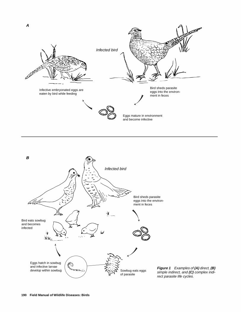
190 Field Manual of Wildlife Diseases: Birds
Figure 1 Examples of (A) direct, (B)simple indirect, and (C) complex indi-rect parasite life cycles.
A
B
Infected bird
Eggs mature in environment and become infective
Infective embryonated eggs are eaten by bird while feeding
Bird sheds parasiteeggs into the environ-ment in feces
Infected bird
Bird eats sowbug and becomes infected
Sowbug eats eggs of parasite
Eggs hatch in sowbug and infective larvae develop within sowbug
Bird sheds parasiteeggs into the environ-ment in feces

Introduction to Parasitic Diseases 191
CInfected bird
Parasite eggs are passed in feces and hatch in water
Larvae (miracidium) swims to a snail and penetrates it, undergoing further larval developmentwithin the host
New larval stage emerges from snail (cercaria) andswims to new host where itpenetrates and encysts
First intermediate host
Second intermediate host
Other bird species eat secondintermediate host andbecome infected

192 Field Manual of Wildlife Diseases: Birds
Figure 2 Hypothetical parasite life cycle illustrating the role of paratenic (transport) hosts.
Infected bird
Bird sheds parasiteeggs into the environ-ment in feces
Birds feed on fish and become infected to complete life cycle
Second intermediate hostAmphipod is eaten by amphibian where infective stages of larvae develop
First intermediate hostEggs eaten by amphipod where first- and second- stage larvae develop
Paratenic hostFish eats the amphibian and larvae encyst in body of fish. No further development of the parasite

Hemosporidiosis 193
Chapter 24
Hemosporidiosis
SynonymsAvian malaria
CauseHemosporidia are microscopic, intracellular parasitic pro-
tozoans found within the blood cells and tissues of their avianhosts. Three closely related genera, Plasmodium, Haemopro-teus, and Leucocytozoon, are commonly found in wild birds.Infections in highly susceptible species and age classes mayresult in death.
Life CycleHemosporidia are transmitted from infected to uninfected
birds by a variety of biting flies that serve as vectors, includ-ing mosquitoes, black flies, ceratopogonid flies (bitingmidges or sandflies) and louse flies (Fig. 24.1) (Table 24.1).When present, infective stages of the parasites (sporozoites)are found in the salivary glands of these biting flies. Theygain entry to the tissues and blood of a new host at the site ofthe insect bite when these vectors either probe or laceratethe skin to take a blood meal. Insect vectors frequently feed
Figure 24.1 The complexgeneral life cycle of hemo-sporidian parasites beginswith (A), an infected insectbiting a susceptible bird.Separate infectious and de-velopmental stages occurin (B), the bird host, and(C), the insect vectors.
A. Infected insect #1 bites bird #1
Infected insect #2 bites a different bird
C. Stages within insect
Infective sporozoitespresent in salivary glands of infected insect vector.Sporozoites gain entry at site of bite
Sporozoites invade tissues and reproduce as schizonts to produce numerous merozoites
Merozoites penetratered blood cells andmature into infectiousgametocytes
New vector (insect #2) feeds on bird and becomes infected
Gametocytes mature, undergo sexual reproductionin midgut
Oocysts become encapsulated on the outer midgut wall
Oocysts rupture andsporozoites invadesalivary gland
B. Stages within bird

194 Field Manual of Wildlife Diseases: Birds
on exposed flesh around the eyes (Fig. 24.2), the beak, andon the legs and feet, although black flies, ceratopogonid flies,and louse flies can crawl beneath the bird’s feathers to reachthe skin surface. Immediately after they infect a bird, sporo-zoites invade the tissues and reproduce for one or more gen-erations before they become merozoites. Merozoites penetratethe red blood cells and become mature, infectious gameto-cytes. The cycle is completed when the gametocytes in thecirculating blood cells of the host bird are ingested by an-other blood-sucking insect, where they undergo both sexualand asexual reproduction to produce large numbers of sporo-zoites. These invade the salivary glands of the vector and aretransmitted to a new host bird during the vector’s next bloodmeal.
Species AffectedThe avian hemosporidia are cosmopolitan parasites of
birds, and they have been found in 68 percent of the more
than 3,800 species of birds that have been examined. Mem-bers of some avian families appear to be more susceptiblethan others. For example, ducks, geese and swans are com-monly infected with species of Haemoproteus, Leucocyto-zoon, and Plasmodium, and more than 75 percent of water-fowl species that were examined were hosts for one or moreof these parasites. Wild turkeys in the eastern United Statesare also commonly infected by these parasites. Pigeons anddoves have similar high rates of infection, but members ofother families, such as migratory shorebirds, are less fre-quently parasitized.
Differences in the prevalence, geographic distribution, andhost range of hemosporidia are associated with habitat pref-erences of the bird hosts, the abundance and feeding habitswithin those habitats of suitable insect vectors, and innatephysiological differences that make some avian hosts moresusceptible than others. For example, some species of blackflies (Simulium sp.) prefer to feed on waterfowl within a lim-
Table 24.1 Avian hemosporidia parasites and their documented vectors.
Parasite Vector type Common name
Haemoproteus Ceratopogonidae Punkies, no-see-ums, sand flies(Culicoides sp.)Hippoboscidae Hippoboscid or louse flies(Ornithomyia sp.)
Plasmodium Culicidae Mosquitoes(Culex, Aedes sp.)
Leucocytozoon Simulidae Black flies(Simulium sp.)
Figure 24.2 A Culex mosquito feeding on the unfeatheredarea surrounding the eye of an apapane, a native Hawaiianhoneycreeper.
Pho
to b
y Ja
ck J
effre
y, U
.S. F
ish
and
Wild
life
Ser
vice

Hemosporidiosis 195
ited distance of the shoreline. Ducks and geese that spendmore of their time in this zone will be more likely to be ex-posed to bites that carry infective stages of Leucocytozoonsimondi. Biting midges or no-see-ums (Culicoides sp.) thattransmit species of Haemoproteus are more active at dusk inthe forest canopy. Birds that roost here, for example, increasetheir chances for being infected with this parasite. Finally,some avian hosts are more susceptible to hemosporidian para-sites than others, but the physiological basis for this is stillpoorly understood.
Species of Plasmodium and Leucocytozoon are capableof causing severe anemia, weight loss, and death in suscep-tible birds. Young birds are more susceptible than adults, andthe most serious mortality generally occurs within the firstfew weeks of hatching. This is also the time of year whenincreasing temperatures favor the growth of the populationsof insect vectors that transmit hemosporidia. Major outbreaksof L. simondi that caused high mortality in ducks and geesein Michigan and subarctic Canada have been documented.Species of Haemoproteus are generally believed to be lesspathogenic, with only scattered reports of natural mortalityin wild birds.
Penguins and native Hawaiian forest birds are highly sus-ceptible to Plasmodium relictum, a common parasite of song-birds that is transmitted by Culex mosquitoes. This parasitecauses high mortality in both captive and wild populationsof these hosts, and it is a major factor in the decline of nativeforest birds in the Hawaiian Islands.
DistributionSpecies of Plasmodium, Haemoproteus, and Leucocyto-
zoon have been reported from most parts of the world withthe exception of Antarctica, where cold temperatures pre-vent the occurrence of suitable insect vectors. Studies of thedistribution of hemosporidia in North America have shownthat areas of active transmission of the parasites coincidewith the geographic distribution of their vectors. Leuco-cytozoon is most common in mountainous areas of Alaskaand the Pacific Northwest where abundant fast-movingstreams create suitable habitat for aquatic black fly larvae.Species of Haemoproteus and Plasmodium are more evenlydistributed across the continent because their ceratopogonidand mosquito vectors are less dependent on the presence offlowing water for larval development. Migratory birds maywinter in habitats that lack suitable vectors; therefore, thesimple presence of infected birds may not be evidence thatthe parasites are being transmitted to birds at the winteringgrounds.
SeasonalityInfections with Plasmodium, Haemoproteus, and Leuco-
cytozoon are seasonal because transmission depends uponthe availability of vector populations. In temperate NorthAmerica, most birds become infected with hemosporidia
during the spring when conditions for transmission becomeoptimal. Some of these conditions include the onset ofwarmer weather; increases in vector populations; the reap-pearance or relapse of chronic, low-level infections in adultbirds; and the hatching and fledging of susceptible,nonimmune juvenile birds. In warmer parts of the UnitedStates, these parasites may be transmitted at other times ofthe year. In Hawaii, P. relictum in forest bird populationsmay be transmitted throughout the year in warm low-eleva-tion forests, but transmission is more seasonal at elevationsabove 3,000 ft. where cooler winter temperatures limit mos-quito populations.
Field SignsBirds with acute infections of Plasmodium, Haemopro-
teus, and Leucocytozoon, may exhibit similar signs in thefield. These include emaciation, loss of appetite, listlessness,difficulty in breathing, and weakness and lameness in one orboth legs. Survivors develop persistent, low-level infectionsin the blood and tissues that stimulate immunity to reinfec-tion. These survivors do not exhibit any signs of disease, butthey serve as reservoirs of infection, allowing the parasitesto survive droughts and cold winter weather when vectorpopulations have died off.
Gross LesionsGross lesions associated with acute infections include
enlargement of the liver and spleen (Fig. 24.3) and the ap-pearance of thin and watery blood as a result of infected bloodcells being destroyed and removed from circulation (Fig.24.4). In Plasmodium and Haemoproteus infections, para-sites within the red blood cells produce an insoluble blackpigment called hemozoin when they digest the host’s oxygen-bearing, iron-laden red blood cell protein or hemoglobin. Thehemozoin is deposited extensively in the host’s spleen andliver tissue as the host’s immune system responds to the in-fection. In very heavy infections, the kidneys may also beaffected. These organs typically appear chocolate brown orblack at necropsy and they may be two or more times theirnormal size (Fig. 24.3). Hemozoin pigment is not producedin Leucocytozoon infections; therefore, organs will not be asdiscolored and dark at necropsy, but they will still appearenlarged. Some species of Haemoproteus form large, cyst-like bodies in muscle tissue that superficially resemble tis-sue cysts produced by species of Sarcocystis (Fig. 24.5).
DiagnosisDefinitive diagnosis of hemosporidian infections is de-
pendent on microscopic examination of a stained blood smearor on an organ impression smear to detect the presence andform of the parasites within the red blood cells (Figs. 24.6,7, 8). Species of Leucocytozoon frequently produce dramaticchanges in the host’s cell structure (Fig. 24.6). Parasitizedred blood cells are often enlarged and elongated so that they

196 Field Manual of Wildlife Diseases: Birds
Figure 24.3 Gross lesions caused by Plasmodium relictum in an apapane. Enlargement and discol-oration of the (A), liver and (B), spleen are typical in acute infections when large numbers of parasites arefound in the circulating red blood cells.
Pho
tos
by
Car
ter
Atk
inso
n, B
RD
-PIE
RC
A
B

Hemosporidiosis 197
Figure 24.4 Thin and watery blood from anapapane infected with Plasmodium relictum before(left) and after (right) centrifugation. In uninfectedsongbirds, approximately half of the blood volume isoccupied by red blood cells. Note that most of theblood cells have been destroyed by the parasite(right).
Figure 24.5 Pectoral muscles of a turkey infected with Haemoproteus meleagridis. Note the whitestreaks and bloody spots in the muscle tissue of this bird (arrows). The tissue stages of this hemosporidianform large, cystlike bodies that may superficially resemble those caused by species of Sarcocystis.
Pho
to b
y C
arte
r A
tkin
son,
BR
D-P
IER
C
Pho
to b
y C
arte
r A
tkin
son,
BR
D-P
IER
C
➡
➡
➡

198 Field Manual of Wildlife Diseases: Birds
Figure 24.8 Stained blood smear from an apapane in-fected with Plasmodium relictum. Some red blood cellscontain multinucleated, asexually-reproducing stages ofthe parasite called schizonts (S). These are diagnosticfor Plasmodium infections and contain one or more cen-trally-located pigment granules (arrows).
Figure 24.6 Stained blood smear from a turkey infectedwith Leucocytozoon smithi. This parasite causes en-largement and distortion of the infected blood cell. Thered blood cell nucleus (N) is divided in two halves that layon either side of the parasite (P). The membrane of theinfected cell is stretched into two hornlike points (arrows).
Figure 24.7 Stained blood smear from a turkey infectedwith Haemoproteus meleagridis. Gametocytes (G) con-tain a single pink-staining nucleus and contain black orgolden brown pigment granules (arrows).
Pho
to b
y C
arte
r A
tkin
son,
BR
D-P
IER
CP
hoto
by
Car
ter
Atk
inso
n, B
RD
-PIE
RC
Pho
to b
y C
arte
r A
tkin
son,
BR
D-P
IER
C
NP
➡
➡
G
➡
➡
S
S
➡
➡

Hemosporidiosis 199
form a pair of horn-like extensions from either end of thecell. Species of Plasmodium and Haemoproteus producefewer changes in their host’s red blood cells, but these para-sites may cause slight enlargement of infected host cells anddisplacement of the red blood cell nucleus to one side (Figs.24.7, 8). Unlike Leucocytozoon, Plasmodium and Haemo-proteus produce golden brown or black deposits of hemozoinpigment in the parasite cell (Figs. 24.7, 8). Further differen-tiation of Plasmodium from Haemoproteus may be difficult.Diagnosis of a Plasmodium infection is dependent on de-tecting the presence of asexually reproducing stages of itslife cycle (schizonts) in the red blood cells of the infectedhost (Fig. 24.8).
ControlControl of the avian hemosporidia is dependent on re-
ducing transmission from infected birds to healthy birdsthrough reduction or elimination of vector populations. Manyof the same techniques that were developed for control ofvector-transmitted human diseases can be used effectively,but few agencies have the resources or manpower to applythem over large areas. Most techniques rely on habitat man-agement to reduce vector breeding sites or depend on theapplication of pesticides that affect larval or adult vectors toreduce vector populations. Large-scale treatment of infectedsurvivor birds could prevent disease outbreaks by reducingsources of infection, but the logistics and practicality of treat-ing sufficient numbers of birds to interrupt transmission areprohibitive. Although some experimental vaccines for theseparasites have been developed, none are currently availablefor general use.
Human Health ConsiderationsThe avian hemosporidia are closely related to the malarial
parasites of humans, but are not capable of infecting people.
Carter T. AtkinsonPacific Island Ecosystems Research CenterKilauea Field Station
Supplementary ReadingAtkinson, C.T., 1991, Vectors, epizootiology, and pathogenicity of
avian species of Haemoproteus: Bulletin of the Society forVector Ecology, vol.16, p. 109–126.
Atkinson, C.T., and van Riper, C., III, 1991, Pathogenicity andepizootiology of avian haematozoa: Plasmodium, Leucocyto-zoon, and Haemoproteus, in Loye, J.E., and Zuk, M., eds.,Bird-parasite interactions, Ecology, evolution, and behavior:New York, Oxford University Press, p. 19–48.
Bennett, G.F., Whiteway, M., and Woodworth-Lynas, C.B., 1982,Host-parasite catalogue of the avian haematozoa: MemorialUniversity of Newfoundland Occasional Papers in BiologyNumber 5, p. 243.
Greiner, E.C., Bennett, G.F., White, E.M., and Coombs, R.F.,1975, Distribution of the avian hematozoa of North America,v. 53, p. 1,762–1,787.
Greiner, E.C., 1991, Leucocytozoonosis in waterfowl and wildgalliform birds: Bulletin of the Society for Vector Ecology,v. 16, p. 84–93.

200 Field Manual of Wildlife Diseases: Birds

Trichomoniasis 201
Chapter 25
Trichomoniasis
SynonymsCanker (doves and pigeons), frounce (raptors), aviantrichomoniasis
CauseAvian trichomoniasis is caused by a single celled proto-
zoan, Trichomonas gallinae. Avirulent T. gallinae strains thatdo not cause disease and highly virulent strains are found innature and circulate within bird populations. The factors thatmake a strain virulent are not known, but they are thought tobe controlled genetically within the parasite. Similarly, thereasons why an avirulent or a virulent form of the parasite isfound within a bird population at any period of time alsoremain unknown. Virulent strains of T. gallinae have causedmajor mortality events or epizootics in doves and pigeons in
addition to less visible, chronic losses (Table 25.1). Infec-tion typically involves the upper digestive tract of doves andpigeons but other species have also been infected (Fig. 25.1).
Trichomoniasis in doves and pigeons, but not in otherspecies, is generally confined to young birds. The parasitewas introduced to the U.S. with the introduction of pigeonsand doves brought by European settlers. It has been reportedthat 80 to 90 percent of adult pigeons are infected, but theyshow no clinical signs of disease. It is speculated that mostof these birds became immune as a result of exposure to aviru-lent strains of the parasite or because they survived mild in-fections. In pigeons and mourning doves, the parasites aretransmitted from the adults to the squabs in the pigeon milkproduced in the crop of the adult. Squabs usually becomeinfected with the first feeding of pigeon milk, which is gen-
Table 25.1 Examples of wild bird mortalities reported in the scientific literature due to trichomoniasis.
Year Magnitude Geographic area Comments
1949–51 Tens of thousands of Southeastern United Trichomoniasis broke out inmourning doves States virtually all States in the
region; the magnitude oflosses focused attention onthe devastation that could becaused by this disease andstimulated research on theecology of this disease.
1950–51 25,000 to 50,000 Alabama Breeding birds were the focusmourning doves each of infection; mortality wasyear thought to have been grossly
underestimated.
1972 Several hundred Nebraska Railroad yards and a grainelevator were focal points ofinfection; birds fed on spilledgrain.
1985 Approximately 800 New Mexico Losses at birdfeeders near Lasmourning doves Cruces.
1988 At least 16,000 California First major epizootic ofband-tailed pigeons trichomoniasis in this species.
1991 Approximately 500 North Carolina —mourning doves

202 Field Manual of Wildlife Diseases: Birds
erally within minutes after hatching. The resulting infectionmay range from asymptomatic or mild disease to a rapidlyfatal course resulting in death within 4–18 days after infec-tion. Other modes for infection are through feed, perhapscontaminated drinking water, and feeding on infected birds(Fig. 25.2).
There is no cyst or resistant stage in the parasite’s lifecycle; therefore, infection must be passed directly from onebird to another, in contaminated feed or water. Feed and waterare contaminated when trichomonads move from the mouthof infected birds, not from their feces. Lesions in the mouthor the esophagus or both of an infected bird (see below) of-ten prevent the passage of ingested grain seeds and cause thebird to regurgitate contaminated food items. Water becomescontaminated by contact with the contaminated bill andmouth. Pigeons that feed among domestic poultry are oftenblamed for contaminating feed and water and passing thedisease to the poultry. Similar transmission has been associ-ated with dove mortality at grain elevators and at birdfeed-ers. Doves and pigeons cross-feed and bill during courtship,and this behavior facilitates direct transmission as does theconsumption of infected birds by raptors. It has been reportedthat some moist grains can maintain viable T. gallinae for atleast 5 days and that parasite survival in water can rangefrom 20 minutes to several hours. These conditions are ad-equate for disease transmission at birdfeeders and waterersbecause of the gregarious habits of doves and pigeons.
Species AffectedTrichomoniasis is considered by many avian disease spe-
cialists to be the most important disease of mourning dovesin North America. Band-tailed pigeons have also sufferedlarge-scale losses from trichomoniasis. This disease has beenreported as a cause of mortality in birds of prey for hundredsof years prior to the causative organism being identified.Songbirds are less commonly reported to be infected, but T.gallinae is reported to be the most important trichomonad ofcaged birds; it is often responsible for epizootics among cap-tive collections. Domestic turkeys and chickens also becomeinfected.
DistributionIt is likely that T. gallinae is found wherever domestic
pigeons and mourning doves are found. Disease in free-ranging wild birds is grossly underreported. Outbreaks atbirdfeeding stations and similar locations reported to theNational Wildlife Health Center have occurred from coast-to-coast within the United States (Fig. 25.3).
SeasonalityEpizootics due to T. gallinae can happen yearround, but
most outbreaks have been reported during late spring, sum-mer, and fall.
Field SignsBecause oral lesions often affect the ability of the bird to
feed, infected birds lose weight, appear listless, and standgrouped together. These birds often appear ruffled. Caseousor cheesy, yellowish lesions may be seen around the beak oreyes of mourning doves and the face may appear “puffy”and distended (Fig. 25.4). Severely infected pigeons may fallover when they are forced to move.
Gross LesionsThe severity and appearance of lesions varies with the
virulence of the strain of the parasite, the stage of infection,and the age of the bird. The most visible lesions from mildlypathogenic strains may simply appear as excess salivationand inflammation of the mucosa or lining of the mouth andthroat. Early oral lesions appear as small, well defined, creamto yellowish spots on the mucosal surface (Fig. 25.5A). Asthe disease progresses the lesions become larger, thicker, and
Pigeons/doves
Falcons/hawks
Owls
Songbirds
Pheasants/quail
Waterfowl
FrequentCommon
OccasionalInfrequentRare
Figure 25.1 Relative frequency of trichomoniasis in free-ranging birds.

Trichomoniasis 203
Birds in nest being fed by dove or pigeon
Billing/feeding courtship
Wild raptor catching or eating an infected dove
Captive raptor being provided infected dove or pigeon
Birds at birdfeederand birdbath
Figure 25.2 Transmission of trichomoniasis.

204 Field Manual of Wildlife Diseases: Birds
EXPLANATIONTrichomoniasis outbreak sites, 1983–97*
Outbreak site
* Outbreaks of trichomoniasis involve doves and pigeons with mortality ranging from tens to hundreds.
Figure 25.3 Locations of outbreaks of trichomoniasis in free-ranging birds,January 1983 through March 1997.
Figure 25.4 Mourning doves at a backyard waterbath. Note the puffy appearance(arrow) of the face of a T. gallinae infected dove.
Pho
to b
y U
.S. F
ish
and
Wild
life
Ser
vice
➡

Trichomoniasis 205
are caseous (consistency of cheese) in appearance (Fig.25.5B). In more advanced lesions, a wet, sticky type of dis-charge and nodules within the mouth are characteristic ofacute disease. Hard, cheesy lesions are most often seen inmore chronic infections. Although lesions are generally con-fined to the inside of the mouth and esophagus, they canextend externally to the beak and eyes and be confused withavian pox (see Chapter 19).
Early lesions of the pharynx to the crop are also cream toyellow in color and caseous. As the disease progresses, theselesions may spread to the esophagus (Fig. 25.5C), and caneventually block its opening (Fig. 25.5D). A bird can suffo-cate if the blockage is severe enough. A bird will starve whenthese masses prevent it from swallowing food and water.These large, caseous masses may invade the roof of the mouthand sinuses (Fig. 25.5E) and even penetrate through the baseof the skull into the brain. Also, a large amount of fluid mayaccumulate in the crop of severely infected birds. Lesionsmay extend down the alimentary tract and the parasite mayinvade the liver, particularly in domestic pigeons. Other or-gans such as the lungs, occasionally become involved. Thedigestive tract below the proventriculus is rarely involved.
DiagnosisA tentative diagnosis can be made for doves and pigeons
on the basis of finding caseous, obstructive lesions withinthe upper areas of the digestive tract. However, other diseaseagents such as pox virus, Aspergillus sp. fungi, Candida sp.yeasts, nematodes of the genus Capillaria, and vitamin Adeficiency can produce similar lesions. Diagnosis is estab-
Figure 25.5 Gross lesions of trichomoniasis in mourningdoves. (A) Small, cream-colored lesion on oral mucosa (arrow).(B) Large, caseous lesions in back of mouth (arrow). (C) Largelesion in upper esophagus (arrow). (D) Occlusion of esopha-gus by a large, caseous lesion. (E) Lesions on the roof of themouth, in the region of the sinuses (arrow). P
hoto
by
J. C
hris
tian
Fran
son
Pho
to b
y Ja
mes
Run
ning
enP
hoto
by
Jam
es R
unni
ngen
Pho
to b
y Ja
mes
Run
ning
en
Pho
to b
y Ja
mes
Run
ning
en
A
B
C
DE➡
➡➡
➡
206 Field Manual of Wildlife Diseases: Birds
lished by finding the trichomonads in the saliva or smears ofthe caseous lesions of infected birds. Specimens are best takenfrom sick birds, or from recently dead birds that are keptchilled and reach the diagnostic laboratory within 48 hoursafter death. Samples of tissues with lesions preserved in 10percent buffered formalin or frozen whole carcasses can beused if fresh carcasses cannot be provided.
ControlThe removal of infected birds is recommended for com-
bating trichomoniasis in poultry and captive pigeons and incaptive collections of wild birds. The focus in both instancesis on birds that harbor virulent strains of the parasite. Elimi-nation of infection from adult birds by drug treatment hasalso been recommended, but this is not a practical approachfor wild birds. Prevention of the build-up of large concentra-tions of doves at birdfeeders and artificial watering areas isrecommended to minimize disease transmission in the wild.Stock tanks, livestock feedlots, grain storage facilities andclusters of urban birdfeeders should be targeted for diseaseprevention activities. Although the environmental persistencefor T. gallinae is rather limited, contaminated feed is sus-pected as a significant source of disease transmission. There-fore, fresh feed should be placed in feeders daily, if it is prac-tical. Platforms and other surfaces where feed may collect,including the area under feeders, should be frequently de-contaminated with 10 percent solution of household bleachin water, preferably just prior to placing clean feed in the
feeder. Pigeons and doves are high risk food sources for birdsof prey; therefore, before they are fed to raptors, pigeonsand doves should be inspected first and found to be free oftrichomoniasis or other infectious diseases.
Human Health ConsiderationsNone. T. gallinae has not been reported to infect humans.
Rebecca A. Cole
Supplementary ReadingConti, J.A., 1993, Diseases, parasites, and contaminants, in
Baskett, T.S., and others, eds., Ecology and management of themourning dove: Harrisburg, Pa., Stackpole Books, p. 205–224.
Levine, N.D., 1985, Flagellates: the trichomonads in veterinaryprotozoology: Ames, Iowa, Iowa State University Press, p. 72–74.
Pokras, M.A., Wheeldon, E.B., and Sedgwick, C.J., 1993, Raptorbiomedicine, in Redig, P.T. and others, eds., Trichomoniasis inowls: report on a number of clinical cases and a survey of theliterature: Minneapolis, Minn., University Minnesota Press,p. 88–91.
Rupier, D.J., and W.M. Harmon, 1988, Prevalence of Trichomonasgallinae in central California mourning doves: California Fishand Game, v. 74, no. 4, p. 471–473.
Stabler, R.M., 1951, A survey of Colorado band-tailed Pigeons,mourning doves, and wild common pigeons for Trichomonasgallinae: Journal of Parasitology, v. 37, p. 471–473.
Intestinal Coccidiosis 207
Chapter 26
Intestinal Coccidiosis
SynonymsCoccidiosis, coccidiasis
CauseCoccidia are a complex and diverse group of protozoan
(single-celled organisms) parasites; the coccidia group con-tains many species, most of which do not cause clinical dis-ease. In birds, most disease-causing or pathogenic forms ofcoccidia parasites belong to the genus Eimeria. Coccidia usu-ally invade the intestinal tract, but some invade other organs,such as the liver and kidney (see Chapter 27).
Clinical illness caused by infection with these parasites isreferred to as coccidiosis, but their presence without diseaseis called coccidiasis. In most cases, a bird that is infected bycoccidia will develop immunity from disease and it willrecover unless it is reinfected. The occurrence of diseasedepends, in part, upon the number of host cells that aredestroyed by the juvenile form of the parasite, and this ismoderated by many factors. Severely infected birds may dievery quickly. Often, tissue damage to the bird’s intestine re-sults in interrupted feeding; disruption of digestive processesor nutrient absorption; dehydration; anemia; and increasedsusceptibility to other disease agents. In cranes, coccidia thatnormally inhabit the intestine sometimes become widely dis-tributed throughout the body. The resulting disease, dissemi-nated visceral coccidiosis (DVC) of cranes, is characterizedby nodules, or granulomas, on the surface of organs and tis-sues that contain developmental stages of the parasite.
Collectively, coccidia are important parasites of domes-tic animals, but, because each coccidia species has a prefer-ence for parasitizing a particular bird species and because ofthe self-limiting nature of most infections, coccidiosis in free-ranging birds has not been of great concern. However, habi-tat losses that concentrate bird populations and the increas-ing numbers of captive-reared birds that are released into thewild enhance the potential for problems with coccidiosis.
Life CycleMost intestinal coccidia have a complex but direct life
cycle in which the infective forms of the parasite invade asingle host animal for development to sexual maturity; thelife cycle is completed in 1–2 weeks (Fig. 26.1). A maturefemale parasite in the intestine of an infected host bird pro-duces noninfective, embryonated eggs or oocysts, which arepassed into the environment in the feces of the host bird. Theoocysts quickly develop into an infective form while theyare in the environment. An uninfected bird ingests the infec-tive oocysts while it is eating or drinking, and the infective
oocysts invade the bird’s intestine. Within the intestine, theoocysts may or may not undergo several stages of develop-ment, depending on the parasite species, before they becomesexually mature male and female parasites. The complex lifecycle for Eimeria (Fig. 26.2) illustrates the exponential rateof infection and destruction of the intestinal epithelial cells,which are the cells that provide the covering of the intestinallining. The mature female parasites release noninfective oo-cysts to the environment, and, thus, the cycle begins anew.
Species AffectedMany animal species, including a wide variety of birds
(Table 26.1) may harbor coccidia. Although disease is notcommon in free-ranging wild birds, several epizootics dueto E. aythyae have been reported among lesser scaup in theUnited States. During those events, predominantly femaleshave died, which suggests that female lesser scaup may bemore susceptible to the disease than male lesser scaup. Le-sions of DVC were first seen in captive sandhill cranes in thelate 1970s. Since then, mortality of captive sandhill andwhooping cranes has been attributed to DVC, and the dis-ease has been found in wild sandhill cranes, including theendangered Mississippi sandhill crane.
Characteristics of Intestinal Coccidiosis
All domestic birds carry more than one species of coc-cidia, and pure infections with a single species are rare.
Different coccidia species are usually found in a spe-cific location within the intestinal tract of the host bird.
After initial exposure to the parasite, the host bird mayquickly develop immunity to it but immunity is notabsolute. A bird can be reinfected by the same or adifferent species of the parasite.
Infections do not generally cause a problem of free-ranging birds; instead, coccidiosis is considered a dis-ease of monoculture and of the raising of birds in con-finement.

208 Field Manual of Wildlife Diseases: Birds
Figure 26.1 Direct life cycle of Eimeria infection in birds.
DistributionCoccidia are found worldwide. The few reported outbreaks
of coccidiosis in free-ranging waterfowl have all occurred inthe Midwestern United States (Fig. 26.3). Recurrent epizoot-ics have broken out at a single reservoir in eastern Nebraska,and coccidiosis is also believed to be the cause of waterfowldie-offs in Wisconsin, North Dakota, Illinois, and Iowa. DVChas been found in migratory sandhill cranes at several loca-tions, and it is a recurring problem in the only free-rangingpopulation of the nonmigratory Mississippi sandhill crane.These birds reside at the Mississippi Sandhill Crane NationalWildlife Refuge in Mississippi.
SeasonalityBirds may be infected with coccidia at any time. Although
little is known about the conditions that may lead to the de-
velopment of clinical disease in wild birds, birds may be-come diseased more frequently during periods of stress. Mostepizootics of intestinal coccidiosis in waterfowl in the Up-per Midwest have broken out in early spring, during a stress-ful staging period of spring migration. Mississippi sandhillcranes also die from DVC most frequently during the spring.
Field SignsField signs for free-ranging wild birds have not been re-
ported. Nonspecific clinical signs reported for captive birdsinclude inactivity, anaemia, weight loss, general unthriftyappearance, and a watery diarrhea that may be greenish orbloody. Tremors, convulsions, and lameness are also occa-sionally seen. Rapid weight loss may lead to emaciation anddehydration followed by death. Young birds that survive se-vere infections may suffer retardation of growth.
lnfected bird
Bird sheds noninfectiveoocysts (eggs) with feces into the environ-ment
Oocysts sporulate within 48 hours and become infective
Susceptible bird ingests infective oocysts while feeding/drinking
Parasite invades intestinal tissue
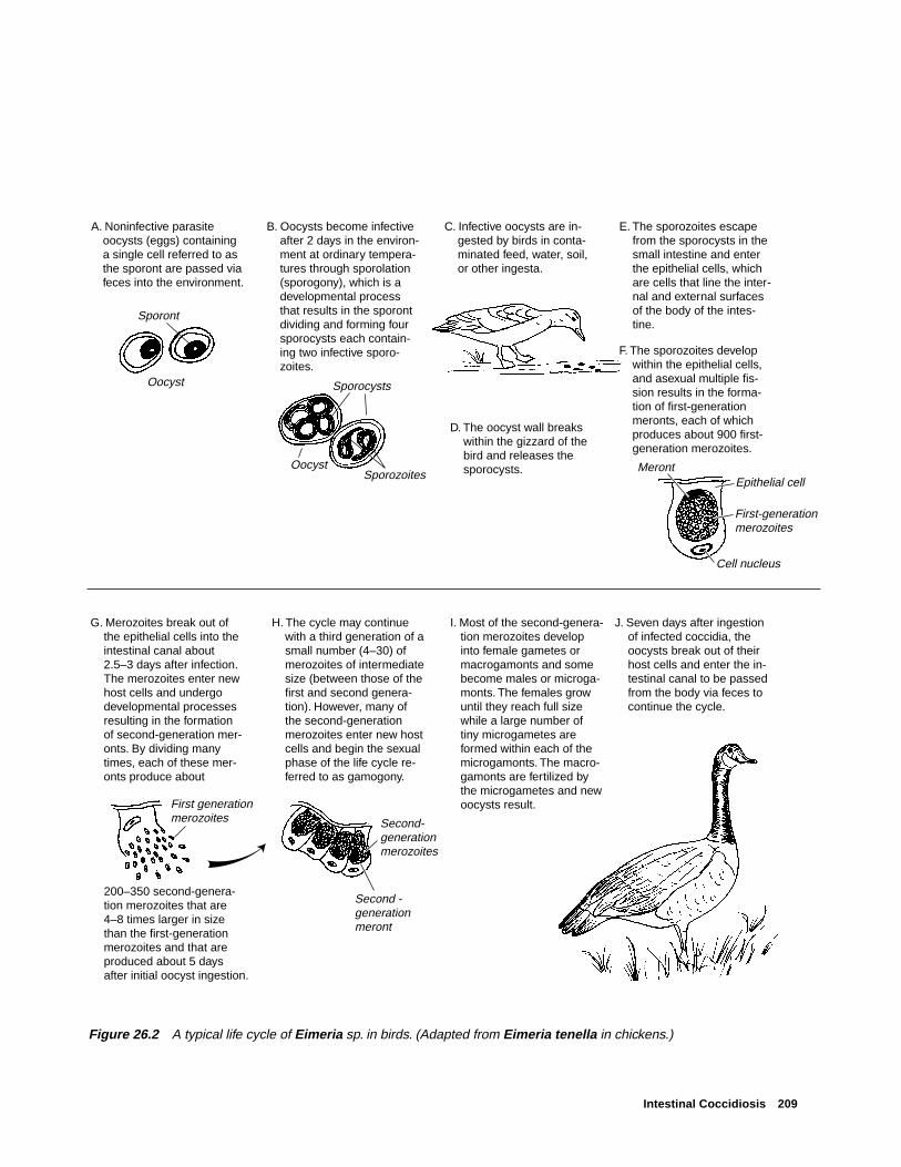
Intestinal Coccidiosis 209
A. Noninfective parasite oocysts (eggs) containing a single cell referred to as the sporont are passed via feces into the environment.
B. Oocysts become infective after 2 days in the environ-ment at ordinary tempera-tures through sporolation (sporogony), which is a developmental process that results in the sporont dividing and forming four sporocysts each contain-ing two infective sporo-zoites.
C. Infective oocysts are in-gested by birds in conta-minated feed, water, soil, or other ingesta.
D. The oocyst wall breaks within the gizzard of the bird and releases the sporocysts.
E. The sporozoites escape from the sporocysts in the small intestine and enter the epithelial cells, which are cells that line the inter-nal and external surfaces of the body of the intes-tine.
F. The sporozoites develop within the epithelial cells, and asexual multiple fis-sion results in the forma-tion of first-generation meronts, each of which produces about 900 first-generation merozoites.
G. Merozoites break out of the epithelial cells into the intestinal canal about 2.5–3 days after infection. The merozoites enter new host cells and undergo developmental processes resulting in the formation of second-generation mer-onts. By dividing many times, each of these mer-onts produce about
200–350 second-genera-tion merozoites that are 4–8 times larger in size than the first-generation merozoites and that are produced about 5 days after initial oocyst ingestion.
H. The cycle may continue with a third generation of a small number (4–30) of merozoites of intermediate size (between those of the first and second genera-tion). However, many of the second-generation merozoites enter new host cells and begin the sexual phase of the life cycle re-ferred to as gamogony.
I. Most of the second-genera-tion merozoites develop into female gametes or macrogamonts and some become males or microga-monts. The females grow until they reach full size while a large number of tiny microgametes are formed within each of the microgamonts. The macro-gamonts are fertilized by the microgametes and new oocysts result.
J. Seven days after ingestion of infected coccidia, the oocysts break out of their host cells and enter the in-testinal canal to be passed from the body via feces to continue the cycle.
Sporont
Oocyst
SporozoitesOocyst
Sporocysts
MerontEpithelial cell
Cell nucleus
First-generation merozoites
First generationmerozoites
Second -generationmeront
Second- generationmerozoites
Figure 26.2 A typical life cycle of Eimeria sp. in birds. (Adapted from Eimeria tenella in chickens.)

210 Field Manual of Wildlife Diseases: Birds
Table 26.1 Relative occurrence of coccidia in different groups of birds. [Frequency of occurrence: ● occasional, ● common,— not reported]
Bird types Coccidia species
(and examples) Eimeria sp. Isospora sp. Tyzzeria sp. Cryptosporidium sp. Wenyonella sp.
Poultry ● — — ● —(Chicken, turkey)
Anseriformes ● ● ● ● ●
(Ducks, geese)
Charadriiformes ● — — — —(Gulls, shorebirds)
Columbiformes ● — — — ●
(Pigeons, doves)
Coraciiformes — ● — — —(Kingfishers)
Falconiformes — ● — — —(Hawks, falcons)
Galliformes ● ● — ● —(Pheasant, quail)
Gruiformes ● — — — —(Cranes, rails)
Passeriformes — ● — — —(Songbirds)
Pelicaniiformes ● — — — —(Pelicans)
Piciformes — ● — — —(Woodpeckers)
Psittaciformes ● ● — ● —(Parrots)
Strigiformes — ● — — —(Owls)
Struthioniformes — ● — — —(Ostriches)
EXPLANATION
Intestinal coccidiosis outbreaks, by State
Figure 26.3 Location of outbreaks ofintestinal coccidiosis in waterfowl.
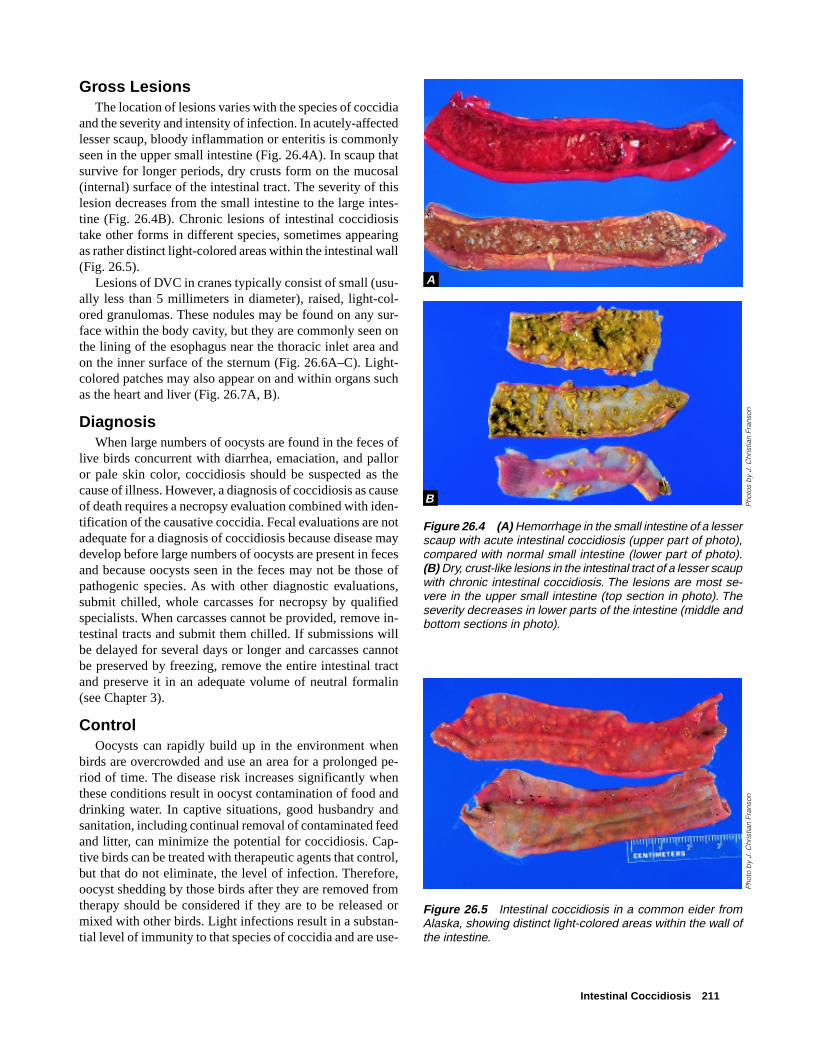
Intestinal Coccidiosis 211
Gross LesionsThe location of lesions varies with the species of coccidia
and the severity and intensity of infection. In acutely-affectedlesser scaup, bloody inflammation or enteritis is commonlyseen in the upper small intestine (Fig. 26.4A). In scaup thatsurvive for longer periods, dry crusts form on the mucosal(internal) surface of the intestinal tract. The severity of thislesion decreases from the small intestine to the large intes-tine (Fig. 26.4B). Chronic lesions of intestinal coccidiosistake other forms in different species, sometimes appearingas rather distinct light-colored areas within the intestinal wall(Fig. 26.5).
Lesions of DVC in cranes typically consist of small (usu-ally less than 5 millimeters in diameter), raised, light-col-ored granulomas. These nodules may be found on any sur-face within the body cavity, but they are commonly seen onthe lining of the esophagus near the thoracic inlet area andon the inner surface of the sternum (Fig. 26.6A–C). Light-colored patches may also appear on and within organs suchas the heart and liver (Fig. 26.7A, B).
DiagnosisWhen large numbers of oocysts are found in the feces of
live birds concurrent with diarrhea, emaciation, and palloror pale skin color, coccidiosis should be suspected as thecause of illness. However, a diagnosis of coccidiosis as causeof death requires a necropsy evaluation combined with iden-tification of the causative coccidia. Fecal evaluations are notadequate for a diagnosis of coccidiosis because disease maydevelop before large numbers of oocysts are present in fecesand because oocysts seen in the feces may not be those ofpathogenic species. As with other diagnostic evaluations,submit chilled, whole carcasses for necropsy by qualifiedspecialists. When carcasses cannot be provided, remove in-testinal tracts and submit them chilled. If submissions willbe delayed for several days or longer and carcasses cannotbe preserved by freezing, remove the entire intestinal tractand preserve it in an adequate volume of neutral formalin(see Chapter 3).
ControlOocysts can rapidly build up in the environment when
birds are overcrowded and use an area for a prolonged pe-riod of time. The disease risk increases significantly whenthese conditions result in oocyst contamination of food anddrinking water. In captive situations, good husbandry andsanitation, including continual removal of contaminated feedand litter, can minimize the potential for coccidiosis. Cap-tive birds can be treated with therapeutic agents that control,but that do not eliminate, the level of infection. Therefore,oocyst shedding by those birds after they are removed fromtherapy should be considered if they are to be released ormixed with other birds. Light infections result in a substan-tial level of immunity to that species of coccidia and are use-
Figure 26.4 (A) Hemorrhage in the small intestine of a lesserscaup with acute intestinal coccidiosis (upper part of photo),compared with normal small intestine (lower part of photo).(B) Dry, crust-like lesions in the intestinal tract of a lesser scaupwith chronic intestinal coccidiosis. The lesions are most se-vere in the upper small intestine (top section in photo). Theseverity decreases in lower parts of the intestine (middle andbottom sections in photo).
Pho
tos
by
J. C
hris
tian
Fran
son
Figure 26.5 Intestinal coccidiosis in a common eider fromAlaska, showing distinct light-colored areas within the wall ofthe intestine.
Pho
to b
y J.
Chr
istia
n Fr
anso
n
A
B

212 Field Manual of Wildlife Diseases: Birds
Figure 26.6 Gross lesions of dissemi-nated visceral coccidiosis of cranes. (A)Granulomas on the lining of the esopha-gus (arrows); and (B) in the area of thethoracic inlet [the tip of the forceps isbetween granulomas on the surface ofa vessel and nerve (left) and on the thy-roid gland (right)]; and (C) on the insidesurface of the sternum (arrow).
Pho
tos
by
Jam
es R
unni
ngen
➡
➡➡
A
B
C

Intestinal Coccidiosis 213
ful in preventing epizootics from this disease. Therefore, theobjective is not to completely eliminate infection with coc-cidia; instead, the focus should be on preventing heavyinfections and the establishment and persistence of highlevels of environmental contamination with coccidia. Forfree-ranging birds, flock dispersal may be warranted whenovercrowding continues for prolonged periods of time.
Human Health ConsiderationsNone. Coccidia of birds are not infectious for humans.
Milton Friend and J. Christian Franson
Figure 26.7 Lesions of disseminated vis-ceral coccidiosis also may include lightpatches as seen here on the (A), surfacesof the heart muscle and (B) on the liver(arrows).
Supplementary ReadingCarpenter, J.W., Novilla, M.N., Fayer, R., and Iverson, G.C.,
1984, Disseminated visceral coccidiosis in sandhill cranes:Journal of the American Veterinary Medical Association,v. 185, no. 11, p. 1,342–1,346.
Courtney, C.H., Forrester, D.J., Ernst, J.V., and Nesbitt, S.A.,1975, Coccidia of sandhill cranes, Grus canadensis: TheJournal of Parasitology, v. 61, no. 4, p. 695–699.
Novilla, M.N., Carpenter, J.W., Spraker, T.R., and Jeffers, T.K.,1981, Parenteral development of Eimerian coccidia in sandhilland whooping cranes: Journal of Protozoology, v., 28, no. 2,p. 248–255.
Parker, B.B., and Duszynski, D.W., 1986, Coccidiosis of sandhillcranes (Grus canadensis) wintering in New Mexico: Journal ofWildlife Diseases, v. 22, no. 1, p. 25–35.
Windingstad, R.M., McDonald, M.C., Locke, L.N., Kerr, S.M.,and Sinn, J.A., 1980, Epizootic of coccidiosis in free-flyinglesser scaup: Avian Diseases 24, p. 1,044–1,049.
Pho
to b
y Ja
mes
Run
ning
enP
hoto
by
J. C
hris
tian
Fran
son
➡
➡
A
B
➡
➡

214 Field Manual of Wildlife Diseases: Birds
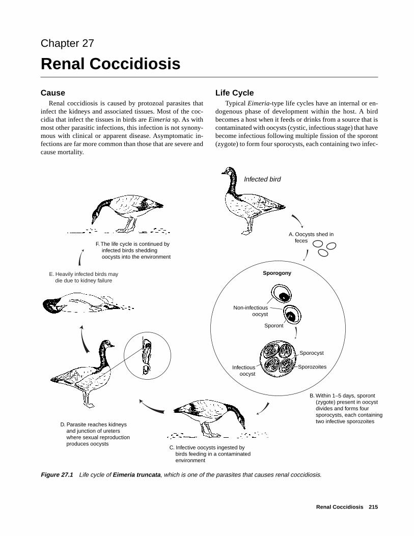
Renal Coccidiosis 215
Chapter 27
Renal Coccidiosis
CauseRenal coccidiosis is caused by protozoal parasites that
infect the kidneys and associated tissues. Most of the coc-cidia that infect the tissues in birds are Eimeria sp. As withmost other parasitic infections, this infection is not synony-mous with clinical or apparent disease. Asymptomatic in-fections are far more common than those that are severe andcause mortality.
Life Cycle Typical Eimeria-type life cycles have an internal or en-
dogenous phase of development within the host. A birdbecomes a host when it feeds or drinks from a source that iscontaminated with oocysts (cystic, infectious stage) that havebecome infectious following multiple fission of the sporont(zygote) to form four sporocysts, each containing two infec-
lnfected bird
Sporogony
Non-infectiousoocyst
Sporont
Sporozoites
Sporocyst
Infectiousoocyst
E. Heavily infected birds may die due to kidney failure
B. Within 1–5 days, sporont (zygote) present in oocyst divides and forms four sporocysts, each containing two infective sporozoites
F. The life cycle is continued by infected birds shedding oocysts into the environment
D. Parasite reaches kidneys and junction of ureters where sexual reproduction produces oocysts
C. Infective oocysts ingested by birds feeding in a contaminated environment
A. Oocysts shed in feces
Figure 27.1 Life cycle of Eimeria truncata, which is one of the parasites that causes renal coccidiosis.
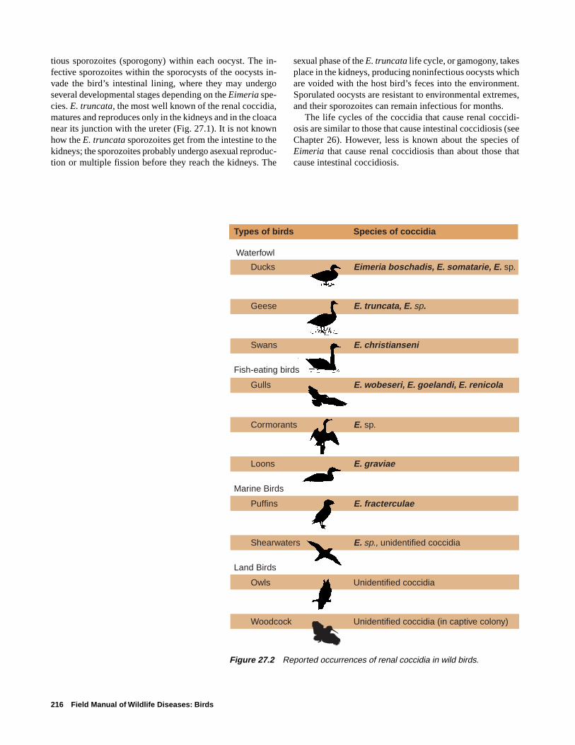
216 Field Manual of Wildlife Diseases: Birds
tious sporozoites (sporogony) within each oocyst. The in-fective sporozoites within the sporocysts of the oocysts in-vade the bird’s intestinal lining, where they may undergoseveral developmental stages depending on the Eimeria spe-cies. E. truncata, the most well known of the renal coccidia,matures and reproduces only in the kidneys and in the cloacanear its junction with the ureter (Fig. 27.1). It is not knownhow the E. truncata sporozoites get from the intestine to thekidneys; the sporozoites probably undergo asexual reproduc-tion or multiple fission before they reach the kidneys. The
sexual phase of the E. truncata life cycle, or gamogony, takesplace in the kidneys, producing noninfectious oocysts whichare voided with the host bird’s feces into the environment.Sporulated oocysts are resistant to environmental extremes,and their sporozoites can remain infectious for months.
The life cycles of the coccidia that cause renal coccidi-osis are similar to those that cause intestinal coccidiosis (seeChapter 26). However, less is known about the species ofEimeria that cause renal coccidiosis than about those thatcause intestinal coccidiosis.
Types of birds Species of coccidia
Waterfowl
Ducks Eimeria boschadis, E. somatarie, E. sp.
Geese E. truncata, E. sp.
Swans E. christianseni
Fish-eating birds
Gulls E. wobeseri, E. goelandi, E. renicola
Cormorants E. sp.
Loons E. graviae
Marine Birds
Puffins E. fracterculae
Shearwaters E. sp., unidentified coccidia
Land Birds
Owls Unidentified coccidia
Woodcock Unidentified coccidia (in captive colony)
Figure 27.2 Reported occurrences of renal coccidia in wild birds.

Renal Coccidiosis 217
Figure 27.3 Kidneys from double-crested cormorants. Top: normal sizeand color. Bottom: enlarged kidneys withdiffuse pale areas from a bird infectedwith renal coccidia.
Figure 27.4 Cut surfaces from thesame two kidneys as in Fig. 27.3. Bot-tom kidney shows chalky material frombuildup of uric acid salts.
Pho
to b
y J.
Chr
istia
n Fr
anso
nP
hoto
by
J. C
hris
tian
Fran
son
Species AffectedAvian coccidiosis was first reported in France. Canadian
investigators have reported that virtually all species of wildducks they examined are susceptible to renal coccidiosis. Dif-ferent species of renal coccidia are found in different spe-cies of birds (Fig. 27.2). Most reports of renal coccidiosisare of asymptomatic birds or birds that show minor physi-ological or pathological changes due to the parasite. Youngbirds and those that have been stressed by various condi-tions are most likely to have clinical cases of renal coccidi-osis. Mortality has occurred in free-ranging wild geese,eider ducklings, and double-crested cormorants. Disease indomestic geese is usually acute, lasts only 2–3 days, and cankill large segments of the flock.
DistributionRenal coccidiosis is found in birds worldwide.
SeasonalityMortality from renal coccidiosis is most common during
periods of the year when birds are densely aggregated ontheir breeding grounds or wintering areas.
Field SignsThere are no specific field signs that indicate that a bird is
infected with renal coccidia. Young birds will often be ema-ciated and weak, but many other diseases cause similar clini-cal signs.
Gross LesionsInfected birds may be emaciated and have a prominent
keel. In severe infections, kidneys may become enlarged andpale, containing multiple spots or foci of infection that coa-lesce into a mottled pattern (Fig. 27.3). Cutting through thesewhite foci may reveal material that has the consistency ofchalk due to the build up of uric acid salts (Fig. 27.4).

218 Field Manual of Wildlife Diseases: Birds
DiagnosisConfirmation of renal coccidiosis requires microscopic
examination of tissue by the trained staff of a diagnostic labo-ratory. Whole carcasses are generally needed to determinethe cause of death unless kidney damage is so severe that itunquestionably would have caused death. When whole re-frigerated carcasses cannot be provided for evaluation be-cause of field circumstances, the kidneys should be removed,preserved in a 10:1 volume of 10 percent buffered neutralformalin and submitted for diagnosis (see Chapter 2).
ControlControl of renal coccidiosis in free-ranging birds is not
feasible. Crowded conditions facilitate transmission of theparasite through fecal contamination of the environment.Prevention of degradation of habitat quantity and quality onbreeding grounds and wintering areas is needed to minimizedisease risks.
Human Health ConsiderationsThere are no reports of human health concerns with this
disease.
Rebecca A. Cole
Supplementary ReadingGajadhar, A.A., and Leighton, F.A., 1988, Eimeria wobeseri sp. n.
and Eimeria goelandi sp. n. (Protozoa: Apicomplexa) in thekidneys of herring gulls (Larus argentatus): Journal of WildlifeDiseases, v. 24, p. 538–546.
Oksanen, A., 1994, Mortality associated with renal coccidiosis injuvenile wild greylag geese (Anser anser anser): Journal ofWildlife Disease, v. 30, p. 554–556.
Wobeser, G., and Stockdale, P.H.G., 1983, Coccidia of domesticand wild waterfowl (Anseriformes): Canadian Journal ofZoology, v. 61, p. 1–24.

Sarcocystis 219
Chapter 28
Sarcocystis
SynonymsRice breast disease, sarcosporidiosis, sarcocystosis
CauseSarcocystis is a nonfatal, usually asymptomatic infection
that is caused by a parasitic protozoan. Various species ofthis parasite affect mammals, reptiles, and birds. The mostcommonly reported species of the parasite in North Americais Sarcocystis rileyi, the species most commonly found inwaterfowl.
Life CycleThe Sarcocystis sp. parasites have an indirect life cycle
(Fig. 28.1) that requires a paratentic or transport host animal(a bird), in which they live for a time before they are trans-
ported to a definitive host animal (a carnivore), in which theyreach maturity. Birds ingest the eggs or oocysts of the ma-ture parasite in food or water that is contaminated by carni-vore feces, which contain the oocysts. The oocysts developin the intestine of the bird into an intermediate form, thesporozoites, that enter the bird’s bloodstream and infect spe-cific cells of the blood vessels. Multiplication of these cellsgives rise to a second intermediate form, merozoites, thatare carried by the blood to the voluntary muscles, where elon-gated cysts or macrocysts are eventually produced (Fig. 28.2).The life cycle is completed when a carnivore ingests the in-fected muscle tissue of a bird and the parasite reaches matu-rity and releases oocysts in the intestines of the carnivore.The carnivore is infected only in its intestine. Macrocysts do
Figure 28.1 General life cycle ofSarcocystis sp.
Oocysts shed in carnivore feces
Cysts ingested by carnivore and parasite reaches maturity within the carnivore
Oocysts ingested by bird in contaminated food or water
Cysts develop in muscle tissues of bird following invasion by merozoites
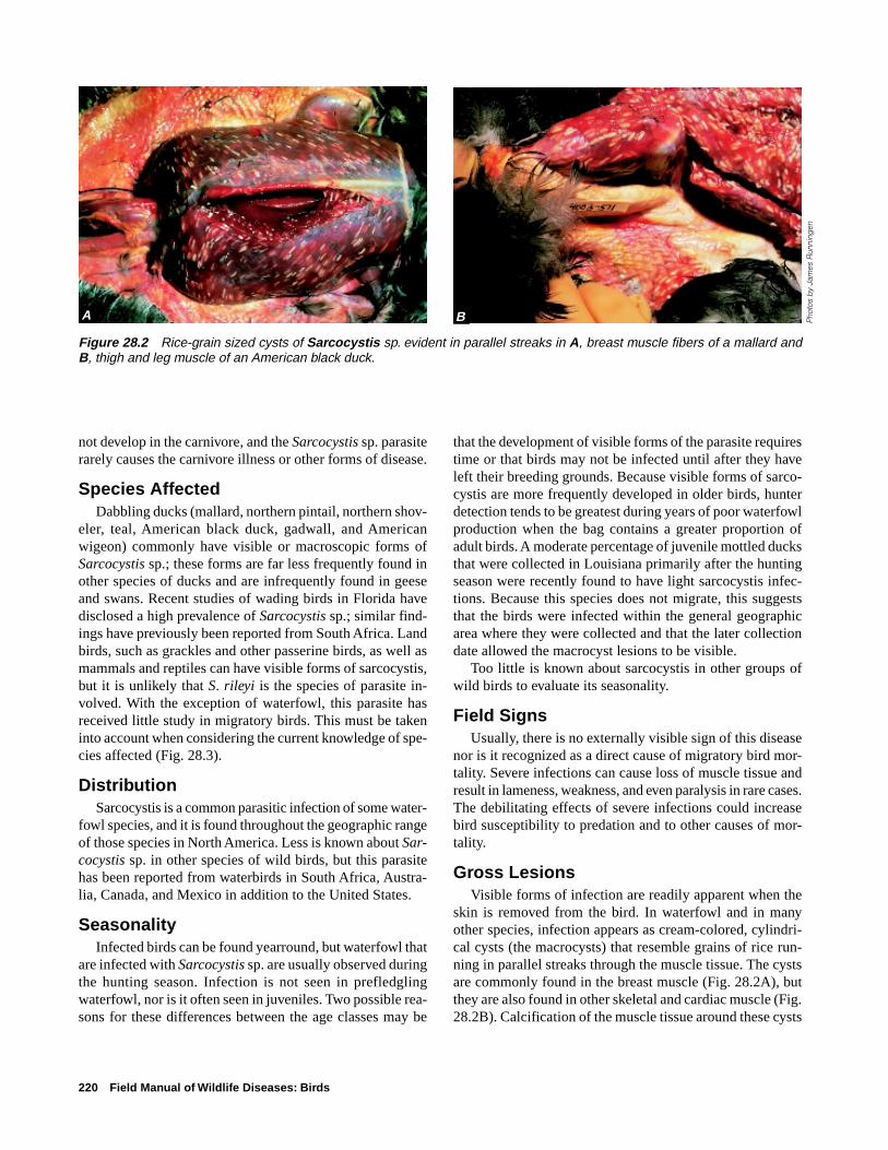
220 Field Manual of Wildlife Diseases: Birds
not develop in the carnivore, and the Sarcocystis sp. parasiterarely causes the carnivore illness or other forms of disease.
Species AffectedDabbling ducks (mallard, northern pintail, northern shov-
eler, teal, American black duck, gadwall, and Americanwigeon) commonly have visible or macroscopic forms ofSarcocystis sp.; these forms are far less frequently found inother species of ducks and are infrequently found in geeseand swans. Recent studies of wading birds in Florida havedisclosed a high prevalence of Sarcocystis sp.; similar find-ings have previously been reported from South Africa. Landbirds, such as grackles and other passerine birds, as well asmammals and reptiles can have visible forms of sarcocystis,but it is unlikely that S. rileyi is the species of parasite in-volved. With the exception of waterfowl, this parasite hasreceived little study in migratory birds. This must be takeninto account when considering the current knowledge of spe-cies affected (Fig. 28.3).
DistributionSarcocystis is a common parasitic infection of some water-
fowl species, and it is found throughout the geographic rangeof those species in North America. Less is known about Sar-cocystis sp. in other species of wild birds, but this parasitehas been reported from waterbirds in South Africa, Austra-lia, Canada, and Mexico in addition to the United States.
SeasonalityInfected birds can be found yearround, but waterfowl that
are infected with Sarcocystis sp. are usually observed duringthe hunting season. Infection is not seen in prefledglingwaterfowl, nor is it often seen in juveniles. Two possible rea-sons for these differences between the age classes may be
that the development of visible forms of the parasite requirestime or that birds may not be infected until after they haveleft their breeding grounds. Because visible forms of sarco-cystis are more frequently developed in older birds, hunterdetection tends to be greatest during years of poor waterfowlproduction when the bag contains a greater proportion ofadult birds. A moderate percentage of juvenile mottled ducksthat were collected in Louisiana primarily after the huntingseason were recently found to have light sarcocystis infec-tions. Because this species does not migrate, this suggeststhat the birds were infected within the general geographicarea where they were collected and that the later collectiondate allowed the macrocyst lesions to be visible.
Too little is known about sarcocystis in other groups ofwild birds to evaluate its seasonality.
Field SignsUsually, there is no externally visible sign of this disease
nor is it recognized as a direct cause of migratory bird mor-tality. Severe infections can cause loss of muscle tissue andresult in lameness, weakness, and even paralysis in rare cases.The debilitating effects of severe infections could increasebird susceptibility to predation and to other causes of mor-tality.
Gross LesionsVisible forms of infection are readily apparent when the
skin is removed from the bird. In waterfowl and in manyother species, infection appears as cream-colored, cylindri-cal cysts (the macrocysts) that resemble grains of rice run-ning in parallel streaks through the muscle tissue. The cystsare commonly found in the breast muscle (Fig. 28.2A), butthey are also found in other skeletal and cardiac muscle (Fig.28.2B). Calcification of the muscle tissue around these cysts
Figure 28.2 Rice-grain sized cysts of Sarcocystis sp. evident in parallel streaks in A, breast muscle fibers of a mallard andB, thigh and leg muscle of an American black duck.
Pho
tos
by
Jam
es R
unni
ngen
BA

Sarcocystis 221
makes them obviously discrete bodies. The degree of calci-fication is often sufficient to give a gritty feeling to the tissuewhen it is cut with a knife.
Lesions that were observed in wading birds differed inappearance; the cysts were white and opaque, and they gen-erally extended throughout the entire length of the infectedmuscle fiber. Cysts were present in the heart muscle and theywere confined to striated muscles.
DiagnosisThe visible presence of sarcosporidian cysts in muscle
tissue is sufficient to diagnose this disease. Visible cysts mayvary in size and shape in different bird species. Good qualitycolor photographs (prints or 35 millimeter slides) of the ex-ternal surface of infected muscle are generally sufficient fora disease specialist to recognize this disease if tissues or awhole carcass cannot be provided. Whole birds should besubmitted if possible. If only tissues can be submitted, thena portion of the infected muscle should be fixed in a 10 per-cent formalin solution. Frozen muscle tissue is also suitablefor diagnosis, and the distinctive appearance of these cystsallows a diagnosis from even partially decomposed carcasses.
ControlThere are no known control methods for this disease, nor
do any seem to be needed or are any being developed. Con-trol of sarcocystis would require interruption of the life cycleof the parasite. Although the life cycles of the Sarcocystissp. that affect wild birds are not precisely known, they areprobably similar to the two-host, indirect life cycle knownfor some other Sarcocystis sp. (Fig. 28.1). The predator-preyrelationship between the intermediate bird hosts and the de-finitive carnivore hosts may be the primary reason that juve-nile birds or some bird species are seldom found to be in-fected. The appropriate carnivores may not be present on thebreeding grounds.
Different species of carnivores seem to be involved in theinfection of different bird species, which suggests that birdsare infected by more than one species of the genus Sarco-cystis sp. If the carnivore-bird cycle is species-specific, thatis, if a specific species of bird can only be infected by oo-cysts that are produced by a parasite in a specific carnivorespecies, then selective control of sarcocystis might be fea-sible. However, current knowledge of the disease does notindicate a need to initiate control because there is little evi-dence that bird health is often compromised by infection.Nevertheless, the role of carnivores in the life cycle of Sar-cocystis sp. infections should be considered when feeding
Figure 28.3 Relative frequency of grossly visible forms ofsarcocystis in selected groups of North American migratorybirds.
Puddle ducks
Grackles
Wading birds
Diving ducks
Mergansers
Sea ducks
Geese
Swans
Shorebirds
Gulls and terns
Pelicans
Raptors
FrequentCommon
OccasionalRare
222 Field Manual of Wildlife Diseases: Birds
uncooked, infected waterfowl to house pets and to farm ani-mals such as hogs.
Human Health ConsiderationsSarcocystis sp. presents no known health hazard to hu-
mans. The primary importance to humans of sarcocystis inwaterfowl is the loss of infected birds for food; the unaes-thetic appearance of parasitized muscle may prompt huntersto discard the carcass. Limited evaluations of hunter responsesto infected carcasses indicate no reduction in carcass con-sumption in areas where the infection is commonly seen.Also, the recognized high prevalence of infection in north-ern shovelers in some areas results in this species often be-ing left unretrieved by some hunters and focuses additionalhunting pressure on other species.
Benjamin N. Tuggle and Milton Friend(Modified from and earlier chapter by Benjamin N. Tuggle)
Supplementary ReadingCawthorn, R.J., Rainnie, D., and Wobeser, G.A., 1981, Experi-
mental transmission of Sarcocystis sp. (Protozoa:Sarcocystidae) between the shoveler (Anas clypeata) duck andthe striped skunk (Mephitis mephitis): Journal of WildlifeDiseases, v. 17, p. 389–394.
Cornwell, G, 1963, New waterfowl host records for Sarcocystisrileyi and a review of sarcosporidiosis in birds: Avian Disease,v. 7, p. 212–216.
Moorman, T.E., Baldassarre, G.A., and Richard, D.M., 1991, Thefrequency of Sarcocystis spp. and its effect on winter carcasscomposition of mottled ducks: Journal of Wildlife Disease,v. 27, no. 3, p. 491–493.
Spalding, M.G., Atkinson, C.T., and Carleton, R.E., 1994,Sarcocystis sp. in wading birds (Ciconiiformes) from Florida:Journal of Wildlife Disease, v. 30, no. 1, p. 29–35.

Eustrongylidosis 223
Chapter 29
EustrongylidosisSynonymsVerminous peritonitis
CauseEustrongylidosis is caused by the nematodes or round-
worms Eustrongylides tubifex, E. ignotus, and E. excisus.Eustrongylides sp. can cause large die-offs of nestlings incoastal rookeries, especially of egrets and other wading birds.
Life CycleThe three species of Eustrongylides that cause disease in
birds have similar indirect life cycles that require two inter-mediate hosts (Fig. 29.1). Four developmental stages of theparasite are required from egg to sexually mature worm. Thefirst larval stage develops within the eggs that are shed in thefeces of the bird host and are eaten by freshwater oligocha-etes or aquatic worms. The oligochaetes serve as the firstintermediate host. The eggs hatch within the oligochaetes,where they develop into second- and third-stage larvae. Min-nows and other small fish, such as species of Fundulus andGambusia, feed upon the infected oligochaetes and serve asthe second intermediate host. The third-stage larvae becomeencapsulated on the internal surface areas of the fish, de-velop into infective fourth-stage larvae, and await ingestionby birds. Predatory fish, which consume infected fish, canserve as paratenic or transport hosts when they are fed uponby birds. Amphibians and reptiles have also been reported assecond-stage intermediate hosts and serve as paratenic hosts.Larvae that are infective for birds can penetrate the ventricu-lus (stomach) within 3–5 hours after a bird ingests an inter-mediate or paratenic host, and the larvae quickly becomesexually mature worms that begin shedding eggs 10–17 dayspostinfection.
Species AffectedE. tubifex has been reported from four different bird fami-
lies, E. ignotus from three, and E. excisus from three (Fig.29.2). Young wading birds are the most common species tohave large mortalities from eustrongylidosis (Table 29.1).Eustrongylides sp. have also been reported in birds of prey.
DistributionEustrongylides sp. have been reported from birds through-
out much of the world. E. tubifex and E. ignotus are the spe-cies reported within the United States (Table 29.2).Eustrongylid infections within the United States have beenreported from many areas (Fig. 29.3). Typical rookeries wherebirds are infected with Eustrongylides sp. are found in coastal
areas and consist of dense populations of birds nesting onlow islands, often surrounded by canals or ditches. Nestinghabitat often includes stands of low trees, such as willows,with an understory that may be submergent, semisubmergent,or upland mixed-prairie species. Inland rookeries are usu-ally adjacent to lakes or rivers, and nesting trees, particu-larly those used by great blue herons, may be much higherthan those in coastal rookeries. Several wading bird speciesmay nest in these areas, but typically one or two species ac-count for most of the birds in the rookery (Fig. 29.4).
SeasonalityBirds can harbor infections yearround. Mortality usually
is reported in spring and summer and birds less than 4 weeksold are more likely to die than adults. Disease in older birdstends to be of a more chronic nature and infection may beseen at any time of the year.
Field SignsDisease results in a variety of clinical or apparent signs
that are not specific to eustrongylidosis. However, consider-ation of the species affected, the age class of birds involved,and the full spectrum of signs may suggest that eustrongyli-diosis is the cause of mortality. Very early in the infection asthe worm is penetrating the ventriculus, some birds will shaketheir heads, have difficulty swallowing, have dyspnea or dif-ficult or labored breathing and, occasionally, regurgitate theirfood. Anorexia or loss of appetite has been noted in experi-mentally infected nestlings. It has been speculated that anor-exia in combination with sibling competition for food maycontribute to the emaciation seen in naturally infected birds.Infected nestlings also may wander from the nest predisposedto predation or trauma or both. Affected nestlings observedduring one mortality event became progressively weakenedand showed abdominal swelling. Palpation of worms on theventriculus has been useful for detecting infection in livenestlings.
Gross LesionsBirds that have been recently infected often have large,
tortuous, raised tunnels that are visible on the serosal sur-face of the proventriculus, ventriculus, or intestines (Fig.29.5A). The nematodes reside within these tunnels, whichare often encased with yellow, fibrous material, and main-tain openings to the lumen of the organ so that parasite eggsmay be passed out with feces into the environment. A fibrino-peritonitis or fibrin-coated inflammation of the surfaces ofthe peritoneal cavity (the area containing the organs below

224 Field Manual of Wildlife Diseases: Birds
Figure 29.1 Life cycle of Eustrongylides sp.
Birds feed on infected transport hosts. Infective larvae reach sexual maturity within bird host
First stage larvae develop in eggs eaten by oligochaetes(freshwater aquatic worms)
First intermediate host
Second intermediate host
Transport (paratenic hosts)
Minnows and other small fish feed upon oligochaetes
Infected minnows are fed upon by species other than birds
Third stage larvae become encapsulated within body of fish
Bird feeds on infected fishthen larvae develop tosexual maturity
Definitive bird host
Bird sheds parasite eggs into the environ-ment in feces
Eggs hatch within oligochaetes; second-and third-stage larvae are produced within oligochaetes
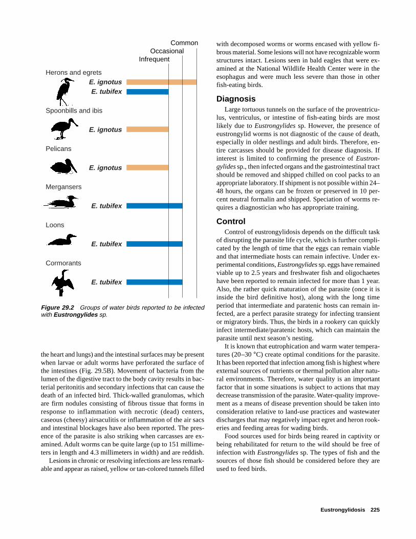
Eustrongylidosis 225
Herons and egretsE. ignotusE. tubifex
Spoonbills and ibis
E. ignotus Pelicans
E. ignotus Mergansers
E. tubifex
Loons
E. tubifex
Cormorants
E. tubifex
CommonOccasional
Infrequent
Figure 29.2 Groups of water birds reported to be infectedwith Eustrongylides sp.
the heart and lungs) and the intestinal surfaces may be presentwhen larvae or adult worms have perforated the surface ofthe intestines (Fig. 29.5B). Movement of bacteria from thelumen of the digestive tract to the body cavity results in bac-terial peritonitis and secondary infections that can cause thedeath of an infected bird. Thick-walled granulomas, whichare firm nodules consisting of fibrous tissue that forms inresponse to inflammation with necrotic (dead) centers,caseous (cheesy) airsaculitis or inflammation of the air sacsand intestinal blockages have also been reported. The pres-ence of the parasite is also striking when carcasses are ex-amined. Adult worms can be quite large (up to 151 millime-ters in length and 4.3 millimeters in width) and are reddish.
Lesions in chronic or resolving infections are less remark-able and appear as raised, yellow or tan-colored tunnels filled
with decomposed worms or worms encased with yellow fi-brous material. Some lesions will not have recognizable wormstructures intact. Lesions seen in bald eagles that were ex-amined at the National Wildlife Health Center were in theesophagus and were much less severe than those in otherfish-eating birds.
DiagnosisLarge tortuous tunnels on the surface of the proventricu-
lus, ventriculus, or intestine of fish-eating birds are mostlikely due to Eustrongylides sp. However, the presence ofeustrongylid worms is not diagnostic of the cause of death,especially in older nestlings and adult birds. Therefore, en-tire carcasses should be provided for disease diagnosis. Ifinterest is limited to confirming the presence of Eustron-gylides sp., then infected organs and the gastrointestinal tractshould be removed and shipped chilled on cool packs to anappropriate laboratory. If shipment is not possible within 24–48 hours, the organs can be frozen or preserved in 10 per-cent neutral formalin and shipped. Speciation of worms re-quires a diagnostician who has appropriate training.
ControlControl of eustrongylidosis depends on the difficult task
of disrupting the parasite life cycle, which is further compli-cated by the length of time that the eggs can remain viableand that intermediate hosts can remain infective. Under ex-perimental conditions, Eustrongylides sp. eggs have remainedviable up to 2.5 years and freshwater fish and oligochaeteshave been reported to remain infected for more than 1 year.Also, the rather quick maturation of the parasite (once it isinside the bird definitive host), along with the long timeperiod that intermediate and paratenic hosts can remain in-fected, are a perfect parasite strategy for infecting transientor migratory birds. Thus, the birds in a rookery can quicklyinfect intermediate/paratenic hosts, which can maintain theparasite until next season’s nesting.
It is known that eutrophication and warm water tempera-tures (20–30 °C) create optimal conditions for the parasite.It has been reported that infection among fish is highest whereexternal sources of nutrients or thermal pollution alter natu-ral environments. Therefore, water quality is an importantfactor that in some situations is subject to actions that maydecrease transmission of the parasite. Water-quality improve-ment as a means of disease prevention should be taken intoconsideration relative to land-use practices and wastewaterdischarges that may negatively impact egret and heron rook-eries and feeding areas for wading birds.
Food sources used for birds being reared in captivity orbeing rehabilitated for return to the wild should be free ofinfection with Eustrongylides sp. The types of fish and thesources of those fish should be considered before they areused to feed birds.
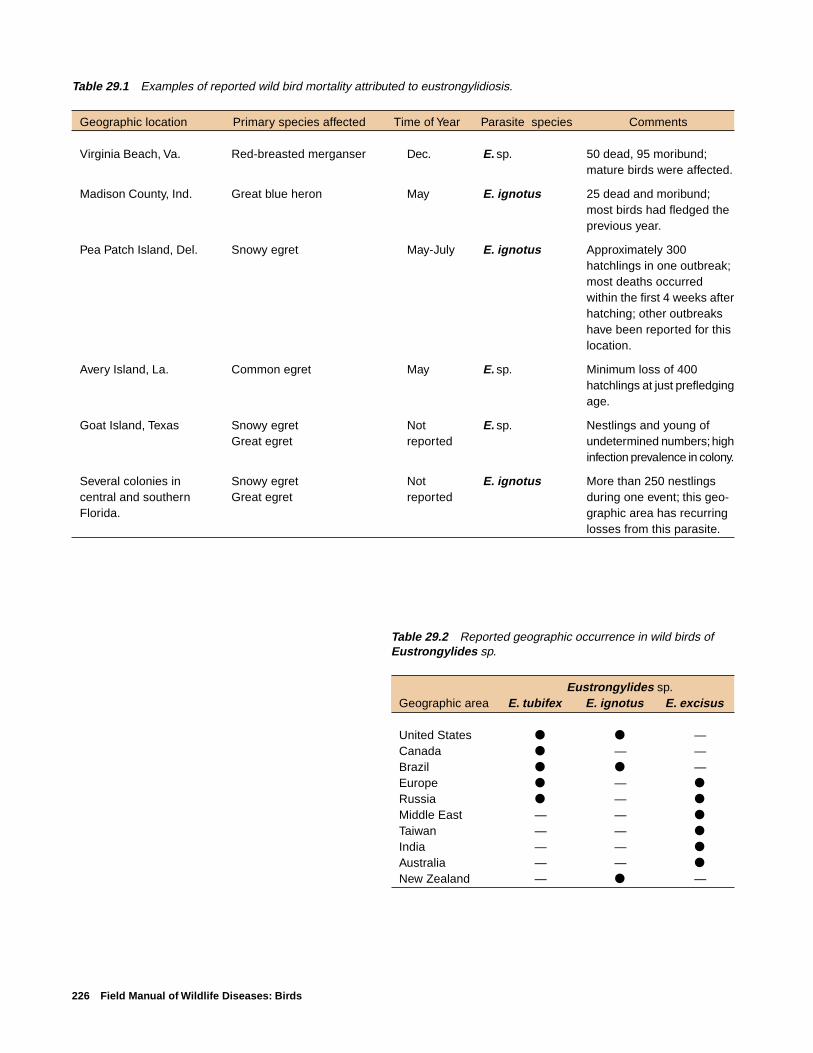
226 Field Manual of Wildlife Diseases: Birds
Table 29.2 Reported geographic occurrence in wild birds ofEustrongylides sp.
Eustrongylides sp.Geographic area E. tubifex E. ignotus E. excisus
United States ● ● —Canada ● — —Brazil ● ● —Europe ● — ●
Russia ● — ●
Middle East — — ●
Taiwan — — ●
India — — ●
Australia — — ●
New Zealand — ● —
Table 29.1 Examples of reported wild bird mortality attributed to eustrongylidiosis.
Geographic location Primary species affected Time of Year Parasite species Comments
Virginia Beach, Va. Red-breasted merganser Dec. E. sp. 50 dead, 95 moribund;mature birds were affected.
Madison County, Ind. Great blue heron May E. ignotus 25 dead and moribund;most birds had fledged theprevious year.
Pea Patch Island, Del. Snowy egret May-July E. ignotus Approximately 300hatchlings in one outbreak;most deaths occurredwithin the first 4 weeks afterhatching; other outbreakshave been reported for thislocation.
Avery Island, La. Common egret May E. sp. Minimum loss of 400hatchlings at just prefledgingage.
Goat Island, Texas Snowy egret Not E. sp. Nestlings and young ofGreat egret reported undetermined numbers; high
infection prevalence in colony.
Several colonies in Snowy egret Not E. ignotus More than 250 nestlingscentral and southern Great egret reported during one event; this geo-Florida. graphic area has recurring
losses from this parasite.

Eustrongylidosis 227
EXPLANATION
Reports of Eustrongylides sp. in birds, by State
Infections reported in birds
Major mortality events reported
Figure 29.3 States where Eustrongylides sp. infections in wild birds have been reported.
Figure 29.4 Although many species may nest in wading bird rookeries, one or two species are oftenpredominant.
Pho
to b
y J.
Chr
istia
n Fr
anso
n

228 Field Manual of Wildlife Diseases: Birds
Human Health ConsiderationsHumans who have consumed raw or undercooked fish that
carry the larval stages of the parasite have experienced gas-tritis or inflammation of the stomach and intestinal perfora-tion requiring surgical removal of worms.
Rebecca A. Cole
Supplementary ReadingFranson, J.C. and Custer, T.W., 1994, Prevalence of
eustrongylidosis in wading birds from colonies in California,Texas, and Rhode Island, United States of America: ColonialWaterbirds, v. 17, p. 168–172.
Measures, L.N., 1988, Epizootiology, pathology and descriptionof Eustrongylides tubifex (Nematoda: Dioctophymatoidea) infish: Canadian Journal of Zoology, v. 66: p . 2212–2222.
Spalding, M.A., Bancroft, G.T., and Forrester, D.J., 1993, Theepizootiology of eustrongylidosis in wading birds(Ciconiiformes) in Florida: Journal of Wildlife Diseases, v. 29,p. 237–249.
Spalding, M.A., and Forrester, D.J., 1993, Pathogenesis ofEustrongylides ignotus (Nematoda: Dioctophymatoidea) inCiconiiformes: Journal of Wildlife Diseases, v. 29, p. 250–260.
Wiese, J.H., Davidson, W.R., and Nettles, V.F., 1977, Large scalemortality of nestling ardeids caused by nematode infection:Journal of Wildlife Diseases, v. 13, no. 4, p. 376–382.
Pho
to b
y Ja
mes
Run
ning
en
Figure 29.5 (A) Raised tunnels caused by Eustrongylidessp. on intestines of a snowy egret. (B) The debris on the intes-tinal surfaces of this snowy egret is characteristic of the peri-tonitis often caused by Eustrongylides sp. infection.
Pho
to b
y J.
Chr
istia
n Fr
anso
n
A
B

Tracheal Worms 229
Chapter 30
Tracheal Worms
SynonymsGape worm, syngamiasis, gapes
CauseInfection by tracheal worms often results in respiratory
distress due to their location in the trachea or bronchi andtheir obstruction of the air passage. Infections by these para-sitic nematodes or roundworms in waterbirds, primarilyducks, geese, and swans, are usually due to Cyathostomabronchialis and infection of land birds are usually due toSyngamus trachea. However, both genera infect a variety ofspecies, including both land and waterbirds. Infections withS. trachea have been more extensively studied than infec-tions with Cyathostoma sp. because of its previous impor-tance as a disease-causing parasite of poultry in many partsof the world. Changes in husbandry practices to modern in-tensive methods for poultry production have essentially elimi-nated S. trachea as an agent of disease in chickens, but it isan occasional cause of disease in turkeys raised on range.
Life CycleTracheal worms have an indirect life cycle (Fig. 30.1) that
requires a paratenic or transport host which transmits the in-fectious larvae to the definitive host bird, where they reachadulthood and reproduce. Adult S. trachea reside within thetrachea. The female releases fertilized eggs, which are swal-lowed by the bird and voided with the feces into the soil.Eggs may also be directly expelled onto the ground from thetrachea. After embryonation (1–2 weeks), infective larvaedevelop within the egg. Birds can become infected by eatinginvertebrate paratenic hosts such as earthworms, snails, slugs,or fly larvae that have consumed the eggs. Infective larvaeare released from the egg and become encysted within thebodies of these invertebrates and can remain infective for upto three and one-half years. Upon ingestion by birds, the lar-vae are believed to penetrate the intestinal wall. Some larvaeenter the abdominal cavity but most enter the bloodstream,where they are carried to the lungs. After further develop-ment in the lungs, the young worms migrate up the bronchito the trachea. Larvae can reach the lungs within 6 hoursafter ingestion and eggs are produced by worms in the tra-chea about 2 weeks after ingestion of those larvae.C. bronchialis is very similar in that earthworms transmitthe infective stage to the bird. Infection of birds withC. bronchialis by direct consumption of fully embryonatedeggs has been documented experimentally; however, wormburdens were extremely low.
Species AffectedDisease caused by tracheal worms is not commonly re-
ported for free-ranging birds within the United States andCanada, but it is common within the United Kingdom and insome other countries. High infection rates within wild birdsin England attest to the potential for this parasite to be aserious pathogen. More than 50 percent of nestling and fledg-ling starlings, more than 85 percent of jackdaws, and 100percent of young rooks were found to be infected in one study.Infection rates in adult birds were considerably lower, butthey still exceeded 30 percent for starlings and rooks. Withinthe United States, S. trachea infections have been reportedfrom wild turkeys, other gamebirds, a variety of passerines,(songbirds), and occasionally from other bird species. Large-scale mortalities have occurred among pheasants and othergamebirds being propagated for sporting purposes. Findingsfrom captive bird collections have led to the conclusion thatalmost any species of cage or aviary bird is susceptible toinfection.
S. trachea has been reported infrequently in waterfowl,but members of the genus Cyathostoma sp. are “characteris-tic” or common parasites of waterfowl. Mortality has beenreported for several species of young geese, leading someinvestigators to suggest that C. bronchialis are potentiallyimportant pathogens for geese. Juvenile free-ranging sand-hill cranes have also been reported to have died fromCyathostoma sp. infection.
DistributionS. trachea and Cyathostoma sp. are found worldwide.
SeasonalityInfected birds can be found yearround. Young birds are
most commonly affected and, therefore, disease is associ-ated with breeding cycles in the spring to summer monthsfor free-ranging birds.
Field SignsMost birds that are infected show no signs of disease. In
general, the severity of disease is dependent upon the degreeof infection and the size of the bird. Small birds are moreseverely affected than larger birds because their narrowertracheal openings result in greater obstruction by the worms.Respiratory distress is the primary clinical sign of disease.Birds with severe infections open their mouth widely and atthe same time stretch out their necks, assuming a “gaping”posture. The adult worms that are attached to the lining of

230 Field Manual of Wildlife Diseases: Birds
6. Bird eats infected invertebrate and larvae penetrate intestinal wall and enter blood stream
Transport hosts
Infected bird
1. Adult worms produce eggs by sexual reproduction within trachea
8. Young worms migrate up the bronchi to the trachea
2. Female worm releases fertilized eggs, which are swallowed by bird and voided with feces
4. Eggs are eaten by earthworms, snails, slugs, or fly larvae
3. Infective larvae develop within egg and hatch
7. Larvae are carried to lungs via blood stream where larvae undergo further development
5. Infective larvae hatch and be-come encysted within bodies of invertebrates
Figure 30.1 Tracheal worm life cycle.

Tracheal Worms 231
the trachea cause irritation and excess mucus production. Thisoften results in agitated bouts of coughing, head shaking,and sneezing as the birds attempt to dislodge the parasites.Severely infected birds may have most or all of the trachealopening obstructed by worms, may stop feeding, and mayrapidly lose body condition.
Gross LesionsSeverely affected birds experience severe weight loss and
have poorer development of body mass than uninfected birds,and they often die from starvation (Fig. 30.2). Anemia mayalso be present due to the blood-feeding habits of the para-sites.
DiagnosisIdentification of the worms (Fig. 30.3) and evaluation of
any associated disease signs are required for a diagnosis.Clinical signs are not diagnostic because similar signs canbe seen with some mite infections, aspergillosis, and wet pox.
ControlThere is no feasible method for controlling tracheal worms
in free-ranging birds. Disease prevention should be practicedby minimizing the potential for captive-propagation and re-lease programs to infect invertebrates that are then fed uponby free-ranging birds. Land-use practices that provide directcontact between poultry rearing and wild birds and the dis-posal of bird feces and litter should also be considered be-cause environmental contamination with infective larvae isa critical aspect of the disease cycle.
Human Health ConsiderationsThere are no reports of these nematodes infecting humans.
Rebecca A. Cole
Supplementary ReadingAnderson, R.C., 1992, Nematode parasites of vertebrates: Their
development and transmission: Wallingford, England, CABInternational, 578 p.
Cram, E.B., 1927, Bird parasites of the nematode subordersStrongylata, Ascaridata, and Spirurata: U.S. National MuseumBulletin No. 140, 465 p.
Fernando, M.A., Hoover, I.J., and Ogungbade, S.G., 1973, Themigration and development of Cyathostoma bronchialis ingeese: Journal of Parasitology, v. 59, p. 759–764.
Threlfall, W., 1965, Life-cycle of Cyathostoma lari: Nature,v. 206, p. 1,167–1,168.
Figure 30.3 Syngamus trachea in the trachea of a ring-necked pheasant.
Figure 30.2 Comparative breast muscle mass of noninfectedpheasants (left) and those with Syngamus trachea infections(right).
Pho
to b
y M
ilton
Frie
ndP
hoto
by
Milt
on F
riend

232 Field Manual of Wildlife Diseases: Birds

Heartworm of Swans and Geese 233
Chapter 31
Heartworm of Swans and Geese
SynonymsFilarial heartworm, Sarconema, Sarconema eurycerca
CauseHeartworm in swans and geese is caused by a filarial
nematode or a roundworm of the superfamily Filarioideawhich is transmitted to the bird by a biting louse. The nema-tode and the louse both are parasites. Sarconema eurycercais the only one of several species of microfilaria or the firststage juvenile of the parasite found in the circulating bloodof waterfowl that is known to be pathogenic or cause clinicaldisease.
Life cycleSarconema eurycerca has an indirect life cycle (Fig. 31.1)
that requires the parasite larvae to develop in an intermedi-ate host before they can become infective for and be trans-mitted to a definitive host, where they mature and reproduce.Female adult heartworms release microfilariae into the blood-stream of the definitive host bird. The microfilariae infect abiting louse, Trinoton anserinum, that subsequently feedsupon the bird. The larvae go through three stages of devel-opment within the louse, and the third stage is infectious tobirds. A new host bird becomes infected when the louse bitesit to feed on its blood and the third-stage larvae move into
Worms migrate through the bloodstream to the heart muscle where they mature and release micro-filariae into the blood-stream
Life cycle is continued when biting lice feed upon the bird's blood containing the microfilariae
Three stages of larval development occur within the biting louse
Infective third-stage larvae are transmitted into the bird's blood-stream when the louse takes another blood meal Microfilariae
Figure 31.1 Indirect life cycle of Sarconema eurycerca.

234 Field Manual of Wildlife Diseases: Birds
the bird’s bloodstream. The larvae migrate through the blood-stream to the myocardium, which is the middle and thickestlayer of the heart wall composed of cardiac muscle. Theyare nourished by and develop to sexual maturity within themyocardium. The cycle continues as this next generation ofmature heartworms release microfilariae into the blood-stream.
Infection with the parasite is not synonymous with dis-ease; that is, the parasite may infect and develop within thebird but not debilitate it.
Species AffectedSarconema eurycerca was first identified from a tundra
swan (whistling swan) in the late 1930s. It has since beenreported from trumpeter, Bewick’s, and mute swans and, fromCanada, snow, white-fronted, and bean geese. Varying per-centages of swans (4–20 percent) have been found to be in-fected on the basis of blood smears that were taken fromapparently healthy birds during field surveys. Canadian in-vestigators have reported a prevalence of approximately 10percent of snow geese that were examined at necropsy andwhich had died from other causes. This parasite has not re-ceived sufficient study for its full host range, its relative fre-quency of occurrence in different species, or its significanceas a mortality factor for wild birds to be determined.
DistributionHeartworm is found throughout the range of its swan and
goose hosts.
SeasonalityIt is suspected that while swans and geese are on the breed-
ing grounds, louse infestation and colonization on birds isprevalent. Therefore, the possibility of infection by heart-worm is highest while birds are on the breeding grounds.
Field SignsField signs are not always present in infected birds, and
infection cannot be determined by the presence of clinicalsigns alone. Chronic types of debilitating diseases, such aslead poisoning, may exacerbate louse infestation becausebirds become lethargic and do not preen. No specific fieldsign is diagnostic for infection.
Gross LesionsThe severity of infection dictates the lesions that are seen
at necropsy. Birds may be emaciated or in comparably goodflesh. The heart may be enlarged and have pale foci or spotswithin the myocardium. The thin, long thread-like wormsmay be visible under the surface layer or epicardium of theheart or the worms may be embedded within the deepermuscle tissue of the myocardium (Fig. 31.2).
DiagnosisA diagnosis of heartworm as the cause of death must be
supported by pathologic lesions seen during examination ofthe heart tissues with a microscope and consideration of othercauses. Therefore, whole carcasses should be submitted fordiagnostic assessments. If the transit time is short enough toavoid significant decomposition of the carcass and if the car-cass can be kept chilled during transit, then chilled wholecarcasses should be submitted to qualified disease diagnos-tic laboratories. If those conditions cannot be met, then car-casses should be submitted frozen.
ControlControl of heartworm is not practical for free-ranging
birds. Decreasing the opportunity for heavy infestation ofthe louse intermediate host will result in reduced opportu-nity for heartworm infection.
Human Health ConsiderationsSarconema eurycerca has not been reported to infect humans.
Rebecca A. Cole
Supplementary ReadingCohen, M., Greenwood, T., and Fowler, J.A., 1991, The louse
Trinoton anserinum (Amblycerca: Pthiraptera), an intermedi-ate host of Sarconema eurycerca (Filarioidea: Nematoda), aheartworm of swans: Medical and Veterinary Entomology, v. 5,p. 101–110.
Scheller, E.L., Sladen, W.L., and Trpis, M., 1976, A Mallophaga,Trinoton anserium, as a cyclodevelopmental vector for aheartworm parasite of waterfowl: Science, v. 194, p. 739–740.
Seegar, W.S., 1979, Prevalence of heartworm, Sarconemaeurycerca, Wehr, 1939 (Nematoda), in whistling swan, Cygnuscolumbianus columbianus: Canadian Journal of Zoology, v. 57,p. 1,500–1,502.
Figure 31.2 Heartworms (arrows) on the inner surface ofthe heart of a tundra swan.
Pho
to b
y Ja
mes
Run
ning
en➡
➡

Gizzard Worms 235
Chapter 32
Gizzard Worms
SynonymsStomach worm, ventricular nematodiasis, amidostomiasis
CauseGizzard worms are comprised of several species of para-
sitic nematodes or roundworms of birds. Severe infectionscan result in birds becoming unthrifty and debilitated to theextent that they are more susceptible to predation and to in-fection by other disease agents. The two gizzard worms thatare emphasized here are trichostrongylid nematodes thatbelong to the genera Amidostomum sp. and Epomidiostomumsp. These long (10–35 millimeter), sometimes coiled, thread-like roundworms are found just beneath the surface liningand the grinding pads of the gizzard, and they are most fre-quently found in waterfowl. Other species of gizzard wormsare found in upland gamebirds such as grouse, in psitticinebirds such as parakeets, and in passerine or perching birdssuch as robins in various parts of the world.
Life CycleAmidostomum sp. and Epomidiostomum sp. have a direct
life cycle in which the infective parasite larvae invade a singlehost animal for development to reproductive maturity (Fig.32.1). Embryonated eggs are passed in the feces of an in-
fected host bird. First-stage larvae hatch from the eggs intothe surrounding environment in about 24–72 hours, depend-ing on the ambient temperature. These larvae molt twiceafter they hatch, and the time between molts also dependson the temperature. Larvae are quite resilient, surviving lowtemperatures and even freezing; they do not, however, sur-vive drying.
After a bird ingests the larvae, most commonly when abird feeds or drinks, they enter the gizzard and burrow intoits surface lining where they molt again before they becomeadult worms. Adult worms become sexually mature in about10–15 days after the final molt, and females shed eggs within15–20 days. The development from egg to adulthood maytake as few as 20 days or as many as 35 days depending onenvironmental conditions. Once a bird is infected, it can har-bor gizzard worms for several years.
In contrast to the direct parasite life cycle, other gizzardworms such as Cheilospirura spinosa have indirect life cycles(Fig. 32.2) in which they undergo one or more stages of de-velopment in an arthropod (insect) intermediate host. C.spinosa is a common gizzard worm of North American ruffedgrouse that also infects partridges, pheasants, quail, and wildturkey. Embryonated C. spinosa eggs that are discharged inthe feces of grouse and other infected upland gamebirds are
Figure 32.1 Direct life cycle of gizzard worms such as Amidostomum sp. and Epomidiostomum sp.
Eggs shed in feces
Eggs hatch, and larvae molt twice to infective stage
Infective larvae ingested while bird feeds
Adults in gizzard lining and/or muscle

236 Field Manual of Wildlife Diseases: Birds
ingested by grasshoppers, the intermediate host, and the eggshatch within the body of the grasshopper. Experimental stud-ies indicate that the larvae then migrate into the body cavityof the grasshopper, where they become loosely encysted orwhere they invade the muscles. They then become third-stagelarvae that are infective for birds; this infective stage isreached about three or three and one-half weeks after thegrasshopper ingests the parasite eggs. Fourth-stage larvae(immature adult worms) have been found underneath thegizzard lining of bobwhite quail 14 days after ingestion ofinfected grasshoppers. Sexual maturity of the parasite is re-ported to be reached in bobwhites 32 days following inges-tion of infected grasshoppers and in 45 days for ruffed grouse
Species AffectedAmidostomum sp. and Epomidiostomum sp. can be found
in a variety of migratory birds, and gizzard worms have beenreported in ducks, geese, swans, American coot, grebes, and
pigeons (Fig. 32.3). Birds can die from gizzard worm infec-tion, and death of very young birds is more common thandeath of adult birds. These worms are among the most com-mon parasites of waterfowl, and they generally are more com-mon in geese than in ducks or swans. However, a very highprevalence of infection of canvasback ducks with Amidosto-mum sp. (80 percent) was reported in one study. Infection ismost severe in snow geese and Canada geese.
SeasonalityMigratory birds first become exposed to gizzard worms
on breeding grounds, and they can continue to be exposedthroughout their lives. Therefore, no seasonality is associ-ated with this parasitism. The loss of young birds may beparticularly high during the fall and winter months becauseof the combined effects of large worm burdens, the stressesof migration, and competition for food.
Figure 32.2 Indirect life cycle of gizzard worms such as Cheilospirura spinosa.
Embryonated eggs are passed in the feces of an infected host bird
Bird host
Gizzard worms are ingested by grasshoppers, where they become third-stage larvae
After ingestion of infected grasshoppers, fourth-stage larvae develop in the gizzard lining of the host bird
Parasite reaches sexual maturity in 32 days (bobwhites) to 45 days (ruffed grouse)

Gizzard Worms 237
Field SignsThere are no field signs that indicate gizzard worm infec-
tion. Heavy worm burdens can result in poor growth of youngbirds, and birds of all ages are subject to emaciation andgeneral weakness. Severe infections can interfere with fooddigestion by the bird as a result of extensive damage to thegizzard lining and muscle.
Gross LesionsObvious changes from the normal appearance of the giz-
zard result from the development, migration, and feeding ofgizzard worms in that organ. The gizzard lining can sloughoff, become inflamed, hemorrhagic, and become ulceratedas a result of erosion of the grinding pads (Fig. 32.4). Largenumbers (greater than 35) of worms can denude the surfacelining of the gizzard, causing the edges of the grinding padsto degenerate and separate the pads from the underlying tis-sue (Fig. 32.5). In geese, portions of the gizzard muscle candie due to the presence of variable numbers of Epomidio-stomum sp., which migrate through the tissue. Oblong tissuecavities 1–4 centimeters long can also be present (Fig. 32.6),and they can contain granular material that results from tis-sue reaction to worm migration through the muscle.
DiagnosisGizzard worm infection can be determined in live birds
by finding and identifying gizzard worm eggs in the feces.The eggs of Amidostomum sp. and Epomidiostomum sp. aresimilar in size and appearance, and they require speciationby trained personnel.
Large numbers of worms and lesions in the gizzard liningor gizzard muscle of carcasses are highly suggestive of deathcaused by gizzard worms. Submit whole carcasses to dis-ease diagnostic laboratories for more thorough evaluation.If it is not possible to submit a whole carcass and you sus-pect gizzard worms as the cause of mortality, then removethe gizzard (see Chap. 2) and ship it chilled or frozen. If thegizzard has been opened, remove with forceps as many wholeworms as possible and place them in a 10 percent formalinsolution or a 70 percent ethanol solution; do not freeze theseworms. Submit the opened gizzard with the worms or pre-serve slices of the gizzard muscle in 10 percent formalin andforward them for microscopic examination (see Chap. 2).
Figure 32.3 Relative frequency of gizzard worms in selectedgroups of North American migratory birds.
Geese
Swans
Coots
Puddle ducks
Diving ducks
Sea ducks
Wading birds
Shorebirds
Gulls and terns
Pelicans
Raptors
FrequentCommon
OccasionalRare or not reported

238 Field Manual of Wildlife Diseases: Birds
Figure 32.4 Canada goose gizzard showing ulcerations in the gizzard lining caused by gizzard worm(Amidostomum sp.) infection.
Figure 32.5 Closeup of Canada goose gizzard showing A, denuded surface lining, and B, degenerationof the edges of the grinding pads. Note also C, the separation of the pads from the gizzard lining and D, thepresence of worms.
AB
C
D
➡
➡
B
Pho
to b
y Ja
mes
Run
ning
enP
hoto
by
Jam
es R
unni
ngen
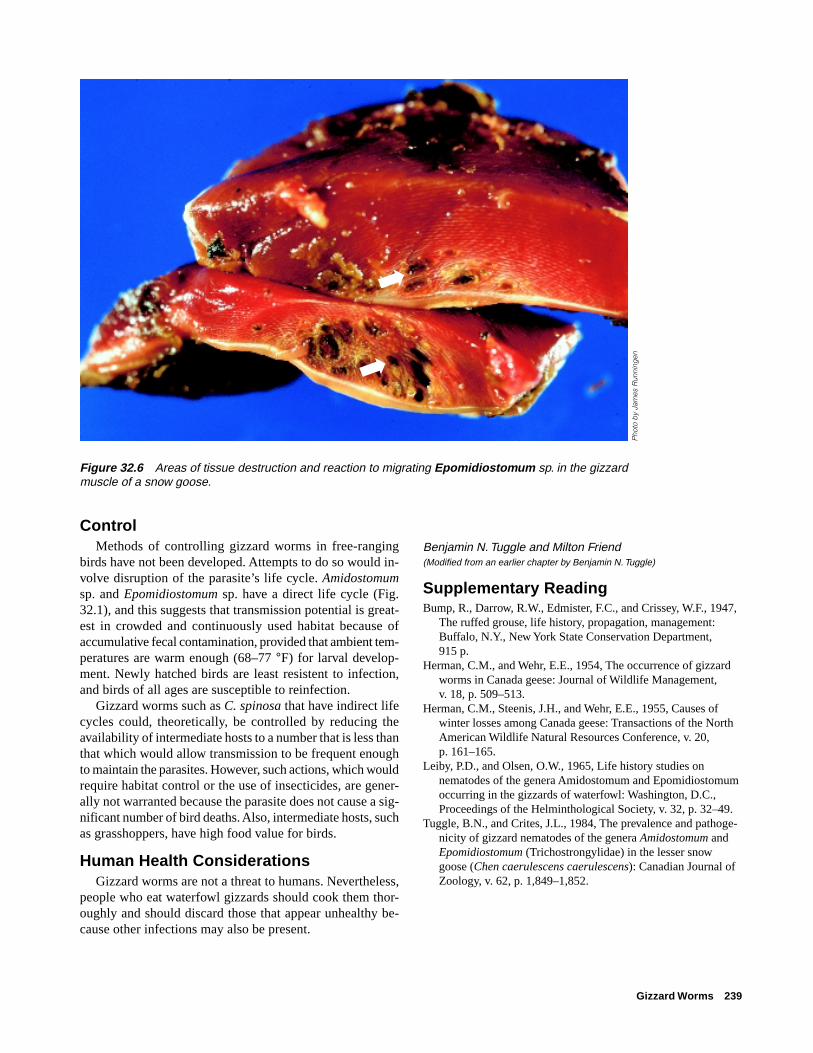
Gizzard Worms 239
Benjamin N. Tuggle and Milton Friend(Modified from an earlier chapter by Benjamin N. Tuggle)
Supplementary ReadingBump, R., Darrow, R.W., Edmister, F.C., and Crissey, W.F., 1947,
The ruffed grouse, life history, propagation, management:Buffalo, N.Y., New York State Conservation Department,915 p.
Herman, C.M., and Wehr, E.E., 1954, The occurrence of gizzardworms in Canada geese: Journal of Wildlife Management,v. 18, p. 509–513.
Herman, C.M., Steenis, J.H., and Wehr, E.E., 1955, Causes ofwinter losses among Canada geese: Transactions of the NorthAmerican Wildlife Natural Resources Conference, v. 20,p. 161–165.
Leiby, P.D., and Olsen, O.W., 1965, Life history studies onnematodes of the genera Amidostomum and Epomidiostomumoccurring in the gizzards of waterfowl: Washington, D.C.,Proceedings of the Helminthological Society, v. 32, p. 32–49.
Tuggle, B.N., and Crites, J.L., 1984, The prevalence and pathoge-nicity of gizzard nematodes of the genera Amidostomum andEpomidiostomum (Trichostrongylidae) in the lesser snowgoose (Chen caerulescens caerulescens): Canadian Journal ofZoology, v. 62, p. 1,849–1,852.
Figure 32.6 Areas of tissue destruction and reaction to migrating Epomidiostomum sp. in the gizzardmuscle of a snow goose.
ControlMethods of controlling gizzard worms in free-ranging
birds have not been developed. Attempts to do so would in-volve disruption of the parasite’s life cycle. Amidostomumsp. and Epomidiostomum sp. have a direct life cycle (Fig.32.1), and this suggests that transmission potential is great-est in crowded and continuously used habitat because ofaccumulative fecal contamination, provided that ambient tem-peratures are warm enough (68–77 °F) for larval develop-ment. Newly hatched birds are least resistent to infection,and birds of all ages are susceptible to reinfection.
Gizzard worms such as C. spinosa that have indirect lifecycles could, theoretically, be controlled by reducing theavailability of intermediate hosts to a number that is less thanthat which would allow transmission to be frequent enoughto maintain the parasites. However, such actions, which wouldrequire habitat control or the use of insecticides, are gener-ally not warranted because the parasite does not cause a sig-nificant number of bird deaths. Also, intermediate hosts, suchas grasshoppers, have high food value for birds.
Human Health ConsiderationsGizzard worms are not a threat to humans. Nevertheless,
people who eat waterfowl gizzards should cook them thor-oughly and should discard those that appear unhealthy be-cause other infections may also be present.
➡
➡
Pho
to b
y Ja
mes
Run
ning
en

240 Field Manual of Wildlife Diseases: Birds

Acanthocephaliasis 241
Chapter 33
Acanthocephaliasis
SynonymsThorny-headed worms, acanths
CauseThe phylum Acanthocephala contains parasitic worms re-
ferred to as thorny-headed worms because both the larvaland adult parasites have a retractable proboscis or a tubularstructure at the head, which has sharp, recurved hooks orspines. Much like the cestodes or tapeworms, they lack di-
gestive tracts and absorb nutrients from the bird’s intestinalcanal. This may weaken the bird and may make it more sus-ceptible to other diseases and to predation.
Adult acanthocephalans are found in a variety of bird spe-cies and in other vertebrates. More than 50 species of acan-thocephalans have been reported in waterfowl, but reevalua-tions of acanthocephalan taxonomy are resulting in revisedspeciation. Nevertheless, numerous species within the phy-lum are found in birds.
Figure 33.1 Indirect life cycle of acanthocephalan worms.Intermediate/transport (paratenic) hosts
Cystacanth develops into adult worm in the bird's intestine andproduces eggs
Definitive bird hosts
Infected birds shed parasite eggs, which are developing embryos, into the environment via the feces
Developing embryos are ingested by inter-mediate/transport host
Infective juvenile stage, or cystacanth, develops within intermediate hosts. Birds feed on intermediate or paratenic hosts

242 Field Manual of Wildlife Diseases: Birds
Life CycleAll acanthocephalan species thus far examined have an
indirect life cycle (Fig. 33.1) that requires at least one inter-mediate host. Intermediate hosts tend to be preferred fooditems of the definitive host; thus, the parasite also uses theintermediate host as a means of transport to the definitivehost. Crustacea of the orders Amphipoda, Isopoda, andDecapoda have been identified as common intermediate hostsof acanthocephalans that infect waterfowl. Some acantho-cephalans that affect passerines or perching birds are reportedto use terrestrial insects as intermediate hosts. Fish, snakes,and frogs have been identified as paratenic hosts in the lifecycle of some acanthocephalans that infect birds.
The adult female parasite within the definitive bird hostproduces eggs that are passed with the bird’s feces into theenvironment. When the egg is ingested by the intermediatehost (insect, crustacean, or centipedes and millipedes), theinfective juvenile stage or cystacanth develops within the in-termediate host. In many life cycles of acanthocephalans, ifthis intermediate host is eaten by a vertebrate host which isunsuitable as a definitive host, the cystacanth will penetratethe vertebrates gut, encyst and cease development. This ver-tebrate is now a paratenic host. If the paratenic or intermedi-ate host is eaten by a suitable definitive host the cystocanthwill attach to the definitive host’s intestinal mucosa via thespined proboscis, mature, mate and produce eggs. A changein body coloration has been noted in some crustaceans in-fected with certain species of acanthocephalan. It is thoughtthat this change in color increases predation by definitivehosts. This might be an evolutionary adaptation which in-creases the chances of life cycle completion by the acantho-cephalan.
Species AffectedAcanthocephalans infect all classes of vertebrates and are
common in birds. Ducks, geese, and swans are considered tobe the most commonly infected birds along with birds ofprey, and some species of passerines. All age classes canbecome infected. Severe disease outbreaks have been repeat-edly reported from common eiders. Eider mortality fromacanthocephalans has been documented throughout the arc-tic areas of their range and has been attributed to food habitsrather than to any increased susceptibility of their species.Historical U.S. Fish and Wildlife Service disease diagnosticrecords reported heavy infections of acanthocephalans andmortality in trumpeter swans from Montana.
DistributionWorldwide.
SeasonalityBirds can be infected with acanthocephalans yearround.
Epizootics usually correspond with food shortages, exhaus-tion (resulting from migration or breeding), or stressful cir-
cumstances. Mortality in immature and adult male eidersis commonly seen in late winter and early spring. Adultfemales experience mortality during or after brooding.Eider ducklings often suffer from both acanthocephalansand renal coccidia. Swan cygnets are also susceptible tolethal infections.
Field SignsLethargy and emaciation are nonspecific but common
clinical signs associated with severe infections.
Gross LesionsGross lesions include white nodules on the serosal or ex-
ternal surface of the intestine (Fig. 33.2). Dissection of thenodules will reveal the proboscis of an acanthocephalan.Examination of the intestinal mucosa or internal lining willreveal white-to-orange colored parasites that are firmly at-tached to the mucosa (Fig. 33.3). Some parasites can pen-etrate the gut wall and project into the abdominal cavity.Adhesions between the loops of intestine are not uncommonin the severe cases where the intestine has been penetrated.
Pho
to b
y R
.A. C
ole
Figure 33.3 Acanthocephalans attached to the inner sur-face of the intestine of a bird.
Figure 33.2 Intestinal loops of a bird infected with acantho-cephalans. Parts of the worms protrude through the intestinalwall.
Pho
to b
y J.
Chr
istia
n Fr
anso
n

Acanthocephaliasis 243
DiagnosisPostmortem examinations are required to reach a diagno-
sis of cause of death. When possible, submit chilled carcassesfor evaluation. Fecal evaluations can be used for determin-ing the presence of infection, but they do not provide a de-finitive diagnosis of disease. Evaluation by a parasitologistis required to differentiate the acanthocephalan species. Ifchilled carcasses cannot be submitted, the following alterna-tives in order of preference are: whole refrigerated intestine,frozen intestine, and formalin-fixed intestine. The formalin-fixed intestine may prohibit species identification but it willallow identification to the genus level as well as assessmentsof worm burden and tissue response to the parasites.
ControlControl of acanthocephaliasis in free-ranging birds is not
practical. Captive flocks can be managed so that aggrega-tions of birds and crustaceans are minimized. One approachthat has been suggested for captive flocks is to limit infec-tion in young birds by segregating them on water areas thatare not used by other birds. These segregated water areaswill presumably have much lower numbers of infected inter-mediate hosts for the birds to be exposed to. Acanthoceph-alans that infect mammals have been successfully treated withivermectin or fenbendazole. Thiabendazole has been recom-mended for use in birds, but treatment is acknowledged tobe difficult and success low.
Human Health ConsiderationsNone
Rebecca A. Cole
Supplementary ReadingBowman, D., 1995, Parasitology for veterinarians (6th ed.):
Philadelphia, Pa., W.B. Saunders, 430 p.Crompton, D.W.T. and Nichol, B.B., 1985, Biology of the
Acanthocephala: Cambridge, England, Cambridge UniversityPress, 519 p.
McDonald, M.E., 1988, Key to acanthocephala reported inwaterfowl: U.S. Fish and Wildlife Service, Resource Publica-tion 173, Washington, D.C., 45 p.
Wobeser, G.A., 1997, Acanthocephala, in Diseases of wildwaterfowl (2nd ed): New York, N.Y., Plenum Press,p. 145–146.

244 Field Manual of Wildlife Diseases: Birds

Nasal Leeches 245
Chapter 34
Nasal Leeches
SynonymsDuck leeches
CauseBloodsucking leeches of the genus Theromyzon sp. are
the only leeches in North America known to feed directly inthe nasal passages, trachea, and beneath the nictitating mem-brane of the eyes of migratory birds. Three species of nasalleeches have been reported from North America, T. rude,T. tessulatum, and T. biannulatum. Other genera of leechesfeed on the exposed surfaces of waterfowl.
Species AffectedNasal leeches affect many aquatic bird species (Fig. 34.1).
Affected waterfowl include northern pintail, teal, Americanwigeon, northern shoveler, ring-necked duck, canvasback,redhead, lesser scaup, bufflehead, gadwall, ruddy duck,white-winged scoter, surf scoter, trumpeter swan, and tun-dra swan. Geese may also be parasitized but they are parasit-ized less frequently than ducks and swans.
DistributionNasal leech infestations of waterfowl and other migra-
tory birds have not been reported south of the 30th paralleland are most commonly observed in northern areas becausethese parasites are better adapted to cold-water lakes (Fig.34.2).
SeasonalityPeak parasitism usually occurs during the spring and sum-
mer months when leeches are actively seeking potential hostsand reproducing. During the winter months, the ambient tem-peratures in frozen ponds and marshes considerably slow theirmetabolic rate and, thus, their activity. In wetlands kept freeof ice during the winter, bird activity may stimulate oppor-tunistic feeding by leeches.
Field SignsBirds that have leeches protruding from the nares or at-
tached externally to the mucous membranes of the eyes areeasily recognized from a distance with the aid of binoculars(Fig. 34.3). Leeches may be so blood-engorged that they re-semble small sacks of blood (Fig. 34.4). Infested birds maybe seen vigorously shaking their heads, scratching at theirbills with their feet, or sneezing in an effort to dislodge theleeches and to force air through blocked nasal passages. Theseefforts are usually unsuccessful. Nasal and respiratory tract
Puddle ducks
Swans
Diving ducks
Mergansers
Sea ducks
Geese
Grebes and loons
FrequentCommon
Occasional
Figure 34.1 Relative frequency of nasal leech infestationsin selected groups of migratory birds.

246 Field Manual of Wildlife Diseases: Birds
Figure 34.2 Reported distribution of nasal leeches in North America.
Figure 34.3 External nasal passage infestations of nasal leeches on (A), the eyes and (B), on the nares of a female redheadduck.
Pho
tos
by
Dav
id T
raug
er, B
iolo
gic
al R
esou
rces
Div
isio
n, U
SG
S
A B
PacificOcean
Arctic Ocean
AtlanticOcean
Gulf of Mexico
HudsonBay
C A N A D A
MEXICO
EXPLANATION
Frequency of occurrence of nasal leaches, by State and Province
Frequent
Common
Occasional
Rare or not reported
➡
➡

Nasal Leeches 247
leech infestations can cause labored breathing and gapingsimilar to that seen among birds suffering from aspergillo-sis.
Gross LesionsSevere leech infestations of the eye can result in tempo-
rary blindness. Eye damage may be seen as an accumulationof a stringy, cheeselike material beneath the nictitating mem-brane, as clouding of the cornea, and, in some instances, ascollapse of the globe of the eye. Nasal passages (Fig. 34.5),throat, and trachea can become blocked by engorged leeches.The feeding action of Theromyzon sp. can cause inflamma-tion and extensive damage to the lining of the nasal cavity.
DiagnosisNasal leeches are 10–45 millimeters long when they are
blood-engorged, are amber or olive colored, and have fourpairs of eyes. Those found in the free-living state are green,with variable patterns of spots on the top surface. Diagnosisof parasitism is usually made by seeing blood-engorgedleeches protruding from the nares or attached to the eyes(Fig. 34.3), especially in birds that cannot be handled. Birdswith internal leech infestations cannot be diagnosed by ob-servation. In cases where the suspected cause of death is nasalleech parasitism, submit the entire carcass for examination.Leeches may depart a dead bird, making diagnosis difficult,or they may move to other areas of the body where they maybe overlooked. Therefore, leeches found on carcasses shouldbe collected and submitted with the carcass. They can beshipped alive in pond water and can be maintained in thatcondition for several months if they are kept refrigerated.
If leeches are to be killed before shipment, they must bepreserved in a relaxed state so that species identification canbe made. Straighten specimens between two glass slides (Fig.34.6A) and flood them with a 10 percent formalin solutionfor 3–5 minutes while applying pressure to the top micro-
Figure 34.4 Blood-engorged nasal leeches removed fromthe nares of a trumpeter swan.
Pho
to b
y Ja
mes
Run
ning
en
Figure 34.5 (A), Nasal passages of trumpeter swan infestedwith blood-engorged Theromyzon sp. leeches. (B), Dissectednasal passages of a mallard showing leech infestation insinus passages.
Pho
tos
by
Jam
es R
unni
ngen
A
B
➡
➡
➡

248 Field Manual of Wildlife Diseases: Birds
scope slide (Fig. 34.6B); then place them in a 10 percentformalin solution for about 12 hours to complete the fixa-tion. Afterwards, transfer leeches to a 5 percent formalinsolution for preservation (Fig. 34.6C).
ControlLeech infestations in waterfowl are common and can be
fatal, especially in cygnets and ducklings. Tissue damageresulting from heavy infestations can facilitate secondarybacterial infections. However, no preventive measures havebeen developed for combating infestations in wild birds.When they are not feeding on birds, nasal leeches occur asfree-living organisms in aquatic environments. Control mea-sures to reduce leech populations might be possible if nasalleeches could be selectively killed. However, leech controlmust be weighed against the value of leeches as an aquaticbird food item.
Leeches protruding externally from the nares or attachedto eyes can be removed with forceps. Leeches in the nasalpassages can be removed by submerging the bird’s bill for5–10 seconds in a 10–20 percent salt solution, and this canbe repeated several times if necessary.
Human Health ConsiderationsTheromyzon sp. feed exclusively on avian hosts and are
not considered a threat to humans.
Benjamin N. Tuggle
Supplementary ReadingDavies, R.W., 1984, Sanguivory in leeches and its effects on
growth, survivorship and reproduction of Theromyzon rude:Journal of Canadian Zoology, v. 62, p. 589–593.
Trauger, D.L., and J.C. Bartonek, 1977, Leech parasitism ofwaterfowl in North America: Wildfowl, v. 28, p. 142–152.
Tuggle, B.N., 1985, The occurrence of Theromyzon rude(Annelida:Hirudinea) In association with mortality oftrumpeter swan cygnets (Cygnus buccinator): Journal ofWildlife Diseases, v. 22, p. 279–280.
Figure 34.6 Leech fixation procedure. (A), Straighten (flat-ten) leech between two glass slides; (B), flood area betweenslides with 10 percent formalin for 3–5 minutes while main-taining downward pressure on top slide; and (C), transfer leechto container of 10 percent formalin (see text for additional de-tails). (Illustration by Randy Stothard Kampen)
A
B
C

Miscellaneous Parasitic Diseases 249
Chapter 35
Miscellaneous Parasitic Diseases
Free-ranging wild birds are afflicted with numerous otherparasites that occasionally cause illness and death. Some ofthese parasites, such as two of the trematodes or flukes high-lighted below, can cause major die-offs. This section aboutparasitic diseases concludes with descriptions of some addi-tional parasites that field biologists may encounter in wildbirds. This listing is by no means complete and it is intendedonly to increase awareness of the diversity of types of para-sites that might be encountered during examinations of wildbirds. One should not assume that the parasites found duringthe examination of bird carcasses caused their death. Be-cause parasites of birds vary greatly in size from a protozoaof a few microns in length to tapeworms of several inches inlength and because they can be found in virtually all tissues,body cavities and other locations within the bird, the obser-vation of the parasites will depend on their visibility and thethoroughness of the examination. Therefore, it is generallybeneficial to submit bird carcasses to qualified disease diag-nostic laboratories to obtain evaluations of the significanceof endoparasites or of ectoparasites. The methods that areused to preserve the carcass, tissues, or other specimens canenhance or compromise the ability of specialists to identifythe parasite to species, and even to genera, in some instances.Therefore, whenever possible, it is best to contact the diag-nostic laboratory that will receive the specimens and obtaininstructions for collecting, preserving, and shipping fieldsamples (See Chapters 2 and 3).
Endoparasites
TrematodesMost trematodes or flukes have complex life cycles that
require two intermediate hosts (Fig. 35.1) in which the para-sites develop before they become infective for the definitive,final bird host. In general, a mollusc is the first intermediatehost, and is often a species that lives in the aquatic environ-ment. Therefore, the aquatic environment brings potentialhosts (waterbirds) and these parasites into close proximityand results in infections of waterfowl and other waterbirdsthat sometimes have fatal consequences.
Sphaeridiotrema globulus is a small trematode less than1 millimeter in length that has been reported to cause die-offs in waterfowl, especially in diving ducks and swans, and,occasionally, in coot. Birds become infected by eating snails(the second intermediate host) that are infected with themetacercarial stage of the parasite (Fig. 35.1). This trema-tode feeds on blood, leading to severe blood loss and ane-mia. Birds often die acutely from shock due to an abnor-
mally decreased volume of circulating plasma in the body orhypovolemic shock. Die-offs usually occur in the late sum-mer or early fall and have been reported in the United Statesmost commonly in the Northeast and North Central States,with a few reports from Western States. Die-offs will oftenrecur yearly at the same locations. For example, mortalityfrom S. globulus is suspected as a recurring event on the St.Lawrence River in southern Quebec. Infections by S. globulushave also been found elsewhere in Canada. The trematode isalso present in the Old World and in Australia. Field signsinclude lethargy and bloodstained vents, although these signsare also found with duck plague (See Chapter 16). Grosslesions can appear as an inflammation or enteritis that is char-acterized by obvious hemorrhages in the lower small intes-tine or as inflammation of the intestinal wall with areas ofulcer-like erosions and the presence of a mixture of bloodand fibrin (Fig. 35.2).
Experimental infections of mute swans with S. globulusindicated that as few as 100 metacercariae could be lethal.Juvenile mallard ducks that had no previous exposure toS. globulus died when infected with 550 metacercariae. Someimmunity has been shown to exist in experimental infectionsof mallards. Adult mallards that were given 100 metacer-cariae and that later were challenged with 2,500 metacer-cariae survived, but those that received only 2,500 metacer-cariae died. Depending on how heavily snail populations areinfected, some birds can receive a lethal dose during lessthan 24 hours of feeding. Susceptible waterfowl generallydie 3–8 days postinfection after ingesting a lethal dose ofS. globulus. Younger birds are generally more susceptiblethan older birds.
No parasite control measures have been developed. Anyattempts at control would need to take into account the factthat the intermediate hosts are different within different geo-graphic areas of the United States. There have been no re-ports of this parasite infecting humans.
Cyathocotyle bushiensis is another trematode that infectswaterfowl and coot in the United States and Canada. Thistrematode can be found in the lower intestine, most com-monly in the cecae or blind pouches extending out from thebeginning of the large intestine (Fig. 35.3). The worms areslightly larger than S. globulus, measuring 1.7–1.8 millime-ters in length. The life cycle for this parasite is similar to thatof S. globulus. Birds become infected by consuming snailsharboring the metacercariae. Disease caused by this trema-tode has been reported in black duck, blue-winged teal, green-winged teal, and coot. This trematode was first described inEngland and it is most likely limited by the geographic dis-
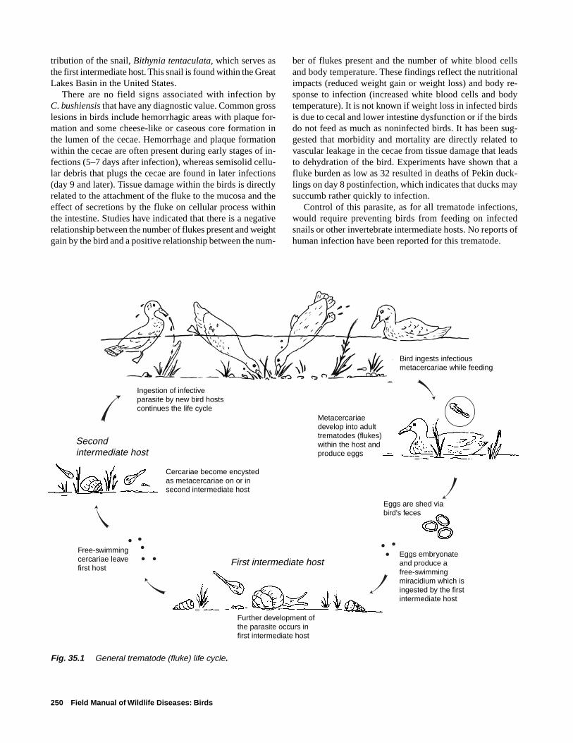
250 Field Manual of Wildlife Diseases: Birds
tribution of the snail, Bithynia tentaculata, which serves asthe first intermediate host. This snail is found within the GreatLakes Basin in the United States.
There are no field signs associated with infection byC. bushiensis that have any diagnostic value. Common grosslesions in birds include hemorrhagic areas with plaque for-mation and some cheese-like or caseous core formation inthe lumen of the cecae. Hemorrhage and plaque formationwithin the cecae are often present during early stages of in-fections (5–7 days after infection), whereas semisolid cellu-lar debris that plugs the cecae are found in later infections(day 9 and later). Tissue damage within the birds is directlyrelated to the attachment of the fluke to the mucosa and theeffect of secretions by the fluke on cellular process withinthe intestine. Studies have indicated that there is a negativerelationship between the number of flukes present and weightgain by the bird and a positive relationship between the num-
ber of flukes present and the number of white blood cellsand body temperature. These findings reflect the nutritionalimpacts (reduced weight gain or weight loss) and body re-sponse to infection (increased white blood cells and bodytemperature). It is not known if weight loss in infected birdsis due to cecal and lower intestine dysfunction or if the birdsdo not feed as much as noninfected birds. It has been sug-gested that morbidity and mortality are directly related tovascular leakage in the cecae from tissue damage that leadsto dehydration of the bird. Experiments have shown that afluke burden as low as 32 resulted in deaths of Pekin duck-lings on day 8 postinfection, which indicates that ducks maysuccumb rather quickly to infection.
Control of this parasite, as for all trematode infections,would require preventing birds from feeding on infectedsnails or other invertebrate intermediate hosts. No reports ofhuman infection have been reported for this trematode.
Eggs are shed via bird's feces
Eggs embryonate and produce a free-swimmingmiracidium which isingested by the firstintermediate host
Metacercariae develop into adult trematodes (flukes) within the host andproduce eggs
Bird ingests infectiousmetacercariae while feeding
First intermediate host
Second intermediate host
Further development of the parasite occurs in first intermediate host
Free-swimming cercariae leave first host
Cercariae become encysted as metacercariae on or in second intermediate host
Ingestion of infective parasite by new bird hosts continues the life cycle
Fig. 35.1 General trematode (fluke) life cycle.
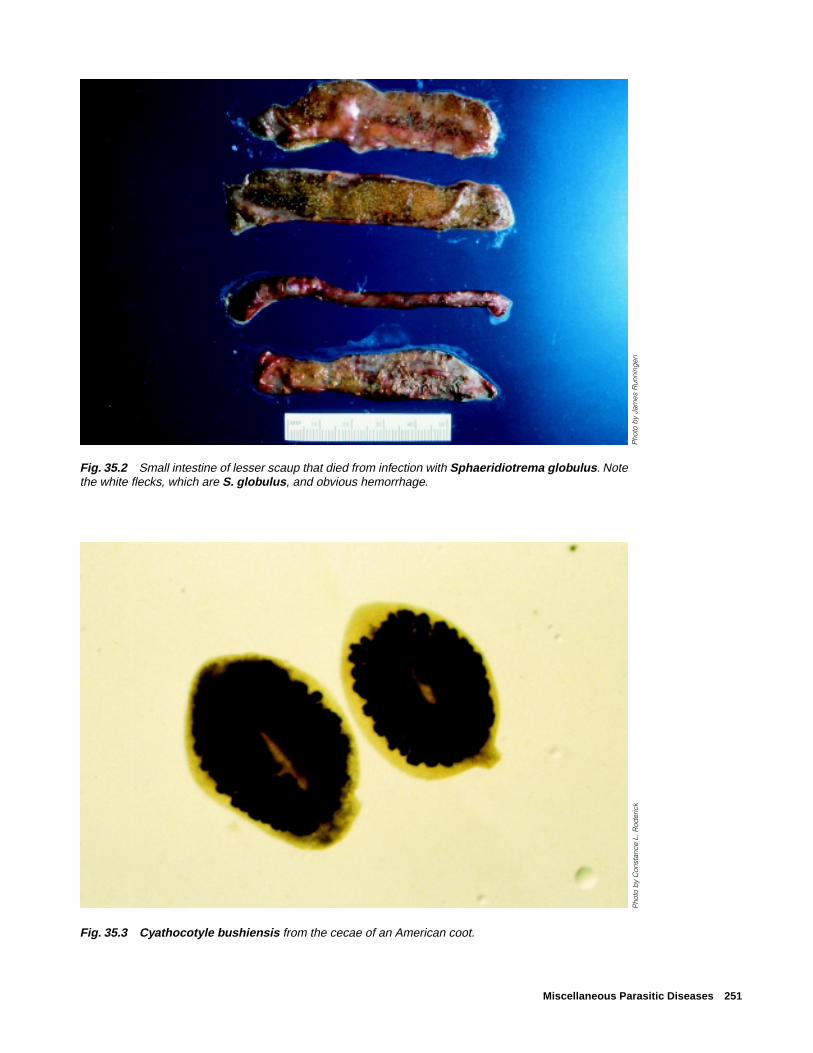
Miscellaneous Parasitic Diseases 251
Fig. 35.3 Cyathocotyle bushiensis from the cecae of an American coot.
Fig. 35.2 Small intestine of lesser scaup that died from infection with Sphaeridiotrema globulus. Notethe white flecks, which are S. globulus, and obvious hemorrhage.
Pho
to b
y Ja
mes
Run
ning
enP
hoto
by
Con
stan
ce L
. Rod
eric
k

252 Field Manual of Wildlife Diseases: Birds
Leyogonimus polyoon (Fig. 35.4) is a newly reportedtrematode in North America that caused the deaths of over1,500 and 11,000 coots in Northeastern Wisconsin duringthe falls of 1996 and 1997, respectively (Fig. 35.5). Thistrematode is of similar small size (0.7–1.0 millimeters inlength) as S. globulus (Fig. 35.2). It is known to cause deathin coot and common moorhen in Europe, but the Wisconsinoutbreaks are the first documentation of this parasite caus-ing mortality in North America. The susceptibility of otherNorth American birds, beside coot, remains unknown. At theWisconsin location, various waterfowl species were dyingfrom infections of S. globulus while the coot were dying fromL. polyoon infections. None of the waterfowl were found tobe infected with L. polyoon.
The life cycle for L. polyoon is not known, although thesuspected first intermediate host is the snail Bithnia tentac-ulata. Investigations at the National Wildlife Health Center(NWHC) have disclosed that snails that were collected in
Wisconsin were infected with a cercariae or a larval form ofthe parasite that fits the literature description of L. polyoon.Additional studies are required to confirm that these larvalforms are L. polyoon. Also, the intermediate host for the in-fective larval form or metacacariae has not yet been found.L. polyoon infects primarily the upper and middle areas ofthe small intestine. No significant field signs are associatedwith infection by L. polyoon. Gross lesions seen at necropsyinclude severe enteritis characterized by thickening of theintestinal wall and a fibrous-to-caseous core of necrotic de-bris that blocks the lumen of the intestine (Fig. 35.6).
The location where these outbreaks occurred is a lake thatis drained by a stream, that is underlain with sand, and thathas substantial growths of water weeds. The shoreline hasbeen extensively developed for home sites and other humanuse, and the lake is used for recreation. L. polyoon has notbeen reported to infect humans.
Fig. 35.5 American coot and various species of waterfowlfrom die-off in Shawano Lake, Wisc. Coot mortality was dueto Leyogonimus polyoon, but waterfowl mortality was dueto Sphaeridiotrema globulus.
Fig. 35.4 Leyogonimus polyoon from thesmall intestine of an American coot.
Pho
to b
y K
. Bro
ckm
an-M
eder
as, W
isco
nsin
Dep
artm
ent o
f Nat
ural
Res
our c
es
Pho
to b
y C
onst
ance
L. R
oder
ick

Miscellaneous Parasitic Diseases 253
Fig. 35.6 (A) Gastrointestinal tract from an American cootthat was infected with Leyogonimus polyoon. Note the en-larged or swollen areas (arrows). (B) Intestinal tract of anAmerican coot. The intestinal tract has been incised to ex-pose cheesy cores of dead tissue debris caused by Leyo-gonimus polyoon (arrows).P
hoto
by
Reb
ecca
A. C
ole
Pho
to b
y R
ebec
ca A
. Col
e
A
B
➡
➡
➡➡➡
➡

254 Field Manual of Wildlife Diseases: Birds
CestodesTapeworms are common in wild birds, but they seldom
cause death. Heavy burdens of these parasites may reducethe vigor of the bird and serve as a predisposing factor forother disease agents, or the parasites may occlude the intes-tine (Fig. 35.7). One genus, Gastrotaenia sp., lives in thegizzard and penetrates the keratohyalin lining or the hornycovering of the gizzard pads, causing inflammation and nec-rosis. Cloacotaenia sp. inhabit the ureter or the tubular areathat transports wastes from the kidneys to the cloaca in somewaterfowl.
NematodesTrichostrongylidosis, the disease that is caused by Tri-
chostrongylus tenius, is not currently a significant problemwithin the United States. However it is included becausetrichostrongylidosis, in natural populations of grouse in theirnative habitat in Scotland and elsewhere in the United King-dom, demonstrates the impact that a parasite can have on thepopulation dynamics of the bird host that it infects. T. teniusis a common nematode or roundworm that is found withinthe ceca of some types of wild birds, primarily grouse, geese,and poultry. This parasite has a direct life cycle that is closelyassociated with host food preferences for terrestrial vegeta-
tion (Fig. 35.8). Trichostrongylidosis outbreaks can occurwhenever birds are hatching because of the synchronousphase of the ecology of the parasite and the feeding habits ofbird hosts.
Hatchability of the parasite eggs and survival of the free-living larval stages are moisture and temperature dependent.The parasite eggs do not develop under dry conditions andthe free-living larvae are generally killed by freezing tem-
Figure 35.7 Large numbers of tape worms may occlude theintestine.
Parasite eggs are shed via bird's feces
Eggs mature in the environment and produce larvae that undergo further development within the environment. Larvae crawl to the tips of vegetation where they accumulate in drops of water
Bird hosts
Birds ingest larvae whilefeeding upon moisture-ladenvegetation
Figure 35.8 Life cycle of the nematodeTrichostrongylus tenius.
Pho
to b
y M
ilton
Frie
nd

Miscellaneous Parasitic Diseases 255
peratures. When the feces that contain the parasite eggs arekept moist and when the ambient air temperature is suitable,first-stage larvae develop within the egg and hatch in about2 days; free-living second-stage larvae develop within an-other 1-1/2-to-2 days; and development to infective third-stagelarvae requires an additional 8–16 days. Studies in Scotlandindicate that infective larvae crawl to the tips of moist heatherand accumulate there in drops of water provided by the mistyweather. Grouse that feed on the tips of the heather ingestthe larvae along with their food. Infective larvae molt twicemore within the ceca of the bird before they become sexu-ally mature adults.
Infections have been reported in chicken, turkey, guineafowl, pheasant, quail, pigeons, ducks, and geese; but infec-tions are most notable for red grouse because this parasitehas clearly been shown to regulate natural populations ofthis species. Because adult worms can survive in their birdhost for more than 2 years, all adult birds evaluated in somepopulations have been found to be infected, thereby, provid-ing a reservoir for infection of young. In addition, larvae canarrest development within the ceca, overwinter, and then re-sume development in the spring. The temperature and mois-ture requirements for hatchability of the parasite eggs andsurvival of larvae results in synchronized availability of para-sites during the period of production of young grouse. Thisresults in the primary occurrence of disease and mortality ingrouse during the spring. The hatchlings are exposed to in-fective larvae soon after the diet for chicks changes frominsects to vegetation.
The impacts of infection by T. tenius are greater than justchick mortality. These parasites also decrease available en-ergy for egg laying by adult birds. The resting metabolic rateis increased and there is a decrease in food intake by thebird. The resulting impact is reduced fecundity within thepopulation (fewer chicks) along with high chick mortality.This combination of impacts controls population levels.
Echinuria uncinata is a common nematode that infectsthe proventriculus of various waterfowl species. The life cycleis indirect, and the parasite uses zooplankton, especiallyDaphnia sp., as intermediate hosts (Fig. 35.9). Wormsmature in a duck approximately 51 days after it eats infectedzooplankton. Adult worms, which are approximately 5 mil-limeters long, burrow headfirst into the mucosa and submu-cosa of the proventriculus, causing tissue swelling and in-flammation. Tumor-like nodules form and can be largeenough to obstruct the lumen of the proventriculus. This para-site can be especially dangerous to waterfowl where zoo-plankton blooms coincide with the hatching of young. Oftenbirds that are late to hatch and do not have fully developedimmune systems can consume enough infected zooplanktonin a very short period of time to become severely infected. Inareas where water is shallow, where zooplankton populationsare numerous, and where birds are crowded into the area,this roundworm can be transmitted to many birds during ashort time.
This occurred within a population of the endangeredLaysan duck on Laysan Island, Hawaii during the fall of 1993.A drought had struck the island and the brine flies that the
Parasite eggs are shed via bird's feces
Infection occurs via ingestion of infected zooplankton
Infective larvae develop in zooplankton
Larvae mature in duck and adult worms burrow headfirst into the proventriculus
Bird hosts
Figure 35.9 Life cycle of the nematodeEchinuria uncinata.

256 Field Manual of Wildlife Diseases: Birds
ducks feed heavily on were believed to be scarce. This de-pressed food base may have resulted in a reduced level ofnutrition and may have compromised the ability of the birdsto withstand infections by E. uncinata. Birds from this die-off were severely emaciated, had thickened proventriculi, andnodules along the proventriculi and intestines. The glandswithin the proventriculus were severely distorted, which sug-gests that the function of the proventriculus was compro-mised. Blood samples taken from sick birds suggested thatthey were emaciated and severely infected with parasiticworms. It was thought that the combination of the drought,aggregation of birds around freshwater seeps, and scarce food
sources, combined with the severe parasitism, caused theducks to die. Other examples exist where the combination ofovercrowded waterfowl, a zooplankton population explosion,large numbers of infected zooplankton, and high retentionof worms within waterfowl resulted in sufficient pathologyby the worms to cause clinical disease and death. Maintain-ing fast water flow to prevent zooplankton explosions hasbeen successfully employed for disease prevention in cap-tive flocks such as those at waterfowl parks. E. uncinata iswidely distributed geographically and has not been reportedto infect humans.
Cecal worm eggs developinto cecal worm larvae inthe soil where they were deposited
Cecal worm larvae containing histomonads
Earthworms feeding insoil ingest cecal wormlarvae containing histo-monads and serve astransport hosts
Histomonas meleagridis infectsovary of cecal worms
Parasite cycle in carrier bird
Cecal worm larvae developinto mature worms
Cecal worms produce eggs within their bird host which contain the protozoanHistomonas meleagridis. Cecal worm eggs are shed into the environment
Carrier bird
Disease cycleSusceptible species ingest the cecal worm larvae and their histomonad "passengers" by (A) directly ingesting cecal worms while feeding or by (B) ingesting earth-worms containing cecal worm larvae with histomonads
AB
Infection by the histomonads often results in death
Figure 35.10 Life cycle of the proto-zoon Histomonas meleagridis, whichcauses histomoniasis.

Miscellaneous Parasitic Diseases 257
ProtozoaHistomoniasis is capable of causing catastrophic losses
in the wild turkey, a species whose restoration has become amajor wildlife management success story. Histomonasmeleagridis, the protozoan that causes histomoniasis, utilizesthe cecal worm, Heterakis gallinarum (a nematode), as avector for entry into the bird hosts (Fig. 35.10). The diseaseis commonly called blackhead because infections sometimescause a bluish or blackish appearance of the skin of the headin some birds due to an excessive concentration of reducedhemoglobin in the blood or cyanosis.
Earthworms and other soil invertebrates can become partof the parasite’s life cycle when they feed on fecal-contami-nated soil that contains cecal worm eggs infected withhistomonads. The cecal worm larvae and histomonads arestored in the body of the earthworm and are transmitted tobirds when worms are fed upon. However, earthworms arenot required for the life cycle; cecal worm larvae that con-tain histomonads may be ingested by birds when they feedin a contaminated environment.
Most, if not all, gallinaceous birds are susceptible hosts. Tur-key, grouse, chicken, and partridge develop severe disease andsuffer high mortality rates that can exceed 75 percent of thoseinfected. Disease is less severe in Hungarian partridge and bob-white quail. In contrast, pheasant and some other species oftendo not exhibit signs of disease, but they instead become carriersthat maintain the disease cycle. Canada geese that were exam-ined at the NWHC have also been found to have a histomonia-sis-like disease. In North America, wild turkey and bobwhitequail are the species most commonly infected in the wild. Thedisease is found worldwide.
There are no clinical signs specific to histomoniasis. Wildturkeys affected with this disease often are listless, have anunthrifty appearance of ruffled feathers, and stand withdrooped wings. The birds may appear depressed, and theirfeces are often sulfur-yellow in color. This fecal colorationgenerally occurs early in the disease and, combined with otherfield signs, it is highly suggestive of histomoniasis. The pri-mary gross lesions seen upon necropsy of infected birds arenumerous large, pale grey, discrete circular crater-like areasof necrosis or tissue death within the liver (Fig. 35.11) andthickened caecal walls that often also become ulcerated andhemorrhagic. The lumen of the ceca may also be obstructedby aggregations of yellowish necrotic debris referred to ascecal cores (Fig. 35.12).
Disease prevention should be the major focus for address-ing histomoniasis. The introduction of gallinaceous bird spe-cies that are disease carriers, such as pheasants, into habitatoccupied by highly susceptible species, such as wild turkey,is unwise and it can have catastrophic results. Similarly,because chickens are often carriers of H. gallinarum (cecalworms), and often shed histomonads, spreading uncompostedchicken manure onto fields can distribute cecal worm eggs towild and susceptible species.
Figure 35.11 Large, pale areas in the liver of a bird infectedwith Histomonas sp.
Figure 35.12 Cecal core (necrotic debris) in cecum of a birdinfected with Histomonas sp.
Histomoniasis has caused the deaths of wild turkeys thatwere provided feed in barnyards frequented by chickens.Therefore, when attempting to reestablish wild turkey flocksin areas where they no longer exist and during periods ofinclement weather that create food shortages for wild birds,placement of feed stations should be done with consider-ation of potential carriers of H. meleagridis or H. gallinarum.
EctoparasitesIn addition to being vectors that transmit disease to birds,
ectoparasites can be direct causes of illness and death. Just afew adult ticks feeding on a small bird can cause anemia,reduced growth, weight loss, and contribute in other ways toa depressed state of health. The fowl tick, a soft-bodied tickof the family Argasidae, is the most important poultry ecto-parasite in many countries and it is often a factor limitingraising chickens and turkeys. Chickens have also been re-ported to suffer tick paralysis, which is a motor paralysis orparalysis of the voluntary muscles, from bites of Argas sp.ticks. Tick paralysis in songbirds has been associated with
Pho
to b
y J.
Chr
istia
n Fr
anso
n
➡
Pho
to b
y M
ilton
Frie
nd

258 Field Manual of Wildlife Diseases: Birds
the bite of the hard-bodied bird tick, Ixodes brunneus. Fatalparalysis from bites by this tick has been reported in numer-ous species of small birds. The engorged ticks in fatal casesare generally found on the bird’s head and they may be at-tached to its eyelids. Death results from a powerful neuro-toxin that is secreted by the tick while it feeds on the bird.Other species of Ixodes ticks have been associated with tickparalysis and mortality in marine birds, including albatrossand petrels. An ascending motor paralysis that starts at thefeet, progresses for 7–10 days, and ends in death has beenreported.
Heavy infestations of lice, mites, fleas, flies, and otherbiting insects have also been responsible for causing illnessand even death of wild birds, especially among nestlings.Conditions caused by these insects range from feather lossand skin damage from acariasis or mange, to myiases or in-festation with fly maggots, and anemia. Mites of the genusKnemidocoptes are the primary cause of mange in birds, andthe mites belong to the same family (Sarcoptidae) of mitesthat cause mange in mammals and humans. The Knemido-coptes sp. mites are specific to birds and they are not a hu-man health hazard.
More knowledge is needed about the role of ectoparasitesas causes of bird death. Proper identification of the speciesassociated with bird mortality is an important component ofsuch assessments; therefore, the presence of insects withinbird nests in which freshly dead nestlings are found shouldbe recorded, representative parasite specimens should be col-lected along with any visible parasites on the carcass, andthe parasites submitted should be with the carcass. Ticks andany heavy infestations of fleas, lice, and other insects on livebirds being handled for banding or other purposes shouldalso be noted, and, when practical, samples should be col-lected and submitted for identification to a parasitologist ordisease diagnostic laboratory. An abundance of such para-sites may be indicators of other health problems for the birds.
Rebecca A. Cole and Milton Friend
Supplementary ReadingAnderson, R.C., 1992, Nematode parasites of vertebrates, their
development and transmission: Wallingford, United Kingdom,C•A•B International, University Press, 578 p.
Arnall, L., and Keymer, I.F., 1975, Bird diseases, an introductionto the study of birds in health and disease: Bailliere Tindall,London, T.F.H. Publications, 528 p.
Calnek, B.W. and others, 1997, Diseases of Poultry (10th ed.):Ames, Iowa, Iowa State University Press, 1,080 p.
Delehay, T.M., Speakman, J.R., and Mass, R., 1995, The energeticconsequences of parasitism: effects of a developing infectionof Trichostrongylus tenus (Nematoda) on red grouse (Lagopuslagopus scoticus) energy balance, body weight and condition:Parasitology, v. 110, p. 473–482.
Davidson, W.R., and Nettles, V.F., 1997, Field manual of wildlifediseases in the Southeastern United States (2d ed.): Athens,Ga., University of Georgia, 417 p.
Gagnon, C., Scott, M.E., and McLaughlin, J.D., 1993, Grosslesions and hematological changes in domesticated mallardducklings experimentally infected with Cyathocotylebushiensis (Digenea): Journal of Parasitology, v. 79, p. 757–762.
Hove, J., and Scott, M.E., 1988, Ecological studies on Cyatho-cotyle bushiensis (Digenea) and Sphaeridiotrema globulus(Digenea), possible pathogens of dabbling ducks in southernQuebec: Journal of Wildlife Diseases, v. 24, p. 407–421.
Hudson, P.J., 1986, The effect of a parasitic nematode on thebreeding production of red grouse: Journal of Animal Ecology,v. 55, p. 85–92.
Huffman, J.E., and Roscoe, D., 1989, Experimental infections ofwaterfowl with Sphaeridiotrema globulus (Digenea): Journalof Wildlife Diseases, v. 25, p. 143–146.
Mucha, K.H., and Huggman, J.E., 1991, Inflammatory cellstimulation and wound healing in Sphearidiotrema globulusexperimentally infected mallard ducks (Anas platyrhynchos):Journal of Wildlife Diseases, v. 27, p. 428–434.

Introduction to Biotoxins 259
Section 6Biotoxins
Algal Toxins
Mycotoxins
Avian Botulism
Aerial view of a large dinoflagellate bloom in near-shore ocean watersPhoto by Peter Frank, Scripps Institute of Oceonography
260 Field Manual of Wildlife Diseases: Birds
Introduction to Biotoxins
Biotoxins are usually defined as poisons that are producedby and derived from the cells or secretions of living organ-isms. These natural poisons include some of the most toxicagents known and they are found within a wide variety oflife forms. Organisms that produce such toxins are generallyclassified as being venomous or poisonous. The classifica-tion of venomous is usually associated with animal life formssuch as poisonous reptiles and insects that have highly de-veloped cellular mechanisms for toxin production and thatdeliver their toxins during a biting (rattlesnake) or stinging(black widow spider) act. Poisonous organisms are gener-ally thought of as those that deliver toxins by being ingestedor by their secretions being ingested by another organism.Therefore, these toxins are essentially forms of food poison-ing. Readers should appreciate that virtually all venomousorganisms are poisonous but many poisonous organisms arenot venomous. This Section will address poisonous, but notvenomous, organisms, and it includes the perspective ofbiotoxins as products of plants and lower life forms.
Birds become poisoned by a broad array of biotoxins. Thechapter about avian botulism involves microbial toxins pro-duced within replicating Clostridium botulinum bacteria. Thepotency of toxins that are produced by the disease-causingClostridia are legendary, and the toxins include such human
diseases as tetanus and lethal botulism food poisoning. Avianbotulism is currently the most important disease of water-fowl and shorebirds, nationally and internationally, and out-breaks of this disease commonly kill tens of thousands ofbirds during a single event. Up to a million birds have re-cently been lost within a single location during the course ofa protracted outbreak.
Because many avian botulism die-offs occur on the samewetlands year after year, one of the primary areas of researchon this disease has focused on identifying and understand-ing the microenvironmental characteristics that contribute toa mortality event. The development of wetland-specific riskassessment tools will enable wildlife disease specialists andnatural resource managers to more effectively manage avianbotulism.
Fungi are an additional source of microbial biotoxins thatcause the death of free-ranging wild birds. Mycotoxins, whichare toxins produced by fungi, have received considerablestudy because of their effects on food animals and humans.In poultry, for example, many types of mycotoxins are knownto cause problems that include mortality, decreased growth,impaired reproduction, immunosuppression, and pathologiceffects on a variety of other organ systems. Although thesetoxins have received little study in wildlife, a growing bodyof literature documents similar effects of mycotoxins in avariety of free-ranging species. The chapter about mycotox-ins illustrates the capabilities of aflatoxins and trichothecenesto cause large-scale bird losses as the result of bird ingestionof food contaminated by molds that produce these toxins. Asmore becomes known about the occurrence of mycotoxins
All quotes from:
Hayes, Wayland, J., Jr., 1991, in Hayes, Wayland J., Jr., and Laws,Edward R., Jr., eds., Handbook of pesticide toxicology, v. 1,General principles: New York, Academic Press, p. 7–8.
“Ecological toxicology is the study of all toxicants produced by living organismsand of the ecological relationships made possible by these poisons.”
(Hayes)
“In all communities chemical interrelations are important aspects of the adapta-
tion of species to one another; in some communities chemical relations seem tobe the principal basis of species niche differentiation and community organiza-tion.” (Whittaker and Feeny)
“Undoubtedly there is much to be learned from finding out how the battle[between toxicants produced by living organisms and host defenses developed in
response to these toxicants] has been fought for the last several million years.”(Hayes)

Introduction to Biotoxins 261
in the natural environment, and as analytical techniques forthe specific toxins become more commonly available, it islikely that more and more cases of mycotoxicosis will bereported in wildlife.
The range of living organisms that cause poisoning in wildbirds is further illustrated by plant toxins in the chapter aboutalgal toxins. Less is known about poisoning of birds fromtoxic plants than is known about poisoning from bacterialand fungal toxins. Plant toxins other than algal toxins thathave caused bird mortality have rarely been reported. Chokecherry seeds contain chemical compounds that release cya-nide upon digestion if the seed capsule is broken during di-gestion. Songbirds have been killed by cyanide poisoningfrom eating these seeds. Waterfowl mortality has been at-tributed to ingestion of castor beans, which results in intoxi-cation from ricin, the active ingredient within the seed thatcauses poisoning. A small number of other reports of planttoxins causing wild bird mortality also exist.
The so-called algal toxins are produced by a variety oforganisms, including true algae, dinoflagellates (aquatic pro-tozoa), and blue-green algae, and are the least understood ofthe biotoxins covered in this Section. Algal blooms, espe-cially red tides and blue-green blooms, wreak aesthetic and
economic havoc in many freshwater and marine environmentsbecause of the potential for toxins to be present. Perhaps oneof the most widely recognized toxins in this group is sax-itoxin, the agent of paralytic shellfish poisoning, which causesoccasional human deaths and renders many tons of shellfishinedible throughout the world.
Algal toxins are likely to become increasingly recognizedas a cause of waterbird mortality. Eutrophication of inlandwaterbodies due to nutrient loads is causing more algalblooms within those waters, many of which are used by largenumbers of water birds. Enhanced technology and increasedstudy are needed to better understand the ecology of algalblooms and the production of toxic components that are haz-ardous to bird life. With the exception of avian botulism,biotoxins as a cause of disease in wild birds have receivedlittle study. However, there should be no debate regardingthe need for study since disease caused by biotoxins extendsbeyond direct mortality. Impaired immune system functionor immunosuppression and cancers caused by biotoxins haveboth been documented in animals and humans. Other effectson wildlife are also likely because of the diversity of diseaseimpacts seen in humans and domestic animals.

262 Field Manual of Wildlife Diseases: Birds

Algal Toxins 263
Chapter 36
Algal ToxinsSynonymsRed tide toxins, phycotoxins
Periodic blooms of algae, including true algae, dinoflagel-lates, and cyanobacteria or blue-green algae have been re-ported in marine and freshwater bodies throughout the world.Although many blooms are merely an aesthetic nuisance,some species of algae produce toxins that kill fish, shellfish,humans, livestock and wildlife. Pigmented blooms of toxin-producing marine algae are often referred to as “red tides”(Fig. 36.1). Proliferations of freshwater toxin-producingcyanobacteria are simply called “cyanobacterial blooms” or“toxic algal blooms.” Cyanobacterial blooms initially appeargreen and may later turn blue, sometimes forming a “scum”in the water (Fig. 36.2).
Although algal blooms historically have been considereda natural phenomenon, the frequency of occurrence of harm-ful algae appears to have increased in recent years. Agricul-tural runoff and other pollutants of freshwater and marinewetlands and water bodies have resulted in increased nutri-ent loading of phosphorus and nitrogen, thus providing con-ditions favorable to the growth of potentially toxic algae.The detrimental impact of red tides and cyanobacterialblooms on wetland, shore, and pelagic species has long beensuspected but not often been substantiated because informa-tion on the effects of these toxins in fish and wildlife speciesis lacking and diagnostic tools are limited.
CauseSome dinoflagellates and cyanobacteria produce toxins
that can affect domestic animals and humans. Some of thesetoxins such as domoic acid, saxitoxin (paralytic shellfishpoisoning or PSP toxin), brevetoxin, and cyanobacterial tox-ins (including anatoxins, microcystins, and nodularins) havebeen suspected, but they have rarely been documented, asthe cause of bird mortality (Table 36.1). Marine algal toxinssuch as domoic acid, saxitoxin, and brevetoxin that bioaccu-mulate or are magnified in the food chain by fish and shell-fish, and anatoxins from freshwater cyanobacteria, affect thenervous system; cyanobacteria that contain microcystins ornodularin cause liver damage.
The effects of some harmful algae are not related to toxinproduction but rather are related to depleted dissolved-oxygen concentrations in water caused by algal prolifera-tion, death, and decay, or night respiration. Other harmfuleffects include occlusion of sunlight by large numbers ofalgae and physical damage to the gills of fish caused by thestructure of some algal organisms. All of these effects can
Figure 36.1 Aerial view of a large dinoflag-ellate bloom in near-shore ocean waters. Theorganism responsible for this bloom is not atoxin producer; however, toxic blooms mayhave a similar appearance.
Figure 36.2 A cyanobacterial or blue-green algal bloom.
Pho
to b
y W
ayne
Car
mic
hael
, Wrig
ht S
tate
Uni
vers
ity
Pho
to b
y P
eter
Fra
nk, S
crip
ps
Inst
itute
of O
cean
ogra
phy

264 Field Manual of Wildlife Diseases: Birds
Table 36.1 Documented instances of wild bird mortality caused by algal toxins.
Migratory bird Route of Toxin Algal species Toxin type(s) species affected exposure
Cyanobacterial Microcystis sp., Hepatotoxins Unidentified ducks, OralAnabaena sp., (microcystins geese, and songbirds, (water)Aphanizomenon sp., and nodularin) Franklin’s gull, AmericanNodularia sp., Neurotoxins coot, mallard, Americanand (anatoxin-a and wigeonOscillatoria sp. anatoxin-a(s))
Domoic acid Pseudonitzschia sp. Neurotoxin Brown pelican, Oral(amnesic shellfish Brandt’s cormorant (food items)poisoning)
Saxitoxin Alexandrium sp. Neurotoxin Shag, northern fulmar, Oral(paralytic great cormorant, (food items)shellfish herring gull, commonpoisoning) tern, common murre,
Pacific loon, and sootyshearwater
Brevetoxin Gymnodinium sp. Neurotoxin Lesser scaup Oral(food items)
lead to mortality of aquatic invertebrates, aquatic plants, orfish and may produce an environment conducive to botu-lism. Other marine algal toxins (okadaic acid, neosaxitoxin,ciguatoxin, and Pfiesteria exotoxin) and cyanobacterial tox-ins (saxitoxin, neosaxitoxin, and cylindrospermopsin) havenot yet been identified as causes of bird mortality events, butincreased awareness and further research may establish arelationship.
Species AffectedMany bird and mammal species can be affected by algal
toxins. Most reports of mortality in birds are of die-offs thatoccur in conjunction with a bloom. Sometimes algal toxinsare found in potential food items; however, there have beenvery few instances in which the algal toxin has been isolatedfrom the ingesta or tissues of affected birds. Domoic acidpoisoning caused mortality in brown pelicans and Brandt’scormorants on the central California coast. Brevetoxin hasbeen suspected as the cause of mortality in lesser scaup, andsaxitoxin has been strongly suspected as the cause of mor-
tality in sea birds (common terns, shags, great cormorants,northern fulmars, herring gulls, common murres, Pacificloons, sooty shearwaters, and others). Cyanobacterial toxi-cosis has been suspected in mortalities of free-ranging ducks,geese, eared grebes, gulls, and songbirds.
DistributionMany of the organisms responsible for red tides are widely
distributed and, in recent years, the organisms seem to bemarkedly spreading. Natural events such as hurricanes candisperse organisms, and it is suspected that some organismsmay be transported long distances in ship ballast waters. An-other factor that may encourage algal proliferation in bothmarine and freshwater systems is increased nutrient load-ing. Certain algae occur more commonly in some areas thanothers and it is useful to know which ones are problems inspecific locations. Good sources of information about algalblooms are the State public health department or the Statedivision of marine resources or marine fisheries.

Algal Toxins 265
SeasonalityThere have not been enough confirmed instances of wild
bird mortality caused by red tides and cyanobacterial bloomsto establish seasonal patterns of occurrence.
Field SignsField signs reported are variable and they depend on the
toxin involved. Domoic acid poisoning of brown pelicanscaused neurologic signs that included muscle tremors, a char-acteristic side-to-side head movement, pouch scratching,awkward flight, toe clenching, twisting of the head over theback, vomiting, and loss of the righting reflex just beforedeath. Brandt’s cormorants that also were involved in thismortality event were easily approached and handled, but theydid not exhibit the neurologic signs seen in the pelicans. Seabirds suspected of having been poisoned by saxitoxin exhib-ited paralysis and vomiting. Clinical signs observed in lesserscaup suspected of having been poisoned by brevetoxin in-cluded lethargy, weakness, reluctance or inability to fly, headdroop, and excessive ocular, nasal, and oral discharge.
White Pekin ducklings that were experimentally exposedto brevetoxins exhibited lethargy, loss of muscle coordina-tion or ataxia, spastic head movements, head droop to oneside, and leg extension to the rear during rest. Clinical signsin muscovy ducks dosed with anatoxin-a(s) included exces-sive salivation, regurgitation of algae, diarrhea, tremors, re-duced responsiveness and activity, incoordination, difficultybreathing, excessive thirst, congestion in foot webs, wingand leg weakness, and recumbency and intermittent seizuresprior to death.
Gross LesionsNo characteristic or diagnostic gross lesions have been
described for most types of algal toxin poisonings of wild
birds. Many of the toxins, particularly the neurotoxins, havea chemical effect that does not produce a grossly observablelesion. Birds that ingest toxic blooms of Microcystis mayhave notable lesions of necrosis or tissue death and hemor-rhage in the liver. These lesions have been reported in do-mestic mammals and birds, including ducks, that died as aresult of exposure to a toxic Microcystis algal bloom or thatwere experimentally dosed with microcystin.
DiagnosisDefinitive diagnosis of algal toxicosis is difficult. Circum-
stantial evidence, such as the occurrence of a marine red tideor freshwater cyanobacterial bloom in conjunction with adie-off, and supportive clinical and pathologic findings, suchas a lack of evidence of the presence of other types of toxinsor infectious disease, are often used to reach a presumptivediagnosis. Analysis of the upper gastrointestinal tract con-tents or tissues of affected birds for algal toxins is possiblebut the tests are not yet widely available. In addition, thereare no established toxic thresholds for wildlife species. Evenwhen levels of particular toxins can be measured it may bedifficult to assess their significance. Recently developedmethods permit detection of microcystins in animal tissuesand gastrointestinal contents by using enzyme linked immu-nosorbent assay (ELISA) technologies. Also, it is now pos-sible to detect saxitoxin in urine and blood samples fromaffected animals by using highly sensitive neuroreceptor as-says.
A sample of organisms from the bloom may be useful ornecessary for diagnosis. Because of the ephemeral nature ofblooms, collect algal samples during the die-off event as soonas possible after carcasses are found. Contact a diagnosticlaboratory for advice on appropriate sample collection.
Figure 36.3 (A and B) These fish were killed by Pfiesteria sp., an organism that has caused neurological problems, includingprolonged amnesia, in people exposed by aerosols in a laboratory. This organism has only been fully described recently, and ithas not been reported to cause mortality in birds; however, it may be encountered by biologists investigating concurrent bird andfish kills.
Pho
tos
by
JoA
nn B
urkh
old
er, N
orth
Car
olin
a S
tate
Uni
vers
ity
A B

266 Field Manual of Wildlife Diseases: Birds
ControlBecause it is difficult to identify algal toxins as the cause
of wildlife mortalities, there has been little opportunity toconsider control measures. Currently, there is much interestin algal toxins and their threat to human water and food sup-plies. Identification of the conditions that trigger harmful algalblooms may aid in developing strategies to prevent red tidesor freshwater cyanobacterial blooms and associated wildlifemortality. Controlling nutrient loading through reduced fer-tilizer use, improved animal waste control, and improvedsewage treatment may reduce the number, or likely locations,of toxic algal blooms. Careful monitoring and early detec-tion of potentially toxic algal blooms could allow time toinitiate actions to prevent or reduce bird mortality.
Human Health ConsiderationsMost red tide and toxic freshwater cyanobacteria are not
harmful unless they are ingested. However, some organismsirritate the skin and others release toxic compounds into thewater and, if aerosolized by wave action, these compoundsmay cause problems when people inhale them (Fig. 36.3).When investigating wildlife mortality that is occurring in con-junction with a known red tide or cyanobacterial bloom, con-tact the local public health department or a diagnostic labo-ratory for information on precautions you may need to take.As in the investigation of all wildlife mortality events, wearrubber or latex gloves when handling carcasses.
Lynn H. Creekmore
Supplementary ReadingAnderson, D.A., 1994, Red tides: Scientific American, August,
p. 62–68._____, 1994, The toxins of cyanobacteria: Scientific American,
January, p. 78–86.Forrester, D.J., Gaskin, J.M., White, F.H., Thompson, N.P., Quick,
J.A., Jr., Henderson, G.E., Woodard, J.C., and Robertson,W.D., 1977, An epizootic of waterfowl associated with a redtide episode in Florida: Journal of Wildlife Diseases, v. 13,p. 160–167.
Nisbet, I.C.T., 1983, Paralytic shellfish poisoning: Effects onbreeding terns; Condor, v. 85, p. 338–345.
Work, T.M., Barr, B., Beale, A. M., Quilliam, M.A., and Wright,J.L.C., 1993, Epidemiology of domoic acid poisoning in brownpelicans (Pelecanus occidentalis) and Brandt’s cormorants(Phalacrocorax penicillatus) in California: Journal of Zoo andWildlife Medicine, v. 24, p. 54–62.

Mycotoxins 267
Chapter 37
Mycotoxins
Mycotoxins are toxins produced by molds (fungi) that, whenthey are ingested, can cause diseases called mycotoxicosis.These diseases are are not infectious. The effects on the ani-mal are caused by fungal toxins in foods ingested, usuallygrains, and are not caused by infection with the fungus. Manydifferent molds produce mycotoxins and many correspond-ing disease syndromes have been described for domestic ani-mals. However, only two types of mycotoxin poisoning,aflatoxicosis and fusariotoxicosis, have been documented infree-ranging migratory birds.
Until recently, sickness or death caused by mycotoxinswere rarely reported in migratory birds. Identification ofmycotoxins as the cause of a mortality event can be difficultfor a number of reasons. The effects may be subtle and diffi-cult to detect or identify, or the effects may be delayed andthe bird may have moved away from the contaminated foodsource before becoming sick or dying. Also, grain contain-ing toxin-producing molds can be difficult or impossible torecognize because it may not appear overtly moldy.
Techniques to detect and quantify a variety of mycotox-ins important to domestic animal and human health are avail-able through many diagnostic laboratories that serve healthneeds for those species. These same techniques are appli-cable for wildlife. Further study and improved diagnostictechnology is likely to result in identification of additionaltypes of mycotoxins as causes of disease and death in water-fowl and other wildlife.
Aflatoxin PoisoningSynonymsAflatoxicosis
CauseAflatoxins are a group of closely related toxic compounds
produced by the fungi Aspergillus flavus or A. parasiticus.Four types of aflatoxins commonly are found in grains con-taminated by these fungi: aflatoxin B1, aflatoxin B2, afla-toxin G1, and aflatoxin G2. These compounds become moretoxic when they are metabolized after they are ingested. Afla-toxin B1 is the most commonly occurring and the most toxic.Aflatoxins present in very low concentrations, in parts perbillion (ppb), can cause toxicosis.
Domestic ducklings are quite sensitive and the effects ofaflatoxin exposure have been studied extensively in this spe-cies. In one study using 1-day-old ducklings, the LD
50, which
is the dose of toxin required to produce death in 50 percent
of the test animals via a single dose or single day’s feeding,of aflatoxin B1 was 360 ppb aflatoxin.
Aflatoxins are often associated with groundnuts (peanuts)and corn, but they also have been found in other grains andnuts. Aspergillus sp. fungi can proliferate in improperly storedgrain that has a moisture content of greater than 14 percent,relative humidity greater than 70 percent, and temperaturegreater than 70 °F. These fungi also can invade grains in thefield, especially when there is drought stress, insect damage,or mechanical damage.
Species AffectedAflatoxins can affect humans, many species of warm-
blooded domestic and wild animals, and fish, most notablyrainbow trout. Animals that consume grain are more likelyto be affected than those that do not. Susceptibility dependson species, age, and diet. In general, birds are more suscep-tible than mammals, and young birds are more susceptiblethan adult birds. Mortality events caused by exposure to afla-toxins have been reported in free-ranging birds including avariety of duck species (mallard, black duck, lesser scaup,gadwall, and blue- and green-winged teal), Canada geese,snow geese, and sandhill cranes.
DistributionWithin the United States, the problem of aflatoxin-con-
taminated grain as a cause of disease in domestic animalsand humans has been associated with the Southeastern andGulf Coast States. Documented wildlife mortality eventscaused by aflatoxicosis are few; of those reported in wildbirds, most occurred in Texas. However, the major fungi thatproduce aflatoxins, A. flavus and A. parasiticus, are wide-spread in temperate and tropical environments.
SeasonalityMost mortalities caused by exposure to acutely toxic lev-
els of aflatoxins are reported in the fall and winter and coin-cide with times during migration and wintering when cranesand waterfowl are consuming waste grain in fields (Fig. 37.1).Mortality can occur at any time of the year when contami-nated grain is provided at birdfeeding stations.
Field SignsField signs of aflatoxicosis reported in waterbirds vary
from depression and lethargy to blindness, lack of aware-ness of surroundings, inability to fly, tremors, and wing flap-ping. Often the birds are simply found dead.

268 Field Manual of Wildlife Diseases: Birds
Gross LesionsLesions of aflatoxicosis can be variable, depending on
the amount of aflatoxin ingested and the length of time theanimal is exposed. Birds exposed to high levels over a shortperiod of time may have an enlarged, swollen pale liver(Fig. 37.2). Liver hemorrhages may be found in multiple fo-cal areas, or may be diffuse and involve most of the livertissue (Fig. 37.3). Hemorrhages and fluid may be observedin many organs in the chest and abdomen. Inflammation andbleeding of the gastrointestinal tract lining, which may causethe intestines to appear blackish-red throughout their entirelength, may also be observed.
Birds that are exposed to small amounts of aflatoxin overa long period of time may not die suddenly, but rather mayhave chronic health problems. Chronic effects, which includeappetite loss, weight loss, and general ill health, can be moreinsidious and difficult to definitively relate to aflatoxin ex-posure. Chronic exposure also may produce a shrunken, fi-brous liver with regenerative nodules or tumors. In labora-tory tests, aflatoxin B1 has been shown to cause geneticmutations, liver cancers, and, possibly, fetal defects. Chroniclow level aflatoxin exposure also is known to suppress theimmune system, which may predispose animals to infectiousdiseases.
DiagnosisDiagnosis is made by examining tissues for gross lesions
and typical microscopic lesions, which include liver tissuedeath or necrosis and proliferation of lesions in the bile duct.Measurement of aflatoxin levels in ingesta and tissues col-lected from affected birds and from the grain suspected ofbeing contaminated is also crucial for confirming the diag-nosis. The samples of choice include whole refrigerated car-casses for necropsy as well as grain that affected birds havebeen eating. Because mycotoxin occurrence can vary widelywithin an agricultural field, it is important to try to obtain arepresentative sample of the suspect grain. If possible, trans-port the grain frozen, and ensure that the sample remainsfrozen so that fungal growth and toxin production secondaryto improper postcollection storage does not occur.
ControlWildlife should not be fed grain that has levels of aflatox-
ins in excess of those allowed for use in human or domesticanimal food (20 ppb for consumption by humans or younganimals and dairy cattle; 100 ppb for mature poultry). In yearswhen aflatoxins are a problem, grain from fields that are fre-quently used by wildlife should be checked for aflatoxin lev-els. If the fields are aflatoxin-contaminated, deep plowing ofthe contents can make the grain unavailable to wildlife. Ifthe fields cannot be plowed, hazing wildlife from the areacan lessen their exposure.
Figure 37.1 Canada geese feeding onstanding waste corn in a winter field.
Nat
iona
l Wild
life
Hea
lth C
ente
r fil
e p
hoto
Figure 37.2 Pale, enlarged liver with multiple, focal hemor-rhages from a bird with acute aflatoxicosis.
Pho
to b
y Ja
mes
Run
ning
en
Figure 37.3 Hemorrhagic liver from a bird with acute aflatoxi-cosis.
Pho
to b
y Ja
mes
Run
ning
en
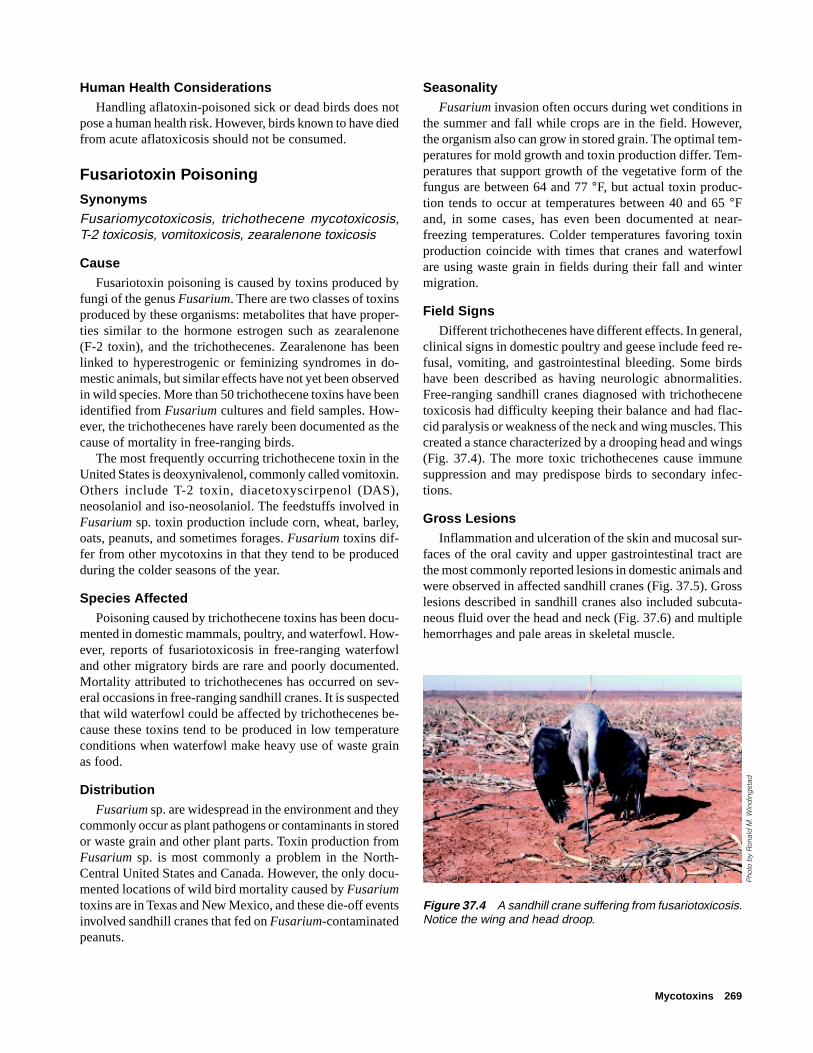
Mycotoxins 269
Human Health ConsiderationsHandling aflatoxin-poisoned sick or dead birds does not
pose a human health risk. However, birds known to have diedfrom acute aflatoxicosis should not be consumed.
Fusariotoxin Poisoning
SynonymsFusariomycotoxicosis, trichothecene mycotoxicosis,T-2 toxicosis, vomitoxicosis, zearalenone toxicosis
CauseFusariotoxin poisoning is caused by toxins produced by
fungi of the genus Fusarium. There are two classes of toxinsproduced by these organisms: metabolites that have proper-ties similar to the hormone estrogen such as zearalenone(F-2 toxin), and the trichothecenes. Zearalenone has beenlinked to hyperestrogenic or feminizing syndromes in do-mestic animals, but similar effects have not yet been observedin wild species. More than 50 trichothecene toxins have beenidentified from Fusarium cultures and field samples. How-ever, the trichothecenes have rarely been documented as thecause of mortality in free-ranging birds.
The most frequently occurring trichothecene toxin in theUnited States is deoxynivalenol, commonly called vomitoxin.Others include T-2 toxin, diacetoxyscirpenol (DAS),neosolaniol and iso-neosolaniol. The feedstuffs involved inFusarium sp. toxin production include corn, wheat, barley,oats, peanuts, and sometimes forages. Fusarium toxins dif-fer from other mycotoxins in that they tend to be producedduring the colder seasons of the year.
Species AffectedPoisoning caused by trichothecene toxins has been docu-
mented in domestic mammals, poultry, and waterfowl. How-ever, reports of fusariotoxicosis in free-ranging waterfowland other migratory birds are rare and poorly documented.Mortality attributed to trichothecenes has occurred on sev-eral occasions in free-ranging sandhill cranes. It is suspectedthat wild waterfowl could be affected by trichothecenes be-cause these toxins tend to be produced in low temperatureconditions when waterfowl make heavy use of waste grainas food.
DistributionFusarium sp. are widespread in the environment and they
commonly occur as plant pathogens or contaminants in storedor waste grain and other plant parts. Toxin production fromFusarium sp. is most commonly a problem in the North-Central United States and Canada. However, the only docu-mented locations of wild bird mortality caused by Fusariumtoxins are in Texas and New Mexico, and these die-off eventsinvolved sandhill cranes that fed on Fusarium-contaminatedpeanuts.
SeasonalityFusarium invasion often occurs during wet conditions in
the summer and fall while crops are in the field. However,the organism also can grow in stored grain. The optimal tem-peratures for mold growth and toxin production differ. Tem-peratures that support growth of the vegetative form of thefungus are between 64 and 77 °F, but actual toxin produc-tion tends to occur at temperatures between 40 and 65 °Fand, in some cases, has even been documented at near-freezing temperatures. Colder temperatures favoring toxinproduction coincide with times that cranes and waterfowlare using waste grain in fields during their fall and wintermigration.
Field SignsDifferent trichothecenes have different effects. In general,
clinical signs in domestic poultry and geese include feed re-fusal, vomiting, and gastrointestinal bleeding. Some birdshave been described as having neurologic abnormalities.Free-ranging sandhill cranes diagnosed with trichothecenetoxicosis had difficulty keeping their balance and had flac-cid paralysis or weakness of the neck and wing muscles. Thiscreated a stance characterized by a drooping head and wings(Fig. 37.4). The more toxic trichothecenes cause immunesuppression and may predispose birds to secondary infec-tions.
Gross LesionsInflammation and ulceration of the skin and mucosal sur-
faces of the oral cavity and upper gastrointestinal tract arethe most commonly reported lesions in domestic animals andwere observed in affected sandhill cranes (Fig. 37.5). Grosslesions described in sandhill cranes also included subcuta-neous fluid over the head and neck (Fig. 37.6) and multiplehemorrhages and pale areas in skeletal muscle.
Figure 37.4 A sandhill crane suffering from fusariotoxicosis.Notice the wing and head droop.
Pho
to b
y R
onal
d M
. Win
din
gst
ad

270 Field Manual of Wildlife Diseases: Birds
DiagnosisDiagnosis is made through observing the appropriate field
signs, finding gross as well as microscopic tissue lesions,and detecting the suspected toxin in grains, forages, or theingesta of affected animals. However, the tests required todetect these toxins are complex and few diagnostic laborato-ries offer tests for multiple trichothecenes. The samples ofchoice include both refrigerated and frozen carcasses fornecropsy examination and a representative sample of thesuspected contaminated grain source. Because the toxin isproduced under cold conditions, the grain sample should befrozen rather than refrigerated for shipment to the diagnos-tic laboratory.
ControlWildlife should not be fed grain with levels of fusario-
toxins in excess of those recommended for use in domesticanimal food. In years when fusariotoxins are a problem, grainfrom fields that are frequently used by wildlife should bechecked for toxins. If the fields are significantly contami-nated, deep plowing of the contents of the field can make thegrain unavailable to wildlife (Fig. 37.7). If the fields cannotbe plowed, hazing wildlife from the area can lessen theirexposure.
Human Health ConsiderationsHandling fusariotoxin-poisoned sick or dead birds does
not pose a human health risk, but birds known to have diedfrom acute fusariotoxicosis should not be consumed. Themore potent trichothecenes in grain or forages may present ahealth hazard when the fungal spores and contaminated plantparts are inhaled or when they contact the skin.
Lynn H. Creekmore
Supplementary ReadingPier, AC., 1981, Mycotoxins and animal health, in Cornelius,
C.E., and Simpson, eds., Advances in veterinary science andcomparative medicine: New York, Academic Press,p. 186–243.
Robinson, R.M., Ray, A.C., Reagor, J.C., and Holland, L.A.,1982, Waterfowl mortality caused by aflatoxicosis in Texas:Journal of Wildlife Diseases, v. 18, p. 311–313.
Roffe, T.J., Stroud, R.K., and Windingstad, R.M., 1989, Suspectedfusariomycotoxicosis in sandhill cranes (Grus canadensis):clinical and pathological findings: Avian Diseases, v. 33,p. 451–457.
Windingstad, R.M., Cole, R.J., Nelson, P.E., Roffe, T.J., George,R.R., and Dorner, J. W., 1989, Fusarium mycotoxins frompeanuts suspected as a cause of sandhill crane mortality:Journal of Wildlife Diseases, v. 25, p. 38–46.
Wobeser, G.A., 1997, Mycotoxins, in Diseases of wild waterfowl(2nd ed): New York, N.Y., Plenum Press, p. 189–193.
Figure 37.5 Inflammation and ulceration of the mucosal sur-face of the esophagus in a sandhill crane with fusariotoxicosis.
Pho
to b
y Ja
mes
Run
ning
en
Figure 37.6 Fluid beneath the skin of the head and neck ofa sandhill crane with fusariotoxicosis.
Pho
to b
y J.
Chr
istia
n Fr
anso
n
Figure 37.7 Plowing of this field made these peanuts un-available to the cranes and ended a fusariotoxicosis mortalityevent.
Pho
to b
y R
onal
d M
. Win
din
gst
ad

Avian Botulism 271
Chapter 38
Avian BotulismSynonymsLimberneck, Western duck sickness, duck disease,alkali poisoning
CauseAvian botulism is a paralytic, often fatal, disease of birds
that results when they ingest toxin produced by the bacte-rium, Clostridium botulinum. Seven distinct types of toxindesignated by the letters A to G have been identified (Table38.1). Waterfowl die-offs due to botulism are usually causedby type C toxin; sporadic die-offs among fish-eating birds,such as common loons and gulls, have been caused by typeE toxin. Type A botulinum toxin has also caused disease inbirds, most frequently in domestic chickens. Types B, D, F,and G are not known to cause avian botulism in NorthAmerica.
Table 38.1 Botulinum toxins and primary species affected.
Toxin type Animals affected Risk for humans
A Poultry, occasionally High B Horses High C Wild birds, cattle, Low
horses, poultry D Cattle Low E Fish-eating birds High F * Unknown G * Unknown
* Rarely detected in nature; too little information for species evaluations.
C. botulinum is an oxygen-intolerant or anaerobic bacte-rium that persists in the form of dormant spores when envi-ronmental conditions are adverse. The spores are resistant toheating and drying and can remain viable for years. Sporesof type C botulism strains are widely distributed in wetlandsediments; they can also be found in the tissues of most wet-land inhabitants, including aquatic insects, mollusks, andcrustacea and many vertebrates, including healthy birds.Botulinum toxin is produced only after the spores germi-nate, when the organism is actively growing and multiply-ing. Although the bacteria provide the mechanism for toxinproduction, the gene that encodes for the toxin protein isactually carried by a virus or phage that infects the bacteria.Unfortunately, little is known about the natural factors thatcontrol phage infection and replication within the bacteria,but several factors may play a role, including the bacterial
host strain and environmental characteristics, such as tem-perature and salinity.
Because botulinum spores and the phages that carry thetoxin gene are so prevalent in wetlands, they are not consid-ered to be a limiting factor in the occurrence of outbreaks inwaterbirds. Other factors are thought to be more critical inthe timing and location of botulism outbreaks; these includeoptimal environmental conditions for spore germination andbacterial growth, suitable material or substrates that provideenergy for bacterial replication, and a means of toxin trans-fer to birds. It is likely that toxin production, toxin availabil-ity to birds and, subsequently, botulism outbreaks in birdsare largely controlled by these ecological factors.
As with other bacteria, temperature plays a critical role inthe multiplication of C. botulinum, with optimal growth inthe laboratory occurring between 25 ° and 40 °C. Most botu-lism outbreaks take place during the summer and fall whenambient temperatures are high (Fig. 38.1). Winter botulismoutbreaks have been documented in some locations, but theseare generally thought to be due to residual toxin producedduring the previous summer. Conditions that elevate wet-land sediment temperatures and decrease dissolved oxygen,including the presence of decaying organic matter and shal-
JAN FEB MAR APRIL MAY JUNE JULY AUG SEPT OCT NOV DEC
PROB
ABIL
ITY
OF A
VIAN
BOT
ULIS
M O
UTBR
EAK
MONTH
HIGH
MODERATE
LOW
Figure 38.1 Relative seasonal probability of type C botu-lism in North American water birds.
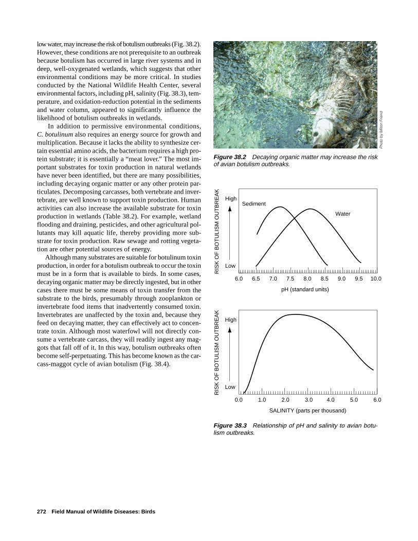
272 Field Manual of Wildlife Diseases: Birds
low water, may increase the risk of botulism outbreaks (Fig. 38.2).However, these conditions are not prerequisite to an outbreakbecause botulism has occurred in large river systems and indeep, well-oxygenated wetlands, which suggests that otherenvironmental conditions may be more critical. In studiesconducted by the National Wildlife Health Center, severalenvironmental factors, including pH, salinity (Fig. 38.3), tem-perature, and oxidation-reduction potential in the sedimentsand water column, appeared to significantly influence thelikelihood of botulism outbreaks in wetlands.
In addition to permissive environmental conditions,C. botulinum also requires an energy source for growth andmultiplication. Because it lacks the ability to synthesize cer-tain essential amino acids, the bacterium requires a high pro-tein substrate; it is essentially a “meat lover.” The most im-portant substrates for toxin production in natural wetlandshave never been identified, but there are many possibilities,including decaying organic matter or any other protein par-ticulates. Decomposing carcasses, both vertebrate and inver-tebrate, are well known to support toxin production. Humanactivities can also increase the available substrate for toxinproduction in wetlands (Table 38.2). For example, wetlandflooding and draining, pesticides, and other agricultural pol-lutants may kill aquatic life, thereby providing more sub-strate for toxin production. Raw sewage and rotting vegeta-tion are other potential sources of energy.
Although many substrates are suitable for botulinum toxinproduction, in order for a botulism outbreak to occur the toxinmust be in a form that is available to birds. In some cases,decaying organic matter may be directly ingested, but in othercases there must be some means of toxin transfer from thesubstrate to the birds, presumably through zooplankton orinvertebrate food items that inadvertently consumed toxin.Invertebrates are unaffected by the toxin and, because theyfeed on decaying matter, they can effectively act to concen-trate toxin. Although most waterfowl will not directly con-sume a vertebrate carcass, they will readily ingest any mag-gots that fall off of it. In this way, botulism outbreaks oftenbecome self-perpetuating. This has become known as the car-cass-maggot cycle of avian botulism (Fig. 38.4).
Figure 38.2 Decaying organic matter may increase the riskof avian botulism outbreaks.
Figure 38.3 Relationship of pH and salinity to avian botu-lism outbreaks.
6.0 6.5 7.0 7.5 8.0 8.5 9.0 9.5 10.0
Sediment
WaterR
ISK
OF
BO
TU
LIS
M O
UT
BR
EA
K
High
Low
pH (standard units)
0.0 1.0 2.0 3.0 4.0 5.0 6.0
RIS
K O
F B
OT
ULI
SM
OU
TB
RE
AK
High
Low
SALINITY (parts per thousand)
Pho
to b
y M
ilton
Frie
nd

Avian Botulism 273
Table 38.2 Human activities speculated to contribute to avianbotulism outbreaks in wetlands.
Action Consequences of action
Fluctuating water levels Deaths of terrestrial and for flooding and drying aquatic invertebrates and
fish
Pesticides and other Deaths of aquatic life chemical inputs into wetlands from agriculture
Raw sewage discharges Nutrient enhancement into wetlands resulting in “boom and
bust” invertebrate popula-tions and oxygen depletioncausing deaths of aquaticand plant life
Carcass-maggot cycle of avian botulism
Maggotsconcentrate toxin
Toxin production takes place indecaying animal carcasses. Flies deposit eggs on carcasses which are fed upon by resulting maggots
Toxic maggots are ingested
Death and additional carcassesfor toxin production and toxicmaggots for other birds tofeed on
Cycle accelerates — major die-off occurs
60 – 92 °F
Figure 38.4 Carcass-maggot cycle of avian botulism.

274 Field Manual of Wildlife Diseases: Birds
Species AffectedMany species of birds and some mam-
mals are affected by type C botulism. In thewild, waterbirds suffer the greatest losses,but almost all birds are susceptible to typeC botulism. The exception is vultures, whichare highly resistant to type C toxin. Forag-ing behavior is probably the most significanthost determinant for botulism. Filter-feedingand dabbling waterfowl and probing shore-birds appear to be among the species at great-est risk (Fig. 38.5). Mortality of wild rap-tors from botulism has been associated withimproper disposal of poultry carcasses.Among captive and domestic birds, pheas-ants, poultry, and waterfowl are the mostfrequently affected (Fig. 38.6). Cattle,horses, and ranch mink are also susceptibleto type C botulism. Although dogs and catsare usually regarded as being resistant totype C toxin, a few cases have been reportedin dogs, which is a factor to consider whendogs are used to retrieve carcasses duringoutbreaks. Also, type C botulism occurredin captive African lions which were fedtoxin-laden chickens.
Losses vary a great deal from year toyear at site-specific locations and from spe-cies to species. A few hundred birds maydie one year and tens of thousands or morethe following year. More than a milliondeaths from avian botulism have been re-ported in relatively localized outbreaks in asingle year, and outbreaks with losses of50,000 birds or more are relatively common(Table 38.3).
On a worldwide basis, avian botulism isprobably the most important disease of mi-gratory birds.
Figure 38.5 Frequency of botulism in major groups of wildbirds.
Waterfowl
Type C
Type E
Shorebirds
Type C
Type E
Gulls
Type C
Type E
Loons
Type C
Type E
Upland gamebirds
Type C
Type E
Herons
Type C
Type E
Raptors
Type C
Type E
Songbirds
Type C
Type E
Common; die-offs occur almost yearlyFrequent
OccasionalInfrequent
Not reported

Avian Botulism 275
Table 38.3 Major waterfowl botulism outbreaks in theUnited States and Canada.
Location Year Estimated loss
Utah and California 1910 “Millions” Lake Malheur, Oregon 1925 100,000 Great Salt Lake, Utah 1929 100,000–
300,000 Tulare Basin, California 1941 250,000 Western United States 1952 4–5 million Montana 1978 50,000 Montana 1979 100,000 Great Salt Lake, Utah 1980 110,000 Canada (Alberta) 1995 100,000 Canada (Manitoba) 1996 117,000 Canada (Saskatchewan) 1997 1 million Great Salt Lake, Utah 1997 514,000
Pheasants
N. America
Europe
Domestic chickens
N. America
Europe
Songbirds
N. America
Europe
Waterfowl
N. America
Europe
FrequentOccasional
InfrequentRare or not reported
Figure 38.6 Frequency of botulism in captive birds.
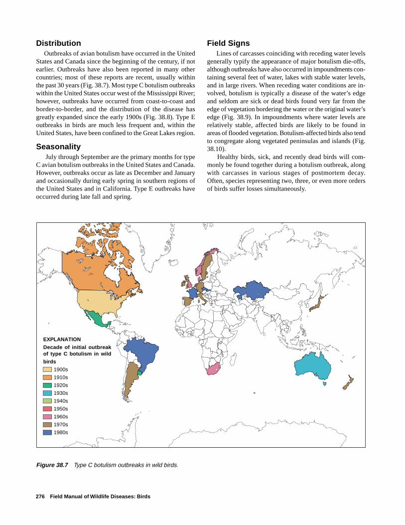
276 Field Manual of Wildlife Diseases: Birds
DistributionOutbreaks of avian botulism have occurred in the United
States and Canada since the beginning of the century, if notearlier. Outbreaks have also been reported in many othercountries; most of these reports are recent, usually withinthe past 30 years (Fig. 38.7). Most type C botulism outbreakswithin the United States occur west of the Mississippi River;however, outbreaks have occurred from coast-to-coast andborder-to-border, and the distribution of the disease hasgreatly expanded since the early 1900s (Fig. 38.8). Type Eoutbreaks in birds are much less frequent and, within theUnited States, have been confined to the Great Lakes region.
Seasonality July through September are the primary months for type
C avian botulism outbreaks in the United States and Canada.However, outbreaks occur as late as December and Januaryand occasionally during early spring in southern regions ofthe United States and in California. Type E outbreaks haveoccurred during late fall and spring.
Field Signs Lines of carcasses coinciding with receding water levels
generally typify the appearance of major botulism die-offs,although outbreaks have also occurred in impoundments con-taining several feet of water, lakes with stable water levels,and in large rivers. When receding water conditions are in-volved, botulism is typically a disease of the water’s edgeand seldom are sick or dead birds found very far from theedge of vegetation bordering the water or the original water’sedge (Fig. 38.9). In impoundments where water levels arerelatively stable, affected birds are likely to be found inareas of flooded vegetation. Botulism-affected birds also tendto congregate along vegetated peninsulas and islands (Fig.38.10).
Healthy birds, sick, and recently dead birds will com-monly be found together during a botulism outbreak, alongwith carcasses in various stages of postmortem decay.Often, species representing two, three, or even more ordersof birds suffer losses simultaneously.
Figure 38.7 Type C botulism outbreaks in wild birds.
EXPLANATION
Decade of initial outbreak of type C botulism in wild
birds
1900s
1910s
1920s
1930s
1940s
1950s
1960s
1970s
1980s
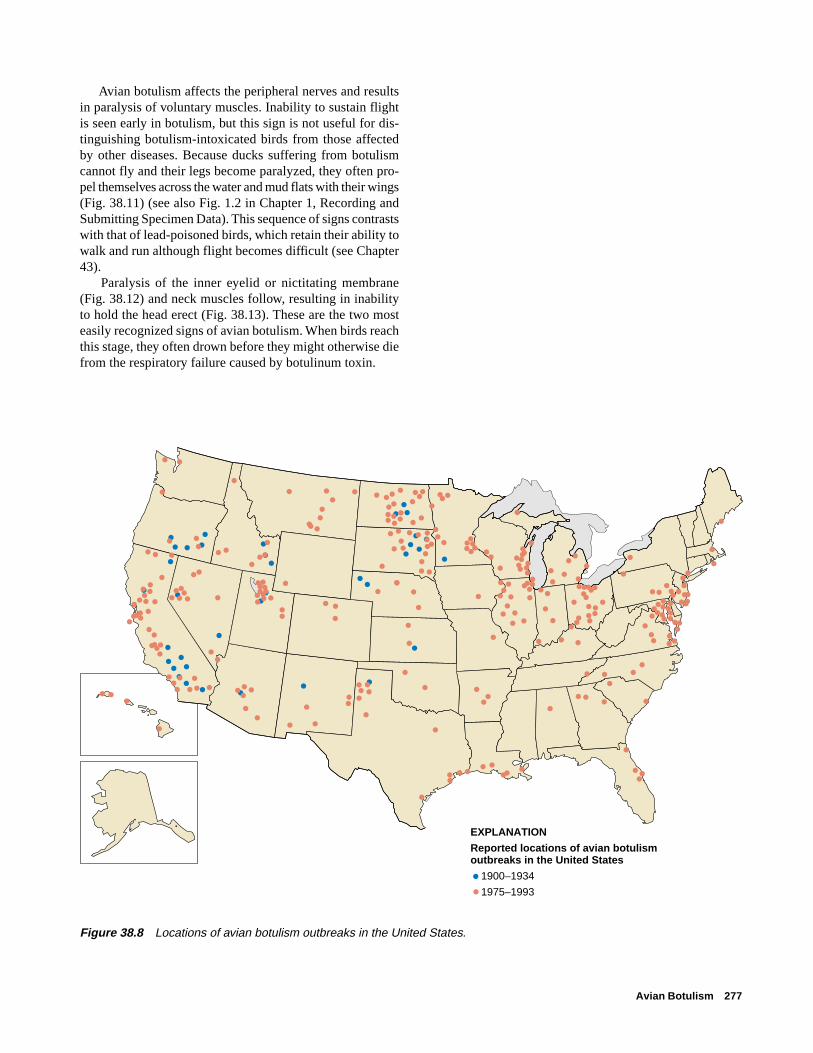
Avian Botulism 277
Avian botulism affects the peripheral nerves and resultsin paralysis of voluntary muscles. Inability to sustain flightis seen early in botulism, but this sign is not useful for dis-tinguishing botulism-intoxicated birds from those affectedby other diseases. Because ducks suffering from botulismcannot fly and their legs become paralyzed, they often pro-pel themselves across the water and mud flats with their wings(Fig. 38.11) (see also Fig. 1.2 in Chapter 1, Recording andSubmitting Specimen Data). This sequence of signs contrastswith that of lead-poisoned birds, which retain their ability towalk and run although flight becomes difficult (see Chapter43).
Paralysis of the inner eyelid or nictitating membrane(Fig. 38.12) and neck muscles follow, resulting in inabilityto hold the head erect (Fig. 38.13). These are the two mosteasily recognized signs of avian botulism. When birds reachthis stage, they often drown before they might otherwise diefrom the respiratory failure caused by botulinum toxin.
EXPLANATION
Reported locations of avian botulism outbreaks in the United States
1900–1934
1975–1993
●
● ●
●
●
●
●
●
●
●
●
●●
●
● ●
●
●
●
●●
●
●
●●
●
●●
●●
●●
●●
● ●●
●
●
●●
●
●
●
●
● ●
●
●
●
●
●●
●
●
● ●
●
●
●
●
●●
●
●●
●
●
●
●
●●
●●
●●
●
●●
●● ● ●
●●●●
●
●●
●
●
●●
●
●
●
●
●● ●
●●●
●
●●●
●
●
●
● ●
●●●
●●
●●
●
●
●
●
●● ●●●●● ●
● ●
●
●
●
●
●
●●
●
●
●●●
●● ●
●● ●
●●
●
●●
●
●●●
●●
●
●
● ●
●
●
●●
●●
●
●
●
●
●
●●
●●●
●●●
●●●●
●
●●
●● ●● ●
●
●
●
●
●
●
●
●
●
●
●●●
●●
●●● ●●●
●
●●
●
●
●● ●●
●●●●●
● ● ●●●
●●
●●●●
●●●
●
●●●
●
●
●
●
●●
●
●
●
●
●
●
●
●
●
●
●
●
●
●
●●
●
●
●
●●
●●●
●●
●
●
●●
●●●
●
●●●●
●
●
●●●●
●
●
●●
●
●●
●
●
●●
●
●●●
●
●
●
● ●
●● ●
●●
●
●
●●
●
Figure 38.8 Locations of avian botulism outbreaks in the United States.

278 Field Manual of Wildlife Diseases: Birds
Figure 38.13 Paralysis of the neck muscles in botulism-intoxicated birds results in inability to hold the head erect(limberneck). Death by drowning often results.
Figure 38.9 Typical scene of avian botulism. Dead birds areoften found along the shore in parallel rows that representreceding water levels.
Figure 38.10 Botulism-affected birds tend to congregatealong vegetated peninsulas and islands. Both dead and sickbirds are evident in the photograph.
Figure 38.11 Botulism-intoxicated birds that have lost thepower of flight and use of their legs often attempt escape bypropelling themselves across water or land using their wings.
Figure 38.12 Paralysis of the inner eyelid is a common signin botulism-intoxicated birds.
Pho
to b
y Ja
mes
Run
ning
en
Pho
to b
y M
ilton
Frie
ndP
hoto
by
Milt
on F
riend
Pho
to b
y M
ilton
Frie
nd
Pho
to b
y M
ilton
Frie
nd

Avian Botulism 279
Gross LesionsThere are no characteristic or diagnostic gross lesions in
waterfowl dying of either type C or type E botulism. Often,affected birds die by drowning, and lesions associated withdrowning may be present.
DiagnosisThe most widely used test for avian botulism is the mouse-
protection test, although an enzyme-linked immunosorbentassay (ELISA) for type C toxin has been developed recently.For the mouse test, blood is collected from a sick or freshlydead bird and the serum fraction is then inoculated into twogroups of laboratory mice, one group of which has been giventype-specific antitoxin. The mice receiving antitoxin will sur-vive, and those that receive no antitoxin will become sickwith characteristic signs or die if botulism toxin is present inthe serum sample. The ELISA is an in vitro test that detectsinactive as well as biologically active toxin.
A presumptive diagnosis is often based on a combinationof signs observed in sick birds and the absence of obviouslesions of disease when the internal organs and tissues ofsick and dead birds are examined. However, this initial diag-nosis must be confirmed by the mouse-protection or ELISAtest to separate avian botulism from algal poisoning, castor-bean poisoning, and other toxic processes that cause similarsigns of disease. Avian botulism should be suspected whenmaggots are found as part of the ingesta of gizzard contentsof dead birds (Fig. 38.14), however, such findings are rare.After a bird ingests toxin, it takes several hours to days be-fore the bird develops signs of the disease and dies. By thistime, most food items ingested at the time of intoxicationhave been eliminated.
Prevention and ControlPrevention of avian botulism outbreaks in waterbirds will
depend on a thorough understanding of the interactions be-tween the agent, the host, and the environment. Because botu-lism spores are so ubiquitous in wetlands and are resilient,attempts to reduce or eliminate the agent are not currentlyfeasible, but some actions can be taken to mitigate environ-mental conditions that increase the likelihood of outbreaks.
Management of EnvironmentAttempts should be made to reduce organic inputs into
wetlands or to eliminate factors that introduce large amountsof decaying matter. For example, in areas that are managedprimarily for migratory waterfowl (ducks, geese, swans),reflooding land that has been dry for a long time is not rec-ommended during the summer. Similarly, avoid sharp waterdrawdowns in the summer because they could result in fish-kills and die-offs of aquatic invertebrates whose carcassescould then become substrates for C. botulinum growth. Inareas managed primarily for shorebirds, water drawdownsprovide essential habitat; thus, botulism control must focuson cleaning up any vertebrate carcasses that may result fromdrawdowns.
Prompt removal and proper disposal of vertebrate car-casses by burial or burning, especially during outbreaks, arehighly effective for removing substrates for toxin produc-tion. The importance of prompt and thorough carcass removaland proper disposal cannot be overemphasized. Several thou-sand toxic maggots can be produced from a single water-fowl carcass (Fig. 38.15). Consumption of as few as two tofour of these toxic maggots can kill a duck, thereby perpetu-ating the botulism cycle. It is not uncommon to find three or
Figure 38.14 Waterfowl and other birds readily feed onmaggots as exemplified by their presence in the gizzard ofthis duck that died from avian botulism.
Figure 38.15 Several thousand toxic maggots can be pro-duced from a single waterfowl carcass.
Pho
to b
y M
ilton
Frie
nd
Pho
to b
y M
ilton
Frie
nd

280 Field Manual of Wildlife Diseases: Birds
four freshly dead birds within a few feet of a maggot-ladencarcass. Failure to carry out adequate carcass removal anddisposal programs can cause a rapid build-up of highly toxicdecaying matter and toxin-laden maggots, thereby acceler-ating losses in waterbirds, as well as seeding the environ-ment with more botulism spores as the carcasses decompose.
Many botulism outbreaks occur on the same wetlands yearafter year and within a wetland there may be localized “hotspots.” Also, outbreaks often follow a fairly consistent andpredictable timeframe. These conditions have direct man-agement implications that should be applied towards mini-mizing losses. Specific actions that should be taken includeaccurately documenting conditions and dates of outbreaksin problem areas, planning for and implementing intensifiedsurveillance and carcass pickup and disposal, and modify-ing habitat to reduce the potential for botulism losses or denybird use of major problem areas during the botulism “sea-son” or both.
Management actions for minimizing lossesfrom avian botulism
Document environmental conditions, specific im-poundments or areas of outbreaks, and dates of occur-rence and cessation.
Plan for and implement intensive surveillance and ver-tebrate carcass pickup and disposal starting 10–15 daysbefore the earliest documented cases until 10–15 daysafter the end of the botulism “season.”
Where possible, monitor and modify environmentalconditions to prevent the pH and salinity of wetlandsfrom reaching or being maintained within high hazardlevels.
Avoid water drawdowns for rough fish and vegetationcontrol during warm weather. Collect vertebrate car-casses (fish) and properly dispose of them if drawdownsare necessary during summer and warm fall months.
Construct wetland impoundments in botulism-proneareas in a manner that facilitates rapid and completedrainage thereby encouraging bird movement to alter-native impoundments.
Because fish carcasses can also serve as sites for C. botu-linum growth, they should be promptly removed during fishcontrol programs in marshes, or fish control programs shouldbe restricted to the cooler months of year (the nonfly sea-son). Also, bird collisions with power lines that cross marsheshave been the source of major botulism outbreaks becausecarcasses from these collisions served as initial substratesfor toxin production within marshes. Therefore, if possible,power lines should not be placed across marshes used bylarge concentrations of waterbirds.
Numerous outbreaks of avian botulism have been associ-ated with sewage and other wastewater discharges intomarshes. This relationship is not presently understood, butoutbreaks have occurred often enough that wetland manag-ers should discourage the discharges of these effluents whenmany waterfowl or shorebirds are using the area or are likelyto use an area during warm weather.
Treatment of Sick BirdsBotulism-intoxicated waterfowl can recover from the dis-
ease. If sick birds are provided with freshwater and shade, orinjected with antitoxin, recovery rates of 75 to 90 percentand higher can result (Fig. 38.16). In contrast to waterfowl,very few coot, shorebirds, gulls, and grebes survive botu-lism intoxication, even after treatment. Experience to datewith these species indicates that rehabilitation efforts maynot be worthwhile.
Because avian botulism most often afflicts waterfowl inthe seasons when they are flightless due to wing molt, biolo-gists and rehabilitators must be careful to distinguish betweenbirds in molt and birds with early stages of botulism, be-cause the behavior of these birds may be similar. Moltingbirds are very difficult to catch, and birds that cannot be cap-tured with a reasonable effort should not be pursued further.Birds that are suffering from botulism can easily be capturedwhen they lose the ability to dive to escape pursuit. Birds atthis level of intoxication still have a high probability of sur-viving if proper treatment is administered.
When botulism-intoxicated birds are treated, they shouldbe maintained under conditions or holding pens that providefree access to freshwater, maximum provision for shade, theopportunity for recovered birds to fly out of the enclosurewhen they choose to, and minimum disturbance (includingthe presence of humans). It is also important to remove car-casses daily from holding pens to prevent the buildup of toxicmaggots.
Costs associated with capturing and treating sick birdsare high. Therefore, the emphasis for dealing with avian botu-lism should be on prevention and control of this disease ratherthan on treatment of intoxicated birds. However, antitoxinshould be available for use in case endangered species areaffected.
Human Health ConsiderationsBotulism in humans is usually the result of eating im-
properly home-canned foods and is most often caused bytype A or type B botulinum toxin. There have been severalhuman cases of type E botulism in North America from eat-ing improperly smoked or cooked fish or marine products.Type C botulism has not been associated with disease in hu-mans, although several outbreaks have been reported in cap-tive primates. Thorough cooking destroys botulinum toxinin food.

Avian Botulism 281
Figure 38.16 Recovery rates of ducks in response to antitoxin therapy.
Tonie E. Rocke and Milton Friend
Supplementary Reading
Eklund, M.W., and Dowell, V.R., eds., 1987, Avian Botulism: AnInternational Perspective: Springfield, Ill., Charles C. Thomas,405 p.
Hariharan, H., and Mitchell, W.R., 1977, Type C botulism: theagent, host spectrum and environment: The VeterinaryBulletin, v. 47, p. 95–103.
Reed, T.M., and Rocke, T.E., 1992, The role of avian carcasses inbotulism epizootics: The Wildlife Society Bulletin, v. 20,p. 175–182
Rocke, T.E., 1993, Clostridium botulinum, in Gyles, C.L. andThoen, C.O., eds., Pathogenisis of bacterial infections inanimals (2d ed.): Ames, Iowa, Iowa State University Press,p. 86–96.
Rocke, T.E., Smith, S.R., and Nashold, S.W., 1998, Preliminaryinvestigation of the in vitro test for the diagnosis of avianbotulism in wild birds: The Journal of Wildlife Diseases, v. 34p. 744–751.
Percentage recoveredAmount of antitoxin Moderate1 Severe1
Duck type (cc) clinical signs clinical signs
Pintail
0.5 91.9 69.0
1.0 93.5 73.3
Green-winged teal
0.5 93.5 57.4
1.0 91.2 58.1
Mallard
0.5 81.6 66.3
1.0 84.9 67.8
Shoveler
0.5 89.2 53.1
1.0 95.1 57.61 Condition of bird when treated with antitoxin.

282 Field Manual of Wildlife Diseases: Birds

Introduction to Chemical Toxins 283
Section 7Chemical ToxinsOrganophosphorus andCarbamate Pesticides
Chlorinated Hydrocarbon Insecticides
Polychlorinated Biphenyls
Oil
Lead
Selenium
Mercury
Cyanide
Salt
Barbiturates
Miscellaneous Chemical Toxins
Oiling disrupts normal feather structures and functionPhoto by Nancy J. Thomas
284 Field Manual of Wildlife Diseases: Birds
Introduction to Chemical Toxins
Many kinds of potentially harmful chemicals are found inenvironments used by wildlife. Some chemicals, such aspesticides and polychlorinated biphenyls (PCBs), are syn-thetic compounds that may become environmental contami-nants through their use and application. Other materials, suchas selenium and salt, are natural components of some envi-ronments, but contaminants of others. Natural and syntheticmaterials may cause direct poisoning and death, but they alsomay have adverse effects on wildlife that impair certain bio-logical systems, such as the reproductive and immune sys-tems. This section provides information about some of theenvironmental contaminants and natural chemicals that com-monly cause avian mortality; microbial and other biotoxinsare addressed in the preceding section.
Direct poisoning and mortality of wildlife caused by ex-posure to chemical toxins are the focus of this section. How-ever, the indirect effects of chemicals may have significantlygreater impacts on wildlife populations than the directeffects. Behavioral changes that affect survival, reproduc-tive success and the survival of young, and that impair thefunctioning of the immune system are examples of indirectchemical toxicity that are known to occur but that are be-yond the scope of this publication. For additional informa-tion readers are directed to more comprehensive treatmentsof environmental toxicology and to publications that focuson specific chemicals and their effects on wildlife.
The diagnosis of chemical poisoning as the cause of wild-life mortality is a challenging task because of the vast arrayof chemicals that wildlife may be exposed to (Table 1), thevariable biological responses following concurrent exposureto multiple chemicals, the absence of tissue residues for some
chemical toxins, and the lack of specific pathological changesassociated with most chemical toxins in tissues. The diag-nostic process can be greatly facilitated by a thorough fieldobservation record, comprehensive background informationabout the circumstances of a mortality event, and by prop-erly collecting, handling, and preserving samples submittedto the diagnostic laboratory (see Section 1). Sources of as-sistance for the investigation of wildlife mortality, when tox-ins are suspected, are listed in Appendix B.
Areas CoveredThe chapters that follow address chemical toxins that are
recurrent causes of avian mortality. The chapters discusschemicals that cause frequent and sometimes large-scalemortality events, as well as some chemicals that are less sig-nificant, because they are restricted to certain geographicareas or have been recently recognized as emerging problems.
PesticidesOrganophosphorus and carbamate compoundsChlorinated hydrocarbons
Polychlorinated biphenylsOilLeadSeleniumMercuryCyanideSaltBarbiturates
“Dosage alone determines poisoning.”(Translation of Paracelsus)
Quote from:
Philipus Aureolus Paracelsus, a German-Swiss physician andalchemist who lived from 1493 to 1541.

Introduction to Chemical Toxins 285
Pesticides
This group includes chemicals that are used to kill or repel organisms that are unwanted in particular situations.Insecticides are generally the best known pesticides but others, their target organisms, and examples of com-pounds within those groups include the following:
Pesticide type Target organisms Compounds
Acaricides Mites, ticks, spiders Permethrin, Phosmet, Methiocarb, Bomyl®, Carbofuran, Demeton(Systox®)
Algacides Algae Copper sulfate, Potassium bromide, Chlorine
Antibiotics Bacteria Phenol, Nitrapyrin
Avicides Birds Avitrol®, Fenthion, Compound 1080, Starlicide®
Fungicides Fungi Thiram, Ziram, Captan, Hexaconazole
Herbicides Plants Diquat®, Alachlor (Lasso®), Atrazine
Molluscicides Snails and slugs Bayluscide®, Methiocarb, Zectran®
Nematocides Nematodes (worms) Terbufos (Counter®), Isazofos (Triumph®), Aldicarb (Temik®),Carbofuran, Diazinon
Piscicides Fish Rotenone, Antimycin
Repellents Mammals ThiramBirds Methiocarb
Rodenticides Rodents Warfarin, Diphacinone, Brodifacoum (Talon®), Chlorophacinone
Metals
Wildlife may be exposed to metals when they are components of pesticides, such as mercury and cadmium infungicides, or through other routes, such as aquatic food chains with high mercury levels.
Metal Source
Arsenic Used as an insecticide and preservative; present in wastes from metal smelting and glassmanufacturing.
Cadmium Used as a fungicide; waste from electroplating and production of plastics and batteries.
Chromium Industrial effluents from ore refinement, chemical processing.
Copper Used as a fungicide, an algicide, and in agriculture.
Lead Mine tailings, ingestion of particulate lead deposited during sporting activities.
Mercury Used as a fungicide in paper mills and other industrial and agricultural uses; combustion offossil fuels.
Selenium Irrigation drain water from soils with high selenium concentrations; combustion of fossil fuel;sewage sludge.
Zinc Found throughout the environment; higher levels in areas of industrial discharge.
Petroleum
Wildlife may be exposed to many forms of petroleum, ranging from crude oils to highly refined forms, such asfuel oil.
Others
Many manufactured compounds, such as antifreeze (ethylene glycol) and certain drugs (such as euthanasiaagents), present hazards to exposed wildlife.
Table 1 Examples of chemical toxins to which wildlife may be exposed.

286 Field Manual of Wildlife Diseases: Birds

Organophosphorus and Carbamate Pesticides 287
Chapter 39
Organophosphorus and CarbamatePesticidesSynonyms
Organophosphates, OPs
The insecticidal properties of organophosphorus (OP) andcarbamate compounds were first discovered in the 1930s,and the compounds were developed for pesticide use in the1940s. They have been used increasingly since the 1970swhen environmentally persistent organochlorine pesticides,such as DDT and dieldrin, were banned for use in the UnitedStates. Organophosphorus and carbamate pesticides are gen-erally short-lived in the environment (usually lasting onlydays to months instead of years) and, generally, chemicalbreakdown is accelerated as temperatures or pH or both in-crease.
CauseThe toxicity of OP and carbamate pesticides is due to the
disruption of the nervous system of an invertebrate or a ver-tebrate through the inhibition of cholinesterase (ChE) en-zymes. These enzymes are involved in transmitting normalnerve impulses throughout the nervous system. An acutepesticide dose reduces the activity of ChEs, and nerve im-pulses cannot be transmitted normally. This can paralyze thenervous system, and it may lead to death, usually from res-piratory failure.
Species AffectedIt is possible for a wide variety of vertebrate species to be
affected by OP or carbamate pesticides. However, birds ap-pear to be more sensitive than other vertebrates to the toxiceffects of OP and carbamate pesticides. More than 100 avianspecies have been poisoned by these pesticides. Waterfowl,passerines, and raptors are the species most commonly iden-tified in reported OP- and carbamate-related mortalities inthe United States (Fig. 39.1). Raptors and other bird speciesbecome victims of secondary poisoning when they scavengedead animals poisoned by pesticides or when they feed onlive animals or invertebrates that are unable to escape preda-tion because of pesticide intoxication.
Age, sex, diet, and body condition all are factors that af-fect a bird’s susceptibility to pesticide poisoning. Generally,embryos and young birds, particularly the dependant or al-tricial birds, appear to be more sensitive to OP or carbamatecompounds than adults. Dietary deficiencies, low fat reserves,poor physiological condition, and high energy needs, suchas migration or high metabolic rates, may increase vulner-
ability to these compounds. Behavioral traits may also in-crease the potential for exposure to OP or carbamate com-pounds. Species at increased risk are those that congregatein areas of treated habitats, gorge on a food source (likegeese), forage in treated substrates, or feed on target organ-isms shortly after applications of these compounds.
Common routes of exposure of birds to OPand carbamate pesticides include:
Consumption of:Treated seedsVegetation with pesticide residuesDead or struggling poisoned insectsGranular formulations as grit, food, or coinciden-
tally with other food itemsCarrion killed by a pesticideFood intentionally baited with pesticideLive animals intoxicated with pesticideWater contaminated with pesticide from runoff or
irrigationInhalationAbsorption through the skin
Also, there can be considerable variability in the sensitiv-ity of individual species to these pesticides (Table 39.1).
Table 39.1 Toxicity for birds of organophosphorus pesti-cides and carbamate pesticides.[Modified from Hoffman and others, 1995. LD50 is the singleoral dose of pesticide in milligrams per kilogram of bodyweight that is required to kill 50 percent of the experimentalpopulation]
Species LD50
Mallard Ring-necked Red-wingedCompound Class duck pheasant blackbird
Aldicarb Carbamate 3.4 5.3 1.8Carbaryl Carbamate >2,000 707 56Carbofuran Carbamate 0.5 4.1 0.4Methiocarb Carbamate 13 270 4.6Mexacarbate Carbamate 3.0 4.6 10
Azinphos-methyl OP 136 75 8.5Dimethoate OP 42 20 6.6Ethion OP >2,000 1,297 45Phorate OP 0.6 7.1 1.0Temephos OP 79 35 42

288 Field Manual of Wildlife Diseases: Birds
Figure 39.1 Frequency of occurrence of major groups of birds in documented organophospho-rus and carbamate pesticide mortality events from 1986–95 (National Wildlife Health Center database).
Passerines
Jays and crowsBlackbirds
Seed-eating birdsThrushes and bluebirds
SwallowsOther
Waterfowl
GeeseSurface-feeding ducks
Diving ducksOther waterfowl
Raptors
EaglesHawks
OwlsVulturesFalcons
Waterbirds
ShorebirdsWading birds
GullsCoot
Upland gamebirds
DovesOther
Common (greater than 45 percent of die-offs)Frequent (11–44 percent of die-offs)
Occasional (4–10 percent of die-offs) Infrequent (less than 3 percent of die-offs)

Organophosphorus and Carbamate Pesticides 289
DistributionOrganophosphorus and carbamate compounds are used
throughout the world as insecticides, herbicides, nemato-cides, acaricides, fungicides, rodenticides, avicides, and birdrepellants. These compounds are applied in a wide varietyof habitats including agricultural lands, forests, rangelands,wetlands, residential areas, and commercial sites. Wild birddeaths from OP and carbamate poisoning have been reportedthroughout the United States (Fig. 39.2). In more than half
of these mortality incidents, the pesticide source is unknown(Fig. 39.3). Known applications of these compounds fall intofive groups: approved applications in 1) agricultural land usessuch as field and row crops, pastures, orchards, and forests;2) residential and urban sites for turf in parks, golf courses,yards, and other urban pest control uses; 3) livestock usessuch as pour-ons or feed products; 4) vertebrate pest control;and 5) malicious pesticide use, such as baiting to intention-ally harm wildlife (Fig. 39.3).
Figure 39.2 Distribution of 181 avian mortality events caused by organophosphorus and carbamate pesticides, 1986–1995(National Wildlife Health Center data base).
Pesticide
Carbofuran
Diazinon
Famphur
Phorate
Parathion
Monocrotophos
Fenthion
Chlorpyrifos
Other
EXPLANATION
Mortality events—Each circle represents the total number of events for a State. Circle size is proportional to number of events
2015
10
5
1

290 Field Manual of Wildlife Diseases: Birds
Clinical signs and bird behaviors that are com-monly associated with acute exposure tocholinesterase-inhibiting pesticides[Modified from Mineau, 1991]
ConvulsionsHyperexcitabilityIncoordination of muscular action (ataxia)Muscular weakness (myasthenia)Difficult breathing (dyspnea)Rapid breathing (tachypnea)VomitingDefecationDiarrheaSpasmodic contraction of anal sphincter (tenesmus)LethargyInduced tranquilityHead and limbs arched back (opisthotonos)Slight paralysis (paresis)BlindnessContraction of pupils (miosis)Dilation of pupils (mydriasis)Drooping of eyelid (ptosis)Protrusion of eyes (exopthalmia)Excessive tear formation (lacrimation)Excessive thirst (polydypsia)Bleeding from nares (epistaxis)Erection of contour feathers (piloerection)
Birds that die rapidly with pronounced neurological signsmay leave evidence of their struggle even after death, suchas vegetation clenched in their talons (Fig. 39.5) or vegeta-tion that they disturbed during thrashing or convulsions. Ani-mals may not have time to disperse before the toxin takeseffect, and carcasses of multiple species, especially preda-tors and granivorous or insectivorous wildlife, may be foundwithin the same area following OP or carbamate exposure.
Birds can also be affected by a sublethal dose of an OP orcarbamate pesticide. Sublethal exposure may contribute toother causes of mortality in birds, such as trauma. In someinstances when birds have died due to trauma from a vehicleimpact, a building strike, or predation, decreased brain ChEhas been demonstrated, which indicates pesticide exposure.The sublethal dose of pesticide likely impaired the nervoussystem enough to alter behavior, thus making the animal morevulnerable to a traumatic cause of death. Special studies thatevaluated sublethal OP or carbamate compound exposure inbirds have found other effects to birds, including a reducedability to regulate body temperature; impaired reproduction;and reduced tolerance to cold stress, which can cause re-duced activity, leading to decreased feeding and weight loss.Altered behaviors such as reduced nest attentiveness andchanges in singing by passerines have also been observed.
Figure 39.3 Applications associated with avian mortalitycaused by organophosphorus and carbamate pesticides from1986–95 (National Wildlife Health Center data base).
Figure 39.4 Avian mortality events due to organophospho-rus and carbamate pesticides by date of onset, 1986–95(National Wildlife Health Center data base).
SeasonalityBecause OP and carbamate pesticides are typically short-
lived in the environment, seasonality of avian mortality isgenerally associated with pesticide applications (Fig. 39.4).In documented mortality events in the United States, Febru-ary was the peak month for the onset of bird die-offs, andmost of these die-offs occurred in the southern United States,where the growing season starts early in the year.
Field SignsMortality can be the first sign noted in a pesticide poi-
soning, but the observer may find other clues at the scene ofa mortality event. Live affected birds may exhibit convul-sions, lethargy, paralysis, tremors, or other nonspecific neu-rological signs.
0
5
10
15
20
25
30
35
JAN FEB MAR APRIL MAY JUNE JULY AUG SEPT OCT NOV DEC
NU
MB
ER
OF
EV
EN
TS
MONTH
Unknown53.6 percent
Pest control1.7 percent
Golf course1.1 percent
Misuse15.1
Residential8.4 percent
Agricultural20 percent

Organophosphorus and Carbamate Pesticides 291
Gross FindingsGranular material or the presence of dye or both in the
gastrointestinal tract are conspicuous findings that implicatepesticide ingestion. The necropsy finding of freshly ingestedfood in the upper gastrointestinal tract of a carcass is a goodindicator of death by intoxication, especially when a largeamount of a uniform food item is present. Feathers, flesh,hair, or other animal parts in the stomachs of raptors or ofscavengers are common in secondary poisoning, whereasingested grain is often found in waterfowl and passerines(Fig. 39.6). The food item may indicate the pesticide source,and the food can then be analyzed for specific chemical com-pounds.
The gross lesions that are associated with acute mortalityfrom pesticide poisoning in birds are nonspecific and areusually minimal. Reddening of the intestinal wall, or evenhemorrhage (Fig. 39.7), is observed occasionally with in-gestion of certain pesticides. Redness and excess fluid in thelungs may be observed; these findings are consistent withrespiratory failure. However, these changes are not uniqueto pesticide poisonings; they can be found in animals thatdied from other causes.
DiagnosisA diagnostic evaluation is essential. A diagnosis of pesti-
cide poisoning in birds is based on evidence of ChE inhibi-tion in the brain or the blood and identification of pesticideresidues in gastrointestinal contents. In many instances, de-pressed ChE activity will be the first indication that OP orcarbamate pesticides caused a mortality event. A necropsy isnecessary to rule out other causes of mortality or to identifycontributing causes.
Brain ChE activity is a reliable indicator of OP and car-bamate exposure in dead birds, but the absence of ChE de-pression does not reliably rule out poisoning. Brain ChEactivity is measured and compared to normal brain ChE
activity of the same species to determine the decrease in en-zyme activity from normal levels (Appendix D). A decreasein brain ChE activity of 25 percent or more from normalindicates exposure to a cholinesterase-inhibiting compound(OP or carbamate pesticide); a decrease of 50 percent or morefrom normal is evidence of lethal exposure. Because of thevariation in results between laboratories and the variabilityeven between methods and procedures within a lab, it is im-portant to compare results with controls from the same labo-ratory using the same method and not interpret analyticalresults from two or more laboratories or from two or moreanalytical methods.
Analyses can be carried one step further to differentiatethe effect of OP from carbamate compounds by measuringthe enzyme activity of a sample after incubation at 37–40 °C and comparing it to the initial measurement. Enzymeactivity that returns toward a normal level after incubation,or that reactivates, indicates that carbamate poisoning is likelybecause carbamates tend to release their bond with ChE overtime at increased temperatures or in aqueous environments.Because reactivation can occur with some pesticides, de-pressed brain ChE activity in a pesticide-poisoned bird maybe difficult to document if the carcass has remained in a warmenvironment for an extended period of time. Another methodthat is used to differentiate an OP from a carbamate com-pound exposure is reactivation analysis, during which 2-PAM, a cholinesterase regenerating agent, is added to thesample and the change in brain ChE activity is then mea-sured. Reactivation of ChE activity using 2-PAM occurs onlywhen an OP compound is bound to the enzyme.
When a pesticide die-off is suspected, it is important tochill carcasses immediately. If diagnostic evaluation cannotbe initiated within 24–48 hours, carcasses should be frozenas soon as possible to prevent further change in brain ChEactivity. Also, when normal brain ChE activity values arenot known for a particular species, control samples collectedfrom normal birds of the same species are needed in order tocompare ChE values.
In birds that recover from OP or carbamate poisoning,brain ChE activity will typically increase but it may remainbelow normal levels for up to 3 weeks, depending on thecompound and on the dose received. Cholinesterase activityin blood from live birds may be used as an indicator of pes-ticide exposure; however, blood ChE activity is more vari-able than brain ChE activity. Cholinesterase enzymes in theblood are more sensitive than brain ChE to OP and carbam-ate pesticides; therefore, pesticide exposure quickly and dra-matically depresses blood ChE activity, which then rapidlyreturns to normal levels.
One advantage of measuring blood ChE activity is that anonlethal sample can be taken to provide evidence of OP orcarbamate pesticide exposure in live birds. A disadvantageof measuring blood ChE activity is that interpretation is dif-ficult because normal blood ChE activity varies among spe-
Figure 39.5 Vegetation clenched in the talons of a bald eagle,which is a finding consistent with organophosphorus or car-bamate poisoning in raptors.
Pho
to b
y Ja
mes
Run
ning
en
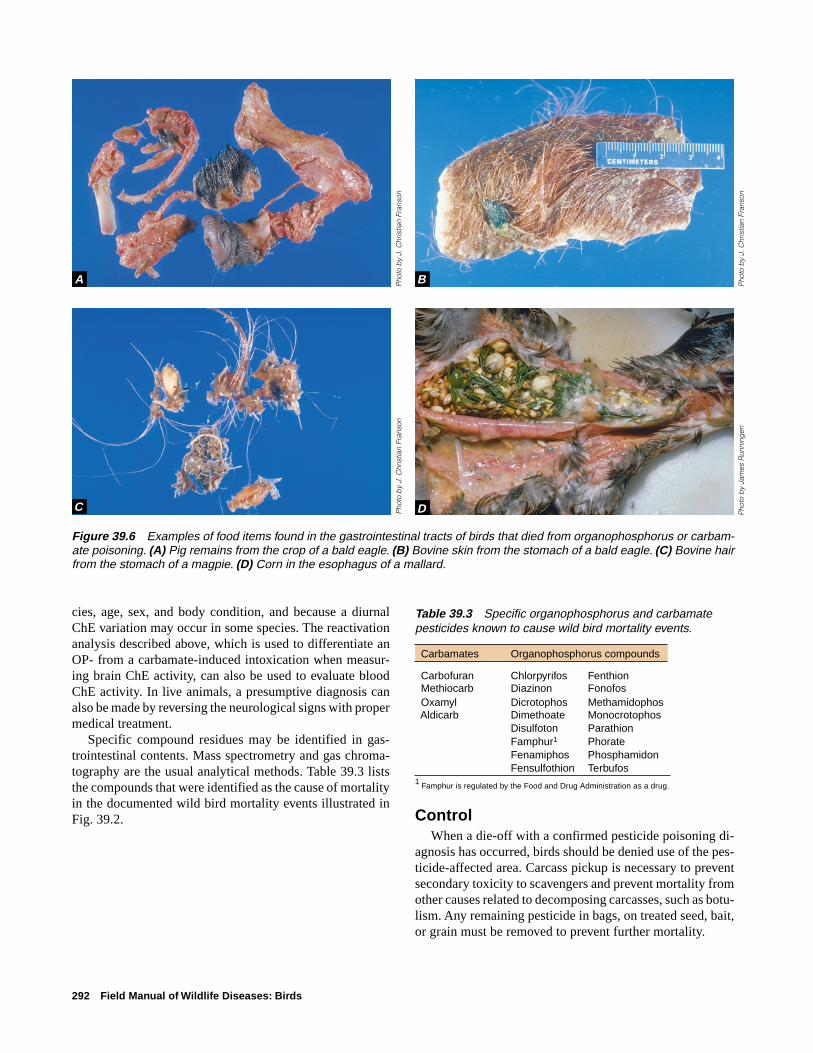
292 Field Manual of Wildlife Diseases: Birds
cies, age, sex, and body condition, and because a diurnalChE variation may occur in some species. The reactivationanalysis described above, which is used to differentiate anOP- from a carbamate-induced intoxication when measur-ing brain ChE activity, can also be used to evaluate bloodChE activity. In live animals, a presumptive diagnosis canalso be made by reversing the neurological signs with propermedical treatment.
Specific compound residues may be identified in gas-trointestinal contents. Mass spectrometry and gas chroma-tography are the usual analytical methods. Table 39.3 liststhe compounds that were identified as the cause of mortalityin the documented wild bird mortality events illustrated inFig. 39.2.
Table 39.3 Specific organophosphorus and carbamatepesticides known to cause wild bird mortality events.
Carbamates Organophosphorus compounds
Carbofuran Chlorpyrifos Fenthion Methiocarb Diazinon Fonofos Oxamyl Dicrotophos Methamidophos Aldicarb Dimethoate Monocrotophos
Disulfoton ParathionFamphur1 PhorateFenamiphos PhosphamidonFensulfothion Terbufos
1 Famphur is regulated by the Food and Drug Administration as a drug.
ControlWhen a die-off with a confirmed pesticide poisoning di-
agnosis has occurred, birds should be denied use of the pes-ticide-affected area. Carcass pickup is necessary to preventsecondary toxicity to scavengers and prevent mortality fromother causes related to decomposing carcasses, such as botu-lism. Any remaining pesticide in bags, on treated seed, bait,or grain must be removed to prevent further mortality.
Figure 39.6 Examples of food items found in the gastrointestinal tracts of birds that died from organophosphorus or carbam-ate poisoning. (A) Pig remains from the crop of a bald eagle. (B) Bovine skin from the stomach of a bald eagle. (C) Bovine hairfrom the stomach of a magpie. (D) Corn in the esophagus of a mallard.
Pho
to b
y Ja
mes
Run
ning
en
A B
C D
Pho
to b
y J.
Chr
istia
n Fr
anso
n
Pho
to b
y J.
Chr
istia
n Fr
anso
nP
hoto
by
J. C
hris
tian
Fran
son

Organophosphorus and Carbamate Pesticides 293
Followup to wildlife mortality incidents due to pesticidepoisoning is important for determining the source and theuse of a chemical. Documented wildlife mortality from ap-proved pesticide applications is considered by regulatoryauthorities for developing label use restrictions and forlicensing pesticide formulations. Malicious use of pesticidesto kill unwanted wildlife is against the law, and legal meanscan be employed to stop illegal use.
Persons who apply pesticides need to consider wildlifeuse and environmental conditions when they apply the chemi-cals. Migration patterns of the wildlife that use the area, thepresence of nesting and breeding species, and weather con-ditions, such as the potential for aerial drift or runoff intowetlands or ponds, are among the factors that should be con-sidered. Pesticides should be applied only as directed; theuse of alternate chemicals or formulations that pose less riskto nontarget species should also be considered. Buffer zonesat crop perimeters will provide more protection to areas usedby wildlife. Agricultural land planted adjacent to wetlandsshould be plowed parallel to a wetland to minimize runoff.
Human Health ConsiderationsHuman exposure to OP or carbamate pesticides can re-
sult in serious illness or even death. Exposure can occurthrough inhalation, absorption through the skin, or by inges-tion. When pesticides that may be associated with wildlifemortality incidents are investigated, field procedures shouldbe scrutinized to avoid inadvertent exposure of personnel topesticides. Persons who collect carcasses or field samplesmust prevent their exposure by wearing nonpermeable gloves,rubber boots, or other appropriate clothing that will preventskin absorption, and respirators should be used if chemicalinhalation is possible.
Poisoning in humans should be treated as a serious medi-cal emergency. When someone seeks medical attention forexposure to an OP or carbamate compound, the attending
physician should be informed that the person may have beenexposed to these chemicals. Patients can be monitored byblood sampling to evaluate their blood ChE levels. Aggres-sive treatment of acute intoxication does not protect againstthe possibility of delayed onset neurotoxicity or persistentneurological defects. Certain compounds have been docu-mented to cause delayed effects in humans. An intermediatesyndrome that occurs within 24–96 hours after exposure hasrecently been described with intoxications of fenthion,dimethoate, monocrotophos, and methamidophos. Musclesof the limbs and those innervated by cranial nerves are af-fected, causing palsies, respiratory depression, and distress.Another delayed neurotoxicity from some OP compoundscan occur 1–2 weeks after exposure. Initially, incoordina-tion develops, and it can progress to moderate to severemuscle weakness and paralysis. This delayed effect wasdocumented with some OP compounds that were rarely usedas pesticides, but the effect may be a potential risk with simi-lar compounds that are more commonly used today if suffi-cient exposure to the compound occurs. These delayed ef-fects could be a problem in wildlife, but they have not beenrecognized yet in any wildlife species.
Linda C. Glaser
Supplementary ReadingAmdur, M.O., Doull, J., and Klaassen, D.C., eds., 1991, Casarett
and Doull's Toxicology, The basic science of poisons, (4thed.): Elmsford, N.Y., Pergamon Press, 1,033 p.
Grue, C.E., Fleming, W.J., Busby, D.G., and Hill, E.F., 1983,Assessing hazards of organophosphate pesticides in wildlife,in Transactions of the 48th North American Wildlife &Natural Resources Conference: Washington, D.C., TheWildlife Management Institute, p 200–220.
Hill, E.F. and Fleming, W.J., 1982, Anticholinesterase poisoningof birds: field monitoring and diagnosis of acute poisoning:Environmental Toxicology and Chemistry 1:27–38.
Hill, E.F., 1995, Organophosphorus and carbamate pesticides, inHoffman, D.H., Rattner, B.A., Burton, G.A., Jr., and Cairns,J., Jr., eds., Handbook of ecotoxicology: Boca Raton, Fla.,Lewis Publishers, p 243–274.
Mineau, P., ed., 1991, Cholinesterase-inhibiting Insecticides,Their impact on wildlife and the environment, chemicals inagriculture v. 2.: Amsterdam, The Netherlands, ElsevierScience Publishing, 348 p.
Smith, G.J., 1987, Pesticide use and toxicology in relation towildlife: Organophosphorus and carbamate compounds:Washington, D.C., U.S. Department of the Interior, Fish andWildlife Service, Resource Publication 170, 171 p.
Figure 39.7 Hemorrhage in the intestine is an occasionalfinding in waterfowl that died of exposure to pesticides, par-ticularly organophosphorus compounds.
Pho
to b
y P
aul S
oler

294 Field Manual of Wildlife Diseases: Birds

Chlorinated Hydrocarbon Insecticides 295
Chapter 40
Chlorinated Hydrocarbon Insecticides
SynonymsOrganochlorines, OCs
Chlorinated hydrocarbon insecticides (OCs) are diverse syn-thetic chemicals that belong to several groups, based onchemical structure. DDT is the best known of these insecti-cides. First synthesized in 1874, DDT remained obscureuntil its insecticidal properties became known in 1939, a dis-covery that earned a Nobel Prize in 1948. The means of syn-thesizing the cyclodiene group, the most toxic of the OCs,was discovered in 1928 and resulted in a Nobel Prize in 1950.The insecticidal properties of cyclodienes, which includealdrin, dieldrin, and endrin (Table 40.1), were discoveredabout 1945. OCs became widely used in the United Statesfollowing World War II. Their primary uses included broadspectrum applications for agricultural crops and forestry and,to a lesser extent, human health protection by spraying todestroy mosquitoes and other potential disease carriers. Thesecompounds also became widely used to combat insect carri-ers of domestic animal diseases.
CauseChlorinated hydrocarbon insecticides are stored in body
fat reserves or are lipophilic, and they remain in the environ-ment for long periods of time after application. Theybioaccumulate or are readily accumulated by animals throughmany exposure routes or repeated exposure and they tend tobiomagnify or accumulate in higher concentrations in ani-mals that are higher in the food chain. This combination ofbioaccumulation and biomagnification can harm or kill wild-life, especially some species of birds. The highly toxic cy-clodiene compounds cause direct mortality of birds as wellas secondary poisoning, which results when birds prey onorganisms dying from insecticide applications. Reproduc-tive impairment is the primary effect of the less acutely toxicDDT and its metabolites, DDD and DDE. The cumulativestorage of OC residues within body fat reserves presents anadditional hazard for birds. Rapid use and depletion or mo-bilization of fat reserves during migration, food shortages,and other stressful conditions release OC residues into theblood. The residues are then carried to the brain, where theycan reach toxic levels resulting in acute poisoning.
Species AffectedAcute mortality from exposure to OCs has been docu-
mented in many bird species (Table 40.1). However, the tox-
icity for birds of different types of these insecticides variesgreatly (Tables 40.2 and 40.3). In general, birds that are higherin the food chain are more likely to be affected by OCs presentin the environment than birds that are lower in the food chain.This is especially true for fish-eating birds and raptors(Fig. 40.1). Environmental biomagnification of these con-taminants can be seen in the mortality of robins and otherbirds from DDT. Leaves from trees that were sprayed withDDT to control Dutch elm disease had high residues of DDT(174–273 parts per million) shortly after spray applications.When the leaves dropped in the fall, they still contained 20–28parts per million of DDT. This leaf litter, along with sprayresidue that reached the ground, produced high DDT resi-dues in the top levels of soil. Earthworms that fed in thosesoils concentrated the residues to a level high enough to killbirds that fed on them. Another hazard is OC seed dressings,which are used to prevent insect damage to agricultural crops,that may be ingested by waterfowl and other seed or grain-eating birds.
DistributionExposure to chlorinated hydrocarbon insecticides is glo-
bal, and residues of these compounds are found in nearlyevery environment, even in Antarctica and the Arctic. Avianmortalities from OCs have been reported from Europe, Asia,North America, and South America. Poisoning may occuranywhere that birds are exposed to point sources of thesechemicals or through bioaccumulation and biomagnification.Because of their environmental persistence and global move-ment, residues of chlorinated hydrocarbon insecticides im-pact bird health long after they become environmental con-taminants and at locations far from the original applicationsites. For example, DDT compounds, polychlorinated biphe-nyls (PCBs), and dioxin-like compounds were recently foundin black-footed albatross adults, chicks, and eggs on Mid-way Atoll in the Pacific.
SeasonalityExposure of birds to OCs is most likely during spring and
summer in countries where these compounds are still usedto control insect pests during the growing season, but expo-sure may occur any time that residues are present in foodsources. For example, waterfowl and other birds that fed on

296 Field Manual of Wildlife Diseases: Birds
Table 40.1 Examples of avian mortality events caused by chlorinated hydrocarbon insecticides.
Purpose of Means of Bird group Principal Event location andInsecticide application bird exposure affected species affected time period
DDT Spray application to Biomagnification in Passerines Robin and other New England,control Dutch elm terrestrial food small birds. Midwest; latedisease. chain. 1940s to 1950s.
DDD Spray application Biomagnification in Grebes Western grebe Clear Lake,to control gnats. aquatic food chain. California, 1950s.
Aldrin Treatment of rice Consumption of Waterfowl Fulvous whistling Texas, 1970sseed to combat treated seeds, use duck, snow goose,agricultural pests. and depletion of blue-winged teal.
stored fat reservesduring migration orperiods of stress.
Heptachlor Treatment of wheat Consumption of Waterfowl Canada goose Oregon, 1970sseed to control treated seeds.agricultural pests.
Toxaphene Spray application to Direct contact with Waterfowl Ducks and coot California, 1960scontrol agricultural and consumption ofpests. contaminated
food.
Spray application for Ingestion of Waterfowl Blue-winged teal, Nebraska, 1960sfisheries management. contaminated food. shoveler, mallard.
Dieldrin Spray application to Biomagnification in Raptors Bald eagle, Nationwide, 1960scontrol agricultural food chain. peregrine falcon and 1970s.pests.
Endrin Spray application to Direct contact with Gallina- Quail, chukar Washington, 1960scontrol orchard spray; consumption of ceous birds, partridge, goshawk, 1970s, 1980s.rodents. contaminated food; raptors, Cooper’s hawk,
biomagnification; geese barn owl,use and depletion Canada goose.of fat reserves.
Chlordane Dry formulations to Consumption of Raptors, Great horned owl, New York, Maryland,control soil pests contaminated food; passerines American kestrel, New Jersey, 1980s.and termites. biomagnification. Cooper’s hawk,
blue jay, robin,starling.

Chlorinated Hydrocarbon Insecticides 297
Table 40.2 Toxicity for the mallard duck of somechlorinated hydrocarbon insecticides.[Modified from Heinz and others, 1979. LC50 is theinsecticide concentration, in parts per million, in feed that isrequired to kill 50 percent of birds during a given period oftime. LD50 is the insecticide amount, in milligrams perkilogram of body weight, in a single dose that is required tokill 50 percent of birds. ppm, parts per million; mg/kg,milligrams per kilogram; >, greater than; ≥, greater than orequal to. — no data available]
Subacute exposure Acute exposureInsecticide LC
50 (ppm) LD
50 (mg/kg)
Aldrin 155 520Chlordane 858 1,200DDT 1,869 >2,240Dieldrin 169 381Endosulfan 1,053 33Endrin 22 5.6Heptachlor 480 ≥2,000Lindane — >2,000Mirex >5,000 >2,400Toxaphene 538 71
Table 40.3 Relative acute toxicity of chlorinated hydrocarbon insecticides for birds. [Modified from Hudson and others, 1984. LD50 is the insecticide amount, in milligrams per kilogram of body weight, in asingle dose that is required to kill 50 percent of birds. mg/kg, milligrams per kilogram; >, greater than; <, less than.]
LD50 (mg/kg)
Species Aldrin Chlordane DDT Dieldrin Endosulfan Endrin Lindane Mirex Toxaphene
Canada goose <141Mallard duck 520 1,200 >2,240 381 31–45 5.6 2,000 2,400 70.7Fulvous whistling 29.2 100–200 99duckSandhill crane >1,200 100–316Pheasant 16.8 24–72 1,334 79 80 to >320 1.8 >2,000 40Sharp-tailed grouse 1.1 19.9Gray partridge 8.8 23.7Chukar partridge 25.3Bobwhite quail 6.6 85.5California quail 14.1 595 8.8 1.2 23.7House sparrow 47.6Horned lark 581
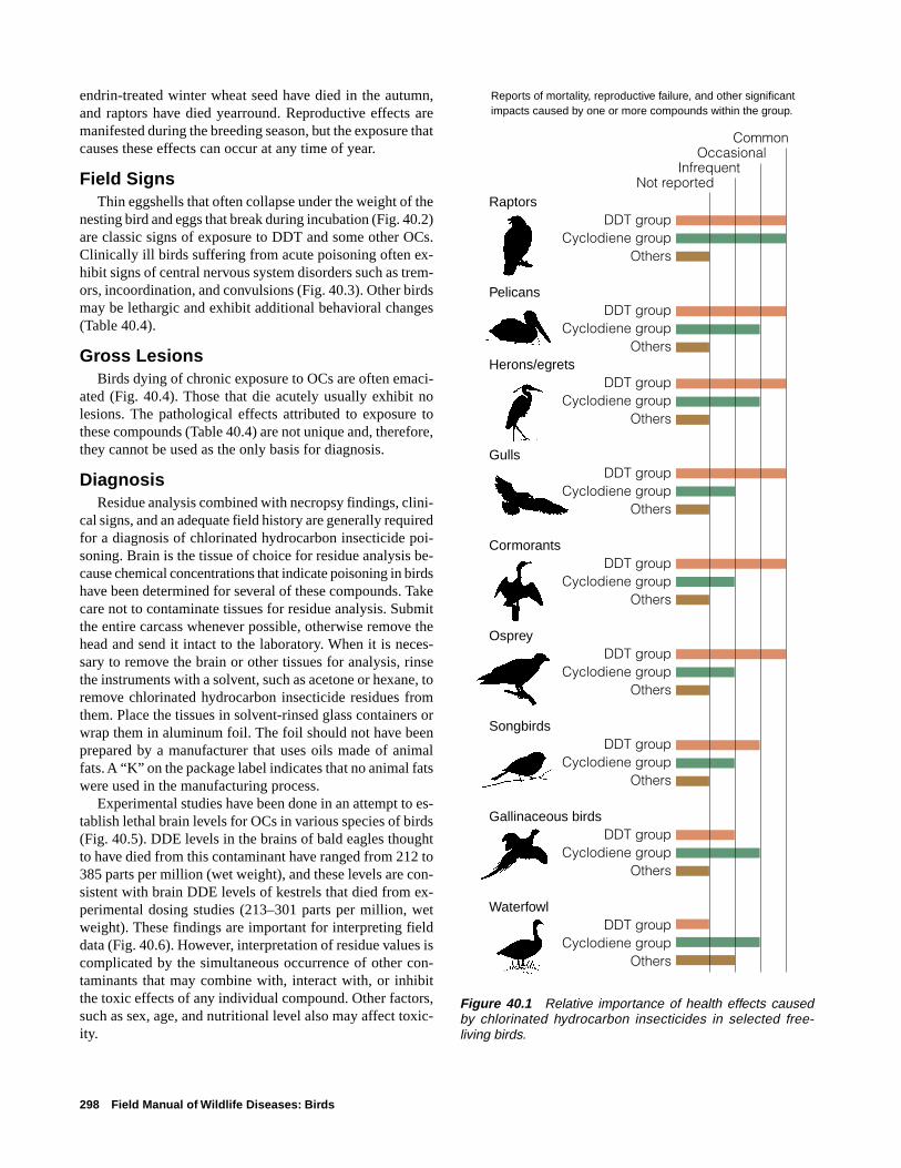
298 Field Manual of Wildlife Diseases: Birds
endrin-treated winter wheat seed have died in the autumn,and raptors have died yearround. Reproductive effects aremanifested during the breeding season, but the exposure thatcauses these effects can occur at any time of year.
Field SignsThin eggshells that often collapse under the weight of the
nesting bird and eggs that break during incubation (Fig. 40.2)are classic signs of exposure to DDT and some other OCs.Clinically ill birds suffering from acute poisoning often ex-hibit signs of central nervous system disorders such as trem-ors, incoordination, and convulsions (Fig. 40.3). Other birdsmay be lethargic and exhibit additional behavioral changes(Table 40.4).
Gross LesionsBirds dying of chronic exposure to OCs are often emaci-
ated (Fig. 40.4). Those that die acutely usually exhibit nolesions. The pathological effects attributed to exposure tothese compounds (Table 40.4) are not unique and, therefore,they cannot be used as the only basis for diagnosis.
DiagnosisResidue analysis combined with necropsy findings, clini-
cal signs, and an adequate field history are generally requiredfor a diagnosis of chlorinated hydrocarbon insecticide poi-soning. Brain is the tissue of choice for residue analysis be-cause chemical concentrations that indicate poisoning in birdshave been determined for several of these compounds. Takecare not to contaminate tissues for residue analysis. Submitthe entire carcass whenever possible, otherwise remove thehead and send it intact to the laboratory. When it is neces-sary to remove the brain or other tissues for analysis, rinsethe instruments with a solvent, such as acetone or hexane, toremove chlorinated hydrocarbon insecticide residues fromthem. Place the tissues in solvent-rinsed glass containers orwrap them in aluminum foil. The foil should not have beenprepared by a manufacturer that uses oils made of animalfats. A “K” on the package label indicates that no animal fatswere used in the manufacturing process.
Experimental studies have been done in an attempt to es-tablish lethal brain levels for OCs in various species of birds(Fig. 40.5). DDE levels in the brains of bald eagles thoughtto have died from this contaminant have ranged from 212 to385 parts per million (wet weight), and these levels are con-sistent with brain DDE levels of kestrels that died from ex-perimental dosing studies (213–301 parts per million, wetweight). These findings are important for interpreting fielddata (Fig. 40.6). However, interpretation of residue values iscomplicated by the simultaneous occurrence of other con-taminants that may combine with, interact with, or inhibitthe toxic effects of any individual compound. Other factors,such as sex, age, and nutritional level also may affect toxic-ity.
RaptorsDDT group
Cyclodiene groupOthers
PelicansDDT group
Cyclodiene groupOthers
Herons/egretsDDT group
Cyclodiene groupOthers
GullsDDT group
Cyclodiene groupOthers
CormorantsDDT group
Cyclodiene groupOthers
OspreyDDT group
Cyclodiene groupOthers
SongbirdsDDT group
Cyclodiene groupOthers
Gallinaceous birdsDDT group
Cyclodiene groupOthers
WaterfowlDDT group
Cyclodiene groupOthers
Common Occasional
InfrequentNot reported
Reports of mortality, reproductive failure, and other significant impacts caused by one or more compounds within the group.
Figure 40.1 Relative importance of health effects causedby chlorinated hydrocarbon insecticides in selected free-living birds.
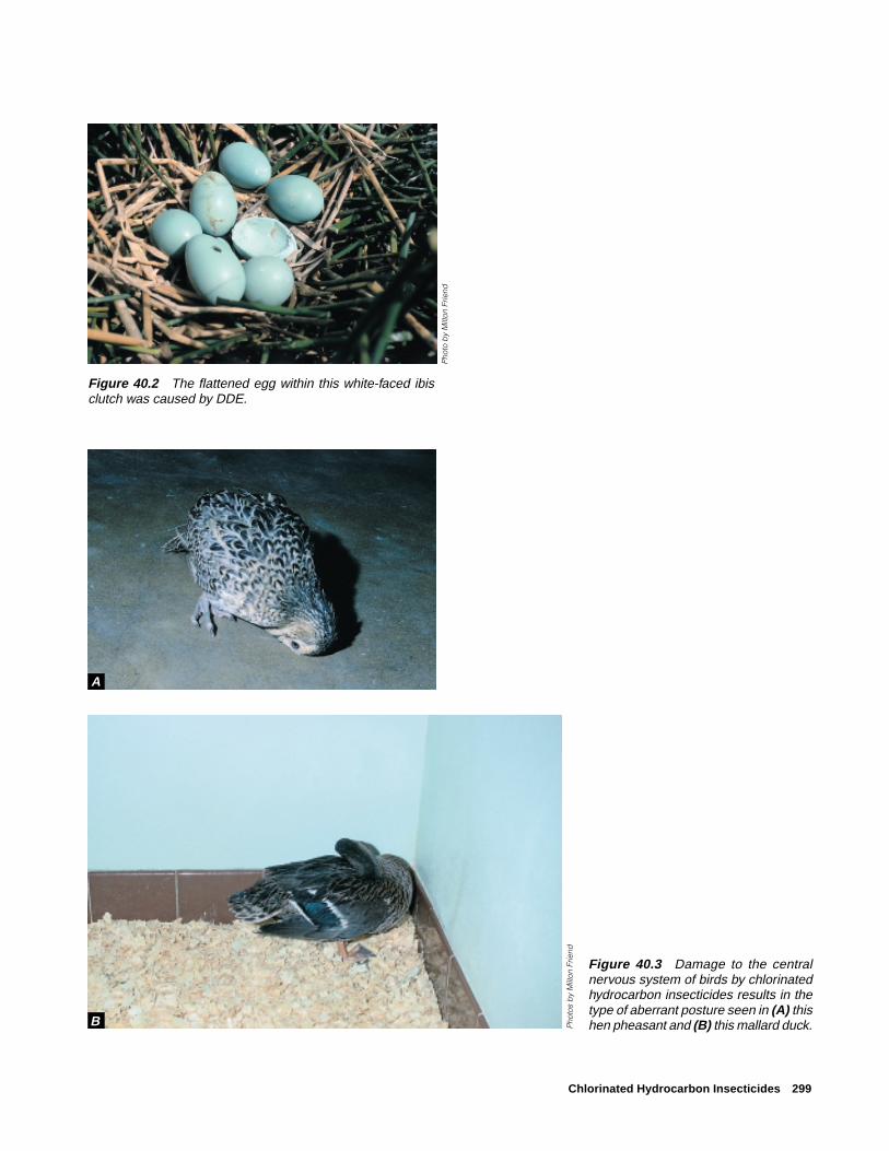
Chlorinated Hydrocarbon Insecticides 299
Figure 40.2 The flattened egg within this white-faced ibisclutch was caused by DDE.
Figure 40.3 Damage to the centralnervous system of birds by chlorinatedhydrocarbon insecticides results in thetype of aberrant posture seen in (A) thishen pheasant and (B) this mallard duck.
Pho
to b
y M
ilton
Frie
nd
Pho
tos
by
Milt
on F
riend
A
B

300 Field Manual of Wildlife Diseases: Birds
Table 40.4 Most commonly reported effects from chlorinated hydrocarbon insecticide exposures ofbirds.
General effect Specific effects
Behavioral Lethargy, slowness, depression
Locomotive and muscle incoordination (ataxia)
Tremors and convulsions
Reduced nest attentiveness and nest abandonment
Violent wing beating
Aberrant wing and body carriage
Muscle spasms causing the body to bend backwardsand become rigid (opisthotonos)
Reproductive Embryo mortality
Decreased egg hatchability
Decreased egg production
Eggshell thinning
Egg breakage during incubation
Pathological Emaciation; muscle wasting and absence of fat
Congestion of the lungs, kidneys, and especially theliver have been reported in pheasants dying fromdieldrin poisoning
Increased liver weight
Small spleens have been reported
In general, pathological changes are not readilyevident at the gross level, and microscopic changesare not diagnostic
Immunological Increased susceptibility to infectious disease
Other Disruption of salt gland function by DDE
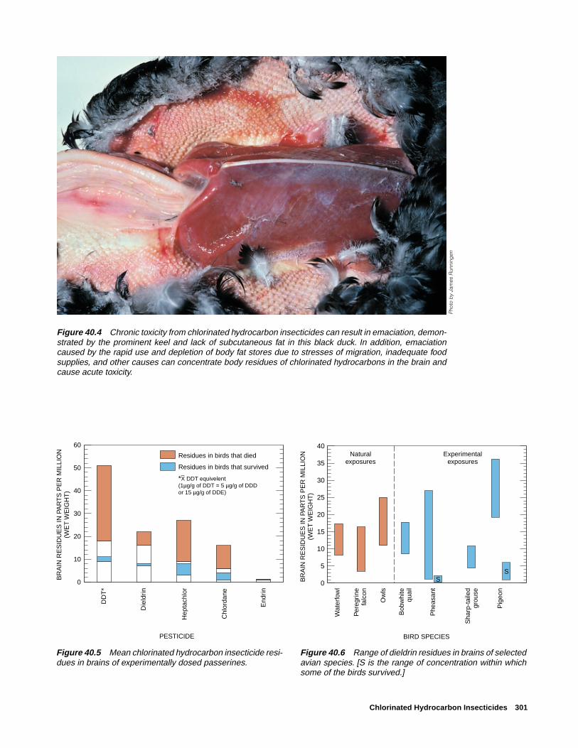
Chlorinated Hydrocarbon Insecticides 301
Figure 40.6 Range of dieldrin residues in brains of selectedavian species. [S is the range of concentration within whichsome of the birds survived.]
Figure 40.4 Chronic toxicity from chlorinated hydrocarbon insecticides can result in emaciation, demon-strated by the prominent keel and lack of subcutaneous fat in this black duck. In addition, emaciationcaused by the rapid use and depletion of body fat stores due to stresses of migration, inadequate foodsupplies, and other causes can concentrate body residues of chlorinated hydrocarbons in the brain andcause acute toxicity.
0
10
20
30
40
50
60
BR
AIN
RE
SID
UE
S IN
PA
RT
S P
ER
MIL
LIO
N(W
ET
WE
IGH
T)
DD
T*
Die
ldrin
Hep
tach
lor
Chl
orda
ne
End
rin
Residues in birds that died
Residues in birds that survived
*X DDT equivelent (1µg/g of DDT = 5 µg/g of DDDor 15 µg/g of DDE)
PESTICIDE
Figure 40.5 Mean chlorinated hydrocarbon insecticide resi-dues in brains of experimentally dosed passerines.
0
5
10
15
20
25
30
35
40Experimental
exposuresNatural
exposures
SS
Wat
erfo
wl
Per
egrin
efa
lcon
Ow
ls
Bob
whi
tequ
ail
Phe
asan
t
Sha
rp-t
aile
dgr
ouse
Pig
eon
BIRD SPECIES
BR
AIN
RE
SID
UE
S IN
PA
RT
S P
ER
MIL
LIO
N(W
ET
WE
IGH
T)
Pho
to b
y Ja
mes
Run
ning
en

302 Field Manual of Wildlife Diseases: Birds
ControlBecause uses of most OCs have been banned or greatly
curtailed in the United States, controlling wildlife exposureto these compounds depends largely on properly disposingof existing stores, preventing leakage into the environment,and preventing malicious use. The spreading of these com-pounds to environments where they are no longer used willcontinue until suitable alternative pest controls are found.Also, migratory wildlife that are exposed to these compoundsin areas where they are still used may not exhibit effects un-til they reach other areas on their migratory route.
Human Health ConsiderationsAs with many of the other toxins discussed in this sec-
tion, residues of chlorinated hydrocarbons in birds are storedin tissues and are not transferred to humans through routinehandling of carcasses. Exceptions include situations wherea person could somehow come in contact with the pesticide,for example, in the stomach contents of a bird or on its skinor feathers. It is always wise to handle carcasses with dis-posable gloves, and to treat unknown mortalities as possiblesources of infectious agents transferrable to humans.
Milton Friend and J. Christian Franson
Supplementary ReadingBlus, L.J., 1995, Organochlorine pesticides, in Hoffman, D.J., and
others, eds., Handbook of ecotoxicology: Boca Raton, Fla.,Lewis Publishers, p. 275–300.
Blus, L.J., Wiemeyer, S. N., and Henny, C.J., 1996, Organochlo-rine pesticides, in Fairbrother, A., and others, eds., Noninfec-tious diseases of wildlife (2nd ed.): Ames, Iowa, Iowa StateUniversity Press, p. 61–70.
Heinz, G.H., Hill, E.F., Stickel, W.H., and Stickel, L.F., 1979,Environmental Contaminant studies by the Patuxent WildlifeResearch Center, in Kenaga, ed., Avian and mammalianwildlife toxicology: Philadelphia, Penn., American Society forTesting and Materials, Special Technical Publication 693,p. 9–35
Hudson, R.H., Tucker, R.K., and Haegele, M.A., 1984, Handbookof toxicity of pesticides to wildlife, (2d ed.): Washington, D.C.,U.S. Department of the Interior, Fish and Wildlife Service,Resources Publication 153, 90 p.
Peakall, D.B., 1996, Dieldrin and other cyclodiene pesticides inwildlife, in Beyer, W.N., and others, eds., Environmentalcontaminants in wildlife: interpreting tissue concentrations:Boca Raton, Fla., Lewis Publishers, p. 73–97.

Polychlorinated Biphenyls 303
Chapter 41
Polychlorinated Biphenyls
SynonymsPCBs, aroclors, chlorinated biphenyls
Polychlorinated biphenyls (PCBs) are industrial compoundswith multiple industrial and commercial uses (Table 41.1).PCBs are chemically inert and stable when heated. Theseproperties contribute greatly to PCBs having become envi-ronmental contaminants. The chemical inertness and heat sta-bility properties that make PCBs desirable for industry alsoprotect them from destruction when the products in whichthey are used are discarded. These same properties alsoenable PCB residues to persist in the environment for longperiods of time and to be transported worldwide when con-taminated particulate matter travels through waters, precipi-tation, wind, and other physical forces.
PCBs have a physical structure similar to DDT, and, likeDDT, they are classified as aromatic hydrocarbons whichcontain one or more benzene rings. The presence of chlorineresults in DDT, PCBs, and other compounds with similarstructures commonly being referred to as chlorinated hydro-carbons. The toxicity of these compounds is associated withthe amount of chlorine they contain. The trade name ofAroclor® for PCBs that were produced by a manufacturer inthe United States contains a numerical designation that speci-fies the amount of chlorine present in a particular formula-tion. For example, Aroclor® 1221 contains 21 percent chlo-rine while Aroclor® 1254 contains 54 percent chlorine. Thefirst two digits designate the number of carbons in the for-mulation. The chemical structure of PCBs results in the pos-sibility of many different forms or isomers, (more commonlycalled congeners) of these compounds. PCBs in other coun-tries have different trade names than Aroclor® (Table 41.2).
Cause Like other chlorinated hydrocarbons, PCBs accumulate
in the fat of animals or are lipophilic, and they tend to be-come concentrated at higher levels of the food chain. In gen-eral, persistence increases for PCBs that are made with higheramounts of chlorine. Birds are most susceptible to PCB com-pounds of the mid-chlorination range (42–54 percent).
Species AffectedMammals, especially mink, are more susceptible than
birds and invertebrates to direct toxicity from PCBs. The high-est tissue concentrations of these compounds are foundamong birds, especially marine species that are at the top ofcomplex oceanic food webs and among fish-eating birds, such
as cormorants, that use large inland water bodies. For ex-ample a 12.9-fold increase has been reported from planktonto fish in a Lake Michigan food web. Although direct toxic-ity for birds is generally low (Table 41.3), PCBs are power-ful inducers of liver enzyme systems that increase the me-tabolism of hormones. PCBs may have caused thin eggshellsin double-crested cormorants and white pelicans, and underexperimental conditions, in ring-doves and (perhaps) inCoturnix quail and mallard ducks. Unfortunately, there isinsufficient knowledge to clearly define the impacts of PCBson bird reproduction, especially in field situations, becausetissue residues are often highly correlated with other lipo-philic compounds, such as organochlorines. Findings havegenerally been inconclusive, but the greatest effects have beenseen in gallinaceous birds such as pheasants, chickens, anddoves.
DistributionPCBs were first identified in the tissues of wildlife in
Sweden, and they are now known to occur in a wide varietyof wildlife and other species, including humans, throughoutthe world. PCBs are clearly global contaminants, and theyare the most abundant of the chlorinated hydrocarbon pol-lutants in the global ecosystem with the possible exceptionof petroleum products. Industrial wastes released into aquaticsystems, point sources of contamination from manufactur-ing facilities, landfills receiving waste from such facilities,and combustion and other disposal of products containingPCBs are generally recognized sources of contamination.Another less well-known source of PCB contamination ofthe environment was the use of PCBs during the 1950s and1960s as additives to extend the residual life and effective-ness of expensive chlorinated insecticides such as chlordane,aldrin, dieldrin, and benzene hexachloride. It is estimatedthat more than 1.5 metric tons of PCBs have been producedworldwide. PCB manufacturing in the United States was dis-continued in 1978.
The variable environmental distribution of PCBs resultsfrom their physical and chemical properties, which influencetheir rates of distribution, retention, and degradation in dif-ferent environments. This results in great differences in therelative concentrations of the different forms of PCBs foundin wildlife samples from different geographic areas and isalso a reflection of the magnitude of local and regional con-

304 Field Manual of Wildlife Diseases: Birds
Table 41.1 Uses of polychlorinated biphenyls (PCBs) in industry and products for society.
Properties
Heat stability
Chemical stability
Ability to be mixed with organic compounds
Slow degradation
Industrial uses
Lubricants, hydraulic fluids, grinding fluids
Heat transfer agents, insulators
Plasticizers
Dielectric sealants
Dedusting agents
Protective coatings
Common products that have contained PCB additives
Wire and cable coating
Impregnants for braided cotton-asbestos insulation
Printing inks and mimeograph inks
Preparation of imitation gold leaf
Pigment vehicle for decoration of glass and ceramics
Essential components of coating for flameproofing cotton drill for outer garments and forrendering olive-drab canvas fire retardant, water-repellant, and rot-proof (tents,tarpaulins)
Moistureproof coating for wood, paper, concrete, and brick
Asphalt, roof coatings
High quality precision casting wax; waxes used in making dentalcastings and costume jewelry
Sealers for masonry, wood, fiberboard, and paper
Window envelopes
Polystyrene, polyethylene, neoprene, polybutene, silicone rubber, crepe rubber
Plasticizers in paints
Life extenders and sometimes toxicity synergists for pesticides containing DDT, dieldrin,lindane, chlordane, aldrin, and benzene

Polychlorinated Biphenyls 305
tamination patterns, environmental transport processes, andthe composition of PCB residues in the food chain.
SeasonalityExposure to PCBs is not seasonally dependent; except
that in warm weather, PCB residues may vaporize or evapo-rate with liquid from contaminated areas, and thus, increasethe risk of airborne exposure.
Field SignsDirect mortality of wild birds from exposure to PCBs
rarely occurs. We are only aware of one such event havingbeen documented. The number of different PCBs present inthe environment further complicates evaluations because ofdifferent impacts and lethality associated with these differ-ent compounds. Nonspecific signs associated with acute ex-posure of birds to toxic levels of PCBs include lethargy, lack
Table 41.2 Trade names for polychlorinated biphenyls (PCBs).
Trade name Country of manufacturer Manufacturer
Aroclor® United States of America Monsanto
Clophens® Germany Bayer
Fenclors® Italy Caffaro
Phenoclors®; Pyralenes® France Prodelec
Kanechlors® Japan Kanegafuchi
Others have been produced in Czechoslovakia and the former USSR
of locomotive and muscle coordination or ataxia, tremors,and other observations. Behavioral modifications and im-paired reproductive performance may also occur and wouldbe more readily detected at the population rather than indi-vidual level (Table 41.4).
Gross LesionsThere are no diagnostic lesions associated with exposure
to PCBs. Enlarged liver and kidneys, atrophy of the spleenand the bursa of Fabricius, emaciation, and excess fluidsaround the heart have been associated with chronic expo-sure.
Excess fluid or edema in tissues has been found in somecases of acute PCB exposure, and this suggests that PCBsmay interfere with tissue permeability or cardiac function orboth. PCBs have been shown to cause physical defects inembryos, or be teratogenic, in chickens, and they also cause
Table 41.3 Relative toxicity of polychlorinated biphenyls (PCBs) for birds.[Adapted from Eisler, 1986. LC50 is the contaminant concentration in the diet thatis required to kill 50 percent of the test animals in a given period of time; bycomparison, the LC50 for mink to Aroclors® 1242 and 1254 is 8.6 and 6.7,respectively. mg/kg, milligrams per kilogram. >, greater than. —, no dataavailable.]
LD50
(mg/kg of Aroclor®)
Species 1221 1242 1254 1260
Bobwhite quail >6,000 2,098 604 747
Mallard duck — 3,182 2,699 1,975
Ring-necked pheasant >4,000 2,078 1,091 1,260
Japanese quail >6,000 >6,000 2,898 2,186
European starling, red-wingedblackbird, brown-headed cowbird — — 1,500 —

306 Field Manual of Wildlife Diseases: Birds
a condition analogous to chick edema disease. This condi-tion results in the leakage of body fluids into various organsand tissues. However, the presence of dioxins as contami-nants within the PCB formulations may be the actual causeof these lesions.
DiagnosisDiagnosis of acute poisoning is based on PCB residues in
tissues, and as for most other chlorinated hydrocarbons,mortality is best diagnosed from residues found in brain tis-sue. However, the concentrations of PCBs that indicate poi-soning vary greatly with the specific formulation of PCBs,the species of bird, and, often, the presence of other environ-mental contaminants. Detection of subacute effects, such aspoor reproductive performance and immunosuppression, isalso confounded by these same factors. Comparison of resi-dues in the tissues of birds suspected of being poisoned withresidues in tissues of normal birds of the same species innearby or regional sites can be diagnostically useful alongwith knowledge of PCB deposition and discharges in the area.Comparisons are sometimes difficult because of the varyingeffects of different PCB mixtures and the interactions thatoccur between PCBs, other pollutants, and other diseaseagents. Many toxic and biochemical responses from PCBexposure occur in multiple species and body organ systems.
Residue levels alone will generally not be sufficient datafor making a diagnosis. Necropsy findings combined withlaboratory analyses, including residue evaluations, knowl-edge of environmental conditions and events at the field site,and response of different species to PCB exposure are allneeded for sound judgements to be reached.
ControlPrevention of the entry of PCBs into the environment and
containment or removal of PCB contamination that is alreadypresent are necessary to reduce exposure of wildlife. PCBsales in the United States were stopped in the 1970s, butlarge amounts are still present in the environment due to en-vironmental persistence and to global transport by winds andother means from locations where PCBs are still used. Im-proper disposal of products that contain PCBs through land-fills and incineration at temperatures that are too low (below1,600 °C) to destroy PCBs can cause further environmentalcontamination. However, more stringent air-quality standardsin the United States and other nations have diminished thepotential that PCBs in incinerated materials will be added tothe environment through combustion.
Bird use of heavily contaminated sites should be preventedto the extent feasible by habitat manipulation, physical bar-riers, scaring devices, and other appropriate means. Knowl-
Table 41.4 Reported effects of polychlorinated biphenyls (PCBs) in birds.
Type of impact Examples
Behavioral Lethargy
Locomotive and muscle incoordination or ataxia
Tremors and convulsions
Reduced nest attentiveness and protection of eggs
Reproductive Embryo mortality resulting in decreased hatchability of eggs
Decreased egg production
Egg shell thinning
Pathological Accumulation of fluid within the pericardial sac or hydropericardium
Excess fluid or edema in body tissues and organs
Atrophy of bursa of Fabricius, spleen, and other lymphoid tissues
Enlarged livers that are firm and light colored
Bill and foot deformities (from embryonic exposure)
Immunological Increased susceptibility to infectious disease
Other Weight loss
Debilitation

Polychlorinated Biphenyls 307
edge of PCB levels in specific environments should be gainedprior to developing those areas for wildlife, including theuse of dredge material to create artificial islands for bird nest-ing habitat. PCB and heavy metal loads in sediments shouldalso be considered in decisions regarding dumping dredgematerials.
Human Health ConsiderationsPCBs are known to accumulate in humans, and health ad-
visories are often issued about consuming wildlife fromheavily contaminated environments. Residues in wildlife canonly be transferred to humans by consuming contaminatedtissues. As with most chlorinated hydrocarbons, the greatestconcentrations of residues are in fat tissue, and removingfatty parts of the carcass prior to cooking can significantlyreduce potential human exposure. Although PCB residuescannot be transferred to humans from wildlife by means otherthan consumption, the cause of death is seldom known whendead wildlife are encountered and the risk of exposure todisease agents that can be transmitted by contact should notbe taken. Always wear gloves or use other physical barriersto prevent personal contact with the carcass.
Milton Friend and J. Christian Franson
Supplementary Reading
Eisler, R., 1986, Polychlorinated biphenyl hazards to fish,wildlife, and invertebrates: a synoptic review: Fish andWildlife Service Biological Report 85(1.7), 72 p.
Hoffman, D.J., Rice, C.P., and Kubiak, T.J., 1996, PCBs anddioxins in birds, in Beyer, W.N., and others, eds., Environmen-tal contaminants in wildlife: interpreting tissue concentrations:Boca Raton, Fla., Lewis Publishers, p. 165–207.
O’Hara, T.M., and Rice, C.D., 1996, Polychlorinated biphenyls, inFairbrother, A., and others, eds., Noninfectious diseases ofwildlife (2nd ed.): Ames, Iowa, Iowa State University Press,p. 71–86.
Rice, C.P., and O’Keefe, P., 1995, Sources, pathways, and effectsof PCBs, dioxins, and dibenzofurans, in Hoffman, D.J., andothers, eds., Handbook of ecotoxicology: Boca Raton, Fla.,Lewis Publishers, p. 424–468.

308 Field Manual of Wildlife Diseases: Birds

Oil 309
Chapter 42
Oil
SynonymsPetroleum
Each year, an average of 14 million gallons of oil from morethan 10,000 accidental spills flow into fresh and saltwaterenvironments in and around the United States. Most acci-dental oil spills occur when oil is transported by tankers orbarges, but oil is also spilled during highway, rail, and pipe-line transport, and by nontransportation-related facilities, suchas refinery, bulk storage, and marine and land facilities(Fig. 42.1). Accidental releases, however, account for only asmall percentage of all oil entering the environment; inheavily used urban estuaries, the total petroleum hydrocar-bon contributions due to transportation activities may be 10percent or less. Most oil is introduced to the environment byintentional discharges from normal transport and refiningoperations, industrial and municipal discharges, used lubri-cant and other waste oil disposal, urban runoff, river runoff,atmospheric deposition, and natural seeps. Oil-laden waste-water is often released into settling ponds and wetlands(Fig. 42.2). Discharges of oil field brines are a major sourceof the petroleum crude oil that enters estuaries in Texas.
CauseBirds that are exposed to spilled or waste petroleum can
be affected both externally and internally. Oil contaminationof feathers (Fig. 42.3) disrupts their normal structure andfunction, and it results in the loss of insulation for warmth
and waterproofing. Oiled birds lose the ability to fly, andthey frequently die from hypothermia, starvation, exhaus-tion, or drowning. Birds that are exposed to oil during theirreproductive season can also transfer lethal doses of the con-taminant to their eggs during incubation. Even small quanti-ties of oil (5–20 microliters) externally applied to eggs cankill embryos. Birds can also ingest, inhale, or absorb oil whenexposed to a spill or while preening contaminated plumage.The toxic effects of ingested oil vary, depending on the typeof oil and on the species of birds affected. These effects in-clude gastrointestinal irritation and hemorrhaging, anemia,reproductive impairment, depressed growth, and osmoregu-latory dysfunction (Table 42.1). Polycyclic aromatic hydro-carbons (PAH) contribute to the toxicity of crude petroleumand refined petroleum products, but the amounts of PAH inpetroleum products vary greatly.
Unfortunately, the effects of petroleum pollution can per-sist long after the visible spill is cleaned or dispersed. Petro-leum persistence in the water column is usually less than 6months, but it can be much longer (more than 10 years) inother components of the environment. Chronic losses mayresult when birds ingest oil in contaminated food items. Forexample, oil from the 1989 Exxon Valdez spill is still se-
Pipelines12 percent
Land transport14 percent
Unknown6 percent
Other3 percent
Fixed facilities(refineries, storage)
36 percent
Marine facilities and vessels29 percent
Figure 42.1 Origin of petroleum spills, 1987–94.
Figure 42.2 Wastewater ladenwith petroleum being dischargedinto a settling pond.
Phot
o by
Milt
on F
riend
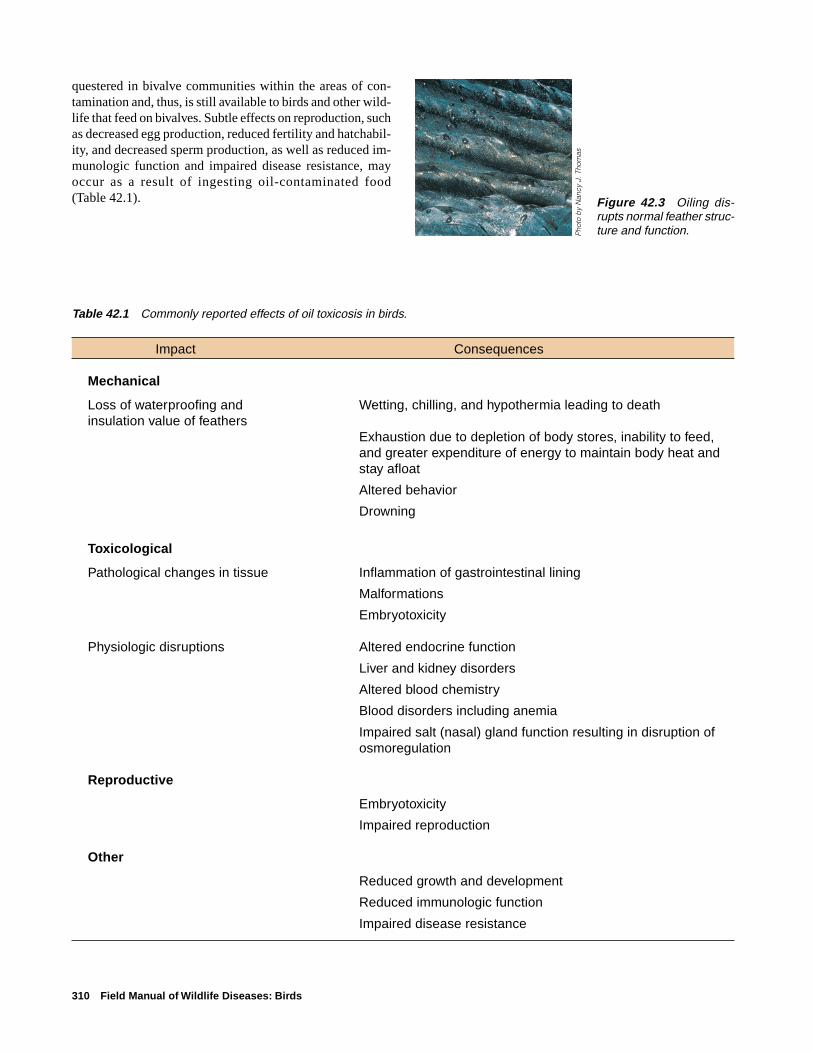
310 Field Manual of Wildlife Diseases: Birds
Table 42.1 Commonly reported effects of oil toxicosis in birds.
Impact Consequences
Mechanical
Loss of waterproofing and Wetting, chilling, and hypothermia leading to deathinsulation value of feathers
Exhaustion due to depletion of body stores, inability to feed,and greater expenditure of energy to maintain body heat andstay afloat
Altered behavior
Drowning
Toxicological
Pathological changes in tissue Inflammation of gastrointestinal lining
Malformations
Embryotoxicity
Physiologic disruptions Altered endocrine function
Liver and kidney disorders
Altered blood chemistry
Blood disorders including anemia
Impaired salt (nasal) gland function resulting in disruption ofosmoregulation
Reproductive
Embryotoxicity
Impaired reproduction
Other
Reduced growth and development
Reduced immunologic function
Impaired disease resistance
questered in bivalve communities within the areas of con-tamination and, thus, is still available to birds and other wild-life that feed on bivalves. Subtle effects on reproduction, suchas decreased egg production, reduced fertility and hatchabil-ity, and decreased sperm production, as well as reduced im-munologic function and impaired disease resistance, mayoccur as a result of ingesting oil-contaminated food(Table 42.1). Figure 42.3 Oiling dis-
rupts normal feather struc-ture and function.P
hoto
by
Nan
cy J
. Tho
mas

Oil 311
Species AffectedA wide variety of birds and other wildlife have been af-
fected by oil. The bird species affected depend on the loca-tion of the oil and the behavior of the birds. Species thatsuffer the greatest losses are gregarious, spend most of theirtime on the water, often near shipping lanes, and dive intothe water to find food or to avoid disturbance. Seabirds, suchas auks, guillemots, murres, puffins, sea ducks, and penguins,are particularly susceptible to contamination from oil spills(Fig. 42.4). In addition, annual losses of marine birds occurdue to natural oil seeps along the Santa Barbara Channel ofthe California coast.
SeasonalitySpecies with high reproductive rates may quickly recover
from a spill, but for species with low reproductive rates, suchas brown pelicans, oil pollution can cause catastrophic lossesand it may take decades for populations to return to prespillnumbers. Even oiled brown pelicans that have been success-fully rehabilitated have reduced reproductive success.
Winter storms increase the likelihood of transport spills,making January, February, and March the peak spill season.This is also the time of year when seabirds and waterfowlcongregate in wintering areas, resulting in an increased po-tential for significant bird losses.
Sea and bay ducks (scoters, scaups, oldsquaws, canvas-back) that tend to concentrate on wintering grounds and div-ing birds (grebes, loons, and mergansers) that overwinter inmarine environments or on large water bodies with commer-cial shipping are quite vulnerable to oil pollution, especiallyduring winter months. Eiders are vulnerable most of the year.
Distribution and Extent of MortalityThe oiling of migratory birds is not limited to specific
geographic areas. Accidental oil spills have occurred in all50 States including inland waters, such as rivers and non-navigable waters, and in open coastal waters, ports and har-bors. Although it is not possible to accurately estimate thenumber of birds lost to oil pollution, in many cases the mor-tality has been substantial (Table 42.2). Bird losses of 5,000or more are common for larger oil spills. Reports are usuallyof the numbers of oiled birds found dead or moribund on theshore, but these estimates may be inaccurate because of searchbiases, accessibility of the shore, losses of birds that havesunk to the bottom, and other factors. An important sourceof error in estimating losses in marine environments is theunknown proportion of oiled birds that die at sea but that donot reach the coast.
In addition to accidental spills, other opportunities foranimal exposure to oil occur in association with oil produc-tion, petroleum refining, and highly industrialized locationsthroughout the United States. Persistent oil pollution is achronic problem around marinas and ports due to dischargesfrom shipping and boating activities and storage tank clean- Figure 42.4 Relative susceptibility of birds to oiling.
Ducks, geese, and swans
Sea and bay ducks
Grebes
Loons
Pelicans
Cormorants
Wading birds (herons, egrets, bitterns)
Gulls
Cranes
Plovers, sandpipers
High susceptibilityMedium susceptiblilityLow susceptibility

312 Field Manual of Wildlife Diseases: Birds
ing, but, unfortunately, the numbers of birds affected by oilpollution in these areas are unknown. In the Playa Lakesregions of eastern New Mexico, northwestern Texas, andwestern Oklahoma, open pits and tanks containing oil andoil-field wastes have been reported to claim the lives of ap-proximately 100,000 birds each year (Fig. 42.5).
Field SignsMajor oil spills are frequently accompanied by intensive
media coverage, and they may be well publicized beforeslicks or affected birds appear. However, small spills, espe-cially those of unknown origin, often go unnoticed exceptfor the appearance of a few contaminated birds. Oiled birdsare frequently wet and chilled because the oil damages featherwaterproofing and insulating properties (Fig. 42.6); birds mayride lower in the water than normal because they have lostfeather buoyancy. Oiling is suggested when water birds leavethe water for islands, rocks, pilings, and other surfaces be-cause they are chilled (Fig. 42.7). Birds that survive for 48hours or more after oiling are often thin, and even close tostarvation, because they have stopped feeding and are usingfirst body fat and then muscle tissue to produce heat in re-sponse to chilling.
Matting of the feathers occurs from external oiling. Oilcan usually be seen or smelled on the feathers, but some light,transparent oils may be difficult to detect. One useful tech-nique for detecting oiling is to place a few feathers from thebird in a pan of water and watch for an oil sheen to appear(Fig. 42.8). An enzyme-linked immunosorbent assay(ELISA), which detects PAH in oil, can provide quick con-firmation of the presence of petroleum products on fur orfeathers.
Gross LesionsNecropsy findings of birds that die from oil exposure are
highly variable. Birds are often emaciated, and oil may bepresent in their trachea, lungs (Fig. 42.9), digestive tract, andaround the vent. The lining of the intestine may be reddened,or the intestine may contain blood. The salt glands, which
Table 42.2 Examples of bird mortality from oil spills.
Vessel or source Year Site Estimated bird mortalities
Exxon Valdez 1989 Prince William Sound, Alaska 350,000–390,000
Nestucca 1988 Grays Harbor, Washington 50,000
Amoco Cadiz 1978 Brittany, France 20,000
Barge STC-101 1976 Chesapeake Bay, Virginia 20,000–50,000
Torrey Canyon 1967 English Channel 30,000
Gerda Maersk 1955 Elbe River, Germany 500,000
Figure 42.6 Oiled birds become wetand chilled because oil damages featherwaterproofing and insulating properties.
Pho
to b
y U
.S. F
ish
and
Wild
life
Ser
vice
Reg
ion
7
Figure 42.5 These oiled birds were recovered from oil-fieldwastewater pits in the southwestern United States.
Pho
to b
y Ja
mes
Run
ning
en

Oil 313
Figure 42.7 Common murre out of waterdue to oiling.
Figure 42.8 If external oiling is suspected, place featherson water and watch for oil sheen.
Pho
to b
y Ja
mes
Run
ning
en
Figure 42.9 In severe cases, oil may be inhaled and maydiscolor the lungs, such as in this Canada goose.
Pho
to b
y J.
Chr
istia
n Fr
anso
n
Figure 42.10 Swollen salt glands.
Pho
to b
y U
.S. F
ish
and
Wild
life
Ser
vice
Reg
ion
7
are located over the eyes, may appear swollen (Fig. 42.10),and the adrenal glands may be enlarged. A variety of otherchanges in the normal appearance of tissues and organs mayalso be present, but no specific or consistent lesion is typicalin animals that are exposed to oil.
DiagnosisDiagnosis of oiling is seldom a problem; visible oil on
the bird or in the environment usually suffices (Fig. 42.11).However, proving that oil has caused mortality is more com-plex. For damage assessments and cause-of-death determi-nations, it must be determined that oiling did not occur afterthe death of the animals in question.
Chemical analyses of tissues or eggs are difficult to usefor diagnosis because the chemical composition of petro-leum products is complex. Therefore, good background in-formation and field observations are an integral part of speci-men submission to diagnostic laboratories (see Chapter 1,Recording and Submitting Specimen History Data). Submitwhole carcasses whenever possible.
ControlTreatment of oil spills within the States, territorial pos-
sessions, and territorial waters of the United States is legis-latively mandated by the Oil Pollution Act of 1990. The Actmandates the inclusion of a fish and wildlife response planwithin the National Contingency Plan and the creation ofArea Contingency Plans. These plans provide for an inte-grated response to a spill with assigned agency responsibili-ties for protecting fish and wildlife and environmentalcleanup.
In the event of a spill, contact the National ResponseCenter at the 24-hour, toll free number 1-(800)-424-8802.The National Response Center will advise the responsibleagencies (Coast Guard, Environmental Protection Agency,
Pho
to b
y M
ilton
Frie
nd
➡
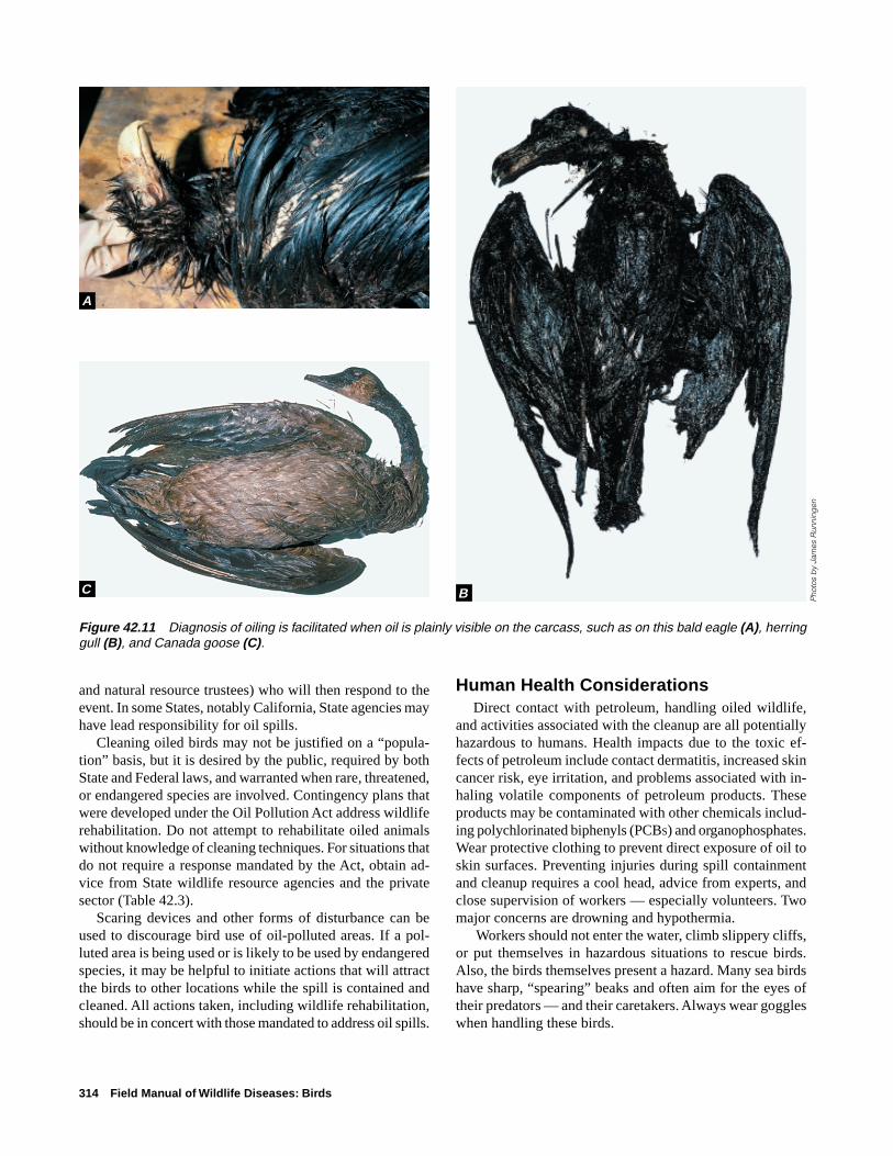
314 Field Manual of Wildlife Diseases: Birds
and natural resource trustees) who will then respond to theevent. In some States, notably California, State agencies mayhave lead responsibility for oil spills.
Cleaning oiled birds may not be justified on a “popula-tion” basis, but it is desired by the public, required by bothState and Federal laws, and warranted when rare, threatened,or endangered species are involved. Contingency plans thatwere developed under the Oil Pollution Act address wildliferehabilitation. Do not attempt to rehabilitate oiled animalswithout knowledge of cleaning techniques. For situations thatdo not require a response mandated by the Act, obtain ad-vice from State wildlife resource agencies and the privatesector (Table 42.3).
Scaring devices and other forms of disturbance can beused to discourage bird use of oil-polluted areas. If a pol-luted area is being used or is likely to be used by endangeredspecies, it may be helpful to initiate actions that will attractthe birds to other locations while the spill is contained andcleaned. All actions taken, including wildlife rehabilitation,should be in concert with those mandated to address oil spills.
Figure 42.11 Diagnosis of oiling is facilitated when oil is plainly visible on the carcass, such as on this bald eagle (A), herringgull (B), and Canada goose (C).
Human Health ConsiderationsDirect contact with petroleum, handling oiled wildlife,
and activities associated with the cleanup are all potentiallyhazardous to humans. Health impacts due to the toxic ef-fects of petroleum include contact dermatitis, increased skincancer risk, eye irritation, and problems associated with in-haling volatile components of petroleum products. Theseproducts may be contaminated with other chemicals includ-ing polychlorinated biphenyls (PCBS) and organophosphates.Wear protective clothing to prevent direct exposure of oil toskin surfaces. Preventing injuries during spill containmentand cleanup requires a cool head, advice from experts, andclose supervision of workers — especially volunteers. Twomajor concerns are drowning and hypothermia.
Workers should not enter the water, climb slippery cliffs,or put themselves in hazardous situations to rescue birds.Also, the birds themselves present a hazard. Many sea birdshave sharp, “spearing” beaks and often aim for the eyes oftheir predators — and their caretakers. Always wear goggleswhen handling these birds.
A
BC
Pho
tos
by
Jam
es R
unni
ngen
Oil 315
Table 42.3 Sources of information for rehabilitation of oiled birds.
Many individuals and groups have expertise in the rehabilitation of oiled birds and otherwildlife. The following are major programs that conduct this type of activity.
Program and address Telephone
Tri-State Bird Rescue and Research, Inc. 302-737-9543110 Possum Hollow Rd., Newark, DE 19711
California Department of Fish and Game 530-752-4167Office of Oil Spill Prevention and ResponseOiled Wildlife Care NetworkWildlife Health CenterUniversity of California, Davis, CA 95616
International Bird Rescue Research Center 510-841-9086699 Potter St., Berkeley, CA 94710
Tonie E. Rocke
Supplementary ReadingAlbers, P.H., 1995, Oil, biological communities and contingency
planning, in Fink, L., and others, eds., Wildlife and oil spills:response, research, and contingency planning: Hanover, Pa.,The Sheridan Press, p. 1–10.
Albers, P.H., 1995, Petroleum and individual polycyclic aromatichydrocarbons, in Hoffman, D.J., and other, eds., Handbook ofEcotoxicology: Boca Raton, Fla., Lewis Publishers, p. 330–355.
Bourne, W.R.P., 1976, Seabirds and pollution in Johnson, R., ed.,Marine Pollution: London, Academic Press, p. 403–502.
Burger, A.E., 1993, Estimating the mortality of seabirds followingoil spills: effects of spill volume. Marine Pollution Bulletin,v. 26, p. 140–143.
Flickinger, E.L., 1981, Wildlife mortality at petroleum pits inTexas: Journal of Wildlife Management, v. 45, p. 560–564.
Hoffman, D.J., 1990, Embryotoxicity and teratogenicity ofenvironmental contaminants to bird eggs: Reviews of Environ-mental Contamination and Toxicology, v. 115, p. 39–89.

316 Field Manual of Wildlife Diseases: Birds

Lead 317
Chapter 43
LeadSynonymPlumbism
Lead poisoning of waterfowl is neither a new disease nor asubject without controversy. The use of lead shot for water-fowl hunting within the United States has been prohibitedand efforts are underway to ban the use of lead fishing sink-ers and prohibit the use of lead shot for nonwaterfowl hunt-ing. The first documented reports within the United Statesof lead-poisoned waterfowl were from Texas in 1874. Nu-merous other reports and studies added to those findingsduring the years and decades that followed. However, strongopposition to nontoxic shot requirements prevented fullimplementation of them until 1991. A full transition to non-toxic shot shells for all hunting and to nontoxic fishing sink-ers and jig heads for fishing within the United States will nothappen easily. The continued use of lead shot and lead fish-ing weights and the large amounts of these materials previ-ously deposited in environments where birds feed assure thatlead poisoning will remain a common bird disease for sometime.
CauseLead poisoning is an intoxication resulting from absorp-
tion of hazardous levels of lead into body tissues. Lead pel-lets from shot shells, when ingested, are the most commonsource of lead poisoning in birds. Other far less commonsources include lead fishing sinkers, mine wastes, paint pig-ments, bullets, and other lead objects that are swallowed.
Species AffectedLead poisoning has affected every major species of
waterfowl in North America and has also been reported in awide variety of other birds. The annual magnitude of leadpoisoning losses for individual species cannot be preciselydetermined. However, reasonable estimates of lead-poisoninglosses in different waterfowl species can be made on thebasis of mortality reports and gizzard analyses. Within theUnited States, annual losses from lead poisoning prior to the1991 ban on the use of lead shot for waterfowl hunting wereestimated at between 1.6 and 2.4 million waterfowl, basedon a fall flight of 100 million birds. Followup studies havenot been conducted since the ban on lead shot to determinecurrent losses from lead poisoning. This disease still affectswaterfowl and other species due to decades of residual leadshot in marsh sediments, continued deposition from allow-able use of lead shot during harvest of other species, non-compliance with nontoxic shot regulations, target shooting
over areas where birds may feed, and from other sources oflead.
Lead poisoning is common in mallard, northern pintail,redhead, and scaup ducks; Canada and snow geese; and tun-dra swan. The frequency of this disease decreases with in-creasing specialization of food habits and higher percent-ages of fish in the diet. Therefore, goldeneye and merganserducks are seldom affected (Fig. 43.1). A surprising recentfinding has been lead poisoning in spectacled and commoneiders on their Alaskan breeding range, where the intensityof hunting is far less than in the contiguous 48 States. Thesefindings demonstrate that lead poisoning can afflict birds evenwithout heavy hunting pressure. Among land birds, eaglesare most frequently reported dying from lead poisoning(Fig. 43.2). Lead poisoning in eagles and other raptors gen-erally is a result of swallowing lead shot embedded in theflesh of their prey. With the exception of waterfowl and rap-tors, lead poisoning from ingesting lead shot is generally aminor finding for other species (Table 43.1). However, leadpoisoning has been reported in partridge, grouse, and pheas-ants subjected to intensive shooting in uplands of Europe.Lead poisoning in pheasants in Great Britain was reportedas early as 1875.
Lead poisoning due to ingesting lead fishing weights hasbeen reported in numerous species. The greatest number ofreports are from swans as a group, common loon, brownpelican, Canada goose, and mallard duck (Fig. 43.3). Laysanalbatross chicks on Midway Atoll suffer high lead exposuresand mortality from ingesting lead-laden paint chips fleckingoff of vacant military buildings (Fig. 43.4).
DistributionLosses occur coast-to-coast and border-to-border within
the United States. Documented lead poisoning in birds var-ies widely between States and does not necessarily reflecttrue geographic differences in the frequency of occurrenceof this condition. For example, although the geographic dis-tribution of lead poisoning in bald eagles is closely associ-ated with their wintering areas, the number of lead poisoningcases from Wisconsin and Minnesota is disproportionatelyhigh. Because submission of bald and golden eagles for ex-amination from different areas is highly variable, no directcomparison can be made between States regarding the num-ber of lead-poisoned eagles (Fig. 43.5A). The reported dis-tribution of lead poisoning in eagles and waterfowl depends

318 Field Manual of Wildlife Diseases: Birds
DucksPuddle ducks
Bay diving ducksTeal, shoveler, wood ducks
Sea ducksMergansers
Whistling ducks
GeeseCanada goose
Snow and Ross' geeseBrant and other geese
Swans
Tundra swanMute swan
Other swans
1Mortality frequently reportedMortality commonly reported
2Mortality occasionally reported3Mortality rarely reported
1Including individual die-offs of hundreds to thousands of birds2Lead shot ingestion studies generally indicate low levels of exposure to lead shot3Lead shot ingestion studies genreally indicate little or no lead shot ingestion
on the numbers of birds submitted for complete disease di-agnostic evaluations. In areas where few birds are examined,the frequency of lead poisoning and other diseases will beunderestimated. Even where many bird carcasses are ad-equately evaluated, the number of diagnoses made reflectsminimum numbers of lead-poisoning cases. The general dis-tribution of this disease in waterfowl on the basis of leadshot-ingestion surveys and documented mortality prior tonontoxic shot requirements is shown in Fig. 43.5B.
Lead poisoning has also been reported as a cause of mi-gratory bird mortality in other countries (Fig. 43.6). Severalof these countries have implemented nontoxic shot require-ments and several others are beginning to address this issue.
SeasonalityBirds can can die from lead poisoning throughout the year,
although birds are most often poisoned by lead after the
waterfowl hunting season has been completed in northernareas and during the later part of the season in southern areasof the United States. January and February are peak monthsfor cases in tundra swans, Canada geese, and puddle ducks.Spring losses are more commonly reported for diving ducks.Tundra swans are also frequently lead poisoned during springmigration.
Field SignsLead-poisoned waterfowl are often mistaken for hunting
season cripples. Special attention should be given to water-fowl that do not take flight when the flock is disturbed andto small groups of waterfowl that remain after most otherbirds of that species have migrated from the area. Lead-poisoned birds become reluctant to fly when approached andthose that can still fly are often noticeably weak flyers —unable to sustain flight for any distance or flying erratically
Figure 43.1 Relative occurrences of lead shot poisoning in North Americanwaterfowl.

Lead 319
RaptorsBald eagle
Golden eagleHawks
Owls
Water birdsCoots and rails
ShorebirdsCranes
GullsOther species
Upland gamebirds
Pheasant and quailsDoves
Turkeys and grouseWoodcock
Mortality frequently reportedMortality commonly reported
1Mortality occasionally reported2Mortality rarely reported
1Lead shot ingestion studies generally indicate low levels of exposure to lead shot2Lead shot ingestion studies genreally indicate little or no lead shot ingestion
and landing poorly. Birds that attempt to escape pursuit byrunning may exhibit an unsteady gait. In lead-poisonedCanada geese, the head and neck position may appear“crooked” or bent during flight; a marked change in the toneof call is also sometimes evident in this species. As the dis-ease progresses and waterfowl become flightless, the wingsare held in a characteristic “roof shaped” position (Fig. 43.7),which is followed by wing droop as the birds become in-creasingly moribund (Fig. 43.8). Fluid may discharge fromthe bill and often a bird may not attempt to escape in thepresence of humans.
Lead-poisoned waterfowl are easily captured during ad-vanced stages of intoxication (Fig. 43.9). Because severelyaffected birds generally seek isolation and protective cover,well-trained retrieving dogs can help greatly to locate andcollect these birds. An abundance of bile-stained feces on anarea used by waterfowl (Fig. 43.10) is suggestive of lead
poisoning and warrants ground searches even if other fieldsigns have not been observed. Green-colored feces can alsoresult from feeding on green wheat and other plants, but thecoloration is somewhat different.
Gross LesionsLead-poisoned waterfowl are often emaciated because of
the prolonged course of the illness and its impact on essen-tial body processes. Therefore, many affected birds appearto be starving; they are light in weight, have a “hatchet-breast”appearance (Fig. 43.11), and the undersurface of their skinis devoid of fat (Fig. 43.12). The vent area of these birds isoften stained with a bright green diarrhea (Fig. 43.13). Theheads of Canada geese may appear puffy or swollen becauseserum-like fluids accumulate in the tissues of the face(Fig. 43.14).
Lesions observed at necropsy of lead-poisoned birds that
Figure 43.2 Relative occurrence of lead shot poisoning in groups of birdsother than waterfowl.

320 Field Manual of Wildlife Diseases: Birds
Table 43.1 Documented North American cases of lead poisoning in free-ranging nonwaterfowl species.
Nonendangered species
Upland gamebirdsRing-necked pheasant Hungarian partridge Bobwhite quail Scaled quailWild turkey Mourning dove
RaptorsGolden eagle Northern harrier Rough-legged hawkRed-tailed hawk Prairie falcon Turkey vulture
Wetland birdsCommon loon Double-crested cormorant Greater sandhill crane Lesser sandhill craneWhite pelican American coot Royal tern FlamingoGreat blue heron White ibis Great egret Snowy egretSora rail American avocet Black-necked stilt Marbled godwitPectoral sandpiper Western sandpiper Long-billed dowitcher Laughing gullHerring gull Glaucous-winged gull California gull Laysan albatross1
Endangered species
California condor Brown pelican Whooping crane2
Bald eagle Mississippi sandhill crane Peregrine falcon
1The cause of poisoning was ingestion of paint chips rather than lead shot, bullets, or fishing tackle.2The cause of poisoning was particulate lead of unknown origin but not lead shot or fishing tackle.
have died after a prolonged illness generally consist of thefollowing:
1. Severe wasting of the breast muscles (Fig. 43.11).2. Absent or reduced amounts of visceral fat(Fig. 43.12).3. Impactions of the esophagus or proventriculus inapproximately 20–30 percent of affected waterfowl.These impactions may contain food items, or combi-nations of food, sand, and mud. The extent of impac-tion may be restricted to the gizzard and proventricu-lus, extend to the mouth, or lie somewhere in between(Fig. 43.l5).4. A prominent gallbladder that is distended, filled withbile, and dark or bright green (Fig. 43.l6).5. The normally yellow gizzard lining is discolored adark or bright green (Fig. 43.l7). Gizzard contents arealso often bile-stained.6. Lead pellets or small particles of lead are oftenpresent among gizzard and proventricular contents.Pellets that have been present for a long time are wellworn, reduced in size, and disk-like rather than spheri-cal (Fig. 43.18). Careful washing of contents is requiredto find smaller lead fragments. X-ray examination isoften used to detect radiopaque objects in gizzards, butrecovery of the objects is necessary to separate leadfrom other metals. Flushing contents through a series
of progressively smaller sieves is one method of pelletrecovery.
Less obvious pathological changes include wasting ofinternal organs such as the liver, kidneys, and spleen; areasof paleness in the heart muscle; a flabby-looking heart; andpaler-than-normal-looking internal organs and muscle tissue.
The above field signs and gross lesions provide a basisfor a presumptive diagnosis of lead poisoning. However, noneof these signs or lesions is diagnostic by itself and all canresult from other causes. Also, many of the above signs andlesions are absent in birds that die acutely following an over-whelming lead exposure.
DiagnosisA definitive diagnosis of lead poisoning as a cause of death
is based on pathological and toxicological findings supple-mented by clinical signs and field observations. The pres-ence or absence of lead shot or lead particles in the gizzardcontents is useful information and should be recorded, but itis not diagnostic. The liver or kidneys are the tissues of choicefor toxicology analysis, with liver tissue being more com-monly used. If you suspect lead poisoning and cannot sub-mit whole birds to the diagnostic laboratory, remove the liveror kidney tissue, wrap the specimens separately in alumi-num foil, and freeze them until they are submitted for analy-sis. Collect the entire liver or one entire kidney. However,

Lead 321
Figure 43.3 Number of reported lead poisoning occurrencesfollowing ingestion of lead sinkers and jigs through 1994.
Waterfowl
Mute swan (Great Britain)1
Whooper swan (Great Britain)
Canada goose
Mallard
Trumpeter swan
Tundra swan
Redhead duck
Wood duck
Black duck
Red-breasted merganser
Mute swan (United States)
Other
Common loon
Brown pelican
Double-crested cormorant
Great blue heron
Sandhill cranes (two species)
White pelican
Royal tern
Gulls (two species)
White ibis
Egrets (two species)
Bald eagle
50 or more26—50
10—2510 or less
1 Great Britain banned the use of lead sinkers in 1987.
Figure 43.4 The droopy wings and unthriftyappearance of this Laysan albatross chickare the result of lead poisoning caused byingestion of lead-laden chips that flecked offabandoned buildings. The paint had highconcentrations of lead.
Pho
to b
y Lo
uis
Sile
o

322 Field Manual of Wildlife Diseases: Birds
EXPLANATIONNumber of eagle deaths, by State
More than 50
25–49
20–24
15–19
10–14
5–9
1–4
0
EXPLANATIONRelative lead exposure in waterfowl
Very frequent
Frequent
Common
Occasional
Figure 43.5 (A) Diagnosed cases of lead poisoning in bald eagles though mid-April, 1996. (B) Relative occurrence of leadexposure in waterfowl prior to the 1991 ban on use of lead shot for waterfowl hunting. Evaluation is based on gizzard analysisand reported mortality.
A
B
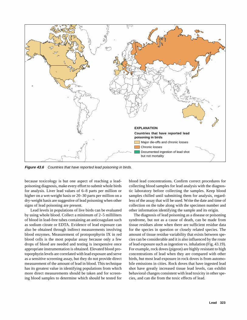
Lead 323
EXPLANATION
Countries that have reported lead poisoning in birds
Major die-offs and chronic losses
Chronic losses
Documented ingestion of lead shot but not mortality
Figure 43.6 Countries that have reported lead poisoning in birds.
because toxicology is but one aspect of reaching a lead-poisoning diagnosis, make every effort to submit whole birdsfor analysis. Liver lead values of 6–8 parts per million orhigher on a wet-weight basis or 20–30 parts per million on adry-weight basis are suggestive of lead poisoning when othersigns of lead poisoning are present.
Lead levels in populations of live birds can be evaluatedby using whole blood. Collect a minimum of 2–5 millilitersof blood in lead-free tubes containing an anticoagulant suchas sodium citrate or EDTA. Evidence of lead exposure canalso be obtained through indirect measurements involvingblood enzymes. Measurement of protoporphyrin IX in redblood cells is the most popular assay because only a fewdrops of blood are needed and testing is inexpensive onceappropriate instrumentation is obtained. Elevated blood pro-toporphyrin levels are correlated with lead exposure and serveas a sensitive screening assay, but they do not provide directmeasurement of the amount of lead in blood. This techniquehas its greatest value in identifying populations from whichmore direct measurements should be taken and for screen-ing blood samples to determine which should be tested for
blood lead concentrations. Confirm correct procedures forcollecting blood samples for lead analysis with the diagnos-tic laboratory before collecting the samples. Keep bloodsamples chilled until submitting them for analysis, regard-less of the assay that will be used. Write the date and time ofcollection on the tube along with the specimen number andother information identifying the sample and its origin.
The diagnosis of lead poisoning as a disease or poisoningsyndrome, but not as a cause of death, can be made fromtissue residues alone when there are sufficient residue datafor the species in question or closely related species. Theamount of tissue residue variability that exists between spe-cies can be considerable and it is also influenced by the routeof lead exposure such as ingestion vs. inhalation (Fig. 43.19).For example, rock doves (pigeon) are highly resistant to highconcentrations of lead when they are compared with otherbirds, but most lead exposure in rock doves is from automo-bile emissions in cities. Rock doves that have ingested leadshot have greatly increased tissue lead levels, can exhibitbehavioral changes consistent with lead toxicity in other spe-cies, and can die from the toxic effects of lead.

324 Field Manual of Wildlife Diseases: Birds
Figure 43.7 Characteristic “roof-shaped”position of the wings in (A) a lead-poisonedmallard (leading bird) and (B) a snow goose.
Figure 43.8 Wing droop in a tundra swan in advanced stagesof lead intoxication.
Pho
to b
y Ja
mes
Run
ning
enP
hoto
by
Milt
on F
riend
B
Figure 43.9 Inability of these lead-poisoned Canada geeseto escape capture by humans illustrates their great vulner-ability to predation.
Pho
to b
y M
ilton
Frie
nd
Pho
to b
y M
ilton
Frie
nd
A

Lead 325
Figure 43.10 Waterfowl feces provide presumptive evidence of lead poisoning. Examination of (A) feces where waterfowl areconcentrating and (B) observations of an abundance of bright green-colored feces should be reason to search for sick birds andcarcasses.
Figure 43.11 “Hatchet-breast” appearance of a lead-poisoned mallard (top bird) and north-ern pintail. The skin has been removed from the breast of the pintail to further illustrate thesevere loss of muscle tissue.
A B
Pho
to b
y M
ilton
Frie
nd
Pho
tos
by
Milt
on F
riend

326 Field Manual of Wildlife Diseases: Birds
Figure 43.12 Loss of subcutaneous fat is often extreme in lead-poisoned birds. (A) The undersurface ofthe skin of this pintail is totally devoid of fat, in contrast with (B) the abundance of yellow fat present in themallard (bottom bird) that had died of avian cholera. Note also the absence of fat in the visceral area andalong the knees of the northern pintail (top bird) in comparison with the mallard.
A
B
➡
➡ ➡
➡
➡
Pho
tos
by
Milt
on F
riend

Lead 327
Figure 43.13 Bright green staining of the vent area is oftenindicative of lead poisoning.
Figure 43.14 The heads of lead-poisoned Canada geeseoften appear puffy or swollen.
Pho
to b
y M
ilton
Frie
ndP
hoto
by
Milt
on F
riend

328 Field Manual of Wildlife Diseases: Birds
Figure 43.15 Examples of impactions in lead-poisoned birds. (A) Impaction of corn in digestive tract ofa hen mallard, extending from the gizzard to the mouth; (B) snow goose with an impaction of grasses.(C) Tundra swan with impaction of grasses and some seeds, extending from the mouth to the gizzard; and(D) a more limited impaction in a drake mallard.
A
B
➡
Pho
tos
by
Milt
on F
riend
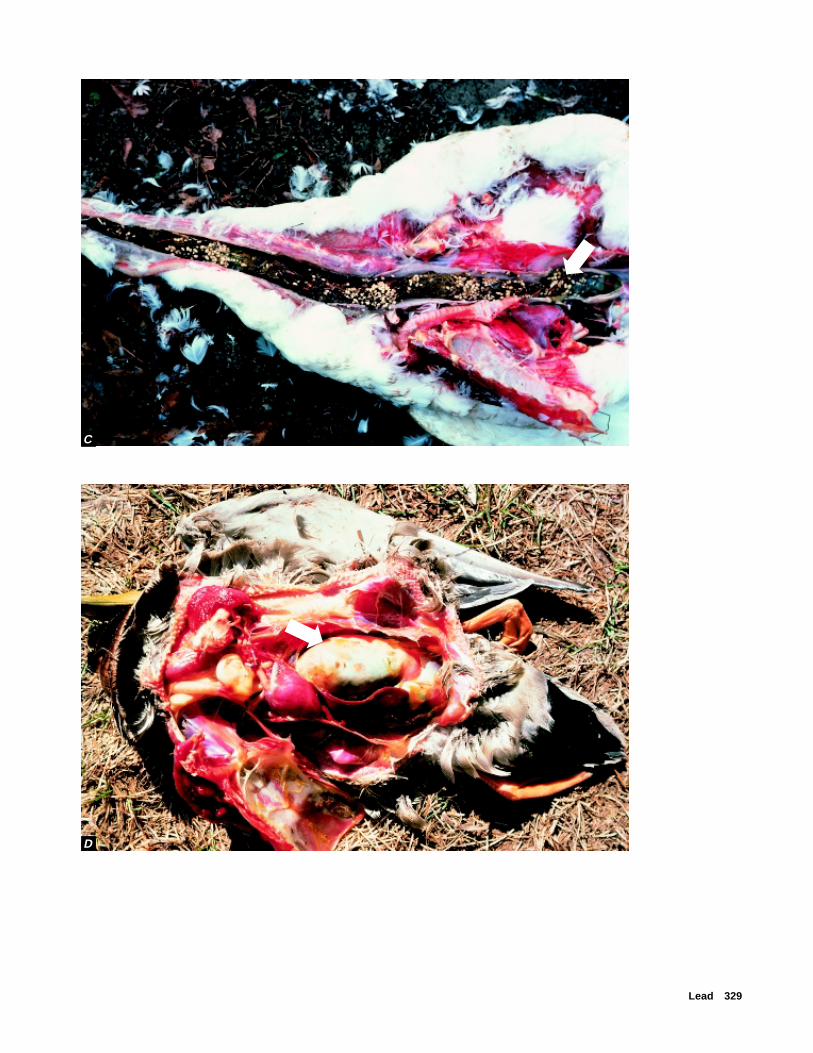
Lead 329
D
C
➡
➡
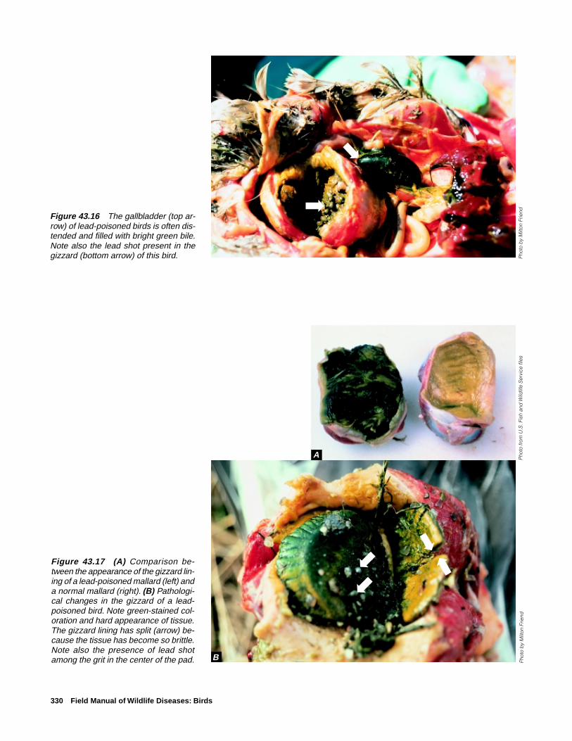
330 Field Manual of Wildlife Diseases: Birds
Figure 43.16 The gallbladder (top ar-row) of lead-poisoned birds is often dis-tended and filled with bright green bile.Note also the lead shot present in thegizzard (bottom arrow) of this bird.
Figure 43.17 (A) Comparison be-tween the appearance of the gizzard lin-ing of a lead-poisoned mallard (left) anda normal mallard (right). (B) Pathologi-cal changes in the gizzard of a lead-poisoned bird. Note green-stained col-oration and hard appearance of tissue.The gizzard lining has split (arrow) be-cause the tissue has become so brittle.Note also the presence of lead shotamong the grit in the center of the pad.
A
B
➡
➡
➡
➡➡
Pho
to b
y M
ilton
Frie
ndP
hoto
by
Milt
on F
riend
Pho
to fr
om U
.S. F
ish
and
Wild
life
Ser
vice
file
s
➡

Lead 331
Figure 43.18 (A) Lead shot in Canadagoose gizzard. Note the presence ofcorn. Corn and other cereal grains in-tensify the toxicity of lead. (B) Lead shot,originally spherical, that have been worndown in the waterfowl gizzard. Note theflattened, disk-like shape of many ofthese pellets.
A
B
➡
➡➡
➡
Pho
to b
y M
ilton
Frie
ndP
hoto
by
Jam
es R
unni
ngen

332 Field Manual of Wildlife Diseases: Birds
ControlTwo actions can often be taken to reduce the magnitude
of mortality from lead poisoning when die-offs occur: deny-ing birds use of problem areas, and rigorous pickup andproper disposal of dead and moribund birds.
Denying birds use of problem areas requires knowingwhere the birds are picking up the lead. This is complicatedby the fact that signs of intoxication may not appear until 1week after lead ingestion, and birds may not start dying un-til 2–3 weeks after lead ingestion. Habitat modification ofcontaminated areas is also useful in some instances, but dif-ferences in feeding habits must be considered. For example,placing additional water on an area may protect puddle ducksfrom reaching lead shot on the bottom of wetlands, but thismay create an attractive feeding area for diving ducks. Simi-larly, draining an area may prevent waterfowl from using anarea and ingesting shot, but it may create an attractive feed-ing area for shorebirds or pheasants. Therefore, control plans
must consider the broad spectrum of wildlife likely to usethe area at the time action will be taken. Rigorous pickupand proper disposal of lead-contaminated waterfowl carcassesis required to prevent raptors and other scavenger speciesfrom ingesting them. The high percentage of waterfowl withembedded body shot provides a continual opportunity forlead exposure in raptors that far exceeds the opportunity foringestion of shot present in waterfowl gizzards.
Other management practices that have been used to re-duce losses from lead poisoning on site-specific areas in-clude tillage programs to turn lead shot below the surface ofsoil so that shot is not readily available to birds, plantingfood crops other than corn and other grains that aggravatethe effects of lead ingestion, and requiring the use of non-toxic shot in hunting areas. The potential contributions ofthe first two practices toward reducing lead-poisoning lossesamong birds are, at best, limited and temporary. Supplemen-tal grit has also been placed in wetlands in the belief that
Waterfowl
Raptors
Pigeons/ doves
Grouse
0 2 4 6 8 10 12 14 16 18 20 22 24 26
LEAD, IN PARTS PER MILLION, WET WEIGHT
Liver
Figure 43.19 Lead residues in the liver and blood associated with subclinical, toxic, and mortalityfindings in several groups of birds. Variability within groups occurs because of differences in species anda variety of factors affecting toxicity within species.
Waterfowl
Raptors
Pigeons/ doves
Grouse
0 2 4 6 8 10 12 14 16 18 20 22 24 26
LEAD, IN PARTS PER MILLION, WET WEIGHT
Blood
EXPLANATION
Tissue lead residues
Subclinical
Toxic
Fatal

Lead 333
birds intentionally ingest shot because grit is not available;as with tillage and food crops, any benefits are limited andtemporary. The use of nontoxic shot is the only long-termsolution for significantly reducing migratory bird losses fromlead poisoning.
The strong correlation between exposure of waterfowl tolead and the use of lead shot for hunting waterfowl was viv-idly demonstrated by National Wildlife Health Center spon-sored studies that compared tissue lead levels and gizzardanalyses in a subpopulation of Canada geese as they mi-grated from their breeding grounds to their winteringgrounds. Nontoxic shot requirements were in place at somesampling sites but not at others. Lead exposure was signifi-cantly less where nontoxic shot requirements existed.
Since lead shot has been banned for hunting waterfowl inthe United States, attention has turned to regulating the useof lead fishing sinkers and lead jig heads. The Environmen-tal Protection Agency has been petitioned to address the prob-lem of bird mortality from these sources (Fig. 43.20). Prohi-bitions against using lead fishing weights below certain sizeshave already been initiated on some Federal lands and otherareas. The number of cases of lead poisoning in swans in theThames Valley of England was reduced by 70 percent in 2years following enactment of the 1987 ban on use of splitlead shot and other fishing sinkers up to 1 ounce in size.Sizes larger than those that can be ingested by birds havenot yet become a focus for concern.
The use of lead shot for target shooting and hunting onuplands is also receiving increased attention. In general, in-gestion rates for lead shot in upland species are far less thanthose for waterfowl, even for doves (Table 43.2). The har-vest of doves is somewhat analogous to waterfowl huntingin that large numbers of shells are often fired over the samelocation year after year (Fig. 43.21.). However, the durationof intense shooting on specific sites tends to be much lessfor doves than for waterfowl and the hunting area is gener-ally tilled annually for agricultural purposes.
Figure 43.20 Fishing weights found in the stomachs and giz-zards of birds that died from lead poisoning.
Pho
to b
y Ja
mes
Run
ning
en
Veterinary treatment of lead-poisoned birds is generallynot a reasonable approach. However, endangered species orother birds of high individual value that are lead poisonedmay warrant treatment. In those instances, treatment shouldbe done only by qualified persons familiar with and skilledin the proper use of lead-chelating chemicals. Under the bestof circumstances, the results of treatment are unpredictableand the success rate low.
Human Health ConsiderationsPeople do inadvertently consume lead-poisoned birds.
Although this is not desirable, no appreciable risks to hu-man health exist. Most lead present in the body of a lead-poisoned bird is in organs such as the liver and kidneys ratherthan in the flesh. The dose relation (milligrams of lead perkilogram of body weight) and lead excretion processes aresuch that a great number of lead-poisoned birds would needto be consumed in a relatively short time before toxic levelsof lead could build up in the human body. Persons who eat
Table 43.2 Percentage of upland gamebirds reported with ingestedlead shot, by State.
Percentage withSpecies State ingested lead shot
Mourning doves Alabama 1.0Eastern seaboard 2.4(Maryland toSouth Carolina)Indiana 2.3Maryland 1.0–6.5
Scaled quail New Mexico 0.4Bobwhite quail New Mexico 1.8

334 Field Manual of Wildlife Diseases: Birds
the liver, kidneys, and other soft tissues from lead-poisonedbirds would consume more lead than those who eat only themuscle tissue of these birds. Persons who consume water-fowl bones would be additionally exposed to lead, becauselead is stored long-term in bone.
There are a few documented cases of humans develop-ing lead poisoning after having accidentally ingested leadshot embedded in the meat they ate. This type of lead poi-soning is rare, perhaps due to caution exercised when eatinghunter-killed wildlife so as to avoid potential damage to teethfrom biting into shot. Lead shot that is ingested can also be-come lodged in the appendix, resulting in appendicitis. Thisdoes not happen often, and it happens most in people whohunt waterfowl for subsistence. It is also possible that humansmay ingest tiny fragments of lead that may be present intissues of wildlife killed with lead shot.
Milton Friend
Figure 43.21 High bag limits and the large number of shellsgenerally expended to reach a bag limit on swift-flying mourn-ing doves results in large amounts of lead shot being depos-ited in uplands. Because most of the doves are harvested overagricultural fields, tillage helps to reduce the potential for thatshot being ingested.
Pho
to b
y M
ilton
Frie
nd
Supplementary ReadingFranson, J.C., 1996, Interpretation of tissue lead residues in birds
other than waterfowl, in Beyer, W. N., and others, eds.,Environmental contaminants in wildlife, interpreting tissueconcentrations: Boca Raton, Fla., Lewis Publishers, p. 265–279.
Franson, J.C., Petersen, M.R., Meteyer, C.U., and Smith, M.R.,1995, Lead poisoning of spectacled eiders (Somateria fischeri)and of a common eider (Somateria mollissima) in Alaska:Journal of Wildlife Diseases, v. 31, no. 2, p. 268–271.
Sanderson, G. C., and Bellrose, F. C., 1986, A review of theproblem of lead poisoning in waterfowl: Illinois NaturalHistory Survey, 172, Special Publication 4, 34 p.
Scheuhammer, A. M., and Norris, S. L., 1996, The ecotoxicologyof lead shot and lead fishing weights: Ecotoxicology, v. 5,p. 279–295.
Scheuhammer, A. M., Perrault, J.A., Routhier, E., Braune, B.M.,and Campbell, G.D., 1998. Elevated lead concentrations inedible portions of game birds harvested with lead shot.Environmental Pollution, v. 102, p. 251–257.

Selenium 335
Chapter 44
Selenium
SynonymsSelenosis
CauseSelenium is a naturally occurring element that is present
in some soils. Unlike mercury and lead, which also are natu-ral environmental components, selenium is an essential nu-trient in living systems. The amount of dietary selenium re-quired by animals depends upon many factors, including theavailability of certain other metals such as zinc and copper,as well as vitamin E and other nutrients. Muscle damageresults if dietary selenium is deficient, but dietary excess canbe toxic.
Species AffectedSelenium poisoning or toxicosis has been documented in
many avian species as well as in mammals and humans. Thevulnerability of animals to selenium poisoning is primarilyassociated with the use of heavily contaminated habitats.Plants and invertebrates in contaminated aquatic systems mayaccumulate selenium in concentrations that are toxic to birdsthat consume them. In an experimental study with mallardducklings, it was demonstrated that exposure to selenium incontaminated food items enhanced the birds’ susceptibilityto infectious diseases.
DistributionThe potential for selenium poisoning exists wherever bird
habitat is created over sites with high soil concentrations ofselenium and where point-source releases of selenium, forexample from smelter emissions and sewage sludge, con-taminate the environment.
Kesterson Reservoir in California is a classic example ofbioaccumulation of selenium in wetlands created in an areawith selenium-rich soils. The reservoir became a sump forwastewater return flows from irrigated soils that were rich inselenium. The continual addition of selenium-laden returnwastewater leads to toxic concentrations of selenium in fooditems of birds. The result is reproductive failure caused byembryonic deformities and death, as well as mortality of adultbirds.
SeasonalityThe seasonality of selenium poisoning depends on when
birds use habitats that have high selenium concentrations.
Field SignsThere are no unique clinical signs of selenium poisoning.
The primary field indications that selenium poisoning maybe occurring in an area are poor avian reproductive perfor-mance, embryonic deaths and deformities, and occasionalmortality of adults.
Gross LesionsDeformities caused by selenium poisoning may include
missing or abnormal body parts, especially wings, legs, eyes,and beaks, as well fluid accumulation in the skull (Fig. 44.1).Affected adults often are emaciated, but other gross lesionsgenerally are absent.
Figure 44.1 Embryonic deformities may be seen in the off-spring of birds exposed to high levels of selenium. (A) A clearedand stained preparation of a coot embryo with fusion of thedigits of both feet (arrow). (B) This mallard embryo has fluidaccumulation over the back of the skull, and the mandible isonly a remnant of normal (arrows).
Pho
tos
by
Dav
id H
offm
an,
U.S
. G
eolo
gic
al S
urve
y ,P
atux
ent W
ildlif
e R
esea
r ch
Cen
ter
A
B
➡
➡
➡

336 Field Manual of Wildlife Diseases: Birds
DiagnosisDiagnosis of selenium poisoning is complicated by its
biological interactions with other elements, particularly mer-cury. These two elements often lessen or prevent the toxicityof each other when both are present. The diagnosis of sele-nium poisoning depends upon a history of potential expo-sure; gross developmental defects; microscopic lesions, pri-marily evidence of chronic liver damage, that are consistentwith selenium toxicosis; and selenium levels in tissues andenvironmental samples such as food items, water, and sedi-ment. In birds found dead at Kesterson Reservoir, mean se-lenium concentrations in livers and kidneys were about 95parts per million dry weight, which is about 10 times higherthan levels found in birds from a control area.
ControlThe construction of artificial wetlands that are likely to
attract bird use in areas of selenium-rich soils should be care-fully evaluated for the potential for bioaccumulation of sele-nium in food items. It is preferable not to create wetlandswhere toxic concentrations of selenium can be expected. Forexisting wetlands, control measures should be directed at pro-viding sources of clean water and at preventing environmen-tal contamination by selenium through carefully disposingof selenium-containing wastes, including irrigation drain-water and sewage. The use of scare devices and other meth-ods to prevent birds from using heavily contaminated areasshould be considered.
Human Health ConsiderationsThe ingestion of high levels of selenium can result in poi-
soning in humans. One should wear gloves when handlingcarcasses, but birds suspected of having died of seleniumpoisoning present no special hazard, because residues arebiologically bound within tissues.
J. Christian Franson
Supplementary Reading
Eisler, R., 1985, Selenium hazards to fish, wildlife, and inverte-brates: a synoptic review: Fish and Wildlife Service BiologicalReport 85(1.5), 57 p.
Heinz, G.H., 1996, Selenium in birds, in Beyer, W.N., and others,eds., Environmental contaminants in wildlife: interpretingtissue concentrations: Boca Raton, Fla., Lewis Publishers,p. 447–458.
Ohlendorf, H.M., and Hothem, R.L., 1995, Agriculturaldrainwater effects on wildlife in central California, inHoffman, D.J., and others, eds., Handbook of ecotoxicology:Boca Raton, Fla., Lewis Publishers, p. 577–595.
Ohlendorf, H.M., 1996, Selenium, in Fairbrother, A., and others,eds., Noninfectious diseases of wildlife (2nd ed.): Ames, Iowa,Iowa State University Press, p. 128–140.

Mercury 337
Chapter 45
Mercury
SynonymsMinamata disease
Mercury has been used by humans for over 2,000 years andwas associated with premature deaths of cinnabar (mercuricsulfide) miners as early as 700 B.C. More recent human poi-sonings have been related to agricultural and industrial usesof mercury. One of the best documented of these casesoccurred in the 1950s in Minamata Bay, Japan, when mer-cury was discharged into the environment and accumulatedin fish and shellfish used as human food. In addition tohuman poisonings, mercury poisoning or toxicosis has beenidentified in many other species.
Mercury is sometimes used to recover gold from streamsediments, and it may pose hazards to wildlife if it is re-leased to the environment during ore recovery. Fungicidaltreatment of seeds with mercury was common in the 1950sand 1960s, but this agricultural practice has been largelyhalted in the Northern Hemisphere.
CauseMercury is a heavy metal that is nonessential and toxic to
vertebrates, and it occurs in both organic and inorganic forms.The organic forms, such as methylmercury, are generally themost toxic. However, inorganic mercury can be transformedinto organic forms through a variety of biological processes.Mercury occurs naturally in soils and sediments, but it isalso introduced into the environment by human activities(Fig. 45.1).
Species AffectedBirds affected by mercury include species that are exposed
to high levels of the metal because of their feeding behavior(Fig. 45.2). Exposure may occur through accumulation ofmercury in the aquatic food chain, agricultural uses of mer-cury as a fungicidal seed treatment, and from point-sourceindustrial and mining discharge to the environment.
Figure 45.1 Sources of mercury contamination.
Mercurytreated seeds
Paper milldischarge
Fossil fuelcombustion
Incineratorwaste
Sewage
Wetland
Industrialdischarge

338 Field Manual of Wildlife Diseases: Birds
Species group and representative species
LoonsCommon loon, red-throated loon
Wading birdsCommon egret, great blue heron,
black-crowned night heron
PelicansBrown pelican, white pelican, gannets
CormorantsDouble-crested cormorant
MergansersCommon merganser, red-breasted merganser
Gulls and ternsHerring gull, common tern
Pelagic seabirdsFulmars, shearwaters
RaptorsBald eagle, osprey, golden eagle, owls
Gallinaceous birdsRing-necked pheasant, chukar partridge, grouse, quail
Figure 45.2 Avian species most likely to be exposed to mercury.
Mercury 339
Table 45.1 Reports of mercury exposure associated with mortality and sublethaleffects in free-ranging birds.
Location Species Effect
Sweden Pheasants, partridge, pigeon, Mortalitymagpie, passerines
Sweden Goshawk, Eurasian sparrow- Mortalityhawk, white-tailed eagle,peregrine falcon
The Netherlands Various raptors Morbidity andmortality
Canada Loons, turkey vulture Mortality
Canada Common tern Poor reproduction
Scotland Golden eagle Poor reproduction
United States Bald eagle Poor reproduction
Canada Loons Poor reproduction
Sources of mercury exposure for birds that live, nest, orfeed in or near aquatic systems include industrial discharge,acid precipitation, and high mercury levels in fish and sedi-ments. Gallinaceous birds, such as turkey and pheasant, maybe exposed when they consume mercury-treated grain. Rap-tors, such as golden eagle and owls, may be secondarily ex-posed when they consume birds or small mammals that diedfrom eating treated grain.
Major bird die-offs from mercury poisoning are rarelyreported. Mortality from mercury poisoning is more of aninsidious problem involving scattered mortalities. Some in-stances where mercury has been associated with mortalityor sublethal effects are listed in Table 45.1.
DistributionMercury is present in fossil fuels and in some soils and
sediments. The release of mercury into the atmosphere fromburning of fossil fuels, the conversion of inorganic mercuryto organic methylmercury and its cycling in aquatic systems,and accumulation through the food chain can expose wild-life to mercury and potential toxicity. Problems with mercurypoisoning in birds traditionally have occurred in northernlatitudes in areas affected by acid precipitation, at point-sources of industrial discharge, and in agricultural areaswhere mercury-based seed treatments have been used.
SeasonalitySeasonality is dependent only on the movement and for-
aging of birds that may put them at risk of mercury exposurewhile they feed in contaminated habitats.
Field SignsClinical signs of mercury poisoning in birds have been
documented primarily from experimental feeding studies, and
they include incoordination, tremors, weakness, ruffled feath-ers, and drooping eyelids. Experimental exposure of birds tohigh levels of mercury has caused acute death in less than 1hour with few signs of intoxication. In free-ranging birds,most cases of mercury poisoning are probably more insidi-ous, resulting in an emaciation syndrome and a variety ofsublethal effects that may act together to cause eventual death(Table 45.2).
Gross LesionsBirds suspected of having died of mercury poisoning of-
ten are emaciated, but no other gross lesions are noted.
DiagnosisA diagnosis of mercury poisoning as cause of death can
seldom be made on the basis of mercury concentrations intissues alone. A complete necropsy examination with appro-priate laboratory evaluations should be done by a qualifieddiagnostic laboratory. A diagnosis is generally based on totalmercury concentrations of 20 parts per million wet weightor more in the liver or the kidneys and by the presence ofmicroscopic lesions in tissues consistent with mercury poi-soning. A definitive diagnosis is difficult, however, becausethe amounts of residues that would indicate mercury poison-ing have not been determined for most bird species. Also,seabirds may naturally accumulate and tolerate higher levelsof mercury than nonmarine birds. Another confoundingfactor is that selenium, which is an element that is essentialto health, has been found to reduce the toxicity of mercury,and residues of both of these elements are often found inbirds. A thorough history of field observations and back-ground information about potential agricultural and indus-trial uses of mercury is an invaluable supplement to the speci-mens submitted.

340 Field Manual of Wildlife Diseases: Birds
ControlPrevention of exposure is required to control the lethal
and sublethal effects of mercury poisoning in avian popula-tions. Elimination of mercury discharge in industrial, min-ing, and sewage wastes, reduction of fossil fuel (especiallycoal) combustion, reduced inputs to (and thus releases from)municipal incinerators, and elimination of agricultural useswill reduce the amount of mercury entering the environmentas a result of human activities. One factor to consider in thedevelopment of new wetlands is that the accumulation ofmercury in aquatic biota is enhanced when terrestrial habi-tats are flooded. Little control is possible over low-level ex-posure to naturally occurring sources of mercury from soilsand sediment.
Human Health ConsiderationsMercury is a well-documented human health hazard.
Avoid exposure to elemental mercury, which is volatile andcan be inhaled in significant amounts in enclosed areas, mer-cury-based seed treatments, and mercury-contaminated food.One should wear gloves when handling carcasses, but birdsthought to have died of mercury poisoning present no spe-cial hazard because the mercury is biologically bound to tis-sues within the carcass.
J. Christian Franson
Table 45.2 Sublethal effects of mercury exposure from experimental studies.
Species Effect(s)
Pheasants Decreased egg weight, fertility, and hatchability
Starling Microscopic kidney lesions
Mallard duck Microscopic brain lesions, skeletal deformities;reduced clutch size, hatchability, embryonicgrowth; behavioral changes
Black duck Reduced clutch size and hatchability
Red-tailed hawk Neurologic signs of weakness and incoordination
Supplementary Reading
Eisler, R., 1987, Mercury hazards to fish, wildlife, and inverte-brates: a synoptic review: U.S. Fish and Wildlife ServiceBiological Report 85(1.10), 90 p.
Hecky, R.E., Ramsey, D.J., Bodaly, R.A., and Strange, N.E.,1991, Increased methylmercury contamination in fish in newlyformed freshwater reservoirs, in Suzuki, T., and others,Advances in mercury toxicology: New York, N.Y., PlenumPress, p. 33–52.
Heinz, G.H., 1996, Mercury poisoning in wildlife, in Fairbrother,A., and others, eds., Noninfectious diseases of wildlife (2nded.): Ames, Iowa, Iowa State University Press, p. 118–127.
Thompson, D.R., 1996, Mercury in birds and terrestrial mammals,in Beyer, W.N., and others, eds., Environmental contaminantsin wildlife: interpreting tissue concentrations: Boca Raton,Fla., Lewis Publishers, p. 341–356.
Wren, C.D., Harris, S., and Harttrup, N., 1995, Ecotoxicology ofmercury and cadmium, in Hoffman, D.J., and others, eds.,Handbook of ecotoxicology: Boca Raton, Fla., LewisPublishers, p. 392–423.

Cyanide 341
Chapter 46
Cyanide
SynonymsHydrocyanic acid poisoning, Prussic acid poisoning
CauseCyanide poisoning of birds is caused by exposure to cya-
nide in two forms: inorganic salts and hydrogen cyanide gas(HCN). Two sources of cyanide have been associated withbird mortalities: gold and silver mines that use cyanide inthe extraction process and a predator control device calledthe M-44 sodium cyanide ejector, which uses cyanide as thetoxic agent.
Most of the cyanide mortality documented in birds is aresult of exposure to cyanide used in heap leach and carbon-in-pulp mill gold or silver mining processes. At these mines,the animals are exposed when they ingest water that con-tains cyanide salts used in mining processes or, possibly, when
they inhale HCN gas. In heap leach mining operations, theore is placed on an impermeable pad over which a cyanidesolution is sprayed or dripped. The cyanide solution dissolvesand attaches to or “leaches out” the gold. The cyanide andgold solution is then drained to a plastic-lined pond, whichis commonly called the pregnant pond. The gold is extracted,and the remaining solution is moved into another lined pond,which is commonly called the barren pond. The cyanide con-centration in this pond is increased so that the solution isagain suitable for use in the leaching process, and the solu-tion is used again on the ore heap (Fig. 46.1). Bird use of theHCN-contaminated water in the ponds (Fig. 46.2) or con-taminated water on or at the base of the heap leach pads (Fig.46.3) can result in mortality.
Figure 46.1 Schematic of a typical heap leach system (Graphic by Robert Hallock, U.S. Fish and Wildlife Service).
OREPREPARATION
PIT/ORESOURCE
SOLUTIONDISTRIBUTION
ORE HEAP SOLUTIONCOLLECTION
PAD
RECOVERY PLANT
WATER CN pH
GOLD/SILVER
PREGNANT POND BARREN POND

342 Field Manual of Wildlife Diseases: Birds
Mill tailings ponds produced by mines using the carbon-in-pulp mill process have also been responsible for migra-tory bird mortalities. In this process, crushed ore, cyanidesolution, and carbon are placed together in a large vat. Thecyanide solution extracts the gold from the ore, and the goldthen adheres to the surface of the carbon. After the gold isextracted from the ore, the spent ore and the cyanide solu-tion slurry are discharged to a mill tailings pond. The cya-nide solution from the pond is drained, recharged, and re-used in the extraction process. Tailings ponds range from 10to several hundred surface acres and, in addition to openwater, frequently have “mud flats” that are attractive to awide variety of migratory birds. Cyanide concentrations aretypically greatest near the spigots where mill slurry is dis-charged into the pond and are lowest in the solution recla-mation areas.
The M-44 is a mechanical device designed to kill mam-malian predators, specifically coyotes, by ejecting sodiumcyanide into the animal’s mouth (Fig. 46.4). Cyanide fromM-44s has occasionally been documented as the cause ofmortality in nontarget bird species, such as eagles and otherscavengers, that are attracted by the bait and trigger the M-44device.
Species AffectedBoth birds and mammals can be killed by cyanide. From
1986–95, more than 3,000 cyanide-related mortalities involv-ing about 75 species of birds representing 23 families werereported to the National Wildlife Health Center (NWHC).Waterbirds and passerines represented the greatest numberof species affected (Fig. 46.5). Exposure to cyanide used ingold mining accounted for almost all of the mortalities; onlyone bird in these submissions, a bald eagle, was killed by anM-44.
DistributionMines that use cyanide in the gold-or silver-extraction
process are located in many areas of the United States. How-ever, most mines are concentrated in western States, particu-larly in arid areas (Fig. 46.6). Because water is limited inthese areas, birds are often attracted to the water sources cre-ated by the mining operations. Bird mortality associated withmining operations in six States has been reported to theNWHC (Fig. 46.7).
The M-44 is used more commonly in the Western states,and its use is restricted by the Environmental ProtectionAgency and individual State regulations.
SeasonalityCyanide toxicosis can occur at any time of the year. How-
ever, most mortalities associated with exposure to cyanide atmines are reported in the spring and fall months when birdsare migrating through areas where mines are located.
Figure 46.2 Aerial view of a heap leach mine. Note the openponds of water (arrows).
Figure 46.3 Heap leach pads at a mine that uses cyanide inthe gold-extraction process. The water puddling at the base ofthe pad in the foreground contains cyanide.
Pho
to b
y D
iane
Frie
s, U
.S. F
ish
and
Wild
life
Ser
vice
Pho
to b
y D
iane
Frie
s, U
.S. F
ish
and
Wild
life
Ser
vice
Figure 46.4 The M-44 device consists of a stake (left), anejector, a top, and a capsule containing cyanide.
Pho
to b
y G
uy C
onno
lly, U
.S. D
epar
tmen
t of A
gric
ultu
r eN
atio
nal W
ildlif
e R
esea
r ch
Cen
ter
➡
➡
0
5
1
0 ce
nti
met
ers

Cyanide 343
Figure 46.5 All of these birds were killed by cyanide-contaminated water at a heapleach gold mine. Note the diversity of the species present.
Figure 46.6 States containing mines that use cyanide in leaching operations.
EXPLANATION
States containing mines that use cyanide in mining opera-tions. (Nevada has the highest concentration of these mines)
Pho
to b
y D
iane
Frie
s, U
.S. F
ish
and
Wild
life
Ser
vice
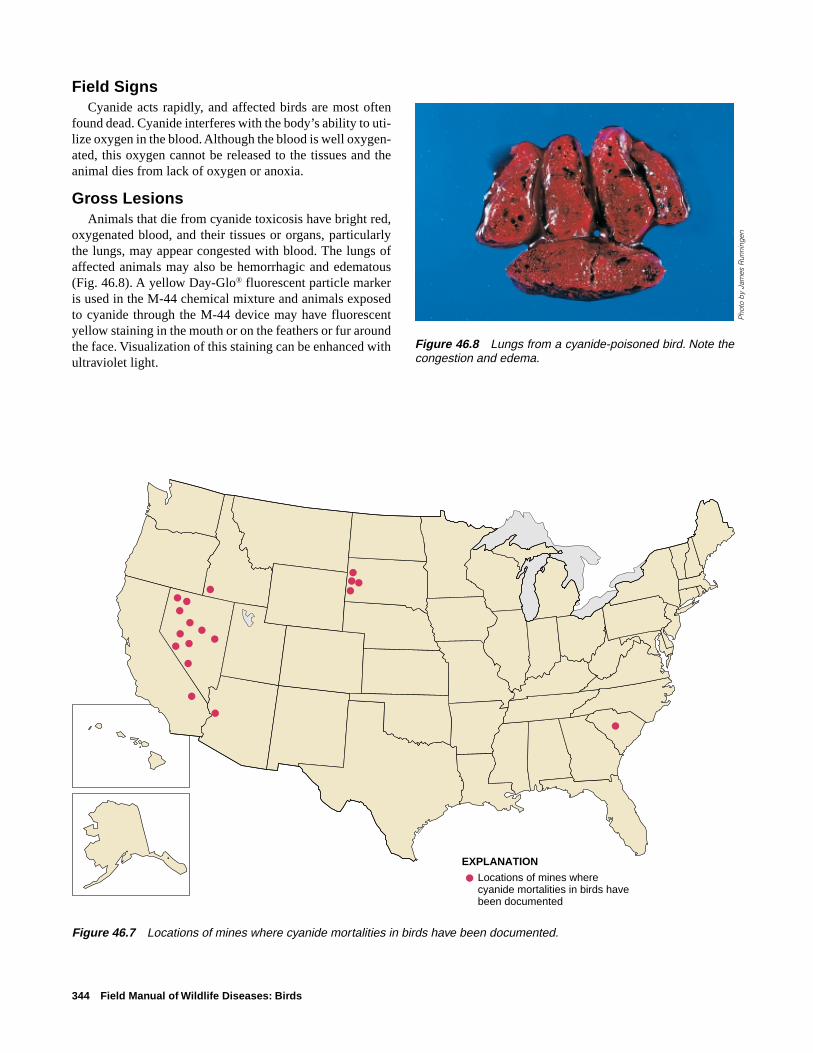
344 Field Manual of Wildlife Diseases: Birds
EXPLANATION
Locations of mines where cyanide mortalities in birds have been documented
●● ●●
●
●
●●
●●
●
●
●
●
●● ●
●
●
Figure 46.7 Locations of mines where cyanide mortalities in birds have been documented.
Figure 46.8 Lungs from a cyanide-poisoned bird. Note thecongestion and edema.
Field SignsCyanide acts rapidly, and affected birds are most often
found dead. Cyanide interferes with the body’s ability to uti-lize oxygen in the blood. Although the blood is well oxygen-ated, this oxygen cannot be released to the tissues and theanimal dies from lack of oxygen or anoxia.
Gross LesionsAnimals that die from cyanide toxicosis have bright red,
oxygenated blood, and their tissues or organs, particularlythe lungs, may appear congested with blood. The lungs ofaffected animals may also be hemorrhagic and edematous(Fig. 46.8). A yellow Day-Glo® fluorescent particle markeris used in the M-44 chemical mixture and animals exposedto cyanide through the M-44 device may have fluorescentyellow staining in the mouth or on the feathers or fur aroundthe face. Visualization of this staining can be enhanced withultraviolet light.
Pho
to b
y Ja
mes
Run
ning
en
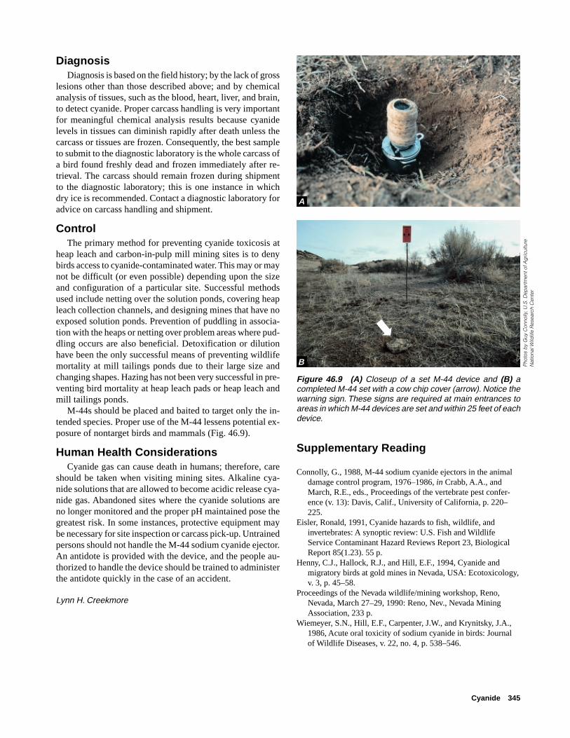
Cyanide 345
Figure 46.9 (A) Closeup of a set M-44 device and (B) acompleted M-44 set with a cow chip cover (arrow). Notice thewarning sign. These signs are required at main entrances toareas in which M-44 devices are set and within 25 feet of eachdevice.
DiagnosisDiagnosis is based on the field history; by the lack of gross
lesions other than those described above; and by chemicalanalysis of tissues, such as the blood, heart, liver, and brain,to detect cyanide. Proper carcass handling is very importantfor meaningful chemical analysis results because cyanidelevels in tissues can diminish rapidly after death unless thecarcass or tissues are frozen. Consequently, the best sampleto submit to the diagnostic laboratory is the whole carcass ofa bird found freshly dead and frozen immediately after re-trieval. The carcass should remain frozen during shipmentto the diagnostic laboratory; this is one instance in whichdry ice is recommended. Contact a diagnostic laboratory foradvice on carcass handling and shipment.
ControlThe primary method for preventing cyanide toxicosis at
heap leach and carbon-in-pulp mill mining sites is to denybirds access to cyanide-contaminated water. This may or maynot be difficult (or even possible) depending upon the sizeand configuration of a particular site. Successful methodsused include netting over the solution ponds, covering heapleach collection channels, and designing mines that have noexposed solution ponds. Prevention of puddling in associa-tion with the heaps or netting over problem areas where pud-dling occurs are also beneficial. Detoxification or dilutionhave been the only successful means of preventing wildlifemortality at mill tailings ponds due to their large size andchanging shapes. Hazing has not been very successful in pre-venting bird mortality at heap leach pads or heap leach andmill tailings ponds.
M-44s should be placed and baited to target only the in-tended species. Proper use of the M-44 lessens potential ex-posure of nontarget birds and mammals (Fig. 46.9).
Human Health ConsiderationsCyanide gas can cause death in humans; therefore, care
should be taken when visiting mining sites. Alkaline cya-nide solutions that are allowed to become acidic release cya-nide gas. Abandoned sites where the cyanide solutions areno longer monitored and the proper pH maintained pose thegreatest risk. In some instances, protective equipment maybe necessary for site inspection or carcass pick-up. Untrainedpersons should not handle the M-44 sodium cyanide ejector.An antidote is provided with the device, and the people au-thorized to handle the device should be trained to administerthe antidote quickly in the case of an accident.
Lynn H. Creekmore
A
B
➡
Supplementary Reading
Connolly, G., 1988, M-44 sodium cyanide ejectors in the animaldamage control program, 1976–1986, in Crabb, A.A., andMarch, R.E., eds., Proceedings of the vertebrate pest confer-ence (v. 13): Davis, Calif., University of California, p. 220–225.
Eisler, Ronald, 1991, Cyanide hazards to fish, wildlife, andinvertebrates: A synoptic review: U.S. Fish and WildlifeService Contaminant Hazard Reviews Report 23, BiologicalReport 85(1.23). 55 p.
Henny, C.J., Hallock, R.J., and Hill, E.F., 1994, Cyanide andmigratory birds at gold mines in Nevada, USA: Ecotoxicology,v. 3, p. 45–58.
Proceedings of the Nevada wildlife/mining workshop, Reno,Nevada, March 27–29, 1990: Reno, Nev., Nevada MiningAssociation, 233 p.
Wiemeyer, S.N., Hill, E.F., Carpenter, J.W., and Krynitsky, J.A.,1986, Acute oral toxicity of sodium cyanide in birds: Journalof Wildlife Diseases, v. 22, no. 4, p. 538–546.
Pho
tos
by
Guy
Con
nolly
, U.S
. Dep
artm
ent o
f Ag
ricul
tur e
Nat
iona
l Wild
life
Res
ear c
h C
ente
r

346 Field Manual of Wildlife Diseases: Birds

Salt 347
Chapter 47
Salt
SynonymsWater deprivation, salt encrustation
CauseAnimals become victims of salt poisoning or toxicosis
when toxic levels of sodium and chloride accumulate in theblood after they ingest large amounts of salt or, in some spe-cies, are deprived of water. For birds, salt sources may in-clude saline water and road salt.
Normally, the salt glands of birds (Fig. 47.1) excrete so-dium and chloride to maintain the proper physiologic chemi-cal balance. However, when there has been insufficient timefor acclimation of the salt gland to the saline environment,or when salt gland function is compromised by exposure tocertain pesticides or oil, the electrolyte balance of the bloodmay be upset by the excess sodium and chloride, resulting intoxicosis. Salt accumulation on the outside of the body, orsalt encrustation, is a greater problem for waterbirds that usevery saline waters than is salt toxicosis. Salt encrustationcan lead to exertion, acute muscle degeneration, and even-tual drowning during the struggle to escape entrapment.
Species AffectedThis infrequently reported toxicosis has affected gallina-
ceous birds, such as pheasants, and rock doves that consumedroad salt and migratory waterbirds forced to use highly sa-line water. Mortality from salt encrustation most often in-volves diving ducks.
DistributionSalt poisoning and salt encrustation can occur anywhere
that birds use saline environments. However, salt poisoningmay be more likely in northern latitudes where saline lakesremain open while nearby freshwater habitats freeze overand where salt is used for removing ice from roadways.
SeasonalitySalt poisoning and salt encrustation may affect birds at
any time of the year. In winter or early spring, terrestrialbirds may consume road salt for grit and mineral content.Migratory waterbirds are more likely to be poisoned duringlate autumn migration after they have spent several monthson freshwater nesting grounds. Cold snaps that freeze fresh-water areas along the migratory route may force birds to usemore saline waters that remain open because of the high saltcontent. High winds can contribute to salt encrustation bycontinually covering birds with salt-laden water.
Figure 47.2 Salt encrustation may completely cover the birdwith salt (A), or salt may accumulate on margins of feathers(B).
Pho
tos
by
Terr
y C
reek
mor
eA
Figure 47.1 The salt glands of birds are locatedjust above the eyes (arrows).
Field SignsClinical signs of salt poisoning may include muscle weak-
ness, partial paralysis, and difficult breathing, all of whichcan be caused by a variety of other toxicoses. Carcasses mayor may not be covered with salt (Fig. 47.2).
B
fpo
Pho
to b
y Ja
mes
Run
ning
en
➡
➡

348 Field Manual of Wildlife Diseases: Birds
Figure 47.4 (A and B) Visceral gout, or accumulation of grittyuric acid deposits on visceral surfaces, is a nonspecific lesionthat is sometimes associated with salt poisoning.
Gross LesionsGross lesions are nonspecific, and they may include red-
dening of the brain surface (Fig. 47.3), visceral gout(Fig. 47.4), fluid accumulation in the lungs, small hemor-rhages on the viscera, and erosions on the surfaces of theeyes.
DiagnosisFinding a source of salt exposure lends support to a some-
times difficult diagnosis of salt poisoning. Salt on the feath-ers provides further evidence, but is not in itself diagnostic.Refrigerated blood and frozen as well as formalin-fixed brainare the best tissues to collect for laboratory analysis. Be-cause the body maintains a constant internal environment orhomeostasis, sodium concentrations in these tissues normallydeviate very little. Therefore, a comparison of sodium con-centrations between suspect and reference specimens can beused to support a diagnosis of salt poisoning. Microscopicexamination of formalin-fixed brain tissue is also useful whensalt poisoning is suspected.
ControlBirds that are on highly saline lakes can be hazed to fresh-
water areas, if such areas exist nearby. Road salt should beused sparingly and should be stored out of reach of wildlife.Management practices that may expose birds to compoundsthat interfere with salt gland function, such as applicationsof organophosphorus and carbamate pesticides, should bedone only when necessary and should be scheduled to allowarriving birds maximum time to adapt to saline environments.
Human Health ConsiderationsNone.
J. Christian Franson and Milton Friend
Supplemental ReadingFriend, M., and Abel, J.H., Jr., 1976, Inhibition of mallard salt
gland function by DDE and organophosphates, in Page, L.A.,ed., Wildlife Disease: New York, N.Y., Plenum Press, p. 261–269.
Trainer, D.O., and Karstad, L., 1960, Salt poisoning in Wisconsinwildlife: Journal of the American Veterinary Associationv. 136, p. 14–17.
Windingstad, R.M., Kartch, F.X., Stroud, R.K., and Smith, M.R.,1987, Salt toxicosis in waterfowl in North Dakota: Journal ofWildlife Diseases, v. 23, p. 443–446.
Wobeser, G.A., 1997, Salt and saline water, in Diseases of wildwaterfowl (2nd ed): New York, N.Y., Plenum Press,p. 204–207.
Figure 47.3 The brains of salt-poisoned birds are sometimesvery red and congested.
Pho
to b
y Ja
mes
Run
ning
enP
hoto
by
J. C
hris
tian
Fran
son
A
Pho
to b
y Ja
mes
Run
ning
en
B

Barbiturates 349
Chapter 48
Barbiturates
SynonymsPentobarbital poisoning, sodium pentobarbital poison-ing
CauseBarbiturate products are commonly used to euthanize do-
mestic animals. The primary active component in euthana-sia solutions is sodium pentobarbital, but some products alsocontain other minor ingredients (Fig. 48.1).
Euthanasia solutions are generally injected intravenouslyin domestic animals; therefore, after death, the solutions willbe most concentrated in the blood and the highly vascular-ized organs, such as the liver or spleen, of the euthanizedanimal.
Euthanized carcasses that are available as carrion pose ahazard to scavenging birds and mammals. Large domesticanimal carcasses, such as horses, that are not used for foodor rendering but that are sufficiently valuable (monetarily orpsychologically) to warrant veterinary services and eutha-nasia drugs are the most common sources of barbiturate poi-soning in scavengers. In one instance in British Columbia, asingle cow carcass was responsible for poisoning 29 baldeagles.
Circumstances that interfere with burial, such as frozenwinter soil or bulky carcasses, result in euthanized carcassesbeing available for scavenger species. This problem couldincrease in the future if more stringent air-quality standardsrestrict carcass incineration.
Species AffectedBald and golden eagles are the only free-living wildlife
species that have been reported to have died of barbituratepoisoning. Raptors generally have a narrow tolerance forbarbiturate compounds; therefore, an anesthetic dose is oftenclose to a fatal dose in these species.
DistributionAs of 1997, the National Wildlife Health Center database
contained records of 17 cases of barbiturate poisoning ineagles from six States (Fig. 48.2). Additional cases have beenreported by other investigators.
SeasonalityCases of barbiturate poisoning have been more frequent
in late winter and early spring, but they are not confined tothat period. Cases of barbiturate poisoning may be corre-lated with the spring thaw in northern climates, when car-casses thaw, and the internal organs become more readilyavailable to scavengers. Residues in those carcasses becomeavailable to scavenger species at that time. Food supplies areoften limited at this time, so scavenging is more common.
Field SignsThe most useful and specific field sign is the proximity of
dead or moribund birds to a euthanized animal carcass thatshows evidence of scavenging. In lieu of that, the proximityof dead or moribund birds to a domestic animal carcass ofunknown origin is a less specific sign, but under that cir-cumstance, barbiturates should be considered along withother poisons, such as pesticides.
Barbiturate-poisoned birds have been found near land-fills in which euthanized animal carcasses were discarded.Landfills are legal disposal sites for carcasses in some Statesor locales.
Barbiturate poisoning may take hours to develop; there-fore, poisoned birds can be found distant from the poisonsource. Eagles have been found beneath their roost trees with-out evident sources of poisoning.
Barbiturate-intoxicated birds are sedated, drowsy, slug-gish, or comatose; have varying degrees of consciousness;and have slow heart and respiration rates. Although they maystruggle to right themselves if they fall from a perch as tox-icity progresses, signs of prolonged or violent struggling areunlikely. They are more likely to be found on undisturbedsubstrate. If more than one bird is exposed, the dose ingestedand susceptibility to the poison may vary with each bird;
Figure 48.1 The active agent in most injectable euthanasiasolutions is sodium pentobarbital.
Pho
to b
y N
ancy
Tho
mas

350 Field Manual of Wildlife Diseases: Birds
therefore, a range of signs from sublethal sedation to comato death may be observed. Birds that are sedated or evencomatose can recover if they are given supportive care untilthey metabolize the drug. Recovery may take several days.
Gross LesionsThere are no specific lesions. Ingesta may be present in
the upper gastrointestinal tract as in other acute poisonings.The ingesta may be recognizable as domestic animal in ori-gin. Barbiturate-poisoned birds are often in good body con-dition, thus reflecting the acute nature of this toxicosis.
DiagnosisAnalysis of liver or upper gastrointestinal contents de-
tects pentobarbital and, sometimes, other components ofeuthanasia drugs. Liver analysis is more definitive for deter-mining that a bird absorbed drug from the ingesta. Bloodsamples from live birds can be analyzed, but the clinical andfield signs and the bird’s recovery may be sufficient for apresumptive diagnosis.
Samples of blood-engorged organs, blood clots, or othertissue from scavenged sites in the suspect domestic animalcarcass can assist in tracing the source of the poison.
Control
TreatmentBirds found alive in the field are often hypothermic (ex-
hibiting low body temperature); warming of less affectedbirds, in itself, may result in recovery. A veterinarian canprovide supportive care, administer cardiac and respiratorystimulants, and remove the undigested crop contents so thatno further drug is absorbed.
3
EXPLANATION
Cases of barbiturate poisoning, by State
3
1
1
1
5 6 Figure 48.2 Seventeen cases of bar-
biturate poisoning in bald and goldeneagles have been reported by theNational Wildlife Health Center from sixStates as of 1997.
ManagementState agricultural departments in the United States gener-
ally regulate carcass disposal to assure that carcasses are notavailable to scavengers. Circumstances such as frozen groundthat prevents burial, poor compliance with regulations, orshallow burial may circumvent these regulations. Landfillregulations or policy can guarantee that carcasses are cov-ered before scavenging is likely.
Cases of barbiturate poisoning are generally inadvertent.Prevention can be greatly enhanced by increasing awarenessof the hazard among the public and veterinary community.
Human Health ConsiderationsNone. Euthanized carcasses are not for human consump-
tion. Barbiturates are not absorbed through the skin.
Nancy J. Thomas
Supplemental ReadingLanglier, K.M., 1993, Barbiturate poisoning in twenty-nine bald
eagles, in Redig, P.T., and others, eds., Raptor Biomedicine:Minneapolis, Minn., University of Minnesota Press, p. 231–232.
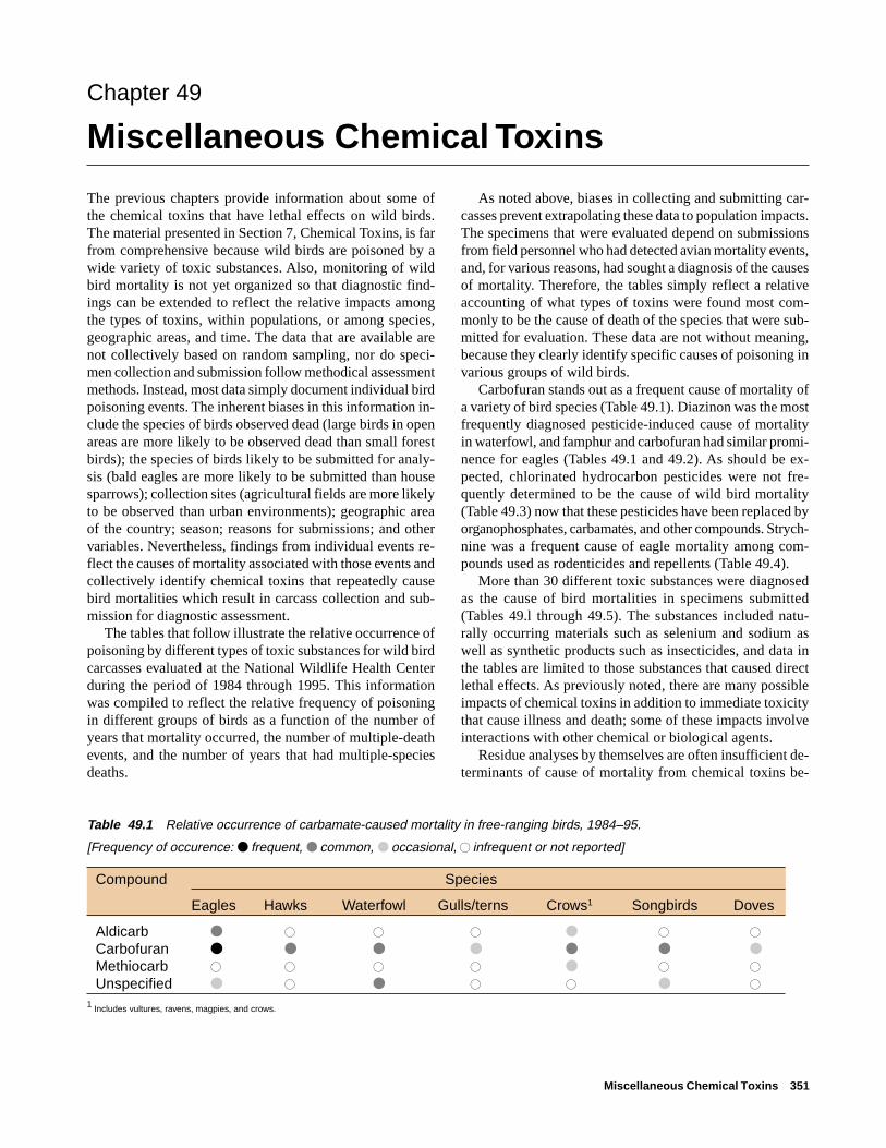
Miscellaneous Chemical Toxins 351
Chapter 49
Miscellaneous Chemical Toxins
The previous chapters provide information about some ofthe chemical toxins that have lethal effects on wild birds.The material presented in Section 7, Chemical Toxins, is farfrom comprehensive because wild birds are poisoned by awide variety of toxic substances. Also, monitoring of wildbird mortality is not yet organized so that diagnostic find-ings can be extended to reflect the relative impacts amongthe types of toxins, within populations, or among species,geographic areas, and time. The data that are available arenot collectively based on random sampling, nor do speci-men collection and submission follow methodical assessmentmethods. Instead, most data simply document individual birdpoisoning events. The inherent biases in this information in-clude the species of birds observed dead (large birds in openareas are more likely to be observed dead than small forestbirds); the species of birds likely to be submitted for analy-sis (bald eagles are more likely to be submitted than housesparrows); collection sites (agricultural fields are more likelyto be observed than urban environments); geographic areaof the country; season; reasons for submissions; and othervariables. Nevertheless, findings from individual events re-flect the causes of mortality associated with those events andcollectively identify chemical toxins that repeatedly causebird mortalities which result in carcass collection and sub-mission for diagnostic assessment.
The tables that follow illustrate the relative occurrence ofpoisoning by different types of toxic substances for wild birdcarcasses evaluated at the National Wildlife Health Centerduring the period of 1984 through 1995. This informationwas compiled to reflect the relative frequency of poisoningin different groups of birds as a function of the number ofyears that mortality occurred, the number of multiple-deathevents, and the number of years that had multiple-speciesdeaths.
As noted above, biases in collecting and submitting car-casses prevent extrapolating these data to population impacts.The specimens that were evaluated depend on submissionsfrom field personnel who had detected avian mortality events,and, for various reasons, had sought a diagnosis of the causesof mortality. Therefore, the tables simply reflect a relativeaccounting of what types of toxins were found most com-monly to be the cause of death of the species that were sub-mitted for evaluation. These data are not without meaning,because they clearly identify specific causes of poisoning invarious groups of wild birds.
Carbofuran stands out as a frequent cause of mortality ofa variety of bird species (Table 49.1). Diazinon was the mostfrequently diagnosed pesticide-induced cause of mortalityin waterfowl, and famphur and carbofuran had similar promi-nence for eagles (Tables 49.1 and 49.2). As should be ex-pected, chlorinated hydrocarbon pesticides were not fre-quently determined to be the cause of wild bird mortality(Table 49.3) now that these pesticides have been replaced byorganophosphates, carbamates, and other compounds. Strych-nine was a frequent cause of eagle mortality among com-pounds used as rodenticides and repellents (Table 49.4).
More than 30 different toxic substances were diagnosedas the cause of bird mortalities in specimens submitted(Tables 49.l through 49.5). The substances included natu-rally occurring materials such as selenium and sodium aswell as synthetic products such as insecticides, and data inthe tables are limited to those substances that caused directlethal effects. As previously noted, there are many possibleimpacts of chemical toxins in addition to immediate toxicitythat cause illness and death; some of these impacts involveinteractions with other chemical or biological agents.
Residue analyses by themselves are often insufficient de-terminants of cause of mortality from chemical toxins be-
Table 49.1 Relative occurrence of carbamate-caused mortality in free-ranging birds, 1984–95.
[Frequency of occurence: ● frequent, ● common, ● occasional, � infrequent or not reported]
Compound Species
Eagles Hawks Waterfowl Gulls/terns Crows1 Songbirds Doves
Aldicarb ● � � � ● � �
Carbofuran ● ● ● ● ● ● ●Methiocarb � � � � ● � �
Unspecified ● � ● � � ● �
1 Includes vultures, ravens, magpies, and crows.

352 Field Manual of Wildlife Diseases: Birds
Table 49.2 Relative occurrence of organophosphorus-caused mortality in free-ranging birds, 1984–95.
[Frequency of occurence; ● frequent, ● common, ● occasional, � infrequent or not reported]
Species
Compound Eagles Hawks Owls Waterfowl Cranes Shorebirds Crows1 Songbirds Doves
Chlorpyrifos � � � � � � ● � �
Coumaphos ● � � � � � � � �
Diazinon � � � ● � � ● ● �
Dimethoate � � � ● � � � � �
Disulfoton � � � � � � � ● �
Famphur ● ● ● � � � ● ● ●
Fenthion ● ● ● � � � � ● �
Fonofos � � � ● � � � � �
Monocrotophos � � � ● � � � ● ●
Parathion ● ● � ● � ● � ● �
Phorate ● � � ● � � � � �
Terbufos ● � � � � � � ● �
Unspecified ● ● ● � ● � � � �1 Includes vultures, ravens, magpies, and crows.
Table 49.3 Relative occurrence of chlorinated-hydrocarbon-caused mortality in free-ranging birds,1984–95.[Frequency of occurence: ● frequent, ● common,● occasional, � infrequent or not reported]
Species
Compound Eagles Owls Songbirds
Dieldrin ● ● ●Heptachlor ● � �
Table 49.4 Relative occurrence of rodenticides and repellents as causes of mortalityin free-ranging birds, 1984–95.
[Frequency of occurence: ● frequent, ● common, ● occasional, � infrequent or notreported]
Species
Compound Eagles Hawks Waterfowl Crows1 Songbirds
Avitrol® � � � � ●
Brodifacoum ● � ● � �
1080 ● � � � �
Strychnine ● ● ● ● ●
Thallium ● � � � �
Zinc phosphide � � ● � �
1 Includes vultures, ravens, magpies, and crows.

Miscellaneous Chemical Toxins 353
cause of species variations, lack of residue for some types ofcompounds, and other variables. Similarly, the often-quoted16th Century statement that, “Dosage Alone Determines Poi-soning” is modified by such factors as route of exposure andother important factors.
Chemical toxins are, and will continue to be, importantcauses of wildlife mortality. Documentation of mortality fromchemical toxins requires rigorous diagnostic work. Deter-mination of wildlife impacts will best be accomplishedthrough methodical monitoring programs that allow soundevaluations of changes in the status and trends of specificcompounds and their impacts on wild bird populations bygeographic area.
Milton Friend
Table 49.5 Relative occurrence of miscellaneous toxicants as causes of mortality in free-ranging birds, 1984–95.
[Frequency of occurence: ● frequent, ● common, ● occasional, � infrequent or not reported]
Compound
Ethylene Hydrogen Penta-Species Chloride Cyanide glycol Fluorine sulfide barbitol Selenium Sodium
Eagles � ● � � � ● � �
Hawks � ● � � � � � �
Owls � � � � ● � � �
Waterfowl ● ● � ● � � ● ●Cranes � ● � � � � � �
Grebes � ● � � � � ● ●Pelicans � � � � � � � ●Gulls/terns � ● � � � � � �
Shorebirds � ● � � � � � �
Egrets1 � � � � � � ● �
Crows2 � � ● � � � � �
Songbirds � ● � � � � � �
Doves � ● � � � � � ●Swallows � ● � � � � � �
Quail � � � � ● � � �
1 Includes long-legged wading birds such as herons and egrets.2 Includes vultures, ravens, magpies, and crows.

354 Field Manual of Wildlife Diseases: Birds

Introduction to Miscellaneous Diseases 355
Section 8Miscellaneous
Electrocution
Miscellaneous Diseases
Vertebral column deformity (scoliosis) in a bald eaglePhoto by James Runningen

356 Field Manual of Wildlife Diseases: Birds
Introduction to Miscellaneous Diseases
“Nature is far from benign; at least it has no special sentimentfor the welfare of the human versus other species.” (Lederberg)
Quote from:
Lederberg, J., 1993, Viruses and humankind: intracellularsymbiosis and evolutionary competition, in Morse, S.S., ed.,Emerging viruses: Oxford, England, Oxford University Press,p. 3.
The fact that “Nature is far from benign” is clearly evidentfrom the preceding chapters of this Manual. The diseasesand other conditions described are the proverbial “tip of theiceberg” relative to the number of specific causes of ill healthand death for free-ranging wild birds, but the wild bird healthproblems described account for most major wild bird dis-ease conditions seen within the United States. However, thefull toll from disease involves many other causes of illnessand death that individually may cause substantial die-offs.Two examples of these other causes of die-offs are the deathsof Canada geese that ingest dry soybeans, which then ex-pand and cause lethal impactions within the moist environ-ment of digestive tract, and the poisoning of ducks from rictin,a naturally occurring toxic component of castor beans. Someof these lesser-known causes of disease and mortality maybecome increasingly important in the future because land-scape and other changes could result in environmental con-ditions that may enhance the interface between specific dis-ease agents and susceptible bird species.
This final Section of the Manual includes some of thelesser-known causes of avian mortality. The first chapter pro-vides an overview of electrocution in birds, with a specialemphasis on eagles. The second chapter is a miscellaneouschapter that highlights a significant disease of domestic duck-lings not yet known to exist in wild birds, disease caused bystress due to improper handling of birds, and several otherconditions that might be encountered by biologists who workwith birds. These other conditions include tumors, traumaticinjuries, weather, nutritional factors, and drowning as causesof avian illness and death. These two chapters expand thescope of disease presented in the previous chapters and pro-vide additional perspectives of the diverse causes of avianmortality. It is our hope that the collective information pro-vided in this Manual will stimulate those interested in theconservation and well-being of avian species to give greaterconsideration to disease in the management strategies em-ployed for the conservation of these species.

Electrocution 357
Chapter 50
Electrocution
CausePower lines and power poles present a potential electro-
cution hazard to wild birds. Many birds, especially raptors,select power poles for perching, and, sometimes, for nesting(Figs. 50.1–3). If a bird’s appendages bridge the gap betweentwo energized parts or between an energized and a groundedmetal part, electricity flows through the “bridge” that is fill-ing the gap and the bird is electrocuted.
Most commonly, birds are electrocuted where conduct-ing wires (conductors) are placed closer together than thewingspan of birds that frequent the poles (Fig. 50.2). Feath-ers are poor electrical conductors, but if contact is madebetween points on the skin, talons, or beak, or if the feathersare wet, conduction can occur. Common anatomical sites ofcontact include conduction between the wrists of each wingor between the skin of one wing and a foot or leg. The result-ing shock causes severe, usually fatal, cardiovascular injury.
Because conductors on distribution lines are placed closertogether than high voltage transmission lines, birds are morefrequently electrocuted on distribution lines despite theirlower voltage.
In addition to one to three conductors, power poles mayalso carry ground wires, transformers, or grounded metalcrossarm braces. Complicated wiring configurations that putmultiple energized and grounded metal parts near attractiveperching or nesting sites are the most hazardous configura-tions (Fig. 50.3).
Species AffectedElectrocution is primarily a problem of large raptors in
open habitat, particularly treeless areas. Golden eagles areby far at greatest risk, but other eagles, large buteos, falcons,and the largest owls, such as the great horned owl, are alsosusceptible. The large wingspan of these birds appears to bethe single most important factor in their susceptibility.
In addition to their size, the perching behavior of thesebird species puts them at greater risk. Species that preferexposed high perches are more likely to be attracted to powerpoles, as are the species that use a “still hunting” techniquein which they perch and visually search the landscape forprey rather than hunting in flight.
Immature and subadult raptors are more commonly elec-trocuted. This predisposition is presumably related to theirinexperience and awkwardness in taking off and landing.
Figure 50.1 A bald eagle using a power poleas a perch.
Figure 50.2 This is a hazardous situation be-cause the eagle’s wings can contact two conduc-tors at once.
Figure 50.3 An eagle nest on the topof a power pole.
Pho
to b
y B
rian
Woo
db
ridg
e, U
.S.
Fish
and
Wild
life
Ser
vice
, Mac
doe
l, C
AP
hoto
by
Mor
lan
Nel
son,
Pac
ifiC
orp
, Boi
se, I
D
Pho
to b
y M
onte
Gar
rett,
Pac
ifiC
orp

358 Field Manual of Wildlife Diseases: Birds
DistributionBird electrocutions are most common in the western plains
of the United States where open shrub and grassland habi-tats are common, and are less prevalent in forested habitat(Fig. 50.4). However, birds may be electrocuted whereverelectrical lines are above ground.
Generally, electrocutions are more prevalent in sites wherea susceptible species’ prey base is present and where suit-able perches, other than power structures, are lacking. In thewestern plains, elevated perches are at a premium, and themore susceptible raptor species are abundant. The combina-tion of golden eagles, jackrabbits, grassland habitat, and dan-gerous power pole configurations can be expected to belethal. Similar conditions exist on the Russian steppes. Elec-trocution is a major cause of mortality for the Russian steppeeagle and for other raptors that nest on power poles and usethem for perches in this largely treeless area (Fig. 50.5).
SeasonalityBirds can be electrocuted during any season, but there
can be seasonal fluctuations in electrocution frequency thatare related to weather conditions or bird behavior. Electro-cutions are more frequent during periods of rain and snowbecause of the increased conductivity of wet feathers. In-clement wet weather may also combine with windy condi-tions so that birds are less stable while landing and takingoff. Where distribution lines are oriented with crossarms per-pendicular or diagonal to the prevailing wind, more electro-cutions occur.
Golden eagles may make greater use of power poles asnight roosts during migration and wintering. This habit maymake them more prone to electrocution as they stretch out todry their wings in the morning sun.
Inattentiveness during seasonal mating behaviors or ter-ritorial conflicts have also been reported to predispose birdsto electrocution.
Field SignsElectrocuted birds often die immediately, so they are found
near a power pole or beneath a power line.The electrical hazard may be apparent in the configura-
tion of the nearby pole. The conductors and other electricalhardware on the pole may be close together. The greatesthazards may be at corner poles where extra wires (jumpers)are required to provide a change in direction, or at poles withtransformers or grounded metal equipment near the conduc-tors (Fig. 50.6).
EXPLANATION
Number of electrocuted eaglesdiagnosed, by State, 1975–97
More than 70
51–70
21–50
11–20
1–10
None
Figure 50.4 Number of electrocuted eagles diagnosed per State from 1975–95. (From unpublished datafrom the National Wildlife Health Center.)
Figure 50.5 Power lines that are not designed to preventelectrocution and that cross largely treeless areas, such asthis line on the Russian steppes, pose a significant hazard forlarge raptors that use the poles as perches for hunting and asnesting platforms.
Phot
o by
Milt
on F
riend

Electrocution 359
Bird electrocutions can cause power outages; therefore, ahistory of electrical power disruption can help confirm thediagnosis and fix the location and time of electrocution.
Electrocuted birds may catch on fire and ignite vegeta-tion beneath the power structures.
If a bird is electrocuted because the prey item or wet nestmaterial it is carrying comes in contact with an energizedpart, then these items may be found with the carcass orclutched in its talons.
Gross LesionsThe hallmark of electrocution is burn marks. Burns are
generally confined to the sites of body contact with the elec-trical source; however, if the feathers are ignited then theentire carcass may be charred (Fig. 50.7). Burn marks fromfatal electrocutions can have a remarkable range in appear-ance from very subtle feather disruption to limb amputation.Burns cause the feather edges to curl or twist (Fig. 50.8),and light-colored feathers may be discolored brown orcharred. Burns on avian skin appear as dry blisters, particu-larly on the scales of the feet or legs (Fig. 50.9A and B). The
margins of these blisters may be brown or charred. Severe,deep burns can extend through the skin, cauterize musclesand tendons, liquefy fat, and even fracture bones.
Sublethal bird electrocutions are uncommon. In thesecases, a single limb is usually affected. Initially, burns maybe seen on the skin or the feathers at the contact site. Later,the only evidence may be the loss of blood supply to a wingor foot and eventual gangrene. If the damage can be removedby surgical amputation, some electrocuted birds can recoverand be kept permanently in captivity.
DiagnosisA diagnosis of electrocution is based on the presence of
burns and an absence of evidence of other causes of death.Hemorrhages in the subcutaneous tissue and internal organssuggest cardiovascular injury and can support the diagnosis.
A field history that includes proximity to an electrical lineis helpful but not sufficient in itself. Birds may collide with
Figure 50.6 Complicated wiring that is configured with trans-formers, jumpers, and additional hardware is often respon-sible for raptor electrocutions. (Graphic provided by MonteGarrett, PacifiCorp)
Figure 50.7 An electrocuted bald eagle that is charred overmost of its body.
Figure 50.8 Electrical burns on the wing feathers of a baldeagle. Note also the fracture and charring of nearby bones.
Pho
to b
y Ja
mes
Run
ning
enP
hoto
by
Jam
es R
unni
ngen
Jumperwires
Lightningarrester
Fusedcutout
Bushings

360 Field Manual of Wildlife Diseases: Birds
Figure 50.9 (A) A large burn on a golden eagle’s foot. (B)Multiple small, subtle burns in the scales on a bald eagle’sfoot.
Pho
tos
by
Jam
es R
unni
ngen
A
B
power lines, be shot while perching, or fall from perchesafter poisoning or illness; therefore, location is not defini-tive for electrocution.
ControlRaptor electrocutions generally can be reduced by adopt-
ing safe electrical pole and line configurations or managingraptor perching. Safe wiring configurations separate the wiresand the grounded metal parts so that raptors cannot simulta-neously touch two of them at once (Fig. 50.10). Existing in-stallations that contain hazardous configurations can be modi-fied by insulating or reconfiguring the wiring. Rather thancomprehensive modifications, an economical but effectiveapproach is to modify selected poles based on field observa-tions of bird use and mortality. If reconfiguring or insulatingthe wires is not feasible, then access to the hazardous perchcan be blocked and safer, alternate perches can be provided.Despite the inherent equipment costs of modification, elec-trical power companies are often proactive in preventing bird
electrocution. Power companies benefit by reducing costlypower outages, by avoiding liability for migratory bird mor-talities, and by the positive public image that is generated bycontrol projects.
When new electrical installations are planned, the designcan take into consideration the likelihood of raptor electro-cution. The risk can be evaluated in advance by consideringraptor concentrations and behavior along the installationroute. Structures in raptor migratory corridors, as well asnesting and wintering ranges, may pose a risk.
Human Health ConsiderationsUnder normal circumstances, there is no exposure.
Figure 50.10 A safe wiring configuration separates the con-ductors and other energized hardware so that large raptorsare unable to touch two pieces of hardware simultaneously.(Graphic provided by Monte Garrett, PacifiCorp)
Nancy J. Thomas
Supplementary ReadingAvian Power Line Interaction Committee, 1996, Suggested
practices for raptor protection on power lines: The state of theart in 1996: Edison Electric Institute/Raptor ResearchFoundation, Washington, D.C., 125 p.
Bevanger, K., 1994, Bird interactions with utility structures: Ibis,v. 136, p. 412–425.
43" 60"
Nonconductingbraces
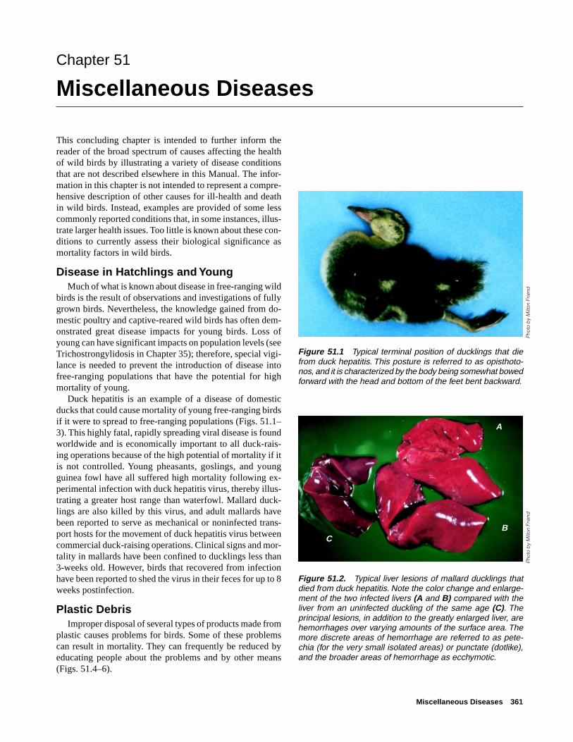
Miscellaneous Diseases 361
Chapter 51
Miscellaneous Diseases
This concluding chapter is intended to further inform thereader of the broad spectrum of causes affecting the healthof wild birds by illustrating a variety of disease conditionsthat are not described elsewhere in this Manual. The infor-mation in this chapter is not intended to represent a compre-hensive description of other causes for ill-health and deathin wild birds. Instead, examples are provided of some lesscommonly reported conditions that, in some instances, illus-trate larger health issues. Too little is known about these con-ditions to currently assess their biological significance asmortality factors in wild birds.
Disease in Hatchlings and YoungMuch of what is known about disease in free-ranging wild
birds is the result of observations and investigations of fullygrown birds. Nevertheless, the knowledge gained from do-mestic poultry and captive-reared wild birds has often dem-onstrated great disease impacts for young birds. Loss ofyoung can have significant impacts on population levels (seeTrichostrongylidosis in Chapter 35); therefore, special vigi-lance is needed to prevent the introduction of disease intofree-ranging populations that have the potential for highmortality of young.
Duck hepatitis is an example of a disease of domesticducks that could cause mortality of young free-ranging birdsif it were to spread to free-ranging populations (Figs. 51.1–3). This highly fatal, rapidly spreading viral disease is foundworldwide and is economically important to all duck-rais-ing operations because of the high potential of mortality if itis not controlled. Young pheasants, goslings, and youngguinea fowl have all suffered high mortality following ex-perimental infection with duck hepatitis virus, thereby illus-trating a greater host range than waterfowl. Mallard duck-lings are also killed by this virus, and adult mallards havebeen reported to serve as mechanical or noninfected trans-port hosts for the movement of duck hepatitis virus betweencommercial duck-raising operations. Clinical signs and mor-tality in mallards have been confined to ducklings less than3-weeks old. However, birds that recovered from infectionhave been reported to shed the virus in their feces for up to 8weeks postinfection.
Plastic DebrisImproper disposal of several types of products made from
plastic causes problems for birds. Some of these problemscan result in mortality. They can frequently be reduced byeducating people about the problems and by other means(Figs. 51.4–6).
Figure 51.1 Typical terminal position of ducklings that diefrom duck hepatitis. This posture is referred to as opisthoto-nos, and it is characterized by the body being somewhat bowedforward with the head and bottom of the feet bent backward.
Pho
to b
y M
ilton
Frie
nd
Figure 51.2. Typical liver lesions of mallard ducklings thatdied from duck hepatitis. Note the color change and enlarge-ment of the two infected livers (A and B) compared with theliver from an uninfected duckling of the same age (C). Theprincipal lesions, in addition to the greatly enlarged liver, arehemorrhages over varying amounts of the surface area. Themore discrete areas of hemorrhage are referred to as pete-chia (for the very small isolated areas) or punctate (dotlike),and the broader areas of hemorrhage as ecchymotic.
Pho
to b
y M
ilton
Frie
nd
A
BC

362 Field Manual of Wildlife Diseases: Birds
Figure 51.3 Mallard duckling infectedwith duck hepatitis. The livers of infectedbirds generally become so swollen thatthey fill much of the bird’s abdominalcavity.
Figure 51.4 (A) Improperly discarded fishing line carried to the top of this tree by a double-crested cormorant became a“hangman’s noose” and strangled the bird in this photograph. The line tangled around the tree top and it also looped around thebird’s neck when it attempted to fly from its perch above a small urban lake. (B) Discarded fishing line wrapped around the bill ofthis white pelican would have resulted in death by starvation had the bird not been captured and the line removed. Note also theconstricted areas of the pouch caused by the line.
Pho
to b
y M
ilton
Frie
nd
Pho
tos
by
Milt
on F
riend
A B
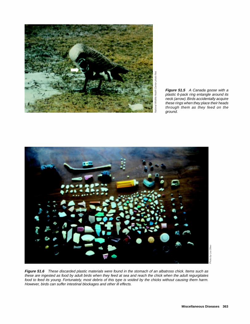
Miscellaneous Diseases 363
Figure 51.6 These discarded plastic materials were found in the stomach of an albatross chick. Items such asthese are ingested as food by adult birds when they feed at sea and reach the chick when the adult regurgitatesfood to feed its young. Fortunately, most debris of this type is voided by the chicks without causing them harm.However, birds can suffer intestinal blockages and other ill effects.
Figure 51.5 A Canada goose with aplastic 6-pack ring entangle around itsneck (arrow). Birds accidentally acquirethese rings when they place their headsthrough them as they feed on theground.
Pho
to b
y Lo
u S
ileo
Nat
iona
l Wild
life
Hea
lth C
ente
r p
hoto
file
s➡

364 Field Manual of Wildlife Diseases: Birds
Disease Due To HandlingImproper judgements and procedures by humans while
they pursue, handle, and transport wild animals, includingbirds, during wildlife management activities can induce cap-ture myopathy (Figs. 51.7–8). More descriptive names in-clude over-straining disease, transport myopathy, exertionalmyopathy, muscle necrosis, white muscle disease, and stressmyopathy. These names convey that improper handling orstress can cause a bird to overexert and result in stress-related injury to its muscles. Tissue damage is a result ofcomplex physiological processes, not physical trauma suchas bruising from impact. Mortality has been reported in awide variety of bird species including flamingos, cranes,waterfowl, raptors, gulls, wild turkey, and other species. Thisdisease of overexertion results in severe damage to striatedmuscles including the heart. Birds may die hours or evenseveral days after they have been released, thereby leavingtheir human captors and handlers unaware of the damagethat was done. The potential for inducing this disease shouldbe carefully considered during the planning phases of wild-life capture, handling, and transportation, and measuresshould be taken to minimize risks. Warm environmental tem-peratures are often a risk factor as are the duration of pur-suit, the method and duration of restraint, placement of birdsin unfamiliar surroundings, and noise associated with hu-man activities. Situations that have induced capture myopa-thy in birds include trapping and handling operations involv-ing drop nets and rocket nets; drive-trapping, handling, andtranslocation of flightless birds; and handling birds so thatmarking devices, including radio transmitters, can be placedon them. All of these needed activities can be done safely ifproper consideration is given to capture myopathy and thesteps that can be taken to avoid inducing this disease.
TumorsNeoplasms or tumors are infrequent findings in free-rang-
ing wild birds, but they are found (Figs. 51.9–12). Tumorsare formed by the abnormal progressive multiplication ofcells into uncontrolled (by the body) new tissue that appearsas various growths within tissues and organs. These growthsmay be noninvasive or benign, or they may spread to othertissues and parts of the body and be malignant. Tumors re-sult from multiple causes. Virus-induced tumors, such as theherpesvirus that causes an important infectious poultry dis-ease known as Marek’s disease, are transmissible. Tumorsformed due to other than infectious agents have been reportedfrom all major body systems of birds, the reproductive, di-gestive, respiratory, nervous, and endocrine systems, in ad-dition to the skin surfaces.
Less than 1 percent of the wild birds for which postmor-tem examinations were done at the National Wildlife HealthCenter (NWHC) over a span of more than 20 years (1975–1998) had tumors. These findings are consistent with those
Figure 51.8 Light colored area in breast muscle (arrow) of aperegrine falcon with capture myopathy.
Figure 51.7 Light colored muscle of leg (arrow) representscapture myopathy in a sandhill crane.
Pho
to b
y Ja
mes
Run
ning
enP
hoto
by
Nan
cy J
. Tho
mas
➡
➡
Figure 51.9 Tumors in breast muscle of a Canada goose.
Pho
to b
y Ja
mes
Run
ning
en

Miscellaneous Diseases 365
of other disease diagnostic laboratories that process largenumbers of free-ranging wildlife. A notable exception at theNWHC has been a high prevalence of tumors in Mississippisandhill cranes received from the wild (Figs. 51.11, 12). Thecause(s) of the tumors in this endangered species remainsundetermined.
TraumaMany wild birds are injured and killed each year from
impacts with buildings, wires, and other products of the hu-man environment (Figs. 51.13, 14). Birds that have large wingspans, such as cranes and eagles, are among those commonlyfound with fractured wings and other injuries from collisionswith power lines and wire fences. Road kills of raptors thatfeed on carrion are common. Whenever it is feasible, birdflight patterns and bird use of local habitat should be consid-ered in the routing of power transmission lines, wind powergeneration units, and roads. Protective measures against birdstrikes should be employed when they are warranted if lesshazardous alternative routings cannot be accomplished.Monitoring for road kills of birds and observations of birdsfeeding on carcasses can indicate food shortages for speciessuch as eagles and can be mitigated by establishing short-term feeding stations that move the birds from the roadwaysto safer locations during the period of food scarcity.
OtherWild birds are subject to major direct losses from weather.
Waterfowl and other species have been frozen to the ice bytheir feet and feathers (Fig. 51.15), and strong winds associ-ated with hurricanes have filled coastal beaches with largenumbers of birds with fractured wings. Heavy snows andstorms that coat vegetation with a thick layer of ice deprive
Figure 51.10 Tumor on the leg of a ruffed grouse.
Figure 51.12 A malignant tumor cov-ering the heart (top arrow) and lungs(bottom arrow) of a Mississippi sandhillcrane.
Figure 51.11 Tumor attached to the kidneys of a Mississippisandhill crane.
Pho
to b
y Ja
mes
Run
ning
enP
hoto
by
Jam
es R
unni
ngen
Pho
to b
y Ja
mes
Run
ning
en
➡
➡
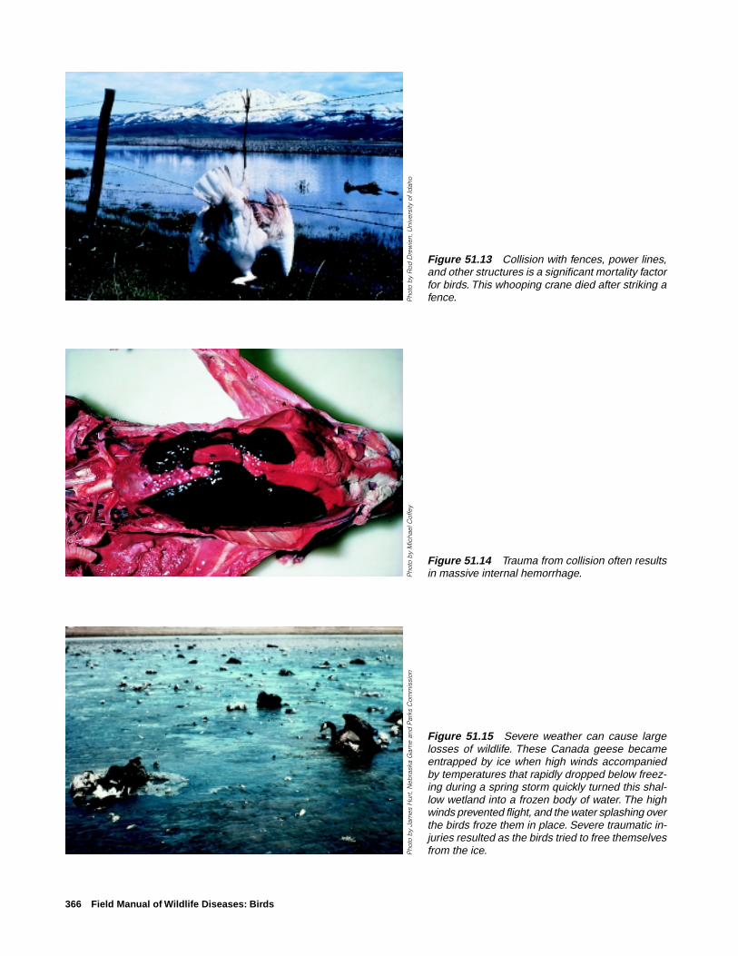
366 Field Manual of Wildlife Diseases: Birds
Figure 51.13 Collision with fences, power lines,and other structures is a significant mortality factorfor birds. This whooping crane died after striking afence.
Figure 51.15 Severe weather can cause largelosses of wildlife. These Canada geese becameentrapped by ice when high winds accompaniedby temperatures that rapidly dropped below freez-ing during a spring storm quickly turned this shal-low wetland into a frozen body of water. The highwinds prevented flight, and the water splashing overthe birds froze them in place. Severe traumatic in-juries resulted as the birds tried to free themselvesfrom the ice.
Figure 51.14 Trauma from collision often resultsin massive internal hemorrhage.
Pho
to b
y R
od D
rew
ien,
Uni
vers
ity o
f Id
aho
Pho
to b
y M
icha
el C
offe
yP
hoto
by
Jam
es H
urt,
Neb
rask
a G
ame
and
Par
ks C
omm
issi
on

Miscellaneous Diseases 367
wild birds and other wildlife of access to food and can resultin starvation (Fig. 51.16). Numerous other weather-relatedsituations also affect bird health.
Malnutrition resulting in starvation is but one aspect ofnutritional diseases that may affect birds. Nutritional diseasesare a complex subject area that is beyond the scope of thisManual, and they are mentioned only to make the readeraware of them. Nutritional diseases involve excess intake aswell as deficiencies. Changes in bird diets associated withlandscape changes due to agriculture can contribute to nutri-tional diseases. For example, excesses of dietary protein andvitamin deficiency may occur due to extensive feeding onagricultural grains rather than natural food sources. Visceralgout may result (Fig. 51.17). Under experimental conditions,substances that are toxic to the kidneys (nephrotoxic agents)and diets deficient in Vitamin A and high in calcium havecaused avian gout.
Wild birds also drown. Drowning may be an outcome ofextreme weather conditions that aquatic birds are sometimessubject to; exhaustion of passerines during migration, whichcauses them to drop into water bodies that they may be tra-versing at the time; and as a result of other factors, such asthe feathers of aquatic birds becoming waterlogged from oilcontamination or nonfunctioning preen glands that preventbirds from “waterproofing” their feathers.
Various deformities due to a variety of causes are alsoseen in wild birds (Fig. 51.18). Some deformities result fromexposure to excess levels of selenium; others may result fromexposure to synthetic compounds, nutritional disorders, orinjury to tissues during early developmental stages of thebird; they may be of genetic origin; or result from other caus-es. Deformities are not commonly observed because birdsthat are afflicted with such conditions are likely to be morevulnerable to factors that reduce their chance for survival.Therefore, clusters of observations of deformities should beviewed as an indication of a larger problem and warrant in-vestigation to determine the underlying cause.
Milton Friend and Nancy J. Thomas
Supplementary ReadingFairbrother, A., Locke, L.N., and Hoff, G.L., 1996, Noninfectious
diseases of wildlife, (2d ed.): Ames, Iowa, Iowa State Univer-sity Press, 219 p.
Wallach, J.D., and Cooper, J.E., 1982, Nutritional diseases of wildbirds, in Hoff, G.L., and others, eds., Noninfectious diseases ofwildlife: Ames, Iowa, Iowa State University Press, p. 113–126.
Pho
to b
y M
ilton
Frie
nd
Figure 51.16 Ice that coats vegetation may prevent accessto food, resulting in starvation.
Figure 51.17 Dietary protein imbalances can cause visceralgout, exhibited by an accumulation of white, gritty deposits onsurfaces of organs, such as the heart (arrow).
Figure 51.18 Vertebral column deformity (scoliosis) in a baldeagle.
Pho
to b
y M
ilton
Frie
ndP
hoto
by
Jam
es R
unni
ngen
➡
➡

368 Field Manual of Wildlife Diseases: Birds

Appendix A 369
Appendix A
Sample specimen history form
Submitter’s name: Affiliation:
Address: Telephone: E-mail:
Date collected:
Method of collection: [found dead, euthanized (describe method) etc.]
Collector’s name:
Specific die-off location:
State: County: Latitude/longitude:
Environmental factors: (Record conditions such as storms, precipitation, temperature changes, or other changes
that may contribute to stress.)
Disease onset: (The best estimate of when the outbreak started.)
Species affected: (The diversity of species affected may provide clues to the disease involved.)
Age/sex: (Any selective mortality related to age and sex.)
Morbidity/mortality: (Ratio of sick animals to dead animals.)
Known dead: (Actual pickup figures.)
Estimated dead: (Consider removal by scavengers or other means.)
Clinical signs: (Any unusual behavior and physical appearance.)
Population at risk: (Number of animals in the area that could be exposed to the disease.)
Population movement: (Recent changes in the number of animals on the area and their source or destination,
if known.)
Problem area description: (Land use, habitat types, and other distinctive features.)
Comments: (Additional information/observations that may be of value such as past occurrences of disease in area.)

370 Field Manual of Wildlife Diseases: Birds
Appendix B
Sources of wildlife diagnostic assistancein the United States
Assistance in obtaining a diagnosis of wildlife illness ordeath is available from a variety of sources. However, it isadvisable to make inquiries before the need arises about avail-able services, the estimated response time for completingwork, and who to contact when assistance is required.
The following wildlife disease programs can offer infor-mation, assistance, and services.
Wildlife Disease Programs
1. U.S. Department of Interior,U.S. Geological Survey,Biological Resources DivisionNational Wildlife Health Center6006 Schroeder RoadMadison, WI 53711Telephone (608) 270-2400Web site: http://www.emtc.usgs.gov/nwhchome.html
2. State fish and game agencies. Several States have wild-life disease programs. Among those are Alaska, Califor-nia, Colorado, Florida, Idaho, Michigan, New Jersey, NewYork, Wisconsin, and Wyoming. Contact the State fish andgame agency headquarters to inquire about assistance.
3. Regional wildlife disease programs. Two regional programsare presently affiliated with universities:
Southeastern Cooperative Wildlife Disease StudyCollege of Veterinary MedicineUniversity of GeorgiaAthens, GA 30602Telephone (706) 542-1741
Northeastern Research Center for Wildlife DiseasesUniversity of ConnecticutDepartment of PathobiologyStorrs, CT 06269-3089Telephone (860) 486-4000
4. University programs. Several other universities, for ex-ample, the University of Florida—Gainesville and VirginiaPolytechnical Institute and State University—Blacksburg,are involved in wildlife disease activities. Inquiries atschools of veterinary medicine and departments of veteri-nary or animal science at universities throughout the UnitedStates will reveal additional sources of wildlife disease di-agnostic assistance.
5. Private sector. Some private consultants also deal withwildlife disease problems.
Domestic Animal Disease Programs
1. U.S. Department of Agriculture,Animal and Plant Health Inspection ServiceNational Veterinary Services LaboratoriesP.O. Box 844, Ames, Iowa 50010Telephone (515) 239-8600.
This facility accepts diagnostic specimens that have beenreferred to it through appropriate State or Federal chan-nels.
2. State departments of agriculture. Animal disease diagnos-tic laboratories exist to serve domestic animal needs, butwill often accept wildlife specimens.
3. Private sector. Veterinarians in private practice often haveboth interest and expertise in wildlife diseases and maybecome involved with these problems.
Additional sources of assistance for investigatingwildlife mortality events when chemical toxins aresuspected.
Federal GovernmentU.S. Fish and Wildlife Service (FWS)
The Division of Environmental Contaminants (DEC) is theFWS focal point for issues associated with chemical tox-ins. Information on contaminants can be obtained from theCentral Office in Washington, D.C.
Telephone: (703) 358-2148Web site: http://www.fws.gov/~r9dec/ecprog.html and fromDEC staff in the seven FWS Regional Offices.
DEC biologists are assigned to field offices throughout theFWS Regions. They work on specific contaminant issuesin each Region and are available to provide informationand assistance regarding mortality event investigations.
Toxic spill coordinators are located in each RegionalOffice, providing a focal point for response actions.
FWS Regional Offices are located in:Portland, Ore.; Albuquerque, N. Mex.; Fort Snelling, Minn.;Atlanta, Ga.; Hadley, Mass.; Denver, Colo.; and Anchor-age, Alaska.

Appendix B 371
U.S. Geological Survey, Biological Resources Division (BRD)
The National Wildlife Health Center, which is the BRDScience Center in Madison, Wis., provides informationabout and assists in investigating wildlife mortality events.
Telephone: (608) 270-2400Web site: http://www.emtc.usgs.gov/nwhchome.html
Research on chemical toxins is carried out at several BRDScience Centers. Those Centers maintain in-depth tech-nical knowledge regarding the fate and impacts of chemi-cals in the environment. The Patuxent Wildlife ResearchCenter in Laurel, Md. is an internationally recognizedsource of information on the effects of contaminants, par-ticularly on avian species.
Telephone: (301) 497-5500Web site: http://www.pwrc.usgs.gov/
U.S. Environmental Protection Agency (EPA)
Office of Solid Waste and Emergency Response
Telephone: (703) 308-8413Web site: http://www.epa.gov/epaoswer/
The Office of Pollution Prevention and Toxics (OPPT) as-sesses the hazards and risks posed by industrial chemi-cals to human health and the environment. The Environ-mental Effects Branch in OPPT can provide informationon the toxicity of chemicals to aquatic and terrestrial or-ganisms.
Telephone: (202) 260-1268
Office of Pesticide Programs maintains the Ecological Inci-dent Information System, which is a data base on mortal-ity of non-target organisms caused by pesticides.
Telephone: (703) 305-5392
State GovernmentMany State natural resource agencies have environmentalcontaminant programs that provide a mechanism to reportsuspected chemical toxin problems. Some States have groupsthat investigate mortality events associated with chemicals,and that may be able to provide field assistance and chemi-cal analysis.
Natural resource agencies in several States maintain wildlifehealth programs, which respond to wildlife mortality events.
State veterinary diagnostic laboratories often have toxicolo-gists on staff who have specific knowledge of toxic problemswithin the region.
Poison Control CentersThe National Animal Poison Control Center at the University
of Illinois College of Veterinary Medicine provides a fee-based service directed to prevention and treatment of ad-verse effects of chemical exposures in animals. This ser-vice is staffed by veterinary health professionals who haveaccess to a wide range of information specific to animalpoisoning.
Telephone (800) 548-2423, (900) 680-0000Web site: http://www.cvm.uiuc.edu/NAPCC/NAPCC.html
The American Association of Poison Control Centers(AAPCC) has certified about 40 regional poison informa-tion centers throughout the U.S. that focus on human ex-posure to chemical toxins. These centers function to pro-vide poison information, telephone management and con-sultation, collect pertinent data, and deliver professionaland public education. The national AAPCC office is inWashington, D.C.Telephone: (202) 362-7217. A directory of the regional cen-ters is available at Web site: http://www.pitt.edu/~martint/pages/rpiclist.htm
Colleges of Veterinary MedicineMost colleges of veterinary medicine have toxicology depart-ments staffed with experts in the area of animal toxicology.
Analytical LaboratoriesChoosing an analytical laboratory requires attention to meth-ods used, quality assurance/quality control (QA/QC), and cost.Laboratories should be using methods that are appropriateto the analysis required in the matrix (material being ana-lyzed) that is submitted. Minimum quality control data pro-vided by the laboratory should include:
(1) The results of analysis of spiked samples, or recov-ery. A known amount of the compound being analyzedfor is added to the appropriate matrix. The recovery isthe amount of the compound that was recovered in theanalysis, and it is expressed as a percentage of theamount of compound added.(2) A replication of results, or an agreement of analy-ses of duplicate samples.(3) The results of blank samples, or an absence of thecompound being analyzed for in a “clean” sample ofthe appropriate matrix.(4) The results of analysis of standard referencesamples. A sample with a known quantity of the com-pound is prepared by an independent laboratory, andthis sample is then analyzed by the laboratory beingevaluated. Although good QA/QC adds to the expenseof analytical work, the alternative may be an incorrectdiagnosis.

372 Field Manual of Wildlife Diseases: Birds
Some of the analytical laboratories that have been used bythe FWS DEC and others include:
For inorganic analyses:Environmental Trace Substance LaboratoryUniversity of Missouri - Rolla101 USBM Bldg., 1300 North Bishop Ave.Rolla, MO 65409-0530Telephone: (314) 341-6607
Research Triangle Institute3040 Cornwallis Road, Bldg. 6Research Triangle Park, NC 27709-2194Telephone: (919) 541-6896
Geochemical & Environmental Research Group833 Graham RoadCollege Station, TX 77845Telephone: (409) 690-0095
For organic analyses:Geochemical & Environmental Research Group (seeabove)
Mississippi State Chemical LaboratoryMississippi State UniversityHand Chemical Lab, Rm 201, Morrill RoadMississippi State, MS 39762Telephone: (601) 325-3251
The above listing is not intended to be comprehensive,nor does it constitute endorsement by the Federal govern-ment. Rather, it illustrates the diversity of possible sourcesof assistance. Individual circumstances and events dictatewhich of these sources will be most useful in specific situa-tions.

Appendix C 373
Appendix C
Sources of supplies used for collecting,
preserving, and shipping specimens
Company Address and telephone Item
Scientific Products 319 West Ontario Whirl-Pak® bags, formalin, wide-mouth, plastic jars, indelible markersChicago, IL 60610Tel: (800)323-4515
Curtis Matheson P.O. Box 1546 Whirl-Pak® bags, wide-mouth, plastic jars, indelible markersScientific, Inc. Houston, TX 77251
Tel: (713)820-9898
Fisher Scientific Co. 711 Forbes Ave. Whirl-Pak® bags, formalin, indelible markersPittsburgh, PA 15219Tel: (800)766-7000
Thomas Scientific P.O. Box 99 Whirl-Pak® bags, wide-mouth, plastic jars, indelible markersSwedesboro, NJ 08095Tel: (609)467-2000
VWR Scientific P.O. Box 66929 Whirl-Pak® bags, indelible markersO’Hare AmpChicago, IL 60666Tel: (800)932-5000
Local hospital and Whirl-Pak® bags, wide-mouth, plastic jarsmedical supplybusinesses
Some pharmacies formalin
Freund Can Co. 167 W. 84th Street metal paint cans with lidsChicago, IL 60620Tel: (312)224-4230
U.S. General Services GSA Customer filament strapping tape, plastic bags, indelible markers(gov’t agencies only) Supply Center
Administration (GAS)5619 W. 115th St.Worth, IL 60482Tel: (800)262-0570
GSA Federal 1500 E. Bannister Rd. filament strapping tape, plastic bags, indelible markersSupply Service Kansas City, MD 64131
Tel: (816)926-7315

374 Field Manual of Wildlife Diseases: Birds
Company Address and telephone Item
Local hardware, sports, Styrofoam®, shipping coolers, indelible markers, ice packs, filamentand discount stores strapping tape
Polyfoam Packers Corp. 2320-T Foster Avenue biomedical shippers and mailersWheeling, IL 60090Tel: (800)225-7443
Protective clothing, gloves, and disinfectants can also be obtained from many of the sources listed above. The above list is not intended to be comprehensive, nordoes it constitute endorsement by the Federal government.

Appendix D 375
Appendix D
Normal brain cholinesterase activity values
StandardSpecies Mean1 deviation Sample size
Avocet, American 19.4 2.9 5Blackbird, Red-winged 24.5 1.2 5Bobwhite, Northern 16.3 2.1 7Brant, Black 14.4 0.8 5Coot, American 20.5 5.0 12Cormorant, Double-crested 29.3 2.4 5Cowbird, Brown-headed 19.7 2.6 5Crane, Mississippi Sandhill 16.6 2.3 15Crane, Sandhill 17.5 1.4 8Crane, Whooping 15.1 1.5 9Dove, Mourning 22.8 3.3 11Duck, American Wigeon 10.5 1.0 12Duck, Blue-winged Teal 19.5 3.6 6Duck, Green-winged Teal 13.5 2.0 17Duck, Kola 11.0 1.2 9Duck, Mallard 11.0 1.6 75Duck, Muscovy 11.6 3.5 8Duck, Northern Shoveler 14.7 1.2 5Duck, Pintail 11.6 1.3 24Duck, Ruddy 13.4 1.4 8Duck, Wood 10.2 1.5 9Eagle, Bald 16.0 2.6 156Eagle, Golden 16.0 2.2 57Egret, Common 17.4 1.9 5Egret, Snowy 25.0 1.3 5Falcon, Peregrine 18.6 3.2 27Goldfinch, American 17.8 1.5 10Goose, Canada 11.9 2.0 36Goose, Canada (Aleutian) 14.0 3.6 8Goose, Ross 14.2 2.3 9Goose, Snow 13.6 2.6 42Goose, White-fronted 12.1 1.2 9Grebe, Eared 14.7 1.4 17Grosbeak, Evening 20.3 3.1 5Gull, Ring-billed 23.9 6.5 8Hawk, Red-tailed 17.5 1.4 7Hawk, Sharp-shinned 21.5 2.0 6Heron, Black-crowned Night 15.6 3.0 5Heron, Great Blue 13.3 2.1 8Loon, Common 17.3 4.4 5Owl, Great-horned 15.5 2.0 7Owl, Screech 18.7 1.4 5Owl, Spotted 14.6 2.0 9

376 Field Manual of Wildlife Diseases: Birds
StandardSpecies Mean1 deviation Sample size
Parrot, Puerto Rican 19.4 2.0 6Pelican, American White 13.0 1.3 13Pelican, Brown 11.2 1.2 16Sandpiper, Semipalmated 14.1 1.1 5Siskin, Pine 16.9 2.3 16Stork, Wood 18.7 4.2 8Swan, Trumpeter 11.3 1.3 10Swan, Tundra 11.6 1.7 14Tern, Least California 44.1 9.1 5Woodcock, American 16.8 1.1 5Woodpecker, Red-cockaded 38.4 4.8 5
1 Cholinesterase activity is expressed as micromoles acetylthiocholine hydrolyzed per minute per gram of wetweight brain tissue (Hill, E.F. and Fleming, W.J., 1982, Environmental toxicology and chemistry 1:27–38).
Milton Smith

Appendix E 377
Appendix ECommon and scientific names of birds in text
Albatrosses, shearwaters, and petrels (Order Procellariiformes)Albatrosses (Family Diomedeidae)
Black-footed albatross (Diomedia nigripes)Laysan albatross (Diomedia immutabilis)
Shearwaters and petrels (Family Procellariidae)Northern fulmar (Fulmarus glacialis)Petrels (Pterodroma sp, Oceanodroma sp, Oceanites sp)Sooty shearwater (Puffinus griseus)
Cormorants, pelicans, and tropicbirds (Order Pelecaniformes)Cormorants and Shags (Family Phalacrocoracidae)
Brandt’s cormorant (Phalacrocorax penicillatus)Cape cormorant (Phalacrocorax capensis)Double-crested cormorant (Phalacrocorax auritus)Great cormorant (Phalacrocorax carbo)Shags (Phalacrocorax sp)
Gannets and boobies (Family Sulidae)Gannet (Morus bassanus)
Pelicans (Family Pelecanidae)Brown pelican (Pelecanus occidentalis)White pelican (Pelecanus erythrorhynchos)
Tropicbirds (Family Phaethontidae)Red-tailed tropicbird (Phaethon rubricauda)White-tailed tropicbird (Phaethon lepturus)
Cranes, coots, and rails (Order Gruiformes)Cranes (Family Gruidae)
Brolga crane (Grus rubicunda)Common crane (Grus grus)Demoiselle crane (Anthropoides virgo)East African crowned crane (Balearica regulorum gibbericeps)Greater sandhill crane (Grus canadensis tabida)Hooded crane (Grus monacha)Lesser sandhill crane (Grus canadensis canadensis)Manchurian crane (red-crowned crane) (Grus japonensis)Mississippi sandhill crane (Grus canadensis pulla)Sarus crane (Grus antigone)Stanley crane (blue crane) (Anthropoides paradisea)White-naped crane (Grus vipio)Whooping crane (Grus americana)
Rails, coots, and gallinules (Family Rallidae) American coot (Fulica americana)Common moorhen (Gallinula chloropos)Sora (Porzana carolina)
Emus and ostriches (Order Struthioniformes)Emu (Family Casuariidae, Dromaius novaehollandiae)Ostrich (Family Struthionidae, Struthio camelus)
Grebes (Order Podicipediformes, Family Podicipedidae)Eared grebe (Podiceps nigricollis)Western grebe (Aechmophorus occidentalis)

378 Field Manual of Wildlife Diseases: Birds
Grouse, quail, and partridges (Order Galliformes, Family Phasianidae)Grouse (Subfamily Tetraoninae)
Blue grouse (Dendragapus obscurus)Ptarmigan (Lagopus sp)Red grouse (Lagopus lagopus scoticus)Ruffed grouse (Bonasa umbellus)Sage grouse (Centrocercus urophasianus)Sharp-tailed grouse (Tympanuchus phasianellus)
Quail (Subfamily Odontophorninae)Bobwhite quail (Northern) (Colinus virginianus)California quail (Callipepla californica)Coturnix quail (Coturnix coturnix)Japanese quail (Coturnix japonica)Scaled quail (Callipepla squamata)
Partridges and pheasants (Subfamily Phasianinae)Chukar partridge (Alectoris chukar)Gray partridge (Hungarian partridge) (Perdix perdix)Common peafowl (Pavo cristatus)Ring-necked pheasant (Phasianus colchicus)
Turkeys (Subfamily Meleagridinae)Wild turkey (Meleagris gallopavo)
Guinea fowl (Subfamily Numidinae, Numida sp)Hawks, falcons, and vultures (Order Falconiformes)
Hawks, eagles, and kites (Family Accipitridae)Eurasian sparrowhawk (Accipiter nisus)Bald eagle (Haliaeetus leucocephalus)Booted eagle (Hieraaetus pennatus)Cooper’s hawk (Accipiter cooperii)Common buzzard (Buteo buteo)Ferruginous hawk (Buteo regalis)Golden eagle (Aquila chrysaetos)Northern goshawk (Accipiter gentilis)Imperial eagle (Aquila heliaca)Northern harrier (marsh hawk) (Circus cyaneus)Red-shouldered hawk (Buteo lineatus)Red-tailed hawk (Buteo jamaicensis)Rough-legged hawk (Buteo lagopus)Sharp-shinned hawk (Accipiter striatus)Steppe eagle (Aquila nipalensis)White-tailed eagle (Haliaeetus albicilla)Osprey (Pandion haliaetus)
Falcons and caracaras (Family Falconidae)American kestrel (sparrowhawk) (Falco sparverius)Gyrfalcon (Falco rusticolus)Peregrine falcon (Falco peregrinus)Prairie falcon (Falco mexicanus)Red-headed falcon (Falco chicquera)Saker falcon (Falco cherrug)
Vultures (Family Cathartidae)California condor (Gymnogyps californianus)King vulture (Sarcoramphus papa)Turkey vulture (Cathartes aura)
Herons, ibises, and storks (Order Ciconiiformes)Flamingos (Family Phoenicopteridae)

Appendix E 379
Greater flamingo (Phoenicopterus ruber)Herons, egrets, and bitterns (Family Ardeidae)
Bitterns (Botaurus sp, Ixobrychus sp)Black-crowned night heron (Nycticorax nycticorax)Great blue heron (Ardea herodias)Great egret (common or american egret) (Casmerodius albus)Snowy egret (Egretta thula)
Ibises and spoonbills (Family Threskiornithidae)Glossy ibis (Plegadis falcinellus)White ibis (Eudocimus albus)White-faced ibis (Plegadis chihi)
Storks (Family Ciconiidae)Wood stork (Mycteria americana)
Loons (Order Gaviiformes, Family Gaviidae)Common loon (Gavia immer)Pacific loon (Gavia pacifica)Red-throated loon (Gavia stellata)
Owls (Order Strigiformes)Typical owls (Family Strigidae)
Barred owl (Strix varia)Eagle owl (Bubo bubo)Eastern screech owl (Otus asio)Great-horned owl (Bubo virginianus)Long-eared owl (Asio otus)Short-eared owl (Asio flammeus)Snowy owl (Nyctea scandiaca)Spotted owl (Strix occidentalis)
Barn-Owls (Family Tytonidae)Barn owl (Tyto alba)
Parrots, parakeets, and macaws (Order Psittaciformes)Parrots (Family Psittacidae)
Budgerigar (Melopsittacus undulatus)Cockatiel (Nymphicus hollandicus)Hawk-headed parrot (Deroptyus accipitrinus)Lories (Lorius sp)Lorikeets (Trichoglossus sp)Lovebirds (Agapornis sp)Macaws (Ara sp)Puerto Rican parrot (Amazona vittata)Rosellas (Platycercus sp)Yellow-naped parrot (Amazona auropalliata)
Perching birds (Order Passeriformes)Finches (Family Fringillidae)
American goldfinch (Carduelis tristis)Apapane (Himatione sanguinea)Cassin’s finch (Carpodacus cassinii)Eurasian bullfinch (Pyrrhula pyrrhula)Evening grosbeak (Coccothraustes vespertinus)Goldfinches (Carduelis sp)House finch (Carpodacus mexicanus)Pine siskin (Carduelis pinus)Purple finch (Carpodacus purpureus)
Wood-warblers, tanagers, grosbeaks, sparrows, and blackbirds (Family Emberizidae)Brown-headed cowbird (Molothrus ater)

380 Field Manual of Wildlife Diseases: Birds
Chipping sparrow (Spizella passerina)Common grackle (Quiscalus quiscula)Dusky seaside sparrow (Ammospiza nigrescens)Northern cardinal (Cardinalis cardinalis)Orioles (Icterus sp)Red-winged blackbird (Agelaius phoeniceus)Rufous-sided towhee (Pipilo erythrophthalmus)
Chickadees ( Family Paridae)Chestnut-backed chickadee (Parus rufescens)
Crows, jays, and magpies (Family Corvidae)American Crow (Corvus brachyrhychos)Blue jay (Cyanocitta cristata)Eurasian jay (Garrulus glandarius)Jackdaw (Corvus monedula)Magpie (Pica sp)Raven (Corvus corax)Rook (Corvus frugilegus)Steller’s jay (Cyanocitta stelleri)
Waxwings (Family Bombycillidae)Cedar waxwing (Bombycilla cedrorum)
Weavers (Family Ploceidae, Ploceus sp)Old world sparrows (Passeridae)
House sparrow (English sparrow) (Passer domesticus)Larks (Family Alaudidae)
Horned lark (Eremophila alpestris)Mockingbirds and thrashers (Family Mimidae)
Northern mockingbird (Mimus polyglottos)Thrashers (Toxostoma sp)
Nuthatches (Family Sittidae, Sitta sp)Shrikes (Family Laniidae, Lanius sp)Starlings (Family Sturnidae)
European starling (Sturnus vulgaris)Mynas (Acridotheres sp, Gracula sp)
Swallows (Family Hirundinidae, Hirundo sp, Tachycineta sp, Riparia sp, Stelgidopteryx sp)Martins (Progne sp)
Thrushes, solitaires, and bluebirds (Family Muscicapidae)American robin (Turdus migratorius)Bluebirds (Sialia sp)Eurasian blackbird (Turdus merula)Thrushes (Turdus sp, Ixoreus sp, Hylocichla sp, Catharus sp)
Estrildid finches (Family Estrildidae)Java finch (Padda oryzivora)
Penguins (Order Sphenisciformes, Family Spheniscidae)Blackfooted penguin (Spheniscus demersus)
Pigeons and doves (Order Columbiformes, Family Columbidae)Band-tailed pigeon (Columba fasciata)Mourning dove (Zenaida macroura)Ringed turtle dove (Streptopelia risoria)Rock dove (common pigeon) (Columba livia)White-winged dove (Zenaida asiatica)Wood pigeon (Columba palumbus)
Shorebirds (Order Charadriiformes)Gulls, terns, skuas, and skimmers (Family Laridae)
California gull (Larus californicus)

Appendix E 381
Common tern (Sterna hirundo)Franklin’s gull (Larus pipixcan)Glaucous-winged gull (Larus glaucescens)Herring gull (Larus argentatus)Kittiwakes (Rissa sp)Laughing gull (Larus atricilla)Least California tern (Sterna antillarum)Ring-billed gull (Larus delawarensis)Royal tern (Sterna maxima)Skua (Catharacta skua)
Auks, murres, and puffins (Family Alcidae)Common murre (Uria aalge)Guillemots (Cepphus sp)Murres (Uria sp)Puffins (Fratercula sp)Razorbill (Alca torda)
Plovers (Family Charadriidae, Charadrius sp, Pluvialis sp)Sandpipers, turnstones, surfbirds, and phalaropes (Family Scolopacidae)
American woodcock (Scolopax minor)Curlews (Numenius sp)Long-billed dowitcher (Limnodromus scolopaceus)Marbled godwit (Limosa fedoa)Pectoral sandpiper (Calidris melanotos)Ruddy turnstone (Arenaria interpres)Semipalmated sandpiper (Calidris pusilla)Spotted sandpiper (Actitis macularia)Western sandpiper (Calidris mauri)
Stilts and avocets (Family Recurvirostridae)American avocet (Recurvirostra americana)Black stilt (Himantopus novaezelandiae)Black-necked stilt (Himantopus mexicanus)
Waterfowl (Order Anseriformes, Family Anatidae)Dabbling ducks
American wigeon (Anas americana)American black duck (Anas rubripes)Blue-winged teal (Anas discors)Gadwall (Anas strepera)Green-winged teal (Anas crecca)Koloa duck (Anas wyvilliana)Laysan duck (Anas laysanensis)Mallard (Anas platyrhynchos)Mottled duck (Anas fulvigula)Muscovy duck (Cairina moschata)Northern pintail (Anas acuta)Northern shoveler (Anas clypeata)White Pekin duck (Anas platyrhynchos)Wood duck (Aix sponsa)
Diving ducksBufflehead (Bucephala albeola)Canvasback (Aythya valisineria)Common goldeneye (American goldeneye) (Bucephala clangula)Greater scaup (Aythya marila)Harlequin duck (Histrionicus histrionicus)Lesser scaup (Aythya affinis)
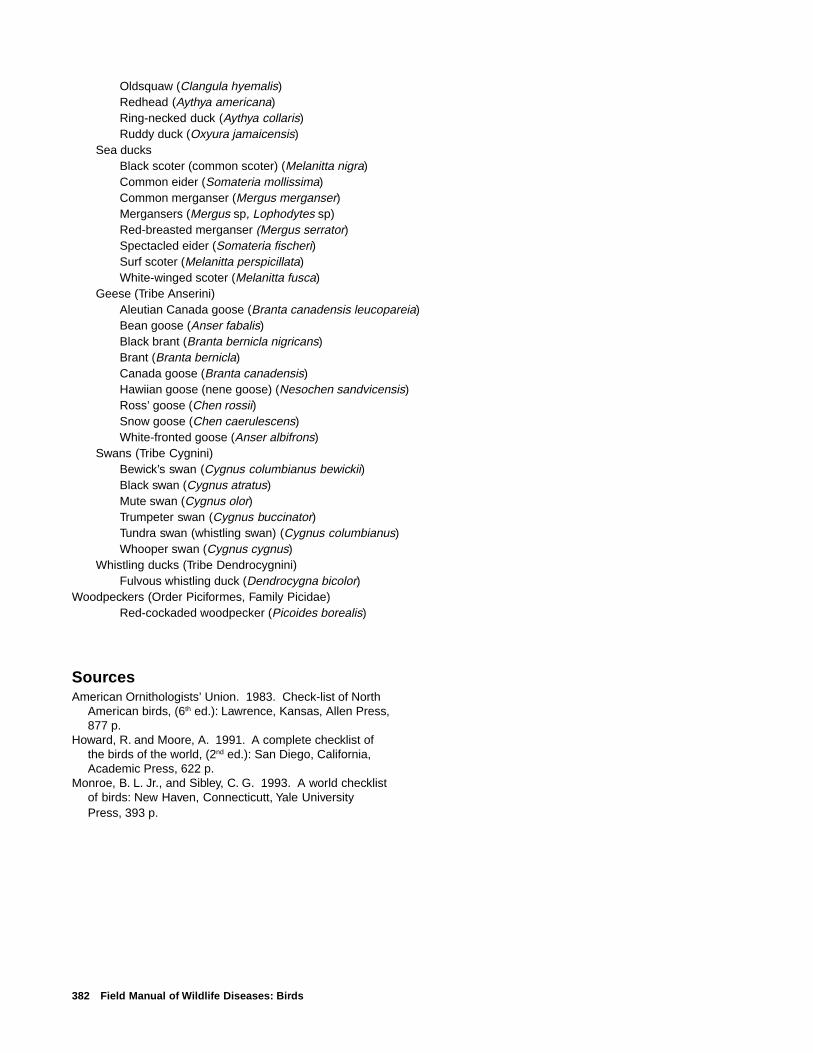
382 Field Manual of Wildlife Diseases: Birds
Oldsquaw (Clangula hyemalis)Redhead (Aythya americana)Ring-necked duck (Aythya collaris)Ruddy duck (Oxyura jamaicensis)
Sea ducksBlack scoter (common scoter) (Melanitta nigra)Common eider (Somateria mollissima)Common merganser (Mergus merganser)Mergansers (Mergus sp, Lophodytes sp)Red-breasted merganser (Mergus serrator)Spectacled eider (Somateria fischeri)Surf scoter (Melanitta perspicillata)White-winged scoter (Melanitta fusca)
Geese (Tribe Anserini)Aleutian Canada goose (Branta canadensis leucopareia)Bean goose (Anser fabalis)Black brant (Branta bernicla nigricans)Brant (Branta bernicla)Canada goose (Branta canadensis)Hawiian goose (nene goose) (Nesochen sandvicensis)Ross’ goose (Chen rossii)Snow goose (Chen caerulescens)White-fronted goose (Anser albifrons)
Swans (Tribe Cygnini)Bewick’s swan (Cygnus columbianus bewickii)Black swan (Cygnus atratus)Mute swan (Cygnus olor)Trumpeter swan (Cygnus buccinator)Tundra swan (whistling swan) (Cygnus columbianus)Whooper swan (Cygnus cygnus)
Whistling ducks (Tribe Dendrocygnini)Fulvous whistling duck (Dendrocygna bicolor)
Woodpeckers (Order Piciformes, Family Picidae)Red-cockaded woodpecker (Picoides borealis)
SourcesAmerican Ornithologists’ Union. 1983. Check-list of North
American birds, (6th ed.): Lawrence, Kansas, Allen Press,877 p.
Howard, R. and Moore, A. 1991. A complete checklist ofthe birds of the world, (2nd ed.): San Diego, California,Academic Press, 622 p.
Monroe, B. L. Jr., and Sibley, C. G. 1993. A world checklistof birds: New Haven, Connecticutt, Yale UniversityPress, 393 p.
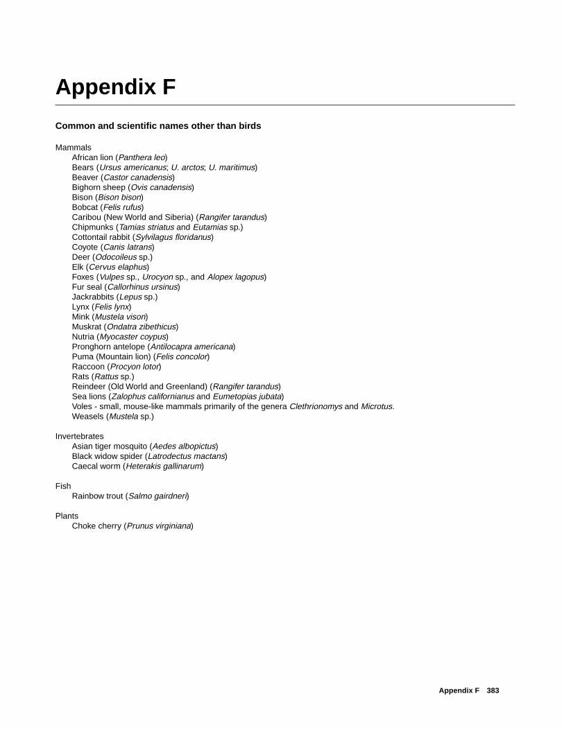
Appendix F 383
Appendix F
Common and scientific names other than birds
MammalsAfrican lion (Panthera leo)Bears (Ursus americanus; U. arctos; U. maritimus)Beaver (Castor canadensis)Bighorn sheep (Ovis canadensis)Bison (Bison bison)Bobcat (Felis rufus)Caribou (New World and Siberia) (Rangifer tarandus)Chipmunks (Tamias striatus and Eutamias sp.)Cottontail rabbit (Sylvilagus floridanus)Coyote (Canis latrans)Deer (Odocoileus sp.)Elk (Cervus elaphus)Foxes (Vulpes sp., Urocyon sp., and Alopex lagopus)Fur seal (Callorhinus ursinus)Jackrabbits (Lepus sp.)Lynx (Felis lynx)Mink (Mustela vison)Muskrat (Ondatra zibethicus)Nutria (Myocaster coypus)Pronghorn antelope (Antilocapra americana)Puma (Mountain lion) (Felis concolor)Raccoon (Procyon lotor)Rats (Rattus sp.)Reindeer (Old World and Greenland) (Rangifer tarandus)Sea lions (Zalophus californianus and Eumetopias jubata)Voles - small, mouse-like mammals primarily of the genera Clethrionomys and Microtus.Weasels (Mustela sp.)
InvertebratesAsian tiger mosquito (Aedes albopictus)Black widow spider (Latrodectus mactans)Caecal worm (Heterakis gallinarum)
FishRainbow trout (Salmo gairdneri)
PlantsChoke cherry (Prunus virginiana)

384 Field Manual of Wildlife Diseases: Birds
Appendix G
Chemical names
Common Names Chemical Names
1080 Sodium monofluoracetate
2-PAM Pralidoxime chloride
Alachlor (Lasso®) 2-Chloro-N-(2,6-diethylphenyl)-N-(methoxymethyl)acetamide
Aldicarb (Temik®) 2-Methyl-2-(methylthio)propanal O-[(methylamino)carbonyl]oxime
Aldrin 1,2,3,4,10,10-Hexachloro-1,4,4a,5,8,8a-hexahydro-1,4:5,8-dimethanonaphthalene
Antimycin 3-Methylbutanoic acid 3-[3-(formylamino)-2-hydroxybenzoyl]amino-8-hexyl-2,6-dimethyl-4,9-dioxo-1,5-dioxonan-7-yl ester
Aroclor® Group of polychlorinated biphenyls
Atrazine 6-Chloro-N-ethyl-N'-(1-methylethyl)-1,3,5-triazine-2,4-diamine
Avitrol® 4-Aminopyridine
Bayluscide® 5-Chloro-N-(2-chloro-4-nitrophenyl)-2-hydroxybenzamide compound respectively with 2-aminoethanol(1:1)
Beuthanasia-D-Special® Sodium pentobarbital
Bomyl® 3-[(Dimethoxyphosphinyl)oxy]-2-pentenedioic acid dimethyl ester
Brodifacoum (Talon®) 3-[3-(4'-Bromo[1-1'-biphenyl]-4-yl)-1,2,3,4-tetrahydro-1-napthalenyl]-4-hydroxy-2H-1-benzopyran-2-one
Captan 3a,4,7,7a-Tetrahydro-2- [(trichloromethyl)thio]-1H-isoindole-1,3(2H)-dione
Carbofuran 2,3-Dihydro-2,2-dimethyl-7-benzofuranol methylcarbamate
Chlorophacinone 2-[(4-Chlorophenyl)phenylacetyl]-1H-indene-1,3(2H)-dione
Chlorpyrifos Phosphorothioic acid O,O-diethyl O-(3,5,6-trichloro-2-pyridinyl) ester
Chlordane 1,2,4,5,6,7,8,8-Octachloro-2,3,3a,4,7,7a-hexahydro-4,7-methano-1H-indene
Clophens® Group of polychlorinated biphenyls
Coumaphos Phosphorothioic acid O-(3-chloro-4-methyl-2-oxo-2H-1-benzopyran-7-yl) O,O-diethyl ester
DDD Dichlorodiphenyl dichloroethane
DDE Dichlorodiphenyldichloroethylene
DDT Dichloro diphenyl trichloroethane
Demeton (Systox®) Phosphorothioic acid O,O-diethyl O-[2-(ethylthio)ethyl] ester mixture with O,O- diethyl S-[2-(ethylthio)ethyl]phosphorothioate
Diazinon Phosphorothioic acid O,O-diethyl O-[6-methyl-2-(1-methylethyl)-4-pyrimidinyl] ester
Dicrotophos Phosphoric acid 3-(dimethylamino)-1-methyl-3-oxo-1-propenyl dimethyl ester
Dieldrin (1aα,2β,2aα,3β,6β,6aα,7β,7aα)-3,4,5,6,9,9-Hexachloro-1a,2,2a,3,6,6a,7,7a-octahydro-2,7:3,6-dimethanonaphth[2,3-b]oxirene

Appendix G 385
Common Names Chemical Names
Dimethoate Phosphorodithioic acid O,O-dimethyl S-[2-(methylamino)-2-oxoethyl] ester
Diphacinone 1,3-Inandione, 2-diphenylacetyl
Diquat® Dipyrido(1,2-a:2',1'-c)pyrazinediium, 6,7-dihydro
Disulfoton Phosphorodithioic acid O,O-diethyl S-[2-(ethylthio)ethyl] ester
EDTA Ethylenediaminetetraacetic acid
Endosulfan 6,7,8,9,10,10-Hexachloro-1,5,5a,6,9,9a-hexahydro-6,9-methano-2,4,3-benzodioxathiepin 3-oxide
Endrin (1aα,2β,2aβ,3α,6α,6aβ,7β,7aα)-3,4,5,6,9,9-Hexachloro-1a,2,2a,3,6,6a,7,7a-octahydro-2,7:3,6-dimethanonaphth[2,3-b]oxirene
Enflurane 2-Chloro-1-(difluoromethoxy)-1,1,2- trifluoroethane
Famphur Phosphorothioic acid O-[4-[(dimethylamino)sulfonyl]phenyl] O,O-dimethyl ester
Fenamiphos (1-Methylethyl)phosphoramidic acid ethyl 3-methyl-4-(methylthio)phenyl ester
Fenbendazole [5-(Phenylthio)-1H-benzimidazol-2-yl]carbamic acid methyl ester
Fenclors® Group of polychlorinated biphenyls
Fensulfothion Phosphorothioic acid O,O-diethyl O-[4-methylsulfinyl)phenyl] ester
Fenthion Phosphorothioic acid O,O-dimethyl O-[3-methyl-4-(methylthio)phenyl ester
Fonofos Ethylphosphonodithioic acid O-ethyl S-phenyl ester
Formalin Formaldehyde solution
Halothane 2-bromo-2-chloro-1,1,1-trifluoroethane
HCN hydrogen cyanide gas
Heptachlor 1H-1,4,5,6,7,8,8-Heptachloro-3a,4,7,7a-tetrahydro-4,7-methanoindene
Hexaconazole (RS)-2-(2,4-dichlorophenyl)-1-(IH-1,2,4,-triazol-1-yl)-hexan-2-ol
Hexane C6H14
Isazophos (Triumph®) Phosphorothioic acid O-[5-chloro-1-(1-methylethyl)-1H-1,2,4-triazol-3-yl] O,O-diethyl ester
Isoflurane 2-Chloro-2-(difluoromethoxy)-1,1,1-trifluoroethane
Ivermectin 22,23-Dihydroabamectin
Kanechlors® Group of polychlorinated biphenyls
Lindane (1α,2α,3β,4α,5α,5β)-1,2,3,4,5,6-Hexachlorocyclohexane
Methamidophos Phosphoramidothioic acid O,S-dimethyl ester
Methiocarb 3,5-Dimethyl-4-(methylthio)phenyl methylcarbamate
Methoxyflurane 2,2-Dichloro-1,1-difluoro-1-methoxyethane
Mirex 1,1a,2,2,3,3a,4,5,5,5a,5b,6-Dodecachlorooctahydro-1,3,4-metheno-1H-cyclobuta[cd]pentalene
Monocrotophos (E)-Phosphoric acid dimethyl [1-methyl-3-(methylamino)-3-oxo-1-propenyl] ester
Nitrapyrin 2-Chloro-6-(trichloromethyl)pyridine
OC Organochlorine
OP Organophosphate

386 Field Manual of Wildlife Diseases: Birds
Common Names Chemical Names
Oxamyl 2-(Dimethylamino)-N-[[(methylamino)-carbonyl]oxy]-2-oxoethanimidothioic acid methyl ester
PAH Polycyclic aromatic hydrocarbon
Parathion Phosphorothioic acid O,O-diethyl O-(4-nitrophenyl) ester
PCB Polychlorinated biphenyl
Permethrin 3-(2,2-Dichloroethenyl)-2,2-dimethylcyclopropanecarboxylic acid (3-phenoxyphenyl)methyl ester
Phenoclors® Group of polychlorinated biphenyls
Phenol C6H6O
Phorate Phosphorodithioic acid O,O-diethyl S- [(ethylthio)methyl] ester
Phosmet Phosphorodithioic acid S-[(1,3-dihydro-1,3- dioxo-2H-isoindol-2-yl)methyl] O,O-dimethyl ester
Phosphamidon Phosphoric acid 2-chloro-3-(diethylamino)-1- methyl-3-oxo-1-propenyl dimethyl ester
Pyralenes® Group of polychlorinated biphenyls
Rotenone [2R-(2α,6aα,12aα)]-1,2,12,12a-Tetrahydro-8,9-dimethoxy-2-(1-methylethenyl)[1]benzopyrano[3,4-b]furo[2,3-h]benzopyran-6(6aH)-one
Sleepaway® Sodium pentobarbital
Starlicide® 3-Chloro-p-toluidine hydrochloride
Strychnine C21H22N2O2
Terbufos (Counter®) Phosphorodithioic acid S-[[(1,1-dimethylethyl)thio]methyl] O,O-diethyl ester
Thiabendazole 2-(4-Thiazoyl)-1H-benzimidazole
Thiram Tetramethylthioperoxydicarbonic diamide
Toxaphene Chlorinated camphene
Warfarin 4-Hydroxy-3-(3-oxo-1-phenylbutyl)-2H-1-benzopyran-2-one
Zectran® 4-(Dimethylamino)-3,5-dimethylphenol methylcarbamate (ester)
Ziram (T-4)-Bis(dimethylcarbamodithioato-S,-S1)zinc
SourcesHoward, P.H., and Neal, M., 1992, Distionary of chemical
names and synonyms: Boca Raton, Florida, LewisPublishers, 1,394 p.
Budavari, S., O’neil, M.J., Smith, A., and others,(eds.),1996, The Merck index, 12th ed.): WhitehouseStation, New Jersey, Merck and Co., 1,741 p.
Meister, R.T. (ed.), 1996, Farm chemicals handbook ‘96:Willoughby, Ohio, Meister Publishing Co., 446 p.

Appendix H 387
Appendix H
Conversion table
The units of measurement that are used by laboratories for recording the results of scientific or diagnostic tests are reportedin this Manual in the metric or SI units (from the French “Le System International d’Unites”) that are customary for thoseresults. Other units of measurement, such as those that are used in the field for reporting area or length, are reported in theinch-pound units that are common in the United States. The lists of conversion factors and abbreviations below are for thosewho are interested in converting the basic units of measurement that are used in this Manual to the system of choice. For
temperature, use the equations provided.
To convert SI to inch-pound
Multiply By To obtainLength
millimeters (mm) 0.039 inch (in.)centimeters (cm) 0.39 inch (in.)
meter (m) 3.28 foot (ft)meter (m) 1.09 yard (yd)
kilometer (km) 0.62 mile (mi)
Weightkilogram (kg) 2.21 pound (lb)
metric ton 1.10 short ton
Volume or capacitymilliliters (mL) 0.03 fluid ounce (fl. oz)
microliters (µL) 0.003 fluid ounce (fl. oz)
Temperature°C, degrees Celsius, to °F, degrees Fahrenheit 9/5 x °C + 32 = °F
To convert inch-pound to SI
Multiply By To obtainLength
inch (in.) 25.4 millimeters (mm)foot (ft) 2.54 centimeters (cm)
mile (mi) 0.3048 kilometer (km)Area
acre 4,047 square meters (m2)Weight
pound (lb) 0.4356 kilogram (kg)short ton 0.907 metric ton
Temperature°F, degrees Fahrenheit, to °C, degrees Celsius
(°F - 32) X 5/9 = °C
Other abbreviated units of measurement used in this Manual:
cc cubic centimeter, a unit of measurement that indicates the dosage of a drug or substancethat is administered intravenously or by injection.
µg/g micrograms per gram
mg/kg milligrams per kilogram
388 Field Manual of Wildlife Diseases: Birds
Conversion table for units of measurement that are frequently used with the toxicology of pesticides:
Concentration of compound in tissue, food, Concentration of compound in tissue, food,or water, in SI units or water, in parts per million (ppm)
1 nanogram per gram (1 ng/g) 0.0011 microgram per 100 grams (1 µg/100 g) 0.011 milligram per kilogram (1 mg/kg) 11 microgram per gram (1 µg/g) 11 microgram per 100 milligrams (1 µg/100 mg) 101 milligram per 100 grams (1 mg/100 g) 101 milligram per gram (1 mg/g) 1,000
Concentration of compound Concentration of compoundin air, in SI units in air, in parts per million (ppm)
1 microgram per liter (1 µg/L) 1

Glossary 389
Glossary
Abdomen — the portion of the body that lies between thethorax and the pelvis.
Abdominal cavity — the space that contains the abdominalviscera (the liver, spleen, intestines, etc.).
Abdominal wall — the layers of muscles lying between theskin and the abdominal cavity.
Absorption — to take in a substance through the pores orcells of a tissue. The substance must pass through the tissueto be absorbed.
Acanthocephalans — cylindrical, unsegmented worms thatattach to the host by a retractable proboscis with sharp hooks.
Acaracides — substances, such as pesticides, that kill mites.
Acariasis — infestation of the body by mites.
Accipiters — short winged, long-tailed hawks; North Ameri-can species are goshawk, Cooper’s hawk, and sharp-shinnedhawk.
Acute — sharp or severe, such as an illness with a suddenonset and a relatively short course.
Air sacs — thin-walled sacs that communicate with the lungsand are part of the avian respiratory system.
Airsacculitis — inflammation of the air sacs in birds.
Alcids — typically, pelagic colonial nesting seabirds, includ-ing species such as auklets, guillemots, murres, murrelets,and puffins.
Algae — a special form of plant life that lacks true roots,stems, or leaves, and that ranges in size from microscopicsingle cells to multicellular structures, such as seaweeds.
Alimentary canal — the digestive tract.
Allergic disease — development of a hypersensitivity ofthe host to substances foreign to the body, primarily antigensand other proteins.
Altricial — refers to newly hatched birds that require care inthe nest for some period of time.
Ambient temperature — room or environmental tempera-ture.
Amino acids — organic compounds of specific compositionfrom which proteins are synthesized.
Amphibians — coldblooded animals characterized by moist,smooth skin that live both on land and in water at various lifestages and that have gills at some stage of development,
that is, frogs, toads, salamanders.
Amplification host — a host in which disease agents, suchas viruses, increase in number.
Amyloid deposit — a complex protein material that grosslyresembles starch and that in certain abnormal conditions ac-cumulates in various body tissues causing cellular damageand injury to the affected organ.
Anaerobic — absence of oxygen; often refers to an organ-ism that grows, lives, or is found in an environment devoid ofoxygen, such as the cellular form of Clostridium botulinum,which causes avian botulism.
Analgesia — the absence of normal sensitivity to pain, typi-cally, being in a semiconscious state induced through an an-esthetic.
Anemia — a reduction in the normal number of red bloodcells, or erythrocytes, in the body.
Anesthetic — a drug used to temporarily deaden pain.
Anesthetic induction time — the time between administer-ing an anesthetic chemical and the actual time when targetnerves are deadened.
Animal pathogens — organisms such as viruses, bacteria,and parasites, that are capable of invading and infecting ani-mal hosts and causing disease.
Annelids — a group of invertebrates characterized by thesegmented worms, including those in marine and freshwaterand earthworms in addition to leeches.
Anorexia — lack of appetite.
Anoxia — a total lack of oxygen caused by several mecha-nisms that prevent oxygen from reaching the mitochondria ofcells. Anoxia indicates a level of oxygen in animal tissuesthat is below normal in the presence of an adequate bloodsupply.
Antibody — a specialized serum protein produced by theimmune system in response to an antigen in an attempt tocounteract the effects of the antigen; antibodies in the bloodindicate exposure to specific antigens or disease agents.
Antidote — substances that counteract or prevent the ac-tion of a poison.
Antigen — any foreign substance (generally proteins) towhich the body reacts by producing antibodies. Antigens maybe soluble substances such as toxins, particulate matter suchas pollen, or microorganisms such as bacteria and viruses.

390 Field Manual of Wildlife Diseases: Birds
Antiserum — a serum containing antibodies to specific anti-gens; can be used to test biological samples for the pres-ence of specific antigens.
Antitoxin — any substance that counteracts the action of atoxin or poison; generally, a specific type of antibody pro-duced in experimental animals as a result of exposure to aspecific toxin. Botulism antitoxin, for example, can be pro-duced by exposing an animal to low levels of botulism toxinover a long period of time and then harvesting serum fromthat animal to treat other animals.
Arbovirus — a virus that is transmitted by invertebrates ofthe phylum Arthropoda [insects, arachnids (spiders, mites,ticks, etc.) and crustaceans].
Arthropod — members of the phylum Arthropoda (insects,arachnids, and crustaceans).
Ascites — accumulation of fluid in the abdominal cavity.
Aseptic — free from infection; sterile.
Asexual reproduction — the formation of new individualswithout the union with cells of the opposite sex and usuallyby an individual.
Asymptomatic — without visible signs of illness; an asymp-tomatic carrier is an organism that harbors a disease agent,but that shows no outward signs.
Ataxia — incoordination.
Avicides — chemical substances used to kill or repel birds.
Avirulent — not virulent, does not cause disease.
Bacterin — a vaccine consisting of killed bacteria that is usedfor protection against infection by a specific bacterial disease.
Bacteriophage — a virus that infects a bacterium.
Bacterium — singular for bacteria. Any of a group of micro-scopic, unicellular organisms that have distinct cell mem-branes and that lack a distinct nucleus surrounded by anuclear membrane.
Barbiturate — a type of sedative or anesthetic that is chemi-cally derived from barbituric acid.
Bay diving ducks — typically, ducks that feed in deep bod-ies of water, usually in coastal bays and deep lakes. Speciesinclude canvasback, goldeneyes, redhead, and scaup.
Benign — noninvasive, that is, tumors that do not spread toother parts of the body; not malignant.
Big game — hunted species of large mammals (from deer toelephants).
Bile — yellow-brown to greenish liquid secreted by the liverand stored in the gallbladder before excretion by way of theintestine. Bile is composed of metabolic breakdown productsderived from hemoglobin and other metabolic waste products.
Bioaccumulation — the accumulation of long-lived toxins,such as chlorinated hydrocarbons, as a result of repeatedexposure or of exposure from a variety of sources.
Biomagnification — an increase in concentrations of long-lived contaminants in animals at higher positions in the foodchain.
Biota — the plant and animal life of an area.
Biotoxins — poisons produced by and derived from the cellsor secretions of a living organism, either plant or animal.
Birds of prey — synonymous with raptors; includes eagles,hawks, falcons, kites, and owls.
Biting louse — see Hippobascid flies.
Black flies — small, bloodsucking, biting flies of the genusSimulium; vectors for Leucocytozoon infections.
Blood flukes — trematode parasites that are found in theblood cells of the host.
Brine flies — species of flies whose larvae live in brine.
Brooding — care of young birds by the adult.
Buffered formalin — a 3.7 percent solution of formaldehyde(equal to 10 percent formalin) to which sodium phosphatebuffers have been added. Buffered formalin is the best over-all fixative for tissue for later microscopic study.
Bumblefoot — an inflammation and, often, swelling of thefoot of birds as the result of a bacterial infection.
Bursa of Fabricious — a saclike outgrowth of the cloaca ofbirds that is part of the avian immune system.
Buteos — a subfamily of the hawks characterized by soar-ing behavior, broad, rounded wings, and a broad, fanned tail,such as the red-tailed hawk.
Caecum (British; plural caeca) or cecum (American; pluralceca) — a large, blind pouch or sac (often a pair) at the junc-tion of the small intestine and the large intestine.
Calcification — the process by which tissues become hard-ened by the deposition of calcium salts.
Canidia — fungal spores.
Canker — synonymous with trichomoniasis in doves and pi-geons.
Capture myopathy — a state of immobility resulting fromdamage to skeletal and cardiac muscles caused by extremephysical exertion, struggle, or stress; may occur in wildlife asthey are chased in capture attempts; may appear later whencaptured wildlife are under physical restraint; or may appearafter they have been released.
Cardiac muscle — heart muscle.
Cardiovascular system — the heart and blood vessels by

Glossary 391
which blood is pumped and circulated through the body.
Carnivores — refers to flesheating mammals in the OrderCarnivora and includes dogs, skunks, weasels, cats, rac-coons, etc.
Carrion — dead and decaying flesh.
Caseous — resembling cheese or curd.
Central nervous system — the brain and spinal cord.
Ceratopogonid flies — very small, bloodsucking gnats com-monly known as punkies, no-see-ums, or sand flies.
Cercaria — the final free-swimming larval stage of a trema-tode parasite.
Cestodes — flattened, usually segmented, parasitic worms;tapeworms.
Chelating chemical — a chemical that combines with a metalion in a firm, ringlike band and that prevents the metallic ionfrom having any further biochemical effect.
Chlorinated hydrocarbons — organic compounds charac-terized by the presence of chlorine; commonly refers to per-sistent chemicals with insecticidal properties; DDT and dield-rin are common examples.
Choana — one of the paired openings on the inner side ofthe maxilla (upper beak), near the back of the oral cavity, thatopens into the nasal cavity.
Cholinesterase enzymes — enzymes that are particularlyimportant in the transmission of nerve impulses; the activityof these enzymes is inhibited by exposure to organophos-phorus and carbamate compounds, and death results whenactivity is greatly reduced.
Chronic — persisting for a relatively long time.
Chronic losses — mortality of attrition; small numbers ofcontinual losses over extended periods of time.
Clinical sign — an abnormal physiological change or be-havior pattern that is indicative of illness. Signs are exter-nally observable, as contrasted with symptoms, which aresubjective.
Cloaca — a common passage for the fecal, urinary, and re-productive discharges of most lower vertebrates (birds, rep-tiles, and amphibians).
Coalescence — the fusion or growing together of tissue dam-age from a disease agent.
Coldblooded vertebrates — species such as fishes andreptiles, which have blood that varies in temperature to ap-proximately that of the surrounding environment.
Colibacillosis — infection with the bacterium Escherichiacoli.
Colon — the large intestine.
Colonial nesters — birds that nest in large groups.
Comatose — in a coma or comalike state; an abnormal stateof continuous deep unconsciousness.
Congener — a member of the same taxonomic grouping,such as polychlorinated biphenyls, that possess similar chemi-cal structures.
Congenital abnormality — usually an anatomical malfor-mation that results from incomplete growth during embryonicdevelopment. Also refers to an abnormal biochemical path-way caused by a genetic factor.
Congestion — the abnormal accumulation of blood in a tis-sue or organ; often causes a reddening of the affected area.
Contagious — capable of being transmitted from animal toanimal, such as a contagious disease.
Coccidiasis — the presence of coccidia, protozoa of the sub-phylum Sporozoa.
Coccidiosis — a disease caused by coccidia, protozoa ofthe subphylum Sporozoa.
Cornea — the transparent tissue on the front of the eyeballthat covers the iris and pupil, through which light passes tothe interior.
Coronary band — a fatty band encircling the heart; in hoovedanimals, the germinal layer beneath skin at the junction ofthe skin and hoof.
Cracker shell — a shotgun shell that is loaded to produce avisible burst and loud sound in order to frighten animals.
Crop — a dilation of the esophagus at the base of the neckof some birds.
Crustacea — a specialized group of invertebrates that in-cludes such diverse species as lobster, shrimp, barnacles,wood lice, and water fleas.
Cygnet — a young swan.
Cyanobacteria — a genus of bacteria composed of the blue-green algae; like the dinoflagellates, cyanobacteria are im-portant sources of environmental toxins that can cause ill-ness and death in humans and wildlife.
Cyanosis — a bluish discoloration of the skin and mucousmembranes due to an excessive concentration of deoxygen-ated hemoglobin in the blood.
Cystocanth — an infective juvenile stage of thorny-headedworms (acanthocephalan parasites).
Cytoplasm — the aqueous part of the cell that is outside ofthe nucleus but that is contained within the cell wall. The cy-toplasm is the site of most of the chemical activities of thecell.
Dabbling ducks — Ducks that feed on the surface or in shal-
392 Field Manual of Wildlife Diseases: Birds
low water, including mallard, American black duck, gadwall,American wigeon, northern pintail, northern shoveler, and teal.Also referred to as puddle ducks.
Definitive host — an organism in which sexually maturestages of a parasite occur.
Dehydration — a condition that results from excessive lossof body fluids.
Depopulation — the destruction of an exposed or infectedgroup of animals.
Dermatophytosis — a fungal infection of the skin.
Dessication — the act or process of drying a substance.
Digestive tract (alimentary canal) — the organs associ-ated with the ingestion, digestion, and absorption of food,such as the esophagus, stomach, and intestines.
Dinoflagellates — aquatic protozoa that are an importantcomponent of plankton. These single-celled organisms maybe present in vast numbers, causing discoloration of the wa-ter referred to as “red tide.” Some species secrete powerfulneurotoxins.
Dioxins — a chemical component of defoliants, such as agentorange, that are considered to be carcinogenic (cause can-cer), teratogenic (cause fetal abnormalities), and mutagenic(cause abnormal mutation rate).
Direct life cycle — a parasitic life cycle that requires only asingle host for its completion.
Diurnal — active during the day.
Diving ducks — synonymous with bay diving ducks.
Domestic duck — ducks typically raised for market, such asthe white Pekin.
Drive-trapping — capture of flightless birds during the moltand of other animals by herding them into a netted or fencedcontainment area.
Drop nets — suspended nets used to capture animals byremote release of the nets or triggering mechanisms at thenet site.
Dyspnea — labored breathing.
Ecchymotic — a hemorrhagic, irregular-shaped area in tis-sues that is bruise-like in appearance and, often, in color.
Ecology — the study of the interrelationships between livingorganisms and their environment.
Ectoparasite — a parasite that lives on the external surface,or in the integument, of its host.
Ectotherms — species that rely on sources of heat outsidethemselves (i.e., coldblooded species).
Edematous — swelling of tissues due to abnormal accumu-lation of fluid in the intercellular tissue spaces; seepage of
these fluids may result in accumulations within the body cavity.
EDTA — ethylenediamine tetra-acetic acid; a chelating agentthat binds with lead and that is used in the treatment of leadpoisoning.
EEE — eastern equine encephalomyelitis; a viral disease.
ELISA — a molecular-based enzyme-linked immonosorbentassay; a type of test used to detect either antigen or anti-body.
Emaciation — a wasted condition of the body; excessiveleanness.
Emasculatome — a veterinary instrument designed for blood-less castration of cattle or sheep; has been used for eutha-nasia of birds by cervical dislocation.
Encrustation — forming a crust or a covering; for example,salt encrustation.
Endemic — a disease that commonly is present within apopulation or a geographical area.
Endogenous phase — developmental phase of the life cycleof a parasite that occurs within the host.
Endoparasite — a parasite that lives within the body of itshost.
Endotherms — warmblooded vertebrates; species able tointernally regulate their body temperatures.
Enteritis — inflammation of the intestine.
Enzootic — an animal disease that commonly is presentwithin a population or geographical area.
Epicardium — the outer covering of the heart.
Epidemic — the presence of a disease in a population or inan area in a higher than expected prevalence, or rate.
Epithelial cells — cells that cover the external and internalsurfaces of the body.
Epizootic — a disease affecting a greater number of ani-mals than normal; typically, occurrences involving many ani-mals in the same region at the same time.
Epizootiology — the study of the natural history of diseasein animal populations.
Erosion — wearing away; gradual disintegration.
Erythrocytes — red blood cells; serve to transport oxygenthroughout the body.
Esophagus — the passage extending from the mouth to thestomach.
Estrogenic — possessing characteristics of the hormone es-trogen; estrogenic compounds may elicit the development offeminine characteristics in male animals.
Etiologic agent — any living or nonliving thing, power, or

Glossary 393
substance capable of causing a disease.
Eutrophication — the excessive growth, caused by an over-supply of nutrients, of plants and algae in bodies of water.
Exotic disease — a disease that normally does not occurwithin a particular area.
Exotoxin — a toxin formed and excreted by bacterial cells.
Exsanguination (bleeding out) — the draining of blood froman animal.
Fastidious — refers to the very specific requirements for theculture of some bacteria.
Fauna — the animals of an area.
“Feather edge” — a long, shallow edge of a body of waterthat gradually deepens offshore.
Femur — the thigh bone of humans; the upper legbone inhooved mammals and birds. The bone between the pelvisand the knee.
Feral pigeon — rock dove.
Fibrin — an insoluble protein that forms a network of fibersduring clotting of the blood.
Fibrinoperitonitis — fibrin-coated inflammation of the sur-faces of the peritoneal cavity.
Fibrinous — a pathologic term referring to a threadlike sheetof material that may occur on surfaces of organs in somedisease conditions; clotting factors in blood contribute to thestructure of this material.
Flatworms — the common name for parasites of the phylumPlatyhelminthes, flukes or trematodes.
Flukes — parasitic flatworms; also referred to as trematodes.
Fly larvae — maggots.
Fomite — an object that is not in itself harmful, such as awooden object or article of clothing, but that may harbor patho-genic microorganisms and serve to transmit an infection to aliving organism.
Food chain — ascending trophic levels within an ecosystemin which species at the lower level are the primary food basefor the species at the next highest level.
Formalin — a liquid solution of formaldehyde that is used asa tissue fixative, usually to prepare tissues for microscopicexamination.
Fossorial — refers to digging animals that live in burrows.
Frounce — synonymous with trichomoniasis in raptors.
Fungicides — chemicals that kill fungi.
Gallinaceous birds — heavy-bodied, chickenlike land birds.Includes ring-necked pheasant, quails, grouse, and wild turkey.
Gamete — one of two cells produced by a gametocyte; theunion of male and female gametes initiates the developmentof a new individual during sexual reproduction
Gametocyte — an undifferentiated cell that develops into agamete.
Gangrene — tissue death due to a failure of the blood sup-ply to that tissue area followed by bacterial invasion andputrefication.
Gapes — see gapeworm.
Gapeworm — parasites of the trachea of birds; synonym fortracheal worms.
Gastrointestinal tract — the tubular organs that form a di-gestive pathway from the mouth to the vent, including thestomach and intestines.
Geographic information system — a specialized computersystem for storage, manipulation, and presentation of layersof geographical information.
Gizzard — the enlarged muscular ventriculus (stomach) ofmany birds.
Granuloma — refers to a tumorlike mass or nodule; oftenassociated with a response to an infection.
Haemoproteus — blood parasites transmitted by louse fliesof the family Hippoboscidae and midges of the familyCeratopogonidae.
Hatchet-breast — a common term to describe the promi-nent, protruding breast keel seen as the result of the atrophyof the breast muscles. “The keel appears as sharp and asprominent as the back of a hatchet.”
Hawaiian forest birds — native and introduced avifauna ofthe forested areas of the Hawaiian Islands. Includes suchspecies as sparrows, finches, cardinals, honeycreepers, andthrushes.
Helminths — parasitic worms.
Hemosporidia — protozoan blood parasites.
Hemoglobin — the oxygen-carrying pigment of red bloodcells.
Hemozoin — a dark pigment produced from the hemoglobinin the host’s red blood cells by malarial parasites that collectin tissues, such as the spleen and liver, causing those or-gans to appear grayish to dark brown or black.
Hepatitis — inflammation of the liver.
Hepatomegaly — enlargement of the liver.
Herbicides — chemicals used to kill unwanted vegetation.
Hermaphroditic — organisms that possess both male andfemale functional reproductive organs.
394 Field Manual of Wildlife Diseases: Birds
Herpesvirus — one of the major groups of related virusesthat have DNA nucleic acids and that are further character-ized by similar size, shape, and physiochemical reactions.
Herpetologists — those who study the natural history andbiology of reptiles.
Heterogenous organism — one that is derived from a com-bination of different types of parent organisms.
Hippoboscid flies — a group of wingless and winged para-sitic flies found on birds and mammals.
Histoplasmosis — a disease of humans caused by inhala-tion of the fungus Histoplasma capsulatum.
History — as it refers to wildlife disease investigations, arecord of background information and chronological eventsassociated with a die-off.
Homeostasis — the tendency toward equilibrium; refers tothe capacity of living organisms to maintain internal body en-vironmental conditions necessary for survival.
Husbandry practice — the care and maintenance of ani-mals.
Hydropericardium — an excessive amount of fluid withinthe sac surrounding the heart.
Hypersensitivity — greater than normal sensitivity to stimulior to biological agents.
Hypothermia — greatly reduced body temperature.
Hypovalemic shock — shock resulting from insufficient bloodvolume to maintain adequate cardiac output and blood pres-sure; caused by acute hemorrhage or excessive fluid loss.
Icthyologists — those who study the natural history and bi-ology of fishes.
Immune — being resistant to a disease.
Immunosuppressive therapy — a medical treatment thatsuppresses the normal immune response.
Impaction — an abnormal accumulation of food or other in-gested materials that become lodged in a section of the di-gestive tract.
Immune system — the combination of host body defensesthat guard against infectious disease.
Inapparent — an infection in which the infectious agent ex-ists within the host but that causes no recognizable signs ofillness; the infectious agent may or may not be shed at ir-regular times.
Incidence — the number of new cases of a disease occur-ring in a population within a certain time period.
Inclusion body — a structure within the cytoplasm or nucleusof a cell; a characteristic of some viral diseases, inclussionbodies occur in only a few species.
Incubation period — the time interval required for the de-velopment of disease; the time between the invasion of thebody by a disease agent and the appearance of the first clini-cal signs.
Indigenous — native to a particular area.
Indirect life cycle — a life cycle that requires more than onehost for its completion.
Infection — the invasion and multiplication of an infectiousagent in host body tissues.
Infectious agent — a living organism capable of invadinganother.
Infective — capable of producing infection.
Infestation — parasitic invasion of external surfaces of a host.
Insecticides — pesticides used to kill insects.
Intermediate host — an organism in which a parasite un-dergoes a stage of asexual development.
Intracellular parasite — a parasitic organism, usually mi-croscopic, that lives within the cells of the host animal.
Involuntary muscle — muscle that is not under the controlof the individual.
Isolate — refers to microorganisms; the separation of a popu-lation of organisms that occur in a particular sample (verb);for example, to isolate a bacterial or viral organism from asample. As a noun, refers to the organism that was isolated;for example, a bacterial isolate was obtained from a sample.
Isopods — crustaceans with flattened bodies, such assowbugs, pillbugs, and wood lice.
Joint capsule — the thick, fibrous capsule surrounding ajoint, as around the knee.
Keel — the narrow middle portion of a bird's sternum.
Kites — hawk-like birds.
Lacrimal discharge — a discharge from the tear glands nearthe eye.
Laparotomy — a surgical procedure in which an incision ismade into the abdominal cavity, often to determine the sex ofbirds for which plumage and other characteristics cannot beused for that purpose.
Larva — an immature parasitic life cycle stage; typically, theform of the parasite is unlike the mature stage.
Larynx — the musculocartilaginous structure at the upperpart of the trachea; it guards the entrance to the trachea andsecondarily serves as the organ of voice.
Latent — dormant or concealed; a latent infection refers tothe situation in which a disease condition is not apparent.
Lentogenic — refers to a form of Newcastle disease virus

Glossary 395
that is mildly virulent as measured in chickens.
Leucocytozoon — blood parasites transmitted by black fliesof the family Simulidae.
Lesion — an abnormal change in tissue or an organ due todisease or injury.
Lethargy — abnormal drowsiness or stupor.
Louse fly — see Hippoboscid flies.
Lyme disease — an infectious disease that is caused by thespirochete Borrelia burgdorferi and transmitted by ticks.
Lipophilic — having an affinity for fat; such as chemicalsthat accumulate in fat and fatty tissues.
Livestock — domestic animals raised for food and fiber com-monly refers to animals such as hogs, sheep, cattle, andhorses.
M-44 — a predator-control device that uses cyanide as thetoxic component.
Macrocyst — a large cyst; a large spore case (fungi); anencapsulated reproductive cell of some slime molds.
Macrogamete — the female sexual form of the malaria para-site that is found in the gut of the mosquito vector.
Maggot — a soft-bodied larva of an insect, especially a formthat lives in decaying flesh.
Malarias — infectious diseases caused by protozoan para-sites that attack the red blood cells.
Malignant — spread from the location of origin to other ar-eas; that is, tumors that are invasive and that spread through-out the body.
Marek’s disease — an important infectious disease of poul-try, that is caused by infection with a herpesvirus.
Marine birds — birds of the open ocean, typically pelagic,and often colonial nesters, such as alcids, shearwaters, stormpetrels, gannets, boobies, and frigatebirds.
Meningoencephalitis — inflammation of the transparent cov-ering (meninges) of the brain.
Mergansers — a group of waterfowl that are commonly re-ferred to as “fish ducks” due to their food habits.
Meront — an asexual stage in the development of some pro-tozoan parasites that gives rise to merozoites.
Merozoite — a stage in the life cycle of some protozoan para-sites.
Mesogenic — refers to a form of Newcastle disease virusthat is moderately virulent as measured in chickens.
Metabolic rate — an expression of the rate at which oxygenis used by cells of the body.
Metacercaria — the encysted resting or maturing stage of atrematode (fluke) parasite in the tissues of an intermediatehost.
Microgamete — the male sexual form of the malaria para-site found in the gut of the mosquito vector.
Migratory birds — all birds listed under the provisions of theMigratory Bird Treaty Act.
Minamata disease — mercury poisoning of humans; namedafter an incident resulting from contamination within MinamataBay, Japan.
Miracidium — the first larval stage of a trematode parasite,which undergoes further development in the body of a snail.
Mobilization — refers to the tendency of lipophilic chemi-cals [environmental contaminants, such as chlorinated hy-drocarbons, that have an affinity for storage in adipose (fat)tissue] to be released into the bloodstream as fat stores aredepleted.
Mollusks — species of the phylum Mollusca; includes snails,slugs, mussels, oysters, clams, octopuses, nautiluses, squids,and similar species.
Molt — the normal shedding of hair, horns, feathers, andexternal skin before replacement by new growth.
Moribund — a visible, debilitated state resulting from dis-ease; appearing to be suffering from disease and close todeath.
Motility/motile/nonmotile — these terms refer to whetheror not a bacterial organism moves on a particular culture me-dium; such movement reflects the presence of flagellae. Thus,the absence or presence of motility is a useful characteristicfor identifying bacteria.
Motor paralysis — paralysis of the voluntary muscles.
Mucosa — a mucous membrane.
Mucous membrane — the layer of tissue that lines a cavityor the intestinal tract and that secretes a mixture of salts,sloughed cells, white blood cells, and proteins.
Mucosal surface — a layer of cells lining the inside of theintestinal tract or other body part that secretes mucus.
Myocarditis — inflammation of heart muscle.
Myocardium — the middle and thickest layer of the heartwall; composed of cardiac muscle.
Mycosis — fungal infection.
Mycotoxin — a poison produced by various species of molds(fungi).
Myiasis — infestation of the body by fly maggots.
Nares — the external openings on the top of the bill of birds;the external orifices of the nose; the nostrils.

396 Field Manual of Wildlife Diseases: Birds
Nasal gland — a specialized gland of birds and some otherspecies that serves to concentrate salt and secrete it fromthe body.
Nasal cavity — the forward (proximal) portion of the pas-sages of the respiratory system, extending from the nares tothe pharynx and separated from the oral cavity by the roof ofthe mouth.
NDV — Newcastle disease virus.
Necropsy — the methodical examination of the internal or-gans and tissues of an animal after death to determine thecause of death or to observe and record pathological changes.
Necrosis — the death of cells in an organ or tissue.
Necrotic — dead; exhibiting morphological changes indica-tive of cell death; in this Manual, necrotic lesions refer to ar-eas of dead tissue.
Nematocides — chemicals used to kill nematode worms.
Nematodes — unsegmented, cylindrical parasitic worms;roundworm.
Neoplasm — see tumor.
Nervous system — specialized components of vertebrates,and, to a lesser extent invertebrates, that control body ac-tions and reactions to stimuli and the surrounding environ-ment.
Neurotoxin — toxins that cause damage to or destroy nervetissue.
Nictitating membrane — the so-called third eyelid, a fold oftissue connected to the medial (side closest to the midline)side of the eye, which moves across the eye to moisten andprotect it.
Nocturnal — species that are active during evening(nondaylight) hours.
Nodule — a small mass of tissue that is firm, discrete, anddetectable by touch.
Nontoxic shot — shotshells with shotpellets that are notmade of lead or other toxic metals; typically, soft iron is used,and is referred to as steel shot.
No-see-ums — see Ceratopognid flies.
Occlusion — a blockage or obstruction; the closure of teeth.
Oligochaetes — the earthworms and aquatic forms of theclass Oligochaeta.
Oocyst — the encysted or encapsulated zygote in the stageof some protozoan parasites; often highly resistant to envi-ronmental conditions.
Opisthotonos — abnormal spasm of the neck and backmuscles resulting in a body position in which the head andheels are involuntarily thrown back and the body is archedforward.
Osmoregulation — adjustment of osmotic pressure in rela-tion to the surrounding environment.
Osteoporosis — loss of bone structure.
PAH — an acronym for polycyclic aromatic hydrocarbons.
Panzootic — a disease involving animals within a wide geo-graphic area such as a region, continent, or globally.
Parasitism — an association between two species in whichone (the parasite) benefits from the other (the host), often byobtaining nutrients.
Paratenic host — a host that has been invaded by a para-site, but within which no morphological or reproductive de-velopment of the parasite takes place; a “transport” host.
Paresis — partial paralysis.
Passerines — small- to medium-sized perching birds.
Pathogenic — the ability to cause disease.
Pathological — an adjective used to describe structural orfunctional changes that have occurred as the result of a dis-ease.
PCB — acronym for polychlorinated biphenyls, a group ofchlorinated aromatic hydrocarbons used in a variety of com-mercial applications. These compounds have long environ-mental persistence and have been a source for various toxiceffects in a wide variety of fauna.
Pelagic — refers to living in or near large bodies of water,such as oceans or seas; typically, this term refers to avianspecies that only come to land areas during the breedingseason.
Pericardium — the fibrous sac surrounding the heart.
Pericarditis — inflammation and thickening of the sac sur-rounding the heart.
Peritoneal cavity — the abdominal cavity, which containsthe visceral organs.
Phage — a virus that has been isolated from a prokaryote(an organism without a defined nucleus, having a singledouble-standard DNA molecule, a true cell wall, and othercharacteristics). Most phages are bacterial viruses.
Pharynx — the musculomembranous passage between themouth and the larynx and esophagus.
Pigeon milk — the regurgitated liquid that an adult pigeonfeeds its young.
Pinnipeds — aquatic mammals that include the sea lions,fur seals, walruses, and earless seals.
Plaque — a patch or a flat area, often on the surface of anorgan.
Plasmodium — blood parasites transmitted by mosquitos ofthe family Culicidae.

Glossary 397
Plumage — the feather covering of birds.
Postmortem — examination and dissection of animal car-casses performed after the death of the animal. Also, changesthat occur in tissues after death.
Poultry — domestic avian species, such as chickens anddomestic ducks, geese, and turkeys.
Prefledglings — birds of the current hatch year that havenot become feathered enough to fly.
Prevalence — the number of cases of a disease occurring ata particular time in a designated or defined area; rate.
Proboscis — a tubular process or structure of the head orsnout of an animal, usually used in feeding; in this Manual,the tubular process of Acanthocephalan parasites is used forattachment to the host and feeding from it.
Protoporphyrin — a component of hemoglobin; useful inthe diagnosis of exposure to lead.
Protozoan — a one-celled animal with a recognizablenucleus, cytoplasm, and cytoplasmic structures.
Psittacines — parrots, parakeets, and other species withinthe family Psittacidae.
Puddle ducks — see dabbling ducks.
Proventriculus — the first, or “glandular,” stomach of a bird.
Puddle ducks — synonymous with dabbling or surface-feed-ing ducks.
Punkies — small, biting midges of the genus Culicoides; vec-tors for Haemoproteus infections. See ceratopognid flies.
Purulent — containing pus, as in a purulent discharge.
Range — the geographic distribution of a population or thearea within which an individual animal moves (as in homerange).
Raptors — synonymous with birds of prey. Birds, includinghawks, owls, falcons, and eagles, that feed on flesh.
Reactivation — refers to the process by which cholinest-erase enzyme activity returns to normal after carbamate ex-posure.
Rendering — a process by which animal carcasses are con-verted into fats and fertilizer.
Reptiles — coldblooded vertebrates that belong to the classReptiles; such as., snakes, turtles, lizards.
Reservoir host — the host that maintains the disease agentin nature and that provides a source of infection to suscep-tible hosts.
Respiratory system — the collection of organs that provideoxygen to the organism and result in the release of carbondioxide; typically, the trachia and lungs.
Rice breast disease — synonym for sarcocystis.
Rocket nets — remotely triggered, weighted firing devicesthat are propelled through the air by an explosive force carry-ing the netting to which they are attached over the birds orother animals being captured.
Rodenticides — toxic substances used to kill rodents.
Rodents — mammals that have chisel-like, ever growing in-cisor teeth that are used for gnawing; i.e., mice to beavers.
Rookery — a nesting area for some colonial birds, such asherons and egrets.
Roost sites — typically, locations where birds congregate atnight in trees and other locations.
Rough fish — a term given to bottom-feeding freshwaterfish with large scales, such as carp, buffalo, and similar spe-cies.
Roundworms — see nematodes.
Ruminants — hooved mammals possessing a rumen or firststomach, from which food or a cud is regurgitated for furtherchewing. Includes deer, elk, sheep, cattle, etc.
Salivary glands — the glands of the mouth that producesaliva.
Salt gland — see nasal gland.
Sand flies — see punkies.
Scavengers — animals that feed on dead, sick or injuredprey. Includes crows, vultures, gulls, eagles, hawks, etc.
Schizogony — a type of asexual reproduction in some pro-tozoan parasites in which daughter cells are produced by mul-tiple nuclear divisions of the parasite (schizont).
Schizonts — the multinucleate, intermediate parasite stagethat develops into merozoites within a host cell.
Scoliosis — an abnormal lateral curvature of the spine.
Sea ducks — ducks that frequent open ocean, although somespecies may be found on coastal bays or inland waters. In-cludes oldsquaw, eiders, scoters, and harlequin duck.
Secondary poisoning — intoxication of an animal as a re-sult of eating a poisoned animal; for example, the poisoningof an eagle after it has fed on a duck that was poisoned by achemical in treated grain. This differs from biomagnification,which involves increasing concentrations of toxic compoundswithin the body of organisms at increasing higher levels of afood chain.
Section 7 consultations — the Endangered Species Act re-quires discussion and evaluation of any proposed Federalactivity, program, or permit that might affect an endangeredspecies.
Sedated — chemically quieted.
398 Field Manual of Wildlife Diseases: Birds
Septicemia — the presence of pathogenic microorganismsor toxins in the blood.
Serosa — refers to the outside layer of an organ, such as theserosal surface of the intestine, or the lining of a body cavity.
Serosal surface — the external surface of an organ or atissue within the body.
Serotype — a taxonomic subdivision of a microorganism,based on characteristic antigens or proteins.
Serovar — a taxonomic subdivision of a microorganism simi-lar to serotype (above) but usually more specific.
Shorebirds — birds that feed at the edge of shallow water,along mudflats, and in shallow wetlands. Typically, these birdsfeed on invertebrates and include such species as Americanavocet, black-necked stilt, curlews, plovers, phalaropes, sand-pipers, yellowlegs, and sanderling.
Signs — observable evidence of disease in animals (similarto symptoms in humans).
Sloughing — shedding of dead cells or dead tissue fromliving structures or tissues.
Slugs — terrestrial, snail-like mollusks that have a long, fleshybody and only a rudimentary shell.
Small mammals — mice to rabbits, racoons etc.; a generalterm used in wildlife management to group species of smallto moderate size.
Small rodents — see rodents; rodents of small size, such asrats and mice.
Songbirds — small perching and singing birds, typically ofthe order Passeriformes, including sparrows, finches, andcardinals.
Sowbugs — see isopods.
Splenomegaly — enlargement of the spleen.
Spore — refers to a resistant stage, usually of bacteria orfungi, by which some microorganisms survive unfavorableenvironmental conditions and then develop into active lifeforms during favorable environmental conditions.
Sporogony — sporulation that involves multiple fission of asporont (schizogony), resulting in the production of a sporo-cysts and sporozoites.
Sporont — a zygote of coccidian protozoa.
Sporozoite — the elongate nucleated infective stage of coc-cidian protozoan parasites.
Sporulation — the formation or libertion of spores.
Squab — a nestling pigeon that has not fledged.
Sternum — the breastbone.
Subcutaneous — under the skin.
Systemic — affecting the entire body.
Tapeworms — segmented parasitic flatworms; also referredto as cestodes.
Teal — small, swift-flying waterfowl of the genus Anas.
Tegument — the covering of an organ or the body.
Tenosynovitis — inflammation of the tendon sheath.
Teratogenic — causing embryonic deformities due to ab-normal differentiation and development of cells.
Thermoregulation — regulation of the internal temperatureof the body by various physiological processes.
Thorax — the part of the body between the neck and therespiratory diaphragm (in mammals), encased by the ribs.
Thorny-headed worms — acanthocephalan parasites.
Thymus gland — a lymph-gland-like organ involved in cellu-lar immunity, located in the neck or upper thoracic cavity.
Torticollis — twisting or rotation of the neck causing an un-natural position of the head.
Toxic — poisonous.
Toxicosis — the condition of being poisoned.
Trematodes — flat, unsegmented parasitic worms; flatworms,flukes.
Trichomonids — protozoan parasites of the genus Trichomo-nas.
Trophic level — refers to an animal’s position in the foodchain. Species at higher trophic levels are, to a greater orlesser extent, dependent upon species in preceding trophiclevels as sources of energy.
Tumor (neoplasm) — growths within organs and tissues ofthe body that result from the abnormal progressive multipli-cation of cells in a manner uncontrolled by the body.
Ubiquitous — found everywhere.
Ulceration — crater-like lesions in the skin and other tis-sues.
Ungulates — hoofed mammals.
Unthrifty appearance — an expression used in animal hus-bandry to describe an animal that is unkempt and dirty. Usu-ally hair or feathers are soiled by excrement.
Upland gamebirds — game birds found in terrestrial habi-tats. Includes species such as ring-necked pheasant, quails,grouse, wild turkey, etc.
Upper digestive tract — the portion of the gastrointestinaltract that extends from the anterior opening of the esopha-gus in the region of the mouth to the stomach, but not includ-ing the intestines.

Glossary 399
Ureter — the tubular structure that transports urates fromthe kidneys to the cloaca of birds.
Vascular system — blood circulation system.
Vector — an insect or other living organism that carries andtransmits a disease agent from one animal to another.
Vegetative form — in bacteria, an active, growing, multiply-ing stage of development as opposed to a “spore,” or a resis-tant resting stage.
Velogenic — refers to highly virulent strains of Newcastledisease virus that are capable of producing severe diseasein the host.
Ventriculus — the stomach of a bird.
Verminous peritonitis — inflammation of the peritoneal cav-ity caused by parasites, usually nematodes.
Vertebrates — animals with backbones.
Viremia — the presence of virus in the blood.
Virulence — the disease-producing ability of a microorgan-ism, generally indicated by the severity of the infection in thehost and the ability of the agent to invade or cause damageor both to the host’s tissues.
Virulent — the degree to which an infectious agent producesadverse effects on the host; a highly virulent organism mayproduce severe disease, including death.
Virus shedding — discharge of virus from body openingsby way of exudate, excrement, or other body wastes or dis-charges.
Viscera — the internal organs, particularly of the thoracicand abdominal cavities.
Viscerotropic — possessing an affinity for visceral organs;a disease that acts primarily on the soft internal tissues of thebody such as the heart, lungs, liver, and digestive tract.
Voluntary muscle — muscle normally under control of theindividual.
Voucher specimen — specimens deposited in scientific col-lections that are representative of a species or a subgroupingof a species.
Wading birds — long-necked, long-legged birds that feedby wading in wetlands and catching prey with their bills. In-cludes egrets, herons, ibises, roseate spoonbills, flamingos,and bitterns.
Waterbirds — birds that require aquatic habitat.
Waterfowl — species of the Family Anatidae; ducks, geese,and swans. Does not include American coot.
Whistling ducks — the fulvous whistling duck or the tropicalblack-bellied tree duck.
Yeasts — single-celled, usually rounded fungi that produceby budding.
Zooplankton — minute animal organisms that in combina-tion with counterparts from the plant kingdom constitute theplankton (minute free-floating organisms) of natural waters.
Zygote — a cell resulting from the union of a male and afemale gamete, until it divides; the fertilized ovum.

400 Field Manual of Wildlife Diseases: Birds
Index
AAcanthocephaliasis 189, 241–243Acanths, see AcanthocephaliasisAcaricides 285Acid precipitation 339Aflatoxicosis, see Aflatoxin poisoningAflatoxin poisoning 129, 260, 267–269Agricultural waste 131Air, gasping for 105Air sacs, cheesy plaques in 131–132Alachlor 285Albatross
plastic debris in stomach of 363tick paralysis in 258
Alcids, chlamydiosis in 111Aldicarb 285, 287, 292, 351Aldrin 295–297, 303Algacides 285Algal bloom 261, 263–264Algal toxins 261, 263–266Alkali poisoning, see Avian botulismAllergic disease 128All-terrain vehicle 21, 27
disinfection of 33transport of live animals in 64–65
American avocet, lead poisoning in 320American black duck
duck plague in 143sarcocystis in 220
American cootalgal toxin poisoning in 264aspergillosis in 130gizzard worms in 236lead poisoning in 320salmonellosis in 103trematodes in 251, 253
American egret, chlamydiosis in 112American goldeneye, avian pox in 163American goldfinch, mycoplasmosis in 115American kestrel
chlorinated hydrocarbon poisoning in 296staphylococcosis in 123
American wigeonalgal toxin poisoning in 264nasal leeches in 245sarcocystis in 220
American woodcock, woodcock reovirus
disease in 185–186Amidostomiasis, see Gizzard wormsAmidostomum, see Gizzard wormsAmphibians, Eustrongylides in 223–224Analytical laboratories 371–372Anatoxin 263, 265Anemia 195, 207–208, 231, 257–258, 309–310Anesthesia
as chemical restraint 58euthanasia with inhalant anesthetics 50
Animal Care and Use Committees, institutional 54Animal containers 65–66Animal lists 19Animal marking 44, 61–63
criteria for 61–62professional and ethical considerations in 62–63
Animal population data 19, 22Animal release guidelines 70Animal relocation 32–34, 69Animal Welfare Act (1985) 53–54, 71Anoxia 344Antimycin 285Apapane, hemosporidiosis in 194, 196–197Appearance, unthrifty 95, 105, 208, 257Appetite, loss of 118, 154, 195, 223, 268Arbovirus 171Aroclor® 303, 305Arsenic 285Arthritis 107, 185Ascites 172Aspergillosis 96, 127–133Aspergillus, see AspergillosisAspergillus flavus, see Aflatoxin poisoningAspergillus niger 137Aspergillus parasiticus, see Aflatoxin poisoningAsper mycosis, see AspergillosisAsphyxiation 129Ataxia 172, 177, 290Atrazine 285Auk
avian cholera in 79oil toxicosis in 311
Avian botulism 4, 55, 74, 260, 271–281Avian cholera 74–92Avian diphtheria, see Avian poxAvian hemorrhagic septicemia, see Avian choleraAvian influenza 140, 181–184

Index 401
Avian malaria, see HemosporidiosisAvian pasteurellosis, see Avian choleraAvian pox 140, 163–169Avian trichomoniasis, see TrichomoniasisAvicides 285Avitrol® 285Azinphos-methyl 287
BBacillary white diarrhea, see SalmonellosisBacterial diseases 74Bald eagle
avian pox in 163, 165, 168barbiturate poisoning in 349–350chlorinated hydrocarbon poisoning in 296, 298cyanide poisoning in 342electrocution of 359eustrongylidiosis in 225herpesvirus disease in 159lead poisoning in 317, 319–321mercury poisoning in 338–339oil toxicosis in 314pesticide poisoning in 292scoliosis in 355, 367staphylococcosis in 123
Ballast waters 264Band-tailed pigeon, trichomoniasis in 201, 203Barbiturate poisoning 349–350, 353Barn owl, chlorinated hydrocarbon poisoning in 296Barred owl, avian pox in 165Bay ducks
lead poisoning in 318oil toxicosis in 311
Bayluscide® 285Bean goose, heartworm of swans and geese in 234Beaver, tularemia in 124Behavior, altered 290Benzene hexachloride 303Beuthanasia-D Special® 51Bewick’s swan, heartworm of swans and geese in 234Big game, Pasteurella multocida in 77Bill
blood-stained 146cheesy lesions around 203discharge from 122, 319rapid opening and closing of 131scratching at 245
Bioaccumulation 295Biomagnification 295Biotoxins 260–261Bird banding activity 109, 123Bird feeding stations 100, 103–105, 107–109, 116, 119,
130–131, 164–165, 202–203, 206Birds of prey
acanthocephaliasis in 242avian cholera in 75eustrongylidiosis in 223mycoplasmosis in 115–116trichomoniasis in 203, 206tuberculosis in 93–94
Bird tick 257Biting fly 193Biting louse 233–234Biting midge 193, 195Bittern, oil toxicosis in 311Blackbird
aspergillosis in 130erysipelas in 121pesticide poisoning in 288salmonellosis in 100, 103
Black-crowned night heron, mercury poisoning in 338Black duck
aflatoxin poisoning in 267chlorinated hydrocarbon poisoning in 301duck plague in 143–144lead poisoning in 321mercury poisoning in 340necrotic enteritis in 122trematodes in 249
Black fly 193–195Black-footed albatross, chlorinated hydrocarbon poisoning
in 295Black-footed penguin, herpesvirus disease in 159Blackhead, see HistomoniasisBlack-necked stilt, lead poisoning in 320Blindness 105, 118, 247, 267, 290Blood, thin and watery 195, 197Blood lead level 323, 332Blood sample 8
collection from live animals 57–59shipment of blood tubes 14–15
Blood smear, in hemosporidiosis 198Bluebird, pesticide poisoning in 288Blue-green bloom 261Blue grouse, tularemia in 124Blue jay
chlorinated hydrocarbon poisoning in 296mycoplasmosis in 119
Blue-winged tealaflatoxin poisoning in 267avian pox in 164chlorinated hydrocarbon poisoning in 296duck plague in 141trematodes in 249
Bobwhite quailavian pox in 164, 169chlorinated hydrocarbon poisoning in 297, 301gizzard worms in 236

402 Field Manual of Wildlife Diseases: Birds
herpesvirus disease in 159histomoniasis in 257lead poisoning in 320, 333polychlorinated biphenyl poisoning in 305tularemia in 124
Body temperature, reduced ability to regulate 290Bomyl® 285Booted eagle, herpesvirus disease in 159Botulinum toxin 271–281Botulism, avian, see Avian botulismBrachial vein, blood collection from live animals 57Brain
cholinesterase levels in 375–376reddening of surface of 348
Brandt’s cormorant, algal toxin poisoning in 264Brant’s goose, lead poisoning in 318Breathing
impaired 165, 168, 195, 223, 247, 290rapid 105, 290
Brevetoxin 263–265Broadifacoum 285Brolga crane, inclusion body disease of cranes in 153Brooder pneumonia, see AspergillosisBrown-headed cowbird
polychlorinated biphenyl poisoning in 305salmonellosis in 104
Brown pelicanalgal toxin poisoning in 264–265erysipelas in 121lead poisoning in 317, 321mercury poisoning in 338oil toxicosis in 311
Budgerigar, candidiasis in 135Bufflehead, nasal leeches in 245Buildings, collisions with 365Bullfinch, avian pox in 169Bumblefoot 123Burial of carcasses 21, 32Burn marks 359–360Bursa of Fabricus, atrophy of 305–306Buteo, electrocution of 357Buzzard, tuberculosis in 93–94
CCadmium 285California gull, lead poisoning in 320California herring gull, Newcastle disease in 175California quail, chlorinated hydrocarbon poisoning in 297Call, change in tone of 319Canada goose
aflatoxin poisoning in 267–268aspergillosis in 130avian cholera in 81, 91avian pox in 163–164
caught in plastic debris 363chlorinated hydrocarbon poisoning in 296–297duck plague in 141, 143, 146gizzard worms in 236, 238heartworm of swans and geese in 234histomoniasis in 257ingestion of dry soybeans 356lead poisoning in 317–319, 321, 324, 327, 331, 333necrotic enteritis in 122oil toxicosis in 313–314transport of live animals 66tumor in 364weather-related losses of 366
Canaryerysipelas in 121herpesvirus disease in 159salmonellosis in 100
Candida albicans, see CandidiasisCandidiasis 128, 135–136Canker, see TrichomoniasisCannon net 35Canvasback
gizzard worms in 236nasal leeches in 245oil toxicosis in 311
Cape cormorant, avian cholera in 81Captan 285Captive-reared animals, release of 70Capture devices 33, 35–36Capture myopathy 58, 60, 364Carbamate pesticides 4, 287–293, 351Carbaryl 287Carbofuran 285, 287, 289, 292, 351Carbon dioxide, euthanasia with 50–51Carbon dioxide projected dart 61Carbon-in-pulp mill process 342, 345Carbon monoxide, euthanasia with 50–51Carcass, see also Specimen
disposal of 21–32, 43dogs to aid in locating 21euthanized, barbiturate poisoning in scavengers
349–350fish 280labeling of 12lines of dead birds 276, 278removal of 21, 41, 43plastic bags/containers 21, 25, 43specimen to submit to laboratory 7transport of 21, 25vertebrate 272, 279–280
Carcass-maggot cycle, of avian botulism 272–273, 279Carnivores
Pasteurella multocida in 77Sarcocystis in 219–222

Index 403
Cassin’s finch, salmonellosis in 104Castor beans 261, 356Cat(s), Mycobacterium avium in 93Catalogued collections 56–58Cattle, see LivestockCecal cores 257Cedar waxwing
avian cholera in 80salmonellosis in 104
Ceratopagonid fly 193–195Cervical dislocation, as euthanasia method 49–50Cestodes 189, 254Cheilospirura spinosa, see Gizzard wormsChemical euthanasia 50–51Chemical ice pack 14Chemical names 382–384Chemical restraint 58, 61Chemical toxins 284–285
miscellaneous 351–353sources of assistance for investigating mortality events
370–372Chestnut-backed chickadee, salmonellosis in 104Chicken
aspergillosis in 130avian botulism in 275avian cholera in 82avian influenza in 181, 183avian pox in 165candidiasis in 135erysipelas in 121herpesvirus disease in 157–158histomoniasis in 257inclusion body disease of cranes in 153–154intestinal coccidiosis in 210mycoplasmosis in 115–116nematodes in 255Newcastle disease in 175–179new duck disease in 122salmonellosis in 100ticks on 257tracheal worms in 229trichomoniasis in 203tuberculosis in 94ulcerative enteritis in 123
Chipping sparrow, salmonellosis in 104Chlamydia psittaci, see ChlamydiosisChlamydiosis 74, 111–114Chlordane 296–297, 301, 303Chloride poisoning 353Chlorinated hydrocarbon poisoning 295–302, 352Chlorine 285Chlorine bleach 33–34Chlorophacinone 285Chlorpyrifos 289, 292, 352
Choke cherry 261Cholera, avian, see Avian choleraCholinesterase, brain levels, normal values 375–376Cholinesterase inhibitors 287–293Chromium 285Chronic respiratory disease, see MycoplasmosisChukar partridge
chlorinated hydrocarbon poisoning in 296–297eastern equine encephalomyelitis in 171mercury poisoning in 338mycoplasmosis in 117–118tuberculosis in 96ulcerative enteritis in 122
Ciguatoxin 264Circular movements, involuntary 172Cloaca, cheesy plaques in 151Cloacotaenia 254Clophens® 305Clostridial enterotoxemia 74Clostridium botulinum, see Avian botulismClostridium colinum, see Ulcerative enteritisClostridium perfringens, see Necrotic enteritisClosure of area 33–34Clothing, protective, see Protective clothingCoccidian 189Coccidiasis, see Intestinal coccidiosisCoccidiosis
intestinal, see Intestinal coccidiosisrenal, see Renal coccidiosis
Cockatiel, chlamydiosis in 111Cold stress, reduced tolerance to 290Colibacillosis 125–126Colonial nesting birds
avian pox in 164salmonellosis in 99, 103–105
Comatose bird 349Common buzzard, herpesvirus disease in 159Common crane, inclusion body disease of cranes in
153–154Common egret
chlamydiosis in 114eustrongylidiosis in 225mercury poisoning in 338
Common eideracanthocephaliasis in 242intestinal coccidiosis in 211lead poisoning in 317
Common goldeneye, avian pox in 164Common loon
lead poisoning in 317, 320–321mercury poisoning in 338
Common mergansererysipelas in 121

404 Field Manual of Wildlife Diseases: Birds
mercury poisoning in 338Common moorhen, trematodes in 252Common murre
algal toxin poisoning in 264avian pox in 164, 169
Common names of wildlife 377–381Common scoter, avian pox in 164Common tern
algal toxin poisoning in 264avian influenza in 183mercury poisoning in 338–339
Composting of carcasses 21, 32–33Conjunctivitis, herpesvirus 158Contagious epithelioma, see Avian poxConversion table, of units of measurement 385–386Convulsions 83, 85, 105, 107, 146, 208, 265, 290, 298,
300, 306Cooper’s hawk, chlorinated hydrocarbon poisoning in
296Coot
avian cholera in 78–81, 91chlorinated hydrocarbon poisoning in 296gizzard worms in 236–237inclusion body disease of cranes in 153lead poisoning in 319mycoplasmosis in 117pesticide poisoning in 288salmonellosis in 100, 103selenium poisoning in 335trematodes in 249, 252
Copper 285Copper sulfate 285Cormorant
avian cholera in 79chlorinated hydrocarbon poisoning in 298erysipelas in 121eustrongylidiosis in 225herpesvirus disease in 158–159mercury poisoning in 338Newcastle disease in 175–176oil toxicosis in 311polychlorinated biphenyl poisoning in 303renal coccidiosis in 216salmonellosis in 100, 103
Cornaflatoxin in 267–268fusariotoxin in 269
Cornea, cloudy 247Coturnix quail, polychlorinated biphenyl poisoning in 303Coughing 231Coumaphos 352Counter® 285Cowbird
aspergillosis in 130
salmonellosis in 103Cracker shell 32, 34Crane
avian cholera in 79–80avian influenza in 181capture myopathy in 364chemical poisoning in 353chlamydiosis in 112disseminated visceral coccidiosis of cranes in 207, 212eastern equine encephalomyelitis in 171–173erysipelas in 121herpesvirus disease in 157–159inclusion body disease of cranes in 140, 153–156intestinal coccidiosis in 207, 210–211lead poisoning in 319oil toxicosis in 311organophosphate poisoning in 352salmonellosis in 100, 103tuberculosis in 93–94
Crane herpes, see Inclusion body disease of cranesCrop, sour, see CandidiasisCrow
aspergillosis in 130avian cholera in 78–80, 85, 91candidiasis in 135carbamate poisoning in 351chemical poisoning in 353erysipelas in 121organophosphate poisoning in 352pesticide poisoning in 288rodenticide and repellent poisoning in 353salmonellosis in 103tuberculosis in 93–94
Crustaceans, acanthocephalans in 242Cryptosporidium, see Intestinal coccidiosisCurlew, salmonellosis in 100Cyanide poisoning 261, 341–345, 353Cyanobacterial bloom 263, 265Cyanobacterial toxins 264Cyanosis 257Cyathocotyle buchiensis 249–251Cyathostoma bronchialis, see Tracheal wormsCylindrospermopsin 264
DDabbling ducks, sarcocystis in 220Dactylaria gallopova 137Daphnia, as nematode host 255DDD 295–296DDE 295, 298, 300DDT 287, 296–298, 301Decapitation 50Deer, Mycobacterium avium in 93Definitive host 188

Index 405
Deformity 306, 335, 367Dehydration 207–208, 250Demeton 285Demoiselle crane, inclusion body disease of cranes in 153Deoxynivalenol 269Depression 105, 118, 122, 157, 172, 257, 267, 300Diacetoxyscirpenol 269Diagnostic assistance, sources of 370–372Diarrhea 95, 105, 122, 172, 290
bright green 319greenish 122rust-colored 112watery 208
Diazinon 285, 289, 292, 351–352Dicrotophos 292Dieldrin 287, 295–297, 300–301, 303, 352Die-off, description of location of 5–6Digestive tract, see Gastrointestinal tract; IntestineDimethoate 287, 292, 352Dinoflagellate bloom 259, 263Diphacinone 285Diphtheria, avian, see Avian poxDiquat® 285Direct life cycle 188–190Disease control operations
analyses 42disease outbreak summary 48field communications systems 44during field research 67–69planning phase ofbiological data records 19, 22–23, 41identification of needs 19–20, 41response activitiesanimal relocation 32–34carcass disposal 21–32, 43carcass removal 21, 41, 43disinfection 33–34, 38–39, 43establishing control of area 41personnel for 34, 40problem identification 21, 41response modifications 40for specific diseasesfor acanthocephaliasis 243for aflatoxin poisoning 268for algal toxins 266for aspergillosis 131for avian botulism 279–280for avian cholera 88–91for avian influenza 184for avian pox 169for barbiturate poisoning 350for chlamydiosis 113for chlorinated hydrocarbon poisoning 302for cyanide poisoning 345
for duck plague 39, 151for eastern equine encephalomyelitis 174in electrocutions 360for eustrongylidiosis 225for fusariotoxin poisoning 270for gizzard worms 239for hemosporidiosis 199for herpesvirus disease 160for inclusion body disease of cranes 154–156for intestinal coccidiosis 211–213for lead poisoning 332–333for mercury poisoning 340for mycoplasmosis 119for nasal leeches 248for Newcastle disease 178–179for oil toxicosis 313–314for pesticide poisoning 292–293for polychlorinated biphenyl poisoning 306–307for renal coccidiosis 218for salmonellosis 107–109for salt poisoning 348for sarcocystis 221–222for selenium poisoning 336for tracheal worms 231for trichomoniasis 206for tuberculosis 95–96for woodcock reovirus disease 186station disease contingency plan 45–47surveillance activities 42, 44wildlife population and habitat management 44wildlife sampling and monitoring 44
Disease introduction 67–69Disease onset, estimation of 4Disinfectant 33–34Disinfection 33–34, 38–39, 43Dissection of bird 10–11Disseminated visceral coccidiosis of cranes 207, 212Disulfoton 292, 352Diving birds
mercury poisoning in 338oil toxicosis in 311
Diving ducksaspergillosis in 130gizzard worms in 237lead poisoning in 318, 332nasal leeches in 245pesticide poisoning in 288sarcocystis in 221trematodes in 249
Doglocation of carcasses by 21Mycobacterium avium in 93
Domestic animal disease programs 370Domoic acid 263–265

406 Field Manual of Wildlife Diseases: Birds
Double-crested cormorantentangled in fishing line 362lead poisoning in 320–321mercury poisoning in 338Newcastle disease in 175–178polychlorinated biphenyl poisoning in 303renal coccidiosis in 217salmonellosis in 103
Doveavian cholera in 79carbamate poisoning in 351chemical poisoning in 353chlamydiosis in 111erysipelas in 121hemosporidiosis in 194herpesvirus disease in 158intestinal coccidiosis in 210lead poisoning in 319, 332–333Newcastle disease in 176organophosphate poisoning in 352pesticide poisoning in 288salmonellosis in 103trichomoniasis in 201–203, 206
Drive trap 36Droopiness 105, 122, 146Drop net 33Droppings
blood-stained 83fawn-colored or yellow 83
Drowning 148, 277–279, 309–310, 356, 367Drowsiness 83, 172, 349Dry ice
euthanasia with 51for specimen shipment 14, 17
Duckacanthocephaliasis in 242aflatoxin poisoning in 267algal toxin poisoning in 264aspergillosis in 130avian botulism in 277avian cholera in 78–79candidiasis in 135chlamydiosis in 111, 114chlorinated hydrocarbon poisoning in 296duck hepatitis in 361duck plague in 141–151erysipelas in 121gizzard worms in 236–237hemosporidiosis in 194–195herpesvirus disease in 157intestinal coccidiosis in 210lead poisoning in 318mycoplasmosis in 115, 117nasal leeches in 248
necrotic enteritis in 122nematodes in 255Newcastle disease in 175new duck disease in 122oil toxicosis in 311renal coccidiosis in 216–217rictin poisoning in 356salmonellosis in 100, 103, 107salt encrustation of 347staphylococcosis in 123tracheal worms in 229tularemia in 124
Duck disease, see Avian botulismDuck hepatitis 361–362Duck leeches, see Nasal leechesDuck plague 39, 140–151Duck virus enteritis, see Duck plagueDung piles 101Dusky song sparrow, salmonellosis in 104DVE, see Duck plague
EEagle
avian cholera in 78–79, 91carbamate poisoning in 351chemical poisoning in 353chlorinated hydrocarbon poisoning in 352cyanide poisoning in 342electrocution of 357erysipelas in 121herpesvirus disease in 158lead poisoning in 317, 322organophosphate poisoning in 352pesticide poisoning in 288rodenticide and repellent poisoning in 353salmonellosis in 100staphylococcosis in 123strychnine poisoning in 351tularemia in 124
Eagle owl, herpesvirus disease in 159Eared grebe
algal toxin poisoning in 264erysipelas in 121salmonellosis in 103
Earthwormschlorinated hydrocarbons in 295histomonads in 257tracheal worm carriage 229–230
East African crowned crane, inclusion body disease ofcranes in 153Eastern equine encephalomyelitis 140, 171–174Eastern screech owl, avian pox in 165Eastern sleeping sickness of horses, see Eastern equine
encephalomyelitis

Index 407
Echinuria uncinata 255–256Ectoparasites 188, 249, 257–258Edema 305–306, 344EEE, see Eastern equine encephalomyelitisEgg production, decreased 183Eggshell, thin 298–300, 303, 306Egret
avian cholera in 79chemical poisoning in 353chlamydiosis in 112chlorinated hydrocarbon poisoning in 298eustrongylidiosis in 225lead poisoning in 321oil toxicosis in 311salmonellosis in 99–100, 103
Eideraspergillosis in 131avian cholera in 80, 82, 91oil toxicosis in 311renal coccidiosis in 217
Eimeria, see Intestinal coccidiosis; Renal coccidiosisElectrocution 356–360Emaciation 95, 131, 165, 185–186, 195, 208, 217, 223,
237, 242, 256, 298, 301, 305, 312, 319, 339Embryo mortality 306, 335Embryonic deformity 335Emerging infectious disease 140Emu, eastern equine encephalomyelitis in 171Encephalomyelitis 158Endoparasites 188, 249Endosulfan 297Endrin 295–297, 301Enflurane 50English sparrow, salmonellosis in 104, 106Enteritis 211, 249
duck virus, see Duck plaguenecrotic, see Necrotic enteritisulcerative, see Ulcerative enteritiswoodcock reovirus 185
Environmental factors, records of 4Epithelioma, contagious, see Avian poxEpizootiology 3Epomidiostomum, see Gizzard wormsErysipelas 121Erysipilothrix rhusopathiae, see ErysipelasEscherichia coli, see ColibacillosisEsophagus
cheesy, raised plaques along 146, 150–151hard, cheesy lesions of 205impaction of 320, 328–329light-colored granulomas in 211–212yellow, cheesy nodules in 106
Ethion 287Ethylene glycol poisoning 285, 353
European blackbird, erysipelas in 121European bullfinch, salmonellosis in 100European jay, aspergillosis in 130European starling
mycoplasmosis in 116, 118polychlorinated biphenyl poisoning in 305salmonellosis in 103
European thrush, acanthocephaliasis in 242Eustrongylides, see EustrongylidiosisEustrongylidiosis 223–228Euthanasia 7, 32, 49–51
chemical 50–51guidelines for field research 54, 67, 70physical 49–50selection of method of 49
Euthanasia agents 50–51, 285barbiturate poisoning of scavengers 349–350
Eutrophication 225, 261Evening grosbeak, salmonellosis in 104, 106Exertional myopathy, see Capture myopathyExhaustion 309–310Exsanguination 50Eye
cheesy plaques in 131discharge from 112, 122drainage from 118erosions on surface of 348inflamed 118leeches in 245, 247protrusion of 290puffy 118swollen 118
Eyelidscrusty 118drooping of 290, 339paralysis of 277–278pasted 105swollen 118
FFace, puffy 203–204Falcon
avian cholera in 78–79electrocution of 357herpesvirus disease in 157–159intestinal coccidiosis in 210pesticide poisoning in 288salmonellosis in 100, 103trichomoniasis in 203tuberculosis in 93
Famphur 289, 292, 351–352Favus, fowl, see RingwormFeathers
burn marks on 359

408 Field Manual of Wildlife Diseases: Birds
loss of 137, 258oil contamination of 309–310, 312ruffled 105, 112, 122, 135, 183, 257, 339
Fecesbile-stained 319green-colored 319, 325
Feeding difficulties 112, 165Fenamiphos 292Fences, collisions with 365Fenclors® 305Fensulfothion 292Fenthion 285, 289, 292, 352Ferruginous hawk
avian pox in 165staphylococcosis in 123
“Field hospital,” for waterfowl 55Field research
animal disposal at completion of study 70animal marking 61–63blood and tissue collections 57–59conditions for confined wildlife 54–55, 63–64disease considerations 67–69euthanasia 67, 70housing and maintenance of field sites 54–55, 63–64investigator disturbance and impacts 56professional society guidelines for 71restraint and handling of wildlife 58–60safety of personnel 70surgical and medical procedures 65–67transport of live animals 64–65wildlife in 53–71wildlife observations and collections 56–58Wildlife Society guidelines for 53–71
Filarial heartworm, see Heartworm of swans and geeseFinch
avian cholera in 79avian pox in 164erysipelas in 121herpesvirus disease in 159
Fishacanthocephalans in 242Eustrongylides in 223–225mercury in 339Pfiesteria mortality in 265
Fish-eating birdschlorinated hydrocarbon poisoning in 295eustrongylidiosis in 225polychlorinated biphenyl poisoning in 303renal coccidiosis in 216
Fishing line, discarded 362Fishing sinkers 333Flamingo
capture myopathy in 364lead poisoning in 320
Flea 258Flight
awkward 265erratic 83, 319inability to fly 146, 148, 267, 277–278, 309reluctance to fly 318
Flukes, see TrematodesFluorine poisoning 353Fly 258Fly larvae, tracheal worm carriage 229–230Fonofos 292, 352Food chain 295, 303, 337Foot webs, congestion in 265Formalin 8Fowl cholera, see Avian choleraFowl favus, see RingwormFowl pest, see Avian influenzaFowl plague, see Avian influenzaFowl pox, see Avian poxFowl tick 257Fowl typhoid, see SalmonellosisFrancisella tularensis, see TularemiaFranklin’s gull, algal toxin poisoning in 264Frogs, acanthocephalans in 242Frounce, see TrichomoniasisFulmar, mercury poisoning in 338Fulvous whistling duck, chlorinated hydrocarbon poisoning
in 296–297Fungal disease 128
cutaneous 137miscellaneous 137subcutaneous 137systemic 137
Fungicides 285Funnel trap 35–36Fusariomycotoxicosis, see Fusariotoxin poisoningFusariotoxin poisoning 269–270Fusarium, see Fusariotoxin poisoning
GGadwall
aflatoxin poisoning in 267duck plague in 141nasal leeches in 245sarcocystis in 220
Gaitabnormal 112unsteady 319
Gallbladder, distended and bile-filled 320, 330Gallinaceous birds
chlorinated hydrocarbon poisoning in 296, 298mercury poisoning in 338
Gannetmercury poisoning in 338

Index 409
Newcastle disease in 177salmonellosis in 103
Gapes, see Tracheal wormsGape worm, see Tracheal wormsGaping 131, 229, 247Gastiotaenia 254Gastrointestinal tract, see also Intestine
dye in 291hemorrhage in 146, 149, 151, 269, 309inflammation and ulceration of 269–270inflammation of 310moist, necrotic lesions of 165
Geographic distributionof aflatoxin poisoning 267–269of aspergillosis 130of avian botulism 275–277of avian cholera 76, 80–84of avian influenza 182–183of avian pox 164–166of candidiasis 135of chlamydiosis 111of chlorinated hydrocarbon poisoning 295of cyanide poisoning 342–344of duck plague 141–145of eastern equine encephalomyelitis 171of electrocutions 358of eustrongylidiosis 223, 226–227of fusariotoxin poisoning 269of heartworm of swans and geese 234of hemosporidiosis 195of herpesvirus disease 157, 159of inclusion body disease of cranes 153–154of intestinal coccidiosis 208, 210of lead poisoning 322–323, 333of mercury poisoning 339of mycoplasmosis 117of nasal leeches 245–246of Newcastle disease 176–178of oil toxicosis 311–312of pesticide poisoning 289of polychlorinated biphenyl poisoning 303–305of renal coccidiosis 217of salmonellosis 100, 104of salt poisoning 347of sarcocystis 220of selenium poisoning 335of tracheal worms 229of trichomoniasis 201, 203–204of tuberculosis 94of woodcock reovirus disease 185
Gizzardgreen-stained 320, 330hemorrhages on surface of 83lead pellets in 320, 331
Gizzard pads, inflammation of 254Gizzard worms 235–239Glaucous-winged gull, lead poisoning in 320Globe, collapse of 247Glossy ibis, eastern equine encephalomyelitis in 171Gloves 7, 21
disposable and reusable 21, 24Goats, see LivestockGolden eagle
avian pox in 165barbiturate poisoning in 349–350electrocution of 357–358herpesvirus disease in 159lead poisoning in 317, 319–320mercury poisoning in 338–339staphylococcosis in 123
Goldfinch, salmonellosis in 100, 104Gold mining 341–345Goose
acanthocephaliasis in 242algal toxin poisoning in 264avian cholera in 79candidiasis in 135chlamydiosis in 111duck plague in 141–151erysipelas in 121fusariotoxin poisoning in 269gizzard worms in 236–237heartworm of swans and geese in 233–234hemosporidiosis in 194–195intestinal coccidiosis in 210lead poisoning in 318mycoplasmosis in 115nasal leeches in 245nematodes in 254–255oil toxicosis in 311pesticide poisoning in 288renal coccidiosis in 216–217salmonellosis in 100, 103sarcocystis in 221tracheal worms in 229tularemia in 124
Goshawkchlorinated hydrocarbon poisoning in 296mercury poisoning in 339
Gout 367visceral 172–173, 348
Grackleaspergillosis in 130avian cholera in 79–80salmonellosis in 103
Grainaflatoxin in 267drugged, to capture birds 36

410 Field Manual of Wildlife Diseases: Birds
fusariotoxin in 269–270mercury-treated 339moldy 129, 131
Grain storage facility 206Granuloma 131, 225Grasshopper, gizzard worm carriage 236, 239Gray partridge, chlorinated hydrocarbon poisoning in 297Great blue heron
eustrongylidiosis in 223, 226lead poisoning in 320–321mercury poisoning in 338
Great cormorant, algal toxin poisoning in 264Great egret
eustrongylidiosis in 225lead poisoning in 320
Greater sandhill crane, lead poisoning in 320Great horned owl
chlorinated hydrocarbon poisoning in 296electrocution of 357herpesvirus disease in 159–160
Grebeavian cholera in 79chemical poisoning in 353chlorinated hydrocarbon poisoning in 296erysipelas in 121gizzard worms in 236nasal leeches in 245oil toxicosis in 311
Green-winged tealaflatoxin poisoning in 267avian botulism in 281avian pox in 163–164erysipelas in 121trematodes in 249
Grinding pads, degeneration of 237–238Grouse
candidiasis in 135gizzard worms in 235histomoniasis in 257lead poisoning in 317, 319, 332mercury poisoning in 338nematodes in 254–255salmonellosis in 100, 103tularemia in 124ulcerative enteritis in 123
Growth retardation 135Guillemot
oil toxicosis in 311salmonellosis in 103
Guinea fowlcandidiasis in 135duck hepatitis in 361erysipelas in 121herpesvirus disease in 157
mycoplasmosis in 115–116nematodes in 255salmonellosis in 100
Gullalgal toxin poisoning in 264aspergillosis in 130avian botulism in 274avian cholera in 78–80, 91avian influenza in 181, 183candidiasis in 135capture myopathy in 364carbamate poisoning in 351chemical poisoning in 353chlamydiosis in 111–112chlorinated hydrocarbon poisoning in 298eastern equine encephalomyelitis in 171–172erysipelas in 121gizzard worms in 237intestinal coccidiosis in 210lead poisoning in 319, 321mercury poisoning in 338oil toxicosis in 311pesticide poisoning in 288renal coccidiosis in 216salmonellosis in 99–100, 103–104, 107sarcocystis in 221tuberculosis in 93–94tularemia in 124
Gunshot, as euthanasia method 50, 56Gyrfalcon, herpesvirus disease in 159
HHaemoproteus, see HemosporidiosisHalothane 50Handling of live animals 58–60
disease due to 364Harlequin duck, avian pox in 164Hatchet-breast appearance 319, 325Hatchlings, disease in 361Hawaiian forest birds
avian pox in 166hemosporidiosis in 195
Hawaiian goose, avian pox in 163Hawk
avian cholera in 79carbamate poisoning in 351chemical poisoning in 353erysipelas in 121intestinal coccidiosis in 210lead poisoning in 319organophosphate poisoning in 352pesticide poisoning in 288rodenticide and repellent poisoning in 353salmonellosis in 100, 103

Index 411
trichomoniasis in 203tuberculosis in 94tularemia in 124
Hawk-headed parrot, salmonellosis in 100Hazardous Materials Regulations 17Hazing 32Head
arched back 290fluid beneath skin of 269–270inability to hold erect 277puffy or swollen 319, 327pulled back and twisted to side 148resting on back 83, 85shaking of 157, 223, 231, 245side-to-side movement of 265twisting of 177–179, 265unnatural positioning of 131
Head droop 265, 269Head restraint 59Heap leach system 341–345Heart
enlarged 234excess fluids around 305fibrinous covering on surface of 122hemorrhage on surface of 83, 86–87, 146, 150light patches on 211, 213pale foci or spots on 234
Heartworm 188Heartworm of swans and geese 233–234Hemorrhage
in gastrointestinal tract 146, 149, 151, 269, 309on gizzard surface 83on heart surface 83, 86–87, 146, 150in intestine 154, 249–251, 268, 291, 293in liver 265, 268, 361in thymus gland 154trauma-related 366
Hemorrhagic septicemia, avian, see Avian choleraHemosporidiosis 187–189, 193–199Hemozoin pigment 195, 199Hepatitis
duck, see Duck hepatitisherpesvirus 157–158woodcock reovirus 185
Hepatomegaly 112–113, 172Heptachlor 296–297, 301, 352Herbicides 285Heron
aspergillosis in 130avian botulism in 274avian cholera in 79chlamydiosis in 111–112chlorinated hydrocarbon poisoning in 298
eustrongylidiosis in 225oil toxicosis in 311salmonellosis in 99–100, 103
Herpesvirusduck plague 141–151inclusion body disease of cranes 153–156
Herpesvirus disease 157–161Herring gull
algal toxin poisoning in 264aspergillosis in 131avian influenza in 183erysipelas in 121lead poisoning in 320mercury poisoning in 338oil toxicosis in 314salmonellosis in 99staphylococcosis in 123
Heterakis gallinarum, as vector for Histomonas 257Hexaconazole 285High voltage transmission lines 357–360Histomonas meleagridis, see HistomoniasisHistomoniasis 256–257History, see Specimen historyHolding devices 58–59Hooded crane, inclusion body disease of cranes in 153Horned lark, chlorinated hydrocarbon poisoning in 297Horse, see LivestockHouse finch
avian pox in 169mycoplasmosis in 115–119salmonellosis in 104
House finch conjunctivitis, see MycoplasmosisHouse sparrow
chlorinated hydrocarbon poisoning in 297eastern equine encephalomyelitis in 171salmonellosis in 104, 107
Housing, of confined wildlife 54–55, 63Huddling together 105Human health risks
of aflatoxin 269of algal toxins 266of aspergillosis 133of avian botulism 280of avian cholera 91–92of avian influenza 184of avian pox 169of bacterial diseases 74of candidiasis 136of chlamydiosis 113–114of chlorinated hydrocarbons 302of cyanide 345of eastern equine encephalomyelitis 174of eustrongylidiosis 227of fusariotoxin 270

412 Field Manual of Wildlife Diseases: Birds
of lead 333–334of mercury 340of Newcastle disease 179of oil pollution 314of pesticides 293of polychlorinated biphenyls 307of salmonellosis 100, 109of selenium 336of staphylococcosis 123–124of tuberculosis 98
Hungarian partridgehistomoniasis in 157lead poisoning in 320Newcastle disease in 175transport of live animals 66
Hydrocyanic acid poisoning, see Cyanide poisoningHydrogen sulfide poisoning 353Hydropericardium 185Hypothermia 309–310, 312, 350Hypovolemic shock 249
IIbis
chlamydiosis in 112eustrongylidiosis in 225
Ice-related problems 365–367Inactivity 208Incineration of carcasses 21, 28–31
above-ground methods for 21, 29–30in-trench methods for 21, 29layering of carcasses for 32
Incinerator, portable 21, 28Inclusion body, intranuclear 160Inclusion body disease of cranes 140, 153–156Inclusion body disease of falcons, see Herpesvirus diseaseIncoordination 105, 172, 265, 290, 298, 300, 305–306,339–340Indian blue peafowl, mycoplasmosis in 118Indirect life cycle 188–191Infectious laryngotracheitis 158Infectious sinusitis, see MycoplasmosisInfluenza, avian, see Avian influenzaInhalant anesthetics, euthanasia with 50Insecticide poisoning 295–302Intermediate host 188Intestinal coccidiosis 207–213Intestine
fibrous-to-caseous core of necrotic debris in 252–253hemorrhage in 154, 249–251, 268, 291, 293lesions of enteritis 122–123light-colored areas in 211necrosis of 157necrotic, crumbly cores in 106–107nodules in 95
raised tunnels on 223, 225, 227reddened 291, 312thick, yellowish fluid in 88white nodules on 242
Isazophos 285Isoflurane 50Iso-neosolaniol 269Isospora, see Intestinal coccidiosis
JJackdaw
candidiasis in 135salmonellosis in 100tracheal worms in 229
Japanese quail, polychlorinated biphenyl poisoning in 305Java finch, chlamydiosis in 112Jay
avian cholera in 79pesticide poisoning in 288
Jugular vein, blood collection from live animals 57
KKanechlors® 305Keel, prominent 217Kesterson Reservoir 335Kestrel
chlorinated hydrocarbon poisoning in 298herpesvirus disease in 159tuberculosis in 93
Kidneycongested 300enlarged 217, 305
Kill trap 56Kingfisher, intestinal coccidiosis in 210King parrot, chlamydiosis in 112Kite, candidiasis in 135Kittiwake, avian influenza in 183
LLameness 95, 195, 208, 220Landfill 101, 107, 303, 349–350Laryngotracheitis, infectious 158Lasso 285Laughing gull, lead poisoning in 320Laysan albatross
avian pox in 164–165, 167–169lead poisoning in 317, 320–321
Laysan duck, nematodes in 255–256Lead poisoning 285, 317–334Leaf litter 295Leeches, nasal, see Nasal leechesLeech fixation procedure 248Leg, extended to rear 265Lesser sandhill crane, lead poisoning in 320

Index 413
Lesser scaupaflatoxin poisoning in 267algal toxin poisoning in 264–265intestinal coccidiosis in 207nasal leeches in 245trematodes in 251
Lethal injection 51Lethargy 83, 85, 95, 118, 154, 242, 249, 267, 290, 298,
300, 305–306Leucocytozoon, see HemosporidiosisLeyogonimus polyoon 252–253Lice 258Life cycle
of acanthocephalans 241–242of coccidia 207–209, 215–216direct 188–190of Echinuria uncinata 255of Eustrongylides 223–224of gizzard worms 235–236of hemosporidia 193–194of Histomonas meleagridis 256indirect 188–191of Sarcocystis 219–220of Sarconema 233–234of tracheal worms 229–230of trematodes 249–252of Trichostrongylus tenuis 254
Light sensitivity 146Limberneck, see Avian botulismLindane 297Listlessness 122, 135, 195, 203Little blue heron, chlamydiosis in 113Liver
amyloid deposits in 96bile stained 125–126congested 123, 300enlarged 195–196, 305enlarged, swollen pale 268fibrinous covering on surface of 122gray spots within 123hemorrhage in 265, 268, 361large, pale grey areas of necrosis in 257light patches on 211, 213necrotic lesions of 150, 160nodules in 95paratyphoid nodules in 106swollen 125–126, 154white-to-yellow spots on 83, 87, 124, 154–155yellow areas of tissue death in 123
Livestockavian influenza virus in 184botulism in 274eastern equine encephalomyelitis virus in 172–173
euthanized carcasses 349–350Mycobacterium avium in 93–94Pasteurella multocida in 77Salmonella in 107–108
Live trap 56–57Living conditions, of confined wildlife 54–55, 63Long-billed dowitcher, lead poisoning in 320Long-eared owl, herpesvirus disease in 159Loon
aspergillosis in 130avian botulism in 274avian cholera in 79eustrongylidiosis in 225mercury poisoning in 339nasal leeches in 245oil toxicosis in 311renal coccidiosis in 216ringworm in 137
Lorikeet, candidiasis in 135Lory, candidiasis in 135Louisiana pneumonitis, see ChlamydiosisLouse fly 193–194Lovebird, candidiasis in 135Lung
cheesy plaques in 131–132congested 113, 300, 344dark red, granular 131, 133fluid accumulation in 348nodules in 95purple areas of firmness in 183
MM-44 sodium cyanide ejector 341–345Macaw
chlamydiosis in 111tuberculosis in 93
Maggots, carcass-maggot cycle of avian botulism 272–273, 279
Magpieavian cholera in 79mercury poisoning in 339pesticide poisoning in 292salmonellosis in 100, 103
Malaria, avian, see HemosporidiosisMallard
aflatoxin poisoning in 267aspergillosis in 129, 131avian botulism in 281avian cholera in 85avian influenza in 181, 183avian pox in 163–164chlorinated hydrocarbon poisoning in 296–297, 299duck hepatitis in 361–362duck plague in 141, 143–144, 146, 151

414 Field Manual of Wildlife Diseases: Birds
lead poisoning in 317, 321, 324, 328, 330mercury poisoning in 340mycoplasmosis in 117nasal leeches in 247necrotic enteritis in 122Newcastle disease in 175, 177, 179pesticide poisoning in 287, 292polychlorinated biphenyl poisoning in 303, 305sarcocystis in 220selenium poisoning in 335staphylococcosis in 123trematodes in 249
Malnutrition 367Manchurian crane, inclusion body disease of cranes in 153Mange 258Man-made structures, collisions with 365–366Manure, contaminated 97, 107Maps 19, 23Marbled godwit, lead poisoning in 320Marek’s disease 158, 364Marine birds
aspergillosis in 130avian influenza in 181, 183avian pox in 163, 166, 169eastern equine encephalomyelitis in 171herpesvirus disease in 158oil toxicosis in 311renal coccidiosis in 216tick paralysis in 258
Marking, see Wildlife markingMartin, avian cholera in 79Measurement, units of, conversion table 385–386Medial-metatarsal vein, blood collection from live animals
57Medical procedures, wildlife 65–67
major procedures 67minor procedures 67
Meningoencephalitis 131, 183Mercury poisoning 337–340Merganser
eustrongylidiosis in 225lead poisoning in 318mercury poisoning in 338nasal leeches in 245oil toxicosis in 311sarcocystis in 221
Metals 285Methamidiphos 292Methiocarb 285, 287, 292, 351Methoxyflurane 50Mexacarbate 287Microcystin 263Microsporum gallinae 137Minimata disease, see Mercury poisoning
Minkbotulism in 274Mycobacterium avium in 93polychlorinated biphenyl poisoning in 303
Minnows, Eustrongylides in 223–224Mirex 297Mite 258Mollusc(s), as trematode hosts 249Molluscicides 285Moniliasis, see CandidiasisMonocrotophos 289, 292, 352Mosquito 163–165, 171–174, 193–195Motor vehicle
disinfection of 33, 38transport of live animals in 64–65
Mottle duck, sarcocystis in 220Mourning dove
avian pox in 164, 169lead poisoning in 320, 333–334salmonellosis in 103trichomoniasis in 201, 203–205
Mouthhard, cheesy lesions of 205inflammation of 205inflammation and ulceration of 269–270lesions of 205moist, necrotic lesions of 165mucous discharge from 83
Murreavian influenza in 183oil toxicosis in 311
Musclecyst-like bodies in 195, 197necrosis of 364rice-grain cysts in 220–221wasting of 95
Muscovy duckalgal toxin poisoning in 265duck plague in 141, 151inclusion body disease of cranes in 153–154
Museum collections 56–57Mute swan
avian pox in 163–164, 169heartworm of swans and geese in 234lead poisoning in 318, 321salmonellosis in 103staphylococcosis in 123trematodes in 249
Mycobacteriosis, see TuberculosisMycobacterium avium, see TuberculosisMycoplasma gallisepticum, see MycoplasmosisMycoplasma meleagridis, see MycoplasmosisMycoplasma synovia, see MycoplasmosisMycoplasmosis 74, 115–119
Index 415
Mycosis, see Fungal diseaseMycotic pneumonia, see AspergillosisMycotoxicosis 128Mycotoxins 137, 260–261, 267–270Myiasis 258Mynah, chlamydiosis in 111Myocarditis 185Myopathy, capture, see Capture myopathy
NNares, bleeding from 290Nasal discharge 265
thick, mucous-like, roppy 83Nasal leeches 188, 245–248National Response Center 313National Wildlife Health Center, shipment of specimens to
14–16ND, see Newcastle diseaseNeck
abnormal position of 319fluid beneath skin of 269–270paralysis of muscles of 277–278twisting of 157, 177–179
Necropsy kit 8Necrotic enteritis 74, 122Nematocidices 285Nematodes 189, 254–256
eustrongylidiosis 223–228gizzard worms 235–239tracheal worms 229–231
Nene goose, tuberculosis in 93Neosaxitoxin 264Neosolaniol 269Nervous system disorder 122Nesting, on power poles 357–360Neurologic symptoms 172–173, 265, 269, 290Newcastle disease 140, 175–179
asymptomatic lentogenic 176lentogenic 175–176mesogenic 176neurotrophic velogenic 175–176velogenic 175viscerotrophic velogenic 176
New duck disease 122Nictitating membrane
paralysis of 277stringy, cheeselike material beneath 247
Nitrapyrin 285Nitrous oxide 50Nodularin 263Northern cardinal, salmonellosis in 104Northern fulmar, algal toxin poisoning in 264Northern harrier
avian cholera in 78
lead poisoning in 320Northern mockingbird, salmonellosis in 104Northern pintail
avian cholera in 85lead poisoning in 317, 325nasal leeches in 245sarcocystis in 220
Northern shovelererysipelas in 121nasal leeches in 245sarcocystis in 220, 222
No-see-ums 194–195Nuthatch, avian cholera in 79Nutritional disease 356, 367Nutritional needs, of confined wildlife 63–64NVND, see Newcastle disease
OObservation of wildlife, guidelines for field research 56–58OCs, see Chlorinated hydrocarbon poisoningOcular discharge 265Oil Pollution Act (1990) 313–314Oil spill 130, 164, 283, 309–315Oil toxicosis 285, 309–315Okadaic acid 264Oldsquaw
avian cholera in 81oil toxicosis in 311
Oligochaetes, Eustrongylides in 223–225Opisthotonos 361Opportunistic infection 128, 137Oral discharge 265Organochlorines, see Chlorinated hydrocarbon poisoningOrganophosphorus pesticides 4, 287–293, 352Oriole, avian cholera in 79Ornithosis, see ChlamydiosisOsmoregulatory dysfunction 309–310Osprey
chlorinated hydrocarbon poisoning in 298mercury poisoning in 338
Osteoporosis 185Ostrich
avian influenza in 181eastern equine encephalomyelitis in 171intestinal coccidiosis in 210salmonellosis in 100
Over-straining disease, see Capture myopathyOwl
avian cholera in 79chemical poisoning in 353chlorinated hydrocarbon poisoning in 301, 352electrocution of 357herpesvirus disease in 157–159intestinal coccidiosis in 210

416 Field Manual of Wildlife Diseases: Birds
lead poisoning in 319mercury poisoning in 338–339organophosphate poisoning in 352pesticide poisoning in 288renal coccidiosis in 216salmonellosis in 100, 103trichomoniasis in 203tuberculosis in 94
Owl herpesvirus, see Herpesvirus diseaseOxamyl 292
PPacific loon, algal toxin poisoning in 264Parakeet
candidiasis in 135chlamydiosis in 111, 113erysipelas in 121gizzard worms in 235
Paralysis 157, 172, 177, 179, 220, 265, 269, 277, 290Paralytic shellfish poisoning 261, 263–264Paramyxovirus-1, see Newcastle diseaseParasitic disease 188–189Paratenic host 188, 192Parathion 289, 292, 352Paratyphoid, see SalmonellosisParrot
candidiasis in 135chlamydiosis in 111–113erysipelas in 121intestinal coccidiosis in 210mycoplasmosis in 115salmonellosis in 100tuberculosis in 93
Parrot disease, see ChlamydiosisParrot-fever, see ChlamydiosisPartridge
candidiasis in 135gizzard worms in 235histomoniasis in 257lead poisoning in 317mercury poisoning in 339mycoplasmosis in 115–116salmonellosis in 100, 103tuberculosis in 94ulcerative enteritis in 123
Passerineschlorinated hydrocarbon poisoning in 296mercury poisoning in 339pesticide poisoning in 287–288
Pasteurella anatipestifer, see New duck diseasePasteurella multocida, see Avian choleraPasteurellosis, avian, see Avian choleraPCBs, see Polychlorinated biphenylsPeafowl
candidiasis in 135erysipelas in 121herpesvirus disease in 157mycoplasmosis in 115, 117salmonellosis in 100
Peanutsaflatoxin in 267fusariotoxin in 269–270
Pectoral sandpiper, lead poisoning in 320Pelican
avian cholera in 79chemical poisoning in 353chlorinated hydrocarbon poisoning in 298erysipelas in 121eustrongylidiosis in 225gizzard worms in 237intestinal coccidiosis in 210mercury poisoning in 338oil toxicosis in 311sarcocystis in 221
Penguinaspergillosis in 130avian cholera in 79erysipelas in 121hemosporidiosis in 195oil toxicosis in 311salmonellosis in 103–104
Penis, prolapsed 146–147Pentobarbital poisoning, see Barbiturate poisoningPeregrine falcon
avian pox in 165capture myopathy in 364chlorinated hydrocarbon poisoning in 296, 301herpesvirus disease in 159–160lead poisoning in 320mercury poisoning in 339mycoplasmosis in 115
Pericarditis 113, 125, 185Perihepatitis 125Peritonitis 225, 227
verminous, see EustrongylidiosisPermethrin 285Pesticides 4, 284–285, 287–293, 351Petrel, tick paralysis in 258Petroleum, see Oil toxicosisPfiesteria, in fish 265Pfiesteria toxin 264Pheasant
avian botulism in 274–275avian cholera in 79avian influenza in 181, 183candidiasis in 135chlorinated hydrocarbon poisoning in 297, 299, 301duck hepatitis in 361
Index 417
eastern equine encephalomyelitis in 171–173erysipelas in 121gizzard worms in 235herpesvirus disease in 157–158histomoniasis in 257intestinal coccidiosis in 210lead poisoning in 317, 319, 332mercury poisoning in 339–340mycoplasmosis in 115, 117–118Newcastle disease in 175new duck disease in 122salmonellosis in 100, 103salt poisoning in 347tracheal worms in 229, 231trichomoniasis in 203tuberculosis in 93–94, 96tularemia in 124ulcerative enteritis in 123
Phenoclors® 305Phenol 285Phorate 287, 289, 292, 352Phosmet 285Phosphamidon 292Photographic record 3–4, 8Phycotoxins, see Algal toxinsPhysical euthanasia 49–50Physical restraint 58–59Pigeon
avian cholera in 79candidiasis in 135chlamydiosis in 111–112chlorinated hydrocarbon poisoning in 301eastern equine encephalomyelitis in 171erysipelas in 121gizzard worms in 236hemosporidiosis in 194herpesvirus disease in 157–159intestinal coccidiosis in 210lead poisoning in 332mercury poisoning in 339mycoplasmosis in 115–116nematodes in 255Newcastle disease in 176salmonellosis in 103, 107trichomoniasis in 201–203, 205–206tuberculosis in 94ulcerative enteritis in 123
Pigeon herpes encephalomyelitis virus, see Herpesvirusdisease
Pigs, see LivestockPiloerection 290Pine sisken, salmonellosis in 104Pinnipeds, Pasteurella multocida in 77Pintail
avian botulism in 281duck plague in 141lead poisoning in 326
Piscicides 285Pitfall, as live trap 57Plague, fowl, see Avian influenzaPlant toxins 261Plasmodium, see HemosporidiosisPlastic debris 361–363Plover, oil toxicosis in 311Plumbism, see Lead poisoningPneumonia
brooder, see Aspergillosismycotic, see Aspergillosis
Pneumonitis, Louisiana, see ChlamydiosisPododermatitis, see BumblefootPoison Control Centers 371Poisonous organism 260Polychlorinated biphenyls 284, 303–307Population movement, documentation of 5Posture, aberrant 299Potassium bromide 285Pouch scratching 265Poultry
avian botulism in 274avian cholera in 77–78, 80, 82, 89avian influenza in 183–184candidiasis in 135erysipelas in 121fusariotoxin poisoning in 269herpesvirus disease in 158intestinal coccidiosis in 210mycoplasmosis in 115, 117–118nematodes in 254Newcastle disease in 175–179salmonellosis in 99–100, 103–105, 107, 109subcutaneous mycosis in 137tracheal worms in 229trichomoniasis in 203, 206tuberculosis in 94tumor in 364
Poultry slaughterhouse 114Power lines 280, 357–360Power poles 357–360Pox, avian, see Avian poxPrairie falcon
herpesvirus disease in 159lead poisoning in 320
Problem areaclosure of 33–34description of location of 5–6
Propane exploder 32, 34Protective clothing 7, 21, 24
disinfection of 33, 38

418 Field Manual of Wildlife Diseases: Birds
Protein imbalance 367Protozoan parasites 189, 256–257Proventriculus
cheesy, raised plaques along 146, 150impaction of 320, 328–329nematodes in 255–256raised tunnels on 223, 225
Prussic acid poisoning, see Cyanide poisoningPseudotuberculosis, see AspergillosisPsittacine herpesvirus, see Herpesvirus diseasePsittacosis, see ChlamydiosisPtarmigan, tularemia in 124Puddle ducks
avian cholera in 81gizzard worms in 237lead poisoning in 318, 332nasal leeches in 245sarcocystis in 221
Puffinavian influenza in 183oil toxicosis in 311renal coccidiosis in 216
Pullorum disease, see SalmonellosisPunkies 194Pupils, contraction of 290Purple finch, salmonellosis in 104Pyralenes® 305
QQuail
avian cholera in 80avian influenza in 181avian pox in 164candidiasis in 135chemical poisoning in 353chlorinated hydrocarbon poisoning in 296gizzard worms in 235herpesvirus disease in 158intestinal coccidiosis in 210lead poisoning in 319mercury poisoning in 338mycoplasmosis in 115nematodes in 255salmonellosis in 100, 103trichomoniasis in 203tuberculosis in 93–94ulcerative enteritis in 122–123
Quail disease, see Ulcerative enteritis
RRabbit
Mycobacterium avium in 93Pasteurella multocida in 77
Raccoon, Pasteurella multocida in 78
Railavian cholera in 79intestinal coccidiosis in 210lead poisoning in 319
Raptorsaspergillosis in 130avian botulism in 274avian cholera in 80, 89avian influenza in 181avian pox in 163, 166chlamydiosis in 112chlorinated hydrocarbon poisoning in 295, 298eastern equine encephalomyelitis in 171electrocution of 357gizzard worms in 237herpesvirus disease in 158–159lead poisoning in 317, 319, 332mercury poisoning in 338–339mycoplasmosis in 119pesticide poisoning in 287–288, 291sarcocystis in 221traumatic injuries in 365trichomoniasis in 202–203tuberculosis in 94, 97tularemia in 124
Ratites, avian influenza in 181Raven
aspergillosis in 130erysipelas in 121
Razorbill, salmonellosis in 103Red blood cells, in hemosporidiosis 198–199Red-breasted merganser
eustrongylidiosis in 226lead poisoning in 321mercury poisoning in 338
Red-crowned crane, inclusion body disease of cranes in154
Red grouse, nematodes in 255Redhead
avian cholera in 81avian pox in 164duck plague in 141lead poisoning in 317, 321nasal leeches in 245–246staphylococcosis in 123
Red-headed falcon, herpesvirus disease in 159Red-tailed hawk
avian pox in 165lead poisoning in 320mercury poisoning in 340staphylococcosis in 123
Red-tailed tropicbird, avian pox in 164–165Red-throated loon, mercury poisoning in 338Red tide 261, 265

Index 419
Red tide toxins, see Algal toxinsRed-winged blackbird
pesticide poisoning in 287polychlorinated biphenyl poisoning in 305
Regurgitation of food 223Rehabilitation, of oiled birds 314–315Renal coccidiosis 215–216Rendering of carcasses 21, 32Reovirus, woodcock 140, 185–186Repellents 353Reproductive impairment 290, 309–310, 335Reptiles
Eustrongylides in 223–224Salmonella in 99
Research, see Field researchRespiratory distress 118, 131, 157, 183, 229Respiratory syndromes 125, 185Respiratory tract, moist, necrotic lesions of 165Restraint 58–60
for blood and tissue collection from live animals 57chemical 58, 61physical 58–59
Rhea, avian influenza in 181Rice breast disease, see SarcocystisRictin poisoning 356Righting reflex, loss of 265Ring-billed gull
chlamydiosis in 113Newcastle disease in 175staphylococcosis in 123
Ring-dovepolychlorinated biphenyl poisoning in 303salmonellosis in 100
Ringed turtle dove, herpesvirus disease in 159Ring-necked duck, nasal leeches in 245Ring-necked pheasant
eastern equine encephalomyelitis in 172lead poisoning in 320mercury poisoning in 338mycoplasmosis in 118pesticide poisoning in 287polychlorinated biphenyl poisoning in 305tracheal worms in 231
Ringworm 137Road kill 365Road salt 347–348Robin
chlorinated hydrocarbon poisoning in 296gizzard worms in 235salmonellosis in 104ulcerative enteritis in 123
Rock dovelead poisoning in 323salmonellosis in 100
salt poisoning in 347Rocket net 33, 35Rodent(s)
eastern equine encephalomyelitis virus in 172Pasteurella multocida in 77–78Salmonella in 99, 107, 109
Rodenticides 285, 353Rook
salmonellosis in 100, 103tracheal worms in 229
Rosella, candidiasis in 135Ross’ goose, lead poisoning in 318Rotenone 285Rough-legged hawk
avian pox in 165lead poisoning in 320
Roundworm, see NematodesRoyal tern, lead poisoning in 320–321Ruddy duck, nasal leeches in 245Ruddy turnstone, avian influenza in 183Ruffed grouse
avian pox in 169gizzard worms in 235–236tularemia in 124tumor in 365
Rufous-sided towhee, salmonellosis in 104
SSage grouse, tularemia in 124Saker falcon, mycoplasmosis in 117–118Salmonellosis 74, 99–109Salt encrustation 347–348Salt gland 347
disruption of 300impaired functioning of 310swollen 312–313
Salt poisoning 284, 347–348, 353Sandfly 193Sandhill crane
aflatoxin poisoning in 267chlorinated hydrocarbon poisoning in 297fusariotoxin poisoning in 269–270inclusion body disease of cranes in 153–154intestinal coccidiosis in 207–208lead poisoning in 321tracheal worms in 229tumor in 365
Sandpiper, oil toxicosis in 311Sarcocystis 188, 219–222Sarcocystis rileyi, see SarcocystisSarcocystosis, see SarcocystisSarconema, see Heartworm of swans and geeseSarcosporidiosis, see SarcocystisSarus crane, inclusion body disease of cranes in 153

420 Field Manual of Wildlife Diseases: Birds
Saxitoxin, see also Paralytic shellfish poisoning 264–265Scaled quail, lead poisoning in 320, 333Scare devices 32, 34Scaup
aspergillosis in 130lead poisoning in 317oil toxicosis in 311
Scavenger speciesavian cholera in 78, 89barbiturate poisoning in 349–350cyanide poisoning in 342lead poisoning in 332Pasteurella multocida in 75tuberculosis in 94tularemia in 124
Scientific names of wildlife 377–381Scoliosis 355, 367Scoter
avian cholera in 81avian pox in 163oil toxicosis in 311
Seabirdsmercury poisoning in 338–339oil toxicosis in 311
Sea ducksavian cholera in 82gizzard worms in 237lead poisoning in 318nasal leeches in 245oil toxicosis in 311sarcocystis in 221tuberculosis in 96
Seasonalityof acanthocephaliasis 242of aflatoxin poisoning 267–269of aspergillosis 130of avian botulism 271, 276of avian cholera 82of avian influenza 183of avian pox 164–166of barbiturate poisoning 349of chlamydiosis 112of chlorinated hydrocarbon poisoning 295–298of cyanide poisoning 342of duck plague 144–145of eastern equine encephalomyelitis 172of electrocutions 358of eustrongylidiosis 223of fusariotoxin poisoning 269of heartworm of swans and geese 234of hemosporidiosis 195of herpesvirus disease 157of intestinal coccidiosis 208of lead poisoning 318
of mercury poisoning 339of mycoplasmosis 118of nasal leeches 245–246of Newcastle disease 177of oil toxicosis 311of pesticide poisoning 290of renal coccidiosis 217of salmonellosis 104–105of salt poisoning 347of sarcocystis 220of tracheal worms 229of tuberculosis 94–95of woodcock reovirus disease 185
Sedation 58, 349Seed-eating birds, pesticide poisoning in 288Seed treatment 295, 337, 339Seizures, see ConvulsionsSelenium poisoning 284–285, 335–336, 353, 367Selenosis, see Selenium poisoningSerovars, Salmonella 99Sewage effluent 107Sewage lagoon 101Sewage sludge 99Sewage treatment plant 108Sewage wastewater 107Shag
algal toxin poisoning in 264Newcastle disease in 177
Sharp-tailed grousechlorinated hydrocarbon poisoning in 297, 301tularemia in 124
Shearwatermercury poisoning in 338renal coccidiosis in 216
Sheep, see LivestockShipment of live animals 64–66Shipment of specimen 13–17
basic supplies for 13, 373–374blood tubes 14–15by commercial carrier 17containers for 13–16cooling and refrigeration 14–16federal regulations for 17labeling requirements for 17to National Wildlife Health Center 14–16preventing breakage and leakage 13
Shivering 105Shorebirds
aspergillosis in 130avian botulism in 274avian cholera in 79–80avian influenza in 181, 183–184avian pox in 163candidiasis in 135

Index 421
chemical poisoning in 353chlamydiosis in 111–112eastern equine encephalomyelitis in 171–172gizzard worms in 237intestinal coccidiosis in 210lead poisoning in 319, 332necrotic enteritis in 122organophosphate poisoning in 352pesticide poisoning in 288sarcocystis in 221
Short-eared owl, avian cholera in 78Shoveler
avian botulism in 281chlorinated hydrocarbon poisoning in 296lead poisoning in 318mycoplasmosis in 117
Shrike, tularemia in 124Silver mining 341–345Sinusitis, infectious, see MycoplasmosisSI units, conversion table 385–386Six-pack ring 363Skin
burn marks on 359–360nodular tubercular lesions on 95–96wart-like nodules on featherless areas 165, 167
Skuaavian cholera in 79salmonellosis in 104
Skull, fluid accumulation in 335Skunk 219Sleepaway® 51Slug, tracheal worm carriage 229–230Slurry runoff 99Snails
tracheal worm carriage 229–230as trematode hosts 249–250, 252
Snake, acanthocephalans in 242Sneezing 231, 245Snow goose
aflatoxin poisoning in 267avian cholera in 75, 77, 79, 81, 91chlamydiosis in 114chlorinated hydrocarbon poisoning in 296gizzard worms in 236, 239heartworm of swans and geese in 234lead poisoning in 317–318, 324, 328necrotic enteritis in 122new duck disease in 122
Snowy egretchlamydiosis in 112, 114eustrongylidiosis in 226–227lead poisoning in 320
Snowy owl, herpesvirus disease in 159Sodium pentobarbital poisoning, see Barbiturate poisoning
Songbirdsalgal toxin poisoning in 264aspergillosis in 130avian botulism in 274–275avian cholera in 80avian influenza in 181avian pox in 163, 166, 169candidiasis in 135carbamate poisoning in 351chemical poisoning in 353chlamydiosis in 111–112chlorinated hydrocarbon poisoning in 298, 352eastern equine encephalomyelitis in 171–173erysipelas in 121herpesvirus disease in 158intestinal coccidiosis in 210mycoplasmosis in 115–116organophosphate poisoning in 352rodenticide and repellent poisoning in 353salmonellosis in 103–107tick paralysis in 257tracheal worms in 229trichomoniasis in 203tuberculosis in 94
Sooty shearwater, algal toxin poisoning in 264Sora rail, lead poisoning in 320Sour crop, see CandidiasisSparrow
avian cholera in 79–80candidiasis in 135salmonellosis in 100, 107tuberculosis in 93
Sparrowhawk, mercury poisoning in 339Specimen
choice of 7–9collection of 7–12basic supplies and equipment for 8, 373–374killed specimens for catalogued collections/museums
56–58dissection of bird 10–11labeling of 8, 12preservation of 7–12, 373–374shipment of, see Shipment of specimen
Specimen history 3–6Specimen history form 369Spectacled eider, lead poisoning in 317Sphaeridiotrema globulus 249, 251–252Spleen
amyloid deposits in 96atrophy of 305–306congested 123necrotic lesions of 160nodules in 95swollen 154

422 Field Manual of Wildlife Diseases: Birds
yellow-white spots on 154–155Splenomegaly 112–113, 172, 195–196Spoonbill, eustrongylidiosis in 225Spore, Clostridium botulinum 271Squeeze chute 59Staggering 105Stanley crane, inclusion body disease of cranes in 153Staphylococcosis 123–124
bumblefoot 123septicemic 123–124
Starlicide® 285Starling
avian cholera in 79–80candidiasis in 135chlorinated hydrocarbon poisoning in 296erysipelas in 121mercury poisoning in 340salmonellosis in 103tracheal worms in 229tuberculosis in 93
Starvation 309Station brochures 19Stellar’s jay, aspergillosis in 130Sternum, light-colored granulomas of 211–212Stilt, avian cholera in 79Stomach worm, see Gizzard wormsStork
erysipelas in 121herpesvirus disease in 158–159
Strychnine 351Stunning and exsanguination 50Styrofoam coolers, for shipment of specimens 13–15Subcutaneous fat, loss of 319–320, 326Suffocation 205Surf scoter, nasal leeches in 245Surgical procedures, wildlife 65–67Swallow
chemical poisoning in 353pesticide poisoning in 288
Swallowing difficulty 205, 223Swan
acanthocephaliasis in 242avian cholera in 79duck plague in 141, 151erysipelas in 121gizzard worms in 236–237heartworm of swans and geese in 233–234hemosporidiosis in 194lead poisoning in 317–318, 333nasal leeches in 245, 248oil toxicosis in 311renal coccidiosis in 216salmonellosis in 100, 103sarcocystis in 221
tracheal worms in 229trematodes in 249
Swift, avian cholera in 79Swim-in trap 33Swimming, circular 83, 85, 148Syngamiasis, see Tracheal wormsSyngamus trachea, see Tracheal wormsSystox® 285
TT-2 toxicosis, see Fusariotoxin poisoningTailings ponds 342Tail vein, blood collection from live animals 59Talon (claw), vegetation clenched in 290–291Talon® (pesticide) 285Tapeworms, see CestodesTarsal vein, blood collection from live animals 59TB, see TuberculosisTeal
lead poisoning in 318nasal leeches in 245sarcocystis in 220
Tears, excessive 290Temephos 287Temik 285Tenosynovitis 185Terbufos 285, 292, 352Tern
avian cholera in 91carbamate poisoning in 351chemical poisoning in 353chlamydiosis in 111–112erysipelas in 121gizzard worms in 237mercury poisoning in 338salmonellosis in 99–100, 103–104sarcocystis in 221tularemia in 124
Theromyzon, see Nasal leechesThiram 285Thirst 146, 290Thorny-headed worms, see AcanthocephaliasisThrasher, avian cholera in 79Throat, inflammation of 205Thrush (bird)
avian cholera in 79pesticide poisoning in 288
Thrush (disease), see CandidiasisThymus gland
atrophy of 185hemorrhage in 154
Tick 69, 124, 257–258Tick paralysis 257–258Tissue sample

Index 423
collection of 7–9collection from live animals 57–58containers for 8–9fixation of 8labeling of 12
Toes, clenched 177, 265Tongue, ulcerative “cold sore” under 146–148Toxaphene 296–297Toxic gas, euthanasia with 50–51Tracheal worms 229–231Tranquilizer gun 58Transformers 357–360Transmission lines 357–360Transport host 188, 192Transport myopathy, see Capture myopathyTransport of live animals 64–65
confinement during shipping 65–66Trapping of wildlife 33, 35Traumatic injury 290, 356, 365–366Tree sparrow, salmonellosis in 104Trematodes 189, 249–253Tremors 105, 172, 177, 208, 265, 267, 290, 298, 300,305–306, 339Trichomonas gallinae, see TrichomoniasisTrichomoniasis 188–189, 201–206Trichophyton gallinae, see RingwormTrichostrongylidosis 254–255Trichostrongylus tenuis, see TrichostrongylidosisTrichothecene 260, 269Trichothecene mycotoxicosis, see Fusariotoxin poisoningTriumph® 285Trumpeter swan
acanthocephaliasis in 242avian pox in 163–164heartworm of swans and geese in 234lead poisoning in 321nasal leeches in 245, 247
Tuberculosis 93–98Tularemia 124Tumbling 107Tumors 356, 364–365
herpesvirus-related 157Tundra swan
aspergillosis in 130avian pox in 163–164heartworm of swans and geese in 234lead poisoning in 317–318, 321, 324, 328nasal leeches in 245
Turkeyavian cholera in 82avian influenza in 181, 183avian pox in 165candidiasis in 135chlamydiosis in 111
erysipelas in 121hemosporidiosis in 197–198herpesvirus disease in 158histomoniasis in 257intestinal coccidiosis in 210lead poisoning in 319mercury poisoning in 339mycoplasmosis in 115–116nematodes in 255Newcastle disease in 176new duck disease in 122salmonellosis in 99–100ticks on 257tracheal worms in 229trichomoniasis in 203tuberculosis in 93–94wild, see Wild turkey
Turkey vulturelead poisoning in 320mercury poisoning in 339
Turtles, Salmonella in 99Typhoid, fowl, see Salmonellosis
UUlcerative enteritis 74, 122–123Upland gamebirds
aspergillosis in 130avian botulism in 274avian cholera in 80avian influenza in 181, 183avian pox in 163, 166, 169chlamydiosis in 111–112eastern equine encephalomyelitis in 171–172lead poisoning in 319, 333mycoplasmosis in 115, 119pesticide poisoning in 288ulcerative enteritis in 123
Uric acid salts 217, 348
VVenipuncture 57–58Venomous organism 260Vent
bloodstained 249bloody discharge from 146bright green staining of 327pasted 105
Ventricular nematodiasis, see Gizzard wormsVentriculus, raised tunnels on 223, 225Verminous peritonitis, see EustrongylidiosisViral disease 140Visceral gout 172–173Vision, impaired 118Vitamin deficiency 367
424 Field Manual of Wildlife Diseases: Birds
Voice changes 172Vomiting 265, 269, 290Vomitoxicosis, see Fusariotoxin poisoningVomitoxin 269Vulture
avian botulism in 274avian cholera in 78pesticide poisoning in 288tuberculosis in 94
VVND, see Newcastle disease
WWading birds
avian cholera in 79–80avian pox in 163eustrongylidiosis in 223, 225, 227gizzard worms in 237mercury poisoning in 338necrotic enteritis in 122oil toxicosis in 311pesticide poisoning in 288sarcocystis in 220–221
Walk-in trap 33Warfarin 285Wariness, loss of 146Warty nodules, on featherless areas 165, 167Wastewater discharge 97, 101, 107, 225, 280, 309, 335Wastewater pits, oil-field 312Wastewater sites 94Watercraft
for carcass collection 21, 26disinfection of 33transport of live animals in 64–65
Water deprivation, see Salt poisoningWater drawdown 279Waterfowl
aspergillosis in 130avian botulism in 274–275avian cholera in 75, 78, 80, 91avian influenza in 181, 183–184avian pox in 163, 169capture myopathy in 364carbamate poisoning in 351chemical poisoning in 353chlamydiosis in 111–112chlorinated hydrocarbon poisoning in 296, 298, 301eastern equine encephalomyelitis in 171–172fusariotoxin poisoning in 269gizzard worms in 235hemosporidiosis in 194herpesvirus disease in 158–159intestinal coccidiosis in 208lead poisoning in 317, 321–322, 332mycoplasmosis in 116
nasal leeches in 245necrotic enteritis in 122new duck disease in 122organophosphate poisoning in 352pesticide poisoning in 287–288, 293, 351renal coccidiosis in 216rodenticide and repellent poisoning in 353sarcocystis in 220tracheal worms in 229trematodes in 249trichomoniasis in 203tuberculosis in 94
Water quality 4Waxwing, avian cholera in 79Weakness 112, 195, 220, 237, 269, 290, 339Weather 356, 365–367Weaver, herpesvirus disease in 159Weight loss 112, 118, 195, 203, 208, 231, 250, 257, 268,
290, 306Wenyonella, see Intestinal coccidiosisWestern duck sickness, see Avian botulismWestern grebe, chlorinated hydrocarbon poisoning in 296Western sandpiper, lead poisoning in 320Wet pox 165, 168–169Whirl-Pak bags 8–9Whistling duck, lead poisoning in 318White-faced ibis, chlorinated hydrocarbon poisoning in 299White-fronted goose
avian cholera in 80heartworm of swans and geese in 234necrotic enteritis in 122
White ibis, lead poisoning in 320–321White-naped crane, inclusion body disease of cranes in
153White Pekin duck
algal toxin poisoning in 265duck plague in 141, 143–144, 151eastern equine encephalomyelitis in 171inclusion body disease of cranes in 153–154trematodes in 250
White pelicanentangled in fishing line 362lead poisoning in 320–321mercury poisoning in 338Newcastle disease in 175polychlorinated biphenyl poisoning in 303
White-tailed eagle, mercury poisoning in 339White-tailed tropicbird, avian pox in 169White-winged dove, chlamydiosis in 114White-winged scoter, nasal leeches in 245Whooper swan, lead poisoning in 321Whooping crane
avian cholera in 91capture myopathy in 60
Index 425
eastern equine encephalomyelitis in 171–173intestinal coccidiosis in 207traumatic injuries in 366tuberculosis in 97
Wildlife, in field research 53–71Wildlife disease programs 370Wildlife marking 44, 61–63Wildlife Society, guidelines for use of wildlife in fieldresearch 53–71Wild turkey
avian pox in 164–165capture myopathy in 364gizzard worms in 235hemosporidiosis in 194herpesvirus disease in 159histomoniasis in 257lead poisoning in 320mycoplasmosis in 115tracheal worms in 229
Wing(s)flapping of 267roof-shaped 319, 324violent beating of 300
Wing droop 105, 131, 177–179, 257, 269, 319, 321, 324Woodcock
lead poisoning in 319renal coccidiosis in 216woodcock reovirus in 185–186
Woodcock reovirus 140, 185–186Wood duck
aspergillosis in 131avian pox in 164duck plague in 141lead poisoning in 318, 321
Woodpeckeravian cholera in 79intestinal coccidiosis in 210
Wood pigeoncandidiasis in 135erysipelas in 121salmonellosis in 100tuberculosis in 95
Written history 3
YYellowleg, avian cholera in 79Yellow-naped Amazon parrot, mycoplasmosis in 115Young birds, disease in 361
ZZearalenone 269Zearalenone toxicosis, see Fusariotoxin poisoningZectran® 285Zinc 285
Ziram 285Zooplankton bloom 255–256

426 Field Manual of Wildlife Diseases: Birds
REPORT DOCUMENTATION PAGE Form Approved
OMB No. 0704-0188
Public reporting burden for this collection of information is estimated to average 1 hour per response, including the time for reviewing instructions, searching existing data sources, gathering andmaintaining the data needed and completing and reviewing the collection of information. Send comments regarding this burden estimate or any other asses of this collection of information, includingsuggestions for reducing this burden, to Washington Headquarters Services, Directorate for Information Operations and Reports, 1215 Jefferson Davis Highway, Suite 1204, Arlington, VA 22202-4302, and to the Office of Management and Budget, Paperwork Reduction Project (0704-0188), Washington, DC 20503.
1. AGENCY USE ONLY (Leave Blank) 2. REPORT DATE 3. REPORT TYPE AND DATES COVERED 1999 Final.
4. TITLE AND SUBTITLE 5. FUNDING NUMBERSField Manual of Wildlife Diseases, 99/00-3204-40A40
General Field Procedures and Diseases of Birds
6. AUTHOR(S)Milton Friend and J. Christian Franson, Technical Editors
7. PERFORMING ORGANIZATION NAME(S) AND ADDRESS(ES) 8. PERFORMING ORGANIZATION REPORT NUMBERU.S. Geological Survey, Biological Resources Division, ITR 1999-001
National Wildlife Health Center, 6006 Schroeder Rd.,
Madison, WI 53711
9. SPONSORING/MONITORING AGENCY NAMES(S) AND ADDRESS(ES) 10. SPONSORING/MONITORINGU.S. Geological Survey, 12201 Sunrise Valley Dr., AGENCY REPORT NUMBER
Reston, VA 20192 ITR 1999-001
11. SUPPLEMENTARY NOTESSupersedes U.S. Fish and Wildlife Service Resource Publication 167 (1987).
12a. DISTRIBUTION/AVAILABILITY STATEMENT 12b. DISTRIBUTION CODE
13. ABSTRACT (Maximum 200 words).The “Field Manual of Wildlife Diseases, General Field Procedures and Diseases of Birds” presents practical, current informa-
tion and insights about wild bird illnesses and the procedures to follow when ill birds are found or epidemics occur. Section
1 of the Manual provides information about general field procedures. Sections 2 through 5 describe various bird diseases.
Sections 6 and 7 provide information about toxins that affect birds, and Section 8 describes miscellaneous diseases and
hazards that affect birds. Manual lists institutions and laboratories that offer diagnostic services; sources of supplies for
collecting, preserving, and shipping specimens; and it contains color illustrations for dissection.
14. SUBJECT TERMS 15. NUMBER OF PAGESBird diseases; wildlife diseases; bacterial diseases; fungal diseases; viral diseases; 440
parasitic diseases; biotoxins; chemical toxins; general field procedures. 16. PRICE CODE
17. SECURITY CLASSIFICATION 18. SECURITY CLASSIFICATION 19. SECURITY CLASSIFICATION 20. LIMITATION OF OF REPORT OF THIS PAGE OF ABSTRACT ABSTRACT
Unclassified
MSN 7540-01-280-5500 STANDARD FORM 298 (Rev. 2-89)Prescribed by ANSI Std 439-18298-102