fiebre sin foco clini a
TRANSCRIPT

8/3/2019 Fiebre Sin Foco Clini a
http://slidepdf.com/reader/full/fiebre-sin-foco-clini-a 1/28
Fever Without Source in Children
0 to 36 Months of Age
Paul Ishimine, MD Department of Emergency Medicine, University of California, San Diego Medical Center,
200 West Arbor Drive, San Diego, CA 92103-8676, USA
Fever, one of the most common chief complaints of children seeking medical
attention [1,2], prompted over 5 million emergency department (ED) visits in
2002 [3]. Most of these children have identifiable causes of their fevers, but many
will have fever without an apparent source (FWS) after conclusion of the history
and physical examination. Despite the frequency of fever as a chief complaint,there is considerable controversy in the management of the young child who
has FWS [4–8]. The challenge in the evaluation of the febrile young child lies
in balancing the minimization of risk to the patient with the costs of testing
and treatment.
Definition of fever
A variety of temperatures have been used to define fever, but the most commonly accepted definition of fever is a temperature of 38.08C (100.48F), a
value derived from studies by Wunderlich, who took 1 million measurements on
25,000 patients and determined that this temperature was the upper limit of
normal [9]. Although less invasive means of measuring temperature exist, such as
axillary and aural thermometry, the variability of measurements at these sites
[10–12] warrants using the current outpatient reference standard, rectal ther-
mometry, when measuring temperatures in young children. An accurate tempera-
ture measurement is especially important if a practitioner chooses to use fever
guidelines because the implementation of these guidelines is initiated once a patient meets a certain temperature threshold.
0031-3955/06/$ – see front matter D 2006 Elsevier Inc. All rights reserved.
doi:10.1016/j.pcl.2005.09.012 pediatric.theclinics.com
E-mail address: [email protected]
Pediatr Clin N Am 53 (2006) 167–194

8/3/2019 Fiebre Sin Foco Clini a
http://slidepdf.com/reader/full/fiebre-sin-foco-clini-a 2/28
Once it is determined that a child has a fever, measured in the emergency
department or in the practitioner’s office, further evaluation can then proceed.
However, a child who presents with a reported fever at home but who is afebrilein the ED or in the office poses more of a challenge. Parents may not be able to
accurately define fever [13], and subjective assessment by parents has been
shown to have generally good but variable sensitivity in the detection of fever
[14–16]. Parental assessment is often colored by ‘‘fever phobia,’’ inaccurate
concerns and misconceptions about the potential danger of fever [17,18].
Additionally, bundling of infant creates confusion for both providers and parents
because bundling of infants may raise the skin temperature but not rectal tem-
perature [19]. However, a fever measured at home with rectal thermometry gen-
erally warrants the same concern as a fever measured in the ED or in the office.Six of 63 patients with bacteremia or bacterial meningitis in a large office-based
study of young febrile infants were found to be afebrile in physicians’ offices but
were febrile at home [20].
Epidemiology
The management of the febrile young child continues to evolve. Contributingto this confusion is the changing epidemiology of bacterial infection in young
children. Haemophilus influenzae previously presented a significant burden of
disease, resulting in substantial morbidity and mortality in young children.
H influenzae represented 19% of all positive cultures in febrile children who
presented to a pediatric walk-in clinic in 1972 [21], but after widespread use of
the H influenzae type b vaccine starting in 1991, the epidemiology of invasive
bacterial disease changed dramatically. H influenzae type b has been nearly
eliminated [22,23], with a 94% decline in H influenzae meningitis shortly after
the introduction of the Hib vaccine [24]. Combining the results of two largestudies of occult bacteremia in patients seen in the mid 1990s in Boston and
Philadelphia, there were no blood cultures that grew H influenzae from 15,366
patients seen in these pediatric emergency departments [25,26].
Corresponding to the decrease in invasive disease caused by H influenzae,
there has been an increase in the percentage of invasive diseases caused by
Streptococcus pneumoniae. The burden of disease caused by S pneumoniae has
been significant. S pneumoniae represented 83% to 92% of positive blood
cultures taken from young febrile children presenting to EDs in the mid 1990s,
and the overall prevalence of occult bacteremia was 1.6% to 1.9% [25,26]. In1998, there were an estimated 12,560 cases of invasive pneumococcal disease
(bacteremia, meningitis, and pneumonia) and 110 deaths in children younger than
2 years of age, with a case fatality rate of 1.4% [27]. This low overall case fatality
rate likely reflects the generally good outcomes in patients with bacteremia,
which represented 75% of the invasive disease in this population [27]. However,
the case fatality rate resulting from S pneumoniae meningitis is higher than
ishimine168

8/3/2019 Fiebre Sin Foco Clini a
http://slidepdf.com/reader/full/fiebre-sin-foco-clini-a 3/28

8/3/2019 Fiebre Sin Foco Clini a
http://slidepdf.com/reader/full/fiebre-sin-foco-clini-a 4/28
by the Centers for Disease Control and Prevention, accompanied by a decline
in penicillin-resistant pneumococcal isolates [35]. There was a 66% decline in
the incidence of invasive pneumococcal infections (77% decline in vaccine-covered serotypes) noted from a network study of children’s hospitals [36]. Three
likely mechanisms are involved in the PCV7-associated decrease in disease:
individual risk decline, decline in antibiotic-resistant bacteria, and herd immunity.
Caveats
Although the differential diagnosis of fever is quite broad and includes both
infectious and noninfectious causes [37], the majority of febrile children haveunderlying infectious causes of fever. For the purposes of this article, patients
are presumed to be febrile from infectious sources. Additionally, diagnostic
strategies emphasize the detection of bacterial disease because bacterial diseases
are more likely to be associated with worse outcomes, but viral infections can
also be associated with significant morbidity and mortality, especially in youn-
ger children.
Most large studies addressing serious bacterial illness use children from large,
urban, tertiary care children’s hospital emergency departments. Physicians in
primary care settings are less compliant with ED-derived recommendations for the evaluation and treatment of febrile children, but compared with ED patients,
outcomes for these patients are similar [20,38]. This similarity in outcome may
be the result of several causes: the sickest patients may preferentially present to
the ED, patients may get closer follow-up by their primary care providers, the
judgment of primary care providers may be more sensitive than criteria put forth
in various guidelines, or because the likelihood of serious disease in these chil-
dren is low [39].
Finally, most studies of febrile young children exclude patients who have
potentially complicating risk factors. These studies typically have excluded chil-dren who are immunocompromised (eg, sickle cell disease, cancer, or long-term
steroid use), have indwelling medical devices (eg, ventriculoperitoneal shunts and
indwelling venous access catheters), are currently taking antibiotics, or have
prolonged fevers ( 5 days).
Approach to the young febrile child
History and physical examination
The history and physical examination are invaluable in the assessment of the
febrile child. The level and duration of a child’s fever as well as the mode of
temperature measurement are important to note. There is an increase in the
prevalence of pneumococcal bacteremia with an increase in temperature [40], and
this is more pronounced in young children. In children less than 3 months of age
ishimine170

8/3/2019 Fiebre Sin Foco Clini a
http://slidepdf.com/reader/full/fiebre-sin-foco-clini-a 5/28

8/3/2019 Fiebre Sin Foco Clini a
http://slidepdf.com/reader/full/fiebre-sin-foco-clini-a 6/28
Young infants: 0 to 3 months old
The traditional approach to young infants has included aggressive inves-tigation, antibiotic administration, and hospital admission [54]. However, the
hospitalization of young infants can result in iatrogenic complications, financial
ramifications, and parental stress [55,56]. Recently, this approach has been chal-
lenged, and the current recommendations are not as strict regarding mandatory
admission in well-appearing infants over 28 days old.
Neonates: birth to 28 days old
Neonates are at a particularly high risk for SBI. The majority of febrileneonates presenting to the ED are diagnosed ultimately as having a nonspe-
cific viral illness, but approximately 12% of all febrile neonates presenting to
a pediatric emergency department have serious bacterial illness [57,58]. When
they are infected, neonates are infected typically by more virulent bacteria
(eg, Streptococci group B, Escherichia coli, and L monocytogenes) and are more
likely to develop serious sequelae from viral infections (eg, herpes simplex virus
meningitis). Streptococci group B, a common bacteria pathogen in this age
group, is associated with high rates of meningitis (39%), non-meningeal foci of
infection (10%), and sepsis (7%) [59]. This age group is the least likely to beaffected by the use of the pneumococcal vaccine because only a small percentage
of neonates are infected by this pathogen. Although infection is uncommon, those
neonates who are infected with S pneumonia have a mortality rate of 14% [60].
The most common bacterial infections in this are group are urinary tract in-
fections (UTIs) and occult bacteremia [57,58].
Evaluation of the febrile neonate
Traditional risk-stratification strategies have used ancillary testing to supple-ment the limited information available from the history and physical exami-
nation. Unfortunately, it is difficult to predict accurately which neonates have
invasive disease, even when laboratory testing is used. Initial studies by Dagan
and colleagues [61,62] appeared promising. These ‘‘Rochester criteria’’
(Rochester, Boston, and Philadelphia criteria are discussed below) were applied
to infants less than 90 days old, and neonates were included. Using the Rochester
criteria, Jaskiewicz and colleagues [63] found that 2 of 227 children younger than
30 days old who met low-risk criteria had SBI. However, Ferrera and colleagues
[64] found that 6% of neonates who were retrospectively classified as low risk bythe Rochester criteria had SBI.
Baker and colleagues [65] retrospectively stratified neonates into high- and
low-risk patients based on the ‘‘Philadelphia criteria’’ they had derived for older
infants. The neonates who were placed in the high-risk category had a higher
incidence of bacterial disease (18.6%), but 4.6% of neonates who were classified
as low-risk patients had a serious bacterial infection. Additionally, 11 different
ishimine172

8/3/2019 Fiebre Sin Foco Clini a
http://slidepdf.com/reader/full/fiebre-sin-foco-clini-a 7/28
bacterial pathogens were identified in 32 patients with SBI, and only one of these
32 patients was infected with S pneumoniae. Kadish and colleagues [58] found a
similar rate of SBI in neonates whom they categorized as low risk when theyretrospectively applied both the Philadelphia criteria and similar criteria created
by Baskin and colleagues (the ‘‘Boston criteria’’). They also found a wide range
of bacterial pathogens, but only two cultures in 55 patients with SBI were
positive from S pneumoniae.
Because of the inability to accurately predict serious infections in this age
group, the recommendations for these patients include obtaining blood cul-
tures, urine for rapid urine testing, urine cultures, and cerebrospinal fluid (CSF)
[66,67]. A peripheral white blood cell (WBC) count is often ordered in the
evaluation of febrile neonates, but the discriminatory value of the WBC count isinsufficient to differentiate between patients with SBI versus nonbacterial
infection [68–70]. Because of the inability of the white blood cell count to
predict SBI, blood cultures should be ordered on all patients. Although various
options for rapidly testing for urinary tract infection exist (eg, urine dipstick,
standard urinalysis, and enhanced urinalysis), no rapid test detects all cases of
UTI, so urine cultures must be ordered in all of these patients [71,72]. Urine
should be collected by bladder catheterization or suprapubic aspiration because
bag urine specimens are associated with unacceptably high rates of contamination
[73,74]. A lumbar puncture should be performed in all febrile neonates. Chest radiographs are indicated only in the presence of respiratory symptoms, and stool
analyses are indicated only in the presence of diarrhea. In neonates, the presence
of signs suggestive of viral illness does not negate the need for a full diagnostic
evaluation. Unlike older children, in whom documented respiratory syncytial
virus (RSV) infections decrease the likelihood of serious bacterial illness, RSV-
infected neonates have the same rate of SBI compared with RSV-negative
neonates [75].
Treatment and disposition of the febrile neonate
Because of the high rates of serious bacterial infections, all febrile neonates
should receive antibiotics. Typically, these patients are treated with a third-
generation cephalosporin or gentamicin. Ceftriaxone is not recommended for
neonates who have jaundice because of the concern for inducing unconjugated
hyperbilirubinemia [76–78]. Other third-generation cephalosporins, such as cefo-
taxime, 50 mg/kg intravenously (IV) (100 mg/kg if there is a concern for
meningitis based on CSF results), or gentamicin, 2.5 mg/kg IV, are used in this
age group. Additionally, although the incidence of L monocytogenes is quite low[79], ampicillin, 50 mg/kg IV (100 mg/kg IV if there is a concern for meningitis)
is still recommended in the empiric treatment of these patients [80].
Neonatal herpes simplex virus (HSV) infections occur in approximately 1 per
3200 deliveries in the United States [81]. Neonates with HSV infections usually
present within the first 2 weeks of life, and only a minority of infected children
have fever [82]. Rates of morbidity and mortality are high with neonatal HSV, but
fever without source in children 0–36 months of age 173

8/3/2019 Fiebre Sin Foco Clini a
http://slidepdf.com/reader/full/fiebre-sin-foco-clini-a 8/28

8/3/2019 Fiebre Sin Foco Clini a
http://slidepdf.com/reader/full/fiebre-sin-foco-clini-a 9/28
503 children (5.4%) were later found to have serious bacterial infection (bacterial
gastroenteritis, urinary tract infection, and occult bacteremia). Only one of nine
patients with occult bacteremia in this study were infected S pneumoniae [88].Baker and colleagues [65] similarly sought to identify low-risk patients
between 29 and 56 days old with temperatures of 38.28C. Patients who ap-
peared to be well (as defined by an Infant Observation Score of 10 or less), had a
peripheral WBC count of 15,000/mm3, a band-to-neutrophil ratio of 0.2, a
urinalysis (UA) with fewer than 10 WBC/hpf, few or no bacteria on a centrifuged
urine specimen, CSF with fewer than 8 WBC/mm3, a gram-negative stain,
negative results on chest radiographs (obtained on all patients), and stool negative
for blood and few or no WBCs on microscopy (ordered on those patients with
watery diarrhea) were considered to have a negative screen and were not treatedwith antibiotics. Of the 747 consecutively enrolled patients, 65 (8.7%) had SBI.
All 65 patients who had serious bacterial infection were identified using these
screening criteria. These 65 patients had a total of 70 bacterial infection sites
where a bacterial pathogen was identified, and four of these 70 infections were
caused by S pneumoniae [65]. In a follow-up study (in which fever was defined
as 38.08C rectally) of 422 consecutively enrolled febrile young infants,
43 (10%) had SBI, and all 101 patients who were identified as low risk had
no SBI. All 43 patients who had SBI were identified prospectively as high risk
using the Philadelphia criteria [89].In the large studies by Baskin and Baker and colleagues, only a minority of
patients with SBI had pneumococcal infection, and thus, children in this age
group are unlikely to benefit directly from the PCV7 vaccine [65,88].
Evaluation of the febrile young infant
The clinical evaluation alone will result in a substantial number of missed SBI,
so laboratory testing is required in this age group. The white blood cell count with
differential, catheterized urinalysis, and blood and urine cultures should beobtained in all patients. Stool studies for white blood cell counts and stool cul-
tures should be ordered in patients with diarrhea. Chest radiographs should be
obtained only in young febrile infants with signs of pulmonary disease (tachypnea
50 breaths/minute, rales, rhonchi, retractions, wheezing, coryza, grunting, nasal
flaring, or cough) [90,91].
Controversies in this age group surround the need for lumbar puncture.
Although the Boston and Philadelphia criteria require CSF analysis, the Roch-
ester criteria do not mandate lumbar puncture. The rarity of bacterial meningitis
contributes to the controversy surrounding the utility of the lumbar puncture.However, the prevalence of bacterial meningitis in febrile infants less than
3 months old is 4.1 per 1000 patients, and neither the clinical examination nor the
peripheral white blood cell count is reliable in diagnosing meningitis in this age
group [68,92]; therefore, the LP should be strongly considered. Additional
controversy surrounds the need for antibiotics in patients who are identified as
low risk. Patients identified as low risk by the Philadelphia protocol were not given
fever without source in children 0–36 months of age 175

8/3/2019 Fiebre Sin Foco Clini a
http://slidepdf.com/reader/full/fiebre-sin-foco-clini-a 10/28
antibiotics, whereas patients enrolled in the Boston studies were given intramuscular
ceftriaxone. There is some concern that performing a lumbar puncture in a
bacteremic patient may lead to meningitis [93,94], and published recommendationsstate that parenteral antibiotics should be ‘‘considered’’ if a lumbar puncture is
performed [66].
The results of these tests help to risk-stratify these young children. The WBC
count is considered abnormal if the count is 15,000/mm3 or 5000/mm3 and
the band- to-neutrophil ratio is 0.2. The urine is considered abnormal if the
urine dipstick is positive for nitrite or leukocyte esterase; or there are 5 WBC/hpf
on microscopy; or organisms are seen on a Gram-stained sample of un-
centrifuged urine. If obtained, there should be fewer than 5 WBC/hpf on the
stool specimen, no evidence of pneumonia on chest x-ray, and fewer than8 WBC/mm3 and no organisms on Gram stain of the cerebrospinal fluid [66].
Of note, however, one recent study reported that four of 8300 children who
underwent CSF analysis had bacterial meningitis and 8 WBC/mm3 in the
CSF [95].
The presence of a documented viral infection lowers but does not eliminate
the likelihood of a serious bacterial infection in this age group. Young infants
classified as high-risk patients using the Rochester criteria who had documented
viral infection (enterovirus, respiratory virus, rotavirus, and herpesvirus) were at
lower risk for SBI compared with patients who did not have an identified source(4.2% versus 12.3%) [96]. Similarly, a subgroup analysis of 187 febrile infants
28 to 60 days old showed a significantly lower rate of SBI in RSV-positive
patients compared with RSV-negative patients (5.5% versus 11.7%) [75], con-
firming the results of similar studies in young infants who had bronchiolitis.
Most of these bacterial infections were urinary tract infections [97,98]. Patients
less than 90 days old who have enteroviral infections have a rate of concurrent
serious bacterial infections (mostly UTI) of 7% [99].
Treatment and disposition of the febrile young infant
Assuming that the patient is an otherwise healthy term infant who appears to
be well and who does not have any lab abnormalities, outpatient management
may be considered. If the patient undergoes a reliable follow-up within 24 hours,
the parents have a way of immediately accessing health care if there is a change
in the patient’s condition, and the parents and the primary care physician
understand and agree with this plan of care, then the patient may be discharged
home. The use of ceftriaxone, 50 mg/kg IV or IM, before discharge is acceptable,
as is withholding antibiotics in these low-risk patients. Patients who did not undergo lumbar puncture in the ED should not receive antibiotics because this
will confound the evaluation for meningitis if the patient is still febrile on follow-
up examination. Close follow-up reevaluation must be assured before discharge.
For those patients who have abnormal test results or who appear to be ill,
antibiotic therapy and hospitalization are warranted. Ceftriaxone, 50 mg/kg IM or
IV (100 mg/kg if meningitis is suspected), is commonly used for these patients.
ishimine176

8/3/2019 Fiebre Sin Foco Clini a
http://slidepdf.com/reader/full/fiebre-sin-foco-clini-a 11/28
Additional antibiotics should be considered in select circumstances (eg,
ampicillin or vancomycin for suspected infection by Listeria, gram-positive
cocci, or enterococcus). Some studies suggest that patients in this age group whohave urinary tract infections may be treated on an outpatient basis [100,101];
however, there are no prospective studies with a large number of young infants
that address this question.
Older infants and toddlers: 3 to 36 months old
A temperature of
38.08
C defines a fever, and in younger children, thistemperature is the usual threshold beyond which diagnostic testing is initiated.
However, in febrile children between 3 and 36 months old (some studies extend
this group to include 2-month-old infants), a temperature of 39.08C is com-
monly used as the threshold temperature for initiating further evaluation. This
higher temperature cutoff is used because of the increasing risk of occult bac-
teremia with increasing temperatures [40]. Large studies of occult bacteremia,
widely referenced in the medical literature, use this temperature as the study entry
criteria [25,26,102].
Evaluation of the child 3 to 36 months old
The history is often helpful in this age group. Patients are more likely to be
able to communicate complaints, and the physical examination is more in-
formative. Clinical assessment as to whether a child appears to be well, ill, or
toxic is important. A well appearance does not completely exclude bacteremia
[103], but children who appear toxic are much more likely to have serious illness
compared with ill- or well-appearing children (92% versus 26% versus 3%, re-
spectively) [104]. Many bacterial infections can be identified by history and physical examination alone, but some infections may be occult. The serious
bacterial infections that may not be clinically apparent are bacteremia, urinary
tract infection, and pneumonia. If no focal source of infection is identified and the
cause is not believed to be viral, then diagnostic testing in this age group is
undertaken for the purposes of identifying these occult bacterial infections.
Occult bacteremia
In the era before universal PCV7 vaccination, the pathogen that most com-
monly caused occult bacteremia was S pneumoniae [25,26]. The children at
greatest risk for pneumococcal bacteremia are children between 6 and 24 months
old. There has been much controversy about the role of blood testing in the
evaluation of the febrile child, specifically regarding the value of blood testing in
the identification of occult bacteremia. There is an increased risk of bacteremia
fever without source in children 0–36 months of age 177

8/3/2019 Fiebre Sin Foco Clini a
http://slidepdf.com/reader/full/fiebre-sin-foco-clini-a 12/28
with an increasing white blood cell count [26,105,106], but the sensitivity and
specificity of a white blood cell count 15,000/mm3 is only 80% to 86% and
69% to 77%, respectively. An absolute neutrophil count (ANC) of 10,000/mm3
is a stronger predictor of occult bacteremia than an elevated white blood cell
count. Eight percent of patients who have an ANC 10,000/mm have occult
pneumococcal bacteremia, whereas 0.8% of patients who have an ANC
10,000/mm3 have occult pneumococcal bacteremia [40]. Nevertheless, using
an elevated WBC or ANC as a surrogate marker for occult bacteremia means that
many patients will unnecessarily receive antibiotics.
The shifting epidemiology of bacteremia has prompted cost-effectiveness
analyses of various management strategies. Using pre-PCV7 data, Lee and col-
leagues [107] analyzed five strategies for the 3- to 36-month-old febrile childwho did not have an identifiable source of infection. Using a bacteremia preva-
lence rate of 1.5%, the authors concluded that the most cost-efficient strategy was
to obtain CBCs and to selectively send blood cultures and treat patients
empirically for WBC counts N15,000/mm3. In their sensitivity analysis, the
authors found that when the prevalence rate of pneumococcal bacteremia dropped
to 0.5%, then clinical judgment (eg, the patient who was deemed to be at low risk
clinically for occult pneumococcal bacteremia received no testing) was a more
cost-effective strategy.
The role of antibiotics in children believed to be at high-risk for bacteremiais controversial as well. There is currently no way of prospectively identifying
bacteremic patients, and practically, this means that at the time of the ED or office
visit, many febrile children who are at risk for bacteremia must be treated to
prevent a single serious bacterial infection. The use of both amoxicillin [108] and
ceftriaxone [102,105] appears to shorten the duration of fever in bacteremic
febrile children. However, there is a paucity of randomized, placebo-controlled
data demonstrating that the use of either oral or parenteral antibiotics prevents
significant, adverse infectious sequelae in these children. One study compared
amoxicillin with placebo for the treatment of febrile children and showed nodifference in the rates of subsequent focal infection [108]. Another retrospective
study demonstrated that, in patients ultimately found to have bacteremia, treat-
ment with oral or parenteral antibiotics reduced persistent fever, persistent bac-
teremia, and hospital admission [109]. A subsequent meta-analysis has shown
that, although ceftriaxone prevents serious bacterial infection in patients with
proven occult bacteremia, 284 patients at risk for bacteremia would need to be
treated with antibiotics to prevent one case of meningitis [110]. Although oral
antibiotics also decrease the risk of SBI in patients with occult bacteremia caused
by S pneumoniae, it is unclear whether antibiotics reduce the risk of meningitis inthese patients [111]. Additionally, there is no apparent difference in rates of
serious bacterial infection in patients with occult pneumococcal bacteremia
who are treated with oral versus parenteral antibiotics [112]. Complicating this
analysis is the fact that in a majority of patients with pneumococcal bactere-
mia, the bacteremia will resolve spontaneously [25]. Focal infections develop in
17% of bacteremic children [25], and 2.7% to 5.8% of patients with occult
ishimine178

8/3/2019 Fiebre Sin Foco Clini a
http://slidepdf.com/reader/full/fiebre-sin-foco-clini-a 13/28
pneumococcal bacteremia develop meningitis [111,113]. These analyses were
conducted on data obtained in the pre-PCV7 era, and it is likely, with the
significant decrease in invasive pneumococcal disease, that many more febrile patients will need to be treated to prevent SBI.
There are relatively few data on occult bacteremia in the post-PCV7 era. In
one retrospective cohort study of pediatric emergency department patients, three
of 329 blood cultures in children between 2 to 36 months old were positive for
S pneumoniae. One patient was infected with a nonvaccine serotype, one was not
immunized with PCV7, and a third patient was infected with an unknown
serotype [114].
Although pneumococcus has been the most common cause of occult bac-
teremia, other causes of bacteremia can be occult as well. Salmonella causes 4%of occult bacteremia, occurring in 0.1% of all children 3 to 36 months old who
have temperatures 39.08C [25,26,102], and whereas the majority of patients
with Salmonella bacteremia have gastroenteritis, 5% will have primary bac-
teremia [115]. One large retrospective study of non-typhi Salmonella bacteremia
in children showed that 54% of bacteremic children had a temperature 39.08C
and a median WBC count of 10,000/mm3. These children had a 41% rate of
persistent bacteremia on follow-up cultures, and the rates of persistent bacter-
emia were the same in patients who were treated with antibiotics at the initial visit
and those who were not. Among immunocompetent patients, 2.5% of patientswith Salmonella bacteremia had focal infections, and no difference in rates of
focal infection were noted in children older and younger than 3 months of
age [116].
Meningococcal infections are infrequent causes of bacteremia but are as-
sociated with high rates of morbidity and mortality. Combining the data from
Boston and Philadelphia occult bacteremia studies, 0.02% of children who ap-
peared to be nontoxic and had temperatures 39.08C had meningococcal
disease [25,26]. Usually, these patients are overtly sick; however, 12% to 16%
of patients with meningococcal disease have unsuspected infection [117,118].Although there is an association between younger age and elevated band count
with meningococcal disease, the positive predictive values of these variables are
quite low, given the low prevalence of this disease, and authors of one large
meningococcal disease study believe that routine screening for all young febrile
children with CBCs for meningococcal bacteremia is not useful [117]. Patients
who had unsuspected meningococcal disease who were treated empirically with
antibiotics had fewer complications than patients who were untreated, but there
were no differences in rates of permanent sequelae or death [119]. However,
testing and empiric treatment may be warranted for children at higher risk for meningococcal disease. Risk factors for meningoccal bacteremia include contact
with patients with meningoccal disease, periods of meningoccal disease out-
breaks, and presence of fever and petechiae (although the majority of children
with fever and petechiae do not have invasive bacterial disease) [120–122]. A
new tetravalent meningococcal conjugate vaccine was licensed for use in the
United States in 2005. Although clinical trials in infants and young children are in
fever without source in children 0–36 months of age 179

8/3/2019 Fiebre Sin Foco Clini a
http://slidepdf.com/reader/full/fiebre-sin-foco-clini-a 14/28
progress, this vaccine has been licensed and recommended for routine admin-
istration only in children 11 years old and older [123].
Children who have positive blood cultures need to be reexamined. A patient who appears ill needs a repeat blood culture, lumbar puncture, intravenous
antibiotics, and hospital admission. Patients with pneumococcal bacteremia who
are afebrile on repeat evaluation can be followed on an outpatient basis [124]
after repeated blood cultures and antibiotics. Children who have pneumococcal
bacteremia and who are persistently febrile need repeat blood cultures and
generally should undergo lumbar puncture and require hospital admission. The
treatment and disposition for well-appearing children with Salmonella bacteremia
are less clear, but patients with meningococcal bacteremia should be hospitalized
for parenteral antibiotics [106].Contaminated blood cultures are common, and in younger children, the rate
of contaminated cultures frequently exceeds the rate of true positive cultures
[25,26,114,125,126]. Although the average cost to the patient of a false-positive
blood culture is rather small [127], false-positive blood cultures lead to further
testing, use of antibiotics, and hospitalizations [128], along with the attendant
iatrogenic complications [129]. The rates of blood culture contamination decline
when cultures are drawn from a separate site rather than through a newly inserted
intravenous catheter [126].
Given the observed decline in invasive pneumococcal disease, the relativeinfrequency of meningococcemia and Salmonella bacteremia, and the limited
value of the white blood cell count in predicting the latter two diseases, the need
for routine CBC, blood cultures, and empiric antibiotics have been called into
question in fully immunized children [130,131]. Baraff, the author of the com-
monly referenced fever algorithms [66,132], has recently stated that children
who have received three does of vaccine are at sufficiently low risk that they do
not need blood testing or antibiotics and that patients who have received only two
doses of the Hib and PCV7 vaccines are not at any significant risk for occult
bacteremia [133]. It is reasonable to address parental preferences when devising a‘‘risk-minimizing’’ versus a ‘‘test-minimizing’’ [134] approach to these children
because parental perceptions and preferences regarding risk may differ from
those of the treating clinician.
Occult urinary tract infection
UTIs are common sources of fever in young children, and children are at risk
for permanent renal damage from UTIs. In older children, historical and ex-amination features such as dysuria, urinary frequency, and abdominal and flank
pain may suggest urinary tract infection. However, in young children, symptoms
are usually nonspecific. Although the overall prevalence in children is 2% to 5%
[135–137], certain subgroups of children are at higher risk for UTIs. Whites,
girls, uncircumcised boys, no alternative source of fever, and temperatures
39.08C were associated with a higher risk; 16% of white girls less than 2 years
ishimine180

8/3/2019 Fiebre Sin Foco Clini a
http://slidepdf.com/reader/full/fiebre-sin-foco-clini-a 15/28
old with temperatures 39.08C and fever without source had urinary tract
infections [135,136]. UTIs were found in 2.7% to 3.5% of febrile children, even
when there were other potential sources of fever (eg, gastroenteritis, otitis media,upper respiratory tract infection, and nonspecific rash) [135,136].
Based on these prevalence data, a clinical decision rule was derived and
validated for febrile girls less than 24 months of age. Urine testing is indicated if
two or more of the following risk factors are present: age less than 12 months,
fever for 2 or more days, temperature 39.08C, white, and no alternative source
of fever [138]. This rule has a sensitivity of 95% to 99% and a false-positive rate
69% to 90% in detecting girls with UTI [138,139]. No similar clinical decision
rules exist for boys, but because the prevalence in boys less than 6 months old is
2.7% [136], urine should be collected in all boys in this age group. The preva-lence of UTIs in uncircumcised boys is 8 to 9 times higher than circumcised boys,
so uncircumcised boys younger than 12 months old should also undergo urine
testing [136,140,141].
Urine culture is the gold standard for the diagnosis of urinary tract infection,
but results are not immediately available. Several rapid urine tests have very good
sensitivity for detecting UTIs. Enhanced urinalysis (10 WBC/hpf or bacteria on
Gram stained, uncentrifuged urine) [71,142] or a combination of 10 WBC/hpf
and bacteriuria (on either centrifuged or uncentrifuged urine) [143] are both
excellent screening tests. The more readily available urine dipstick (positive for either leukocyte esterase or nitrites) has a sensitivity of 88% [71]. Importantly,
however, because no rapid screening test detected all UTIs, urine cultures should
be ordered on all of these patients [74]. Any positive test results from a rapid test
should lead to a presumptive diagnosis of a urinary tract infection, and antibiotic
treatment should be initiated. Most patients with urinary tract infection who
appear well can be treated on an outpatient basis. Empiric antibiotic therapy
should be tailored to local bacterial epidemiology, but reasonable outpatient medi-
cations include cefixime (8 mg/kg twice on the first day of treatment, then
8 mg/kg/d, starting from the second day) or cephalexin (25–100 mg/kg/ddivided into four doses). The duration of therapy should be from 7 to 14 days.
Occult pneumonia
Young children commonly develop pneumonia, and the most common patho-
gens are viruses and (based on pre-PCV7 data) S pneumoniae [144]. The diagno-
sis of pneumonia based on clinical examination can be difficult [145]. Multiple
attempts have been made at deriving clinical decision rules for the accuratediagnosis of pneumonia, but none has been successfully validated [146–148].
The presence of any pulmonary findings on examination (eg, tachypnea, crackles,
respiratory distress, or decreased breath sounds) increases the likelihood of pneu-
monia, and conversely, the absence of these findings decreases the likelihood of
pneumonia [149–151]. The role of pulse oximetry in detecting pneumonia is
unclear [152,153], and although the chest radiograph is often believed to be the
fever without source in children 0–36 months of age 181

8/3/2019 Fiebre Sin Foco Clini a
http://slidepdf.com/reader/full/fiebre-sin-foco-clini-a 16/28
gold standard, there is var iability in the interpretation of radiographs even by
pediatric radiologists [154]. Radiographic findings cannot be used to distinguish
reliably between bacterial and nonbacterial causes [155,156]. In one South Afri-can study, chest radiographs did not affect the clinical outcome in children meet-
ing the World Health Organization definition of pneumonia [157].
Some cases of pneumonia are likely to be clinically occult. Bachur and
colleagues [158] found that 19% to 26% of children younger than 5 years old
who had a temperature of 39.08C, a WBC count 20,000/mm3, and no other
source or only a ‘‘minor’’ bacterial source on examination had a pneumonia in-
fection as seen on a chest radiograph. A clinical policy by the American College
of Emergency Physicians states that a chest radiograph should be considered in
children older than 3 months who have a temperature 398
C and a WBC count 20,000/mm3 and that a chest radiograph is usually not indicated in febrile
children older than 3 months who have a temperature 398C without clinical
evidence of acute pulmonary disease [90]. The British Thoracic Society similarly
recommends that a chest radiograph should be considered in children younger
than 5 years old who have a temperature 398C caused by an unclear source of
infection [159]. These recommendations may change based on the decline of the
prevalence of pneumococcal pneumonia [160]. No decision rules exist for
pediatric pneumonia that help with disposition decisions in children who have
pneumonia, but the majority of patients are treated on an outpatient basis. Bothamoxicillin (80 mg/kg/d divided twice or three times daily) and macrolide anti-
biotics (eg, azithromycin, 10 mg/kg by mouth on the first day, then 5 mg/kg/d
for 4 more days) are acceptable. Treatment duration is usually from 7 to 10 days
(with the exception of azithromycin), but no definitive evidence supports a spe-
cific duration of therapy [159].
Future directions and questions
The pneumococcal vaccine has already had a significant impact on the epi-
demiology of bacterial infection in young children, and this vaccine has already
seems to have had some impact on the practice patterns of pediatricians.
Pediatricians who were surveyed were found to order fewer blood and urine
tests and were less likely to prescribe antibiotics in a hypothetical scenario of an
8-month-old febrile but otherwise healthy infant when the child had been fully
immunized with PCV7 compared with a nonimmunized child [161]. Some
authors have begun advocating a less aggressive approach to the evaluation of
the immunized febrile child, given the decline in invasive pneumococcal diseasewith PCV7 [131,133]. Other investigators, however, are urging caution before
changing evaluation and management strategies, postulating that invasive pneu-
mococcal disease will persist for several reasons: not all serotypes are covered by
vaccine, some children will not be able to mount an adequate immune response
to form protective antibodies, and some children still will be incompletely im-
munized [162].
ishimine182

8/3/2019 Fiebre Sin Foco Clini a
http://slidepdf.com/reader/full/fiebre-sin-foco-clini-a 17/28
Other questions regarding PCV7 have arisen. Among the seven serotypes,
the amount of disease reduction is variable [34–36]. Furthermore, although the
overall rate of invasive pneumococcal disease is lower, there is an increase in the percentage of invasive pneumococcal disease caused by nonvaccine serogroups
[33–36]. The clinical implications of this serotype replacement remains unclear
but will depend on the capacity of the PCV7 vaccine to protect against these
noncovered serotypes as well as the virulence of the nonvaccine strains. Pneu-
mococcal conjugate vaccines intended to cover nine and 11 serotypes are in
development [163]. Another question that remains unanswered is the duration of
protection afforded to patients who are immunized. Finally, the approach to the
patient who is not fully immunized is still unclear. Partial immunization likely
provides some protection against pneumococcus; the majority of patients in the post-surveillance PCV7 studies were not fully immunized (ie, three vaccinations),
but there was still a decline in invasive pneumococcal disease [33].
Despite the use of the PCV7 vaccine, patients will still develop bacteremia,
and there will be still be a need for better tests to diagnose invasive bacterial
disease. Several additional tests are being studied as potential surrogate markers
for bacterial disease in young children: procalcitonin (not yet available in the
United States), C-reactive protein, and interleukin-6 [164–171].
Summary
Most children 0 to 36 months of age who have fever without an obvious
source have viral infections, but certain subsets of febrile children are at higher
risk for more serious bacterial disease. The child who appears to be toxic, re-
gardless of age, needs a comprehensive work-up, antibiotic coverage, and
admission to the hospital. Generally, this entails a complete blood count with
differential, blood culture, urinalysis and urine culture, lumbar puncture withcerebrospinal fluid analysis, Gram stain and culture, and, when indicated, chest
radiographs and stool studies. These patients should receive broad-spectrum
parenteral antibiotics before hospital admission. The febrile neonate (0–28 days
old) is at high risk for serious bacterial infection, even with benign examination
and normal screening laboratory results. Therefore, these patients also need a
complete blood count with differential, blood culture, urinalysis and urine cul-
ture, lumbar puncture with cerebrospinal fluid analysis, Gram stain and culture,
and, when indicated, chest radiographs and stool studies. Febrile neonates should
receive empiric antibiotic coverage, typically with ampicillin (50 mg/kg IV, or 100 mg/kg if meningitis is suspected) and cefotaxime (50 mg/kg IV, or 100 mg/
kg if meningitis is suspected) or gentamicin (2.5 mg/kg IV).
The febrile young infant (1–3 months old) is also at significant risk for
bacterial infection. These patients need complete blood counts, blood cultures,
urinalyses and urine cultures. A lumbar puncture with cerebrospinal fluid analy-
sis, Gram stain, and culture should be strongly considered because laboratory
fever without source in children 0–36 months of age 183
8/3/2019 Fiebre Sin Foco Clini a
http://slidepdf.com/reader/full/fiebre-sin-foco-clini-a 18/28
tests such as the white blood cell count are inaccurate in predicting which patients
have meningitis. When they are clinically indicated, chest radiographs and stool
studies should be obtained as well. If any of these test findings are abnormal(including peripheral WBC 15,000/mm3 or 5000/mm3, band-to-neutrophil
ratio 0.2, a urine dipstick test positive for nitrite or leukocyte esterase, or
5 WBCs/hpf, or organisms seen on Gram stain; cerebrospinal fluid with
8 WBC/mm3 or organisms on Gram stain; or 5 WBC/hpf on the stool speci-
men or evidence of pneumonia on a chest radiograph), these patients should re-
ceive ceftriaxone (50 mg/kg IVor IM, or 100 mg/kg IV if meningitis is suspected)
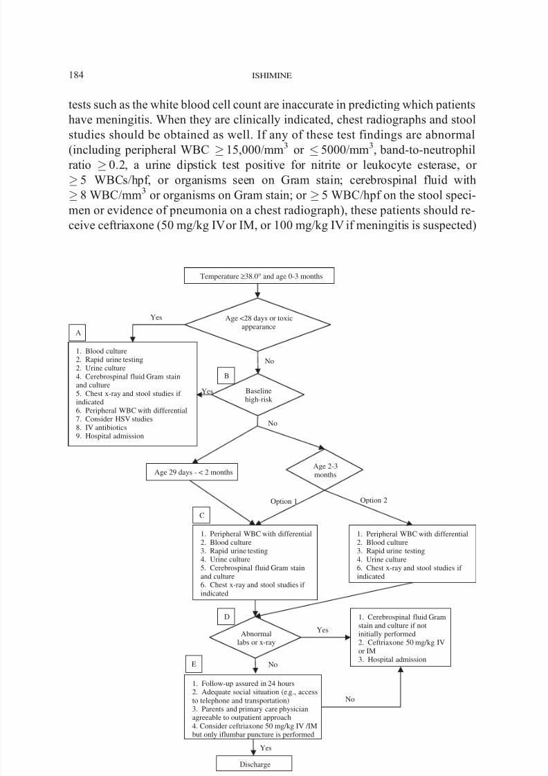
Temperature ≥38.0° and age 0-3 months
Age <28 days or toxicappearance
1. Blood culture2. Rapid urine testing2. Urine culture4. Cerebrospinal fluid Gram stainand culture5. Chest x-ray and stool studies if indicated
6. Peripheral WBC with differential7. Consider HSV studies8. IV antibiotics9. Hospital admission
Baselinehigh-risk
1. Peripheral WBC with differential
2. Blood culture3. Rapid urine testing4. Urine culture6. Chest x-ray and stool studies if indicated
1. Peripheral WBC with differential
2. Blood culture3. Rapid urine testing4. Urine culture5. Cerebrospinal fluid Gram stainand culture6. Chest x-ray and stool studies if indicated
Abnormallabs or x-ray
1. Cerebrospinal fluid Gramstain and culture if notinitially performed2. Ceftriaxone 50 mg/kg IVor IM3. Hospital admission
1. Follow-up assured in 24 hours2. Adequate social situation (e.g., accessto telephone and transportation)3. Parents and primary care physicianagreeable to outpatient approach4. Consider ceftriaxone 50 mg/kg IV /IMbut only iflumbar puncture is performed
Discharge
Age 29 days - < 2 months
A
B
C
D
Yes
No
Age 2-3months
Option 1 Option 2
E
Yes
No
No
No
Yes
Yes
ishimine184

8/3/2019 Fiebre Sin Foco Clini a
http://slidepdf.com/reader/full/fiebre-sin-foco-clini-a 19/28
and should be admitted to the hospital. If these initial laboratory results are normal,
a patient can be discharged if follow-up within 24 hours (or sooner if clinically
worse) can be assured. The administration of ceftriaxone, 50 mg/kg IV or IM,should be considered if a lumbar puncture is performed, but if a lumbar puncture is
not performed, antibiotics should be withheld. If a patient is 2 to 3 months old and
the practitioner is comfortable with pediatric assessment skills, these children can
be treated similarly to older febrile children.
The older infant or toddler (3–36 months old) who has a temperature of
39.08C may be treated more selectively. In this age group, if no febrile source
is identified definitively, a catheterized urine specimen for evaluation (dipstick,
urinalysis, microscopy, or Gram stain) and urine culture should be obtained in
girls less than 2 years old, if two or more of the following risk factors are present:age less than 12 months old, fever for 2 or more days, temperature 39.08C,
white, and no alternative source of fever. All boys younger than 6 months old and
all uncircumcised boys younger than 12 months old should also have catheterized
urine sent for rapid urine testing and culture. Based on pre-PCV7 data, the most
cost-effective approach to the child who has not had at least three PCV7 doses is
to obtain a CBC. If the WBC count is 15,000/mm 3, a blood culture should be
ordered and the administration of ceftriaxone should be considered. Other options
(eg, blood culture only or CBC and blood culture with selective antibiotic ad-
ministration) are reasonable. However, in nontoxic children who have had threePCV7 immunizations and who are not at risk for meningococcal disease, some
practitioners believe that obtaining any blood work is unnecessary. The current
Fig. 1. ( A) Urine testing can be accomplished either by microscopy, Gram stain, or urine dipstick.
Chest radiographs are indicated in patients with hypoxia, tachypnea, abnormal lung sounds, or
respiratory distress. Stool studies are indicated in patients with diarrhea. Herpes simplex virus testing
should be considered in the presence of risk factors (see text for details). HSV testing is best
accomplished by polymerase chain reaction or viral culture. Neonates should receive both ampicillin(50 mg/kg IV, or 100 mg/kg IV if meningitis is suspected) and cefotaxime (50 mg/kg, or 100 mg/kg IV
if meningitis is suspected) or gentamicin (2.5 mg/kg IV). Additionally, neonates with findings sug-
gestive of HSV infection should receive acyclovir (20 mg/kg IV). Older children should receive
ceftriaxone (50 mg/kg IV, or 100 mg/kg IV if meningitis is suspected). A WBC count with differen-
tial may be ordered, but the results should not dissuade the clinician from pursuing a full evaluation
and treatment with antibiotics. ( B) Young patients who have increased underlying risk include chil-
dren who were premature, had prolonged hospital stays after birth, those with underlying medical
conditions, patients with indwelling medical devices, fever lasting longer than 5 days, or patients
already on antibiotics. (C ) Urine testing can be accomplished either by microscopy, Gram stain, or
urine dipstick. Chest radiographs are indicated in patients with hypoxia, tachypnea, abnormal lung
sounds, or respiratory distress. Stool studies are indicated in patients with diarrhea. ( D) Abnormallaboratory findings: peripheral WBC count 5,000/mm3 or 15,000/mm3 or band-to-neutrophil
ratio 0.2; urine testing, 5 WBC/hpf, bacteria on Gram stain, or positive leukocyte esterase or
nitrite; cerebrospinal fluid, 8 WBC/mm3 or bacteria on Gram stain; stool specimen, 5 WBC/hpf;
and chest radiograph, infiltrate detected. ( E ) Administering ceftriaxone (50 mg/kg IV or IM) is
optional but should only be considered in patients who have undergone lumbar puncture. Patients
who have not undergone lumbar puncture should not get ceftriaxone. ( Adapted from Baraff L.
Management of fever without source in infants and children. Ann Emerg Med 2000;36(6):602–14.)
fever without source in children 0–36 months of age 185

8/3/2019 Fiebre Sin Foco Clini a
http://slidepdf.com/reader/full/fiebre-sin-foco-clini-a 20/28
evidence suggests that this may become a reasonable approach, but studies
addressing this specific approach have not yet been published (Figs. 1 and 2).
Finally, it is critically important to recognize that there is no combination of clinical assessment and diagnostic testing that will successfully identify all
patients with serious infection at the time of initial presentation. Therefore, the
importance of timely reassessment cannot be overemphasized, and caretakers
must be instructed to return to the ED or the office immediately for any
deterioration in the child’s condition. While strategies such as that described
above may help guide the evaluation and treament of febrile young infants,
Age 3-36 months
Healthy without underlying medical conditionsNontoxic appearance and no obvious source
1. No diagnostic testingrequired
2. Discharge home withfollow-up in 48 hours if feverpersists
3. Reevaluation if conditionworsens
Evaluate for occult infection
Occult pneumonia
1. Obtain CXR if patient hashypoxia, tachypnea,
respiratory distress,abnormal breath sounds
2. Consider CXR if no other
source identified and WBC>20,000/mm3 (if obtained)
Occult UTI
1. Obtain rapid urine testing andculture in girls <24 months if
1 risk factor present:• Fever ≥2 days
• Age < 12 months
• White race
• No alternative source of fever2. Strongly consider rapid urine
testing and culture foruncircumcised boys <12mand circumcised boys <6m
Rapid urine test positive?
1. Outpatient oral antibiotics (e.g.,
cefixime, cephalexin)
2. Consider giving first dose of parenteral antibiotics (e.g.,
ceftriaxone) in ED or office
Findings suggestive of pneumonia?
1. Assess clinical stability for discharge
2 Ensure ability to obtain follow-up care2. Follow-up in 24-48 hours for persistent
symptoms3. Immediate follow-up for worsening condition4. Immediate follow-up for positive blood culture5. Discharge home
1. Outpatient oral antibiotics(e.g., amoxicillin, azithromycin)2. Consider giving first dose of
parenteral antibiotics (e.g.,ceftriaxone) in ED or office
Occult Bacteremia
1. Increased risk with
• Temp ≥40°C
• WBC > 15,000/mm3 orANC >10,000/mm3
• Age 6-24 months
• Contacts withmeningococcal disease
• Petechiae
• Prolonged gastroenteritis
Several acceptable options:
Option 1
• CBC with diff
• If WBC >15K, bloodculture and ceftriaxone
Option 2
• Blood culture
Option 3
For patients with ≥3 PCV7doses:
• No blood testing
Temp ≥39°C
Yes No
Yes No
No
Yes
Fig. 2. Algorithm for treating children aged 3 to 36 months old (may be used for patients 2 to 3 months
old as well; see text). ( Adapted from Baraff L. Management of fever without source in infants and
children. Ann Emerg Med 2000;36(6):602–14.)
ishimine186

8/3/2019 Fiebre Sin Foco Clini a
http://slidepdf.com/reader/full/fiebre-sin-foco-clini-a 21/28
no single approach can capture the nuances of all febrile young patients.
Therefore, this approach should serve as an adjunct to, and not a replacement
for clinician judgment.
References
[1] Nelson DS, Walsh K, Fleisher GR. Spectrum and frequency of pediatric illness presenting to
a general community hospital emergency department. Pediatrics 1992;90(1 Pt 1):5–10.
[2] Krauss BS, Harakal T, Fleisher GR. The spectrum and frequency of illness presenting to a
pediatric emergency department. Pediatr Emerg Care 1991;7(2):67–71.
[3] McCaig LF, Burt CW. National Hospital Ambulatory Medical Care Survey: 2002 emergency
department summary. Adv Data 2004;340:1–34.
[4] Wittler RR, Cain KK, Bass JW. A survey about management of febrile children without source
by primary care physicians. Pediatr Infect Dis J 1998;17(4):271–7 [discussion: 7–9].
[5] Baraff LJ, Schriger DL, Bass JW, et al. Management of the young febrile child: commentary on
practice guidelines. Pediatrics 1997;100(1):134– 6.
[6] Belfer RA, Gittelman MA, Muniz AE. Management of febrile infants and children by pediatric
emergency medicine and emergency medicine: comparison with practice guidelines. Pediatr
Emerg Care 2001;17(2):83–7.
[7] Isaacman DJ, Kaminer K, Veligeti H, et al. Comparative practice patterns of emergency
medicine physicians and pediatric emergency medicine physicians managing fever in young
children. Pediatrics 2001;108(2):354– 8.[8] Kramer MS, Shapiro ED. Management of the young febrile child: a commentary on recent
practice guidelines. Pediatrics 1997;100(1):128– 34.
[9] Mackowiak PA. Concepts of fever. Arch Intern Med 1998;158(17):1870– 81.
[10] Craig JV, Lancaster GA, Taylor S, et al. Infrared ear thermometry compared with rectal
thermometry in children: a systematic review. Lancet 2002;360(9333):603– 9.
[11] Craig JV, Lancaster GA, Williamson PR, et al. Temperature measured at the axilla compared
with rectum in children and young people: systematic review. BMJ 2000;320(7243):1174–8.
[12] Jean-Mary MB, Dicanzio J, Shaw J, et al. Limited accuracy and reliability of infrared axil-
lary and aural thermometers in a pediatric outpatient population. J Pediatr 2002;141(5):
671–6.
[13] Gittelman MA, Mahabee-Gittens EM, Gonzalez-del-Rey J. Common medical terms defined by parents: are we speaking the same language? Pediatr Emerg Care 2004;20(11):754–8.
[14] Banco L, Veltri D. Ability of mothers to subjectively assess the presence of fever in their
children. Am J Dis Child 1984;138(10):976–8.
[15] Hooker EA, Smith SW, Miles T, King L. Subjective assessment of fever by parents: compari-
son with measurement by noncontact tympanic thermometer and calibrated rectal glass mer-
cury thermometer. Ann Emerg Med 1996;28(3):313–7.
[16] Graneto JW, Soglin DF. Maternal screening of childhood fever by palpation. Pediatr Emerg
Care 1996;12(3):183–4.
[17] Crocetti M, Moghbeli N, Serwint J. Fever phobia revisited: have parental misconceptions
about fever changed in 20 years? Pediatrics 2001;107(6):1241–6.
[18] Schmitt BD. Fever phobia: misconceptions of parents about fevers. Am J Dis Child 1980;134(2):176–81.
[19] Grover G, Berkowitz CD, Lewis RJ, et al. The effects of bundling on infant temperature.
Pediatrics 1994;94(5):669 – 73.
[20] Pantell RH, Newman TB, Bernzweig J, et al. Management and outcomes of care of fever in
early infancy. JAMA 2004;291(10):1203– 12.
[21] McGowan Jr JE, Bratton L, Klein JO, et al. Bacteremia in febrile children seen in a ‘‘walk-in’’
pediatric clinic. N Engl J Med 1973;288(25):1309–12.
fever without source in children 0–36 months of age 187

8/3/2019 Fiebre Sin Foco Clini a
http://slidepdf.com/reader/full/fiebre-sin-foco-clini-a 22/28
[22] Bisgard KM, Kao A, Leake J, et al. Haemophilus influenzae invasive disease in the United
States, 1994–1995: near disappearance of a vaccine-preventable childhood disease. Emerg
Infect Dis 1998;4(2):229–37.
[23] Wenger JD. Epidemiology of Haemophilus influenzae type b disease and impact of Hae-
mophilus influenzae type b conjugate vaccines in the United States and Canada. Pediatr Infect
Dis J 1998;17(9 Suppl):S132–6.
[24] Schuchat A, Robinson K, Wenger JD, et al. Bacterial meningitis in the United States in 1995:
Active Surveillance Team. N Engl J Med 1997;337(14):970–6.
[25] Alpern ER, Alessandrini EA, Bell LM, et al. Occult bacteremia from a pediatric emergency
department: current prevalence, time to detection, and outcome. Pediatrics 2000;106(3):505– 11.
[26] Lee GM, Harper MB. Risk of bacteremia for febrile young children in the post- Haemophilus
influenzae type b era. Arch Pediatr Adolesc Med 1998;152(7):624–8.
[27] Robinson KA, Baughman W, Rothrock G, et al. Epidemiology of invasive Streptococcus
pneumoniae infections in the United States, 1995–1998: opportunities for prevention in the
conjugate vaccine era. JAMA 2001;285(13):1729–35.
[28] Whitney CG, Farley MM, Hadler J, et al. Increasing prevalence of multidrug-resistant
Streptococcus pneumoniae in the United States. N Engl J Med 2000;343(26):1917–24.
[29] Kaplan SL, Mason Jr EO, Wald E, et al. Six year multicenter surveillance of invasive
pneumococcal infections in children. Pediatr Infect Dis J 2002;21(2):141–7.
[30] American Academy of Pediatrics. Pneumococcal infections. In: Pickering L, editor. Red book:
2003 report of the Committee on Infectious Diseases. 26th edition. Elk Grove Village (IL)7
American Academy of Pediatrics; 2003. p. 490–500.
[31] Wise RP, Iskander J, Pratt RD, et al. Postlicensure safety surveillance for 7-valent pneu-
mococcal conjugate vaccine. JAMA 2004;292(14):1702 – 10.
[32] Black S, Shinefield H, Fireman B, et al for the Northern California Kaiser Permanente VaccineStudy Center Group. Efficacy, safety and immunogenicity of heptavalent pneumococcal con-
jugate vaccine in children. Pediatr Infect Dis J 2000;19(3):187–95.
[33] Black S, Shinefield H, Baxter R, et al for the in Northern California Kaiser Permanente Vaccine
Center Group. Postlicensure surveillance for pneumococcal invasive disease after use of
heptavalent pneumococcal conjugate vaccine. Pediatr Infect Dis J 2004;23(6):485–9.
[34] Hsu K, Pelton S, Karumuri S, et al. Population-based surveillance for childhood invasive
pneumococcal disease in the era of conjugate vaccine. Pediatr Infect Dis J 2005;24(1):17–23.
[35] Whitney CG, Farley MM, Hadler J, et al. Decline in invasive pneumococcal disease after
the introduction of protein-polysaccharide conjugate vaccine. N Engl J Med 2003;348(18):
1737–46.
[36] Kaplan SL, Mason Jr EO, Wald ER, et al. Decrease of invasive pneumococcal infections inchildren among 8 children’s hospitals in the United States after the introduction of the 7-valent
pneumococcal conjugate vaccine. Pediatrics 2004;113(3 Pt 1):443–9.
[37] Alpern E, Henretig F. Fever. In: Fleisher G, Ludwig S, Henretig F, et al, editors. Textbook of
pediatric emergency medicine. 5th edition. Philadelphia7 Lippincott Williams & Wilkins; 2006.
p. 295–306.
[38] Finkelstein JA, Christiansen CL, Platt R. Fever in pediatric primary care: occurrence, man-
agement, and outcomes. Pediatrics 2000;105(1 Pt 3):260–6.
[39] Roberts KB. Young, febrile infants: a 30-year odyssey ends where it started. JAMA 2004;
291(10):1261–2.
[40] Kuppermann N, Fleisher G, Jaffe D. Predictors of occult pneumococcal bacteremia in young
febrile children. Ann Emerg Med 1998;31(6):679–87.[41] Stanley R, Pagon Z, Bachur R. Hyperpyrexia among infants younger than 3 months. Pediatr
Emerg Care 2005;21(5):291–4.
[42] Teach SJ, Fleisher GR for the Occult Bacteremia Study Group. Duration of fever and its
relationship to bacteremia in febrile outpatients three to 36 months old. Pediatr Emerg Care
1997;13(5):317–9.
[43] Li SF, Lacher B, Crain EF. Acetaminophen and ibuprofen dosing by parents. Pediatr Emerg
Care 2000;16(6):394–7.
ishimine188

8/3/2019 Fiebre Sin Foco Clini a
http://slidepdf.com/reader/full/fiebre-sin-foco-clini-a 23/28
[44] McErlean MA, Bartfield JM, Kennedy DA, et al. Home antipyretic use in children brought
to the emergency department. Pediatr Emerg Care 2001;17(4):249–51.
[45] Huang SY, Greenes DS. Effect of recent antipyretic use on measured fever in the pediatric
emergency department. Arch Pediatr Adolesc Med 2004;158(10):972–6.
[46] Baker MD, Fosarelli PD, Carpenter RO. Childhood fever: correlation of diagnosis with tem-
perature response to acetaminophen. Pediatrics 1987;80(3):315 – 8.
[47] Baker RC, Tiller T, Bausher JC, et al. Severity of disease correlated with fever reduction in
febrile infants. Pediatrics 1989;83(6):1016 – 9.
[48] Torrey SB, Henretig F, Fleisher G, et al. Temperature response to antipyretic therapy in
children: relationship to occult bacteremia. Am J Emerg Med 1985;3(3):190–2.
[49] Yamamoto LT, Wigder HN, Fligner DJ, et al. Relationship of bacteremia to antipyretic therapy
in febrile children. Pediatr Emerg Care 1987;3(4):223–7.
[50] Greenes DS, Harper MB. Low risk of bacteremia in febrile children with recognizable viral
syndromes. Pediatr Infect Dis J 1999;18(3):258–61.
[51] Smitherman HF, Caviness AC, Macias CG. Retrospective review of serious bacterial infections
in infants who are 0 to 36 months of age and have influenza A infection. Pediatrics 2005;
115(3):710–8.
[52] Schutzman SA, Petrycki S, Fleisher GR. Bacteremia with otitis media. Pediatrics 1991;87(1):
48–53.
[53] Turner D, Leibovitz E, Aran A, et al. Acute otitis media in infants younger than two months of
age: microbiology, clinical presentation and therapeutic approach. Pediatr Infect Dis J 2002;
21(7):669–74.
[54] DeAngelis C, Joffe A, Willis E, et al. Hospitalization v outpatient treatment of young, febrile
infants. Am J Dis Child 1983;137(12):1150–2.
[55] DeAngelis C, Joffe A, Wilson M, et al. Iatrogenic risks and financial costs of hospitalizingfebrile infants. Am J Dis Child 1983;137(12):1146–9.
[56] Paxton RD, Byington CL. An examination of the unintended consequences of the rule-out
sepsis evaluation: a parental perspective. Clin Pediatr (Phila) 2001;40(2):71–7.
[57] Baker MD, Bell LM. Unpredictability of serious bacterial illness in febrile infants from birth
to 1 month of age. Arch Pediatr Adolesc Med 1999;153(5):508–11.
[58] Kadish HA, Loveridge B, Tobey J, et al. Applying outpatient protocols in febrile infants 1–28
days of age: can the threshold be lowered? Clin Pediatr (Phila) 2000;39(2):81–8.
[59] Pena BM, Harper MB, Fleisher GR. Occult bacteremia with group B streptococci in an out-
patient setting. Pediatrics 1998;102(1 Pt 1):67–72.
[60] Hoffman JA, Mason EO, Schutze GE, et al. Streptococcus pneumoniae infections in the
neonate. Pediatrics 2003;112(5):1095–102.[61] Dagan R, Powell KR, Hall CB, et al. Identification of infants unlikely to have serious bacterial
infection although hospitalized for suspected sepsis. J Pediatr 1985;107(6):855–60.
[62] Dagan R, Sofer S, Phillip M, et al. Ambulatory care of febrile infants younger than 2 months of
age classified as being at low risk for having serious bacterial infections. J Pediatr 1988;112(3):
355–60.
[63] Jaskiewicz JA, McCarthy CA, Richardson AC, et al for the Febrile Infant Collaborative Study
Group. Febrile infants at low risk for serious bacterial infection–an appraisal of the Rochester
criteria and implications for management. Pediatrics 1994;94(3):390 – 6.
[64] Ferrera PC, Bartfield JM, Snyder HS. Neonatal fever: utility of the Rochester criteria in
determining low risk for serious bacterial infections. Am J Emerg Med 1997;15(3):299–302.
[65] Baker MD, Bell LM, Avner JR. Outpatient management without antibiotics of fever in selectedinfants. N Engl J Med 1993;329(20):1437–41.
[66] Baraff L. Management of fever without source in infants and children. Ann Emerg Med
2000;36(6):602–14.
[67] Steere M, Sharieff GQ, Stenklyft PH. Fever in children less than 36 months of age: questions
and strategies for management in the emergency department. J Emerg Med 2003;25(2):149 – 57.
[68] Bonsu BK, Harper MB. Utility of the peripheral blood white blood cell count for identifying
sick young infants who need lumbar puncture. Ann Emerg Med 2003;41(2):206–14.
fever without source in children 0–36 months of age 189

8/3/2019 Fiebre Sin Foco Clini a
http://slidepdf.com/reader/full/fiebre-sin-foco-clini-a 24/28
[69] Bonsu BK, Harper MB. Identifying febrile young infants with bacteremia: is the peripheral
white blood cell count an accurate screen? Ann Emerg Med 2003;42(2):216–25.
[70] Brown L, Shaw T, Wittlake WA. Does leucocytosis identify bacterial infections in febrile
neonates presenting to the emergency department? Emerg Med J 2005;22(4):256–9.
[71] Gorelick MH, Shaw KN. Screening tests for urinary tract infection in children: a meta-analysis.
Pediatrics 1999;104(5):e54.
[72] Shaw KN, McGowan KL, Gorelick MH, et al. Screening for urinary tract infection in infants
in the emergency department: which test is best? Pediatrics 1998;101(6):E1.
[73] Al-Orifi F, McGillivray D, Tange S, et al. Urine culture from bag specimens in young children:
are the risks too high? J Pediatr 2000;137(2):221–6.
[74] Committee on Quality Improvement, Subcommittee on Urinary Tract Infection. Practice pa-
rameter: the diagnosis, treatment, and evaluation of the initial urinary tract infection in febrile
infants and young children. Pediatrics 1999;103(4):843 – 52.
[75] Levine DA, Platt SL, Dayan PS, et al. Risk of serious bacterial infection in young febrile infants
with respiratory syncytial virus infections. Pediatrics 2004;113(6):1728–34.
[76] Wadsworth SJ, Suh B. In vitro displacement of bilirubin by antibiotics and 2-hydroxybenzoyl-
glycine in newborns. Antimicrob Agents Chemother 1988;32(10):1571–5.
[77] Martin E, Fanconi S, Kalin P, et al. Ceftriaxone–bilirubin-albumin interactions in the neonate:
an in vivo study. Eur J Pediatr 1993;152(6):530–4.
[78] Robertson A, Fink S, Karp W. Effect of cephalosporins on bilirubin-albumin binding. J Pediatr
1988;112(2):291–4.
[79] Sadow KB, Derr R, Teach SJ. Bacterial infections in infants 60 days and younger: epi-
demiology, resistance, and implications for treatment. Arch Pediatr Adolesc Med 1999;153(6):
611–4.
[80] Brown JC, Burns JL, Cummings P. Ampicillin use in infant fever: a systematic review. ArchPediatr Adolesc Med 2002;156(1):27–32.
[81] Brown ZA, Wald A, Morrow RA, et al. Effect of serologic status and cesarean delivery
on transmission rates of herpes simplex virus from mother to infant. JAMA 2003;289(2):
203–9.
[82] Kimberlin DW, Lin CY, Jacobs RF, et al. Natural history of neonatal herpes simplex virus
infections in the acyclovir era. Pediatrics 2001;108(2):223–9.
[83] Kimberlin DW, Lin CY, Jacobs RF, et al. Safety and efficacy of high-dose intravenous acyclovir
in the management of neonatal herpes simplex virus infections. Pediatrics 2001;108(2):230 – 8.
[84] Kimberlin D. Herpes simplex virus, meningitis and encephalitis in neonates. Herpes 2004;
11(Suppl 2):A65–76.
[85] Kimberlin DW. Neonatal herpes simplex infection. Clin Microbiol Rev 2004;17(1):1– 13.[86] Chiu CH, Lin TY, Bullard MJ. Identification of febrile neonates unlikely to have bacterial
infections. Pediatr Infect Dis J 1997;16(1):59–63.
[87] Baker MD, Avner JR, Bell LM. Failure of infant observation scales in detecting serious illness
in febrile, 4- to 8-week-old infants. Pediatrics 1990;85(6):1040–3.
[88] Baskin MN, O’Rourke EJ, Fleisher GR. Outpatient treatment of febrile infants 28 to 89 days
of age with intramuscular administration of ceftriaxone. J Pediatr 1992;120(1):22–7.
[89] Baker MD, Bell LM, Avner JR. The efficacy of routine outpatient management without
antibiotics of fever in selected infants. Pediatrics 1999;103(3):627–31.
[90] American College of Emergency Physicians Clinical Policies Committee and Subcommittee
on Pediatric Fever. Clinical policy for children younger than three years presenting to the
emergency department with fever. Ann Emerg Med 2003;42(4):530–45.[91] Bramson RT, Meyer TL, Silbiger ML, et al. The futility of the chest radiograph in the febrile
infant without respiratory symptoms. Pediatrics 1993;92(4):524– 6.
[92] Bonsu BK, Harper MB. A low peripheral blood white blood cell count in infants younger than
90 days increases the odds of acute bacterial meningitis relative to bacteremia. Acad Emerg
Med 2004;11(12):1297– 301.
[93] Shapiro ED, Aaron NH, Wald ER, et al. Risk factors for development of bacterial meningitis
among children with occult bacteremia. J Pediatr 1986;109(1):15–9.
ishimine190

8/3/2019 Fiebre Sin Foco Clini a
http://slidepdf.com/reader/full/fiebre-sin-foco-clini-a 25/28
[94] Teele DW, Dashefsky B, Rakusan T, et al. Meningitis after lumbar puncture in children with
bacteremia. N Engl J Med 1981;305(18):1079–81.
[95] Bonsu BK, Harper MB. Accuracy and test characteristics of ancillary tests of cerebrospinal
fluid for predicting acute bacterial meningitis in children with low white blood cell counts in
cerebrospinal fluid. Acad Emerg Med 2005;12(4):303–9.
[96] Byington CL, Enriquez FR, Hoff C, et al. Serious bacterial infections in febrile infants 1 to
90 days old with and without viral infections. Pediatrics 2004;113(6):1662–6.
[97] Liebelt EL, Qi K, Harvey K. Diagnostic testing for serious bacterial infections in infants aged
90 days or younger with bronchiolitis. Arch Pediatr Adolesc Med 1999;153(5):525–30.
[98] Titus MO, Wright SW. Prevalence of serious bacterial infections in febrile infants with
respiratory syncytial virus infection. Pediatrics 2003;112(2):282–4.
[99] Rittichier KR, Bryan PA, Bassett KE, et al. Diagnosis and outcomes of enterovirus infections
in young infants. Pediatr Infect Dis J 2005;24(6):546–50.
[100] Dayan PS, Hanson E, Bennett JE, et al. Clinical course of urinary tract infections in infants
younger than 60 days of age. Pediatr Emerg Care 2004;20(2):85–8.
[101] Hoberman A, Wald ER, Hickey RW, et al. Oral versus initial intravenous therapy for urinary
tract infections in young febrile children. Pediatrics 1999;104(1 Pt 1):79–86.
[102] Fleisher GR, Rosenberg N, Vinci R, et al. Intramuscular versus oral antibiotic therapy for the
prevention of meningitis and other bacterial sequelae in young, febrile children at risk for occult
bacteremia. J Pediatr 1994;124(4):504–12.
[103] Teach SJ, Fleisher GR for the Occult Bacteremia Study Group. Efficacy of an observation scale
in detecting bacteremia in febrile children three to thirty-six months of age, treated as
outpatients. J Pediatr 1995;126(6):877 – 81.
[104] McCarthy PL, Sharpe MR, Spiesel SZ, et al. Observation scales to identify serious illness
in febrile children. Pediatrics 1982;70(5):802–9.[105] Bass JW, Steele RW, Wittler RR, et al. Antimicrobial treatment of occult bacteremia: a mul-
ticenter cooperative study. Pediatr Infect Dis J 1993;12(6):466 – 73.
[106] Kuppermann N. Occult bacteremia in young febrile children. Pediatr Clin North Am 1999;
46(6):1073–109.
[107] Lee GM, Fleisher GR, Harper MB. Management of febrile children in the age of the conjugate
pneumococcal vaccine: a cost-effectiveness analysis. Pediatrics 2001;108(4):835–44.
[108] Jaffe DM, Tanz RR, Davis AT, et al. Antibiotic administration to treat possible occult
bacteremia in febrile children. N Engl J Med 1987;317(19):1175–80.
[109] Harper MB, Bachur R, Fleisher GR. Effect of antibiotic therapy on the outcome of outpatients
with unsuspected bacteremia. Pediatr Infect Dis J 1995;14(9):760–7.
[110] Bulloch B, Craig WR, Klassen TP. The use of antibiotics to prevent serious sequelae in childrenat risk for occult bacteremia: a meta-analysis. Acad Emerg Med 1997;4(7):679–83.
[111] Rothrock SG, Harper MB, Green SM, et al. Do oral antibiotics prevent meningitis and serious
bacterial infections in children with Streptococcus pneumoniae occult bacteremia? a meta-
analysis. Pediatrics 1997;99(3):438 – 44.
[112] Rothrock SG, Green SM, Harper MB, et al. Parenteral vs oral antibiotics in the prevention
of serious bacterial infections in children with Streptococcus pneumoniae occult bacteremia:
a meta-analysis. Acad Emerg Med 1998;5(6):599–606.
[113] Baraff LJ, Oslund S, Prather M. Effect of antibiotic therapy and etiologic microorganism on the
risk of bacterial meningitis in children with occult bacteremia. Pediatrics 1993;92(1):140–3.
[114] Stoll ML, Rubin LG. Incidence of occult bacteremia among highly febrile young children in the
era of the pneumococcal conjugate vaccine: a study from a children’s hospital emergencydepartment and urgent care center. Arch Pediatr Adolesc Med 2004;158(7):671–5.
[115] Yang YJ, Huang MC, Wang SM, et al. Analysis of risk factors for bacteremia in children with
nontyphoidal Salmonella gastroenteritis. Eur J Clin Microbiol Infect Dis 2002;21(4):290–3.
[116] Zaidi E, Bachur R, Harper M. Non-typhi Salmonella bacteremia in children. Pediatr Infect
Dis J 1999;18(12):1073–7.
[117] Kuppermann N, Malley R, Inkelis SH, et al. Clinical and hematologic features do not reliably
identify children with unsuspected meningococcal disease. Pediatrics 1999;103(2):E20.
fever without source in children 0–36 months of age 191

8/3/2019 Fiebre Sin Foco Clini a
http://slidepdf.com/reader/full/fiebre-sin-foco-clini-a 26/28
[118] Wang VJ, Kuppermann N, Malley R, et al. Meningococcal disease among children who live
in a large metropolitan area, 1981–1996. Clin Infect Dis 2001;32(7):1004–9.
[119] Wang VJ, Malley R, Fleisher GR, et al. Antibiotic treatment of children with unsuspected
meningococcal disease. Arch Pediatr Adolesc Med 2000;154(6):556–60.
[120] Mandl K, Stack A, Fleisher G. Incidence of bacteremia in infants and children with fever and
petechiae. J Pediatr 1997;131(3):398.
[121] Nielsen HE, Andersen EA, Andersen J, et al. Diagnostic assessment of haemorrhagic rash and
fever. Arch Dis Child 2001;85(2):160–5.
[122] Wells LC, Smith JC, Weston VC, Collier J, et al. The child with a non-blanching rash: how
likely is meningococcal disease? Arch Dis Child 2001;85(3):218–22.
[123] American Academy of Pediatrics. Committee on Infectious Diseases. Prevention and control of
meningococcal disease. recommendations for use of meningococcal vaccines in pediatric
patients. Pediatrics 2005;116(2):496–505.
[124] Bachur R, Harper MB. Reevaluation of outpatients with Streptococcus pneumoniae bacteremia.
Pediatrics 2000;105(3 Pt 1):502–9.
[125] Bandyopadhyay S, Bergholte J, Blackwell CD, et al. Risk of serious bacterial infection in
children with fever without a source in the post- Haemophilus influenzae era when antibiotics
are reserved for culture-proven bacteremia. Arch Pediatr Adolesc Med 2002;156(5):512–7.
[126] Norberg A, Christopher NC, Ramundo ML, et al. Contamination rates of blood cultures
obtained by dedicated phlebotomy vs intravenous catheter. JAMA 2003;289(6):726–9.
[127] Waltzman ML, Harper M. Financial and clinical impact of false-positive blood culture results.
Clin Infect Dis 2001;33(3):296–9.
[128] Thuler LC, Jenicek M, Turgeon JP, et al. Impact of a false positive blood culture result on the
management of febrile children. Pediatr Infect Dis J 1997;16(9):846–51.
[129] DeAngelis C, Joffe A, Wilson M, et al. Iatrogenic risks and financial costs of hospitalizingfebrile infants. Am J Dis Child 1983;137(12):1146–9.
[130] Evidence based clinical practice guideline for outpatient management of fever of uncertain
source in children 2 to 36 months of age. 2003. Available at http://www.cincinnatichildrens.org/
svc/alpha/h/health-policy/ev-based/feverii.htm. Accessed August 1, 2005.
[131] Kuppermann N. The evaluation of young febrile children for occult bacteremia: time to
reevaluate our approach? Arch Pediatr Adolesc Med 2002;156(9):855–7.
[132] Baraff LJ, Bass JW, Fleisher GR, et al for the Agency for Health Care Policy and Research.
Practice guideline for the management of infants and children 0 to 36 months of age with fever
without source. Ann Emerg Med 1993;22(7):1198–210.
[133] Baraff LJ. Clinical policy for children younger than three years presenting to the emergency
department with fever [editorial]. Ann Emerg Med 2003;42(4):546–9.[134] Green S, Rothrock S. Evaluation styles for well-appearing febrile children: are you a ‘‘risk-
minimizer’’ or a ‘‘test-minimizer’’? Ann Emerg Med 1999;33(2):211– 4.
[135] Hoberman A, Chao HP, Keller DM, et al. Prevalence of urinary tract infection in febrile infants.
J Pediatr 1993;123(1):17–23.
[136] Shaw KN, Gorelick M, McGowan KL, et al. Prevalence of urinary tract infection in febrile
young children in the emergency department. Pediatrics 1998;102(2):E16.
[137] Bachur R, Harper MB. Reliability of the urinalysis for predicting urinary tract infections in
young febrile children. Arch Pediatr Adolesc Med 2001;155(1):60–5.
[138] Gorelick MH, Shaw KN. Clinical decision rule to identify febrile young girls at risk for urinary
tract infection. Arch Pediatr Adolesc Med 2000;154(4):386–90.
[139] Gorelick MH, Hoberman A, Kearney D, et al. Validation of a decision rule identifyingfebrile young girls at high risk for urinary tract infection. Pediatr Emerg Care 2003;19(3):
162–4.
[140] Schoen EJ, Colby CJ, Ray GT. Newborn circumcision decreases incidence and costs of urinary
tract infections during the first year of life. Pediatrics 2000;105(4 Pt 1):789–93.
[141] Task Force on Circumcision. Circumcision policy statement. Pediatrics 1999;103(3):686 – 93.
[142] Zorc JJ, Kiddoo DA, Shaw KN. Diagnosis and management of pediatric urinary tract infec-
tions. Clin Microbiol Rev 2005;18(2):417–22.
ishimine192

8/3/2019 Fiebre Sin Foco Clini a
http://slidepdf.com/reader/full/fiebre-sin-foco-clini-a 27/28
[143] Huicho L, Campos-Sanchez M, Alamo C. Metaanalysis of urine screening tests for determin-
ing the risk of urinary tract infection in children. Pediatr Infect Dis J 2002;21(1):1–11.
[144] Wubbel L, Muniz L, Ahmed A, et al. Etiology and treatment of community-acquired
pneumonia in ambulatory children. Pediatr Infect Dis J 1999;18(2):98–104.
[145] Margolis P, Gadomski A. Does this infant have pneumonia? JAMA 1998;279(4):308– 13.
[146] Jadavji T, Law B, Lebel MH, et al. A practical guide for the diagnosis and treatment of pediatric
pneumonia. CMAJ 1997;156(5):703.
[147] Lynch T, Platt R, Gouin S, et al. Can we predict which children with clinically suspected
pneumonia will have the presence of focal infiltrates on chest radiographs? Pediatrics 2004;
113(3 Pt 1):E186–9.
[148] Rothrock SG, Green SM, Fanelli JM, et al. Do published guidelines predict pneumonia in
children presenting to an urban ED? Pediatr Emerg Care 2001;17(4):240–3.
[149] Leventhal JM. Clinical predictors of pneumonia as a guide to ordering chest roentgenograms.
Clin Pediatr (Phila) 1982;21(12):730–4.
[150] Taylor JA, Del Beccaro M, Done S, et al. Establishing clinically relevant standards for tachy-
pnea in febrile children younger than 2 years. Arch Pediatr Adolesc Med 1995;149(3):
283–7.
[151] Zukin DD, Hoffman JR, Cleveland RH, et al. Correlation of pulmonary signs and symptoms
with chest radiographs in the pediatric age group. Ann Emerg Med 1986;15(7):792–6.
[152] Mower WR, Sachs C, Nicklin EL, et al. Pulse oximetry as a fifth pediatric vital sign. Pediatrics
1997;99(5):681–6.
[153] Tanen DA, Trocinski DR. The use of pulse oximetry to exclude pneumonia in children. Am J
Emerg Med 2002;20(6):521–3.
[154] Davies HD, Wang EE, Manson D, et al. Reliability of the chest radiograph in the diagnosis
of lower respiratory infections in young children. Pediatr Infect Dis J 1996;15(7):600–4.[155] McCarthy PL, Spiesel SZ, Stashwick CA, et al. Radiographic findings and etiologic diagnosis
in ambulatory childhood pneumonias. Clin Pediatr (Phila) 1981;20(11):686 – 91.
[156] Courtoy I, Lande AE, Turner RB. Accuracy of radiographic differentiation of bacterial from
nonbacterial pneumonia. Clin Pediatr (Phila) 1989;28(6):261– 4.
[157] Swingler GH, Hussey GD, Zwarenstein M. Randomised controlled trial of clinical outcome
after chest radiograph in ambulatory acute lower-respiratory infection in children. Lancet 1998;
351(9100):404–8.
[158] Bachur R, Perry H, Harper MB. Occult pneumonias: empiric chest radiographs in febrile
children with leukocytosis. Ann Emerg Med 1999;33(2):166–73.
[159] British Thoracic Society of Standards of Care Committee. BTS Guidelines for the manage-
ment of community acquired pneumonia in childhood. Thorax 2002;57:1–24.[160] Black SB, Shinefield HR, Ling S, et al. Effectiveness of heptavalent pneumococcal conjugate
vaccine in children younger than five years of age for prevention of pneumonia. Pediatr Infect
Dis J 2002;21(9):810–5.
[161] Lee KC, Finkelstein JA, Miroshnik IL, et al. Pediatricians’ self-reported clinical practices and
adherence to national immunization guidelines after the introduction of pneumococcal
conjugate vaccine. Arch Pediatr Adolesc Med 2004;158(7):695– 701.
[162] Klein JO. Management of the febrile child without a focus of infection in the era of universal
pneumococcal immunization. Pediatr Infect Dis J 2002;21(6):584 – 8 [discussion: 613–4].
[163] O’Brien KL, Santosham M. Potential impact of conjugate pneumococcal vaccines on pediatric
pneumococcal diseases. Am J Epidemiol 2004;159(7):634–44.
[164] Fernandez Lopez A, Luaces Cubells C, Garcia Garcia JJ, et al. Procalcitonin in pediatricemergency departments for the early diagnosis of invasive bacterial infections in febrile infants:
results of a multicenter study and utility of a rapid qualitative test for this marker. Pediatr Infect
Dis J 2003;22(10):895–903.
[165] Galetto-Lacour A, Zamora SA, Gervaix A. Bedside procalcitonin and C-reactive protein tests in
children with fever without localizing signs of infection seen in a referral center. Pediatrics
2003;112(5):1054–60.
[166] Gendrel D, Raymond J, Coste J, et al. Comparison of procalcitonin with C-reactive protein,
fever without source in children 0–36 months of age 193
8/3/2019 Fiebre Sin Foco Clini a
http://slidepdf.com/reader/full/fiebre-sin-foco-clini-a 28/28
interleukin 6 and interferon-alpha for differentiation of bacterial vs. viral infections. Pediatr
Infect Dis J 1999;18(10):875–81.
[167] Hsiao AL, Baker MD. Fever in the new millennium: a review of recent studies of markers
of serious bacterial infection in febrile children. Curr Opin Pediatr 2005;17(1):56–61.
[168] Isaacman DJ, Burke BL. Utility of the serum C-reactive protein for detection of occult bacterial
infection in children. Arch Pediatr Adolesc Med 2002;156(9):905–9.
[169] Pulliam PN, Attia MW, Cronan KM. C-reactive protein in febrile children 1 to 36 months of age
with clinically undetectable serious bacterial infection. Pediatrics 2001;108(6):1275 – 9.
[170] van Rossum AM, Wulkan RW, Oudesluys-Murphy AM. Procalcitonin as an early marker of
infection in neonates and children. Lancet Infect Dis 2004;4(10):620–30.
[171] Carrol ED, Newland P, Riordan FA, et al. Procalcitonin as a diagnostic marker of me-
ningococcal disease in children presenting with fever and a rash. Arch Dis Child 2002;86(4):
282–5.
ishimine194