fiber length variability within the flexor carpi ulnaris and flexor carpi radialis muscles:...
TRANSCRIPT

F habilitationS VeteransA
0539, TheS al Instito
s article.ornia San
D
Fiber Length Variability Withinthe Flexor Carpi Ulnaris and
Flexor Carpi Radialis Muscles:Implications for Surgical
Tendon Transfer
Jan Fridén, MD, PhD, Göteborg, Sweden,Richard M. Lovering, PhD, Baltimore, MD,
Richard L. Lieber, PhD, San Diego, CA
Purpose: The purpose of this study was to understand the detailed architectural properties of thehuman flexor carpi radialis (FCR) and flexor carpi ulnaris (FCU) muscles and their implications fortendon transfer surgery.Methods: Muscle fiber length was measured in 6 separate regions of the FCU and FCR from 10cadaveric specimens. Sarcomere length was measured by laser diffraction for normalization.Moment arms were estimated by measuring tendon excursion with respect to joint angle. Theposition of entry of the motor nerve branches into each muscle also was measured to establishlimits for the safe length of muscle mobilization.Results: Muscle fiber length varied significantly along both the FCU and FCR. Fiber lengthvariability in the FCU was twice that of the FCR. Although the average fiber length for both musclesacross all regions was similar (62.6 � 2.1 mm for the FCR and 63.1 � 4.0 mm for the FCU), theproximal fibers of the FCU were longer compared with the proximal fibers of the FCR and the distalfibers of the FCU were shorter compared with the distal fibers of the FCR. The 99% confidenceinterval for the second nerve branch entry into the muscles was located �69 mm distal to themedial epicondyle for the FCU and approximately 73 mm distal for the FCR.Conclusions: These data show different designs of both the FCU and the FCR. The functionalsignificance of fiber length variability is not clear but imply that, when used in tendon transfer, theproperly mobilized FCU has a much greater excursion. (J Hand Surg 2004;29A:909–914. Copy-right © 2004 by the American Society for Surgery of the Hand.)Key words: Hand surgery, muscle, muscle architecture, tendon transfer.
rom the Department of Hand Surgery, Sahlgrenska University Hospital, Göteborg, Sweden; the Department of Physical Therapy and Recience, University of Maryland, Baltimore, MD; and the Departments of Orthopaedics and Bioengineering, University of California anddministration Medical Centers, San Diego, CA.Received for publication December 10, 2003; accepted in revised form April 20, 2004.Supported by the Department of Veteran’s Affairs, Rehabilitation Research and Development, National Institutes of Health grant AR4
wedish Research Council grant 11200, and Göteborg University. Travel supported by the International Society for Biomechanics and Nationutesf Health grant T32-AR07592 (R.M.L.).No benefits in any form have been received or will be received from a commercial party related directly or indirectly to the subject of thiReprint requests: Richard L. Lieber, PhD, Department of Orthopaedics (9151), Veteran’s Affairs Medical Center and University of Califiego, 3350 La Jolla Village Dr, San Diego, CA 92161.Copyright © 2004 by the American Society for Surgery of the Hand0363-5023/04/29A05-0020$30.00/0
doi:10.1016/j.jhsa.2004.04.028The Journal of Hand Surgery 909

Sfisumaelisofimefitttibralnft
nTpichFtutvmmlmccnau
dfi
wo
MTtmvewrrwstrw1tawinotafificS
tlnltFpomalbrcFivSafi
910 The Journal of Hand Surgery / Vol. 29A No. 5 September 2004
keletal muscle architecture profoundly influencesunction.1 Previous measurements of upper-extrem-ty architecture showed a wide range of muscle de-igns across the arm and forearm.2–4 Based on thisnderstanding specific recommendations have beenade regarding the use of donor muscles that are
ppropriate to restore lost function.5 Because thexperimental method of determining architecture re-ies on the relatively tedious method of microdissect-ng individual fibers or fiber bundles from fixed tis-ue, most architectural studies base their conclusionsn just a few samples across the entire muscle.6,7 Ifber dimensions are consistent along and across auscle this probably is acceptable. There are, how-
ver, reasons to be concerned about this practice:rst, muscles with relatively broad origins, such as
he pectoralis major and even the teres minor, haveremendous fiber length variation, presumably owingo the multifunctional nature of these muscles.8 Evenn the forearm, the brachioradialis muscle, with itsroad humeral origin, shows fiber length variationanging from 100 mm to 180 mm,9 although theverage fiber length was reported as 121 mm3. Fiberength heterogeneity has considerable implicationsot only for understanding muscle function but alsoor choosing which muscles should be selected forransfer.
Major donor muscles include the flexor carpi ul-aris (FCU) and the flexor carpi radialis (FCR).10,11
hese synergistic muscles are reported to have com-lementary architectural designs, with the FCR hav-ng a higher excursion and lower force potentialompared with the FCU with its lower excursion andigher force potential.2,4 Because both the FCU andCR originate at the medial epicondyle and act on
he wrist in 2 planes (flexion-extension and radial-lnar deviation), one might suspect that their archi-ectural properties would be more complex than pre-iously appreciated. On the other hand because theseuscles do not have a broad origin their architectureay be simplified compared with muscles with fan-
ike origins such as the brachioradialis, pectoralisajor, and teres minor. Unfortunately, detailed ar-
hitectural analysis of human upper extremity mus-les, with the exception of the brachioradialis9 hasever performed; thus it is currently impossible toddress this issue of fiber length variability for mostpper extremity muscles.In light of the importance of the FCU and FCR as
onor muscles and the lack of information regarding
ber length heterogeneity, the purpose of this study oas to measure the detailed architectural propertiesf the FCU and FCR.
aterials and Methodsen fresh cadaveric specimens (n � 10 for each of
he FCR and FCU) were used for this study anduscle architecture was determined as described pre-
iously.4,9 At the time of dissection the wrist andlbow flexion moment arms of the FCR and FCUere estimated by measuring the excursion of the
espective tendon while the appropriate joint wasotated through an arc of 1 to 2 radians.12 Joint angleas measured with a goniometer and tendon excur-
ion was measured with a ruler. By using this methodhe tendon excursion corresponding to 1 radian jointotation is equal to the moment arm.12 Muscles thenere harvested, weighed, and immersion-fixed in0% buffered formaldehyde for 72 hours in a flat-ened position corresponding to the supinated fore-rm and extended elbow. After fixation the musclesere rinsed in phosphate-buffered saline and stored
n fresh phosphate-buffered saline at 4° C untileeded. Muscle length (Lm) was measured as therigin of the most proximal fibers to the insertion ofhe most distal fibers. Surface muscle fiber pennationngle was measured with a goniometer. Althoughber pennation typically is measured for superficialbers, many of the fiber bundles originated superfi-ially but inserted into deep tendon aponeurosis.uch fibers were excluded from this analysis.Muscle fiber bundles were isolated under a dissec-
ion microscope (�10 to �20) and fiber bundleength (Lf) was measured using digital calipers to theearest hundredth of a millimeter. To define fiberength heterogeneity the muscles were divided arbi-rarily into 6 equal regions from proximal to distal.iber bundles were dissected from each of the 3roximal regions (denoted P1, P2, P3) and from eachf the 3 distal regions (denoted D1, D2, D3). Theuscular origins of these 2 muscles are at similar
natomic levels so regions P1, P2, and P3 wereocated at about the same anatomic level. In contrast,ecause the FCU musculature extends more distally,egions D1, D2, and D3 extended approximately 2m more distally for the FCU compared with theCR. Approximately 9 separate fiber bundles were
solated from each region. Fiber length coefficient ofariation within a region ranged from 5% to 12%.arcomere length (Ls) was measured in 3 locationslong each bundle by laser diffraction using the 0 torst order diffraction angle and also the 0 to second
13
rder when possible. This method has been used
pptlgtpm(fo
ppl
P
wctlsawsnWlf
DFaclcsesieswqa
wT
tn
RBttvcocmFaws(miomt6tctwfif
Fridén, Lovering, and Lieber / Wrist Flexor Muscle Design 911
reviously to measure sarcomere length in fixed up-er-extremity specimens and relies on the construc-ive interference that occurs between incident laseright and skeletal muscle sarcomeres.3,4 To provideuidelines regarding safe limits for muscle mobiliza-ion the motor nerves were traced as distally asossible under the dissecting microscope to deter-ine the point at which they entered the muscle
denoted Ln). This was expressed as the distancerom the muscle origin for the 1 or 2 nerve branchesbserved.In addition to the measured data the following
arameters were calculated: the Lf/Lm ratio and thehysiologic cross-sectional area according to the fol-owing equation6:
hysiologic cross-sectional area (mm2)
� M(g) ·cos � ⁄ �(g ⁄ mm3) ·Lf (mm)
here M represents muscle mass, � represents mus-le density (1.056 g/mm3), � represents fiber penna-ion angle, and Lf represents fiber length. Muscleength and fiber bundle length were normalized to atandard (Ls � 2.7 �m) to compensate for variationmong specimens in muscle length during fixationith joints in different configurations. The actual
arcomere length value chosen for normalization isot critical in making comparisons between muscles.e chose 2.7 �m because this is the sarcomere
ength in human muscle that results in maximumorce generation.14
ata Analysisiber length was analyzed by 2-way analysis of vari-nce (ANOVA) with repeated measures using mus-le and region as grouping factors. The significanceevel was set to � � .05 and statistical power was notalculated because all differences were statisticallyignificant and thus no potential for type II errorxisted.15 Statistical power is only an important con-ideration when the null hypothesis is accepted, thats, when a study concludes no significant differencexisted. Because we observed significant effects theample size and thus the statistical power of the studyas adequate. Coefficient of variation was used touantify fiber length variability for each specimennd was calculated as:
Coef ficient of variation � (s ⁄ X̄) ·100%
here X� is the sample mean and s is the sample SD.
o define the safe limits for surgical muscle release nhe 99% confidence intervals were calculated for theerve branches of both the FCU and FCR.16
esultsecause both the FCU and FCR were harvested from
he same specimens, any differences measured be-ween muscles were not caused by interspecimen sizeariation but were true differences between the mus-les (Table 1). Much more fiber length variation wasbserved for the FCU compared with the FCR. Spe-ifically the coefficient of variation for the FCU wasore than twice that of the FCR (p � .001; Fig. 1).or both muscles studied a notable fiber length vari-tion was observed along the entire muscle length,ith fibers in the distal regions being markedly
horter compared with fibers in the proximal regionsFig. 2). Two-way ANOVA revealed no significantain effect of muscle (p � .9), but a highly signif-
cant muscle � region interaction (p � 0.0001). Inther words although average fiber length for bothuscles across all regions was similar (reflected by
he main effect of the ANOVA being insignificant:2.6 � 2.1 mm for the FCR and 63.1 � 4.0 mm forhe FCU) the proximal fibers of the FCU were longerompared with the proximal fibers of the FCR andhe distal fibers of the FCU were shorter comparedith the distal fibers of the FCR (Fig. 2). Because theber length variation of the 2 muscles was oppositerom proximal to distal, this yielded the highly sig-
Table 1. Descriptive Properties of WristFlexor Muscles
Parameter FCR FCU
Forearm length(mm) 262.7 � 7.3 262.7 � 7.3
Muscle length (Lm,mm) 158.5 � 10.0 236.5 � 5.4
Muscle mass (M, g) 20.2 � 2.5 25.9 � 2.4Tendon length (Lt,
mm)External 149.6 � 6.3 147.5 � 10.1Internal 101.7 � 4.9 88.0 � 8.1
Moment arm (mm)Elbow 3.0 � 0.4 0.0 � 0.0Wrist 12.4 � 0.6 10.6 � 0.6
Motor branch entrypoint (Ln)*
First 46.6 � 4.4 29.7 � 1.9Second 56.8 � 3.8 50.4 � 4.1
Values are presented are mean � SEM, n � 10.*Measured as the distance from muscle origin near the medial
epicondyle to the point where the nerve branch enters the musclebelly.
ificant interaction term mentioned (p � .001).

tpcm.bnc9wetebm
FatseiFw
DTatg
Flpdlbibtfitattsm
pfimfiddiufiiopwotT
F(s
FFaa
912 The Journal of Hand Surgery / Vol. 29A No. 5 September 2004
Two main nerve branches were observed for all ofhe FCUs and 7 of 10 of the FCRs (Fig. 3). Theroximal nerve branch entered the muscle significantlyloser to the medial epicondyle for the FCU (29.7 � 1.9m) compared with the FCR (44.0 � 4.1 mm, p �
005). For the 7 paired specimens in which the secondranch always was observed, they entered the muscle atearly the same location (FCU at 50.9 � 4.8 mmompared with FCR at 56.3 � 4.3 mm, p � .4). The9% confidence interval for the second nerve branchas located approximately 69-mm distal to the medial
picondyle for the FCU and approximately 73-mm dis-al for the FCR. Based on the number of regions withinach muscle and the overall muscle length (Table 1)oth nerve branches in both muscles would enter theuscle belly proximally in regions P1 and P2.There was no elbow flexion moment arm for the
CU (0 � 0 mm; Table 1) whereas the FCR momentrm was 3.0 � 0.36 mm, showing that the sole action ofhe FCU was at the wrist and even the FCR had a verymall influence on elbow joint function. (The zero FCUlbow moment arm calls into question the practice ofmmobilizing the elbow after surgery involving theCU.) The wrist flexion moment arms of the 2 musclesere similar at 11 to 12 mm (Table 1).
iscussionhe purpose of this study was to quantify the detailedrchitectural properties of 2 synergistic wrist flexors:he FCR and the FCU. Although the FCU showed
igure 1. Fiber length variability observed in the FCU andCR as expressed by coefficient of variation (see Materialsnd Methods section). Note that the FCU fiber length vari-bility is more than twice that of the FCR.
reater fiber length variability compared with the P
CR (Fig. 1), both muscles showed significant fiberength variability from proximal to distal, with theroximal fibers being significantly longer than theistal fibers (Fig. 2). Although the average fiberength of the FCU and FCR was the same the distri-ution of fiber lengths along the muscles was signif-cantly different. Specifically, the proximal FCU fi-ers were longer than the proximal FCR fibers, buthe distal FCU fibers were shorter than the distal FCRbers. Because the general consensus seems to be
hat the FCU has shorter fibers than the FCR,2,4,17
pparently most investigators preferentially sampledhe distal musculature. This is likely owing to the facthat the proximal flexor mass is very difficult toeparate into discrete muscles and thus the proximalusculature is studied less often.The reason that fiber length distribution is so im-
ortant for hand surgeons to take into account is thatber length is the primary determinant of skeletaluscle excursion.2,18 Taken at face value the longerbers of the proximal FCU and FCR would be pre-icted to have a greater excursion compared with theistal fibers. The rules that govern the intramuscularnteractions among fiber populations, however, arenknown at this time. With regard to whole-muscleunction it is not clear whether skeletal muscle fibersn various regions of the same muscle act completelyn parallel, completely in series, or some combinationf the two. If muscle fibers were acting completely inarallel then the proximal portion of the muscleould have a greater excursion than the distal portionf the muscle and this would result in various por-ions of the muscle “fi ghting” against one another.he shorter fibers even might restrict the range of the
igure 2. Fiber length distribution in the FCU (�) and FCR) muscles along their length. Each region represents one
ixth of the muscle from the proximal origin (regions P1, P2,
3) to the distal insertion (regions D1, D2, D3).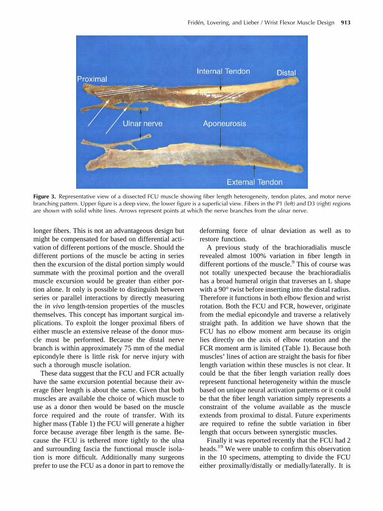
lmvdtsmtsttpecbes
hemufhfcatp
dr
rdnhwTrfsFlFmlcrbbceal
hi
Fba
Fridén, Lovering, and Lieber / Wrist Flexor Muscle Design 913
onger fibers. This is not an advantageous design butight be compensated for based on differential acti-
ation of different portions of the muscle. Should theifferent portions of the muscle be acting in serieshen the excursion of the distal portion simply wouldummate with the proximal portion and the overalluscle excursion would be greater than either por-
ion alone. It only is possible to distinguish betweeneries or parallel interactions by directly measuringhe in vivo length-tension properties of the muscleshemselves. This concept has important surgical im-lications. To exploit the longer proximal fibers ofither muscle an extensive release of the donor mus-le must be performed. Because the distal nerveranch is within approximately 75 mm of the medialpicondyle there is little risk for nerve injury withuch a thorough muscle isolation.
These data suggest that the FCU and FCR actuallyave the same excursion potential because their av-rage fiber length is about the same. Given that bothuscles are available the choice of which muscle to
se as a donor then would be based on the muscleorce required and the route of transfer. With itsigher mass (Table 1) the FCU will generate a higherorce because average fiber length is the same. Be-ause the FCU is tethered more tightly to the ulnand surrounding fascia the functional muscle isola-ion is more difficult. Additionally many surgeons
igure 3. Representative view of a dissected FCU muscle shranching pattern. Upper figure is a deep view, the lower figure shown with solid white lines. Arrows represent points at
refer to use the FCU as a donor in part to remove the e
eforming force of ulnar deviation as well as toestore function.
A previous study of the brachioradialis muscleevealed almost 100% variation in fiber length inifferent portions of the muscle.9 This of course wasot totally unexpected because the brachioradialisas a broad humeral origin that traverses an L shapeith a 90° twist before inserting into the distal radius.herefore it functions in both elbow flexion and wrist
otation. Both the FCU and FCR, however, originaterom the medial epicondyle and traverse a relativelytraight path. In addition we have shown that theCU has no elbow moment arm because its origin
ies directly on the axis of elbow rotation and theCR moment arm is limited (Table 1). Because bothuscles’ lines of action are straight the basis for fiber
ength variation within these muscles is not clear. Itould be that the fiber length variation really doesepresent functional heterogeneity within the muscleased on unique neural activation patterns or it coulde that the fiber length variation simply represents aonstraint of the volume available as the musclextends from proximal to distal. Future experimentsre required to refine the subtle variation in fiberength that occurs between synergistic muscles.
Finally it was reported recently that the FCU had 2eads.19 We were unable to confirm this observationn the 10 specimens, attempting to divide the FCU
fiber length heterogeneity, tendon plates, and motor nervesuperficial view. Fibers in the P1 (left) and D3 (right) regionsthe nerve branches from the ulnar nerve.
owingre is awhich
ither proximally/distally or medially/laterally. It is
pas2ts
R
1
1
1
1
1
1
1
1
1
1
914 The Journal of Hand Surgery / Vol. 29A No. 5 September 2004
ossible that the previous investigators reported annomaly that would not be apparent given our limitedample size. If the FCU were innervated dually and-headed then it certainly would provide new oppor-unities regarding the choice of donor muscles forurgical reconstruction.
eferences1. Lieber RL, Fridén J. Functional and clinical significance of
skeletal muscle architecture. Muscle Nerve 2000;23:1647–1666.
2. Brand PW, Beach RB, Thompson DE. Relative tension andpotential excursion of muscles in the forearm and hand.J Hand Surg 1981;6:209–219.
3. Lieber RL, Jacobson MD, Fazeli BM, Abrams RA, BotteMJ. Architecture of selected muscles of the arm andforearm: anatomy and implications for tendon transfer.J Hand Surg 1992;17A:787–798.
4. Lieber RL, Fazeli BM, Botte MJ. Architecture of selectedwrist flexor and extensor muscles. J Hand Surg 1990;15A:244–250.
5. Fridén J, Lieber RL. Tendon transfer surgery: clinical im-plications of experimental studies. Clin Orthop 2002;403S:S163–S170.
6. Sacks RD, Roy RR. Architecture of the hind limb muscles ofcats: functional significance. J Morphol 1982;173:185–195.
7. Gans C. Fiber architecture and muscle function. In: Exerciseand Sport Science Reviews. Vol 10. Lexington, MA: Frank-lin University Press, 1982:160–207.
8. Van der Helm FCT, Veenbaas R. Modelling the mechan-ical effect of muscles with large attachment sites: appli-
cation to the shoulder mechanism. J Biomech1991;24:1151–1163.
9. Fridén J, Albrecht D, Lieber RL. Biomechanical analysis ofthe brachioradialis as a donor in tendon transfer. Clin Orthop2001;383:152–161.
0. Beasley RW. Tendon transfers for radial nerve palsy. OrthopClin North Am 1970;1:439–445.
1. Boyes JH. Tendon transfers for radial palsy. Bull Hosp JointDis 1960;21:97–105.
2. An KN, Ueba Y, Chao EY, Cooney WP, Linscheid RL.Tendon excursion and moment arm of index finger muscles.J Biomech 1983;16:419–425.
3. Lieber RL, Yeh Y, Baskin RJ. Sarcomere length determina-tion using laser diffraction. Effect of beam and fiber diam-eter. Biophys J 1984;45:1007–1016.
4. Lieber RL, Loren GJ, Fridén J. In vivo measurement ofhuman wrist extensor muscle sarcomere length changes.J Neurophysiol 1994;71:874–881.
5. Sokal RR, Rohlf FJ. Biometry. The principles and practiceof statistics in biological research. 2nd ed. San Francisco:W.H. Freeman and Company, 1981;244–261.
6. Fridén J, Lieber RL. Quantitative evaluation of the posteriordeltoid to triceps tendon transfer based on muscle architec-tural properties. J Hand Surg 2001;26A:147–155.
7. Smith RJ, Hastings H II. Principles of tendon transfers to thehand. AAOS Instr Course Lect 1993;21:129–149.
8. Bodine SC, Roy RR, Meadows DA, Zernicke RF, Sacks RD,Fournier M, et al. Architectural, histochemical, and contrac-tile characteristics of a unique biarticular muscle: the catsemitendinosus. J Neurophysiol 1982;48:192–201.
9. Lim AY, Kumar VP, Pereira BP, Hua J. Independent func-tion in a tendon transfer of the split flexor carpi ulnaris. PlastReconstr Surg 1999;104:1739–1741.