ff-10502, an antimetabolite with novel activity on dormant...
TRANSCRIPT

JPET #248740
1
Title Page
Title
FF-10502, an antimetabolite with novel activity on dormant cells, is superior to gemcitabine for
targeting pancreatic cancer cells
Author names and affiliations
Shinji Mima, Chihaya Kakinuma, Tamami Higuchi, Kazunori Saeki, Takayuki Yamada, Rena
Uematsu, Miki Ishino, Nobuko Kito, Hiroki Nishikawa, Hidenobu Kuniyoshi, Takuya Matsumoto,
Hideyasu Fujiwara, Linda J. Paradiso, Yasuhiro Shimada, and Hiroyuki Iwamura
FUJIFILM Corporation, Tokyo, Japan (S.M., C.K., T.H., K.S., T.Y., R.U., M.I., N.K., H.N., H.K.,
T.M., H.F., Y.S., and H.I.); Strategia Therapeutics, Inc., Houston, Texas, United States (L.J.P.)
This article has not been copyedited and formatted. The final version may differ from this version.JPET Fast Forward. Published on April 13, 2018 as DOI: 10.1124/jpet.118.248740
at ASPE
T Journals on January 14, 2020
jpet.aspetjournals.orgD
ownloaded from
JPET #248740
2
Running title: FF-10502 suppresses dormant cells via DNA repair inhibition
Corresponding author:
Hiroyuki Iwamura, Ph.D.,
Pharmaceutical Products Division, FUJIFILM Corporation, 7-3 Akasaka 9-Chome, Minato-ku,
Tokyo 107-0052, Japan
Phone: +81-80-8426-8876
Fax: +81-3-6271-3191
E-mail: [email protected]
Number of text pages: 25
Number of tables: 3
Number of figures: 7
Number of references: 38
Number of words in the Abstract: 247
Number of words in the Introduction: 750
Number of words in the Discussion: 1454
Nonstandard abbreviations:
BER, base excision repair; CSCs, cancer stem cells; DDIs, DNA damage inducers; DMSO,
dimethylsulfoxide; EdU, 5-ethynyl-2-deoxyuridine; EFS, event-free survival; FBS, fetal bovine
This article has not been copyedited and formatted. The final version may differ from this version.JPET Fast Forward. Published on April 13, 2018 as DOI: 10.1124/jpet.118.248740
at ASPE
T Journals on January 14, 2020
jpet.aspetjournals.orgD
ownloaded from

JPET #248740
3
serum; FF-10502, 1-(2-deoxy-2-fluoro-4-thio-β-D-arabinofuranosyl) cytosine; FF-10502TP,
FF-10502-triphosphate; 5-FU, 5-fluorouracil; gemTP, gemcitabine-triphosphate; PBS, phosphate
buffered saline; PDX, patient-derived xenograft; polα, polymerase alpha; polβ, polymerase beta;
ROS, reactive oxygen species.
Section assignment: Chemotherapy, Antibiotics, and Gene Therapy
This article has not been copyedited and formatted. The final version may differ from this version.JPET Fast Forward. Published on April 13, 2018 as DOI: 10.1124/jpet.118.248740
at ASPE
T Journals on January 14, 2020
jpet.aspetjournals.orgD
ownloaded from

JPET #248740
4
Abstract
We report in this paper that FF-10502, a pyrimidine nucleoside antimetabolite with a
chemical structure similar to gemcitabine, shows beneficial anticancer activity via novel
mechanism of action on dormant cells. The growth-inhibition of pancreatic cancer cell lines by
FF-10502 (IC50, 60–330 nM) was moderately weaker than that by gemcitabine in vitro. In contrast,
an in vivo orthotopic implantation model in mice with established human pancreatic cancer cell line,
SUIT-2, revealed no mortality with FF-10502 intravenous treatment, which was related with
regression of implanted tumor and little metastasis, whereas 75% of the mice treated with
gemcitabine died by day 128. Two in vivo patient-derived xenograft models with
gemcitabine-resistant pancreatic cancer cells also demonstrated complete tumor growth suppression
with FF-10502, but only partial inhibition with gemcitabine. We also investigated the mechanism of
action of FF-10502 by using dormant cancer cells, which are reportedly involved in the development
of resistance to chemotherapy. In vitro serum starvation–induced dormant SUIT-2 cells developed
resistance to gemcitabine even in combination with DNA damage inducers (DDIs; H2O2, cisplatin,
and temozolomide). Interestingly, FF-10502 in combination with DDIs significantly induced
concentration-dependent cell death in accordance with enhanced DNA damage. FF-10502 was far
more potent than gemcitabine in inhibiting DNA polymerase β, which may explain the difference in
dormant cell injury, although further investigations for direct evidences are necessary. In conclusion,
This article has not been copyedited and formatted. The final version may differ from this version.JPET Fast Forward. Published on April 13, 2018 as DOI: 10.1124/jpet.118.248740
at ASPE
T Journals on January 14, 2020
jpet.aspetjournals.orgD
ownloaded from
JPET #248740
5
our study demonstrated the beneficial antitumor effects of FF-10502 in clinically relevant in vivo
models, and suggests the importance of preventing DNA repair unlike gemcitabine.
This article has not been copyedited and formatted. The final version may differ from this version.JPET Fast Forward. Published on April 13, 2018 as DOI: 10.1124/jpet.118.248740
at ASPE
T Journals on January 14, 2020
jpet.aspetjournals.orgD
ownloaded from

JPET #248740
6
Introduction
Gemcitabine, an antitumor chemotherapy drug classified as a pyrimidine nucleoside
antimetabolite, was approved by the United States Food and Drug Administration in 1996 primarily
as a result of an enhanced clinical benefit response. The current standard of care for patients with
advanced or metastatic pancreatic cancer is gemcitabine-based chemotherapy. However, the
antitumor effect of gemcitabine is modest, with a partial response rate of 5.4% and median survival
time 1.3 months longer than that with 5-fluorouracil (5-FU) (4.2 months) (Burris et al., 1997).
Following the development of combination therapy with nab-paclitaxel, median survival time was
extended by 2.1 months compared to treatment with gemcitabine alone (6.6 months) (Goldstein et al.,
2015). Another combination chemotherapy regimen consisting of oxaliplatin, irinotecan, 5-FU, and
leucovorin (FOLFIRINOX) also extended the survival, and the median overall survival was 11.1
months in the FOLFIRINOX group as compared with 6.8 months in the gemcitabine group (Conroy
et al., 2011). Although these new regimens represent significant improvements, the five-year
survival rate for patients with pancreatic cancer remains only 7%, the lowest among all types of
cancer (Siegel et al., 2015). It is especially concerning that a poor prognosis is associated with poor
response to chemotherapy and tumor recurrence. Specifically, human pancreatic cancer can acquire
resistance to conventional chemotherapeutics including paclitaxel, 5-FU, cisplatin, and gemcitabine
(Arumugam et al., 2009). One such mechanism of drug resistance in pancreatic cancer is the
This article has not been copyedited and formatted. The final version may differ from this version.JPET Fast Forward. Published on April 13, 2018 as DOI: 10.1124/jpet.118.248740
at ASPE
T Journals on January 14, 2020
jpet.aspetjournals.orgD
ownloaded from

JPET #248740
7
enrichment of dormant cell populations (cancer stem cells and/or non-proliferating, quiescent cells).
Stanton et al. reported that 28 ± 15% of pancreatic cancer cells were positive for nuclear Ki67 in a
study of 33 pancreatic adenocarcinomas, suggesting that over 70% of pancreatic tumor cells are
dormant (Stanton et al., 2003). Those dormant cells - including cancer stem cells - possess
“robustness,” a property that encompasses the characteristics of a slow cell cycle, resistance to
oxidative stress, and a rapid response to DNA damage, all of which contribute to the development of
therapeutic resistance (Yoshida and Saya, 2016). Thus, in order to advance the treatment of
pancreatic cancer, it is necessary to develop novel approaches that target these dormant cells.
FF-10502 (1-(2-deoxy-2-fluoro-4-thio-β-D-arabinofuranosyl) cytosine) , formerly known
as 4’-thio-FAC, was discovered by the Yamasa Corporation in Japan and developed as an anticancer
chemotherapy drug (Miura et al., 1998). FF-10502 is classified as a pyrimidine nucleoside
antimetabolite and has a chemical structure similar to gemcitabine (Fig. 1). FF-10502 has a sulfur
atom in its sugar ring instead of oxygen, and has one fluorine atom compared to gemcitabine’s two
fluorine atoms at the 2′ position in the sugar moiety (Miura et al., 1999). The antiproliferative and
cytotoxic effects of FF-10502 have been tested in vitro on many types of solid tumors, including
pancreas, lung, stomach, colon, breast, ovary, bladder, melanoma, osteosarcoma, and head and neck
cancers (Miura et al., 1998; Miura et al., 1999; Zajchowski et al., 2005). It is noteworthy that
FF-10502 has shown superior efficacy compared to gemcitabine in all in vivo studies reported thus
This article has not been copyedited and formatted. The final version may differ from this version.JPET Fast Forward. Published on April 13, 2018 as DOI: 10.1124/jpet.118.248740
at ASPE
T Journals on January 14, 2020
jpet.aspetjournals.orgD
ownloaded from
JPET #248740
8
far (Miura et al., 1998; Miura et al., 1999; Miura et al., 2002; Zajchowski et al., 2005).
Previous reports have revealed elements of the mechanism of action of FF-10502. After its
uptake into cells, FF-10502 is metabolized to FF-10502-triphosphate (FF-10502TP), an active
metabolite of FF-10502 similar to gemcitabine-triphosphate (gemTP) (Heinemann et al., 1988).
Compared with gemTP, the inhibitory activity of FF-10502TP on DNA polymerase alpha (polα) and
polymerase beta (polβ) was approximately 1,000 and 100 times higher, respectively. Therefore, it
was hypothesized that the efficacy of FF-10502TP can be attributed to the inhibition of polα (Miura
et al., 2001; Miura and Izuta, 2004). However, this potent inhibition of polα is not consistent with
tumor cell growth inhibition in vitro, as the IC50 values for FF-10502 in tumor cell growth assays are
generally higher than those of gemcitabine. This does not explain why FF-10502 has shown efficacy
superior to gemcitabine in animal models of tumors in vivo.
To explain the discrepancy between the in vitro and in vivo efficacy of FF-10502 and
gemcitabine, we further examined the pharmacological profile of FF-10502, with an objective to
elucidate the mechanisms involved. Pancreatic cancer is focused on in this study, because dormancy
of this type of tumor has been suggested to be the key factor in the differences in efficacy. The effect
of FF-10502 in relation to the inhibition of DNA repair after exposure of dormant cells with
chemotherapy resistance to DNA damage inducers (DDIs), and the importance of the novel
mechanism involved in the effect is discussed.
This article has not been copyedited and formatted. The final version may differ from this version.JPET Fast Forward. Published on April 13, 2018 as DOI: 10.1124/jpet.118.248740
at ASPE
T Journals on January 14, 2020
jpet.aspetjournals.orgD
ownloaded from

JPET #248740
9
Materials and Methods
Chemicals and Reagents
FF-10502 methanesulfonate (FF-10502-01) and FF-10502TP were synthesized and provided by
FUJIFILM Corporation (Tokyo, Japan). For simplicity, FF-10502 methanesulfonate is referred to as
“FF-10502” in this study. Gemcitabine hydrochloride was purchased from Teva Pharmaceutical
Industries (Netanya, Israel) and is designated as “gemcitabine” in this study. The formulation used in
the in vivo study represents the free base for both compounds. FF-10502 and gemcitabine were
dissolved in phosphate buffered saline (PBS) for in vitro studies, in dimethylsulfoxide (DMSO) for
combination study with DDIs, or in saline for in vivo studies. GemTP was purchased from Jena
Bioscience (Jena, Germany). Three DDIs, H2O2, cisplatin, and temozolomide, were purchased from
Kanto Chemical Co., Inc. (Tokyo, Japan), Wako Pure Chemical Industries, Ltd., (Osaka, Japan), and
LKT Laboratories, Inc. (St. Paul, MN), respectively. All DDIs were dissolved in culture medium.
Cell Lines
BxPC-3 cells and Capan-1 cells were obtained from the American Type Culture Collection
(Manassas, VA), and SUIT-2 cells and MIA PaCa-2 cells were obtained from the Japanese Collection
of Research Bioresources (Osaka, Japan). BxPC-3 and SUIT-2 cells were cultured in RPMI 1640
medium (Thermo Fisher Scientific Inc. Waltham, MA) with 10% heat-inactivated fetal bovine serum
(FBS) and 1% penicillin-streptomycin solution. Capan-1 and MIA PaCa-2 cells were cultured in
Iscove's Modified Dulbecco's Medium (Thermo Fisher Scientific Inc. Waltham, MA) with 20%
This article has not been copyedited and formatted. The final version may differ from this version.JPET Fast Forward. Published on April 13, 2018 as DOI: 10.1124/jpet.118.248740
at ASPE
T Journals on January 14, 2020
jpet.aspetjournals.orgD
ownloaded from

JPET #248740
10
heat-inactivated FBS, and Minimum Essential Media with 10% heat-inactivated FBS plus 1%
nonessential amino acids, respectively. All culture media contained 100 units/mL penicillin and 100
µg/mL streptomycin. The cultures were incubated in a CO2 incubator at 37°C with 5% CO2 in a
humidified atmosphere. All cells were subcultured every 3–4 days.
Cell Growth Inhibition Assay
Cells were seeded at 1,000 cells/well (BxPC-3, SUIT-2, and MIA PaCa-2) or 3,000 cells/well
(Capan-1) into 96-well culture plates. After a 24-hour culture, FF-10502, gemcitabine, or PBS alone
(control) was added to the wells. The cells were incubated for approximately 72 hours. Cell growth
inhibition was evaluated using CellTiter-Glo® Luminescent Cell Viability Assay kit (Promega,
Madison, WI). Luminescence was measured using an EnVision plate reader (Perkin Elmer, Waltham,
MA). The IC50 value of the test substance against cell growth was calculated using Microsoft®
Office Excel 2003 (Microsoft Corporation, Redmond, WA).
Subcutaneous Implantation Model with Human Pancreatic Cancer Cell Line Capan-1
Experiment was performed in accordance with the Guide for the Care and Use of Laboratory
Animals and was approved by the FUJIFILM animal experiment committee. Five-week-old female
nude mice (BALB/c-nu/nu) were purchased from CLEA Japan SLC Inc. (Tokyo, Japan). A Capan-1
cell suspension of 1 × 10⁸ cells/mL was prepared with serum-free medium; 100 µL of the cell
suspension was subcutaneously injected into the right flank region of each animal. Nine days after
This article has not been copyedited and formatted. The final version may differ from this version.JPET Fast Forward. Published on April 13, 2018 as DOI: 10.1124/jpet.118.248740
at ASPE
T Journals on January 14, 2020
jpet.aspetjournals.orgD
ownloaded from

JPET #248740
11
implantation, the mice were randomized into nine groups (10 mice/group), and 100 µL/kg of saline
(vehicle solution), gemcitabine, or FF-10502 was administered by tail vein injection once weekly for
4 weeks. Tumor diameter and body weight were measured twice weekly. For calculation of tumor
volume, both long and short diameters (mm) were measured by Vernier caliper (Mitutoyo, Kawasaki,
Japan). The formula for calculating tumor volume was as follows: tumor volume (mm3) = long
diameter (mm) × short diameter (mm) × short diameter (mm) × 0.5.
Orthotopic Implantation Model with Human Pancreatic Cancer Cell Line SUIT-2
Experiment was performed in accordance with the Guide for the Care and Use of Laboratory
Animals and was approved by the FUJIFILM animal experiment committee. Five-week-old female
nude mice (BALB/c-nu/nu) were purchased from CLEA Japan SLC Inc.(Tokyo, Japan). A SUIT-2
cell suspension of 1 × 10⁸ cells/mL was prepared in serum-free medium, and 10 µL of the cell
suspension was injected into the pancreas of each animal under isoflurane anesthesia. After
confirmation that there was no hemorrhage at the site of implantation, the abdominal wall incision
was closed with absorbable sutures, and the skin was clamped. Seven days after implantation, at
which metastases were already observed (Higuchi et al., 2018), the mice were randomized into five
groups (20 mice/group), and 100 µL/kg of saline (vehicle solution), gemcitabine, or FF-10502 was
administered by tail vein injection once weekly for 18 weeks.
Antitumor activity of the orthotopic implantation model was evaluated by event-free
This article has not been copyedited and formatted. The final version may differ from this version.JPET Fast Forward. Published on April 13, 2018 as DOI: 10.1124/jpet.118.248740
at ASPE
T Journals on January 14, 2020
jpet.aspetjournals.orgD
ownloaded from

JPET #248740
12
survival (EFS) time, defined as the day from SUIT-2 cell injection until death or moribundity (e.g.,
marked decrease in body weight, hypothermia, or other conditions requiring euthanasia).
Histopathological evaluation was also performed for various tissues of surviving animals stained
with hematoxylin and eosin.
Patient-Derived Xenograft (PDX) Model
Animal welfare for this experiment complied with the U.S. Department of Agriculture’s Animal
Welfare Act (9 Code of Federal Regulations Parts 1, 2, and 3), as applicable. Female NOD-SCID
mice were purchased from Harlan Laboratories (Indianapolis, IN). Two patient-derived pancreatic
cancer cells (PA5364 and PA5365) were used. These are adenocarcinoma and carcinoma cells
derived from ascites of 78 years old female and omentum of 54 years old female, respectively.
PA5364 shows high resistance to 5-FU and carmustine, intermediate resistance to gemcitabine and
mitomycin C, and low resistance to cisplatin and doxorubicin in vitro. PA5365 shows high resistance
to gemcitabine and doxorubicin, intermediate resistance to ifosfamide and mitomycin C, and low
resistance to 5-FU, docetaxel and SN38 (an active metabolite of irinotecan). The cell suspensions of
2.5 × 10⁶ cells/mL (PA5364) or 1.2 × 10⁶ cells/mL (PA5365) were prepared with PBS, mixed with an
equal volume of Cultrex® extracellular matrix (ECM) (Trevigen, Gaithersburg, MD), and 200
µL of the cell suspension in ECM was injected subcutaneously into the rear flank under isoflurane
anesthesia. The animals were randomized into three groups (10 mice/group) when the average tumor
This article has not been copyedited and formatted. The final version may differ from this version.JPET Fast Forward. Published on April 13, 2018 as DOI: 10.1124/jpet.118.248740
at ASPE
T Journals on January 14, 2020
jpet.aspetjournals.orgD
ownloaded from
JPET #248740
13
size reached approximately 200 mm3. Vehicle (saline), gemcitabine, or FF-10502 was administered
by tail vein injection once weekly for 4 weeks, followed by 4 weeks of observation. Animals were
monitored weekly for palpable tumors and any changes in appearance or behavior. Once tumors
were palpable, they were measured using calipers. For calculation of tumor volume, both long and
short diameters (mm) were measured by Vernier caliper (Mitutoyo, Kawasaki, Japan). The formula
for calculating tumor volume was as follows: tumor volume (mm3) = long diameter (mm) × short
diameter (mm) × short diameter (mm) × 0.5.
Inhibitory Activity against Polα and Polβ
The inhibitory activity of FF-10502TP and gemTP for polα was measured by a DNA synthesis assay
in vitro (Podust et al., 1989). Purified human polα (EuRx, Gdansk, Poland) and FF-10502TP or
gemTP were incubated for 30 minutes at 37°C in 50 µL of reaction mixture (60 mM Tris-HCl, pH
8.0, 5.0 mM magnesium acetate, 0.3 mg/mL bovine serum albumin, 1.0 mM dithiothreitol, 0.1 mM
spermine, 20 µM each dCTP, dGTP, and dATP, 5 µM [3H-methyl]dTTP, and 20 µg activated calf
thymus DNA). Likewise, purified human polβ (EuRx, Gdansk, Poland) and FF-10502TP or gemTP
were incubated for 15 minutes at 37°C in 50 µL of reaction mixture (50 mM Tris-HCl, pH 8.7, 10.0
mM MgCl2, 0.4 mg/mL bovine serum albumin, 1.0 mM dithiothreitol, 100 mM KCl, 15% glycerol,
50 µM each dCTP, dGTP, and dATP, 5 µM [3H-methyl]dTTP, and 10 µg activated calf thymus DNA).
After incubation, 20 µL of the reaction solution was passed through DEAE-cellulose paper and the
This article has not been copyedited and formatted. The final version may differ from this version.JPET Fast Forward. Published on April 13, 2018 as DOI: 10.1124/jpet.118.248740
at ASPE
T Journals on January 14, 2020
jpet.aspetjournals.orgD
ownloaded from

JPET #248740
14
membrane was washed with 5% Na2HPO4 (1 mL × 6), Milli-Q water (1 mL), ethanol (1 mL × 2),
and diethyl ether (1 mL). The DEAE-cellulose paper was transferred into a scintillation vial and 5
mL of scintillation fluid, PICO-FLUORTM PLUS (PerkinElmer, Waltham, MA) was added;
radioactivity was measured with a liquid scintillation counter (PerkinElmer, Waltham, MA).
The inhibition rate (individual value) was calculated as 100 − response ratio (formula
shown below). In addition, the mean inhibition rate of duplicate samples was calculated.
Response ratio: [(B − N) / (B0 − N)] × 100 (%)
where B = radioactivity of the sample measuring inhibitory activity (individual value), B0 =
radioactivity of the sample measuring total activity (mean value), and N = radioactivity of the
sample measuring non-specific activity (mean value). For data processing, Microsoft® Excel 2003
(Microsoft Corporation, Redmond, WA) was used.
Inhibitory Activity of DNA Synthesis in a SUIT-2 Orthotopic Implantation Tumor Model
Mice with SUIT-2 orthotopic implantation were prepared in accordance with the procedure
described above. On day 18, vehicle (saline), gemcitabine, or FF-10502 was administered by tail
vein injection (100 µL/kg). The tumors were harvested 4, 24, 48, and 72 hours after drug
administration. Mice were injected intraperitoneally with 100–200 µg of 5-ethynyl-2-deoxyuridine
(EdU) (Baseclick GmbH, Neuried, Germany) in PBS 4 hours before tumor harvest (Salic and
Mitchison, 2008). Pieces of the tumor were formalin-fixed, embedded in paraffin, and sectioned.
This article has not been copyedited and formatted. The final version may differ from this version.JPET Fast Forward. Published on April 13, 2018 as DOI: 10.1124/jpet.118.248740
at ASPE
T Journals on January 14, 2020
jpet.aspetjournals.orgD
ownloaded from

JPET #248740
15
After paraffin removal, sections on glass slides were stained with 10 µM Alexa568-azide (Thermo
Fisher Scientific, Waltham, MA) for 30 minutes at room temperature. Sections were counterstained
with Hoechst 33342 (Thermo Fisher Scientific, Waltham, MA) and mounted for fluorescence
microscopy (CQ1: Yokogawa Electric Corporation, Tokyo, Japan) to quantify total cell number and
DNA synthesizing cell number. The rate of EdU-incorporated cells was calculated as follows:
EdU-incorporated cells = NE / NH × 100 (%)
where NE = number of EdU-incorporated cells (individual value), and NH = number of
Hoechst-stained cells (individual value). For data processing, Microsoft® Excel 2003 (Microsoft
Corporation, Redmond, WA) was used.
SUIT-2 Dormant Cell Model
SUIT-2 cells were seeded at 15,000 cells/well into 96-well culture plates (CellBIND 96 well Clear
Flat Bottom, Corning, NY) with medium containing 10% FBS. After a 24-hour culture, the cells
were washed twice with 150 µL of serum-free medium and then cultured in serum-free medium for
72 hours. FF-10502, gemcitabine, or 0.1% DMSO alone (control) was added to the wells. H2O2
(final concentration of 300 µM), temozolomide (final concentration of 200 µM), or cisplatin (final
concentration of 5 µM) was also added for combination treatment. The cells were incubated in a CO2
incubator at 37°C with 5% CO2 in a humidified atmosphere for approximately 72 hours, and cell
growth inhibition was evaluated using the CellTiter-Glo® Luminescent Cell Viability Assay Kit
This article has not been copyedited and formatted. The final version may differ from this version.JPET Fast Forward. Published on April 13, 2018 as DOI: 10.1124/jpet.118.248740
at ASPE
T Journals on January 14, 2020
jpet.aspetjournals.orgD
ownloaded from

JPET #248740
16
(Promega, Madison, WI). Luminescence was measured using an EnVision plate reader (Perkin
Elmer, Waltham, MA).
Comet Assay in SUIT-2 Dormant Cell Model
The SUIT-2 cell culture and test article treatments were performed according to the same procedure
described above, with the exception that SUIT-2 cells were seeded at 100,000 cells/well into 24-well
culture plates and combined with DDIs and FF-10502 or gemcitabine for 4 or 24 hours. After
treatment, the cells were detached and resuspended in PBS at over 100,000 cells/mL. The cell
suspension was mixed with Comet LMAgarose (Trevigen, Gaithersburg, MD) and placed onto slides
(MAS coat slide 5 hole, Matsunami Glass Ind., Ltd., Osaka, Japan). Cells on the slides were lysed
with lysis solution (2.5 M NaCl, 0.1 M EDTA, 0.01 M Tris, 10% DMSO, and 1% Triton X-100, pH
10) at 4°C for at least 30 minutes, and slides were immersed in electrophoresis solution (0.3 M
NaOH, 1 mM EDTA, pH > 13) for 20 minutes. Electrophoresis was then carried out at 21 V/cm for
30 minutes. The slides were washed twice with distilled water and immersed in 70% ethanol, then
dried and stained with SYBR Gold® (Thermo Fisher Scientific, Waltham, MA). Fluorescence
microscopic images were scored quantitatively by an image analyzer system (Comet Assay IV
version 4.2, Perceptive Instruments Ltd., Edmunds, UK). Percentage of fluorescence intensity in tail
DNA region per whole DNA is expressed as % tail DNA. The experiment was performed in
duplicate and 60 comet cells/well were analyzed; the mean % tail DNA was calculated using the
This article has not been copyedited and formatted. The final version may differ from this version.JPET Fast Forward. Published on April 13, 2018 as DOI: 10.1124/jpet.118.248740
at ASPE
T Journals on January 14, 2020
jpet.aspetjournals.orgD
ownloaded from

JPET #248740
17
Comet Assay Spreadsheet Generator version 1.3.1 (Perceptive Instruments Ltd., Edmunds, UK).
Statistical Analysis
Mouse EFS was graphically represented using Kaplan–Meier analysis, and was analyzed between
groups with the log-rank test using SAS 9.2 (SAS Institute Japan Ltd., Tokyo, Japan) and the
interlocking system Exsus ver. 7.7.1 (CAC EXICARE Corporation, Tokyo, Japan). PDX model was
analyzed by One-way ANOVA and Tukey’s test using the GraphPad Prism 5.04 software (GraphPad
Software, Inc., CA, USA). In comparison testing, the significance level was set at 5%.
This article has not been copyedited and formatted. The final version may differ from this version.JPET Fast Forward. Published on April 13, 2018 as DOI: 10.1124/jpet.118.248740
at ASPE
T Journals on January 14, 2020
jpet.aspetjournals.orgD
ownloaded from

JPET #248740
18
Results
Antitumor Activity of FF-10502 In Vitro and In Vivo with Human Pancreatic Cancer Cell
Lines
The inhibitory activities of gemcitabine and FF-10502 in four human pancreatic cancer cell lines
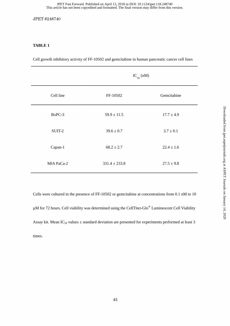
(BxPC-3, SUIT-2, Capan-1, and MIA PaCa-2) were evaluated in vitro (Table 1). The average IC50
values (nmol/L) of gemcitabine in these cell lines were 17.7, 3.7, 22.4, and 27.5, respectively, and
those of FF-10502 were 59.9, 39.6, 68.2, and 331.4, respectively. These results were consistent with
those reported in previous studies (Miura et al., 1999; Zajchowski et al., 2005).
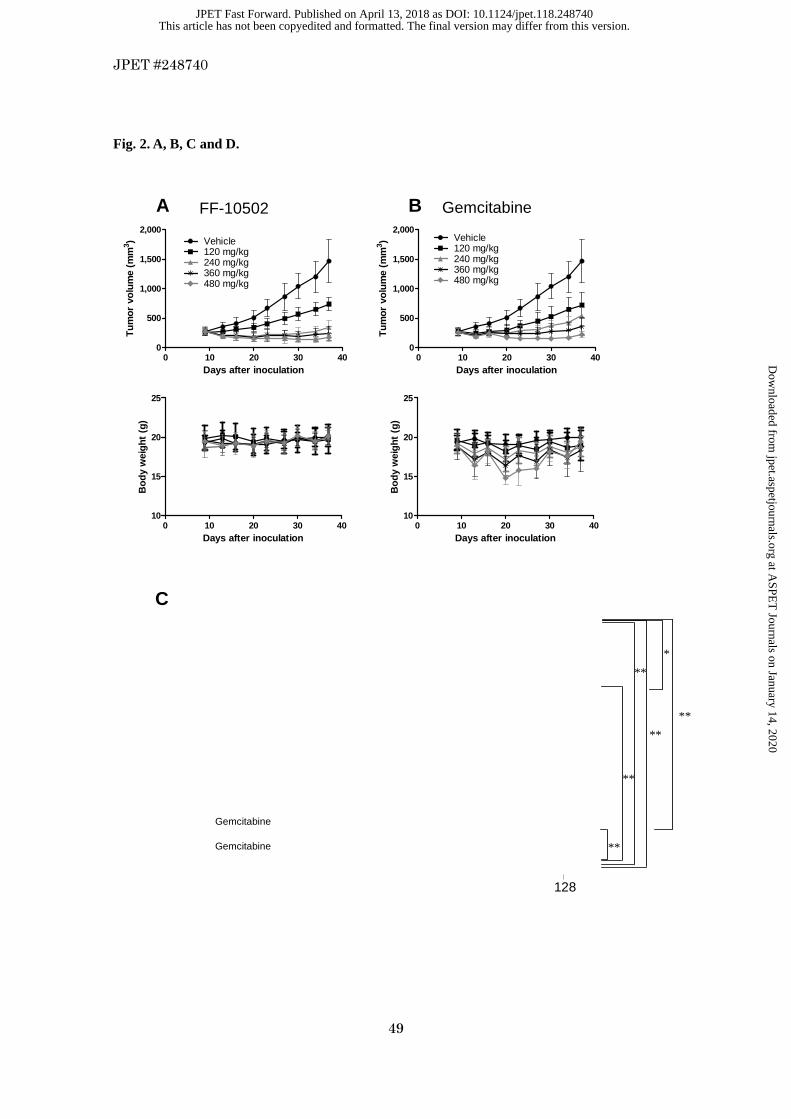
The antitumor effect of FF-10502 was evaluated in a mouse xenograft model with the
subcutaneously implanted human pancreatic cancer cell line Capan-1. Intravenous administrations of
FF-10502 or gemcitabine at 120, 240, 360, and 480 mg/kg once weekly suppressed tumor growth in
a dose-dependent manner (Fig. 2, A and B). The maximum decrease in body weight in FF-10502-
and gemcitabine-treated mice was 14.5% at 360 mg/kg and 28.3% at 480 mg/kg, respectively. No
deaths were observed in either treatment group.
The antitumor effect of FF-10502 was further evaluated in a mouse model of orthotopic
implantation with the human pancreatic cell line, SUIT-2. The SUIT-2 cell line is derived from liver
metastasis of pancreatic cancer patients with Kras and TP53 gene mutations, produces at least two
tumor markers, carcinoembryonic antigen and carbohydrate antigen 19-9, and has been widely used
since its establishment (Iwamura et al., 1987; Iwamura et al., 1992; Moore et al., 2001). The SUIT-2
This article has not been copyedited and formatted. The final version may differ from this version.JPET Fast Forward. Published on April 13, 2018 as DOI: 10.1124/jpet.118.248740
at ASPE
T Journals on January 14, 2020
jpet.aspetjournals.orgD
ownloaded from

JPET #248740
19
cells can survive in mice after orthotopic implantation, which is known to reproduce the pattern of
local tumor growth and distant metastasis observed in human pancreatic cancer (Shono et al., 2001;
Higuchi et al., 2018). The first weekly intravenous administration of each dosing solution (20
mice/group) was performed 7 days after implantation, the same day that the implanted SUIT-2 cells
began to metastasize to the mesenteric and spleen. Treatments continued until day 128.
Vehicle-treated mice started to die at day 19; their survival rate at day 128 (the final day of the study)
after tumor implantation was 5%, and median survival was 54.5 days (Fig. 2C; Table 2).
Gemcitabine treatment showed a dose-dependent and statistically significant prolongation of mouse
survival compared to that of vehicle-treated mice. The survival rate at day 128 was 25% at the 240
mg/kg dose and 75% at the 480 mg/kg dose (Table 2). In contrast, all mice treated with 240 or 480
mg/kg of FF-10502 survived until the end of the study, yielding a survival rate of 100% at both
doses. The effects were statistically significant compared with not only the vehicle-treated group but
also the dose-matched gemcitabine-treated group.
A histopathological assessment of five surviving mice in each group revealed further
contrast between gemcitabine and FF-10502 treatments (Fig. 2D). Increased sizes of implanted
tumors at pancreas were observed in the gemcitabine-treated all mice at 240 and 480 mg/kg. In
contrast, all mice treated with FF-10502 at 240 mg/kg showed regressions of the implanted tumors,
and 3 out of 5 of the FF-10502-treated mice at 480 mg/kg showed no implanted tumors. Metastases
This article has not been copyedited and formatted. The final version may differ from this version.JPET Fast Forward. Published on April 13, 2018 as DOI: 10.1124/jpet.118.248740
at ASPE
T Journals on January 14, 2020
jpet.aspetjournals.orgD
ownloaded from

JPET #248740
20
at mesenterium were observed in all gemcitabine-treated mice in the 240 mg/kg group, and in 2 out
of 5 in the 480 mg/kg group, whereas metastases were observed after FF-10502 treatment at the
same doses in only 1 out of 5 mice in each group. Liver metastases were also observed in 5 out of 5,
and 2 out of 5 of the gemcitabine-treated mice at 240 and 480 mg/kg, respectively. However, liver
metastases were observed in only 1 out of 5, and 0 out of 5 of the FF-10502–treated mice at 240 and
480 mg/kg, respectively.
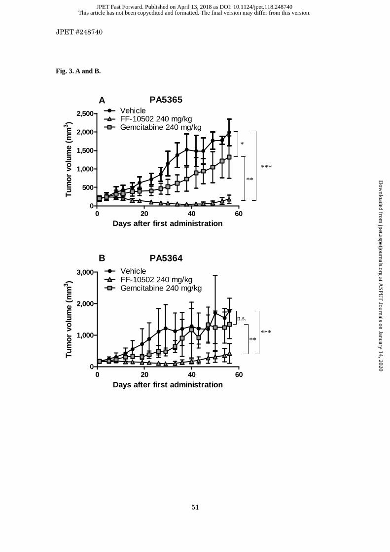
Antitumor Activity of FF-10502 in Pancreatic Patient-Derived Cancer Cells In Vivo
We next evaluated the antitumor effects of gemcitabine and FF-10502 in a PDX model in vivo, in
which two lines of gemcitabine-resistant, patient-derived pancreatic tumor cells were used. Four
intravenous weekly treatments of gemcitabine or FF-10502 were initiated when the average tumor
size reached 200 mm3. The tumors receiving gemcitabine treatment showed slow growth, which
turned into rapid growth after the last drug administration on day 22. In contrast, the
FF-10502–treated groups showed statistically significant tumor shrinkage at 240 mg/kg, and the
effect continued even after the termination of treatment (Fig. 3, A and B).
Mechanism of Action of FF-10502
While the principal mechanisms of FF-10502 antitumor activity remain to be determined, some
studies indicate a potential role of DNA polα inhibition, an enzyme involved in initiation of DNA
replication (Miura et al., 2001; Miura and Izuta, 2004). Miura et al reported that the inhibitory
This article has not been copyedited and formatted. The final version may differ from this version.JPET Fast Forward. Published on April 13, 2018 as DOI: 10.1124/jpet.118.248740
at ASPE
T Journals on January 14, 2020
jpet.aspetjournals.orgD
ownloaded from

JPET #248740
21
activity of FF-10502TP for polα was approximately 1,000 times more potent than that of gemTP
(Miura et al., 2001). This was confirmed in our current study, as shown in Fig. 4A; the inhibitory
activity of FF-10502TP against polα was more potent than that of gemTP, although the potency was
14-fold higher (IC50 was 23 µM for FF-10502TP and 547 µM for gemTP). It was therefore
hypothesized that the antitumor activity of FF-10502 is dependent on its inhibitory activity for polα;
consequently, we evaluated DNA synthesis activity in tumors using a mouse model of orthotopically
implanted SUIT-2 cells by measuring the percentage of EdU incorporated into DNA after FF-10502
or gemcitabine administration. Both FF-10502 and gemcitabine inhibited DNA synthesis from 4 to
48 hours, but DNA synthesis recovered by 72 hours. No significant differences were observed in the
inhibitory activities for DNA synthesis between FF-10502 and gemcitabine (Fig. 4, C and D).
We next examined polβ, in light of a previous study that reported the potency of
FF-10502TP against polβ to be 23 times higher than that of gemTP (Miura et al., 2001). This trend
was confirmed in our current study, although gemTP did not reach 50% inhibition while the IC50 of
FF-10502TP was 10 µM (Fig. 4B). Polβ is known to be a DNA-repairing enzyme that plays a key
role in base excision repair (BER); therefore, DDIs causing DNA damage (repaired by BER) were
combined with FF-10502 or gemcitabine to investigate whether polβ inhibitory activity contributes
to its cytotoxic effect. To avoid antiproliferation effects by polα inhibition with FF-10502 or
gemcitabine, we established a SUIT-2 dormant cell model that was induced by serum-free medium
This article has not been copyedited and formatted. The final version may differ from this version.JPET Fast Forward. Published on April 13, 2018 as DOI: 10.1124/jpet.118.248740
at ASPE
T Journals on January 14, 2020
jpet.aspetjournals.orgD
ownloaded from

JPET #248740
22
(Fig. 5A). SUIT-2 cells did not proliferate, and cell death was not evident by 72 hours in serum-free
conditions. Individual treatment of gemcitabine or FF-10502, or DDIs alone, did not affect cell
viability (Fig. 5B). Under these conditions, gemcitabine did not affect, or weakly affected, cell
viability in combination with DDIs. However, FF-10502 induced significant cell death in
combination with H2O2, cisplatin, or temozolomide (Fig. 5, C–E). The comet assay was applied to
confirm DNA damage; the results clearly indicated that the combination of FF-10502 with DDIs
caused synergistic DNA damage, whereas the combination of gemcitabine with DDIs yielded little
response (Fig. 6; Fig. 7; Table 3).
This article has not been copyedited and formatted. The final version may differ from this version.JPET Fast Forward. Published on April 13, 2018 as DOI: 10.1124/jpet.118.248740
at ASPE
T Journals on January 14, 2020
jpet.aspetjournals.orgD
ownloaded from
JPET #248740
23
Discussion
In most of the previous studies, FF-10502 was evaluated in mice models of subcutaneously
implanted pancreatic cancer cell lines in vivo, and showed superior efficacies compared to
gemcitabine (Miura et al., 1998; Miura et al., 1999; Miura et al., 2002; Zajchowski et al., 2005). We
further evaluated FF-10502 in our study using more clinically relevant models in vivo, i.e.,
orthotopic implantation of human pancreatic cancer SUIT-2 cells and subcutaneous implantation of
gemcitabine-resistant patient-derived cells. The SUIT-2 cell line has been widely used in vitro and in
vivo since its establishment from metastasis in liver of human pancreatic patients. The in vivo
orthotopic implantation model with SUIT-2 in mice has been validated, and shows similarities to
human pancreatic patients in terms of metastases at peritoneum, diaphragm, liver and lungs, and the
partial response to gemcitabine (Tomioka et al., 2001; Higuchi et al., 2018). Our study using the
model revealed that FF-10502-treated mice showed prominent inhibition of implanted tumors, and
little or no hepatic or intraperitoneal metastasis, while increased sizes of implanted tumors and
metastases were observed in gemcitabine-treated mice (Fig. 2D). The results suggested that these
histopathological observations were related to survival of all mice treated with FF-10502, unlike
gemcitabine (Fig. 2C). The PDX models with pancreatic cancer cells from patients with resistance to
gemcitabine also showed clear difference between FF-10502 and gemcitabine (Fig. 3, A and B).
These are new findings showing the superior efficacy of FF-10502 to gemcitabine in those clinically
relevant animal models of pancreatic cancer.
This article has not been copyedited and formatted. The final version may differ from this version.JPET Fast Forward. Published on April 13, 2018 as DOI: 10.1124/jpet.118.248740
at ASPE
T Journals on January 14, 2020
jpet.aspetjournals.orgD
ownloaded from
JPET #248740
24
The growth-inhibitory activity of FF-10502 in vitro human pancreatic cancer cell lines
(BxPC-3, SUIT-2, Capan-1, and MIA PaCa-2) was lower than that of gemcitabine (Table 1).
However, the antitumor activity of FF-10502 in vivo was superior to that of gemcitabine, as
demonstrated in mouse models of Capan-1 xenografts, SUIT-2 orthotopic implantation, and PDX
with pancreatic cancer cells in vivo.
We hypothesized that the lower efficacy of gemcitabine in vivo is caused by the presence of slow
growth or dormant cells. It is empirically known that implanted cells in vivo grow much slower than
cultured cells in vitro. Amikura et al reported that the doubling time of metastatic pancreatic tumor
cells in the liver in vivo was approximately 15 times longer than their doubling time in culture
(Amikura et al., 1995). In this study, we demonstrated that 90% of implanted SUIT-2 cells did not
proliferate (Fig. 4, C and D). Gemcitabine is not cytotoxic to cells that grow very slowly or acquire
dormancy, since the implanted target of gemcitabine is the DNA synthesis enzyme polα. For
example, the potency of gemcitabine is approximately 100 times lower in a quiescent Capan-2
spheroid culture than in a proliferative monolayer culture (Dufau et al., 2012). In accordance with
this prior report, we demonstrated that gemcitabine did not affect dormant cells cultured in
serum-free medium (Fig. 5B). Accordingly, we hypothesized that the higher efficacy of FF-10502
observed in the in vivo model can be attributed to cytotoxicity against slow-growing or dormant
cells, which emerge due to acquired heterogeneity in vivo, and that FF-10502 may have an
This article has not been copyedited and formatted. The final version may differ from this version.JPET Fast Forward. Published on April 13, 2018 as DOI: 10.1124/jpet.118.248740
at ASPE
T Journals on January 14, 2020
jpet.aspetjournals.orgD
ownloaded from

JPET #248740
25
additional unique mechanism of action that is distinct from the mechanism of gemcitabine.
In previous studies also conducted in mice, the pharmacokinetics of FF-10502 and gemcitabine were
comparable, and FF-10502 showed a high degree of inhibitory activity against polα (Miura et al.,
2001; Zajchowski et al., 2005). It was hypothesized, therefore, that the superior antitumor activity of
FF-10502 was caused by high polα inhibitory activity. However, there was no significant difference
in the inhibition of DNA synthesis between FF-10502 and gemcitabine in the SUIT-2 orthotopic
implantation model (Fig. 4, C and D). These results suggest that inhibitory activity against polα
cannot account for the pronounced antitumor activity of FF-10502 in vivo. Thus, we hypothesized
that FF-10502 may have additional mechanisms of action.
Although a previous study suggested that FF-10502 did not affect the RNA transcription in
the growing cell (Miura et al., 2001), a question was raised as to whether it would be the same in
dormant cells. 5-FU is known as a DNA and RNA synthesis inhibitor, and we evaluated the
cytotoxicity at 0–100 µM in our serum starvation assay in combination with 10 µM cisplatin. As the
result, 5-FU did not affect the cisplatin-induced cytotoxicity even at the highest concentration
(100 µM) (supplement data). The results suggest that transcriptional inhibition is less likely as the
mechanism of action in the enhanced cisplatin-induced cytotoxicity by FF-10502 in dormant cells.
We then focused on polβ inhibitory activity, as FF-10502 showed a much greater degree of
inhibition against polβ than gemcitabine (Fig. 4B). The observed inhibition of polβ with FF-10502
This article has not been copyedited and formatted. The final version may differ from this version.JPET Fast Forward. Published on April 13, 2018 as DOI: 10.1124/jpet.118.248740
at ASPE
T Journals on January 14, 2020
jpet.aspetjournals.orgD
ownloaded from

JPET #248740
26
was much more prominent in this study than in previous reports. The reason for this difference is not
clear, except perhaps differing sources of polβ (calf thymus in the previous report and recombinant
human protein in this study). Polβ, which is induced by H2O2 or temozolomide, is a principal DNA
polymerase in BER and has been explored as a cancer therapeutic target (Lange et al., 2011; Kim
and Wilson, 2012). In previous studies, small-molecule polβ inhibitors enhanced the cytotoxicity of
bleomycin or temozolomide in lung and colon cancers (Gao et al., 2008; Jaiswal et al., 2009), and
interference in polβ expression increased sensitivity to oxaliplatin (Yang et al., 2010). Therefore, we
further investigated the relationship between the inhibitory activity of FF-10502 against polβ and
dormant cell viability, using a SUIT-2 serum-starvation model. The cell viability of SUIT-2 dormant
cells was not affected by treatment with H2O2 alone, but was dramatically reduced by combining
H2O2 with FF-10502; however, this effect was not observed with gemcitabine (Fig. 5C). Similar
results were obtained with temozolomide, which causes DNA damage that is repaired by BER (Fig.
5E). These results suggest that FF-10502 inhibits BER of dormant tumor cells and causes cell death
in the presence of DDIs.
It is known that high amounts of reactive oxygen species (ROS) are generated in tumors and cause
DNA damage, such as base oxidation (Szatrowski and Nathan, 1991; Trachootham et al., 2006).
Therefore, the high efficacies of FF-10502 in vivo may be explained by endogenous oxidative DNA
damage in tumors plus polβ inhibition by FF-10502.
This article has not been copyedited and formatted. The final version may differ from this version.JPET Fast Forward. Published on April 13, 2018 as DOI: 10.1124/jpet.118.248740
at ASPE
T Journals on January 14, 2020
jpet.aspetjournals.orgD
ownloaded from

JPET #248740
27
DNA polymerase δ, ε and κ, as well as polβ, are known to be involved in cisplatin-induced DNA
damage (Ogi et al., 2010). The observation that FF-10502 also enhanced the cytotoxicity of cisplatin
in dormant cells (Fig. 5D) further suggests that there are other target polymerases for FF-10502.
It may be argued that polβ inhibition causes deleterious effects because of the essential role it plays
in BER, but there were no serious toxicities observed with FF-10502 treatment in our animal models,
and a clinical trial of FF-10502 currently on-going in the United States has demonstrated good
tolerability in patients with solid tumors (Falchook et al., 2017; Janku et al., 2018).
Taken together, these results imply that polβ inhibition may play a role in the mechanism
contributing to the difference in efficacy between FF-10502 and gemcitabine, although further
experiments will be necessary to show direct evidences for the contribution of polβ inhibition as well
as the involvement of other DNA polymerases.
Cancer stem cells (CSCs) are accepted as the cause of relapse and distant metastasis in
cancer progression (Rasheed et al., 2011; Podberezin et al., 2013; Li and Li, 2014). SUIT-2 cells
express CSC markers (CD133+ and CXCR4+) (Moriyama et al., 2010), and our study indicated that
orthotopically implanted SUIT-2 cells metastasize aggressively and are resistant to gemcitabine.
Despite repeated high-dose administration of gemcitabine (480 mg/kg), tumor progression was not
prevented. In contrast, FF-10502 treatment resulted in no deaths at doses of both 240 and 480 mg/kg;
FF-10502 also inhibited dissemination of tumor cells and reduced tumor cell growth at the
This article has not been copyedited and formatted. The final version may differ from this version.JPET Fast Forward. Published on April 13, 2018 as DOI: 10.1124/jpet.118.248740
at ASPE
T Journals on January 14, 2020
jpet.aspetjournals.orgD
ownloaded from

JPET #248740
28
inoculated site. These results suggest that FF-10502 displays a wide spectrum of antitumor activity
that includes targeting rapidly growing and dormant cells (including CSCs); this new insight may
encourage the exploration of novel research directions for cancer stem cell therapeutics.
The effect of FF-10502 on tumor microenvironment should also be considered as other mechanisms
such as interactions with immune cells, blood vessels, fibroblasts and extracellular matrix in the
process of metastasis, however, further investigations are required to clarify this.
In conclusion, we have demonstrated new findings that FF-10502 is significantly more
efficacious than gemcitabine in a variety of clinically relevant mouse models of pancreatic cancer,
including PDX models, which may be explained by inhibition of dormant cancer cells through a
higher inhibition of polβ unlike gemcitabine. A clinical trial of FF-10502 is currently underway in
the United States to assess the usefulness of FF-10502 in patients with solid tumors, including
pancreatic tumors. FF-10502 is expected to be a promising agent for pancreatic cancer with acquired
resistance to gemcitabine.
This article has not been copyedited and formatted. The final version may differ from this version.JPET Fast Forward. Published on April 13, 2018 as DOI: 10.1124/jpet.118.248740
at ASPE
T Journals on January 14, 2020
jpet.aspetjournals.orgD
ownloaded from

JPET #248740
29
Acknowledgments
The authors are deeply grateful to Dr. Michihiko Wada and Dr. Junji Furuse for useful suggestions
and advice. The authors thank Takaaki Nakamura, Shin-ichi Watanabe, Takeaki Suzuki, Yuki Awa,
Ken Okada, Hiroko Fujisaki, Hiroko Nemoto, Ayako Kamei, and Dr. Thomas Myers for technical
assistance and helpful discussion, and Jill Ricono and Jonathan Nakashima for their help in
experiments with PDX models.
This article has not been copyedited and formatted. The final version may differ from this version.JPET Fast Forward. Published on April 13, 2018 as DOI: 10.1124/jpet.118.248740
at ASPE
T Journals on January 14, 2020
jpet.aspetjournals.orgD
ownloaded from

JPET #248740
30
Authorship Contributions
Participated in study design, performing experiments and data analysis: Mima, Kakinuma, Higuchi,
Saeki, Yamada, Uematsu, Ishino, Kito, Nishikawa, Paradiso, and Iwamura.
Contributed to synthesizing new agents: Kuniyoshi, Matsumoto, Fujiwara, and Shimada.
Contributed to the writing of the manuscript: Mima, Yamada, Paradiso, and Iwamura.
This article has not been copyedited and formatted. The final version may differ from this version.JPET Fast Forward. Published on April 13, 2018 as DOI: 10.1124/jpet.118.248740
at ASPE
T Journals on January 14, 2020
jpet.aspetjournals.orgD
ownloaded from

JPET #248740
31
References
Amikura K, Kobari M and Matsuno S (1995) The time of occurrence of liver metastasis in
carcinoma of the pancreas. Int J Pancreatol 17:139-146.
Arumugam T, Ramachandran V, Fournier KF, Wang H, Marquis L, Abbruzzese JL, Gallick
GE, Logsdon CD, McConkey DJ and Choi W (2009) Epithelial to mesenchymal
transition contributes to drug resistance in pancreatic cancer. Cancer Res
69:5820-5828.
Burris HA, 3rd, Moore MJ, Andersen J, Green MR, Rothenberg ML, Modiano MR, Cripps
MC, Portenoy RK, Storniolo AM, Tarassoff P, Nelson R, Dorr FA, Stephens CD and
Von Hoff DD (1997) Improvements in survival and clinical benefit with gemcitabine
as first-line therapy for patients with advanced pancreas cancer: a randomized trial.
J Clin Oncol 15:2403-2413.
Conroy T, Desseigne F, Ychou M, Bouche O, Guimbaud R, Becouarn Y, Adenis A, Raoul JL,
Gourgou-Bourgade S, de la Fouchardiere C, Bennouna J, Bachet JB,
Khemissa-Akouz F, Pere-Verge D, Delbaldo C, Assenat E, Chauffert B, Michel P,
Montoto-Grillot C and Ducreux M (2011) FOLFIRINOX versus gemcitabine for
metastatic pancreatic cancer. N Engl J Med 364:1817-1825.
Dufau I, Frongia C, Sicard F, Dedieu L, Cordelier P, Ausseil F, Ducommun B and Valette A
This article has not been copyedited and formatted. The final version may differ from this version.JPET Fast Forward. Published on April 13, 2018 as DOI: 10.1124/jpet.118.248740
at ASPE
T Journals on January 14, 2020
jpet.aspetjournals.orgD
ownloaded from

JPET #248740
32
(2012) Multicellular tumor spheroid model to evaluate spatio-temporal dynamics
effect of chemotherapeutics: application to the gemcitabine/CHK1 inhibitor
combination in pancreatic cancer. BMC Cancer 12:15.
Falchook GS, Bramwell L, Hannan L, Vishwamitra D, Yamada T, Rosner M, Wages D,
Myers T, Paradiso L and Janku F (2017) Abstract CT100: First-in-human phase 1
trial of pyrimidine anti-metabolite FF-10502-01 in patients with advanced cancer.
Cancer research 77:CT100-CT100.
Janku F, Kurman M, Vishwamitra D, Shroff R, Hong D, Javle M, Fogelman D, Bramwell L,
Hannan L, Ward M, Tiefenwerth K, Denton S, Bolognese J, Subach RA, Madden T,
Maier G, Yamada T, Iwamura H, Paradiso L and Falchook G (2018) Preliminary
Activity of FF-10502-01 in Patients with Refractory Advanced Cholangiocarcinoma.
Cholangiocarcinoma Foundation Annual Conference.
Gao Z, Maloney DJ, Dedkova LM and Hecht SM (2008) Inhibitors of DNA polymerase beta:
activity and mechanism. Bioorg Med Chem 16:4331-4340.
Goldstein D, El-Maraghi RH, Hammel P, Heinemann V, Kunzmann V, Sastre J, Scheithauer
W, Siena S, Tabernero J, Teixeira L, Tortora G, Van Laethem JL, Young R,
Penenberg DN, Lu B, Romano A and Von Hoff DD (2015) nab-Paclitaxel plus
gemcitabine for metastatic pancreatic cancer: long-term survival from a phase III
This article has not been copyedited and formatted. The final version may differ from this version.JPET Fast Forward. Published on April 13, 2018 as DOI: 10.1124/jpet.118.248740
at ASPE
T Journals on January 14, 2020
jpet.aspetjournals.orgD
ownloaded from
JPET #248740
33
trial. J Natl Cancer Inst 107.
Heinemann V, Hertel LW, Grindey GB and Plunkett W (1988) Comparison of the cellular
pharmacokinetics and toxicity of 2',2'-difluorodeoxycytidine and
1-beta-D-arabinofuranosylcytosine. Cancer Res 48:4024-4031.
Higuchi T, Yokobori T, Naito T, Kakinuma C, Hagiwara S, Nishiyama M and Asao T (2018)
Investigation into metastatic processes and the therapeutic effects of gemcitabine on
human pancreatic cancer using an orthotopic SUIT-2 pancreatic cancer mouse model.
Oncology letters 15:3091-3099.
Iwamura T, Katsuki T and Ide K (1987) Establishment and characterization of a human
pancreatic cancer cell line (SUIT-2) producing carcinoembryonic antigen and
carbohydrate antigen 19-9. Japanese journal of cancer research : Gann 78:54-62.
Iwamura T, Taniguchi S, Kitamura N, Yamanari H, Kojima A, Hidaka K, Setoguchi T and
Katsuki T (1992) Correlation between CA19-9 production in vitro and histological
grades of differentiation in vivo in clones isolated from a human pancreatic cancer
cell line (SUIT-2). Journal of gastroenterology and hepatology 7:512-519.
Jaiswal AS, Banerjee S, Panda H, Bulkin CD, Izumi T, Sarkar FH, Ostrov DA and Narayan
S (2009) A novel inhibitor of DNA polymerase beta enhances the ability of
temozolomide to impair the growth of colon cancer cells. Mol Cancer Res
This article has not been copyedited and formatted. The final version may differ from this version.JPET Fast Forward. Published on April 13, 2018 as DOI: 10.1124/jpet.118.248740
at ASPE
T Journals on January 14, 2020
jpet.aspetjournals.orgD
ownloaded from

JPET #248740
34
7:1973-1983.
Kim YJ and Wilson DM 3rd (2012) Overview of base excision repair biochemistry. Curr Mol
Pharmacol 5:3-13.
Lange SS, Takata K and Wood RD (2011) DNA polymerases and cancer. Nat Rev Cancer
11:96-110.
Li S and Li Q (2014) Cancer stem cells and tumor metastasis (Review). Int J Oncol
44:1806-1812.
Miura S, Endo Y, Yoshimura Y, Endo M, Yonemura Y and Sasaki T (2002) Potent antitumor
effect of 1-(2-deoxy-2-fluoro-4-thio-beta-D-arabinofuranosyl)cytosine on peritoneal
dissemination models of gastrointestinal cancers. Oncol Rep 9:1319-1322.
Miura S and Izuta S (2004) DNA polymerases as targets of anticancer nucleosides. Curr
Drug Targets 5:191-195.
Miura S, Yoshimura Y, Endo M, Machida H, Matsuda A, Tanaka M and Sasaki T (1998)
Antitumor activity of a novel orally effective nucleoside,
1-(2-deoxy-2-fluoro-4-thio-beta-D-arabinofuranosyl)cytosine. Cancer Lett
129:103-110.
Miura S, Yoshimura Y, Endo M, Satoh H, Machida H and Sasaki T (1999) Comparison of
1-(2-deoxy-2-fluoro-4-thio-beta-D-arabinofuranosyl)cytosine with gemcitabine in its
This article has not been copyedited and formatted. The final version may differ from this version.JPET Fast Forward. Published on April 13, 2018 as DOI: 10.1124/jpet.118.248740
at ASPE
T Journals on January 14, 2020
jpet.aspetjournals.orgD
ownloaded from

JPET #248740
35
antitumor activity. Cancer Lett 144:177-182.
Miura S, Yoshimura Y, Satoh H and Izuta S (2001) The antitumor mechanism of
1-(2-deoxy-2-fluoro-4-thio-beta-D-arabinofuranosyl)-cytosine: effects of its
triphosphate on mammalian DNA polymerases. Japanese journal of cancer
research : Gann 92:562-567.
Moore PS, Sipos B, Orlandini S, Sorio C, Real FX, Lemoine NR, Gress T, Bassi C, Kloppel G,
Kalthoff H, Ungefroren H, Lohr M and Scarpa A (2001) Genetic profile of 22
pancreatic carcinoma cell lines. Analysis of K-ras, p53, p16 and DPC4/Smad4.
Virchows Archiv : an international journal of pathology 439:798-802.
Moriyama T, Ohuchida K, Mizumoto K, Cui L, Ikenaga N, Sato N and Tanaka M (2010)
Enhanced cell migration and invasion of CD133+ pancreatic cancer cells cocultured
with pancreatic stromal cells. Cancer 116:3357-3368.
Ogi T, Limsirichaikul S, Overmeer RM, Volker M, Takenaka K, Cloney R, Nakazawa Y,
Niimi A, Miki Y, Jaspers NG, Mullenders LH, Yamashita S, Fousteri MI and
Lehmann AR (2010) Three DNA polymerases, recruited by different mechanisms,
carry out NER repair synthesis in human cells. Mol Cell 37:714-727.
Podberezin M, Wen J and Chang CC (2013) Cancer stem cells: a review of potential clinical
applications. Arch Pathol Lab Med 137:1111-1116.
This article has not been copyedited and formatted. The final version may differ from this version.JPET Fast Forward. Published on April 13, 2018 as DOI: 10.1124/jpet.118.248740
at ASPE
T Journals on January 14, 2020
jpet.aspetjournals.orgD
ownloaded from

JPET #248740
36
Podust VN, Lavrik OI, Nasheuer HP and Grosse F (1989) DNA polymerase alpha-DNA
primase from human placenta. Immunoaffinity purification and preliminary
characterization. FEBS Lett 245:14-16.
Rasheed ZA, Kowalski J, Smith BD and Matsui W (2011) Concise review: Emerging concepts
in clinical targeting of cancer stem cells. Stem Cells 29:883-887.
Salic A and Mitchison TJ (2008) A chemical method for fast and sensitive detection of DNA
synthesis in vivo. Proc Natl Acad Sci U S A 105:2415-2420.
Shono M, Sato N, Mizumoto K, Maehara N, Nakamura M, Nagai E and Tanaka M (2001)
Stepwise progression of centrosome defects associated with local tumor growth and
metastatic process of human pancreatic carcinoma cells transplanted orthotopically
into nude mice. Laboratory investigation; a journal of technical methods and
pathology 81:945-952.
Siegel RL, Miller KD and Jemal A (2015) Cancer statistics, 2015. CA Cancer J Clin 65:5-29.
Stanton KJ, Sidner RA, Miller GA, Cummings OW, Schmidt CM, Howard TJ and Wiebke EA
(2003) Analysis of Ki-67 antigen expression, DNA proliferative fraction, and survival
in resected cancer of the pancreas. Am J Surg 186:486-492.
Szatrowski TP and Nathan CF (1991) Production of large amounts of hydrogen peroxide by
human tumor cells. Cancer Res 51:794-798.
This article has not been copyedited and formatted. The final version may differ from this version.JPET Fast Forward. Published on April 13, 2018 as DOI: 10.1124/jpet.118.248740
at ASPE
T Journals on January 14, 2020
jpet.aspetjournals.orgD
ownloaded from

JPET #248740
37
Tomioka D, Maehara N, Kuba K, Mizumoto K, Tanaka M, Matsumoto K and Nakamura T
(2001) Inhibition of growth, invasion, and metastasis of human pancreatic
carcinoma cells by NK4 in an orthotopic mouse model. Cancer research
61:7518-7524.
Trachootham D, Zhou Y, Zhang H, Demizu Y, Chen Z, Pelicano H, Chiao PJ, Achanta G,
Arlinghaus RB, Liu J and Huang P (2006) Selective killing of oncogenically
transformed cells through a ROS-mediated mechanism by beta-phenylethyl
isothiocyanate. Cancer Cell 10:241-252.
Yang J, Parsons J, Nicolay NH, Caporali S, Harrington CF, Singh R, Finch D, D'Atri S,
Farmer PB, Johnston PG, McKenna WG, Dianov G and Sharma RA (2010) Cells
deficient in the base excision repair protein, DNA polymerase beta, are
hypersensitive to oxaliplatin chemotherapy. Oncogene 29:463-468.
Yoshida GJ and Saya H (2016) Therapeutic strategies targeting cancer stem cells. Cancer
Sci 107:5-11.
Zajchowski DA, Biroc SL, Liu HL, Chesney SK, Hoffmann J, Bauman J, Kirkland T,
Subramanyam B, Shen J, Ho E, Tseng JL and Dinter H (2005) Anti-tumor efficacy of
the nucleoside analog 1-(2-deoxy-2-fluoro-4-thio-beta-D-arabinofuranosyl) cytosine
(4'-thio-FAC) in human pancreatic and ovarian tumor xenograft models. Int J
This article has not been copyedited and formatted. The final version may differ from this version.JPET Fast Forward. Published on April 13, 2018 as DOI: 10.1124/jpet.118.248740
at ASPE
T Journals on January 14, 2020
jpet.aspetjournals.orgD
ownloaded from
JPET #248740
38
Cancer 114:1002-1009.
This article has not been copyedited and formatted. The final version may differ from this version.JPET Fast Forward. Published on April 13, 2018 as DOI: 10.1124/jpet.118.248740
at ASPE
T Journals on January 14, 2020
jpet.aspetjournals.orgD
ownloaded from

JPET #248740
39
Footnotes
a) Financial support: None
b) Citation of meeting abstracts where the work was previously presented:
(1) Shinji Mima, Hiroki Nishikawa, Shinichi Watanabe, Tamami Higuchi, Takeaki Suzuki, Hiroyuki
Iwamura, Chihaya Kakinuma, Takaaki Nakamura, Yasuhiro Shimada. Abstract #5127: In vitro and in
vivo evaluation of FF-10502-01, a new pyrimidine nucleoside analogue. American Association for
Cancer Research (AACR), Washington DC, USA. April (2017).
(2) Takeaki Suzuki, Linda J. Paradiso, Jill Ricono, Jonathan Nakashima, Yoshihide Iwaki, Shinji
Mima, Takayuki Yamada, Chihaya Kakinuma, Hiroyuki Iwamura, Shinichi Watanabe. Abstract
#5112: Evaluation of FF-10502-01, a new pyrimidine nucleoside analogue, in pancreatic (PANC)
patient-derived xenograft (PDX) models compared to gemcitabine and in combination with
nab-paclitaxel. AACR, Washington DC, USA. April (2017).
(3) Falchook GS, Bramwell L, Hannan L, Vishwamitra D, Yamada T, Rosner M, Wages D, Myers T,
Paradiso L and Janku F. Abstract CT100: First-in-human phase 1 trial of pyrimidine anti-metabolite
FF-10502-01 in patients with advanced cancer. AACR, Washington DC, USA. April (2017).
(4) Janku F, Kurman M, Vishwamitra D, Shroff R, Hong D, Javle M, Fogelman D, Bramwell
L, Hannan L, Ward M, Tiefenwerth K, Denton S, Bolognese J, Subach RA, Madden T, Maier
G, Yamada T, Iwamura H, Paradiso L and Falchook G. Abstract #6: Preliminary Activity of
This article has not been copyedited and formatted. The final version may differ from this version.JPET Fast Forward. Published on April 13, 2018 as DOI: 10.1124/jpet.118.248740
at ASPE
T Journals on January 14, 2020
jpet.aspetjournals.orgD
ownloaded from

JPET #248740
40
FF-10502-01 in Patients with Refractory Advanced Cholangiocarcinoma.
Cholangiocarcinoma Foundation Annual Conference. Salt Lake City, USA .February (2018).
c) Name, full address and e-mail address of person to receive reprint requests to:
Hiroyuki Iwamura, Ph.D.,
Pharmaceutical Products Division, FUJIFILM Corporation, 7-3 Akasaka 9-Chome, Minato-ku,
Tokyo 107-0052, Japan
E-mail: [email protected]
This article has not been copyedited and formatted. The final version may differ from this version.JPET Fast Forward. Published on April 13, 2018 as DOI: 10.1124/jpet.118.248740
at ASPE
T Journals on January 14, 2020
jpet.aspetjournals.orgD
ownloaded from

JPET #248740
41
Figure Legends
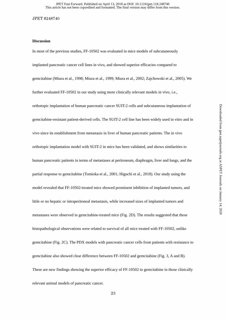
Fig. 1. Chemical structures of FF-10502 and gemcitabine.
Fig. 2. Antitumor effects of FF-10502 and gemcitabine on human pancreatic xenograft models.
Capan-1 cells were subcutaneously implanted into BALB/c-nu/nu mice, and when the average
tumor size reached approximately 200 mm3, treatment with vehicle (saline), FF-10502 (A), or
gemcitabine (B) was initiated and administered intravenously once weekly for 4 weeks. Tumor
volume (upper plots) and body weights (lower plots) are shown. Error bars show standard deviation
(n = 10). (C) SUIT-2 cells were orthotopically implanted into the subcapsular region of the
pancreas of 20 BALB/c-nu/nu mice and after 7 days, treatment with vehicle (saline), FF-10502, or
gemcitabine was initiated and administered intravenously once weekly for 18 weeks (treatment
days are indicated by arrows on the x-axis). Kaplan-Meier survival plots are presented. Significant
differences are indicated; * P<0.05 and ** P<0.01 between groups. (D) Histopathological features
of pancreas (D-1,2,5 and 6), mesenterium (D-3 and 7), and liver (D-4 and 8) from SUIT-2
orthotopically implanted mice treated with gemcitabine (D-1,2,3 and 4) or FF-10502 (D-5,6,7 and
8). D-2 and 6 are higher magnification of D-1 and 5, respectively. In gemcitabine-treated mice,
SUIT-2 showed widespread increase (D-1) with inter- and intra-acinar invasions (D-2) in pancreas,
metastasis into mesenterium (D-3) and portal area of liver (D-4) were remarkable (►). In
This article has not been copyedited and formatted. The final version may differ from this version.JPET Fast Forward. Published on April 13, 2018 as DOI: 10.1124/jpet.118.248740
at ASPE
T Journals on January 14, 2020
jpet.aspetjournals.orgD
ownloaded from
JPET #248740
42
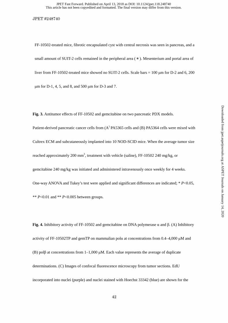
FF-10502-treated mice, fibrotic encapsulated cyst with central necrosis was seen in pancreas, and a
small amount of SUIT-2 cells remained in the peripheral area (*). Mesenterium and portal area of
liver from FF-10502-treated mice showed no SUIT-2 cells. Scale bars = 100 µm for D-2 and 6, 200
µm for D-1, 4, 5, and 8, and 500 µm for D-3 and 7.
Fig. 3. Antitumor effects of FF-10502 and gemcitabine on two pancreatic PDX models.
Patient-derived pancreatic cancer cells from (A) PA5365 cells and (B) PA5364 cells were mixed with
Cultrex ECM and subcutaneously implanted into 10 NOD-SCID mice. When the average tumor size
reached approximately 200 mm3, treatment with vehicle (saline), FF-10502 240 mg/kg, or
gemcitabine 240 mg/kg was initiated and administered intravenously once weekly for 4 weeks.
One-way ANOVA and Tukey’s test were applied and significant differences are indicated; * P<0.05,
** P <0.01 and ** P<0.005 between groups.
Fig. 4. Inhibitory activity of FF-10502 and gemcitabine on DNA polymerase α and β. (A) Inhibitory
activity of FF-10502TP and gemTP on mammalian polα at concentrations from 0.4–4,000 µM and
(B) polβ at concentrations from 1–1,000 µM. Each value represents the average of duplicate
determinations. (C) Images of confocal fluorescence microscopy from tumor sections. EdU
incorporated into nuclei (purple) and nuclei stained with Hoechst 33342 (blue) are shown for the
This article has not been copyedited and formatted. The final version may differ from this version.JPET Fast Forward. Published on April 13, 2018 as DOI: 10.1124/jpet.118.248740
at ASPE
T Journals on January 14, 2020
jpet.aspetjournals.orgD
ownloaded from

JPET #248740
43
indicated treatment times. Gem, gemcitabine; 502, FF-10502. (D) Percentage of EdU-incorporated
cells counted in three image fields (mean ± standard deviation). Time points were 4, 24, 48, and 72
hours. Mice were treated with vehicle (at 24 hours), gemcitabine, or FF-10502.
Fig. 5. Cytotoxic effect of DDI treatment in combination with either FF-10502 or gemcitabine on
pancreatic cancer dormant cells. (A) Study design of the SUIT-2 dormant cell model. After 3 days of
culture in serum-free medium, cells were incubated with FF-10502 or gemcitabine at concentrations
from 1–10 µM in the presence or absence of DDIs for 3 days. The viability of the remaining cells
was evaluated by the CellTiter-Glo® Luminescent Cell Viability Assay kit. Cell viability was plotted
at each concentration of FF-10502 and gemcitabine in (B) the absence of DNA damaging inducers,
and the presence of (C) 300 µM H2O2, (D) 5 µM cisplatin, and (E) 200 µM temozolomide.
Fig. 6. DNA damage induced by the combination of DDIs with FF-10502 or with gemcitabine was
evaluated by a comet assay in SUIT-2 cells under serum starvation. Serum-starved SUIT-2 cells were
exposed to FF-10502 or gemcitabine (100 or 1,000 nM) in the presence or absence of DDIs
(cisplatin, H2O2, or temozolomide). After 4 hours and 24 hours of exposure, cells were subjected to
the comet assay as described in Materials and Methods. Results are expressed as the percentage of
DNA with comet-like tail (% tail DNA) and the mean % tail DNA per 60 comet cells/well is
This article has not been copyedited and formatted. The final version may differ from this version.JPET Fast Forward. Published on April 13, 2018 as DOI: 10.1124/jpet.118.248740
at ASPE
T Journals on January 14, 2020
jpet.aspetjournals.orgD
ownloaded from

JPET #248740
44
indicated. 502, FF-10502; GEM, gemcitabine; TMZ, temozolomide.
Fig. 7. Representative images in comet assay in combination of DDIs with FF-10502 or gemcitabine
at 1,000 nM for 24 hours in human pancreatic SUIT-2 cells under serum starvation. 502, FF-10502;
DDIs, DNA damage inducers; GEM, gemcitabine; TMZ, temozolomide.
This article has not been copyedited and formatted. The final version may differ from this version.JPET Fast Forward. Published on April 13, 2018 as DOI: 10.1124/jpet.118.248740
at ASPE
T Journals on January 14, 2020
jpet.aspetjournals.orgD
ownloaded from
JPET #248740
45
TABLE 1
Cell growth inhibitory activity of FF-10502 and gemcitabine in human pancreatic cancer cell lines
IC50
(nM)
Cell line FF-10502 Gemcitabine
BxPC-3
SUIT-2
Capan-1
MIA PaCa-2
59.9 ± 11.5
39.6 ± 0.7
68.2 ± 2.7
331.4 ± 233.8
17.7 ± 4.9
3.7 ± 0.1
22.4 ± 1.6
27.5 ± 9.8
Cells were cultured in the presence of FF-10502 or gemcitabine at concentrations from 0.1 nM to 10
µM for 72 hours. Cell viability was determined using the CellTiter-Glo® Luminescent Cell Viability
Assay kit. Mean IC50 values ± standard deviation are presented for experiments performed at least 3
times.
This article has not been copyedited and formatted. The final version may differ from this version.JPET Fast Forward. Published on April 13, 2018 as DOI: 10.1124/jpet.118.248740
at ASPE
T Journals on January 14, 2020
jpet.aspetjournals.orgD
ownloaded from

JPET #248740
46
TABLE 2
Summary of SUIT-2 orthotopic implantation model
Group Dose
Median survival
time
Number of animals surviving at
the end of observation
(mg/kg) (days) (Surviving animals/n)
Vehicle - 54.5 1/20
FF-10502 240 >128a, b 20/20
FF-10502 480 >128a, c 20/20
Gemcitabine 240 109a 5/20
Gemcitabine 480 >128a 15/20
a P < 0.01 for each group versus vehicle. b P < 0.01 for FF-10502 240 mg/kg versus gemcitabine 240
mg/kg. c P < 0.05 for FF-10502 480 mg/kg versus gemcitabine 480 mg/kg.
This article has not been copyedited and formatted. The final version may differ from this version.JPET Fast Forward. Published on April 13, 2018 as DOI: 10.1124/jpet.118.248740
at ASPE
T Journals on January 14, 2020
jpet.aspetjournals.orgD
ownloaded from

JPET #248740
47
TABLE 3
Comet assay of DDI treatment in combination with FF-10502 or gemcitabine in human
pancreatic SUIT-2 cells under serum starvation
Group % Tail DNA
No DDIs
Cisplatin
5 µmol/L
H2O2
300 µmol/L
TMZ
200 µmol/L
4h 24h 4h 24h 4h 24h 4h 24h
Vehicle control (0.1% DMSO) 5.5 7.4 12.1 11.3 38.9 6.1 40.3 39.5
FF-10502 100 nmol/L 8.9 17.1 20.1 45.1 83.1 89.7 71.8 93.7
FF-10502 1000 nmol/L 6.8 22.2 19.9 49.7 93.8 95.8 88.1 97.4
Gemcitabine 100 nmol/L 5.6 6.5 19.5 13.1 37.4 6.9 39.0 39.5
Gemcitabine 1000 nmol/L 5.2 6.8 13.4 15.9 43.2 7.5 48.0 38.6
Percentage of fluorescence intensity in tail DNA region per whole DNA is expressed as % tail DNA.
The experiment was performed in duplicate, and the mean % tail DNA was calculated. DDI, DNA
damage inducer; DMSO, dimethylsulfoxide; TMZ, temozolomide.
This article has not been copyedited and formatted. The final version may differ from this version.JPET Fast Forward. Published on April 13, 2018 as DOI: 10.1124/jpet.118.248740
at ASPE
T Journals on January 14, 2020
jpet.aspetjournals.orgD
ownloaded from

JPET #248740
48
Fig. 1.
FF-10502 Gemcitabine
(Gemzar®)
This article has not been copyedited and formatted. The final version may differ from this version.JPET Fast Forward. Published on April 13, 2018 as DOI: 10.1124/jpet.118.248740
at ASPE
T Journals on January 14, 2020
jpet.aspetjournals.orgD
ownloaded from
JPET #248740
49
Fig. 2. A, B, C and D.
Days after inoculation
Bo
dy
wei
ght
(g
)
0 10 20 30 4010
15
20
25
Days after inoculation
Bo
dy
wei
ght
(g
)
0 10 20 30 4010
15
20
25
Days after inoculation
Tu
mor
vo
lum
e (m
m3 )
0 10 20 30 400
500
1,000
1,500
2,000Vehicle120 mg/kg240 mg/kg360 mg/kg480 mg/kg
Days after inoculationT
um
or v
olu
me
(mm
3 )
0 10 20 30 400
500
1,000
1,500
2,000Vehicle120 mg/kg240 mg/kg360 mg/kg480 mg/kg
FF-10502 Gemcitabine
C
B A
128
**
**
**
**
**
*
Gemcitabine
Gemcitabine
This article has not been copyedited and formatted. The final version may differ from this version.JPET Fast Forward. Published on April 13, 2018 as DOI: 10.1124/jpet.118.248740
at ASPE
T Journals on January 14, 2020
jpet.aspetjournals.orgD
ownloaded from

JPET #248740
50
D-1 D-2
D-5 D-6
D-3
D-7
D-4
D-8
********
****
D
This article has not been copyedited and formatted. The final version may differ from this version.JPET Fast Forward. Published on April 13, 2018 as DOI: 10.1124/jpet.118.248740
at ASPE
T Journals on January 14, 2020
jpet.aspetjournals.orgD
ownloaded from
JPET #248740
51
Fig. 3. A and B.
PA5364
Days after first administration
Tu
mo
r vo
lum
e (m
m3 )
0 20 40 600
1,000
2,000
3,000 VehicleFF-10502 240 mg/kgGemcitabine 240 mg/kg
**
n.s.
***
B
PA5365
Days after first administration
Tum
or v
olum
e (m
m3 )
0 20 40 600
500
1,000
1,500
2,000
2,500 VehicleFF-10502 240 mg/kgGemcitabine 240 mg/kg
**
*
***
A
This article has not been copyedited and formatted. The final version may differ from this version.JPET Fast Forward. Published on April 13, 2018 as DOI: 10.1124/jpet.118.248740
at ASPE
T Journals on January 14, 2020
jpet.aspetjournals.orgD
ownloaded from

JPET #248740
52
Fig. 4. A, B, C and D
mol/L
% in
hib
itio
n
10- 7 10- 6 10 - 5 10- 4 10- 3 10- 2
0
50
100 FF-10502TPGemTP
mol/L10- 6 10- 5 10- 4 10- 3
0
50
100 FF-10502TPGemTP
% in
hib
ition
Vehicle 24 h
Gem 72 h
502 24 h 502 72 h
Gem 24 h
B polα polβ A
C
This article has not been copyedited and formatted. The final version may differ from this version.JPET Fast Forward. Published on April 13, 2018 as DOI: 10.1124/jpet.118.248740
at ASPE
T Journals on January 14, 2020
jpet.aspetjournals.orgD
ownloaded from

JPET #248740
53
Ed
U in
corp
ora
ted
cel
ls (
%)
4 h 24 h 48 h 72 h 4 h 24 h 48 h 72 h0
5
10
15
20
Gemcitabine FF-10502
D
This article has not been copyedited and formatted. The final version may differ from this version.JPET Fast Forward. Published on April 13, 2018 as DOI: 10.1124/jpet.118.248740
at ASPE
T Journals on January 14, 2020
jpet.aspetjournals.orgD
ownloaded from

JPET #248740
54
Fig. 5. A, B, C, D and E.
SUIT-2 seeding
Change medium to Serum -free medium
1 Day 3 Days
Gemcitabine or FF-10502&DNA damage inducer
Cell viability
3 DaysSerum-free
Concentration (nM)
Cel
l via
bilit
y (%
)
1 10 100 1,000 10,0000
20
40
60
80
100
120GemcitabineFF-10502
Concentration (nM)
Cel
l via
bilit
y (%
)
1 10 100 1,000 10,0000
20
40
60
80
100
120 GemcitabineFF-10502
Concentration (nM)
Cel
l via
bili
ty (
%)
1 10 100 1,000 10,0000
20
40
60
80
100
120 GemcitabineFF-10502
Concentration (nM)
Cel
l via
bili
ty (
%)
1 10 100 1,000 10,0000
20
40
60
80
100
120GemcitabineFF-10502
A
B No DDIs C H2O2
D Cisplatin E Temozolomide
This article has not been copyedited and formatted. The final version may differ from this version.JPET Fast Forward. Published on April 13, 2018 as DOI: 10.1124/jpet.118.248740
at ASPE
T Journals on January 14, 2020
jpet.aspetjournals.orgD
ownloaded from

JPET #248740
55
Fig. 6.
0
20
40
60
80
100
% t
ail
DN
A
4 h 24 h
GEM 502
(nM) (nM)
GEM 502
(nM) (nM)
GEM 502
(nM) (nM)
GEM 502
(nM) (nM)
+Cisplatin 5 µM +H2O2 300 µM +TMZ 200 µM
This article has not been copyedited and formatted. The final version may differ from this version.JPET Fast Forward. Published on April 13, 2018 as DOI: 10.1124/jpet.118.248740
at ASPE
T Journals on January 14, 2020
jpet.aspetjournals.orgD
ownloaded from
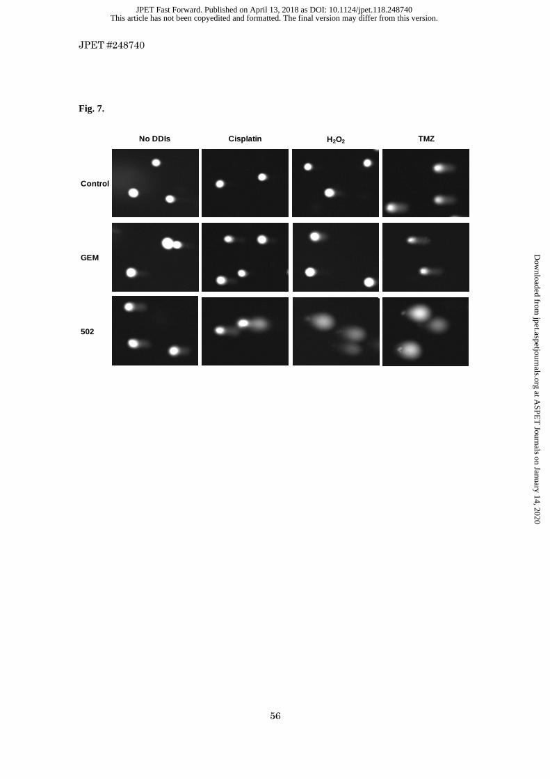
JPET #248740
56
Fig. 7.
No DDIs Cisplatin H2O2 TMZ
Control
GEM
502
This article has not been copyedited and formatted. The final version may differ from this version.JPET Fast Forward. Published on April 13, 2018 as DOI: 10.1124/jpet.118.248740
at ASPE
T Journals on January 14, 2020
jpet.aspetjournals.orgD
ownloaded from