fetal programming of insulin-like growth factor (igf)-i and igf-binding protein-3: evidence for an...
TRANSCRIPT
Fetal programming of insulin-like growth factor (IGF)-I andIGF-binding protein-3: evidence for an altered response toundernutrition in late gestation following exposure topericonceptual undernutrition in the sheep
B W Gallaher, B H Breier, C L Keven, J E Harding andP D GluckmanResearch Centre for Developmental Medicine and Biology, Faculty of Medicine and Health Science, University of Auckland, Auckland, New Zealand
(Requests for offprints should be addressed to B H Breier, Research Centre for Developmental Medicine and Biology, Faculty of Medicine and Health Science,University of Auckland, Private Bag 92019, Auckland, New Zealand)
Abstract
It has been demonstrated in several animal models thatundernutrition in utero has significant long lasting effects onsubsequent fetal and postnatal development. To address thehypothesis that the insulin-like growth factors (IGFs) maymediate such effects, our study examined whether a periodof periconceptual maternal undernutrition could have alasting influence on the IGF axis in the fetal sheep. Eweswere either allowed to feed ad libitum or kept undernour-ished from day 60 prior to mating until day 30 afterconception, and then both groups were allowed to feed adlibitum. These groups were further divided at day 105 ofgestation, either being fed ad libitum or undernourisheduntil day 115 of gestation. Fetal and maternal blood sampleswere obtained at both day 105 and 115 of gestation. Wedescribe the development of a specific homologous RIA tomeasure ovine IGF-binding protein-3 (IGFBP-3) in fetaland maternal sheep plasma. Fetal plasma IGFBP-3 and
IGF-I concentrations were significantly (P<0·05) reducedat day 115 of gestation after maternal undernutrition. Thefetal plasma IGFBP-2 levels were unchanged. The degreeof reduction in fetal plasma IGFBP-3 and IGF-I betweenday 105 and 115 of gestation as a response to acute maternalundernutrition was significantly greater (P<0·05) in fetusesof mothers receiving low periconceptual nutrition. Theresponse of maternal plasma IGFBP-3 and IGF-I to under-nutrition did not depend on the level of periconceptualnutrition. Western blot data indicate that changes in eithermaternal or fetal plasma IGFBP-3 concentrations were notthe result of increased proteolytic activity. These resultssuggest that exposure to maternal periconceptual under-nutrition reprograms IGFBP-3 and IGF-I regulation inthe developing sheep fetus, altering its response to under-nutrition in late gestation.Journal of Endocrinology (1998) 159, 501–508
Introduction
Fetal growth and development is limited both by theintrauterine environment (e.g. nutrient uptake by theplacenta and its availability to the fetus) and by the geneticpotential. In the sheep, inadequate nutrient supply to thefetus (Mellor & Matheson 1979, Owens et al. 1987) leadsto fetal growth retardation. While some consequences offetal undernutrition are transitory, others are observableeven into adulthood. For example, undernutrition in uteroleads to elevated blood pressure in adult guinea pigs(Persson & Jansson 1992) and rats (Woodall et al. 1996b).In humans, poor fetal growth as reflected by factors such aslow birthweight, low fetal/placental weight ratio or areduced neonatal growth rate is associated with anincreased risk of developing adult pathophysiologiessuch as cardiovascular disease and non insulin-dependent
diabetes mellitus (Barker 1997). Such permanent alter-ations of developmental and homeostatic processes havebeen termed ‘fetal reprogramming’ (Barker et al. 1995).
It has long been suggested that insulin-like growthfactor (IGF)-I may play a key role during fetal growth (fora review see Gluckman 1995), since a number of studiesreport a close association between birth weight and plasmaconcentrations of IGF-I. However, the physiologicmechanisms which link fetal growth retardation with adultsequelae have not yet been defined, although alterations inendocrine pathways such as the IGF/IGF-binding protein(IGFBP) axis have been implicated (Barker et al. 1993,Woodall et al. 1997). The role of IGFs in embryonic andfetal development (Baker et al. 1993, Han et al. 1994) andthe sensitivity of IGFs to acute changes in nutritional statushave been well defined (Strauss et al. 1991, Osborn et al.1992, Gallaher et al. 1994).
501
Journal of Endocrinology (1998) 159, 501–508 ? 1998 Society for Endocrinology Printed in Great Britain0022–0795/98/0159–0501 $08.00/0

Harding & Johnston (1995) and Harding (1997)demonstrated that sheep fetuses growing slowly in lategestation did not further slow their growth rate whensubjected to 10 days of severe maternal undernutrition. Itwas proposed that these fetuses, as a result of exposure topericonceptual undernutrition, had adapted to reducednutrient supply which subsequently allowed them tocontinue growth during later periods of maternal under-nutrition. We have reported in a preliminary communi-cation (Gallaher et al. 1995b) that fetal plasma levels ofglucose and insulin are markedly reduced as an acuteresponse to undernutrition in late gestation without anylong-term effects of periconceptional undernutrition.However, periconceptual undernutrition altered theresponse to maternal undernutrition in late gestation inapparent fetal plasma levels of IGFBP-1 analyzed by ligandblot analysis (Gallaher et al. 1995b).
Using specific RIAs for ovine IGFBP-3, IGFBP-2and IGF-I, we examined in the present study whetheradaptation to periconceptual undernutrition causes 1) per-sistent changes in either the fetal or maternal plasma IGFaxis during late gestation, and/or 2) a reprogramming ofthe response of the fetal IGF axis to acute maternalundernutrition in late gestation. Such changes in homeo-static regulatory mechanisms could provide a potentiallink between aberrant fetal growth and associatedpathophysiologies.
Materials and Methods
Experimental design
Coopworth–Border ewes were divided into two groupswhich were either fed ad libitum (AL) or undernourished(UN) for a period of 60 days prior to mating through until30 days after conception (see Harding 1997 for exper-imental details). The undernutrition caused a 10%decrease in ewe body weight over the first 30 days whichwas then maintained for the subsequent 60 days. Betweenday 30 and 90 of gestation, both groups were allowed tofeed ad libitum. Singleton-bearing ewes (AL, n=9; UN,n=12) were then brought into the laboratory and given adiet of concentrates and barley straw and allowed to feedad libitum with free access to water. Ewes and their fetuseswere chronically catheterized on day 95–100 of gestation(Oliver et al. 1992), allowed a 5-day recovery period andthen each periconceptual nutrition group was furtherdivided to receive either AL or UN diets for 10 days untilday 115 of gestation, creating four treatment groups(AL-AL, n=4; AL-UN, n=5; UN-AL, n=5; UN-UN,n=7). During the late period of maternal undernutrition,feed intake was adjusted individually to maintain maternalpreprandial blood glucose concentrations at 1·4–1·6 mM.Blood glucose concentrations in AL-fed ewes were ap-proximately 2·5 mM. The maternal energy intake in thefour different groups during the differential feeding period
from day 105 to 115 of gestation was: AL-AL 15·2&1·7;UN-AL 16·5&2·0, AL-UN 0·6&0·3, UN-UN0·8&0·5 (MJ/day). The detailed experimental proceduresof this study have been described previously (Harding1997). Fetal and maternal blood samples were collected atday 105 and 115 of gestation, placed on ice for no morethan 15 min, centrifuged at 3000 g for 15 min at 4 )C andthe plasma stored at "20 )C. These animal studies wereapproved by the University of Auckland Animal EthicsCommittee.
Measurements
Ovine IGFBP-3 in fetal and maternal plasma wasmeasured by a newly developed homologous oIGFBP-3RIA described below. Immunoblots of oIGFBP-3 wereperformed using antiserum B12 (1:500 dilution) andan alkaline phosphatase detection system (Bio-RadLaboratories, Richmond, CA, USA, see Gallagher et al.1995a for details). IGF-I and IGFBP-2 were measured byRIA as previously described (Breier et al. 1994, Gallagheret al. 1995a).
oIGFBP-3 radioimmunoassay
A semi-purified oIGFBP preparation from adult sheepserum was prepared following SP-Sephadex C25 extrac-tion and IGF-II affinity chromatography (Gallagher et al.1995a). The eluate was loaded onto a Pharmacia HR 5/10C8 reverse phase column and proteins were eluted usinga water–acetonitrile (ACN) gradient. Screening foroIGFBPs was performed by ligand blotting (Gallagheret al. 1992). Fractions containing oIGFBPs were separatedusing a Serva C18 reverse phase column using a 0·1 Mtriethylamine phosphate pH 3–ACN gradient. Fractionscontaining the 39–42 kDa IGFBP doublet were used forraising antibodies, as assay standard or for N-terminalamino acid sequence analysis. Amino acid sequence analy-sis was performed using a gas-phase sequencer (740 A,Applied Biosystems, Foster City, CA, USA) equippedwith an on-line PTH-amino acid analyser (120 A, AppliedBiosystems) with chemicals and the program supplied bythe manufacturer. The 15 N-terminal amino acids of thisprotein (Table 1) showed strong homology with corre-sponding IGFBP-3 sequences from other species (Sprattet al. 1991).
Antisera against oIGFBP-3 were raised in New ZealandWhite rabbits following four immunizations with purifiedoIGFBP-3 (25 µg) conjugated to keyhole limpet hemo-cyanin (Breier et al. 1991) in Freund’s complete adjuvant.After immunoblotting with antiserum B12, a doublet of39–42 kDa was seen with adult sheep plasma and purifiedoIGFBP-3 (Fig. 1). We detected additional low intensitybands at approximately 30 kDa after extended exposure
B W GALLAHER and others · Fetal programming of IGF-I and IGFBP-3502
Journal of Endocrinology (1998) 159, 501–508

which may indicate minor cross-reactivity with otherIGFBPs or represent degraded or deglycosylatedoIGFBP-3.
The 125I-oIGFBP-3 was prepared by iodinating 5 µgpurified oIGFBP-3 with 0·5 mCi 125I using thechloramine-T method (Gallaher et al. 1995a) and purifiedby size separation chromatography. In the RIA, antiserumB12 gave 29% specific binding of 125I-oIGFBP-3 at a finaldilution of 1:4500. Purified oIGFBP-3 for use as a standardand sheep plasma samples were diluted appropriately inassay buffer (0·05 M phosphate pH 7·4, 0·1 M NaCl,0·05% NaN3, 0·1% Triton X-100, 0·2% BSA). Afterpre-incubation of sample (100 µl) with diluted antiserum(50 µl in assay buffer) and recombinant hIGF-I (batchG080AB R9821AX, Genentech Inc, San Francisco, CA,USA; 50 ng/50 µl assay buffer) for 1 h at 20 )C, 125I-
oIGFBP-3 (20 000 c.p.m. in 100 µl assay buffer) wasadded and the assay incubated overnight at 4 )C. Recom-binant hIGF-I was added to correct the potency differencefor the antiserum between free oIGFBP-3 (tracer andstandard curve) and IGF-associated oIGFBP-3 in theplasma samples. Second antibody solution (1 ml 0·5%sheep á-rabbit ã globulin antiserum, 0·1% normal rabbitserum, 5% PEG 6000 in 0·01 M PBS pH 7·4) was addedto each tube and the assay was incubated for 1 h at 20 )Cbefore centrifuging at 3000 g for 30 min at 4 )C. Thepellets were counted by gamma counter.
The homologous oIGFBP-3 RIA exhibited paralleldisplacement between purified oIGFBP-3 and sheepplasma. The ED50 for the assay was 1·5 ng oIGFBP-3/tube and the minimal detectable dose was 5 ng/ml. Theintra- and interassay coefficients of variation were 6 and12% respectively. There was no significant cross-reactivitywith human, porcine or rat serum but strong cross-reactivity with bovine serum (Fig. 2). Further addition of
Table 1 N-terminal amino acid sequence analysis (residues 1–15) of (a) purified ovine IGFBP-3 and (b–d) N-terminal sequences deducedfrom bovine, porcine and human IGFBP-3 cDNA (Spratt et al. 1991). Residues in bold indicate species-specific differences, in comparisonwith the purified ovine IGFBP-3.
1 2 3 4 5 6 7 8 9 10 11 12 13 14 15
a Gly Ala Gly Thr Val Gly Ala Gly Pro Val Val Arg Cys Glu Pro—
b Gly Ala Gly Thr Met Gly Ala Gly Pro Val Val Arg Cys Glu Pro—
c Gly Ser Gly Ala Val Gly Thr Gly Pro Val Val Arg Cys Glu Pro—
d Gly Ala Ser Ser Ala Gly Leu Gly Pro Val Val Arg Cys Glu Pro
Figure 1 Specificity of antiserum B12 for oIGFBP-3-screening ofadult sheep plasma (lanes 1, 3 and 5) and purified oIGFBP-3(lanes 2, 4 and 6) using (A) ligand or Western blot with(B) antiserum B12 and (C) non-immune serum.
Figure 2 The oIGFBP-3 RIA was assessed for sensitivity andparallelism of ovine plasma relative to purified oIGFBP-3 andcross-reactivity with plasma from other species. The displacementcurves are as follows: purified oIGFBP-3 5, ovine plasma ,,human plasma ., bovine plasma /, rat plasma 1, porcineplasma -. Each point is the mean of duplicates expressed aspercentage bound (% B/Bo) corrected for non-specific binding.Buffer-diluted plasma volumes are expressed in terms of neatplasma volume equivalents.
Fetal programming of IGF-I and IGFBP-3 · B W GALLAHER and others 503
Journal of Endocrinology (1998) 159, 501–508
IGF-I, IGF-II or heparin to control sheep plasma samplesdid not affect the values obtained (Table 2).
Statistical analysis
Differences between day 105 and 115 of gestation andbetween the treatment groups for IGFBP-2, IGFBP-3and IGF-I were analyzed by two way repeated measuresANOVA followed by Neuman–Keuls post-hoc test(Sigmastat, Jandel Scientific, Chicago, IL, USA).IGFBP-3 and IGF-I values for each animal on day 115 ofgestation were also expressed as a ratio over the corre-sponding day 105 value to eliminate the effect of baselinevariation at day 105 of gestation. Changes in peptideconcentrations between day 105 and 115 of gestation werethen analyzed for inter-group differences by one wayANOVA followed by Scheffe’s test (Sigmastat, JandelScientific). Statistical significance was accepted at P<0·05.Data are expressed as means&...
Results
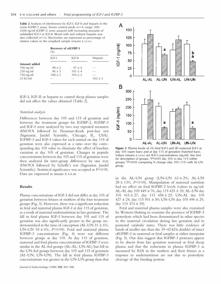
Plasma concentrations of IGF-I did not differ at day 105 ofgestation between fetuses or mothers of the four treatmentgroups (Fig. 3). However, there was a significant reductionin fetal and maternal plasma IGF-I at day 115 of gestation,as a result of maternal undernutrition in late gestation. Thefall in fetal plasma IGF-I between day 105 and 115 ofgestation was also significantly greater in the group un-dernourished at the time of conception (AL-UN 31&6%;UN-UN 54&6%, P<0·05). Fetal and maternal plasmaIGFBP-3 concentrations (Fig. 4) were not differentbetween groups at day 105. At day 115 of gestation,maternal and fetal plasma concentrations of IGFBP-3 weresimilar in the AL-fed groups (AL-AL; UN-AL) but fell inthe UN-fed groups between day 105 and 115 of gestation(AL-UN; UN-UN). The fall in fetal plasma IGFBP-3concentrations was greater in the UN-UN group than that
in the AL-UN group (UN-UN 63&2%; AL-UN28&13%, P<0·05). Manipulation of maternal nutritionhad no effect on fetal IGFBP-2 levels (values in ng/ml:AL-AL day 105 449&76, day 115 425&35; AL-UN day105 415&27, day 115 458&22; UN-AL day 105427&24, day 115 501&50; UN-UN day 105 496&29,day 115 474&39).
Fetal and maternal plasma samples were also examinedby Western blotting to examine the presence of IGFBP-3proteolysis which had been demonstrated in other speciesin the maternal circulation during late gestation and inpostnatal catabolic states. There was little evidence ofbands of smaller size than the 39–42 kDa doublet of intactoIGFBP-3 in maternal or fetal samples at either timepoint(Fig. 5). Our data suggest that IGFBP-3 proteases appearto be absent from late gestation maternal or fetal sheepplasma and that the reductions in plasma IGFBP-3 asmeasured by RIA in the mothers and their fetuses as aresponse to undernutrition are not due to proteolyticcleavage of this binding protein.
Table 2 Analysis of interference by IGF-I, IGF-II and heparin in theovine IGFBP-3 assay. Serum control pools (n=4; range: 500–2500 ng/ml IGFBP-3) were assayed with increasing amounts ofunlabelled IGF-I or IGF-II. Blood with and without heparin wasalso collected (n=2). Recoveries are expressed as percentage ofrelative values to the unspiked sample (means&S.E.M.).
Recovery of oIGFBP-3(%)
IGF-I IGF-II Heparin
Amount added100 ng/ml 98&2 97&4 —250 ng/ml 96&5 101&4 —750 ng/ml 100&3 105&2 —25 IU/ml — — 102&5
Figure 3 Plasma levels of (A) fetal IGF-I and (B) maternal IGF-I atday 105 (open bars) and at day 115 of gestation (hatched bars).Values (means&S.E.M.) are IGF-I concentrations (ng/ml). See textfor description of groups. aP<0·05 day 105 vs day 115 withingroups; bP<0·05 comparing % change (day 105–115) with AL-UNgroup.
B W GALLAHER and others · Fetal programming of IGF-I and IGFBP-3504
Journal of Endocrinology (1998) 159, 501–508

Discussion
This paper reports an alteration in regulation of the IGF-Iand IGFBP-3 in the developing fetal sheep followingexposure of the mother to periconceptual undernutrition.While basal IGF-I and IGFBP-3 concentrations in bothmaternal and fetal sheep plasma during late gestation werenot altered, periconceptual undernutrition of the eweamplified the reduction of IGF-I and IGFBP-3 in fetalplasma in response to 10 days of maternal undernutritionin late gestation. Our results therefore suggest that peri-conceptual undernutrition may reprogram (persistentlyalter the normal developmental pattern and homeostaticregulatory mechanisms) the fetal IGF-I and IGFBP-3system in its ability to respond to acute changes insubstrate supply. This reprogramming of IGF-I andIGFBP-3 during fetal development may be of particularphysiologic significance for the long-term regulation offuel homeostasis, since the fetal response to late gestationmaternal undernutrition in plasma glucose and insulin was
not altered by periconceptual maternal undernutrition(Gallagher et al. 1995b).
Whether other members of the IGF axis in thefetal sheep are similarly reprogrammed by maternal peri-conceptual undernutrition awaits further investigations.We found no evidence for long-term consequences ofpericonceptual maternal undernutrition on fetal plasmaconcentrations of IGFBP-2 (present study). However,preliminary data from this laboratory (Gallagher et al.1995b) using ligand blot analysis, would suggest that theincrease in fetal plasma IGFBP-1 as a response to acutematernal undernutrition in late gestation may similarly bealtered by reprogramming during early fetal life (Gallagheret al. 1995b).
The physiologic significance of reprogramming of fetalIGF-I and IGFBP-3 is not clearly defined at present.Harding (1997) measured fetal growth rates in vivo in thefirst part of this study using growth-measuring devices,which had been surgically placed at day 95 of gestation.Her study showed that maternal undernutrition from 60days before until 30 days after conception resulted infetuses with relatively slow growth between day 98 and105. Undernutrition in late gestation (day 105–115) mark-edly reduced fetal growth rate in the AL-UN group, butdid not change the already slow growth in the UN-UNgroup. This slower rate of fetal growth in the UN-UNgroup during late pregnancy appeared to partially protectthe fetus against further reduction in growth and against arelative increase in heart, kidney and liver size caused by10 days of severe maternal undernutrition in late gestation(Harding 1997). It is therefore tempting to speculate thatthe amplified reduction in fetal plasma IGF-I andIGFBP-3 during late gestation maternal undernutrition (inthe UN-UN group at day 115) may be an adaptivemechanism of the reprogrammed fetal somatotropic axiswhich partly protects the fetus against a further slowing ofgrowth and a relative over-growth of the heart, kidney andliver. The amplified reduction in fetal plasma IGF-I andIGFBP-3 in response to acute maternal undernutrition inlate gestation in the UN-UN group may reflect alterationsin hepatic synthesis or clearance induced by maternalpericonceptual undernutrition.
Reprogramming of postnatal endocrine parameters as aresult of endocrine manipulations during fetal develop-ment has already been described. For example, passiveimmunization of the fetal rat against growth hormone(GH)-releasing hormone permanently alters adult GHsecretion (Cella et al. 1994). Muaku et al. (1996) showed inthe rat that offspring from mothers fed on a low proteindiet throughout gestation are growth retarded at birth andhave reduced plasma levels of IGF-I postnatally until theyreach puberty. Our own studies in the rat showed signifi-cant growth retardation at birth and reduced postnatalgrowth and plasma concentrations of IGF-I in offspring ofmothers given a nutritionally-balanced diet and allowed tofeed just 30% ad libitum (Woodall et al. 1996a). However,
Figure 4 Plasma levels of (A) fetal ovine IGFBP-3 or (B) maternalovine IGFBP-3 at day 105 (open bars) and at day 115 of gestation(hatched bars). Values (means&S.E.M.) are IGFBP-3 concentrations(ng/ml). See text for description of groups. aP<0·05 day 105 vsday 115 within group; bP<0·05 comparing % change (day105–115) with AL-UN group.
Fetal programming of IGF-I and IGFBP-3 · B W GALLAHER and others 505
Journal of Endocrinology (1998) 159, 501–508

in this model of fetal growth retardation plasma IGF-Iconcentrations had recovered at weaning, but we observedsignificant hypertension during adult life (Woodall et al.1996b).
The present study provides the first example of repro-gramming of the somatotropic axis during fetal life causedby manipulation of maternal nutrition. A related phenom-enon has been previously suggested in fetal sheep. Theeffect of maternal starvation in late gestation on ovineplacental lactogen (oPL) concentrations in fetal plasmadiffers depending on the level of maternal nutritionimmediately prior to starvation (Oliver et al. 1992). Themechanisms responsible for the amplified reduction of fetalplasma IGF-I and IGFBP-3 in response to the maternalundernutrition in late gestation in the UN-UN group areunclear. One possible explanation may relate to the effectsof periconceptual undernutrition on fetal responsiveness tocorticosteroids in late gestation. Although we did notmeasure cortisol in this study, it is known that maternalundernutrition can elevate cortisol in the late-gestationfetal sheep (Binienda et al. 1990) and that cortisol exposurecan increase blood pressure (Tangalakis et al. 1992) in thefetal sheep. Blood pressure was elevated in fetuses of ourstudy (Harding & Johnston 1995). Corticosteroid-inducedgrowth retardation of the fetal rat also lowers IGFBP-3expression and plasma IGF-I (Zhou-Li et al. 1991, Priceet al. 1992). The cortisol secretory response to hypoxemiadecreases with advancing gestational age (Akagi & Challis1990). Any delay in this maturation process caused by
exposure to periconceptual undernutrition could then leadto a greater fetal cortisol response to undernutrition in lategestation and therefore a greater decrease in IGF-I andIGFBP-3 seen in the UN-UN group. Whether alteredplasma cortisol levels are causally related to reprogrammingof the fetal IGF axis awaits further investigations.
This paper also reports the development of a homolo-gous RIA for oIGFBP-3 and its use in examining theeffects of periconceptual undernutrition on the maternaland fetal IGF axes in the late gestation sheep. TheoIGFBP-3 39–42 kDa doublet, purified from adult sheepserum by IGF-II affinity chromatography and reversephase chromatography, showed significant N-terminalsequence homology with equivalent cDNA-derivedsequences for bovine, porcine and human IGFBP-3(Spratt et al. 1991). The antiserum raised against thisprotein was specific for oIGFBP-3. Both fetal and post-natal plasma samples exhibited parallel displacement topurified oIGFBP-3 in the RIA with no interference fromIGFs or heparin.
Of interest is the observation that, when analyzed byspecific RIA, IGFBP-3 concentrations were not sup-pressed in day 105 fetal plasma in groups whose mothersreceived periconceptual undernutrition, in contrast to theresults of earlier studies from this laboratory using ligandblotting where apparent IGFBP-3 values were reduced atday 105 in the UN-AL and UN-UN groups (Gallagheret al. 1995b). This discrepancy reinforces previous debateindicating that ligand blot and immunologically based
Figure 5 Representative oIGFBP-3 Western blots of (A) fetal and (B) maternal plasma from each group (see text for description) at day105 (odd-numbered lanes) and day 115 (even-numbered lanes) of gestation.
B W GALLAHER and others · Fetal programming of IGF-I and IGFBP-3506
Journal of Endocrinology (1998) 159, 501–508
analyses can produce disparate results (Gargosky et al.1992), reflecting the different properties on which themethodology is based, either immunoreactive peptide orIGF binding activity. A functional difference such asreduced affinity for the IGFs by IGFBP-3 in fetusesfrom the periconceptual undernutrition groups (UN-AL,UN-UN) may explain the apparent suppression ofIGFBP-3 on ligand blots at day 105 gestation. While thiscould potentially be achieved by the induction ofIGFBP-3 protease activity following periconceptualmaternal undernutrition, our Western blotting analysisrevealed minimal levels of low molecular weightimmunoreactive IGFBP-3 in all experimental groups atday 105 gestation, implying that IGFBP-3 is intact and ofnormal size in fetal sheep plasma. IGFBP-3 proteaseactivity is also not clearly demonstrable in mid-to-lategestation fetal plasma of some other species (Lewitt et al.1995). The lack of degradation products at day 115 in fetalplasma following severe undernutrition also suggests thatinduction of IGFBP-3 protease activity is not respon-sible for the greater fall in immunoreactive IGFBP-3plasma levels in the UN-UN fetuses. Further analysisusing IGFBP-3 proteolysis assays would confirm thisobservation.
We were not able to demonstrate any changes inresponsiveness to late gestation undernutrition in maternalplasma IGF-I and IGFBP-3. However, we did note inWestern blots that IGFBP-3 in late gestation maternalsheep plasma was essentially intact which is in contrast toother species such as man and rodents (Lewitt et al. 1995)where little IGFBP-3 is intact at this stage of pregnancy.Such species specific differences could reflect differencesin placental structure between these species. Sincematernal plasma IGF-I is a key element in enhancingplacental function in the sheep (Liu et al. 1994), the eweis likely to have alternative means of increasing IGF-Iavailability to the placenta.
Several postnatal endocrine consequences have alreadybeen ascribed to the effects of growth retardation in utero.Humans who are born small exhibit an altered glucosemetabolism such that, as adults, they have an increased riskof developing non-insulin dependent diabetes (Hales et al.1991). Rats, growth retarded in utero, were unresponsiveto GH treatment between day 11 and 21 postnatally butnot after puberty, suggesting the presence of transient GHresistance during the neonatal period due to the intra-uterine growth retardation (Woodall et al. 1997). Thepostnatal consequences of changes in the fetal IGF axisdescribed in this study are not known but they do supportthe hypothesis that nutritional reprogramming of the fetalIGF axis during early gestation might establish alteredhomeostatic patterns to subsequent nutritional limitationwhich could then contribute to postnatal pathophysiology.Further studies are clearly required to examine anypostulated changes in postnatal growth and/or thedevelopment of the somatotropic axis in offspring of
mothers who experienced major nutritional restrictionduring the periconceptual period.
Acknowledgements
This work was supported by the Health Research Councilof New Zealand and the National Child Health ResearchFoundation. The authors thank Dr David Christie, Schoolof Biological Sciences, University of Auckland, for per-forming the oIGFBP-3 N-terminal amino acid sequenceanalysis.
References
Akagi K & Challis JR 1990 Threshold of hormonal and biophysicalresponses to acute hypoxemia in fetal sheep at different gestationalages. Canadian Journal of Physiology and Pharmacology 68 549–555.
Baker J, Liu J, Robertson EJ & Efstratiadis A 1993 Role of insulin-likegrowth factors in embryonic and postnatal growth. Cell 75 73–82.
Barker DJP 1997 Fetal undernutrition and adult disease. Endocrinologyand Metabolism 4 (Suppl. B) 39–46.
Barker DJP, Gluckman PD, Godfrey KM, Harding JE, Owens JA &Robinson JS 1993 Fetal nutrition and cardiovascular disease in adultlife. Lancet 341 938–941.
Barker DJP, Gluckman PD & Robinson JS 1995 Conference report:fetal origins of adult disease: report of the First International StudyGroup, Sydney, 29–30 October 1994. Placenta 16 317–320.
Binienda Z, Rosen ED, Kelleman A, Sadowsky DW, Nathanielsz PW& Mitchell MD 1990 Maintaining fetal normoglycemia preventsthe increase in myometrial activity and uterine 13,14-dihydro-15-keto-prostaglandin F2 alpha production during food withdrawal inlate pregnancy in the ewe. Endocrinology 127 3047–3051.
Breier BH, Gallaher BW & Gluckman PD 1991 Radioimmunoassayfor insulin-like growth factor-I: solutions to some potential problemsand pitfalls. Journal of Endocrinology 128 347–357.
Breier BH, Ambler GR, Sauerwein H, Surus A & Gluckman PD1994 The induction of hepatic somatotrophic receptors after birthin sheep is dependent on parturition associated mechanisms. Journalof Endocrinology 141 101–108.
Cella SG, Locatelli V, Broccia ML, Meregola E, Giavini E, DeGennaro Colonna V, Torsello A, Wehrenberg WB & Muler EE1994 Long term changes in somatotrophic function induced bydeprivation of growth hormone releasing hormone during the fetallife of the rat. Journal of Endocrinology 140 111–117.
Gallaher BW, Breier BH, Oliver MH, Harding JE & Gluckman PD1992 Ontogenic differences in the nutritional regulation ofcirculating IGF binding proteins in sheep plasma. ActaEndocrinologica 126 49–54.
Gallaher BW, Oliver MH, Eichhorn K, Kessler U, Kiess W, HardingJE, Gluckman PD & Breier BH 1994 The circulatingIGF-II/mannose-6-phosphate receptor and insulin-like growthfactor binding proteins in fetal sheep plasma are regulated byglucose and insulin. European Journal of Endocrinology 131 398–404.
Gallaher BW, Breier BH, Blum WF, McCutcheon SN & GluckmanPD 1995a An homologous radioimmunoassay for ovine insulin-likegrowth factor binding protein 2 (oIGFBP-2): ontogenesis and theresponse to growth hormone, placental lactogen and IGF-Itreatment. Journal of Endocrinology 144 75–82.
Gallaher BW, Breier BH, Harding JE & Gluckman PD 1995bPericonceptual undernutrition resets plasma IGFBP levels and altersthe response of IGFBP-1, IGFBP-3 and IGF-I to subsequentmaternal undernutrition in the sheep. Progress in Growth FactorResearch 6 189–195.
Fetal programming of IGF-I and IGFBP-3 · B W GALLAHER and others 507
Journal of Endocrinology (1998) 159, 501–508

Gargosky SE, Pham HM, Wilson KF, Liu F, Giudice LC &Rosenfeld RG 1992 Measurement and characterization ofinsulin-like growth factor binding protein-3 in human biologicalfluids: discrepancies between radioimmunoassay and ligand blotting.Endocrinology 131 3051–3060.
Gluckman PD 1995 The endocrine regulation of fetal growth in lategestation: the role of insulin-like growth factors. Journal of ClincalEndocrinology and Metabolism 80 1047–1050.
Hales CN, Barker DJ, Clark PM, Cox LJ, Fall C, Osmond C &Winter PD 1991 Fetal and infant growth and impaired glucosetolerance at age 64. British Medical Journal 303 1019–1022.
Han VKM, Asano H, Matsell DG, Debhumty PJD, Hayatsu J, BassettN, Phillips ID, Nygard K & Stepaniuk O 1994 Insulin-like growthfactors (IGFs) and IGF binding protein (IGFBPs) in fetal develop-ment: physiology and pathophysiology. In The Insulin-like GrowthFactors and their Regulatory Proteins, pp 263–273. Eds RC Baxter,PD Gluckman & RG Rosenfeld. Amsterdam: Elsevier.
Harding JE 1997 Periconceptual nutrition determines the fetal growthresponse to acute maternal undernutrition in fetal sheep of lategestation. Prenatal and Neonatal Medicine 2 310–319.
Harding JE & Johnston BM 1995 Nutrition and fetal growth.Reproduction, Fertility and Development 7 539–547.
Lewitt MS, Scott FP, Clarke NM & Baxter RC 1995 Developmentalregulation of circulating insulin-like growth factor binding proteinsin normal pregnancies and in pre-eclampsia. Progress in GrowthFactor Research 6 475–480.
Liu L, Harding JE, Evans PC & Gluckman PD 1994 Maternalinsulin-like growth factor-I infusion alters feto–placentalcarbohydrate and protein metabolism in pregnant sheep.Endocrinology 135 895–900.
Mellor DJ & Matheson IC 1979 Daily changes in the curvedcrown-rump length of individual sheep fetuses during the last60 days of pregnancy and effects of different levels of maternalnutrition. Quarterly Journal of Experimental Physiology 64119–131.
Muaku SM, Beauloye V, Thissen JP, Underwood LE, Fossion C,Gerard G, Ketelslegers JM & Maiter D 1996 Long-term effects ofgestational protein malnutrition on postnatal growth, insulin-likegrowth factor (IGF)-I, and IGF-binding proteins in rat progeny.Pediatric Research 39 649–655.
Oliver MH, Harding JE, Breier BH, Evans PC & Gluckman PD 1992The nutritional regulation of circulating placental lactogen in fetalsheep. Pediatric Research 31 520–523.
Osborn BH, Fowlkes J, Han VKM & Freemark M 1992 Nutritionalregulation of insulin-like growth factor-binding protein gene
expression in the ovine fetus and the pregnant ewe. Endocrinology131 1743–1750.
Owens JA, Falconer J & Robinson JS 1987 Effect of restriction ofplacental growth on fetal and utero–placental metabolism. Journal ofDevelopmental Physiology 9 225–238.
Persson E & Jansson T 1992 Low birth weight is associated withelevated adult blood pressure in the chronically catheterised guineapig. Acta Physiologica Scandinavica 145 195–196.
Price WA, Stiles AD, Moats-Staats BM & D’Ercole AJ 1992 Geneexpression of insulin-like growth factors (IGFs), the type I IGFreceptor, and IGF-binding proteins in dexamethasone-induced fetalgrowth retardation. Endocrinology 130 1424–1432.
Strauss DS, Ooi GT, Orlowski CC & Rechler MM 1991 Expressionsof the genes for IGF-I, IGF-II and IGFBP-1 and -2 in the fetal ratunder conditions of intra-uterine growth retardation caused bymaternal fasting. Endocrinology 128 518–525.
Spratt SK, Tatsuno GP & Sommer A 1991 Cloning andcharacterization of bovine insulin-like growth factor bindingprotein-3 (bIGFBP-3). Biochemical and Biophysical ResearchCommunications 177 1025–1032.
Tangalakis K, Lumbers ER, Moritz KM, Towstoless MK & WintourEM 1992 Effect of cortisol on blood pressure and vascular reactivityin the ovine fetus. Experimental Physiology 77 709–717.
Woodall SM, Breier BH, Johnston BM & Gluckman PD 1996a Amodel of intrauterine growth retardation caused by chronicmaternal undernutrition in the rat: effects on the somatotrophic axisand postnatal growth. Journal of Endocrinology 150 231–242.
Woodall SM, Johnston BM, Breier BH & Gluckman PD 1996bChronic maternal undernutrition in the rat leads to delayedpostnatal growth and elevated blood pressure of offspring. PediatricResearch 40 438–443.
Woodall SM, Breier BH, Johnston BM & Gluckman PD 1997Reduced maternal nutrition during gestation in the rat leads totemporary growth hormone resistance during the neonatal period.Proceedings of the 79th Annual Meeting of the Endocrine Society P1.123.
Zhou-Li F, Joly-Pharaboz MO, Bouillard B, Albaladejo V, Nicolas B& Andre J 1991 Multihormonal control of cell proliferation:opposite effects of two stimulators (17 beta-estradiol andL-triiodothyronine) and one inhibitor (dexamethasone) on F4Z2pituitary tumor cells. Endocrinology 128 2761–2768.
Received 9 December 1997Accepted 8 July 1998
B W GALLAHER and others · Fetal programming of IGF-I and IGFBP-3508
Journal of Endocrinology (1998) 159, 501–508