female pelvic viscera
TRANSCRIPT

Female Pelvic
Viscera
Khaleel Alyahya, PhD, MedKing Saud University
@khaleelya

Khaleel Alyahya, PhD, MEd

Pelvic Viscera
Pelvic Colon (Sigmoid Colon)
lies in the upper and posterior part of the pelvic cavity
Rectum
lies in the lower and posterior part of the pelvic cavity .
Anal Canal
begins at the lower end of the rectum, directeddownward and backward.
Uterus
projects between rectum and urinary bladder
its free end (fundus) overhangs the urinary bladder
its lower end (cervix) opens into the vagina
Vagina
descends between the bladder and urethra anteriorlyand the rectum and anal canal posteriorly.
Urinary Bladder
lies in the lower and anterior part of the pelvic cavity,behind the symphysis pubis.
Khaleel Alyahya, PhD, MEd

Pelvic Peritoneum
The peritoneum on the dorsal wall ofthe pelvis is reflected, to form themedial limb of pelvic mesocolon.
The rectum has partial covering;
the upper third on front andsides.
the middle third only in front.
no covering for the lower third.
From the sides of the rectum, to theside wall of the pelvis forms the floorof the pararectal fossae.
From front of the rectum, it reflects onthe upper part of posterior wall ofvagina, forming the rectovaginalpouch.
Khaleel Alyahya, PhD, MEd

Pelvic Peritoneum
From back of vagina, to the posteriorsurface of uterus, then over the fundusto cover its anterior surface.
From anterior surface of the uterus atjunction with cervix, on to superiorsurface of the bladder (utero-vesical)pouch.
From upper surfaces of the bladder, itcovers the upper part of the lateralsurface, then extends laterally to theside wall of the pelvis forming theparavesical fossae.
From upper surfaces of the bladder, itreflects anteriorly to line the posterioraspect of the anterior abdominal wall.
Khaleel Alyahya, PhD, MEd

Pelvic Viscera
The sigmoid colon
It is the continuation of thedescending colon at pelvicbrim.
It ends at middle piece ofsacrum continuous with therectum.
Khaleel Alyahya, PhD, MEd

Rectum
It is a continuation of the sigmoidcolon and ends at recto analangle to be continuous with analcanal.
Related anteriorly to rectovaginalpouch and posterior wall ofvagina, and posteriorly to sacrum,sacral vessels and sympathetictrunks.
Khaleel Alyahya, PhD, MEd

Uterus
The uterus is a hollow, thick-walled,muscular organ
Situated deeply in the pelvic cavitybetween the bladder and rectum.
Into its upper part, the uterine tubesopen, one on either side.
Below, its cavity communicates withthat of the vagina.
Khaleel Alyahya, PhD, MEd
Uterus
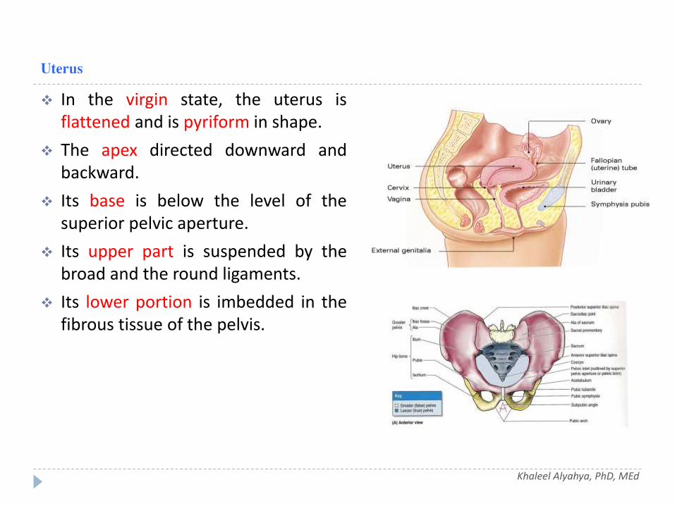
In the virgin state, the uterus isflattened and is pyriform in shape.
The apex directed downward andbackward.
Its base is below the level of thesuperior pelvic aperture.
Its upper part is suspended by thebroad and the round ligaments.
Its lower portion is imbedded in thefibrous tissue of the pelvis.
Khaleel Alyahya, PhD, MEd

Uterus
the isthmus, (a slight constriction,corresponding to the internal os),divides the uterus into;
The portion above theisthmus is the body, and thatbelow it is the cervix.
The part of the body whichlies above a plane passingthrough the points ofentrance of the uterine tubesis known as the fundus.
Khaleel Alyahya, PhD, MEd

Fundus
The fundus is convex in all directions.
It is covered by peritoneum continuouswith that on the vesical and intestinalsurfaces.
It is related to some coils of smallintestine, and occasionally the distendedsigmoid colon.
Khaleel Alyahya, PhD, MEd

Body
Gradually narrows from the fundus tothe isthmus.
The vesical or anterior surface isflattened and covered by peritoneum,which is reflected on to the bladder toform the vesicouterine pouch .
The intestinal or posterior surface isconvex transversely and is covered byperitoneum, which is continued down onto the cervix and vagina.
Khaleel Alyahya, PhD, MEd

Body
The lateral margins are slightlyconvex.
The uterine tube pierces the uterinewall, at the upper end of each margin.
Below and in front of this point theround ligament of the uterus is fixed,while behind it is the attachment ofthe ligament of the ovary.
These three structures lie within thetwo layers of the broad ligament.
Khaleel Alyahya, PhD, MEd

Cervix
Is the lower constrictedsegment of the uterus.
It is conical in shape, with itstruncated apex directeddownward and backward, but isslightly wider in the middle.
It is less freely movable than thebody.
It projects through the anteriorwall of the vagina, which dividesit into an upper, supravaginalportion, and a lower, vaginalportion.
Khaleel Alyahya, PhD, MEd

Cervix
The supravaginal portion is separatedin front from the bladder by fibroustissue (parametrium), which extendsbetween the layers of the broadligaments.
The uterine arteries reach the marginsof the cervix.
Posteriorly, it is covered byperitoneum, which is prolonged belowon to the posterior vaginal wall, whenit is reflected on to the rectum,forming the rectouterine pouch.
It is in relation with the rectum, fromwhich it may be separated by coils ofsmall intestine.
Khaleel Alyahya, PhD, MEd

Cervix
The vaginal portion of the cervixprojects free into the anterior wallof the vagina between the anteriorand posterior fornices.
On its rounded extremity is a small,depressed, circular aperture, theexternal orifice of the uterus,through which the cavity of thecervix communicates with that ofthe vagina.
The external orifice is bounded bytwo lips, an anterior and a posterior,which lie in contact with theposterior vaginal wall.
Khaleel Alyahya, PhD, MEd

Ligaments of Uterus
The ligaments of the uterus are eight innumber: one anterior; one posterior;two lateral or broad; two uterosacral;and two round ligaments.
The anterior ligament consists of thevesicouterine fold of peritoneum, whichis reflected on to the bladder from thefront of the uterus, at the junction of thecervix and body.
The posterior ligament consists of therectovaginal fold of peritoneum, which isreflected from the back of the posteriorfornix of the vagina on to the front ofthe rectum.
Khaleel Alyahya, PhD, MEd

Ligaments of Uterus
The two lateral or Broad ligaments pass from thesides of the uterus to the lateral walls of thepelvis. Together with the uterus they form aseptum across the female pelvis, dividing thatcavity into two portions.
In the anterior part is contained the bladder
in the posterior part the rectum
The Round ligaments are two flattened bandsbetween 10 and 12 cm. in length.
Situated between the layers of the broadligament in front of and below the uterinetubes.
The round ligaments consists of muscular tissue,fibrous and blood vessels, lymphatics; andnerves, enclosed in a fold of peritoneum.
Khaleel Alyahya, PhD, MEd

Position of Uterus
The long axis of the uterus usuallylies approximately in the axis of thesuperior pelvic aperture.
Position of the uterus in relation tothe vagina;
The body is ante-flexed on thecervix, and the whole uterus isante-verted on the vagina .
Khaleel Alyahya, PhD, MEd

Uterine Tube
One on each side of the uterus, run inthe free border of the broad ligament.
Its medial end opens into the superiorangle of the uterine cavity.
The lateral end opens into theperitoneal cavity close to the ovaries,surrounded by fimbriae.
It is divided into; infundibulum,ampulla, isthmus and intramural parts.
Khaleel Alyahya, PhD, MEd

Ovaries
One on each side of the uterus, run inthe free border of the broadligament,
Its medial end opens into the superiorangle of the uterine cavity.
The lateral end opens into theperitoneal cavity close to the ovaries,surrounded by fimbriae.
It is divided into; infundibulum,ampulla, isthmus and intramuralparts.
Khaleel Alyahya, PhD, MEd

Ovaries
The tubal end is near the externaliliac vein; to it are attached thesuspensory ligament of the ovary,which contains the ovarian vessels.
The uterine end is directeddownward toward the pelvic floor,is attached to the lateral angle ofthe uterus, by the ligament of theovary, which lies within the broadligament.
Khaleel Alyahya, PhD, MEd

Ovaries
The lateral surface is in contactwith the parietal peritoneum, ofthe ovarian fossa.
the medial surface is covered bythe fimbriated end of the uterinetube.
Khaleel Alyahya, PhD, MEd

Vagina
Strong muscular canal, (7.5cm)
It connects the uterus with the vestibuleof the external genitalia,
Its long axis is almost parallel with that ofthe lower part of the sacrum.
Its anterior wall is 1.5- 2cm shorter thanthe posterior wall.
Relations;
Anterior wall directly related to thebladder base.
Posterior wall is related to rectovaginalpouch, rectum, its lower part separatedfrom the anal canal by the perineal body.
Laterally related to ureters and uterinevessels .
Khaleel Alyahya, PhD, MEd

Urinary Bladder
It lies in the lower and anterior part of thepelvis.
It is a three sided pyramid.
Its apex; is directed anteriorly.
the base; lies posteriorly, directly relatedto the cervix and anterior wall of vagina.
It rests on its neck inferiorly, where it iscontinuous with the urethra.
Khaleel Alyahya, PhD, MEd

Urinary Bladder
It has three surfaces; superior and twoinferolateral.
Superior surface is related to anterior wallof uterus.
Inferolateral surfaces; are related to retro-pubic fat which separates them frompubis, muscle wall , obturator vessels andnerve.
The neck, is directly continuous withurethra and related to anterior wall ofvagina.
Khaleel Alyahya, PhD, MEd

Pelvic Part of Ureters
Enter the pelvis by crossing theends of common iliac arteries.
It passes below the broad ligamenton reaching the bladder.
Khaleel Alyahya, PhD, MEd

Urethra
It is 4 cm long,
Begins at neck of bladder close to theanterior wall of vagina.
It opens into the vestibule in front ofvaginal orifice.
Khaleel Alyahya, PhD, MEd

QUESTIONS?