feline hypertrophic cardiomyopathy · • normal vertebral heart score 7.5 ± 0.3 (litster and...
TRANSCRIPT

Feline Hypertrophic Cardiomyopathy
An Update Agnieszka Kent, DVM, MS DACVIM
(Cardiology) Bayer Conference
Centre Vétérinaire DMV January, 2014

Hypertrophic Cardiomyopathy
• Concentric hypertrophy of the left ventricle • Rule out other causes
• Hyperthyroidism
• Systemic hypertension
• Aortic stenosis
• Acromegaly
• Myocardial disease • Defect of the sarcomere
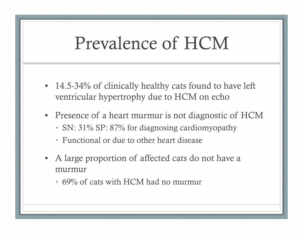
Prevalence of HCM
• 14.5-34% of clinically healthy cats found to have left ventricular hypertrophy due to HCM on echo
• Presence of a heart murmur is not diagnostic of HCM • SN: 31% SP: 87% for diagnosing cardiomyopathy
• Functional or due to other heart disease
• A large proportion of affected cats do not have a murmur • 69% of cats with HCM had no murmur

Predisposed Breeds
• Maine coon • MYBPC3 mutation (autosomal dominant trait)
• Incomplete penetrance and variable expressivity • Not all cats with mutation will show disease
• Homozygous cats more likely to show disease and have severe form
• Ragdoll • MYBPC mutation different from Maine coon
• Homozygous cats very severely affected
• Can develop early onset severe disease • Mean age at diagnosis of 15 months (Lefbom BK, et al. 2001)
• Persian, Himalayan, Birman, Bengal, Sphynx
• Most commonly diagnosed breed • Domestic shorthair
North Carolina State University College of Veterinary Medicine www.ncstatevets.org/genetics

Pathophysiology
• Left ventricular hypertrophy
• Diastolic dysfunction • Decreased ability of the left ventricle to fill with blood during
relaxation and passive ventricular filling • Increased ventricular stiffness
• Ventricular filling pressures become increased • Increased left atrial pressures • Increased pulmonary venous pressures
• If > ~25 mmHg cardiogenic edema develops
• High heart rates can worsen diastolic dysfunction
• End-stage HCM (“burnt-out” HCM) • Systolic dysfunction, LV dilation, wall thinning

Physical Examination
• Cardiac auscultation • Heart murmur – most often systolic and dynamic
• Does not predict severity of disease
• Gallop sound – Not an arrhythmia!
• Arrhythmia – premature beats, irregular rhythm

Physical Examination
• Jugular veins • Distention, pulsations
• Arterial Pulses • Strong, weak, present?
• Synchronous with heart sounds
• Pulse deficits
• Thoracic auscultation • Crackles, wheezes, absence of lung sounds

How many of you automatically recommend
referral to a cardiologist upon detection of a heart murmur
or gallop?

What you can do in a general practice
• Thoracic radiographs
• Electrocardiogram
• NT-pro-BNP (Idexx Cardiopet)
• Rule out common causes of left ventricular hypertrophy • Total T4 (cats > 7 years)
• Blood pressure

Thoracic Radiographs
• Used for assessing • Overt cardiomegaly
• May be normal in mild to moderate HCM • Normal vertebral heart score 7.5 ± 0.3 (Litster and Buchanan 2000)
• Pulmonary vascular changes • Pulmonary parenchyma
• Congestive heart failure versus primary respiratory disease
• Limitations • Cannot provide a specific diagnosis • Cardiac disease can be missed
• Cannot evaluate risk for thromboembolism

Mild HCM

Mild HCM

What to look for to diagnose congestive heart failure
• Cardiomegaly • Left atrial/auricular enlargement*
• Pulmonary venous congestion*
• Pulmonary infiltrates consistent with cardiogenic edema • Interstitial and/or alveolar pattern
• Most commonly in the caudal and accessory lung lobes but in cats edema can go anywhere!
• Pleural effusion

Mild Pulmonary Edema

Mild Pulmonary Edema

Moderate Pulmonary Edema

Moderate Pulmonary Edema
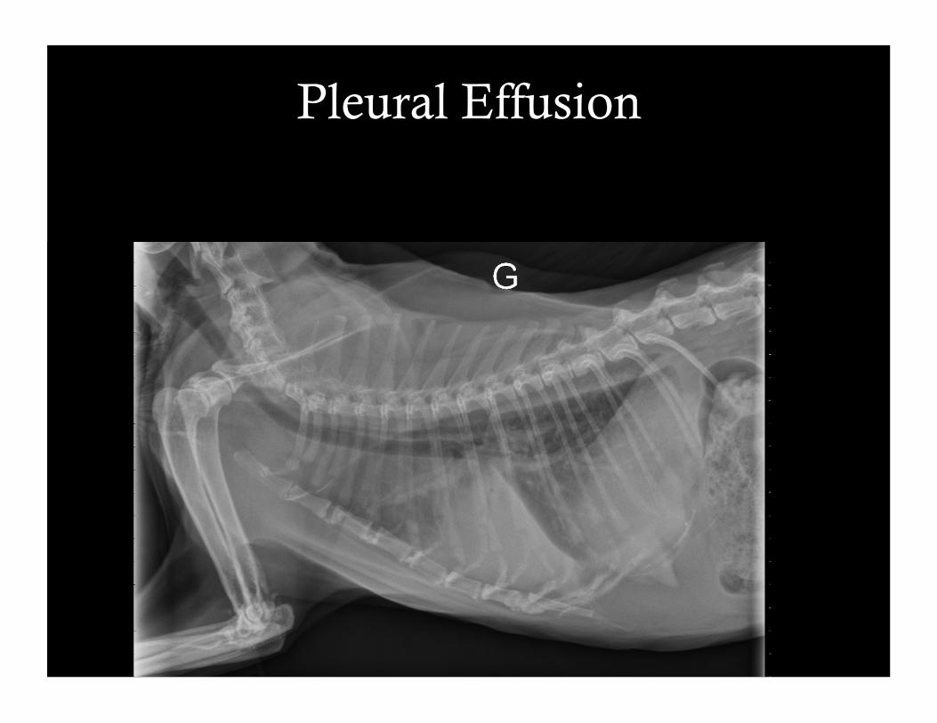
Pleural Effusion

Pleural Effusion
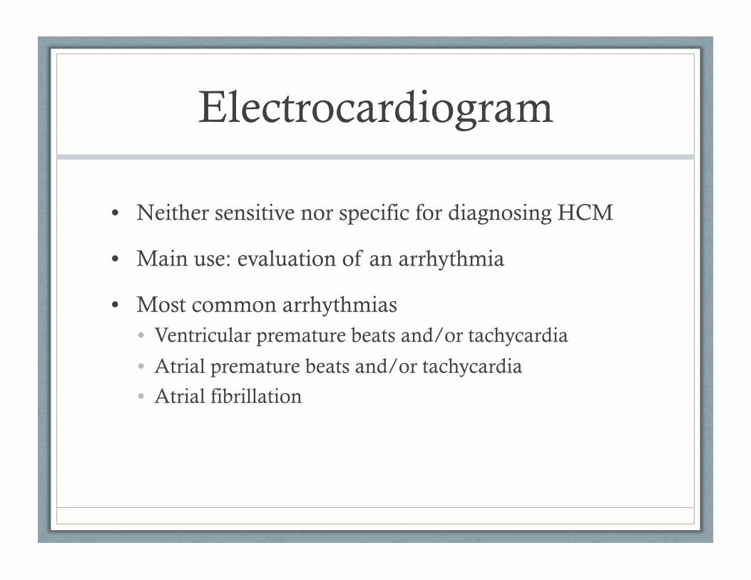
Electrocardiogram
• Neither sensitive nor specific for diagnosing HCM
• Main use: evaluation of an arrhythmia
• Most common arrhythmias • Ventricular premature beats and/or tachycardia
• Atrial premature beats and/or tachycardia
• Atrial fibrillation

Ventricular Premature Contractions

Atrial Fibrillation
• Irregularly irregular
• No P-waves
• Rapid • 250-300/min
• Uncommon in cats
• Severe left atrial dilation

Cardiac Biomarkers
• NT-pro Brain Natriuretic Peptide (Idexx CardiopetTM) • BNP produced due to increased ventricular wall stress or
pressure load • Causes natriuresis and vasodilation and counterbalance RAAS
• Positively correlates with disease severity • Utility in cats
• Differentiating between cardiac and non-cardiac causes of respiratory signs • Low: congestive heart failure unlikely • High values suggestive of congestive heart failure (careful of
concurrent respiratory and cardiac diseases)
• Detection of occult HCM

Use of NT-proBNP to detect occult cardiomyopathy
• Fox PR, et al. Multicenter evaluation of plasma N-terminal probrain natriuretic peptide (NT-pro BNP)as a biochemical screening test for asymptomatic (occult) cardiomyopathy in cats. JVIM. 2011.
• NT-pro BNP significantly higher in occult CM (186 pmol/L) versus normal (24 pmol/L) • 99 pmol/L was highest recorded in normals
• Correlated with atrial size, caudal wall thickness
• If use cut-off of >99 pmol/L • Sensitivity:70.8%; Specificity: 100%
• If use cut-off of 46 pmol/L • Sensitivity: 86%; Specificity: 91%
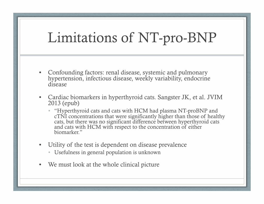
Limitations of NT-pro-BNP
• Confounding factors: renal disease, systemic and pulmonary hypertension, infectious disease, weekly variability, endocrine disease
• Cardiac biomarkers in hyperthyroid cats. Sangster JK, et al. JVIM 2013 (epub) • “Hyperthyroid cats and cats with HCM had plasma NT-proBNP and
cTNI concentrations that were significantly higher than those of healthy cats, but there was no significant difference between hyperthyroid cats and cats with HCM with respect to the concentration of either biomarker.”
• Utility of the test is dependent on disease prevalence • Usefulness in general population is unknown
• We must look at the whole clinical picture

Cardiac Biomarkers
• Cardiac troponin I (cTnI) • Marker of myocardial injury
or necrosis • Has been shown to be elevated in cats with cardiomyopathy
• When should you use cTnI • Generally not useful in general practice • Elevations seen with mycoardial infarction, myocarditis, chest trauma,
congestive heart failure • Assessment of the diagnostic accuracy of circulating cardiac troponin I
concentration to distinguish between cats with cardiac and non-cardiac causes of respiratory distress. Connolly DJ, et al. J Vet Cardiol, 2009 Dec;11(2):71-8
• “Serum cTnI concentrations were different in RD+CHF compared to RD-NC cats. However the overlap in cTnI concentrations between the 2 groups reduced the clinical efficacy of the assay “

Echocardiography
• What are the most important findings? • Left ventricular hypertrophy
• Focal, multifocal, diffuse
• Left atrial dilation**
• Evidence of fibrosis
• Evidence of myocardial infarction
• Left ventricular systolic and diastolic function
• Left auricular function
• Signs thrombus development

Normal Left ventricle
Left atrium
Mitral Valve
Papillary muscles
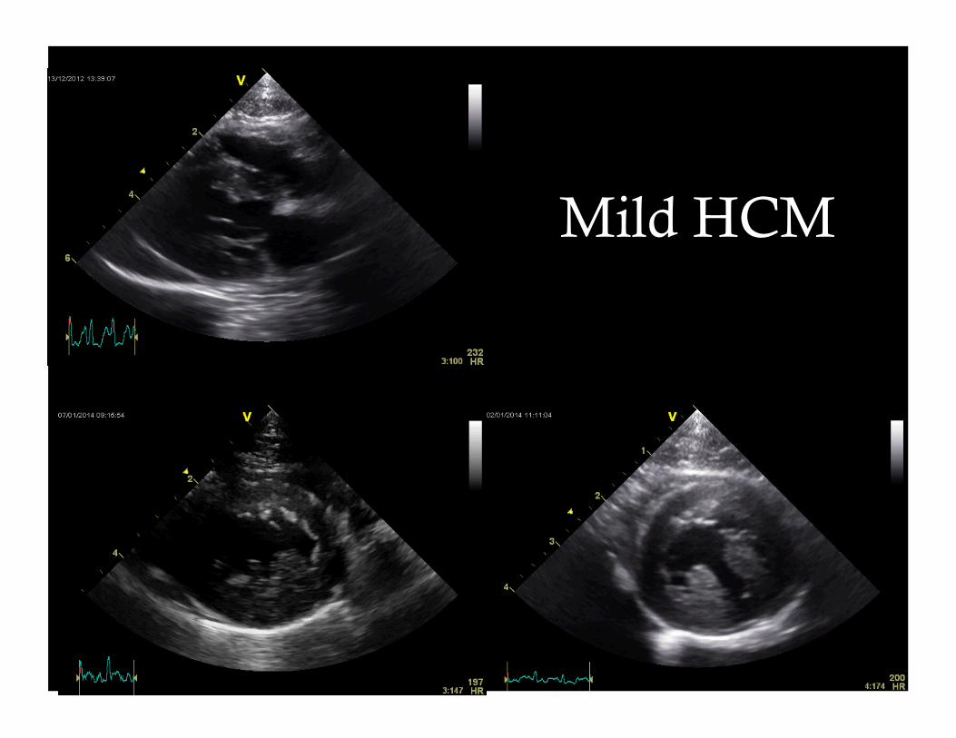
Mild HCM

Severe HCM
Images courtesy of Dr. Lynne O’Sullivan

Thrombus Formation
Spontaneous echocardiographic contrast “smoke” • Platelet/red cell aggregation
Left atrium
Left auricle with soft thrombus
Smoke
Organized thrombus Big Trouble!!!!

The Echo Report

Systolic Anterior Motion of the Mitral Valve
• SAM results in a dynamic left ventricular outflow tract (LVOT) obstruction and mitral regurgitation • Hypertrophic Obstructive Cardiomyopathy
Complications
• Congestive heart failure
• Arterial thromboembolism
• Sudden cardiac death

Arterial Thromboembolism
• Which patients are at risk? • Spontaneous echocardiographic contrast (“smoke”)
• Left atrial and/or auricular thrombus
• Severe left atrial dilation?
• Reduced left auricular ejection velocities on echo • Velocities ,0.2 m/s were associated with smoke

Arterial Thromboembolism
• Acute paresis or paralysis of one or more limbs • Lower motor neuron signs
• Generally intensely painful
• Paw pads of affected limbs are cyanotic • Compare to unaffected limbs,
look at nail beds
• Paw pads of affected limbs are
cold
• Absent pulses in affected limb • Can use Doppler to try to find
pulse
Smith SA, et al. JVIM 2003;17:73-83

Are these patients a lost cause?
• Borgeat K, et al. Arterial thromboembolism in 250 cats in general practice: 2004-2012. 2014; 28:102-108
• 61.2% euthanized at presentation
• 27.2% survived 24 hours • 55.9% were dead in <7 days (47% euthanized)
• Of 30 cats that lived ≥ 7 days 6 alive at 1 year
• At least 1 recurrence of ATE reported in 46.7% • Median time to recurrence 118 d

Are these patients a lost cause?
• Smith SA, et al. Arterial thromboembolism in cats: acute crisis in 127 cases (1992-2001) and long-term management with low-dose aspirin in 24 cases. J Vet Intern Med. 2003;17-73-83
• 45% of cats survived to discharge over the 9 year period
• Survival times improved over time
• 73% survived to discharge within the last year of the study
• Median survival times for discharged cats was 117 d
• 11 cats had recurrence
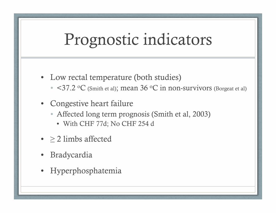
Prognostic indicators
• Low rectal temperature (both studies) • <37.2 oC (Smith et al); mean 36 oC in non-survivors (Borgeat et al)
• Congestive heart failure • Affected long term prognosis (Smith et al, 2003)
• With CHF 77d; No CHF 254 d
• ≥ 2 limbs affected
• Bradycardia
• Hyperphosphatemia

Treatment
• 1st Analgesia! • Fentanyl, hydromorphone, morphine, buprenorphine • Butorphanol is not strong enough unless pain is mild
• 24-48 hours post-event pain generally subsides
• Treatment to prevent further thrombus formation • Clopidogrel**, unfractionate heparin, aspirin, low molecular weight heparin
• External warming not necessary • Peripheral vasodilation and could worsen core perfusion
• Continuous ECG • Monitor for reperfusion injury (hyperkalemia and atrial standstill) and for other
arrhythmias.
• Monitor electrolytes q 12h ideally
• Average hospitalization time 2-3 days • Clients should expect estimate of $2500-3500
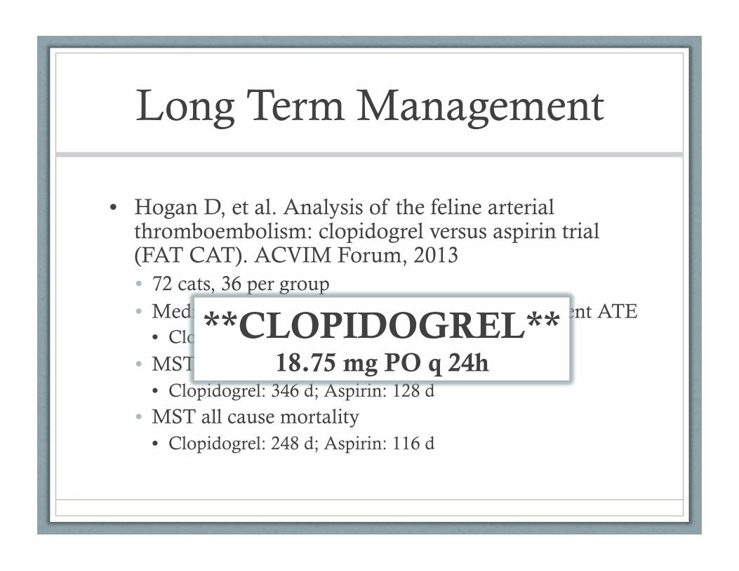
Long Term Management
• Hogan D, et al. Analysis of the feline arterial thromboembolism: clopidogrel versus aspirin trial (FAT CAT). ACVIM Forum, 2013 • 72 cats, 36 per group • Median survival time (MST) for cats with recurrent ATE
• Clopidogrel: 443 d; Aspirin: 192 d
• MST with recurrent ATE or cardiac death • Clopidogrel: 346 d; Aspirin: 128 d
• MST all cause mortality • Clopidogrel: 248 d; Aspirin: 116 d
**CLOPIDOGREL** 18.75 mg PO q 24h

Congestive Heart Failure
• Acute: • Furosemide 1-2 mg/kg SC/IM*/IV*
• Repeat q 30-60 minutes depending on response
• If really severe and responding poorly furosemide CRI is ideal
• Oxygen
• Nitroglycerine 1/8-1/4 inch on inner pinna
• Sedation • Butorphanol 0.2-0.25 mg/kg IM/IV

In-hospital Monitoring
• Ideally hourly respiratory rate and effort
• Blood pressure
• Thoracic radiographs in 12-24 hours • More frequent results in excessive patient stress and cost for the owner
• Repeat more frequently if have a specific reason
• ECG monitoring • Only if detectable arrhythmia unless monitoring post ATE
• Renal profile 12-24 hours after initiating furosemide
• If only mild CHF and patient discharged right away:
• Recheck renal profile, thoracic radiographs +/- blood pressure in 1 week

Congestive Heart Failure
• Chronic treatment • Furosemide 1-2 mg/kg PO q 12h starting dose
• ACE inhibitor: benazepril
• Clopidogrel 18.75 mg PO q 24h
• Spironolactone rarely used • ~30% of cats get severe facial ulcerative dermatitis
• Beta-blockers not used in “wet” patients • If already receiving may need to be decreased
• Pimobendan?

Pimobendan in Cats
• MacGregor JM, et al. Use of pimobendan in 170 cats (2006-2010). J Vet Cardiol. 2011;13:251-260
• Median dose: 0.24 mg/kg q 12h
• Well tolerated; 5 cats (3%) had potential side effects • Treatment discontinued in one cat
• Ideal dose not established • Longer elimination half life (almost 3x) and maximal
drug plasma concentration (>10x) than dogs. (Hanzlicek, AS. 2012)

Sudden Cardiac Death
• Fatal arrhythmia
• Myocardial infarction
• Difficult to predict, patient may have no preceding signs
• If significant ventricular arrhythmias • Consider antiarrhythmic therapy with sotalol or beta-
blocker (atenolol)

Treatment of Asymptomatic Disease
• Echocardiography is possible
• No systolic anterior motion of the mitral valve • Mild to moderate disease with no left atrial enlargement
• No treatment
• Severe disease with severe left atrial enlargement • No smoke or thrombus
• Benazepril +/- clopidogrel
• Smoke and/or thrombus • Benazepril + clopidogrel

Atenolol
• Not recommended if no echocardiogram
• Generally used in the setting of systolic anterior motion with moderate to severe LVOT obstruction • Potential benefits
• Reduction or control of obstruction and mitral regurgitation • Prevention of tachycardia • Improved diastolic filling • Improved coronary perfusion • Antiarrhythmic effects • Some cats can have regression of hypertrophy • Reduction of symptoms

Atenolol and Survival
• Schober KE, et al. Effect of treatment with atenolol on 5-year survival in cats with preclinical (asymptomatic) hypertrophic cardiomyopathy. J Vet Cardiol. 2013;15: 93-104
• Atenolol: 42 cats; No atenolol: 21 cats
• No difference in cardiac death between atenolol (24%) and no atenolol (19%) over 5 y
• No difference in time to death
• Study did not address quality of life
• Small number of cats with LVOT obstruction in the untreated group

Atenolol versus Diltiazem
• Atenolol
• Q 12-24 h dosing
• Low risk for side effects
• Better decrease in LVOT obstruction
• Prolongs early diastolic relaxation
• Diltiazem
• Q 8 h dosing
• Higher risk for side effects
• Improve early diastolic relaxation
• Not as effective to control heart rate
Treatment of Asymptomatic Disease
• Echocardiography is not possible
• Thoracic radiographs • Severe cardiomegaly • Severe left atrial/auricular enlargement
• Consider ACE inhibition • Blood pressure and evaluation of renal function first
• Consider antiplatelet therapy (clopidogrel) • Have onwers monitor sleeping respiratory rate (<30 rpm)
• Mild to moderate cardiomegaly • No treatment
Sedation Considerations
• Mild sedation • Butorphanol
• More significant sedation needed • No perfect answer!
• Butorphanol + midazolam
• Butorphanol + low dose acepromazine
• Butorphanol + low dose acepromazine + low dose ketamine
• Dexmedetomidine could reduce severity of dynamic left ventricular outflow tract obstruction (Lamont LA, et al 2002)

Anesthetic Considerations • If receiving beta-blocker or ACE inhibitor
• Consider dose reduction by half the morning of anesthesia
• Careful titration of IV fluids • Depends on severity of disease
• 2013 AAHA/AAFP guidelines • Authors suggest 3ml/kg/h in normal cats
• Avoid excessively deep anesthetic plane and maintain blood pressure
• Avoid tachycardia inducing drugs • Ketamine; anticholinergics only if needed
• Multimodal approach is best
• Ideal patient monitoring • Blood pressure, heart rate, ECG, pulse oximetry
• Monitor respiratory rate for 12-24 hours post-recovery

Prognosis
• Highly variable survival times • Asymptomatic 1129 d median survival time (MST) • With congestive heart failure 563 d MST
(Rush JE, et al. 2002)
• Cats with systolic anterior motion of the mitral valve reported to live longer
• Increased left atrial size and age are negative prognostic indicators
• More recent study showed multiple echocardiographic findings that negatively affected prognosis (Payne JR, et al 2013)

Follow Up
• Asymptomatic HCM • Recheck echocardiography and blood pressure every
6-12 months
• If receiving benazepril +/- furosemide • Regular renal function testing
• If severe disease or history of congestive heart failure • Regular thoracic radiographs • Periodic echocardiography

We are here to work with you!
• If you have a case please do not hesitate to call!
• Basic follow-up generally recommended with the family vet. • Call or email with update/questions
• Fax or email blood results
Thank you!

References Côté E, et al. Hypertrophic cardiomyopathy. In Feline Cardiology. Wiley-Blackwell. 2011:103-175.
Paige CF, et al. Prevalence of cardiomyopathy in apparently healthy cats. JAVMA 2009;234:1398-1403
Lefbom BK, et al. Severe hypertrophic cardiomyopathy in 10 young Ragdoll cats. J Vet Intern Med. 2011; 15:308.
Meurs KM. Genetics of cardiac disease in the small animal patient. Vet Clin Small Anim Prac 2009 July 40(4): 701-715
Litster AL, Buchanan JW. Vertebral scale system to measure heart size in radiographs of cats, JAVMA 2000; 216:210-214)
Sangster JK, et al. Cardiac biomarkers in hyperthyroid cats. J Vet Intern Med, 2013 Dec (epub)
Connolly DJ, et al. Assessment of the diagnostic accuracy of circulating cardiac troponin I concentration to distinguish between cats with cardiac and non-cardiac causes of respiratory distress. J Vet Cardiol, 2009 Dec;11(2):71-8
Oyama MA, et la. The use of NT-proBNP assay in the management of canine patients with heart disease. Vet Clin Small Anim Prac 2009 July 40(4): 545-558
Oyama MA. Using Cardiac Biomarkers in Veterinary Practice. Vet Clin Small Anim Prac 2013 Nov 43:1261-1272
Fox PR, et al. Utility of plasma N-terminal pro-brain natriuretic peptide (NT-proBNP) to distinguish between congestive heart failure and non-cardiac causes of acute dyspnea in cats. J Vet Cardiol. 2009 11, S51-S61.

References
Fox PR, et al. Utility of plasma N-terminal pro-brain natriuretic peptide (NT-proBNP) to distinguish between congestive heart failure and non-cardiac causes of acute dyspnea in cats. J Vet Cardiol. 2009 11, S51-S61.
Fox PR, et al. Multicenter evaluation of plasma N-terminal probrain natriuretic peptide (NT-pro BNP)as a biochemical screening test for asymptomatic (occult) cardiomyopathy in cats. J Vet Intern Med. 2011;25:1010-1016.
Schober KE, Maerz I. Assessment of left atrial appendage flow velocity and its relation to spontaneous echocardiographic contrast in 89 cats with myocardial disease. J Vet Intern Med. 2006; 20:12—130.
Borgeat K, et al. Arterial thromboembolism in 250 cats in general practice: 2004-2012. 2014; 28:102-108.
Hogan D, et al. Analysis of the feline arterial thromboembolism: clopidogrel versus aspirin trial (FAT CAT). ACVIM Forum, 2013
MacGregor JM, et al. Use of pimobendan in 170 cats (2006-2010). J Vet Cardiol. 2011;13:251-260
Hanzlicek AS, et al. Pharmacokinetics of oral pimobendan in healthy cats. J Vet Cardiol. 2012;14:489-496.

References
Schober KE, et al. Effect of treatment with atenolol on 5-year survival in cats with preclinical (asymptomatic) hypertrophic cardiomyopathy. J Vet Cardiol. 2013;15: 93-104.
Lamont LA, et al. Doppler echocardiographic effects of medetomidine on dynamic left ventricular outflow tract obstruction in cats. JAVMA 2002;221:1276-1281.
Bednarski R, et al. AAHA anesthesia guidelines for dogs and cats. JAAHA. 2011;47(6): 377-385
Davis H, et al. 2013 AAHA/AAFP fluid therapy guidelines for dogs and cats. JAAHA. 2013;49(3):149-159.
Rush JE, et al. Population and survival characteristics of cats with hypertrophic cardiomyopathy:260 cases (1990-1999). JAVMA 2002;220:202-207.
Payne J, et al. Population characteristics and survival in 127 referred cats with hypertrophic cardiomyopathy (1997-2005). J Small Anim Prac 2010;51:540-547.
Payne JR, et al. Prognostic indicators in cats with hypertrophic cardiomyopathy. J Vet Intern Med. 2013;27:1427-1436.
Pyendop BH. In Côté E, et al. Anesthesia in the patient with cardiac disease. Feline Cardiology. Wiley-Blackwell. 2011:411-421