feasibility and efficacy of transcranial motor … research feasibility and efficacy of transcranial...
TRANSCRIPT

ORIGINALRESEARCH
Feasibility and Efficacy of TranscranialMotor-Evoked Potential Monitoring inNeuroendovascular Surgery
T.G. HortonM. Barnes
S. JohnsonP.C. Kalapos
A. LinkK.M. Cockroft
BACKGROUND AND PURPOSE: Neurophysiological monitoring for neuroendovascular procedures typi-cally involves EEG and SSEP monitoring via cutaneous electrodes. MEP monitoring has been used lessfrequently because, traditionally, this has required subdural electrode placement. With the advent oftranscutaneous techniques, MEP monitoring use has increased. However, little has been publishedregarding the use of this technique in therapeutic neuroendovascular procedures. The purpose of thisstudy was therefore to determine whether TcMEP monitoring is feasible and efficacious in therapeuticneuroendovascular procedures.
MATERIALS AND METHODS: We retrospectively reviewed our data base of therapeutic neuroendovas-cular procedures performed with the use of TcMEP monitoring. We specifically determined theincidence of TcMEP changes compared with changes in either SSEP or EEG. We then correlated thesechanges to actual adverse neurologic events.
RESULTS: Although TcMEP monitoring was technically successful in all of the 140 patients in which itwas attempted, we observed significant changes in TcMEP signals in only 1 patient. This patientexperienced changes involving all 3 monitoring modalities after intraprocedural aneurysm rupture. Incontrast, changes in SSEP tracings alone were found in 9 patients. Of these, 2 patients were knownto be moribund before their procedures and neither recovered. Among the remaining 7 patients,temporary SSEP changes tended to correlate with temporary neurologic deficits, while permanentchanges were associated with permanent or long-lasting deficits.
CONCLUSIONS: These results suggest that TcMEP monitoring is feasible in therapeutic neuroendo-vascular procedures. However, it appears that the addition of TcMEP monitoring provides no addedbenefit to SSEP and EEG monitoring alone.
ABBREVIATIONS: BAER � brain stem auditory evoked response; EEG � electroencephalogram;EVD � external ventricular drain; GOS � Glasgow outcome score; MEP � motor-evoked potential;SSEP � somatosensory-evoked potential; TcMEP � transcranial motor-evoked potential; TIVA �total intravenous anesthetic
Neurophysiological monitoring has become an integral in-traoperative tool in the surgical management of neuro-
vascular disease. This has carried over to the treatment of cere-brovascular disease by the endovascular route, with variousgroups demonstrating the usefulness of transcutaneous tech-niques include monitoring of SSEP, EEG, and BAER.1,2 Intra-operative monitoring of MEP has also been shown to be safeand reliable in the setting of spinal surgery and open aneurysmsurgery, though this typically involves placement of subduralelectrodes.3 The use of MEP is of particular interest, givenreported advantages in sensitivity over other neurophysiologicmonitoring techniques.4,5 However, use of MEP in neuroen-dovascular procedures has been limited due to the need to usetranscutaneous stimulation and concerns over excessive pa-tient movement from such stimulation. As a result, little con-sideration has been given to the use and efficacy of MEP mon-itoring in neuroendovascular procedures. A review of the
medical literature found only one previously published workconcerning the use of MEP monitoring in the endovasculartreatment of intracranial vascular disease.6
Over the past 5 years at our institution, we have selectivelyemployed a combination of SSEP, EEG, and TcMEP monitor-ing during therapeutic neuroendovascular procedures. In aneffort to better understand the potential usefulness of theTcMEP technique in this setting, we reviewed our series ofthese patients and hereby present our experience with 140patients.
Materials and MethodsWe retrospectively reviewed our data base of neuroendovascular pro-
cedures to examine the feasibility and efficacy of TcMEP in therapeu-
tic neuroendovascular procedures. This study was approved by our
institutional review board under expedited review.
We routinely use neurophysiological monitoring in the form of
SSEP and EEG for almost all therapeutic neurovascular procedures.
In this report, in addition to our standard protocol, we reviewed a
subset of patients monitored with TcMEP. The decision regarding
whether to use TcMEP was made by the primary operator in each
case, with 1 of the senior authors (P.C.K.) using TcMEP in almost all
interventional cases, and the other senior author (K.M.C.) using the
technique only rarely. Information regarding patient demographics,
procedure type, monitoring changes, and clinical outcome was col-
Received October 6, 2011; accepted after revision December 17.
From the Departments of Neurosurgery (T.G.H., P.C.K., K.M.C.) and Radiology (P.C.K.,K.M.C.), Penn State Hershey Medical Center, Hershey, Pennsylvania; Impulse Monitoring,Inc. (M.B., S.J., A.L.), Columbia, Maryland.
Please address correspondence to Kevin M Cockroft, MD, MSc, Department of Neurosur-gery – EC110, Penn State Hershey Medical Center, PO Box 859, Hershey, PA 17033; e-mail:[email protected]
http://dx.doi.org/10.3174/ajnr.A3017
FUN
CTION
AL
ORIGINAL
RESEARCH
AJNR Am J Neuroradiol 33:1825–31 � October 2012 � www.ajnr.org 1825

lected. Special attention was paid to the monitoring technique expe-
riencing a change and to the sequence of these changes, if more than 1
technique was affected.
Anesthesia ProtocolAll patients received a TIVA with endotracheal intubation. Short-
acting neuromuscular blockage was used during induction. However,
due to the inherent limitations of TcMEP, neuromuscular blockade
was not used during the remainder of the procedure. Typically, pa-
tients were anesthetized using propofol infusion alone or in combi-
nation with dexmedetomidine and/or remifentanil.
Monitoring TechniqueA Digitimer D185 constant voltage stimulator (Digitimer, Letch-
worth Garden City, Hertfordshire, United Kingdom) was used to
elicit a multipulse TcMEP. Stimulation parameters consisted of train
counts that ranged from 3–7 pulses, with a constant duration of
50 microseconds. The interstimulus interval ranged from 1.1– 4.1
milliseconds.
Four transcranial stimulation subdermal needle electrodes were
used in favor of corkscrew electrodes in an effort to reduce the
amount of artifact present. Three stimulation montages were used for
TcMEP stimulation. A lateral C3-C4 montage, based on the Interna-
tional 10 –20 system, was typically used for stimulation. The precise
details of this technique have been previously described.5 The myo-
genic response from TcMEP stimulation was recorded with subder-
mal needle electrodes. Muscle groups selected consisted of the flexor
carpi radials, abductor pollicis brevis, anterior tibialis, and adductor
hallucis brevis muscles. Electromyography activity evoked from
TcMEP stimulation was recorded with a filter of 10 Hz to 3 KHz and
a sweep time of 10 msec/division. Amplifier gain was varied from
50 –100 �sec/division.
Preincision baseline data (before insertion of femoral sheath)
were obtained immediately after induction of anesthesia. If a short-
acting muscle relaxant was used during intubation, then a train of 4
was obtained. Train of 4 was recorded from the gastrocnemius muscle
upon stimulation of the common peroneal nerve at the popliteal
fossa; 4/4 twitches were verified before TcMEP baseline data were
established.
Anodal stimulation was use to evoke a myogenic response. A
threshold technique was used.7,8 Contralateral myogenic recruitment
from TcMEP stimulation was compared with the ipsilateral response
to cathodal stimulation. Stimulation intensity was then adjusted until
cathodal stimulation resulted in no ipsilateral response. The TcMEP
response amplitude, utilizing the described stimulation technique,
ranged from 50 �V to 1 mV.
The techniques used for SSEP and EEG monitoring have been
previously described9,10 and are therefore not described in this report.
ResultsA total of 758 therapeutic neuroendovascular procedures wereperformed in our department between January 2005 and De-cember 2009. Of these, 140 were performed using a combina-tion of SSEP, EEG, and TcMEP monitoring. The patient pop-ulation included 85 females and 55 males. Patients werebetween 6 and 87 years of age (mean age 47). Most patients(111) were treated for intracranial aneurysms. Of the remain-der, 18 patients had arteriovenous malformations, 5 patientshad intracranial arterial stenosis, and 4 patients had dural ar-teriovenous fistulas. In addition, there was 1 patient with in-tracranial vasculitis and 1 patient who underwent balloon testocclusion before a brain tumor resection.
TcMEP monitoring was technically successful in all pa-tients in this series. Monitoring was not abandoned because ofpatient movement in any procedure. A total of 11 patients hadchanges noted in at least 1 of their neurophysiological moni-toring parameters (Table). One patient had simultaneouschanges in TcMEP, EEG, and SSEP. One patient had concur-
Table: Summary of patients demonstrating changes in neurophysiological monitoring parameters
PatientAge (yrs)and Sex Diagnosis Procedure EEG SSEP TcMEP
ImmediateOutcome
(GOS)
Long-TermOutcome
(GOS)1 63 F Anterior communicating
artery aneurysmAnterior communicating artery aneurysm
coilinga� � 4 5 (25 months)
2 55 F Grade 5 SAH Right posterior inferior cerebellar arteryaneurysm coiling
� 2 1 (6 days)
3 56 F Grade 5 SAH Anterior communicating artery aneurysmcoiling
� 2 1 (6 days)
4 69 F Grade 1 SAH Right anterior choroidal and posteriorcommunicating artery aneurysms
� 4 5 (8 months)
5 34 F Grade 1 SAH Anterior communicating artery aneurysmcoilinga
� � � 4 5 (7 months)
6 62 F Grade 1 SAH Anterior communicating artery aneurysmcoiling
� 5 3 (28 months)
7 52 M Grade 1 SAH Left pericallosal artery aneurysm coiling � 4 4 (5 months)8 67 F Grade 3 SAH Right middle cerebral artery aneurysm
coiling� 3 5 (17 months)
9 53 M Grade 4 SAH Diagnostic angiogram � 4 5 (5 weeks)10 71 F Left paraclinoid
region aneurysmPrecoiling stent placement � 5 5 (14 months)
11 57 F Grade 3 SAH Anterior communicating and rightposterior communicating arteryaneurysm coiling
� 3 5 (4 months)
Note:—“Grade” in SAH patients is Hunt-Hess grade. A change is indicated by a “�.”a Changes were simultaneous.
1826 Horton � AJNR 33 � October 2012 � www.ajnr.org
rent SSEP and EEG changes. In the remaining 9 patients, onlychanges in SSEP were detected.
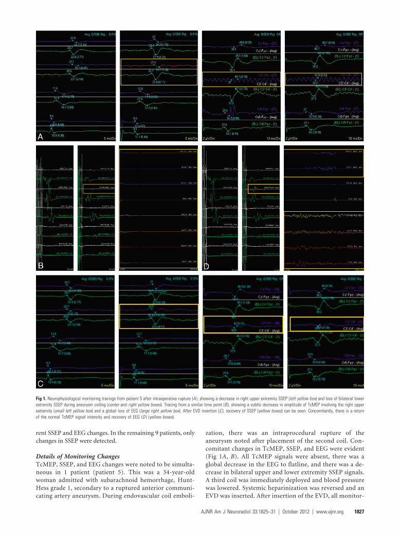
Details of Monitoring ChangesTcMEP, SSEP, and EEG changes were noted to be simulta-neous in 1 patient (patient 5). This was a 34-year-oldwoman admitted with subarachnoid hemorrhage, Hunt-Hess grade 1, secondary to a ruptured anterior communi-cating artery aneurysm. During endovascular coil emboli-
zation, there was an intraprocedural rupture of theaneurysm noted after placement of the second coil. Con-comitant changes in TcMEP, SSEP, and EEG were evident(Fig 1A, B). All TcMEP signals were absent, there was aglobal decrease in the EEG to flatline, and there was a de-crease in bilateral upper and lower extremity SSEP signals.A third coil was immediately deployed and blood pressurewas lowered. Systemic heparinization was reversed and anEVD was inserted. After insertion of the EVD, all monitor-
Fig 1. Neurophysiological monitoring tracings from patient 5 after intraoperative rupture (A ), showing a decrease in right upper extremity SSEP (left yellow box) and loss of bilateral lowerextremity SSEP during aneurysm coiling (center and right yellow boxes). Tracing from a similar time point (B ), showing a subtle decrease in amplitude of TcMEP involving the right upperextremity (small left yellow box) and a global loss of EEG (large right yellow box). After EVD insertion (C ), recovery of SSEP (yellow boxes) can be seen. Concomitantly, there is a returnof the normal TcMEP signal intensity and recovery of EEG (D ) (yellow boxes).
AJNR Am J Neuroradiol 33:1825–31 � October 2012 � www.ajnr.org 1827

ing signals slowly returned to baseline (Fig 1C, D). Thepatient showed gradual improvement in her neurologic sta-tus over the next 2 weeks and was discharged home 2 weeksafter her coiling procedure. She was neurologically intact.She remained free of neurologic deficits through follow-upat 7 months.
The patient with documented SSEP and EEG changes (pa-tient 1) was undergoing coil embolization of an unrupturedanterior communicating artery aneurysm. After detachmentof the final coil in this 63-year-old woman, a control angio-gram demonstrated lack of flow in the right A2 segment. Therewas a simultaneous decrease in the amplitude of the left lowerextremity SSEP as well as the EEG amplitude in the right fron-tal region. The patient’s mean arterial pressure was increasedby the anesthesia staff and abciximab (ReoPro) was adminis-tered by intravenous and intra-arterial routes. There was sub-sequent return of flow within the right A2 segment, and SSEPand EEG signals returned to near baseline. There was no clin-ical deficit noted on examination at the conclusion of theprocedure.
SSEP changes were noted in the absence of TcMEP or EEGchanges in 9 patients. Two of these patients were in poor clin-ical condition (Hunt-Hess grade V) after aneurysmal rupture(patients 2 and 3). Both patients had poor SSEP tracings at thebeginning of their procedures and both experienced a globaldecrease in SSEP amplitude, which did not improve in eitherpatient. Neither patient recovered neurologic function aftertheir procedure. Both of these patients ultimately died. Of theremaining 7 patients with SSEP changes alone, 1 had tempo-rary changes that were not associated with any clinical change(patient 8), 2 had transient changes that corresponded to tem-porary clinical deficits (patients 4 and 11), 2 had sustainedchanges without significant improvement, but no neurologicdeficits (patients 6 and 10), and 2 had sustained changes thatwere associated with new permanent neurologic deficits (pa-tients 7 and 9). In the single patient (patient 8) with a tempo-rary change that was not associated with any clinical change,the decrease in SSEP amplitude was found to be related to localeffects on the monitored extremity, and the changes resolvedafter repositioning of the extremity. In 1 of the patients with atemporary change and transient postoperative deficits (pa-tient 11), SSEP changes were noted in association with tempo-rary balloon occlusion during a balloon remodeling procedurefor aneurysm embolization. Induced hypertension and theshortening of occlusion times led to SSEP improvement. Theother patient in this category (patient 4) developed a thirdnerve palsy in the setting of SAH, and the coiling of tandemanterior choroidal artery and right posterior communicat-ing artery region aneurysms. In 1 patient with an unresolvedSSEP change and no subsequent clinical deficit (patient 6), adistal vessel occlusion, not apparent on the magnified imagesviewed during the aneurysm embolization, was found after theSSEP change, leading to early thrombolysis with IA and IVabciximab (ReoPro).
An example of significant SSEP-alone changes can be seenin patient 7, a 52-year-old man admitted with SAH, Hunt-Hess grade 1, secondary to a ruptured left pericallosal arteryaneurysm. This patient underwent endovascular coil emboli-zation of the ruptured aneurysm on postbleed day 1. Duringthe procedure, there was thromboembolic occlusion of the left
MCA immediately after placement of the guide catheter andbefore placement of any coils within the aneurysm (Fig 2A).There was an associated decrease in SSEP amplitude involvingthe right upper and lower extremities at this time (Fig 2B).Intra-arterial tPA was given, but persistent occlusion of a pos-terior frontal M3 division and slow flow through most otherbranches of the left MCA remained (Fig 2C). However, theSSEP signals improved to baseline following thrombolysis (Fig2D). After coiling of the aneurysm, thrombosis within theparent artery was seen at the aneurysm neck (Fig 2E). Therewas a decrease in SSEP amplitude involving the right upperand lower extremities at this time (Fig 2F). Intra-arterialabciximab (ReoPro) was administered. Angiographic imagesshowed resolution of the thrombus (Fig 2G). The SSEP signalsimproved slightly but remained decreased (below 50% ofbaseline amplitude) for the remainder of the procedure, withno further recovery (Fig 2H). There were no changes onTcMEP or EEG at any time during the procedure. The patientremained intubated overnight after the procedure and wasextubated the next day. He was aphasic and not followingcommands. The patient required placement of a ventriculo-peritoneal shunt later in his admission and was discharged to arehabilitation facility 5 weeks after admission. At 5-month fol-low-up, the patient had a GOS of 4 (modified Rankin scale,grade 2), with minimal weakness and slight expressive aphasia.
Time Requirements and CostThe overall duration of cases was not felt to be relevant becausemany variables impact case length, and patients were not ran-domly assigned to monitoring techniques. Monitoring setuptime was not directly tracked. However, the subjective impres-sion was that the addition of electrodes for TcMEP monitor-ing, including troubleshooting problems, added approxi-mately 5–10 minutes to the case setup time above and beyondSSEP and EEG monitoring alone. As mentioned in the Mate-rials and Methods section, the use of TcMEP requires the useof intravenous anesthetic agents, rather than volatile inhala-tional agents, and neuromuscular blockade cannot be used.Although we did not find patient movement to be a problemin most cases, the use of a TIVA protocol did subjectivelyappear to increase the time to arousal and extubation, partic-ularly after long procedures.
At our institution, intraoperative monitoring is performedby an independent company. The technical fee is the same forevery case monitored, whereas the professional fee is depen-dent on the actual monitoring service performed. In the caseof TcMEP monitoring, the professional fee is approximately$24.00 per extremity pair (upper versus lower). In contrast,the professional fee for EEG is $19.00, and this is the same asthe per pair extremity charge for SSEP.
DiscussionThe use of neuromonitoring has become well established inthe realm of neurosurgical and orthopedic procedures involv-ing intracranial neurosurgery and spinal surgery. The efficacyof specific modalities, including EEG, SSEP, and TcMEP, hasbeen described in the literature.11 The use of TcMEP for mon-itoring during microsurgical management of cerebrovasculardisease was recently described by Szelenyi et al12,13 and wasfound to be useful in a treatment group of 119 patients. Kang
1828 Horton � AJNR 33 � October 2012 � www.ajnr.org
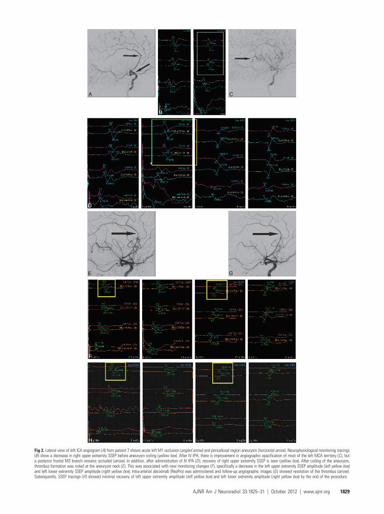
Fig 2. Lateral view of left ICA angiogram (A) from patient 7 shows acute left M1 occlusion (angled arrow) and pericallosal region aneurysm (horizontal arrow). Neurophysiological monitoring tracings(B ) show a decrease in right upper extremity SSEP before aneurysm coiling (yellow box). After IV tPA, there is improvement in angiographic opacification of most of the left MCA territory (C ), buta posterior frontal M3 branch remains occluded (arrow). In addition, after administration of IV tPA (D ), recovery of right upper extremity SSEP is seen (yellow box). After coiling of the aneurysm,thrombus formation was noted at the aneurysm neck (E ). This was associated with new monitoring changes (F ), specifically a decrease in the left upper extremity SSEP amplitude (left yellow box)and left lower extremity SSEP amplitude (right yellow box). Intra-arterial abciximab (ReoPro) was administered and follow-up angiographic images (G ) showed resolution of the thrombus (arrow).Subsequently, SSEP tracings (H ) showed minimal recovery of left upper extremity amplitude (left yellow box) and left lower extremity amplitude (right yellow box) by the end of the procedure.
AJNR Am J Neuroradiol 33:1825–31 � October 2012 � www.ajnr.org 1829

et al4 reported the clinical utility of TcMEP monitoring indetecting motor dysfunction in a small cohort of patients un-dergoing surgery for either lesions adjacent to the brain stemor intracranial aneurysms. When it comes to neuroendovas-cular procedures, reports have tended to focus on SSEP, EEG,and BAER monitoring. Lui et al1 were the first to report a seriesof 35 patients monitored with SSEP, EEG, and BAER whileundergoing endovascular therapy for the treatment of cerebralaneurysms. Nine patients experienced significant changes,and in 2 cases these led to alterations in intraoperative man-agement. In a more recent larger series of 63 patients moni-tored in a similar fashion, Chen and colleagues2 noted signif-icant monitoring changes in 3 patients, all of which led toprocedural changes. Both publications concluded that thesetechniques were useful in detecting intraprocedural ischemia,but neither addressed the monitoring of motor pathways.
After using TcMEP intermittently over the past few years,we decided to analyze our results to determine whether theaddition of TcMEP increased our ability to detect adverse neu-rologic events in our patients undergoing therapeutic neu-roendovascular procedures.
In our series of 140 patients, we observed significantchanges in TcMEP signals in only 1 patient. In this patient,changes involving all 3 monitoring modalities occurred afterintraprocedural aneurysm rupture. In addition, in this case, allmonitoring modalities changed simultaneously, suggesting noadvantage for 1 technique over another for the early detectionof neurologic compromise. In contrast, there were changes inSSEP tracings alone in 9 patients. Of these, 2 patients wereknown to be moribund before their procedures, and their bi-lateral SSEP amplitude changes were more likely due to pro-gression of their initial injury rather than a procedural event.In the end, neither patient recovered. Among the remaining 7patients, temporary SSEP changes tended to correlate withtemporary neurologic deficits, while permanent changes wereassociated with permanent or long-lasting deficits. In the pa-tients with SSEP changes, at least 3 underwent interventions orhad their procedures altered as a result of monitoring con-cerns. Although angiographic findings were noticeable beforeSSEP changes in 4 of the 7 patients, 3 patients experiencedSSEP changes either unrelated to an angiographic finding orbefore the angiographic finding being detected. One patienthad an upper extremity repositioned, possibly preventing aperipheral compression neuropathy. A second patient hadblood pressure increased and balloon occlusion times mini-mized during balloon-assisted coiling, possibly averting a per-manent neurologic deficit. Finally, 1 patient’s SSEP changeswere found to be related to a distal embolus, which was notapparent on the magnified images being viewed during theaneurysm embolization, leading to early thrombolysis and,again, possibly preventing a permanent neurologic deficit.These results lend continued support to previous work con-cluding that neurophysiologic monitoring using at least SSEPis useful. However, our series does not support the conclusionthat the addition of TcMEP monitoring provides any signifi-cant added benefit over SSEP alone.
The use of intraoperative neurophysiologic monitoring hassteadily increased, despite a paucity of clinical data purportingthe sensitivity and specificity of monitoring modalities in pre-dicting neurologic insult. Small case series using TcMEP in
neurovascular surgery have supported the use of this tech-nique, given the relative ease of setup and safety, but actualcorrelation of intraoperative changes to outcome remains elu-sive. Quinones-Hinojosa et al14 advocated the use of TcMEPin basilar artery aneurysm surgery but provided no clear proofof actual efficacy in monitoring alerts versus clinical outcomein specific patients. In the only previous report of TcMEP usein neuroendovascular patients that we could find, Hiraishi etal6 reported a series of 7 patients with anterior choroidal arteryaneurysms monitored with TcMEP while undergoing coil em-bolization. Three of their 7 patients experienced a transientdecrease in TcMEP signals. Of these, 2 patients saw their sig-nals improve after the removal of the coils. One of the 3 pa-tients experienced a transient neurologic deficit. No compar-ison with other modalities was offered, and the very smallnumber of patients makes it difficult to draw any conclusionsrelative to the overall efficacy of the technique.
With regard to the feasibility of TcMEP monitoring, wesubmit that, from a technical standpoint, this technique isstraightforward in implementation. Review of our institu-tional practice found that TcMEP monitoring added little timeto the setup or to the overall cost of the procedure. AlthoughTcMEP monitoring poses no direct risk to the patient, thismonitoring technique does mandate modification of anes-thetic technique. Neuromuscular blockade cannot be used.This translates to increased patient movement during the en-dovascular procedure that may potentially interfere with theprecision of treatment. This has not been a problem in ourexperience. However, it has been our subjective impressionthat the use of TIVA, necessary for TcMEP monitoring, doeslead to a longer time for patients to awaken from their anes-thetic, thus potentially prolonging the overall duration of thecase.
Obviously, this study has its limitations. Patients were notprospectively randomized to the various monitoring methodsand the physicians were not blinded to the protocol beingused. The technique for TcMEP monitoring is extremely sen-sitive to anesthetic changes. As a result, the quality of the dataobtained is likely to have been variable and similar results maynot necessarily be achieved at every institution. Given the rel-atively small number of patients, and the even smaller numberwith monitoring changes, it is difficult to be certain that somebenefit for TcMEP in certain subsets of the neurovascular dis-ease population undergoing endovascular therapy might notbe seen.6 Clearly, not all possible clinical scenarios were en-countered in the present series and further study may beindicated.
ConclusionsTcMEP appears to be feasible in therapeutic neuroendovascu-lar procedures. However, we found little evidence to support aclinical benefit for the routine use of TcMEP as an adjunct toneurophysiological monitoring with SSEP and EEG duringsuch procedures.
Disclosures: Mollie Barnes—UNRELATED: Employment: Employed by Impulse Monitoring,Inc. Samuel Johnson—OTHER RELATIONSHIPS: I serve as the Director of Operations forImpulse Monitoring. Impulse is neurophysiological intraoperative monitoring company. Theclinical research paper that I collaborated on with Dr. Kevin Cockroft was purely to satisfyour intellectual curiosity, and my company did not contribute nor benefit from this
1830 Horton � AJNR 33 � October 2012 � www.ajnr.org

manuscript in any way. Kevin M. Cockroft—OTHER RELATIONSHIPS: Consultant – eV3Endovascular (not directly related to the project/manuscript).
AcknowledgmentThe authors thank Lynne Hamann for her assistance in thepreparation of this manuscript.
References1. Liu AY, Lopez JR, Do HM, et al. Neurophysiological monitoring in the endo-
vascular therapy of aneurysms. AJNR Am J Neuroradiol 2003;24:1520 –272. Chen L, Spetzler RF, McDougal CG, et al. Detection of ischemia in endovascu-
lar therapy of cerebral aneurysms: a perspective in the era of neurophysiolog-ical monitoring. Neurosurg Rev 2011;34:69 –75
3. Neuloh G, Schramm J. Monitoring of motor evoked potentials compared withsomatosensory evoked potentials and microvascular Doppler ultrasonogra-phy in cerebral aneurysm surgery. J Neurosurg 2004;100:389 –99
4. Kang D, Wu Z, Lan Q, et al. Combined monitoring of evoked potentials duringmicrosurgery for lesions adjacent to the brainstem and intracranial aneu-rysms. Chin Med J 2007;120:1567–73
5. Szelenyi A, Kothbauer K, de Camargo A, et al. Motor evoked potential moni-toring during cerebral aneurysm surgery: technical aspects and comparisonof transcranial and direct cortical stimulation. Neurosurgery 2005;57:331–38
6. Hiraishi T, Fukuda M, Oishi M, et al. Usefulness of motor-evoked potential
monitoring during coil embolization of anterior choriodal artery aneurysms:technical reports. Neurol Res 2011;33:360 – 62
7. Novak K, de Camargo A, Neuwirth M, et al. The refractory period of fast con-ducting corticospinal tract axons in man and its implications for intraopera-tive monitoring of motor evoked potentials. Clin Neurophysiol 2004;115:1931– 41
8. Szelenyi A, Kothbauer K, Deletis V. Transcranial electric stimulation for intra-operative motor evoked potential monitoring: stimulation parameters andelectrode montages. Clin Neurophysiol 2007;118:586 –95
9. Grundy B. Intraoperative monitoring of sensory-evoked potentials. Anesthe-siology 1983;58:72– 87
10. Harner R. A recommendation for standard EEG montages. Am J of EEG Tech1977;17:105–14
11. Neuloh G, Schramm J. Motor evoked potential monitoring for the surgery ofbrain tumours and vascular malformations. Adv Tech Stand Neurosurg2004;29:171–228
12. Szelenyi A, Langer D, Beck J, et al. Transcranial and direct cortical stimulationfor motor evoked potential monitoring in intracerebral aneurysm surgery.Neurophysiol Clin 2007;37:391– 89
13. Szelenyi A, Langer D, Kothbauer K, et al. Monitoring of muscle motor evokedpotentials during cerebral aneurysm surgery: intraoperative changes andpostoperative outcome. Neurosurgery 2006;105:675– 81
14. Quinones-Hinojosa A, Alam M, Lyon R, et al. Transcranial motor evoked po-tentials during basilar artery aneurysm surgery: technique application for 30consecutive patients. Neurosurgery 2004;54:916 –24
AJNR Am J Neuroradiol 33:1825–31 � October 2012 � www.ajnr.org 1831