fdg-pet/ct(a) imaging in large vessel vasculitis and ... · polymyalgia rheumatica (pmr) belong to...
TRANSCRIPT

REVIEW ARTICLE
FDG-PET/CT(A) imaging in large vessel vasculitis and polymyalgiarheumatica: joint procedural recommendation of the EANM, SNMMI,and the PET Interest Group (PIG), and endorsed by the ASNC
Writing group*:Riemer H.J.A. Slart1a,d, Andor W.J.M. Glaudemansb,d, Panithaya Chareonthaitaweec,e, Giorgio Tregliab,d, Florent L.Bessond, Thorsten A. Bleyd, Daniel Blockmansd, Ronald Boellaardd, Jan Buceriusa, José Manuel Carrild, WengenChenc, Maria C Cidd, Bhaskar Daguptad, Sharmila Dorbalac,e, Olivier Gheysensb,d, Fabien Hyafila, Shaifali Jaind,Thorsten Klinkd, Conny J. van der Lakend, Francisco Lomeñad, Michela Massollod, Sergio Prieto-Gonzálezd, RaashidLuqmanid, Anne Roivainend, Carlo Salvaranid, Antti Sarasted, Michael Schirmerd, Hein J. Verbernea, Annibale Versarib,d,Alexandre E. Voskuyld, Martin A. Walterd, Dario Camellinod, Elisabeth Brouwerd, Marco A. Cimminod
Reviewer group*:Aiden Abidove, Denis Agostinia, Rob S. Beanlandse, Roberto C. Delgado-Boltonf, Andrew J. Einsteine, Alessia Gimellia,Edward J. Millere, Roberto Sciagràa, Alberto Signoreb
*Members of EANM Cardiovasculara, EANM Infection & Inflammationb Committees, SNMMI Cardiovascularc Council,PET Interest Groupd, and ASNCe, EANM Committee Coordinatorf.
Received: 2 February 2018 /Accepted: 6 February 2018 /Published online: 11 April 2018# The Author(s) 2018
AbstractLarge vessel vasculitis (LVV) is defined as a disease mainly affecting the large arteries, with two major variants, Takayasu arteritis(TA) and giant cell arteritis (GCA). GCA often coexists with polymyalgia rheumatica (PMR) in the same patient, since both belongto the same disease spectrum. FDG-PET/CT is a functional imaging technique which is an established tool in oncology, and has alsodemonstrated a role in the field of inflammatory diseases. Functional FDG-PET combined with anatomical CT angiography, FDG-PET/CT(A), may be of synergistic value for optimal diagnosis, monitoring of disease activity, and evaluating damage progression inLVV. There are currently no guidelines regarding PET imaging acquisition for LVVand PMR, even though standardization is of theutmost importance in order to facilitate clinical studies and for daily clinical practice. This work constitutes a joint proceduralrecommendation on FDG-PET/CT(A) imaging in large vessel vasculitis (LVV) and PMR from the Cardiovascular andInflammation & Infection Committees of the European Association of Nuclear Medicine (EANM), the Cardiovascular Councilof the Society of Nuclear Medicine and Molecular Imaging (SNMMI), and the PET Interest Group (PIG), and endorsed by theAmerican Society of Nuclear Cardiology (ASNC). The aim of this joint paper is to provide recommendations and statements, basedon the available evidence in the literature and consensus of experts in the field, for patient preparation, and FDG-PET/CT(A)acquisition and interpretation for the diagnosis and follow-up of patients with suspected or diagnosed LVV and/or PMR. Thisposition paper aims to set an internationally accepted standard for FDG-PET/CT(A) imaging and reporting of LVVand PMR.
Keywords Large vessel vasculitis . Polymyalgia rheumatica . FDG-PET/CT(A) . Imaging procedure
Introduction
Large vessel vasculitis (LVV) is defined as a disease mainlyaffecting the large arteries, with two major variants, Takayasuarteritis (TA) and giant cell arteritis (GCA) [1]. Vasculitis canbe distributed locally in the branches of the internal and ex-ternal carotid artery or the aorta and its main branches more
* Riemer H. J. A. [email protected]
1 University of Groningen,Medical Imaging Center, Department of NuclearMedicine & Molecular Imaging, University Medical Center Groningen,Hanzeplein 1, P.O. Box 30001, 9700 RB Groningen, The Netherlands
Electronic supplementary material The online version of this article(https://doi.org/10.1007/s00259-018-3973-8) contains supplementarymaterial, which is available to authorized users.
European Journal of Nuclear Medicine and Molecular Imaging (2018) 45:1250–1269https://doi.org/10.1007/s00259-018-3973-8
centrally in the thorax. TA and GCA are different diseases withdifferent age of onset, ethnic distribution, immunogenic back-ground [2], and distribution and therapy response [3, 4] of theaffected arteries. GCA and TA also show some overlap withregard to the histopathology of arterial lesions, reflecting sharedpathways in tissue inflammation [5, 6]. Clinically, GCA andpolymyalgia rheumatica (PMR) belong to a disease spectrum,and both often coexist in the same patient. Nearly half of pa-tients with GCA have evidence of PMR, while approximately20% of patients with PMR have concomitant GCA [7, 8], al-though the frequency of GCA in PMR (either by biopsy orimaging) may vary, depending on the cohort selection criteria.
FDG-PET/CT is a functional imaging technique which is anestablished tool in oncology, and has also demonstrated a role inthe field of inflammatory diseases. FDG-PET is based on theability to detect enhanced glucose uptake from high glycolyticactivity of inflammatory cells in inflamed arterial walls andsynovia/bursa [9]. Thereby, it can identify the presence of sys-temic LVV in patients with GCA and TA, and it can also showinflammation of peri-articular and extra-articular synovial struc-tures in the case of PMR. Approximately 20% of patients withapparently isolated PMR show LVVon FDG-PET/CT [10], andthis percentage can be even higher, depending on the presenceof LVV symptoms [11–13]. It is important to realize that anegative temporal artery biopsy, an ultrasonography without ahalo sign, or magnetic resonance imaging (MRI) without aorticwall thickening or edema does not definitively exclude the pres-ence of LVVand should therefore not limit the use of FDG-PET/CT when LVV is clinically suspected [14, 15]. Furthermore,there is substantial variation in the type of vessels involved(i.e. aortic and cranial large vessels) [16], which can be detectedby FDG-PET, given its whole-body scan nature, with the excep-tion of the temporal artery, due to the high physiological FDGuptake in the brain and limited resolution of the camera system.In addition, FDG-PET may assist in the differential diagnosisbetween PMR and elderly-onset rheumatoid arthritis (EORA) orspondyloarthritis [8], according to the location of inflammation(articular, capsular, or extracapsular). In patients with fever ofunknown origin (FUO), when the diagnosis of systemic LVV isruled out, FDG-PET/CT results enable the identification of othercauses of the inflammatory process, including oncological dis-eases, in the majority of cases. Functional FDG-PET combinedwith anatomical CT angiography, FDG-PET/CT(A), may be ofsynergistic value for optimal diagnosis, disease activity moni-toring, and evaluation of damage progression in LVV [17]. Themain limitation of FDG-PET/CT(A) to becoming a standardizeddiagnostic tool is the lack of an internationally accepted defini-tion of vascular inflammation and/or PMR, based on the inten-sity and pattern of the glucose analogue uptake. Also, FDG-PET/CT is not disease specific and is primarily developed todiagnose malignant and infectious/inflammatory diseases.Results have to be interpreted with caution as inflammatory/metabolic changes in the arterial wall usually precede
Preamble The Society of Nuclear Medicine and Molecular Imaging(SNMMI) is an international scientific and professional organizationfounded in 1954 to promote the science, technology, and practical appli-cation of nuclear medicine. The European Association of NuclearMedicine (EANM) is a professional nonprofit medical association thatfacilitates communication on a global basis between individuals pursuingclinical and research excellence in nuclear medicine. The EANM wasfounded in 1985. SNMMI and EANM members are physicians, technol-ogists, and scientists specializing in the research and practice of nuclearmedicine.The SNMMI and EANM will periodically define new guidelines fornuclear medicine practice to help advance the science of nuclear medicineand to improve the quality of care to patients throughout the world.Existing practice guidelines are reviewed for revision or renewal, as ap-propriate, on their fifth anniversary, or sooner if indicated.Each practice guideline, representing a policy statement by the SNMMI/EANM, has undergone a thorough consensus process in which it has beensubjected to extensive review. The SNMMI and EANM recognize thatthe safe and effective use of diagnostic nuclear medicine imaging requiresspecific training, skills, and techniques, as described in each document.Reproduction or modification of the published practice guideline by en-tities not providing these services is not authorized.These guidelines are an educational tool designed to assist practitioners inproviding appropriate care for patients. They are not inflexible rules orrequirements of practice and are not intended, nor should they be used, toestablish a legal standard of care. For these reasons and those set forthbelow, both the SNMMI and the EANM caution against the use of theseguidelines in litigation in which the clinical decisions of a practitioner arecalled into question.The ultimate judgment regarding the propriety of any specific procedureor course of action must be made by the physician or medical physicist inlight of all the circumstances presented. Thus, there is no implication thatan approach differing from the guidelines, standing alone, is below thestandard of care. To the contrary, a conscientious practitioner may respon-sibly adopt a course of action different from that set forth in the guidelineswhen, in the reasonable judgment of the practitioner, such course ofaction is indicated by the condition of the patient, limitations of availableresources, or advances in knowledge or technology subsequent to publi-cation of the guidelines.The practice of medicine includes both the art and the science of the preven-tion, diagnosis, alleviation, and treatment of disease. The variety and com-plexity of human conditions make it impossible to always reach the mostappropriate diagnosis or to predict with certainty a particular response totreatment. Therefore, it should be recognized that adherence to these guide-lines will not ensure an accurate diagnosis or a successful outcome. All thatshould be expected is that the practitioner will follow a reasonable course ofaction based on current knowledge, available resources, and the needs of thepatient, to deliver effective and safe medical care. The sole purpose of theseguidelines is to assist practitioners in achieving this objective.This joint procedural recommendation paper on 2-[18F]-fluorodeoxyglucose(FDG) positron emission tomography–computed tomography (PET/CT) orPET/CT(A) (with angiography) imaging in large vessel vasculitis (LVV) andpolymyalgia rheumatica (PMR) has been developed under the auspices ofthe Cardiovascular and Inflammation & Infection Committees of theEANM, the Cardiovascular Council of the SNMMI, and the PET InterestGroup (PIG). The purpose of this paper is to assist imaging specialists andclinicians in recommending, performing, and interpreting the results ofFDG-PET in patients with suspected LVVand PMR. In addition, the paperhighlights the importance of standardization and optimal procedural perfor-mance of FDG-PET/CT(A) imaging in LVVand PMR, and emphasizes theimportance of bridging the gap between imaging specialists and cliniciansworking in this field.
Eur J Nucl Med Mol Imaging (2018) 45:1250–1269 1251
anatomic changes [18–23]. Furthermore, whereas increasedFDG uptake is mainly seen in active disease processes, infor-mation of advanced stages, for example calcification in chron-ic or past inflammation, is mainly provided by morphologicalimaging [24]. Atherosclerosis activity may also interfere withthe FDG-PET signal in patients with LVV [25]. Finally, theinstigating inflammatory process may have subsided, leavingresidual arterial stenosis or aortic aneurysms for which FDG-PET is not the best imaging option.
In nuclear medicine, procedural guidelines for FDG-PET im-aging have been published for both cancer [26] and infection/inflammation [27]. However, LVVand PMR are distinct diseaseentities, which require a specific technical approach. The inter-pretation of FDG-PET images for LVV can be challenging, andthere is currently no consensus on how to interpret the images inthe setting of LVV. Furthermore, as previously described, FDGuptake has been demonstrated to respond to glucocorticoids(GC) therapy, which reduces metabolic cell activity. In this set-ting, aortic/arterial wall thickening (visible on CTorMRI) is stillpresent due to a delayed morphological vascular response [28].
There are currently no guidelines regarding PET imagingacquisition for LVVand PMR, even though standardization isof the utmost importance for facilitating clinical studies andfor daily clinical practice.
The aim of this joint paper is to provide recommendationsand statements, based on the available evidence in the litera-ture and consensus of experts in the field, for patient prepara-tion and FDG-PET/CT(A) acquisition and interpretation in thediagnosis and follow-up of patients with suspected or diag-nosed LVV and/or PMR. This position paper aims to set aninternationally accepted standard for FDG-PET/CT(A) imag-ing and reporting of LVV and PMR. An additional aim is tofacilitate prospective clinical studies and pooling of futuremulti-center data. Other imaging modalities applied in LVVdiagnostics, such as MRI angiography and ultrasound, arebeyond the scope of this document.
FDG-PET/CT(A) procedures in LVV and PMR
Patient preparation and FDG-PET/CT(A) imageacquisition
Patient preparation
The main goal of adequate patient preparation is to reducephysiologic tracer uptake in normal tissues (myocardium,skeletal muscle, urinary tract and brown adipose tissue) whilemaintaining uptake in diseased tissues and organs. Patients areinstructed to fast for at least 6 h prior to FDG administrationalthough intake of non-caloric beverages is allowed duringthat period [27]. In addition, strenuous physical activitiesshould be avoided within 24 h before FDG administration.
At the moment of and after administration of FDG, patientsshould relax in an adequately temperature-controlled room(20–22 °C [68–71.6 °F]) to minimize physiologic uptake inmuscles and brown fat [29]. In some cases, FDG uptake inbrown fat can be reduced by beta-blocking drugs, e.g. orallyadministered 20 mg propranolol 1 h before FDG injection[30]. Prior to positioning on the table, patients are asked tovoid urine. Patients with FUO and suspicion of cardiac in-volvement (e.g. endocarditis, sarcoidosis) must prepare witha special diet to reduce physiological myocardial uptake ofFDG. Patient preparation for cardiac FDG-PET imaging isbased on increasing the provision of fatty acids to the heartand decreasing physiological uptake of glucose by the myocar-dium. The SNMMI/ASNC/Society of CardiovascularComputed Tomography (SCCT) guidelines and SNMMI/ASNC consensus document recommend preparation with afat-enriched diet lacking carbohydrates for 12–24 h prior to thescan, a 12–18 h fast, and/or the use of intravenous unfractionatedheparin approximately 15 min prior to FDG injection [31, 32].
Serum glucose levels before FDG administration
Previous studies have shown that FDG uptake is reduced ifserum glucose levels exceed 7 mmol/L (126 mg/dL) [33–35],thereby rapidly and efficiently shunting FDG to organs with ahigh density of insulin receptors (e.g. skeletal and cardiacmuscles), resulting in altered FDG biodistribution and subop-timal image quality [36].
The impact of glucose levels on FDG uptake in inflamma-tory lesions is less well investigated. A study by Rabkin et al.in 123 patients with suspected infection demonstrated thathyperglycemia at the time of study had no significant impacton the false-negative rate [33]. However, a prospective studyin 195 patients evaluating the impact of fasting glucose levelson arterial uptake showed a negative correlation between up-take in the arterial wall and pre-scan glucose levels, as well asincreased blood pool activity with increased glucose levels[35]. In general, efforts should be made to reduce blood glu-cose to the lowest possible level, but glucose levels below7 mmol/L (126 mg/dL) are preferable.
Glucocorticoids and FDG administration
Glucocorticoids (GC) may reduce vascular wall uptake ofFDG; the available data regarding the effect of GCwithdrawalon FDG uptake are scarce. Nielsen et al. recently confirmedthat diagnostic accuracy of LVV with FDG-PET remained for3 days after initiation of GC, after which the signal decreasedsignificantly [37–39]. Thus there may be a diagnostic windowof opportunity within 3 days of initiation of GC.
A brief withdrawal of GC could Brestore^ pathological FDGuptake and reduce the likelihood of a false-negative result, butthis is not known. At the same time, GC withdrawal may pose
1252 Eur J Nucl Med Mol Imaging (2018) 45:1250–1269
risks to the patient. In the case of GCA, especially if temporalartery or ocular involvement is suspected, administration of GCcannot be delayed or withdrawn due to possible ischemic com-plications. In other conditions such as PMR or TA, withdrawingor delaying therapy until after PET can be permitted unless thereis risk of ischemic complications (Table 1).
The use of GC may also increase FDG uptake in the liver,resulting in underestimation and/or under-scoring of vascularFDG uptake [40].
Acquisition time after FDG administration
A minimum of 60 min between intravenous FDG administra-tion and acquisition has been recommended for adequate tracerbiodistribution [27]. Delayed acquisitions increase the vascular-to-blood pool ratio, hence increasing contrast resolution [35],and could make the measured vascular uptake more accurate[41]. However, as the majority of LVV studies have been per-formed at 60 min, PET-positive criteria at delayed time pointshave not yet been evaluated in this setting andmay differ slight-ly from those defined at the standard time interval. In contrast toFDG-PET studies evaluating metabolic activity of atheroscle-rotic lesions, studies comparing early (1 h) versus delayed (3 h)imaging in LVVare scarce [42]. A small prospective study in 23patients with suspicion of LVV concluded that delayed imagingat 3 h yielded a more detailed image of the arterial wall, mainlydue to decreased blood pool activity [43]. The recently pub-lished EANM position paper on the use of FDG-PET in ath-erosclerosis recommends an interval of 2 h between FDG ad-ministration and acquisition [44]. Currently, there is not enoughevidence to apply the same time window for LVV. At this time,
we recommend an uptake interval of at least 60 min.Standardization of the time interval is essential, especially whenusing semiquantitative analyses and when comparing FDG up-take on follow-up studies and between institutes.
Patient positioning and acquisition parameters
There are currently no guidelines for image acquisition in LVVor PMR, but whole-body acquisition from head to knee (op-tionally including the feet) in the supine position with the armsnext to the body is recommended, because (PMR) patients aregenerally unable to hold their arms above their head. For FDG-PET/CT imaging, a low-dose non-contrast CT must be per-formed for attenuation correction and anatomical localization.Alternatively, a diagnostic contrast-enhanced CT may be per-formed according to applicable local or national protocols andguidelines. A contrast-enhanced CTA is useful for identifyingstenotic lesions in TA, but data are insufficient to support itsroutine use for GCA LVV [45]. When using a contrast-enhanced CTA, a low-dose CT scan should be performed priorto intravenous contrast injection for attenuation correction andsubsequent standardized uptake value (SUV) calculations. Theimpact of intravenous contrast agents on the accuracy of atten-uation correction is considered acceptable only when CT dataare collected in the equilibrium or venous phase (i.e. delayedacquisition), with the advantage of radiation dose reduction[26]. Detection of smaller vascular structures in the head andneck region can be improved by increasing the acquisition time(~ doubled) per bed position to improve image quality, andapplying larger image matrices (thus smaller voxels) [46].This will reduce the partial volume effect of smaller structures,
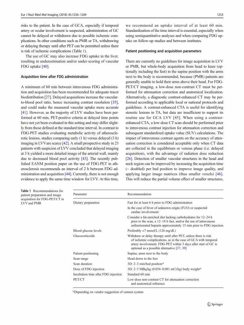
Table 1 Recommendations forpatient preparation and imageacquisition for FDG-PET/CT inLVVand PMR
Parameter Recommendation
Dietary preparation Fast for at least 6 h prior to FDG administration
In the case of fever of unknown origin (FUO) or suspectedcardiac involvement:
Consider a fat-enriched diet lacking carbohydrates for 12–24 hprior to the scan, a 12–18 h fast, and/or the use of intravenousunfractionated heparin approximately 15 min prior to FDG injection
Blood glucose levels Preferably <7 mmol/L (126 mg/dL)
Glucocorticoids Withdraw or delay therapy until after PET, unless there is riskof ischemic complications, as in the case of GCAwith temporalartery involvement. FDG-PETwithin 3 days after start of GC isoptional as a possible alternative [37, 39]
Patient positioning Supine, arms next to the body
Scan range Head down to the feet
Scan duration 3D: 2–3 min/bed position*
Dose of FDG injection 3D: 2–3 MBq/kg (0.054–0.081 mCi/kg) body weight*
Incubation time after FDG injection Standard 60 min
PET/CT Low-dose non-contrast CT for attenuation correctionand anatomical reference
*Depending on vendor suggestion of camera system
Eur J Nucl Med Mol Imaging (2018) 45:1250–1269 1253
provided appropriate high-resolution image reconstruction set-tings are chosen, e.g. minimal image filtering during recon-struction and appropriate number of iterations/subsets to ensuresufficient convergence and/or contrast recovery by the iterativereconstruction process. When available, time-of-flight informa-tion should be used during reconstruction.
Interpretation and reporting of FDG-PET/CT(A)
Interpretation criteria
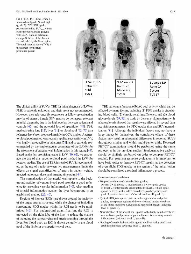
Several factors may significantly influence the arterial wall FDGuptake, andmust be taken into consideration for interpretation ofFDG-PET in LVVand PMR. For clinical routine, interpretationcriteria must be uniform, reproducible, and easy to use. ManyPET interpretation criteria have been proposed (Table 2), andevidence from the last 15 years supports the use of a visualgrading scale (vascular to liver uptake) (Fig. 1). We proposethe use of a standardized 0-to-3 grading system as follows:0 = no uptake (≤ mediastinum); 1 = low-grade uptake (< liver);
2 = intermediate-grade uptake (= liver), 3 = high-grade uptake(> liver), with grade 2 possibly indicative and grade 3 consideredpositive for active LVV (Table 3) [25, 73]. A total vascularscore (TVS) can be determined, for instance, at seven differentvascular regions (thoracic aorta, abdominal aorta, subclavianarteries, axillary arteries, carotid arteries, iliac arteries, andfemoral arteries) as negative (0) or positive, further scoredsemiquantitatively as 1 (minimal but not negligible FDG up-take), 2 (clearly increased FDG uptake), or 3 (verymarked FDGuptake). Therefore, a TVS could be calculated ranging from 0(no vascular FDGuptake in any of the seven vascular regions) to21 (vascular FDG uptake scored 3 in all seven territories).
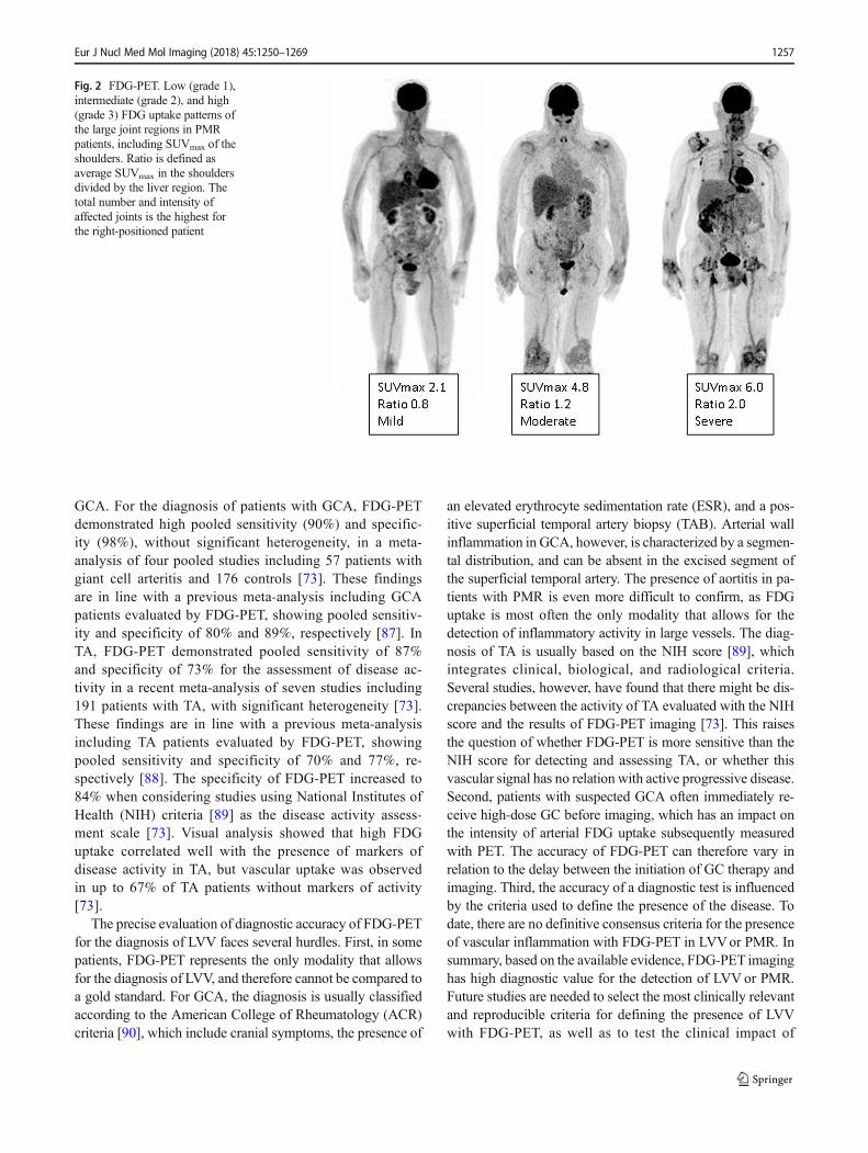
As PMR and GCA frequently overlap, typical FDG jointuptake patterns should be reported, including uptake inglenohumeral synovia, subacromial-subdeltoid bursa,supraspinatus tendinitis and biceps synovitis (shoulder),trochanteric/ischial bursa, hip synovia, interspinous regionsof the cervical and lumbar vertebrae, or the synovial tissueof the knees if present, including the use of a standardized 0-to-3 grading system [74, 75] (Fig. 2).
Atherosclerotic vascular uptake [76, 77], frequent with ag-ing, may be a source of false positivity for LVV evaluation,despite a classical Bpatchy^ uptake pattern. Uptake iniliofemoral arteries should be interpreted with caution, be-cause this is a frequent site of atherosclerosis. Taking theseconsiderations into account, vascular inflammation in LVVonFDG-PET classically appears as a smooth linear pattern, in-volving the aorta and its main branches (subclavian, carotid orvertebral arteries, pulmonary arteries specifically in TA), butnot all main branches have to be involved.
Quantification issues requiring further clarification Severalsemiquantitative methods have also been proposed, from simpleSUV metrics to target-to-background ratios (TBR) (Table 2).
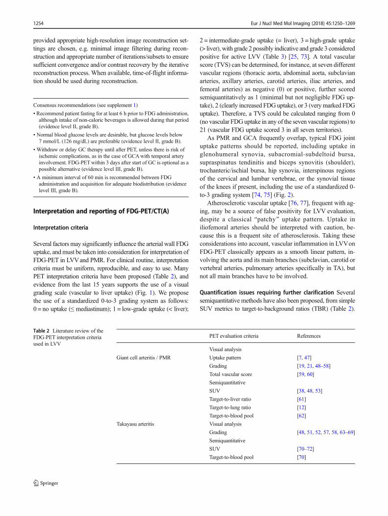
Table 2 Literature review of theFDG-PET interpretation criteriaused in LVV
PET evaluation criteria References
Visual analysis
Giant cell arteritis / PMR Uptake pattern [7, 47]
Grading [19, 21, 48–58]
Total vascular score [59, 60]
Semiquantitative
SUV [38, 48, 53]
Target-to-liver ratio [61]
Target-to-lung ratio [12]
Target-to-blood pool [62]
Takayasu arteritis Visual analysis
Grading [48, 51, 52, 57, 58, 63–69]
Semiquantitative
SUV [70–72]
Target-to-blood pool [70]
Consensus recommendations (see supplement 1)
• Recommend patient fasting for at least 6 h prior to FDG administration,although intake of non-caloric beverages is allowed during that period(evidence level II, grade B).
• Normal blood glucose levels are desirable, but glucose levels below7 mmol/L (126 mg/dL) are preferable (evidence level II, grade B).
• Withdraw or delay GC therapy until after PET, unless there is risk ofischemic complications, as in the case of GCAwith temporal arteryinvolvement. FDG-PETwithin 3 days after start of GC is optional as apossible alternative (evidence level III, grade B).
• A minimum interval of 60 min is recommended between FDGadministration and acquisition for adequate biodistribution (evidencelevel III, grade B).
1254 Eur J Nucl Med Mol Imaging (2018) 45:1250–1269
The clinical utility of SUVor TBR for initial diagnosis of LVVorPMR is currently unknown, and their use is not recommended.However, their relevance for recurrence or follow-up evaluationmay be of interest. Simple SUV metrics do not appear relevantin initial diagnosis, due to the high overlap between patients andcontrols [62] and the potential loss of specificity [48]. TBRmethods using lung [12], liver [61], or blood pool [62, 70] as areference have been proposed, mainly in GCA studies. A target-to-blood pool method was recently applied successfully in LVV,was highly reproducible in atheroma [78], and is currently rec-ommended by the cardiovascular committee of the EANM forthe assessment of vascular wall inflammation in this setting [44].Based on the few promising results in LVV [40, 62], we encour-age the use of this target-to-blood pool method in LVV forresearch studies. The use of TBR instead of SUVis recommend-ed, as the use of a ratio between two measurements limits theeffects on signal quantification of errors in patient weight,injected radiotracer dose, and imaging time point [44].
The normalization of the arterial wall uptake to the back-ground activity of venous blood pool provides a good refer-ence for assessing vascular inflammation [40]. Also, gradingof arterial inflammation against the liver background is anestablished method [25, 40].
Regions of interest (ROIs) are drawn around the majorityof the target arterial structure, while the chance of includingsurrounding FDG uptake within the ROI needs to be mini-mized [40]. For background quantification, the ROI isprojected on the right lobe of the liver to reduce the chanceof including the various veins and arteries running through theliver. For blood pool, an ROI is drawn centrally in the bloodpool of the (inferior or superior) caval vein.
TBR varies as a function of blood pool activity, which can beaffected by many factors, including (1) FDG uptake in circulat-ing blood cells, (2) chronic renal insufficiency, and (3) bloodglucose levels [79, 80]. A study by Lensen et al. in patients withatherosclerosis showed that results were affected by several dataacquisition parameters, i.e. FDG uptake time and SUV normal-ization [81]. Although the individual factors may not have alarge impact by themselves, the cumulative effects of thesefactors may result in substantial differences in reported SUVsthroughout studies and within multi-center trials. RepeatedPET/CT examinations should be performed using the sameprotocol as in the previous studies. Semiquantitative analysisshould be similarly performed (in order to compare PET/CTresults). For treatment response evaluation, it is important tohave basic (prior to therapy) PET/CT results, as the detectionof even slight FDG uptake in the region of the initial lesionshould be considered a residual inflammatory process.
Fig. 1 FDG-PET. Low (grade 1),intermediate (grade 2), and high(grade 3) LVV FDG uptakepatterns including SUVmax valuesof the thoracic aorta in patientswith GCA. Ratio is defined asaverage SUVmax of the thoracicaorta divided by the liver region.The total vascular score (TVS) isthe highest for the right-positioned patient
Consensus recommendations
• We propose the use of a standardized gradingsystem: 0 = no uptake (≤ mediastinum); 1 = low-grade uptake(< liver); 2 = intermediate-grade uptake (= liver), 3 = high-gradeuptake (> liver), with grade 2 considered possibly positive andgrade 3 positive for active LVV (evidence level II, grade B).
• Typical FDG joint uptake patterns including scapular and pelvicgirdles, interspinous regions of the cervical and lumbar vertebrae,or the knees should be evaluated and reported if present (evidencelevel II, grade B).
• Normalization of the arterial wall uptake to the background activity ofvenous blood pool provides a good reference for assessing vascularinflammation (evidence level II, grade B).
• Grading of arterial inflammation against the liver background is anestablished method (evidence level II, grade B).
Eur J Nucl Med Mol Imaging (2018) 45:1250–1269 1255

Diagnostic accuracy of FDG-PET/CT(A) for LVVand PMR
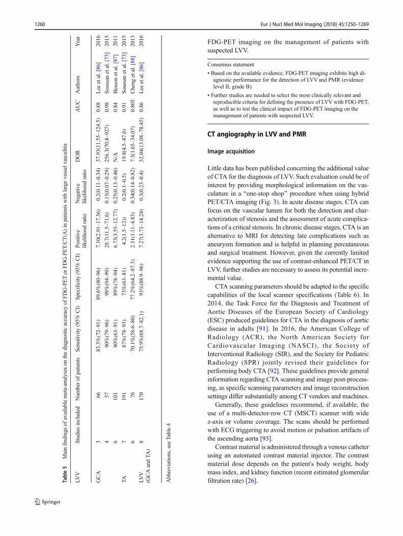
In general, the diagnostic performance of FDG-PET forthe detection of LVV is good; individual studies are sum-marized in Table 4, and meta-analyses are summarized inTable 5. A recent meta-analysis of eight studies including170 LVV patients with GCA or TA and 230 controls
confirmed that FDG-PET offers good diagnostic perfor-mance for the identification of LVV [86]. The diagnosticperformance of FDG-PET was higher for the detection ofGCA than TA (87% vs. 58%, respectively; p < 0.0001)[73, 86], but was impaired in patients under GC and/orimmunosuppressive treatment at the time of imaging [73].Of note, patients with TA are more often receiving long-term treatment at the time of imaging than patients with
Table 3 Proposed standardizedFDG-PET/CT(A) interpretationcriteria in LVV
Recommended PET interpretation criteria
For clinical use LVV visual grading (GCA and TA)
Grade 0: No vascular uptake (≤ mediastinum)
Grade 1: Vascular uptake < liver uptake
Grade 2: Vascular uptake = liver uptake, may be PET-positive
Grade 3: Vascular uptake > liver uptake, considered PET-positive
PMR associated visual assessment (only GCA)
Grade 0: No uptake
Grade 1: Uptake < liver uptake
Grade 2: Uptake = liver uptake
Grade 3: Uptake > liver uptake
Increased metabolic activity of the scapular and pelvic girdles
Increased metabolic activity of the knee bursae and capsule
Increased metabolic activity at the site of the cervical andlumbar interspinous bursae
Increased metabolic activity of the trochanteric and ischial bursae
In general for research only PET semiquantitative analysis*
Target: Average SUVmax artery of the vascular ROIs
Blood pool: Average SUVmean of several vein ROIs
TBR = average SUVmax artery / average SUVmean vein
Liver: SUVmax of a liver region, preferably the right lobe
TBR = average SUVmax artery / SUVmax of a liver region
Vascular targets:
- Carotid arteries
- Subclavia arteries
- Axillary arteries
- Vertebral arteries
- Ascending aorta
- Aortic arch
- Pulmonary arteries
- Descending aorta
- Abdominal aorta
Joints: scapulae and pelvic girdles, knees, cervical andlumbar interspinous bursae, trochanteric and ischial bursae
For clinical use Contrast-enhanced (PET/)CTA
Regular vascular wall thickness (mm)
Contrast enhancement
Presence of stenosis / aneurysm
TBR target-to-background ratio; SUV standardized uptake value; ROI region of interest; TA Takayasu arteritis;PMR polymyalgia rheumatica; GCA giant cell arteritis.
*SUV using EARL criteria [26]
1256 Eur J Nucl Med Mol Imaging (2018) 45:1250–1269
GCA. For the diagnosis of patients with GCA, FDG-PETdemonstrated high pooled sensitivity (90%) and specific-ity (98%), without significant heterogeneity, in a meta-analysis of four pooled studies including 57 patients withgiant cell arteritis and 176 controls [73]. These findingsare in line with a previous meta-analysis including GCApatients evaluated by FDG-PET, showing pooled sensitiv-ity and specificity of 80% and 89%, respectively [87]. InTA, FDG-PET demonstrated pooled sensitivity of 87%and specificity of 73% for the assessment of disease ac-tivity in a recent meta-analysis of seven studies including191 patients with TA, with significant heterogeneity [73].These findings are in line with a previous meta-analysisincluding TA patients evaluated by FDG-PET, showingpooled sensitivity and specificity of 70% and 77%, re-spectively [88]. The specificity of FDG-PET increased to84% when considering studies using National Institutes ofHealth (NIH) criteria [89] as the disease activity assess-ment scale [73]. Visual analysis showed that high FDGuptake correlated well with the presence of markers ofdisease activity in TA, but vascular uptake was observedin up to 67% of TA patients without markers of activity[73].
The precise evaluation of diagnostic accuracy of FDG-PETfor the diagnosis of LVV faces several hurdles. First, in somepatients, FDG-PET represents the only modality that allowsfor the diagnosis of LVV, and therefore cannot be compared toa gold standard. For GCA, the diagnosis is usually classifiedaccording to the American College of Rheumatology (ACR)criteria [90], which include cranial symptoms, the presence of
an elevated erythrocyte sedimentation rate (ESR), and a pos-itive superficial temporal artery biopsy (TAB). Arterial wallinflammation in GCA, however, is characterized by a segmen-tal distribution, and can be absent in the excised segment ofthe superficial temporal artery. The presence of aortitis in pa-tients with PMR is even more difficult to confirm, as FDGuptake is most often the only modality that allows for thedetection of inflammatory activity in large vessels. The diag-nosis of TA is usually based on the NIH score [89], whichintegrates clinical, biological, and radiological criteria.Several studies, however, have found that there might be dis-crepancies between the activity of TA evaluated with the NIHscore and the results of FDG-PET imaging [73]. This raisesthe question of whether FDG-PET is more sensitive than theNIH score for detecting and assessing TA, or whether thisvascular signal has no relation with active progressive disease.Second, patients with suspected GCA often immediately re-ceive high-dose GC before imaging, which has an impact onthe intensity of arterial FDG uptake subsequently measuredwith PET. The accuracy of FDG-PET can therefore vary inrelation to the delay between the initiation of GC therapy andimaging. Third, the accuracy of a diagnostic test is influencedby the criteria used to define the presence of the disease. Todate, there are no definitive consensus criteria for the presenceof vascular inflammation with FDG-PET in LVVor PMR. Insummary, based on the available evidence, FDG-PET imaginghas high diagnostic value for the detection of LVV or PMR.Future studies are needed to select the most clinically relevantand reproducible criteria for defining the presence of LVVwith FDG-PET, as well as to test the clinical impact of
Fig. 2 FDG-PET. Low (grade 1),intermediate (grade 2), and high(grade 3) FDG uptake patterns ofthe large joint regions in PMRpatients, including SUVmax of theshoulders. Ratio is defined asaverage SUVmax in the shouldersdivided by the liver region. Thetotal number and intensity ofaffected joints is the highest forthe right-positioned patient
Eur J Nucl Med Mol Imaging (2018) 45:1250–1269 1257
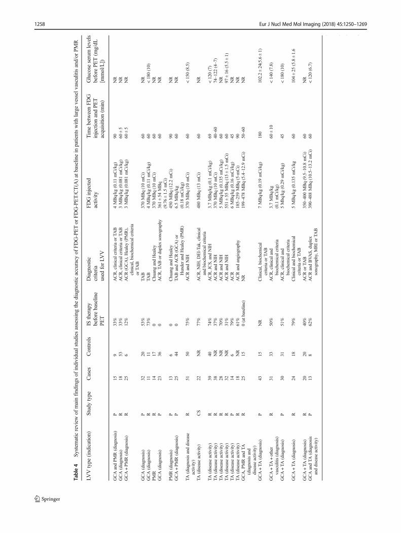
Table4
Systematicreview
ofmainfindings
ofindividualstudiesassessingthediagnosticaccuracy
ofFDG-PETor
FDG-PET/CT(A
)atbaselin
ein
patientswith
largevesselvasculitisand/or
PMR
LVVtype
(indication)
Studytype
Cases
Controls
IStherapy
before
baselin
ePET
Diagnostic
criteria
used
forLV
V
FDGinjected
activ
ityTim
ebetweenFD
GinjectionandPET
acquisition
(min)
Glucose
serum
levels
before
PET(m
g/dL
[mmol/L])
GCAandPMR(diagnosis)
P15
933%
ACR,clinicalcriteriaor
TAB
4MBq/kg
(0.11mCi/kg)
90NR
GCA(diagnosis)
R18
5333%
ACR,clinicalcriteriaor
TAB
3MBq/kg
(0.081
mCi/kg)
60±5
NR
GCA+PMR(diagnosis)
R25
612%
ACR(G
CA),Healey(PMR),
clinical,biochem
icalcriteria
orTA
B
3MBq/kg
(0.081
mCi/kg)
60±5
NR
GCA(diagnosis)
P32
2053%
TAB
370MBq(10mCi)
60NR
GCA(diagnosis)
R11
1173%
TAB
4MBq/kg
(0.11mCi/kg)
60<180(10)
PMR
R14
170
ChuangandHealey
370MBq(10mCi)
60NR
GCA(diagnosis)
P23
360
ACR,T
ABor
duplex
sonography
361±54
MBq
(9.76±1.5mCi)
60NR
PMR(diagnosis)
P13
60
ChuangandHealey
450MBq(12.2mCi)
90NR
GCA+PMR(diagnosis)
P25
440
TABandACR(G
CA)or
HunderandHealey(PMR)
6.5MBq/kg
(0.18mCi/kg)
60NR
TA(diagnosisanddisease
activity)
R51
5075%
ACRandNIH
370MBq(10mCi)
60<150(8.5)
TA(disease
activity)
CS
22NR
77%
ACR,N
IH,D
EI-Tak,clinical
andbiochemicalcriteria
480MBq(13mCi)
60NR
TA(disease
activity
)R
3940
74%
ACR,JCS,and
NIH
3.7MBq/kg
(0.1
mCi/k
g)69
<120(7)
TA(disease
activity)
R38
NR
37%
ACRandNIH
370MBq(10mCi)
40–60
74–122
(4–7)
TA(disease
activity)
R28
NR
70%
ACRandNIH
5MBq/kg
(0.135
mCi/kg)
60NR
TA(disease
activity)
R32
NR
31%
ACRandNIH
551±55
MBq(15±1.5mCi)
6097
±16
(5.5±1)
TA(disease
activity
)P
146
79%
ACR
6MBq/kg
(0.16mCi/k
g)45
NR
TA(disease
activity)
R18
NR
61%
ACRandangiography
185–259MBq(5-m
Ci)
90NR
GCA,P
MRandTA
(diagnosisand
diseaseactiv
ity)
R25
150(atb
aseline)
NR
199–478MBq(5.4–12.9mCi)
50–60
NR
GCA+TA
(diagnosis)
P43
15NR
Clin
ical,biochem
ical
criteriaor
TAB
7MBq/kg
(0.19mCi/kg)
180
102.2±24(5.6±1)
GCA+TA
+other
vasculitis(diagnosis)
R31
3350%
ACR,clinicaland
biochemicalcriteria
3.7MBq/kg
(0.1
mCi/kg)
60±10
<140(7.8)
GCA+TA
(diagnosis)
P30
3151%
ACR,clinicaland
biochemicalcriteria
5Mbq/kg(0.29mCi/kg)
45<180(10)
GCA+TA
(diagnosis)
R24
1879%
Clin
icalandbiochemical
criteriaor
TAB
5MBq/kg
(0.135
mCi/kg
60104±25
(5.8±1.6
GCA+TA
(diagnosis)
R20
2040%
ACRor
TAB
350–400MBq(9.5–10.8mCi)
60NR
GCAandTA
(diagnosis
anddiseaseactiv
ity)
P13
862%
ACRandBVAS,
duplex
sonography,M
RIor
TAB
390–488MBq(10.5–13.2
mCi)
60<120(6.7)
LVVtype
(indication)
PETanalysis
Threshold
used
for
diagnosisof
LVVatPE
TSensitiv
itySp
ecificity
Authors
Year
GCAandPMR(diagnosis)
QA:h
ighvascular
uptake
66.7%
(QA)
100%
(QA)
Lariviere
etal.[82]
2016
1258 Eur J Nucl Med Mol Imaging (2018) 45:1250–1269

Tab
le4
(contin
ued)
LVVtype
(indication)
PETanalysis
Threshold
used
for
diagnosisof
LVVatPET
Sensitiv
itySpecificity
Authors
Year
QA(visual)andSQ
A(vesselw
all
SUVmax/blood
pool
SUVmean)
GCA(diagnosis)
QA(visual)andSQ
A(aortic
SUVmaxandaortic/liver,
aortic/superiorcava,aortic/inferiorcava
SUVmaxratios)
QA1:
firstimpression
QA2:
diffusevascular
uptake
=liver
uptake
QA3:
diffusevascular
uptake
>liver
uptake
56%
(QA1)100%
(QA2)83%
(QA3))
98%
(QA1)51%
(QA2)91%
(QA3)
Stellin
gwerffet
al.[40]
2015
GCA+PM
R(diagnosis)
QA(visual)
QA1:
firstimpression
QA2:
diffusevascular
uptake
=liver
QA3:
diffusevascular
uptake
>liver
QA4:
diffusevascular
uptake
>femoralartery
92%
(QA1)100%
(QA2)100%
(QA3)80%
(QA4)
90%
(QA1)60%
(QA2)98%
(QA3)96%
(QA4)
Lensenet
al.[25]
2015
GCA(diagnosis)
SQA(vesselS
UVmax)
SQA:v
esselS
UVmaxcutoff1.89
80%
(SQA)
79%
(SQA)
Prieto-Gonzalezetal.[38]
2014
GCA(diagnosis)
SQA(aortic/liver,lung,orvenous
bloodpoolSU
Vmaxratio)
SQA:aortic/venousbloodpool
SUVmaxratio
cutoff1.53
81.8%(SQA)
91%
(SQA)
Bessonet
al.[62]
2014
PMR
QA(visual)andSQ
A(vesselS
UVmax)
QA:m
ildvascular
uptake
(<liver
uptake)
64.3%
(QA)
76.5%
(QA)
Yam
ashitaet
al.[83]
2012
GCA(diagnosis)
SQA(vessel/liver
SUVmax)
SQA:v
essel/liver
SUVratio
cutoff1
88.9%
(SQA)
95.1%
(SQA)
Hautzelet
al.[61]
2008
PMR(diagnosis)
QA(visual)andSQ
A(vessel/lunguptake
ratio)
NR
92.3%
(QA)
100%
(QA)
Moosiget
al.[12]
2004
GCA+PM
R(diagnosis)
QA(visual)
QA:m
oderateuptake
(=liver
uptake)
76%
(QA)
77%
(QA)
Blockmanset
al.[7]
2000
TA(diagnosisanddisease
activity)
QA(visual)andSQ
A(vesselS
UVmaxandvessel
SUVmax/liverSU
Vmean)
QA:intense
uptake
(>liver
uptake)in
theascendingaorta,
moderateuptake
(=liv
eruptake)in
theaorticarch
and
largeaorticbranch,and
mild
uptake
(<liver
uptake)in
thedescending
orabdominalaorta
83.3%
(QA)
90%
(QA)
Santhosh
etal.[68]
2014
TA(disease
activity)
QA(visual)andSQ
A(vesselS
UVmaxandvessel
SUVmax/liverSU
Vmean)
QA:m
oderateuptake
(=liver
uptake)
foraortaandmild
uptake
forothervessels
100%
(QA)
88.9%
(QA)
Karapolatet
al.[65]
2013
TA(disease
activity)
QA(visual)andSQ
A(vesselS
UVmaxandvessel
SUVmax/in
ferior
cava
SUVmean)
SQA:v
esselS
UVmaxcutoff2.1
92.6%
(SQA)
91.7%
(SQA)
Tezuka
etal.[70]
2012
TA(disease
activity)
QA(visual)andSQ
A(vessel/liver
SUVmax)
QA:m
oderatevascular
uptake
(=liver
uptake)
75%
(QA)
64.3%
(QA)
Lee
etal.[66]
2012
TA(disease
activity)
QA(visual)andSQ
A(vesselS
UVmaxandvessel
SUVmax/liverSU
Vmean)
QA:m
oderatevascular
uptake
(=liver
uptake)
69.2%
(QA)
33.3%
(QA)
Arnaudet
al.[64]
2009
TA(disease
activity)
QA(visual)
QA:m
oderateuptake
(=liver
uptake)
foraortaandmild
uptake
forothervessels
78%
(QA)
87%
(QA)
Lee
etal.[67]
2009
TA(disease
activity)
SQA(vesselS
UVmax)
SQA:S
UVmaxcutoff1.3
90.9%
(SQA)
88.8%
(SQA)
Kobayashi
etal.[72]
2005
TA(disease
activity)
QA(visual)
QA:m
ildvascular
uptake
(<liver
uptake)
92%
(QA)
100%
(QA)
Webbet
al.[69]
2004
GCA,P
MRandTA
(diagnosisand
diseaseactiv
ity)
QA(visual)andSQ
A(vascularSUVmean)
QA:sum
med
vascular
visualscorecutoff
8SQA:average
vascular
SUVmeancutoff0.697
84%
(QA)96%
(SQA)
86.7%
(QA)86.7%
(SQA)
Castellani
etal.[84]
2016
GCA+TA
(diagnosis)
SQA1(aortic
SUVmax)SQA2(aortic
wallS
UVmax/lm
SUVmax)
SQA1:
aorticSUVmaxcutoff1.74SQ
A2:
aortic
wallS
UVmax/lm
SUVmaxcutoff1.34
80%
(SQA1)100%
(SQA2)
83.3%
(SQA1)94%
(SQA2)
Martín
ez-Rodríguez
etal.
[43]
2014
GCA+TA
+other
vasculitis(diagnosis)
QA(visual)andSQ
A(vesselS
UVmax)or
JA(Q
Aandradiological/clin
icalelem
ents)
QA1:
mild
vascular
uptake
(<liv
eruptake)Q
A2:
moderatevascular
uptake
(=liver
uptake)SQA:v
essel
SUVmaxcutoff2.4
93.5%
(QA1)64.5%
(QA2)74.2%
(SQA)93.5%
(JA)
75.7%
(QA1)84.8%
(QA2)78.8%
(SQA)93.9%
(JA)
Rozzanigo
etal.[85]
2013
GCA+TA
(diagnosis)
QA(visual)
QA:m
oderateuptake
(=liver
uptake)foraorta
andmild
uptake
forothervessels
73.3%
(QA)
83.9%
(QA)
Fuchset
al.[52]
2012
GCA+TA
(diagnosis)
QA(visual)
QA:m
oderatevascular
uptake
(=liver
uptake)
92%
(QA)
91%
(QA)
Försteret
al.[21]
2011
GCA+TA
(diagnosis)
QA(visual)andSQ
A(vesselS
UVmax)
QA:intense
vascular
uptake
(>liv
eruptake)SQA:
SUVmaxcutoff2.24
65%
(QA)90%
(SQA)
80%
(QA)45%
(SQA)
Lehmannet
al.[48]
2011
GCAandTA
(diagnosis
anddiseaseactivity
)QA(visual)andSQ
A(vesselS
UVmax)
NR
92.3%
(QA)
100%
(QA)
Henes
etal.[53]
2008
Abbreviations:G
CA=giantcellarteritis;TA
=Takayasu
arteritis;LVV=largevesselvasculitis;DOR=diagnosticoddratio
;AUC=area
underthecurve;N/A
=notavailable
IS=im
munosuppressive;N
R=notreported.Studytype:P
=prospective;R=retrospective;CS=crosssectional.Ty
peof
vasculitis:LV
V=largevesselvasculitis;PMR=polymyalgiarheumatica;RF=
retroperito
nealfibrosis.D
iagnostic
criteria:ACR=American
College
ofRheum
atology;
NIH
=NationalInstitutes
ofHealth
;TAB=temporalarterybiopsy;M
RI=
magnetic
resonanceim
aging;
JCS=
JapaneseCirculatio
nSo
ciety;BVAS=Birmingham
VasculitisActivity
Score;DEI-Tak=DiseaseExtentIndex—Takayasu.PETanalysis:Q
A=qualitativ
eanalysis;SQA=semiquantitativ
eanalysis;JA=
jointanalysis;SUVmax=maxim
umstandardized
uptake
value;SU
Vmean=meanstandardized
uptake
value
Eur J Nucl Med Mol Imaging (2018) 45:1250–1269 1259
FDG-PET imaging on the management of patients withsuspected LVV.
CT angiography in LVV and PMR
Image acquisition
Little data has been published concerning the additional valueof CTA for the diagnosis of LVV. Such evaluation could be ofinterest by providing morphological information on the vas-culature in a Bone-stop shop^ procedure when using hybridPET/CTA imaging (Fig. 3). In acute disease stages, CTA canfocus on the vascular lumen for both the detection and char-acterization of stenosis and the assessment of acute complica-tions of a critical stenosis. In chronic disease stages, CTA is analternative to MRI for detecting late complications such asaneurysm formation and is helpful in planning percutaneousand surgical treatment. However, given the currently limitedevidence supporting the use of contrast-enhanced PET/CT inLVV, further studies are necessary to assess its potential incre-mental value.
CTA scanning parameters should be adapted to the specificcapabilities of the local scanner specifications (Table 6). In2014, the Task Force for the Diagnosis and Treatment ofAortic Diseases of the European Society of Cardiology(ESC) produced guidelines for CTA in the diagnosis of aorticdisease in adults [91]. In 2016, the American College ofRadiology (ACR), the North American Society forCardiovascular Imaging (NASCI), the Society ofInterventional Radiology (SIR), and the Society for PediatricRadiology (SPR) jointly revised their guidelines forperforming body CTA [92]. These guidelines provide generalinformation regarding CTA scanning and image post-process-ing, as specific scanning parameters and image reconstructionsettings differ substantially among CT vendors and machines.
Generally, these guidelines recommend, if available, theuse of a multi-detector-row CT (MSCT) scanner with widez-axis or volume coverage. The scans should be performedwith ECG triggering to avoid motion or pulsation artifacts ofthe ascending aorta [93].
Contrast material is administered through a venous catheterusing an automated contrast material injector. The contrastmaterial dose depends on the patient's body weight, bodymass index, and kidney function (recent estimated glomerularfiltration rate) [26].Ta
ble5
Mainfindings
ofavailablemeta-analyses
onthediagnosticaccuracy
ofFDG-PETor
FDG-PET/CT(A
)in
patientswith
largevesselvasculitis
LVV
Studiesincluded
Num
berof
patients
Sensitiv
ity(95%
CI)
Specificity
(95%
CI)
Positive
likelihoodratio
Negative
likelihoodratio
DOR
AUC
Authors
Year
GCA
366
83.3%(72–91)
89.6%(80–96)
7.10(2.91–17.36)
0.2(0.11–0.34)
37.93(11.55–124.5)
0.88
Lee
etal.[86]
2016
457
90%(79–96)
98%(94–99)
28.7(11.5–71.6)
0.15(0.07–0.29)
256.3(70.8–927)
0.98
Soussan
etal.[73]
2015
6101
80%(63–91)
89%(78–94)
6.73(3.55–12.77)
0.25(0.13–0.46)
N/A
0.84
Bessonetal.[87]
2011
TA7
191
87%(78–93)
73%(63–81)
4.2(1.5–12))
0.2(0.1–0.5)
19.8(4.5–87.6)
0.91
Soussanetal.[73]
2015
676
70.1%(58.6–80)
77.2%(64.2–87.3)
2.31(1.11–4.83)
0.34(0.14–0.82)
7.5(1.65–34.07)
0.805
Cheng
etal.[88]
2013
LVV
(GCAandTA
)8
170
75.9%(68.7–82.1)
93%(88.9–96)
7.27(3.71–14.24)
0.3(0.23–0.4)
32.04(13.08–78.45)
0.86
Lee
etal.[86]
2016
Abbreviations,see
Table4
Consensus statement
• Based on the available evidence, FDG-PET imaging exhibits high di-agnostic performance for the detection of LVVand PMR (evidencelevel II, grade B).
• Further studies are needed to select the most clinically relevant andreproducible criteria for defining the presence of LVV with FDG-PET,as well as to test the clinical impact of FDG-PET imaging on themanagement of patients with suspected LVV.
1260 Eur J Nucl Med Mol Imaging (2018) 45:1250–1269

CTA images should be reconstructed in thin slices (e.g. 1mmthick) to allow for additional multiplanar reformation (MPR)and 3D image post-processing. Preferably, isotropic voxelsshould be achieved. Both filtered back-projection and iterativereconstruction algorithms can be used, with the latter providingimproved image quality due to noise removal, and dose-savingpotential [94]. Medium-sharp or vascular reconstruction kernelscan be recommended with a reconstruction matrix of 512 × 512pixels and angiographic window setting using a level of 100Hounsfield units (HU) and width of 700 HU.
Interpretation and reporting
According to American College of Radiology guidelines, CTAis indicated to Bdiagnose and localize diseases with primarymanifestations in the arterial wall, including vasculitis, infec-tion, and degenerative disorders^ [92]. Arterial vessel wallthickening is the typical sign of vascular inflammation oncontrast-enhanced CT images (Fig. 4). In vasculitis, muralthickening usually involves the complete circumference of thevessel wall, whereas in atherosclerosis, plaque formation startsfrom a focal point rather than circumferentially. CTA-baseddiagnosis is greatly facilitated in the absence of atheroscleroticplaques and when the thickening is not concentric. A
circumferential aortic wall thickness of more than 2–3mmwithadventitial and peri-adventitial contrast enhancement is sugges-tive of aortitis [95, 96]. It is assumed that the degree of muralcontrast enhancement is associated with the inflammatory ac-tivity, as studies have shown that aortic wall contrast enhance-ment can resolve during GC therapy, while the wall thickeningmay persist [39].
Diagnostic accuracy of CTA
Although CTA itself is helpful for diagnosing LVV, the diag-nostic accuracy of combined FDG-PET/CTA scans remainsundefined. While the inflammatory activity within the vesselwall is displayed with high sensitivity on FDG-PET images,combining FDG-PET with CTA enhances specificity by pro-viding high-resolution anatomical details.
CTA also helps to differentiate different pathologic FDG-PET findings, as both vasculitis and atherosclerosis can demon-strate increased FDG wall uptake. Equally important in the con-tribution of CTA to the diagnostic accuracy of combined FDG-PET/CTA is its role in detecting structural changes and potentialcomplications of vasculitis [96]. During the primary diagnosticwork-up, CTA often helps to identify or exclude acute or symp-tomatic manifestations that require immediate therapy. For
Fig. 3 FDG-PET/CTA. On the left, a transaxial view of a contrast chestCT in a 67-year-old man with GCA, with an enlarged diameter of theascending aorta of 41 × 41 mm and moderately increased wall thicknessof 3.1 mm, and severely increased wall thickness of 4.7 mm of the
descending aorta (diameter of 30 × 31 mm). On the right, the fusedtransaxial images of the contrast chest CT and FDG-PET showing highlyelevated FDG uptake (average SUVmax 5.5) in the ascending and de-scending aorta
Table 6 Recommendations for patient preparation and image acquisition for the CTA scan
Patient positioning Supine, arms next to the body for hybrid PET/CTA; otherwise, arms should be elevated.
Scan volume Entire aorta including the cervical, upper extremity, visceral and renal, pelvic, and proximallower extremity arterial branches
Contrast material administration 80 to 150 mL iodinated low-osmolar or iso-osmolar contrast material with concentrationsof 300 to 400 mg iodine per mL is injected at flow rates of 3.0–5.0 mL/s via antecubital vein.
Specific CTA settings Optimal arterial contrast phase:Bolus tracking or test bolus technique, scanning in cranio-caudal directionAvoidance of aortic motion artifacts:ECG triggering
Specific CT machine settings Refer to individual CT scanner recommendations, as parameters and protocols may differamong vendors and machines.
Eur J Nucl Med Mol Imaging (2018) 45:1250–1269 1261

example, inflammatory vascular stenosis can lead to serioussequelae such as brain infarction or mesenteric ischemia withbowel necrosis. Furthermore, initial CTA may provide informa-tion on current disease stage, distribution, and duration. Duringdisease follow-up, CTA plays a particular role in the detectionand monitoring of complications such as aortic aneurysm anddissection. While stenosis is frequently observed in TA, GCAmay lead to aortic or arterial dilatation. These dilated arteries mayenlarge to aneurysms during the disease course, even thoughinflammatory activity is absent or sufficiently suppressed.
Monitoring the efficacy of immunosuppressivetherapy with FDG-PET/CT(A)
For monitoring of LVVactivity during and after treatment, re-lated biomarker measurements would be helpful.Unfortunately, both the cranial GCA and the extracranial large
vessel type of GCA or TA lack disease-specific serumbiomarkers.
Although FDG-PET/CT(A) has proven to be an importantimaging modality for diagnosis of non-temporal GCA, verylimited data are available on the role of FDG-PET/CT(A) forpatient management once treatment has started.With regard tothe utility of FDG-PET for assessing changes in arterial wallinflammation in response to GC and methotrexate, the resultsare mixed, and represent only small patient cohorts.
In the only prospective study, which was conducted byBlockmans et al. (Table 7), whole-body FDG-PET/CT imageswere acquired at baseline and after 3 and 6 months of GCtherapy [59]. The total vascular score (TVS) decreased from amean (± SD) of 7.9 ± 5.5 at baseline to 2.4 ± 3.5 on repeatPET scan at 3 months (p < 0.0005), but no further decreasewas seen at 6 months. In patients who experienced a relapse(recurrent signs and symptoms, together with an increase inacute-phase reactants), FDG-PETwas performed within 5 days.The authors found no difference in the predictive value ofFDG uptake between relapsing and non-relapsing patients.
A retrospective study by Bertagna et al. (Table 7) includeda total of nine patients, with eight GCA patients having anormalized FDG-PET at follow-up after GC therapy, andone patient without any change in the FDG-PET [97].
Fig. 4 CTangiography of the chest in two patients with GCA.Upper rowCTA of the aorta and the supra-aortic arteries in a 64-year-oldmale patientwith giant cell arteritis. Mural thickening and contrast enhancement of theaortic wall (arrows in B). Please note hypodense inner ring delineatingluminal contrast-enhanced blood from contrast-enhancing thickened aor-tic wall. Mural inflammatory changes are present in both subclavianarteries as visualized in cross section (bold arrow in A) and in a
longitudinal section (light arrows in A). Asterisk in A indicates the leftsubclavian vein. Lower rowAxial view of a CTangiography of a 76-year-old woman with GCA showing severely increased wall thickness of5.2 mm and contrast enhancement of the descending aorta (bold arrow)(A). Contrast CT of the same patient performed 4 years earlier, with nosignificant aortic wall thickening (B)
Consensus recommendation
• CTA and FDG-PET have complementary roles in the diagnosis of LVV(evidence level III, grade B).
• CTA has incremental value in detecting structural vascular changes andpotential complications of vasculitis (evidence level II, grade A).
1262 Eur J Nucl Med Mol Imaging (2018) 45:1250–1269

Table7
Literature
review
ofstudiesusingFDG-PET/CT(A
)formonito
ring
ofpatientswith
LVV/PMR
LVVtype
(indication)
Study
type
Cases
Controls
Therapy
Diagnostic
criteria
used
forLV
VPE
Tanalysis
Threshold
used
for
diagnosisof
LVV
atPET
Follo
w-upinterval
PET(m
onths)
Diagnostic
criteria
Authors
Year
GCA
P35
N/A
GC
TAB,baseline,PE
T,clinicaldata,lab
QA:v
isualu
ptake
intensity,T
VS
Decreasein
vessel
uptake,T
VS,
3and6
Clin
icaldata,lab
Blockmansetal.[59]
2006
GCA
R9
N/A
GC
Clin
icaldata,lab
QA:v
isual
SQA:v
esselS
UVmax,
vessel/liverSU
Vmaxratio
Decreasein:V
essel
/liverSU
Vratio
cutoff1
3Clin
icaldata,lab
Bertagnaetal.[97]
2010
LVV
R13
13GC
Clin
icaldata,lab
QA:v
isualu
ptake
intensity,T
VS
SQA:v
esselS
UVmaxCT:
W,W
/R
Decreasein
TVS,
WandW/R
NR
Clin
icaldata,lab
Mutoetal.[98]
2014
GCAandPM
RR
5N/A
MTX
NR
QA:v
isualu
ptakeintensity,
vesselto
liver
uptake,
TVS,T
JS
Decreasein:T
VS
andTJS
Median10.7
clinicaldata,lab
Cam
ellin
oetal.[99]
2010
GCA,T
AR
10N/A
CYC
NR
QA:v
isualv
esselto
liveruptake
Decreasein
vessel
uptake
3–4
Clin
icaldata,
BVAS,
lab
Henes
etal.[100]
2011
GCA,T
AR
5N/A
GC
Clin
ical,lab,other
imaging*
QA:v
esselu
ptakeintensity
Decreasein
vessel
uptake
Median10
Clin
icaldata,lab,
otherim
aging
DeLeeuw
etal.[51]
2004
GCAandPM
RP
35N/A
TAB,baseline,PE
T,clinicaldata,lab
QA:v
isualu
ptakeintensity,
TVS,T
JSDecreasein:v
essel
uptake,T
VSandTJS
3and6
clinicaldata,lab
Blockmansetal.[60]
2007
N/A
=notavailable
GC=glucocorticoids
CYC=cyclophosphamide
MTX=methotrexate
TVS=totalv
ascularscore
TJS
=totaljoint
score
BVAS=Birmingham
vasculitisactiv
ityindex
W=wallthickness
W/R
=ratio
ofwallthickness
totheradius
TAB=temporalarterybiopsy
Asterisk(*)=CTangiography,magnetic
resonanceangiography(M
RA),duplex
ultrasound
Eur J Nucl Med Mol Imaging (2018) 45:1250–1269 1263
Despite the small number of patients enrolled, the authorsconcluded that FDG-PET/CT might be a useful and accuratetool for evaluating disease progression.
In a study of five patients by Camellino et al. (Table 7),FDG-PET uptake decreased after the addition of methotrexateto the traditional GC treatment [99]. No studies have investi-gated whether GCA disease activity can be monitored byFDG-PET/CT in patients on GC-sparing drugs such as tumornecrotic factor α (TNF-α) blocking agents for TA orinterleukin-6 receptor blockade (tocilizumab) for GCA
Interestingly, a recent abstract by Nielsen et al. [37] report-ed that the FDG-PET/CT score, based on the semiquantitativeapproach described by Meller et al. [55] (score < 3), remainedpositive for vasculitis after 3 days of GC treatment, but be-came negative after 10 days.
Recent studies have shown that at the temporal artery level,granulomatous infiltrates can persist even up to 1 year follow-ing the start of GC treatment [28, 101]. Macrophages andgranulomatous inflammation have been reduced with GCtreatment in experimental models [102], decreasing in atime-dependent manner from 78 to 100% at initial biopsy, to50% at 9 months and 25% at 12months in sequential temporalartery biopsies. Lymphocytes may persist longer [102], andhave been reported to be present in GCA patients treated forup to 1 year [28].
This is in agreement with a study by Brack et al., in whichmacrophages persisted in the vessel walls of severe combinedimmunodeficiency disorder (SCID) mice engrafted with TABafter 1 week of GC treatment [103]. These findings are also inline with the fact that FDG-PET/CT(A) shows arterial walluptake after 6 months in treated patients, although the uptakeis no longer diagnostic for vasculitis.
Prieto-González et al. prospectively assessed GC-inducedchanges in CTA findings of LVV in patients with GCA [39].Forty patients with biopsy-proven GCA evaluated by CTA atdiagnosis were prospectively followed and scheduled for anew CTA after approximately 1 year of treatment. Vessel wallthickening, diameter, and contrast enhancement of the aortaand its tributaries were evaluated. Results were compared withthose obtained at the time of diagnosis. CTAwas repeated for35 patients after a median follow-up of 13.5 months (IQ 25–75% 12.4–15.8). Arterial wall thickening was still present in17 patients (68% of the patients who initially had LVV). Thenumber of affected segments and the wall thickness at variousaortic segments were significantly decreased, and no patientsdeveloped new lesions, new aortic dilation, or an increase inprevious dilation. Contrast enhancement disappeared in 15(94%) of 16 patients in whom this finding could be assessed.Signs of LVV improve with treatment. While contrast en-hancement resolves in the majority of patients, vessel wallthickening persists in two-thirds. However, the number of af-fected aortic segments, as well as the aortic wall thickness,decreases significantly.
For PMR, there is one study with sequential PET/CT,by Blockmans et al. (Table 7), using the same methodol-ogy as that for GCA. The authors found that vascularFDG uptake was present in 11 patients, with slight ormoderate uptake at diagnosis in nine of 35 patients, andthat the uptake decreased after 3 and 6 months [60]. Atbaseline, FDG uptake in the shoulders was present in allbut two patients, and uptake was still present, although toa lesser extent, after 3 and 6 months of GC therapy. Thesame holds true for the hips and the spinous processes.No difference was found in the predictive value of FDGuptake at baseline and after 3 months at the shoulders,hips, or spinous processes between patients who experi-enced a relapse and those who did not.
The optimal length of time before performing FDG-PET in PMR after treatment with GC is unclear. Arecent study by Palard-Novello et al. evaluated the use ofFDG-PET/CT(A) for the assessment of tocilizumab as first-line treatment in patients with PMR [104]. They found thatFDG uptake decreased significantly but moderately after toci-lizumab therapy in PMR patients, and may reflect diseaseactivity.
Consensus statements on open issuesfor future research agenda
Clinical issues
& Further establish the role of FDG-PET/CT in patient man-agement and evaluate its role in treatment monitoring.When to use FDG-PET/CT in the diagnosis, in the fol-low-up, and how often?
& Development of guidelines in LVV and PMR imagingwith FDG-PET/CT(A) similar to those previously devel-oped for FDG-PET/CT in oncology (EARL) criteria [26].Randomized prospective studies are needed for moreevidence.
& Including imaging biomarkers with the current diag-nostic criteria to be considered for TA, GCA, and/orPMR.
& Finding a consensus in the clinical support for performingimaging as early as possible and before starting GC ther-apy if treatment delay can be justified due to non-criticalsymptoms.
& Further investigation of the GC effect on vascular FDGuptake.
Consensus statement
FDG-PET/CT(A) may be of value for evaluating response to treatmentby monitoring functional metabolic information and detectingstructural vascular changes (evidence level III, grade C), butadditional prospective FDG-PET/CT(A) studies are warranted.
1264 Eur J Nucl Med Mol Imaging (2018) 45:1250–1269

& Theranostics (diagnostics for selected therapy) for LVV/PMR, which may open more paths to targeted therapy,resulting in personal/precision medicine. Radiolabeledtocilizumab or other monoclonal antibody PET tracersare potential candidates for this.
& Circumstances of when there may be myocardial involve-ment in patients with LVV should be further investigated(additional myocardial perfusion imaging, CT coronarycalcium assessment, and CTangiography may be needed),including the risk of cardiovascular events due to drugtherapy in LVV [105].
& Worldwide reimbursement for application of FDG-PET/CT(A) in LVV/PMR is needed.
Methodological issues
& Standardization of visual scoring and (semi)quantificationin FDG-PET in LVVand PMR is essential for interpreta-tion, for optimal comparison among centers, especially infuture multicenter trials.
& Decide how much thickening is mild, moderate, or severe(not established in literature). Based on our expertise, wethink that ≥2 mm (up to 2.9) may be mild, ≥ 3 mm (up to3.9) moderate, and ≥4 mm severe.
& Consensus needed on which quantification method to ap-ply in LVV.
& An uptake interval of 60 min after FDG injection is rec-ommended, but 90–120 min intervals can be evaluated forbetter image quality.
& Dual-time-point imaging may improve the target-to-background ratio, resulting in better image quality due togreater FDG blood pool clearance, particularly in patientswith reduced kidney function. However, evidence-baseddata are lacking.
& New techniques for imaging and reconstruction of theskull that enable visualization of the superficial temporalartery, which will result in better comparison of local LVVwith TAB.
& Value of combining FDG-PET with CTA as a standardprocedure in LVVand PMR, single modalities or hybrid.
& Value of FDG-PET/MRI in monitoring LVV and PMR,i.e. reduction of radiation dose [106].
& Development of online trainingmodalities for interpretation.
Technical issues
& Optimization of the application of hybrid imaging in mon-itoring (residual) vascular wall disease in LVV.
& The use of vasculitis-specific tracers, directed againstcells/proteins involved in and unique for the pathophysi-ology of LVVand PMR, should be investigated.
& New developments in camera systems, such as PET/MRI,enable us to combine metabolism or other molecular targets(PET) with vascular tissue layer characterization (MRI),including a reduction in radiation dose and improved cra-nial visualization. The value of these new multi-modalityimaging systemsmay be of interest for LVVassessment andmonitoring.
& Optimal use of (low-dose) CT to distinguish active athero-sclerosis from active vasculitis by pattern recognition, vi-sually as well as with the use of dedicated software meth-odologies (textural feature).
Several open issues are also in line with the recommendationfor the use of imaging in large vessel vasculitis in clinical practiceof the European League Against Rheumatism (EULAR) [107].
Conclusion
The present procedural recommendation paper provides rec-ommendations to assist imaging specialists and clinicians inrequesting, performing, and interpreting the results of FDG-PET in patients with suspected LVVand PMR.
Based on the present clinical data, FDG-PET/CT(A) has animportant role in the diagnosis of extracranial vascular in-volvement in patients with LVV/PMR, but additional random-ized studies are needed to support this.
Improvements in FDG-PET/CT(A) procedures will help tooptimize the diagnostic andmonitoring value of this techniquein LVV/PMR.
Visual qualitative methods are most commonly used, butsemiquantitative methods such as the vascular/blood ratio andvascular/liver ratio using SUVs are increasingly being used.
The addition of CTA to FDG-PET provides high-resolutionimaging of vascular morphology that can potentially improvediagnostic accuracy, but more importantly provides informa-tion on the presence of possible complications such as steno-sis, organ ischemia, aneurysm formation, and dissection.
Further prospective studies involving large cohorts of GCA/PMR patients are needed to investigate and validate the role ofsemiquantitative methods for the assessment of LVV.
Several other open issues, as stated above, need to be stud-ied for optimal performance of FDG-PET/CT(A) in the diag-nosis, (treatment) monitoring, and future theranostics in LVV/PMR, further improving the levels of evidence and grades ofrecommendations.
Acknowledgements This Joint Procedural Recommendation summa-rizes the views of the Cardiovascular and Inflammation & InfectionCommittees of the European Association of Nuclear Medicine (EANM)and reflects recommendations for which the EANM cannot be held re-sponsible. The Recommendations should be taken in the context of goodpractice of nuclear medicine and are not a substitute for national andinternational legal or regulatory provisions.
Eur J Nucl Med Mol Imaging (2018) 45:1250–1269 1265

The Joint Procedural Recommendation was brought to the attention ofall other EANM Committees and to the EANM National Societies ofNuclear Medicine. The comments and suggestions from the EANMCommittees and the EANM National Societies are greatly appreciatedand have been considered for this Joint Procedural Recommendation.Funding No funding was received for this study.
Compliance with ethical standards
Conflict of interest All authors declare that they have no conflict ofinterest with respect to this study.
Ethical approval This article does not contain any studies with animalsor human participants performed by any of the authors.
Open Access This article is distributed under the terms of the CreativeCommons At t r ibut ion 4 .0 In te rna t ional License (h t tp : / /creativecommons.org/licenses/by/4.0/), which permits unrestricted use,distribution, and reproduction in any medium, provided you giveappropriate credit to the original author(s) and the source, provide a linkto the Creative Commons license, and indicate if changes were made.
References
1. Jennette JC, Falk RJ, Bacon PA, Basu N, CidMC, Ferrario F, et al.2012 revised International Chapel Hill Consensus ConferenceNomenclature of Vasculitides. Arthritis Rheum. 2013;65:1–11.
2. Carmona FD, Coit P, Saruhan-Direskeneli G, Hernandez-Rodriguez J, Cid MC, Solans R, et al. Analysis of the commongenetic component of large-vessel vasculitides through a meta-Immunochip strategy. Sci Rep. 2017;7:43953.
3. Langford CA, Cuthbertson D, Ytterberg SR, Khalidi N, MonachPA, Carette S, et al. A randomized, double-blind trial of Abatacept(CTLA-4Ig) for the treatment of Takayasu arteritis. ArthritisRheumatol. 2017;69:846–53.
4. Langford CA, Cuthbertson D, Ytterberg SR, Khalidi N, MonachPA, Carette S, et al. A randomized, double-blind trial of Abatacept(CTLA-4Ig) for the treatment of Giant cell arteritis. ArthritisRheumatol. 2017;69:837–45.
5. Gravanis MB. Giant cell arteritis and Takayasu aortitis: morpho-logic, pathogenetic and etiologic factors. Int J Cardiol.2000;75(Suppl 1):S21–33.
6. Maksimowicz-McKinnon K, Clark TM, Hoffman GS. Takayasuarteritis and giant cell arteritis: a spectrum within the same dis-ease? Medicine (Baltimore). 2009;88:221–6.
7. Blockmans D, Stroobants S, Maes A, Mortelmans L. Positronemission tomography in giant cell arteritis and polymyalgiarheumatica: evidence for inflammation of the aortic arch. Am JMed. 2000;108:246–9.
8. Ernst D, Baerlecken NT, Schmidt RE, Witte T. Large vessel vas-culitis and spondyloarthritis: coincidence or associated diseases?Scand J Rheumatol. 2014;43:246–8.
9. Kubota R, Yamada S, Kubota K, Ishiwata K, Tamahashi N, Ido T.Intratumoral distribution of fluorine-18-fluorodeoxyglucose in vivo:high accumulation in macrophages and granulation tissues studiedby microautoradiography. J Nucl Med. 1992;33:1972–80.
10. Cimmino MA, Zampogna G, Parodi M. Is FDG-PET useful in theevaluation of steroid-resistant PMR patients? Rheumatology(Oxford). 2008;47:926–7.
11. Lavado-Perez C, Martinez-Rodriguez I, Martinez-Amador N,Banzo I, Quirce R, Jimenez-Bonilla J, et al. (18)F-FDG-PET/CT
for the detection of large vessel vasculitis in patients withpolymyalgia rheumatica. Rev Esp Med Nucl Imagen Mol.2015;34:275–81.
12. Moosig F, Czech N, Mehl C, Henze E, Zeuner RA, Kneba M, et al.Correlation between 18-fluorodeoxyglucose accumulation in large ves-sels and serological markers of inflammation in polymyalgiarheumatica: a quantitative PETstudy. Ann RheumDis. 2004;63:870–3.
13. Rehak Z, Vasina J, Nemec P, Fojtik Z, Koukalova R, Bortlicek Z,et al. Various forms of (18)F-FDG-PET and PET/CT findings inpatients with polymyalgia rheumatica. Biomed PapMed Fac UnivPalacky Olomouc Czech Repub. 2015;159:629–36.
14. Einspieler I, Thurmel K, Pyka T, EiberM,WolframS,Moog P, et al.Imaging large vessel vasculitis with fully integrated PET/MRI: apilot study. Eur J Nucl Med Mol Imaging. 2015;42:1012–24.
15. Loffler C, Hoffend J, Benck U, Kramer BK, Bergner R. The valueof ultrasound in diagnosing extracranial large-vessel vasculitiscompared to FDG-PET/CT: a retrospective study. ClinRheumatol. 2017;
16. Lie JT. Aortic and extracranial large vessel giant cell arteritis: areview of 72 cases with histopathologic documentation. SeminArthritis Rheum. 1995;24:422–31.
17. De BH, Dumont A, Liozon E, Lambert M, Boutemy J, Maigne Get al. Giant-cell arteritis: concordance study between aortic CTangiography and FDG-PET/CT in detection of large-vessel in-volvement. Eur J Nucl Med Mol Imaging 2017.
18. Blockmans D, Bley T, Schmidt W. Imaging for large-vessel vas-culitis. Curr Opin Rheumatol. 2009;21:19–28.
19. Both M, Ahmadi-Simab K, Reuter M, Dourvos O, Fritzer E,Ullrich S, et al. MRI and FDG-PET in the assessment of inflam-matory aortic arch syndrome in complicated courses of giant cellarteritis. Ann Rheum Dis. 2008;67:1030–3.
20. Dasgupta B. Concise guidance: diagnosis and management ofgiant cell arteritis. Clin Med (Lond). 2010;10:381–6.
21. Forster S, Tato F, Weiss M, Czihal M, Rominger A, Bartenstein P,et al. Patterns of extracranial involvement in newly diagnosedgiant cell arteritis assessed by physical examination, colour codedduplex sonography and FDG-PET. Vasa. 2011;40:219–27.
22. Pipitone N, Versari A, Salvarani C. Role of imaging studies in thediagnosis and follow-up of large-vessel vasculitis: an update.Rheumatology (Oxford). 2008;47:403–8.
23. Tato F, Hoffmann U. Giant cell arteritis: a systemic vascular dis-ease. Vasc Med. 2008;13:127–40.
24. Ben-Haim S, Gacinovic S, Israel O. Cardiovascular infection andinflammation. Semin Nucl Med. 2009;39:103–14.
25. Lensen KD, Comans EF, Voskuyl AE, van der Laken CJ, BrouwerE, Zwijnenburg AT, et al. Large-vessel vasculitis: interobserveragreement and diagnostic accuracy of 18F-FDG-PET/CT.Biomed Res Int. 2015;2015:914692.
26. Boellaard R, Delgado-Bolton R, Oyen WJ, Giammarile F, TatschK, Eschner W, et al. FDG-PET/CT: EANM procedure guidelinesfor tumour imaging: version 2.0. Eur J Nucl Med Mol Imaging.2015;42:328–54.
27. Jamar F, Buscombe J, Chiti A, Christian PE, Delbeke D, DonohoeKJ, et al. EANM/SNMMI guideline for 18F-FDG use in inflam-mation and infection. J Nucl Med. 2013;54:647–58.
28. Maleszewski JJ, Younge BR, Fritzlen JT, Hunder GG, Goronzy JJ,Warrington KJ, et al. Clinical and pathological evolution of giantcell arteritis: a prospective study of follow-up temporal artery bi-opsies in 40 treated patients. Mod Pathol. 2017;
29. van Marken Lichtenbelt WD, Vanhommerig JW, Smulders NM,Drossaerts JM, Kemerink GJ, Bouvy ND, et al. Cold-activatedbrown adipose tissue in healthy men. N Engl J Med. 2009;360:1500–8.
30. Parysow O, Mollerach AM, Jager V, Racioppi S, San RJ,Gerbaudo VH. Low-dose oral propranolol could reduce brown
1266 Eur J Nucl Med Mol Imaging (2018) 45:1250–1269

adipose tissue F-18 FDG uptake in patients undergoing PETscans. Clin Nucl Med. 2007;32:351–7.
31. Chareonthaitawee P, Beanlands RS, Chen W, Dorbala S, MillerEJ, Murthy VL et al. Joint SNMMI-ASNC expert consensus doc-ument on the role of 18F-FDG-PET/CT in cardiac sarcoid detec-tion and therapy monitoring. J Nucl Cardiol 2017.
32. Dorbala S, Di Carli MF, Delbeke D, Abbara S, DePuey EG,Dilsizian V, et al. SNMMI/ASNC/SCCT guideline for cardiacSPECT/CT and PET/CT 1.0. J Nucl Med. 2013;54:1485–507.
33. Rabkin Z, Israel O, Keidar Z. Do hyperglycemia and diabetesaffect the incidence of false-negative 18F-FDG-PET/CT studiesin patients evaluated for infection or inflammation and cancer?A comparative analysis. J Nucl Med. 2010;51:1015–20.
34. Zhuang HM, Cortes-Blanco A, Pourdehnad M, Adam LE,Yamamoto AJ, Martinez-Lazaro R, et al. Do high glucose levelshave differential effect on FDG uptake in inflammatory and ma-lignant disorders? Nucl Med Commun. 2001;22:1123–8.
35. Bucerius J, Mani V, Moncrieff C, Machac J, Fuster V, FarkouhME, et al. Optimizing 18F-FDG-PET/CT imaging of vessel wallinflammation: the impact of 18F-FDG circulation time, injecteddose, uptake parameters, and fasting blood glucose levels. Eur JNucl Med Mol Imaging. 2014;41:369–83.
36. Wahl RL, Henry CA, Ethier SP. Serum glucose: effects on tumorand normal tissue accumulation of 2-[F-18]-fluoro-2-deoxy-D-glucose in rodents with mammary carcinoma. Radiology.1992;183:643–7.
37. Nielsen BD, Tønder Hansen L, Keller KK, Therkildsen P, Hauge EM,Gormsen LC. Attenuation of fluorine-18-fluorodeoxyglucose uptakein large vessel giant cell arteritis after short-term high-dose steroidtreatment - a diagnostic window of opportunity. Arthritis Rheumatol.2016. 68[suppl 10]. http://acrabstracts.org/abstract/attenuation-of-fluorine-18-fluorodeoxyglucose-uptake-in-large-vessel-giant-cell-arteritis-after-short-term-high-dose-steroid-treatment-a-diagnostic-window-of-opportunity/. Accessed 7 Mar 2018.
38. Prieto-Gonzalez S, Depetris M, Garcia-Martinez A, Espigol-FrigoleG, Tavera-Bahillo I, Corbera-Bellata M, et al. Positron emissiontomography assessment of large vessel inflammation in patientswith newly diagnosed, biopsy-proven giant cell arteritis: a prospec-tive, case-control study. Ann Rheum Dis. 2014;73:1388–92.
39. Prieto-Gonzalez S, Garcia-Martinez A, Tavera-Bahillo I,Hernandez-Rodriguez J, Gutierrez-Chacoff J, Alba MA, et al.Effect of glucocorticoid treatment on computed tomography an-giography detected large-vessel inflammation in giant-cell arteri-tis. A prospective, longitudinal study. Medicine (Baltimore).2015;94:e486.
40. Stellingwerff MD, Brouwer E, Lensen KJ, Rutgers A, Arends S,van der Geest KS, et al. Different scoring methods of FDG-PET/CT in Giant cell arteritis: need for standardization. Medicine(Baltimore). 2015;94:e1542.
41. Tawakol A,Migrino RQ, Bashian GG, Bedri S, VermylenD, CuryRC, et al. In vivo 18F-fluorodeoxyglucose positron emission to-mography imaging provides a noninvasive measure of carotidplaque inflammation in patients. J Am Coll Cardiol. 2006;48:1818–24.
42. Blomberg BA, Bashyam A, Ramachandran A, Gholami S,H o u s hma n d S , S a l a v a t i A , e t a l . Q u a n t i f y i n g[(1)(8)F]fluorodeoxyglucose uptake in the arterial wall: the effectsof dual time-point imaging and partial volume effect correction. EurJ Nucl Med Mol Imaging. 2015;42:1414–22.
43. Martinez-Rodriguez I, Martinez-Amador N, Banzo I, Quirce R,Jimenez-Bonilla J, De Arcocha-Torres M, et al. Assessment ofaortitis by semiquantitative analysis of 180-min 18F-FDG-PET/CT acquisition images. Eur J Nucl Med Mol Imaging. 2014;41:2319–24.
44. Bucerius J, Hyafil F, Verberne HJ, Slart RH, Lindner O, Sciagra R,et al. Position paper of the cardiovascular Committee of the European
Association of nuclear medicine (EANM) on PET imaging of ath-erosclerosis. Eur J Nucl Med Mol Imaging. 2016;43:780–92.
45. Yamada I, Nakagawa T, Himeno Y, Numano F, Shibuya H.Takayasu arteritis: evaluation of the thoracic aorta with CT angi-ography. Radiology. 1998;209:103–9.
46. Huet P, Burg S, Le GD, Hyafil F, Buvat I. Variability and uncer-tainty of 18F-FDG-PET imaging protocols for assessing inflam-mation in atherosclerosis: suggestions for improvement. J NuclMed. 2015;56:552–9.
47. Blockmans D, Maes A, Stroobants S, Nuyts J, Bormans G,Knockaert D, et al. New arguments for a vasculitic nature ofpolymyalgia rheumatica using positron emission tomography.Rheumatology (Oxford). 1999;38:444–7.
48. Lehmann P, Buchtala S, Achajew N, Haerle P, Ehrenstein B,Lighvani H, et al. 18F-FDG-PET as a diagnostic procedure inlarge vessel vasculitis-a controlled, blinded re-examination of rou-tine PET scans. Clin Rheumatol. 2011;30:37–42.
49. Bleeker-Rovers CP, Bredie SJ, van der Meer JW, Corstens FH,Oyen WJ. F-18-fluorodeoxyglucose positron emission tomogra-phy in diagnosis and follow-up of patients with different types ofvasculitis. Neth J Med. 2003;61:323–9.
50. Czihal M, Tato F, Forster S, Rademacher A, Schulze-Koops H,Hoffmann U. Fever of unknown origin as initial manifestation oflarge vessel giant cell arteritis: diagnosis by colour-coded sonog-raphy and 18-FDG-PET. Clin Exp Rheumatol. 2010;28:549–52.
51. de Leeuw K, Bijl M, Jager PL. Additional value of positron emis-sion tomography in diagnosis and follow-up of patients with largevessel vasculitides. Clin Exp Rheumatol. 2004;22:S21–6.
52. Fuchs M, Briel M, Daikeler T, Walker UA, Rasch H, Berg S, et al.The impact of 18F-FDG-PETon the management of patients withsuspected large vessel vasculitis. Eur J Nucl Med Mol Imaging.2012;39:344–53.
53. Henes JC, Muller M, Krieger J, Balletshofer B, Pfannenberg AC,Kanz L, et al. [18F] FDG-PET/CTas a new and sensitive imagingmethod for the diagnosis of large vessel vasculitis. Clin ExpRheumatol. 2008;26:S47–52.
54. Hooisma GA, Balink H, Houtman PM, Slart RH, Lensen KD.Parameters related to a positive test result for FDG-PET(/CT) forlarge vessel vasculitis: a multicenter retrospective study. ClinRheumatol. 2012;31:861–71.
55. Meller J, Strutz F, Siefker U, Scheel A, Sahlmann CO, Lehmann K,et al. Early diagnosis and follow-up of aortitis with [(18)F]FDG-PET and MRI. Eur J Nucl Med Mol Imaging. 2003;30:730–6.
56. Morinobu A, Tsuji G, Kasagi S, Saegusa J, Hayashi H, NakazawaT, et al. Role of imaging studies in the diagnosis and evaluation ofgiant cell arteritis in Japanese: report of eight cases. ModRheumatol. 2011;21:391–6.
57. Scheel AK, Meller J, Vosshenrich R, Kohlhoff E, Siefker U,Muller GA, et al. Diagnosis and follow up of aortitis in the elderly.Ann Rheum Dis. 2004;63:1507–10.
58. Walter MA, Melzer RA, Schindler C, Muller-Brand J, Tyndall A,Nitzsche EU. The value of [18F]FDG-PET in the diagnosis oflarge-vessel vasculitis and the assessment of activity and extentof disease. Eur J Nucl Med Mol Imaging. 2005;32:674–81.
59. Blockmans D, De CL, Vanderschueren S, Knockaert D,Mortelmans L, Bobbaers H. Repetitive 18F-fluorodeoxyglucosepositron emission tomography in giant cell arteritis: a prospectivestudy of 35 patients. Arthritis Rheum. 2006;55:131–7.
60. Blockmans D, De CL, Vanderschueren S, Knockaert D,Mortelmans L, Bobbaers H. Repetitive 18-fluorodeoxyglucosepositron emission tomography in isolated polymyalgiarheumatica: a prospective study in 35 patients. Rheumatology(Oxford). 2007;46:672–7.
61. Hautzel H, Sander O, Heinzel A, Schneider M, Muller HW.Assessment of large-vessel involvement in giant cell arteritis with
Eur J Nucl Med Mol Imaging (2018) 45:1250–1269 1267

18F-FDG-PET: introducing an ROC-analysis-based cutoff ratio. JNucl Med. 2008;49:1107–13.
62. Besson FL, De BH, Parienti JJ, Bouvard G, Bienvenu B, AgostiniD. Towards an optimal semiquantitative approach in giant cellarteritis: an (18)F-FDG-PET/CT case-control study. Eur J NuclMed Mol Imaging. 2014;41:155–66.
63. Andrews J, Al-Nahhas A, Pennell DJ, Hossain MS, Davies KA,Haskard DO, et al. Non-invasive imaging in the diagnosis andmanagement of Takayasu's arteritis. Ann Rheum Dis. 2004;63:995–1000.
64. Arnaud L, Haroche J, Malek Z, Archambaud F, Gambotti L,Grimon G, et al. Is (18)F-fluorodeoxyglucose positron emissiontomography scanning a reliable way to assess disease activity inTakayasu arteritis? Arthritis Rheum. 2009;60:1193–200.
65. Karapolat I, Kalfa M, Keser G, Yalcin M, Inal V, Kumanlioglu K,et al. Comparison of F18-FDG-PET/CT findings with current clin-ical disease status in patients with Takayasu's arteritis. Clin ExpRheumatol. 2013;31:S15–21.
66. Lee KH, Cho A, Choi YJ, Lee SW, Ha YJ, Jung SJ, et al. The roleof (18) F-fluorodeoxyglucose-positron emission tomography inthe assessment of disease activity in patients with takayasu arter-itis. Arthritis Rheum. 2012;64:866–75.
67. Lee SG, Ryu JS, Kim HO, Oh JS, Kim YG, Lee CK, et al.Evaluation of disease activity using F-18 FDG-PET-CT in patientswith Takayasu arteritis. Clin Nucl Med. 2009;34:749–52.
68. Santhosh S,Mittal BR, Gayana S, Bhattacharya A, SharmaA, JainS. F-18 FDG-PET/CT in the evaluation of Takayasu arteritis: anexperience from the tropics. J Nucl Cardiol. 2014;21:993–1000.
69. Webb M, Chambers A, Al-Nahhas A, Mason JC, Maudlin L,Rahman L, et al. The role of 18F-FDG-PET in characterisingdisease activity in Takayasu arteritis. Eur J Nucl Med MolImaging. 2004;31:627–34.
70. Tezuka D, Haraguchi G, Ishihara T, Ohigashi H, Inagaki H, SuzukiJ, et al. Role of FDG-PET-CT in Takayasu arteritis: sensitive detec-tion of recurrences. JACC Cardiovasc Imaging. 2012;5:422–9.
71. Alibaz-Oner F, Dede F, Ones T, Turoglu HT, Direskeneli H.Patients with Takayasu's arteritis having persistent acute-phaseresponse usually have an increased major vessel uptake by 18F-FDG-PET/CT. Mod Rheumatol. 2015;25:752–5.
72. Kobayashi Y, Ishii K, Oda K, Nariai T, Tanaka Y, Ishiwata K, et al.Aortic wall inflammation due to Takayasu arteritis imaged with18F-FDG-PET coregistered with enhanced CT. J Nucl Med.2005;46:917–22.
73. Soussan M, Nicolas P, Schramm C, Katsahian S, Pop G, Fain O,et al. Management of large-vessel vasculitis with FDG-PET: asystematic literature review and meta-analysis. Medicine(Baltimore). 2015;94:e622.
74. Cimmino MA, Camellino D, Paparo F, Morbelli S, Massollo M,Cutolo M, et al. High frequency of capsular knee involvement inpolymyalgia rheumatica/giant cell arteritis patients studied by pos-itron emission tomography. Rheumatology (Oxford). 2013;52:1865–72.
75. Rehak Z, Szturz P. Comment on: FDG-PET in the early diagnosisof large-vessel vasculitis. Eur J Nucl Med Mol Imaging. 2014;41:579–80.
76. Ben-Haim S, Kupzov E, Tamir A, Israel O. Evaluation of 18F-FDG uptake and arterial wall calcifications using 18F-FDG-PET/CT. J Nucl Med. 2004;45:1816–21.
77. Dunphy MP, Freiman A, Larson SM, Strauss HW. Association ofvascular 18F-FDG uptake with vascular calcification. J Nucl Med.2005;46:1278–84.
78. Rudd JH, Myers KS, Bansilal S, Machac J, Pinto CA, Tong C,et al. Atherosclerosis inflammation imaging with 18F-FDG-PET:carotid, iliac, and femoral uptake reproducibility, quantificationmethods, and recommendations. J Nucl Med. 2008;49:871–8.
79. Bai B, Bading J, Conti PS. Tumor quantification in clinical posi-tron emission tomography. Theranostics. 2013;3:787–801.
80. Gholami S, Salavati A, Houshmand S, Werner TJ, Alavi A.Assessment of atherosclerosis in large vessel walls: a comprehen-sive review of FDG-PET/CT image acquisition protocols andmethods for uptake quantification. J Nucl Cardiol. 2015;22:468–79.
81. Lensen KDF, van Sijl AM, Voskuyl AE, van der Laken CJ,HeymansMW, Comans EFI, et al. Variability in quantitative anal-ysis of atherosclerotic plaque inflammation using 18F-FDG-PET/CT. PLoS One. 2017;12:e0181847.
82. Lariviere D, Benali K, Coustet B, Pasi N, Hyafil F, Klein I, et al.Positron emission tomography and computed tomography angi-ography for the diagnosis of giant cell arteritis: a real-life prospec-tive study. Medicine (Baltimore). 2016;95:e4146.
83. Yamashita H, Kubota K, Takahashi Y, Minaminoto R, MorookaM, Ito K, et al.Whole-body fluorodeoxyglucose positron emissiontomography/computed tomography in patients with activepolymyalgia rheumatica: evidence for distinctive bursitis andlarge-vessel vasculitis. Mod Rheumatol. 2012;22:705–11.
84. Castellani M, Vadrucci M, Florimonte L, Caronni M, Benti R,Bonara P. 18F-FDG uptake in main arterial branches of patientswith large vessel vasculitis: visual and semiquantitative analysis.Ann Nucl Med. 2016;30:409–20.
85. Rozzanigo U, Pellegrin A, Centonze M, Casagranda G, Erini M,Donner D. Diagnosis of large-vessel vasculitis using [18F]-FDG-PET-CT. Radiol Med. 2013;118:633–47.
86. Lee YH, Choi SJ, Ji JD, Song GG. Diagnostic accuracy of 18F-FDG-PET or PET/CT for large vessel vasculitis : a meta-analysis.Z Rheumatol. 2016;75:924–31.
87. Besson FL, Parienti JJ, Bienvenu B, Prior JO, Costo S, Bouvard G,et al. Diagnostic performance of (1)(8)F-fluorodeoxyglucose posi-tron emission tomography in giant cell arteritis: a systematic reviewandmeta-analysis. Eur JNuclMedMol Imaging. 2011;38:1764–72.
88. Cheng Y, Lv N, Wang Z, Chen B, Dang A. 18-FDG-PET inassessing disease activity in Takayasu arteritis: a meta-analysis.Clin Exp Rheumatol. 2013;31:S22–7.
89. Kerr GS, Hallahan CW, Giordano J, Leavitt RY, Fauci AS, RottemM, et al. Takayasu arteritis. Ann Intern Med. 1994;120:919–29.
90. Hunder GG, ArendWP, Bloch DA, Calabrese LH, Fauci AS, FriesJF, et al. The American College of Rheumatology 1990 criteria forthe classification of vasculitis. Introduction Arthritis Rheum.1990;33:1065–7.
91. Erbel R, Aboyans V, Boileau C, Bossone E, Bartolomeo RD,Eggebrecht H, et al. 2014 ESC guidelines on the diagnosis and treat-ment of aortic diseases: document covering acute and chronic aorticdiseases of the thoracic and abdominal aorta of the adult. The task forcefor the diagnosis and treatment of aortic diseases of the EuropeanSociety of Cardiology (ESC). Eur Heart J. 2014;35:2873–926.
92. https://www.acr.org/~/media/ACR/Documents/PGTS/guidelines/Body_CTA.pdf. www.acr.org. 2017.
93. Halliburton SS, Abbara S, Chen MY, Gentry R, Mahesh M, RaffGL, et al. SCCT guidelines on radiation dose and dose-optimization strategies in cardiovascular CT. J CardiovascComput Tomogr. 2011;5:198–224.
94. Klink T, Obmann V, Heverhagen J, Stork A, Adam G, BegemannP. Reducing CT radiation dose with iterative reconstruction algo-rithms: the influence of scan and reconstruction parameters onimage quality and CTDIvol. Eur J Radiol. 2014;83:1645–54.
95. Agard C, Barrier JH, Dupas B, Ponge T, Mahr A, Fradet G, et al.Aortic involvement in recent-onset giant cell (temporal) arteritis: acase-control prospective study using helical aortic computedtomodensitometric scan. Arthritis Rheum. 2008;59:670–6.
96. Prieto-Gonzalez S, Arguis P, Garcia-Martinez A, Espigol-FrigoleG, Tavera-Bahillo I, Butjosa M, et al. Large vessel involvement inbiopsy-proven giant cell arteritis: prospective study in 40 newly
1268 Eur J Nucl Med Mol Imaging (2018) 45:1250–1269
diagnosed patients using CT angiography. Ann Rheum Dis.2012;71:1170–6.
97. Bertagna F, Bosio G, Caobelli F, Motta F, Biasiotto G,Giubbini R. Role of 18F-fluorodeoxyglucose positron emis-sion tomography/computed tomography for therapy evalua-tion of patients with large-vessel vasculitis. Jpn J Radiol.2010;28:199–204.
98. Muto G, Yamashita H, Takahashi Y, Miyata Y, Morooka M,Minamimoto R, et al. Large vessel vasculitis in elderly patients:early diagnosis and steroid-response evaluation with FDG-PET/CT and contrast-enhanced CT. Rheumatol Int. 2014;34:1545–54.
99. Camellino D,Morbelli S, Sambuceti G, CimminoMA.Methotrexatetreatment of polymyalgia rheumatica/giant cell arteritis-associatedlarge vessel vasculitis. Clin Exp Rheumatol. 2010;28:288–9.
100. Henes JC, Mueller M, Pfannenberg C, Kanz L, Koetter I.Cyclophosphamide for large vessel vasculitis: assessment of re-sponse by PET/CT. Clin Exp Rheumatol. 2011;29:S43–8.
101. Visvanathan S, Rahman MU, Hoffman GS, Xu S, Garcia-MartinezA, Segarra M, et al. Tissue and serum markers of inflammationduring the follow-up of patients with giant-cell arteritis–a prospec-tive longitudinal study. Rheumatology (Oxford). 2011;50:2061–70.
102. Corbera-Bellalta M, Garcia-Martinez A, Lozano E, Planas-RigolE, Tavera-Bahillo I, Alba MA, et al. Changes in biomarkers aftertherapeutic intervention in temporal arteries cultured inMatrigel: a
new model for preclinical studies in giant-cell arteritis. AnnRheum Dis. 2014;73:616–23.
103. Brack A, Rittner HL, Younge BR, Kaltschmidt C, Weyand CM,Goronzy JJ. Glucocorticoid-mediated repression of cytokine genetranscription in human arteritis-SCID chimeras. J Clin Invest.1997;99:2842–50.
104. Palard-Novello X, Querellou S, Gouillou M, Saraux A,Marhadour T, Garrigues F, et al. Value of (18)F-FDG-PET/CTfor therapeutic assessment of patients with polymyalgiarheumatica receiving tocilizumab as first-line treatment. Eur JNucl Med Mol Imaging. 2016;43:773–9.
105. Misra DP, Shenoy SN. Cardiac involvement in primary systemicvasculitis and potential drug therapies to reduce cardiovascularrisk. Rheumatol Int. 2017;37:151–67.
106. Einspieler I, Thurmel K, Eiber M. Fully integrated whole-body[18F]-fludeoxyglucose positron emission tomography/magneticresonance imaging in therapy monitoring of giant cell arteritis.Eur Heart J. 2016;37:576.
107. Dejaco C, Ramiro S, Duftner C, Besson FL, Bley TA, BlockmansD et al. EULAR recommendations for the use of imaging in largevessel vasculitis in clinical practice. Ann Rheum Dis. 2018.
108. OCEBM Levels of Evidence Working Group. The Oxford 2011levels of evidence. Oxford Center for Evidence-Based Medicine.2016. https://www.cebm.net/index.aspx?o=5653.
Eur J Nucl Med Mol Imaging (2018) 45:1250–1269 1269