fce-18092-lj 1. - core
TRANSCRIPT

Citation: Lu, Jianwei, Lan, Lan, Liu, Terence, Wang, Na and Fan, Xiaolei (2019) Plasmonic Au nanoparticles supported on both sides of TiO2 hollow spheres for maximising photocatalytic activity under visible light. Frontiers of Chemical Science and Engineering. ISSN 2095-0179 (In Press)
Published by: Springer
URL: https://doi.org/10.1007/s11705-019-1815-2 <https://doi.org/10.1007/s11705-019-1815-2>
This version was downloaded from Northumbria Research Link: http://nrl.northumbria.ac.uk/39075/
Northumbria University has developed Northumbria Research Link (NRL) to enable users to access the University’s research output. Copyright © and moral rights for items on NRL are retained by the individual author(s) and/or other copyright owners. Single copies of full items can be reproduced, displayed or performed, and given to third parties in any format or medium for personal research or study, educational, or not-for-profit purposes without prior permission or charge, provided the authors, title and full bibliographic details are given, as well as a hyperlink and/or URL to the original metadata page. The content must not be changed in any way. Full items must not be sold commercially in any format or medium without formal permission of the copyright holder. The full policy is available online: http://nrl.northumbria.ac.uk/policies.html
This document may differ from the final, published version of the research and has been made available online in accordance with publisher policies. To read and/or cite from the published version of the research, please visit the publisher’s website (a subscription may be required.)
brought to you by COREView metadata, citation and similar papers at core.ac.uk
provided by Northumbria Research Link
COMMUNICATION
Plasmonic Au nanoparticles supported on both sides of TiO2
hollow spheres for maximising photocatalytic activity undervisible light
Jianwei Lu1, Lan Lan2, Xiaoteng Terence Liu (✉)3, Na Wang (✉)4, Xiaolei Fan (✉)2
1 School of Chemical Engineering and Technology, Collaborative Innovation Center of Chemical Science and Engineering, Tianjin University,Tianjin 300072, China
2 School of Chemical Engineering and Analytical Science, The University of Manchester, Manchester, M13 9PL, UK3 Department of Mechanical & Construction Engineering, University of Northumbria, Newcastle upon Tyne, NE1 8ST, UK
4 Advanced Manufacturing Institute of Polymer Industry (AMIPI), Shenyang University of Chemical Technology, Shenyang 110142, China
© The Author(s) 2019. This article is published with open access at link.springer.com and journal.hep.com.cn 2019
Abstract A strategy of intensifying the visible lightharvesting ability of anatase TiO2 hollow spheres (HSs)was developed, in which both sides of TiO2 HSs wereutilised for stabilising Au nanoparticles (NPs) through thesacrificial templating method and convex surface-inducedconfinement. The composite structure of single Au NPyolk-TiO2 shell-Au NPs, denoted as Au@Au(TiO2, wasrendered and confirmed by the transmission electronmicroscopy analysis. Au@Au(TiO2 showed enhancedphotocatalytic activity in the degradation of methyleneblue and phenol in aqueous phase under visible lightsurpassing that of other reference materials such asAu(TiO2 by 77% and Au@P25 by 52%, respectively, inphenol degradation.
Keywords TiO2 hollow spheres, plasmonic Au nanopar-ticles, confinement, visible light, photocatalytic degradation
1 Introduction
Titanium dioxide is a semiconductor that has attractedincreasing interests in photocatalytic and photovoltaicapplications [1‒4] because of the great oxidative potentialof its positive holes (E =+ 3.0 V versus standard hydrogenelectrode) [5], especially in the anatase form. However,due to the wide band-gap of 3.2 eV (for the anatase form)[6], TiO2 has trivial response to the visible light(wavelength: 390–700 nm) [7,8] hindering the develop-
ment of visible-light-driven photocatalysts based on TiO2
for energy and environmental applications [6,9]. Variousstrategies [8,10‒14] such as (i) metal (e.g., transitionmetals Cu, Co) [11] with and non-metal doping (e.g., p-block elements N, B, F) [12,15,16] and (ii) crystal structureand morphology engineering [13,14] are being developedto adjust the band structure and trap states of TiO2 with anaim to enhance the response of the TiO2-based photoelec-trochemical materials in the visible region. Recently, theintegration of plasmonic gold or silver nanoparticles (NPs)with TiO2 [8,17‒22] have been proposed to enhance thephotocatalytic and photovoltaic activity in the visibleregion due to the strong surface plasmon resonance (SPR)excitation of such metal NPs, by which can enhance theconcentration of charge carriers. One specific examplepresented by Zhang et al. [22] is the hybrid plasmonic Aunanoparticle (NP)-loaded hierarchical hollow porous TiO2
spheres, which showed the enhanced the overall catalyticactivity in 4-nitrophenol reduction [22]. In general, theenergy-transfer mechanisms SPR were concluded as: (i)direct charge injection from metal to semiconductor, (ii)near-field electromagnetic and (iii) resonant photon-scattering mechanisms [21], depending largely on thegeometric configuration of the composites. The synergiceffect of plasmonic metal NPs and TiO2 semiconductor canbe manipulated by controlling the relative arrangement ofthe two building blocks. For instance, the electric fieldintensity enhancement was found dropped significantly bymore than one order of magnitude when the distancebetween adjacent plasmonic Ag NPs were increased from1 to 10 nm [21]. However, to stabilise nanosized metalparticles on TiO2 absorber against their high tendencies toaggregate presents a challenge to design stable hybridnanostructures for practical settings. TiO2 hollow spheres
Received November 27, 2018; accepted January 7, 2019
E-mails: [email protected] (Liu X T),
[email protected] (Wang N),
[email protected] (Fang X)
Front. Chem. Sci. Eng.https://doi.org/10.1007/s11705-019-1815-2

(HSs) [20,23,24] have attracted increasing interests due totheir tunable structural properties (e.g., size, shell thicknessand crystallinity) and hence their photoelectrochemicalproperties [23,24]. The unique void of TiO2 HSs was alsoemployed to encapsulate and stabilise plasmonic metalNPs [17,20,25‒27] yielding yolk (metal NP)-shell (TiO2)arrangements, e.g., Au(TiO2 HSs (‘(’ represents theencapsulation of Au NP in HSs), with improved thermalstability of the metal cores. Though few excellent samplesof the yolk-shell Au(TiO2 HSs nanoreactors have showngood performance in photocatalytic oxidation of CO [20]and water splitting [26], their applications in liquid phaseenvironmental remediation with bigger organic moleculesare still limited due to (i) the restricted accessibility of themetal core and (ii) the inadequate availability of metalspecies (usually the single core configuration was designedto avoid aggregation).Molecular dynamics simulations have shown that the
surface layer of a metal NP possessed liquidlike properties[28] favouring the aggregation of adjacent metal NPs. Infact, the outer surface of TiO2 HSs is convex in alldirections perfect to provide spatial confinement forseparating metal drops. To date, the confinement effectof metal NPs created by the curvature of the TiO2 HSs hasbeen mostly neglected, and only a few examples showedthe effectiveness of using the outer surface of TiO2 HSs tostabilise plasmonic metal NPs [8]. Herein, we demonstratethe design and synthesis of plasmonic Au-TiO2 HSsnanostructure with Au NPs deposited on both sides of theanatase TiO2 HS, i.e., a single encapsulated Au core andmultiple Au NPs confined by the convex curved outersurface of the TiO2 HS, denoted as Au@Au(TiO2 (@represents the deposition of Au NP on HSs), to improve thephotocatalytic efficiency of the system.
2 Materials and methods
2.1 Synthesis of core-shell Au(SiO2
All glassware was thoroughly cleaned with aqua regia(three parts HCl, one part HNO3) for 12 h and rinsed withdeionised water (DI). 5 mL sodium citrate (1%, TianjinChemical Reagent No 1 Plant) was dispersed into 50 mLHAuCl4∙3H2O (J&K Scientific) aqueous solution (4 �10–4 mol/L) under sonication and then heated to 95°C.After 5 min, 37.5 mL of 400 mmol/L myristyltrimethy-lammonium bromide (TTAB, Sigma-Aldrich,≥99%) wasadded to the solution and the mixture was maintained at95°C for 15 min under vigorous stirring. The obtainedTTAP-capped Au aqueous solution (20 mL), tetraethylorthosilicate (28%, Tianjin Chemical Reagent No 1 Plant,0.96 mL), dehydrated ethanol (99.8%, Tianjin GuangfuFine Chemical Institute, 23 mL) and ammonia solution(26%, Tianjin Guangfu Fine Chemical Institute, 0.62 mL)were then mixed and stirred for 4 h at room temperature.
The precipitated silica nanoparticles were extracted bycentrifugal separation and washed with ethanol then re-dispersed in 5 mL of ethanol.
2.2 Synthesis of yolk-shell Au(TiO2
Au(SiO2 was dispersed in a mixture of hydroxypropylcellulose (HPC, Tokyo Chemical Industry Co., Ltd., 0.1 g),ethanol (20 mL) and DI (0.1 mL) and stirred for 40 min.Titanium tert-butoxide (98%, Tianjin Chemical ReagentNo 1 Plant, 1 mL) in ethanol (5 mL) solution was added inthe mixture at a rate of 0.5 mL/min. The mixture wasstirred at 900 r/min and 85°C under refluxing condition for100 min. The final product was separated by centrifuga-tion, washed with ethanol, and kept in 5 mL of ethanol toform Au (SiO2(TiO2 nanocomposites. The Au(SiO2(TiO2
was calcined in air at 500°C (2°C/min) for 2 h to removeall organic compounds and crystallise the amorphous TiO2.The calcined samples were dispersed in 20 mL water undersonication and heated to 50°C. 1.5 mL aqueous NaOHsolution (2.5 mol/L, Tianjin Guangfu Fine ChemicalInstitute) was added in the aqueous Au(SiO2(TiO2 solutionand the solution was heated to 70°C. Finally, aqueousNaOH solution (2.5 mol/L, 1 mL) was added and theresulting solution was stirred for 6 h to remove the SiO2
core and obtain yolk-shell Au(TiO2.
2.3 Synthesis of Au@Au(TiO2
Yolk-shell Au(TiO2 (0.1 g) and sodium citrate (1%, TianjinChemical Reagent No 1 Plant, 5 mL) were dispersed into50 mL HAuCl4∙3H2O aqueous solution (4 � 10–4 mol/L)under sonication. The mixture was heated to 95°C andmaintained at 95°C for 20 min under vigorous stirring. Theproduct was separated by centrifugal separation, washedwith DI and dried at 100°C for 10 h. The same procedurewas used for depositing Au NPs on other supports.
2.4 Characterisation of materials
N2 adsorption-desorption at -196°C was performed usinga Micromeritics Tristar 3000 analyser to determine thespecific surface area (the Brunauer–Emmett–Tellermethod) and the pore size distribution (the Barrett-Joyner-Halenda method). X-ray diffraction (XRD) patternsof materials were recorded on a Bruker D8 Focusdiffractometer (nickel-filtered CuKα1 radiation, l =1.5406 Å, 40 kV, 40 mA) at a scanning rate of 0.02° perstep and 0.15 s per step in a 2q range of 10°‒85° (and 5°‒65°). Transmission electron microscopy (TEM) imagingwas completed using an FEI Tecnai G2 F20 operating at100 kV. Samples were dispersed in ethanol and pipettedonto the copper grid-supported transparent carbon foil anddried prior to imaging. Scanning electron microscopyimaging of materials was undertaken using a JEM-2100F.Diffuse reflectance spectra (DRS) of materials were
2 Front. Chem. Sci. Eng.

obtained using a SHIMADZU UV-2550 spectrophot-ometer equipped with a 60 nm diameter integrating sphereusing BaSO4 as the reflectance sample.
2.5 Evaluation of photocatalytic activity
The photocatalytic activity of materials was evaluated byphotodegradation of methylene blue (MB, 98%, TianjinDamao Chemical Co., Ltd.) and phenol (Tianjin DamaoChemical Co., Ltd.). 50 mg photocatalyst was firstly addedinto a 100 mL quartz photoreactor containing the solutionwith the substrate (12 mmol/L) and ultrasonicated for 2min. The mixture was magnetically stirred for 30 min inthe dark to ensure good dispersion of the photocatalyst inthe solution the adsorption-desorption equilibriumbetween substrate molecules and the photocatalyst. Thesame 50 mg photocatalyst was also used for phenolsolution (100 µmol/L). A visible light source (420£l£780 nm) was introduced by a 300 W xenon lamp(Beijing Perfectlight Technology Co. Ltd., LS-SXE300-CUV, equipped with an AM 1.5-filter power intensity =100 mW/cm2) 10 cm above the liquid surface. The ambienttemperature of the reaction was maintained by watercooling during the reaction. Every 30 min, a 2 mL samplewas taken and analysed for the concentration of thesubstrate. For MB and phenol, the absorbance intensity attheir maximum absorbance wavelength of lmax = 664, 270nm was monitored using a SHIMADZU UV-2550 spectro-photometer.
3 Results and discussion
The synthesis and morphological evolution of the yolk-shell Au(TiO2 nanostructure and the corresponding TEMimages were illustrated in Figs. 1 and 2. A three-stepmethod was used (Fig. 1) to synthesise the yolk-shell Au(TiO2 nanostructure, i.e., (i) Synthesising Au(SiO2 nano-particles using the Stöber method (Fig. 2(a)) [29]; (ii)Coating Au(SiO2 with anatase TiO2 via hydrolysis-
condensation and calcination (Fig. 2(b)) [17,23]; and (iii)Removing SiO2 core to render the yolk-shell Au(TiO2 withNaOH-based chemical etching (Fig. 2(c)). The methodenabled the solitary inclusion of one myristyltrimethylam-monium bromide-capped Au NP of 7�2 nm. The averageshell thickness and diameter of the anatase TiO2 shell areabout 25�2 and 140�10 nm, respectively. The developedTiO2 shell was highly porous with a specific surface area of187 m2/g (Figs. S1, S2 and Table S1, cf. ElectronicSupplementary Material (ESM)).To further utilise the outer surface of the porous TiO2
shell, Au NPs (ca. 1 wt-%) were deposited on the yolk-shell Au(TiO2 yielding the Au NP core-TiO2 shell-Au NPshybrid structure as shown in Fig. 2(d). Au NPs with similardiameters were found well-dispersed on the outer surfaceof anatase TiO2 shell in the as-prepared Au(TiO2
(Fig. 2(d)). The convex outer surface of TiO2 HSs wasbelieved to avoid the aggregation of Au NPs. High-resolution transmission electron microscopy (HR-TEM)analysis also showed the well-resolved crystalline Au NPswith a fine crystal lattice spacing of 0.2355 nm (inset ofFig. 2(d)), corresponding to (111) plane of the face-centredcubic gold. For the purpose of comparison, TEM images ofpure SiO2 core, core-shell SiO2(TiO2, TiO2 HSs andAu@TiO2 HSs were prepared and shown in Figs. 2(e‒h). Itwas found that the hollow spherical structure of porousTiO2 remained largely intact during the synthesis (Fig. S1).XRD patterns of TiO2 HSs, Au@TiO2, Au(TiO2 andAu@Au(TiO2 (Fig. S3, cf. ESM) showed characteristicdiffraction peaks of the anatase TiO2 phase (JCPDS file no.21-1272) and the Au metal phase (JCPDS file no. 01-1174). As compared to Au(TiO2, the intensity of Au peaksin Au@Au(TiO2 are much stronger, which can beattributed to the extra Au NPs loaded on TiO2 shell.UV-Vis DRS analyses (Fig. S4, cf. ESM) showed that therewas no absorption observed in the visible region for TiO2
HSs. With the decoration of plasmonic Au NPs, the surfaceplasmon resonance (SPR) peaks at around 550 nmemerged [30,31]. In comparison with pure Au nanocrystal(ca. 520 nm), the redshift in SPR peaks of Au-based TiO2
Fig. 1 Scheme of synthesis and formation of the single Au core-anatase TiO2 shell nanostructure. (a‒d) TEM images showing themorphological evolution of the yolk-shell Au(TiO2
Jianwei Lu et al. Maximising the photocatalytic activity of titania hollow spheres 3

materials can be ascribed to the effect of the high-refractive-index anatase TiO2 shell [32,33]. In addition, thevariation in SPR intensities found in Au-based TiO2
composites (Au@Au(TiO2, Au(TiO2 and Au@TiO2)suggests that the optical properties of TiO2 HSs can betuned by controlling the deposition location of Au NPs.Yolk-shell Au@Au(TiO2 showed the strongest SPRintensity among these samples, which is related to asubstantial increase in its scattering coefficient promotedby the Au NPs located on both sides of TiO2 shell. Thephotocatalytic activity of materials was evaluated undervisible light irradiation (420£l£780 nm) using MB andphenol as probe molecules shown in Figs. 3 and 4.Comparative photocatalytic studies were carried out
using materials developed alongside naked Au NPs and Au
NPs supported on P25 catalyst (Au@P25) and results wereshown in Figs. 3(a) and 4(a). The chemical catalysis effectof Au NPs was trivial because naked Au NPs were foundinactive in both cases. Commercial P25 and TiO2 HSsshowed the poor catalytic activity with both MB andphenol (i.e.,< 7% conversions after 180 min illumination)suggesting that the plasmon-enhanced light absorption andplasmonic sensitisation of Au NPs indeed improved thephotocatalytic activity of TiO2 materials. Au@TiO2 HSsshowed better activities than Au@P25 in both photo-catalytic model reactions. This is attributed to the highspecific surface area of TiO2 HSs (187 m2/g) providingabundant active sites for converting the probing molecules,while only 36 m2/g for commercial P25 (Table S1). Inaddition, the light harvesting and scattering ability were
Fig. 2 TEM images of the as-prepared materials: (a) Au(SiO2, (b) core-core-shell Au(SiO2(TiO2, (c) yolk-shell Au(TiO2, (d) Au@Au(TiO2, (e) SiO2 core, (f) SiO2(TiO2 core-shell, (g) TiO2 HSs, (h) Au@TiO2 HSs
Fig. 3 (a) The rate of MB degradation under visible light promoted by various TiO2 photocatalysts; (b) The corresponding pseudo-first-order kinetic rate plot
4 Front. Chem. Sci. Eng.
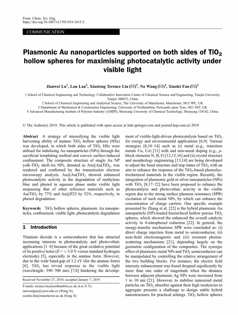
also improved by the HSs structure as explained in theprevious work [17]. For the Au-based TiO2 hybridnanomaterial, the decolorisation of MB and degradationof phenol were improved for the same period of the time,following the order of activity as: Au@Au(TiO2>Au@TiO2 HSs> yolk-shell Au(TiO2.The degradation rate of both probing molecules
followed first-order kinetics concerning their concentra-tions. The rate can, therefore, be interpreted using thepseudo-first-order reaction model (Figs. 3(b) and 4(b)).The previous study by the action spectrum has shown thatthe degradation of MB was induced mainly by the SPRexcitation of Au NPs in Au-TiO2 materials rather than thesensitisation of the dye molecules [30]. By comparing therate constants in the decolourisation of MB, Au@TiO2
HSs showed a better activity (0.030 min–1) than that ofyolk-shell Au(TiO2 (0.013 min–1). In the yolk-shell Au(TiO2, e–h pairs are produced inside the TiO2 shell,whereas the catalytic reactions take place mostly at theouter surface of TiO2 HSs. Therefore, the transportation ofe–h pairs through the TiO2 shell increases the probabilityof e–h recombination leading to the relatively low activityin photocatalysis in comparison with that of Au@TiO2
HSs. To decorate the inner and outer surface of TiO2 HSssimultaneously, the photocatalytic activity of Au@Au(TiO2 increased significantly with a constant of 0.044min–1 for MB decolourisation, more than three timeshigher than that of yolk-shell Au(TiO2. Based on theresults above, the utilisation of both sides of TiO2 shell wasproven to be efficient to boost the photocatalytic activity ofAu NP-TiO2 HS nanostructures. This can be attributedmainly to the improved light-harvesting capacity of TiO2
HSs caused by the multiple intensified local electromag-netic fields across the TiO2 shell. Accordingly, the
interfacial hot electron transfer from Au NPs to TiO2
shell upon the excitation of the LSPR (plasmonicsensitisation) was also intensified effectively as thesynergistic effect. Additionally, due to the confinement ofAu NPs both within the TiO2 HSs and on their convexexternal surfaces, the developed Au@Au(TiO2 photocata-lyst also demonstrated good stability in the model reaction.The photocatalyst remained sufficiently active even afterthree successive recycles in the decolourisation of MB inthe aqueous phase (Fig. S5, cf. ESM)
4 Conclusions
In conclusion, a hybrid structure of Au nanoparticles (NPs)and anatase TiO2 HSs was synthesised, in which Au NPswere confined on both sides of the TiO2 HSs enabled bythe silica templating-NaOH etching and convex surface-induced confinement. The sandwich yolk-shell structuresof Au NPs and TiO2 HSs, termed Au@Au(TiO2, weredetermined by TEM characterisation, in which multiple AuNPs decorated on a single Au yolk-TiO2 shell nanos-tructure were found. According to the UV-Vis DRSanalysis, Au@Au(TiO2 showed the strongest SPR inten-sity at about 550 nm compared to Au(TiO2 and [email protected] studies of Au@Au(TiO2, Au(TiO2,Au@TiO2 and Au@P25 in the photocatalytic degradationof two model molecules (i.e., MB and phenol) undervisible light irradiation were carried out. Au@Au(TiO2
exhibited the enhanced activity for both reactions with thedegradation rate of MB and phenol as 99% and 78%,respectively, after 180 min due to the multiple intensifiedlocal electromagnetic fields across the TiO2 shell. Thestrategy of sequential decorating both sides of TiO2 shells
Fig. 4 (a) The rate of phenol degradation under visible light promoted by various TiO2 photocatalysts; (b) The corresponding pseudo-first-order kinetic rate plot
Jianwei Lu et al. Maximising the photocatalytic activity of titania hollow spheres 5

is generic and applicable to another semiconductor hollowspherical materials (e.g., BiVO4 and SnO2), which isbeneficial to develop highly efficient photocatalysts forphotoelectrochemical applications such as solar cells andwater splitting.
Acknowledgements We thank for the financial support from TheUniversity of Manchester through Higher Education Innovation Funded‘Knowledge and Innovation Hub for Environmental Sustainability’. LLthanks the China Scholarship Council (CSC, file no. 201706950035)-University of Manchester joint studentship for supporting her Ph.D. research.
Electronic Supplementary Material Supplementary material is availablein the online version of this article at https://doi.org/10.1007/s11705-019-1815-2 and is accessible for authorized users.
Open Access This article is licensed under a Creative CommonsAttribution 4.0 International License, which permits use, sharing, adaptation,distribution and reproduction in any medium or format, as long as you giveappropriate credit to the original author(s) and the source, provide a link to theCreative Commons licence, and indicate if changes were made. The imagesor other third party material in this article are included in the article’s CreativeCommons licence, unless indicated otherwise in a credit line to the material.If material is not included in the article’s Creative Commons licence and yourintended use is not permitted by statutory regulation or exceeds the permitteduse, you will need to obtain permission directly from the copyright holder. Toview a copy of this licence, visit http://creativecommons.org/licenses/by/4.0/.
References
1. Schrauben J N, Hayoun R, Valdez C N, Braten M, Fridley L, Mayer
J M. Titanium and zinc oxide nanoparticles are proton-coupled
electron transfer agents. Science, 2012, 336(6086): 1298–1301
2. Gratzel M. Photoelectrochemical cells. Nature, 2001, 414(6861):
338–344
3. Caravaca A, Daly H, Smith M, Mills A, Chansai S, Hardacre C.
Continuous flow gas phase photoreforming of methanol at elevated
reaction temperatures sensitised by Pt/TiO2. Reaction Chemistry &
Engineering, 2016, 1(6): 649–657
4. Caravaca A, Jones W, Hardacre C, Bowker M H. Hydrogen
production by the photocatalytic reforming of cellulose and raw
biomass using Ni, Pd, Pt and Au on titania. Proceedings of the Royal
Society A: Mathematical, Physical and Engineering Science, 2016,
472
5. Palmisano L, Sclafani A. Thermodynamics and kinetics for
heterogeneous photocatalytic processes. In: Schiavello M, ed.
Heterogeneous Photocatalysis. New York: John Wiley & Sons,
1997, 109–132
6. Cong Y, Zhang J, Chen F, Anpo M. Synthesis and characterization
of nitrogen-doped TiO2 nanophotocatalyst with high visible light
activity. Journal of Physical Chemistry C, 2007, 111(19): 6976–
6982
7. Meng Q, Wang T, Liu E, Ma X, Ge Q, Gong J. Understanding
electronic and optical properties of anatase TiO2 photocatalysts co-
doped with nitrogen and transition metals. Physical Chemistry
Chemical Physics, 2013, 15(24): 9549–9561
8. Lu J, Su F, Huang Z, Zhang C, Liu Y, Ma X, Gong J. N-Doped Ag/
TiO2 hollow spheres for highly efficient photocatalysis under
visible-light irradiation. RSC Advances, 2013, 3(3): 720–724
9. Wang H, Zhang L, Chen Z, Hu J, Li S, Wang Z, Liu J, Wang X.
Semiconductor heterojunction photocatalysts: Design, construction,
and photocatalytic performances. Chemical Society Reviews, 2014,
43(15): 5234–5244
10. Pan H, Zhang Y W, Shenoy V B, Gao H. Effects of H-, N-, and (H,
N)-doping on the photocatalytic activity of TiO2. Journal of Physical
Chemistry C, 2011, 115(24): 12224–12231
11. Pelaez M, Nolan N, Pillai S, Seery M, Falaras P, Patrick A G,
Jeremy S M, Hamiltone W J, Byrne J A, O’Shea K, et al. A review
on the visible light active titanium dioxide photocatalysts for
environmental applications. Applied Catalysis B: Environmental,
2012, 125: 331–349
12. Dozzi M V, Selli E. Doping TiO2 with p-block elements: Effects on
photocatalytic activity. Journal of Photochemistry and Photobiology
C, Photochemistry Reviews, 2012, 14: 13–28
13. Kamat P V. TiO2 nanostructures: Recent physical chemistry
advances. Journal of Physical Chemistry C, 2012, 116(22):
11849–11851
14. Li L, Yan J, Wang T, Zhao Z J, Zhang J, Gong J, Guan N. 10 nm
rutile titanium dioxide nanoparticles for efficient visible-light-driven
photocatalytic hydrogen production. Nature Communications,
2015, 6(1): 5881
15. Ansari S A, Khan M M, Ansari M O, Cho M H. Nitrogen-doped
titanium dioxide (N-doped TiO2) for visible light photocatalysis.
New Journal of Chemistry, 2016, 40(4): 3000–3009
16. Li L, Meng F, Hu X, Qiao L, Sun C Q, Tian H, Zheng W. TiO2
band restructuring by B and P dopants. PLoS One, 2016, 11(4):
e0152726
17. Lu J, Zhang P, Li A, Su F, Wang T, Liu Y, Gong J. Mesoporous
anatase TiO2 nanocups with plasmonic metal decoration for highly
active visible-light photocatalysis. Chemical Communications
(Cambridge), 2013, 49(52): 5817–5819
18. Tian Y, Tatsuma T. Mechanisms and applications of plasmon-
induced charge separation at TiO2 films loaded with gold
nanoparticles. Journal of the American Chemical Society, 2005,
127(20): 7632–7637
19. Awazu K, Fujimaki M, Rockstuhl C, Tominaga J, Murakami H,
Ohki Y, Yoshida N, Watanabe T. A plasmonic photocatalyst
consisting of silver nanoparticles embedded in titanium dioxide.
Journal of the American Chemical Society, 2008, 130(5): 1676–
1680
20. Lee I, Joo J B, Yin Y D, Zaera F. A yolk@shell nanoarchitecture for
Au/TiO2 catalysts. Angewandte Chemie International Edition, 2011,
50(43): 10208–10211
21. Linic S, Christopher P, Ingram D B. Plasmonic-metal nanostructures
for efficient conversion of solar to chemical energy. Nature
Materials, 2011, 10(12): 911–921
22. Zhang Q, Jin X, Xu Z, Zhang J, Rendón U F, Razzari L, Chaker M,
Ma D. Plasmonic Au-loaded hierarchical hollow Porous TiO2
spheres: Synergistic catalysts for nitroaromatic reduction. Journal of
Physical Chemistry Letters, 2018, 9(18): 5317–5326
23. Joo J B, Dahl M, Li N, Zaera F, Yin Y. Tailored synthesis of
mesoporous TiO2 hollow nanostructures for catalytic applications.
Energy & Environmental Science, 2013, 6(7): 2082–2092
24. Joo J B, Zhang Q, Dahl M, Lee I, Goebl J, Zaera F, Yin Y. Control
6 Front. Chem. Sci. Eng.
of the nanoscale crystallinity in mesoporous TiO2 shells for
enhanced photocatalytic activity. Energy & Environmental Science,
2012, 5(4): 6321–6327
25. Dillon R J, Joo J B, Zaera F, Yin Y, Bardeen C J. Correlating the
excited state relaxation dynamics as measured by photolumines-
cence and transient absorption with the photocatalytic activity of
Au@TiO2 core-shell nanostructures. Physical Chemistry Chemical
Physics, 2013, 15(5): 1488–1496
26. Lee Y J, Joo J B, Yin Y, Zaera F. Evaluation of the effective
photoexcitation distances in the photocatalytic production of H2
from water using Au@void@TiO2 yolk-shell nanostructures. ACS
Energy Letters, 2016, 1(1): 52–56
27. Lee I, Joo J B, Yin Y, Zaera F. Au@Void@TiO2 yolk-shell
nanostructures as catalysts for the promotion of oxidation reactions
at cryogenic temperatures. Surface Science, 2016, 648: 150–155
28. José-Yacamán M, Gutierrez-Wing C, Miki M, Yang D Q, Piyakis K
N, Sacher E. Surface diffusion and coalescence of mobile metal
nanoparticles. Journal of Physical Chemistry B, 2005, 109(19):
9703–9711
29. Liz-Marzan L M, Giersig M, Mulvaney P. Synthesis of nanosized
gold-silica core-shell particles. Langmuir, 1996, 12(18): 4329–4335
30. Bian Z, Tachikawa T, Zhang P, Fujitsuka M, Majima T. Au/TiO2
Superstructure-based plasmonic photocatalysts exhibiting efficient
charge separation and unprecedented activity. Journal of the
American Chemical Society, 2014, 136(1): 458–465
31. Prikulis J, Hanarp P, Olofsson L, Sutherland D, Käll M. Optical
spectroscopy of nanometric holes in thin gold films. Nano Letters,
2004, 4(6): 1003–1007
32. Seh Z W, Liu S H, Low M, Zhang S Y, Liu Z L, Mlayah A, Han M
Y. Janus Au-TiO2 photocatalysts with strong localization of
plasmonic near-fields for efficient visible-light hydrogen generation.
Advanced Materials, 2012, 24(17): 2310–2314
33. Wu X F, Song H Y, Yoon J M, Yu Y T, Chen Y F. Synthesis of core-
shell Au@TiO2 nanoparticles with truncated wedge-shaped mor-
phology and their photocatalytic properties. Langmuir, 2009, 25
(11): 6438–6447
Jianwei Lu et al. Maximising the photocatalytic activity of titania hollow spheres 7