fatty acid uptake in diabetic rat adipocytes
TRANSCRIPT

51
Molecular and Cellular Biochemistry 167: 51–60, 1997.© 1997 Kluwer Academic Publishers. Printed in the Netherlands.
Fatty acid uptake in diabetic rat adipocytes
Heather Fraser,1 Scott M. Coles,2 Judith K. Woodford,3 Audrey A.Frolov,4 Eric J. Murphy,4 Friedhelm Schroeder,4 David A. Bernlohr5 andVernon Grund61Department of Pharmacology, University of Alberta, Edmonton, Alberta, T6G 2H7, Canada; 2The Cleveland Clinic Founda-tion, 9500 Euclid Avenue, NC10, Cleveland, OH 44195, USA; 3The Andrew Jergens Co., 2535 Spring Grove Avenue,Cincinnati, OH 45214-1773, USA; 4Department of Physiology and Pharmacology, Texas A and M University, TVMC, CollegeStation, Texas 77843-4466, USA; 5Department of Biochemistry, University of Minnesota, Saint Paul, Minnesota 55108, USA;6Department of Pharmaceutical Sciences, School of Pharmacy and Allied Health Sciences, University of Montana, Missoula,Montana 59812-1075, USA
Received 8 September 1995; accepted 2 July 1996
Abstract
The effect of diabetic status and insulin on adipocyte plasma membrane properties and fatty acid uptake was examined. Studieswith inhibitors and isolated adipocyte ghost plasma membranes indicated 9Z, 11E, 13E, 15Z-octatetraenoic acid (cis-parinaricacid) uptake was protein mediated. Cis-parinaric acid uptake was inhibited by trypsin treatment or incubation with phloretin, andcompeted with stearic acid. The initial rate, but not maximal uptake, of cis-parinaric acid uptake was enhanced two-fold inadipocytes from diabetic rats. Concomitantly, the structure and lipid composition of adipocyte ghost membranes was dramati-cally altered. However, the increased initial rate of cis-parinaric acid uptake in the diabetic adipocytes was not explained bymembrane alterations or by a two-fold decrease in cytosolic adipocyte fatty acid binding protein (ALBP), unless ALBP stimu-lated fatty acid efflux. Thus, diabetic status dramatically altered adipocyte fatty acid uptake, plasma membrane structure, lipidcomposition, and cytosolic fatty acid binding protein. (Mol Cell Biochem 167: 51–60, 1997)
Key words: fatty acid, cis-parinaric acid, adipocyte, transport, diabetes, insulin
Address for offprints: V. Grund, Department of Pharmaceutical Sciences, School of Pharmacy and Allied Health Sciences, University of Montana,Missoula, Montana 59812-1075, USA
Introduction
Fatty acid transport across the plasma membrane and its in-tracellular release to cytosolic fatty acid binding proteins orother intracellular binding sites in mammalian cells is not com-pletely understood. Several studies suggest that cellular fattyacid transport may be mediated through one or more recentlyidentified candidate transporters. Transport of fatty acids issaturable, with K
m values range between 10–8 and 10–7 M [1–
3]. The transport of radiolabeled fatty acids was competitivelyinhibited by a variety of unlabeled fatty acids [4]. Uptake wasmore specific (lower K
m) for straight-chain saturated fatty ac-
ids [2]. Fatty acid transport is inhibited by protein (SH rea-gents, phloretin, trypsin) and ATP (dinitrophenol, KCN)
inhibitors [5–7], fatty acid affinity labels [8], and antibodies[2]. In contrast to the poorly defined transport molecules,adipocytes have a well characterized cytoplasmic fatty acidbinding protein, ALBP (adipocyte lipid binding protein) [4, 6,9]. ALBP binds a variety of saturated and unsaturated fattyacids. X-ray crystallographic analysis of ALBP has provideda wealth of information on protein lipid interactions (for a re-view, see [10]}.
Cellular fatty acid uptake may be regulated at several lev-els. First, fatty acid uptake is hormonally regulated. Epine-phrine rapidly stimulates the transport or exchange of longchain fatty acids across the plasma membrane of isolatedadipocytes [11, 12]. Insulin at physiological concentrations(0.01–10 nM) inhibits the epinephrine effect but has no effect

52
on the basal fatty acid uptake [12]. Second, plasma membranestructure can modify plasma membrane transport protein ac-tivity. Insulin rigidifies plasma membranes in vitro when addeddirectly to plasma membranes isolated from control animals[13]. Plasma membranes isolated from erythrocytes or kidneyof animals treated with insulin in vivo were also rigidified [14,15]. Membrane rigidification may have effects on fatty acidtransport activity. Third, whether transport activity [16] orALBP activity are rate limiting in cellular fatty acid uptake isunclear. In chickens [17] and differentiating adipocytes [6, 9]fatty acid uptake correlated with the presence of both cyto-solic ALBP and plasma membrane transport activity. Inter-estingly, ALBP fatty acid binding can be controlled by insulinwhich stimulates phosphorylation of ALBP [10] through in-sulin receptor kinase [18]. Furthermore, levels of ALBP mRNA,protein, and transcription in adipocytes are depressed instreptozotocin diabetic rats [19].
Intracellular esterification and metabolism of fatty acidprobes may complicate resolution of these mechanisms. Hereina fluorescent fatty acid, cis-parinaric acid, was used to examinethe relative role of plasma membrane transport activity andcytosolic ALBP in fatty acid uptake in adipocytes and adipo-cyte ghosts from control and diabetic rats. Cis-parinaric acid isnot oxidized and is only weakly esterified [7]. Therefore, an in-crease in cellular fluorescence largely reflects fatty acid uptakein the absence of intracellular metabolism [7].
Materials and methods
Materials
Cis-parinaric acid and diphenylhexatriene(DPHa), were fromMolecular Probes, Eugene, OR. Bovine serum albumin (BSA),streptozotocin (STZ), trypsin, phloretin, oleic acid and stearicacid were obtained from Sigma Chemical Co., St. Louis, MO.Crystalline porcine insulin (26.8 U/mg) was kindly providedby Dr. Ronald E. Chance, Eli Lilly, Indianapolis, IN. Pentobar-bital was purchased from Abbott Laboratories, Inc., Chicago,IL. Collagenase was purchased from Worthington Biochemi-cal, Freehold, NJ. [l25]I-Protein A was purchased from Amer-sham, Arlington Heights, IL.
Animals
Male (315–380 g) Wistar rats were purchased from Harlan In-dustries (Indianapolis, lN). Type 1 diabetes mellitus was in-duced with streptozotocin (60 mg/kg, i.v., tail vein) in physio-logical saline, pH 7.4, under ether anesthesia. Diabetes wasverified by blood glucose tests at both 3 and 5 days using aGlucose Analyzer 2 (Beckman Instruments, Inc., Fullerton,
CA). Animals were considered diabetic when plasma glucosevalues exceeded 300 mg/dl. Rats were diabetic for 6 weeksbefore use.
Adipocyte isolation and adipocyte ghost preparation
Adipocytes were isolated from the epididymal fat pads of dia-betic and control Wistar rats according to the methods ofRodbell [20] as modified by Grund et al. [21]. Adipocyte ghostswere isolated following the methods of Birnbaumer et al. [22],resuspended in an equal volume of a sucrose-tris-ethyleneglycol-bis-(beta-amino-ethyl ether) N,N,N′,N′-tetraacetic acid(EGTA) solution, and stored at –20°C. Protein content of thefrozen ghost suspensions were determined using the Brad-ford protein assay kit (Sigma Chemical Co., St. Louis, MO).
Lipid extraction, analysis, and liposome preparation
Lipids were extracted from adipocyte ghosts and eitheranalyzed as described earlier [22] or liposomes were prepared[23].
Fluorescent fatty acid uptake assay
The cis-parinaric acid fatty acid uptake assay exhibits a numberof advantages for use in determining fatty acid uptake. First,cis-parinaric acid does not fluoresce in aqueous buffer butonly fluoresces when in the hydrophobic environment of theadipocyte membrane transport system and intracellular sites.Consequently, fatty acid uptake can be measured withoutseparation of cellular bound from free fatty acid. Second, thecontinuous assay is computerized and assimilates up to 540data points in only a few minutes. This permits accurate ki-netic analysis of rapid uptake. Third, the increase in parinaricacid fluorescence upon interaction with cells represents trans-port across the plasma membrane rather than simply partition-ing of the fatty acid into the membrane. In the first minute ofincubation 99+% of the cis-parinaric acid can be back ex-tracted with albumin washes [24, 25]. At 1 min incubation, theinitial rate of cis-parinaric acid uptake was essentially the sameas that of [3H]-oleic acid uptake [25, 26]. With increasing time,less cis-parinaric acid is back-extractable with albumin suchthat at equilibrium about 80% of cis-parinarate is associatedwith the plasma membrane and 20% is associated with intra-cellular sites wherein it is poorly or not metabolized [25]. Thisis in contrast to [3H]-oleic acid which continued to increaseand showed no plateau in the time frame of the assay [25, 26].Fourth, because parinaric acid is a naturally occurring fattyacid [27], it does not have bulky side groups that may inter-fere with its interactions with lipids and proteins [24, 27]. Con-

53
sequently, cis-parinaric acid interacts similarly (similar Kds and
stoichiometry of binding) to radiolabeled fatty acids withcytosolic fatty acid binding proteins [7, 28, 29]. Fifth, unlikeradiolabeled fatty acids, cis-parinaric acid does not undergosignificant metabolism or esterification in murine L-cellfibroblasts [7, 24]. Less than 3% of cis-parinaric acid taken upby L-cells was found in esterified form following 15 min ofincubation with minimal oxidation [4, 7, 24]. Thus after 1 minof incubation used in the present system, the esterificationof cis-parinaric acid would be insignificant. In comparison,radiolabeled fatty acids are significantly (30–80%) esterifiedin both L-cells and adipocytes after 1 min of incubation [7, 24,30, 31]. These properties make cis-parinaric acid a good probefor measuring uptake of fatty acid and intracellular distribu-tion in the absence of the extensive esterification and oxida-tion usually observed with radiolabeled fatty acid. In thepresent investigation, the cis-parinaric acid uptake assay [22,25, 32] was modified for use with adipocytes as follows: A 2ml suspension of 7.5 × 10⁴ cells/ml (or 11.7 µg protein adipo-cyte ghosts) in albumin free KRP were placed in a 1 cm × 1 cmfluorescence cuvette. The suspension was maintained at 37°Cand stirred constantly with a Fisher brand CellSpinBar, FisherScientific, Pittsburgh, PA. Any effectors were added at thispoint and pre-incubated for either 3 min (trypsin and phlore-tin) or 5 min (insulin and norepinephrine) with the fluorimetersample compartment closed. Unless otherwise stated the fi-nal cis-parinaric acid concentration in the sample was 3.62 µM;the final ethanol concentration in the buffer was maintainedat less than 0.5%.
Fluorescence lifetime and differential polarized phasefluorometry
Diphenylhexatriene fluorescence lifetime determinations wereconducted on adipocyte ghosts following the methods ofNemecz and Schroeder [23]. Differential polarized phase fluor-ometry was used to resolve steady state anisotropy into a staticcomponent (limiting anisotropy) and a dynamic component(rotational relaxation time, in ns) as described earlier [33].
Western blots
Isolated rat adipocytes were solubilized in ice-cold solubili-zation buffer (25 mM 4-2-hydroxyethyl)-1-piperazineethane-sulfonic acid (HEPES), pH 7.5; 0.1 mM phenylmethylsulfonylfluoride (PMSF); 10 mM 2-mercaptoethanol; 1 µg/ml eachpepstatin, leupeptin and aprotinin as protease inhibitors).Samples were homogenized before centrifugation at 100,000g for 20 min., 4°C. Supernatants were assayed with the Brad-ford protein assay. Samples (50 µg protein) were separatedby 5–15% SDS-PAGE electrophoresis and transferred to ni-
trocellulose. The proteins were blocked for 2 h with westernbuffer [10 mM HEPES pH 7.5, 150 mM NaCl, 1 mM ethylenediaminetetraacetic acid (EDTA), and 0.5% (v/v) polyoxy-ethylene [23] lauryl ether (Brij 35)] prior to incubation withanti-murine ALBP for 4 h at 24°C. Antibody was detected afterincubation in western buffer (1 µCi/ml of [125]I-Protein A) for 2h followed by exposure to x-ray overnight at –70°C. Theadipocyte cytosol and standard ALBP blots were quantitatedby densitometry [33] except that a ScanJet IIP (HewlettPackard Co., Corvallis, OR) and QuantiScan software (Micro-bial Systems Ltd., distributed by Biosoft, London, England)were used.
Statistical analysis
Statistical analysis was carried out using the standard Stu-dent’s t-test. P < 0.05 was indicative of a statistical differencebetween groups.
Results
Cis-parinaric acid uptake by adipocytes
Cis-parinaric acid in aqueous solution is a very weak fluoro-phore. However, when in a hydrophobic environment asfound in membranes or hydrophobic binding pockets of fattyacid binding proteins, cis-parinaric acid strongly fluoresces[28, 29]. This allows cis-parinaric acid fluorescence to beused as a parameter to continuously monitor fatty acid up-take (Fig. 1). As shown, the initial rate of fluorescence in-crease by isolated adipocytes is rapid and the intensityreaches a maximum in about 40 sec. After 70 sec the fluores-cence intensity decreased. This latter decrease was due tothe slow flotation of the lipid laden adipocytes, despite con-tinuous stirring of the adipocyte suspension with a residentcell spinbar in the cuvette. Although flotation could be pre-vented by vigorous tituration, the concomitant interruptionof the fluorescence signal compromised continuous dataacquisition. Cis-parinaric acid uptake was dependent uponthe initial concentration of cis-parinaric acid over the range0–36.2 µM.
When the initial rate of cis-parinaric acid uptake and themaximal percent fluorescence intensity increase were plottedas a function of cis-parinaric acid concentration, saturationcurves were obtained (Figs 2A and 2B, respectively). K
m (6.0
± 0.9 µM) and Vmax
( 22110 ± 1920 ∆I min–1 µg–1 or 7.74 nmolmg–1 min–1) values for cis-parinaric acid uptake into ratadipocytes were determined from the data using Hanes-Woolfand Lineweaver Burke plots.

54
Inhibition of cis-parinaric acid uptake
The data shown in Figs 1 and 2 demonstrating saturation andconcentration dependence of cis-parinaric acid uptake are con-sistent with but not definitive for cis-parinaric acid uptakebeing protein mediated. A series of protein inhibitors wastherefore used to further define cis-parinaric acid uptake.
Exposure of cells to mild, limited, trypsin treatment resultedin proteolysis of the extracellular portion of membrane pro-teins. Pre-incubation of adipocytes with trypsin prior to ad-dition of cis-parinaric acid inhibited cis-parinaric acid uptake,see Fig. 3. Trypsin maximally inhibited cis-parinaric acid up-take at 0.12 mg trypsin/ml. The maximal intensity increase, in-dicating cis-parinaric acid uptake, was lowered 36% bypre-incubation with trypsin vs non-trypsin treated adipocytes.
The effect of 0.02 µM and 0.2 µM phloretin on cis-parinaricacid uptake was also examined. Phloretin acts to inhibit mem-brane transport processes [1]. The maximal percent intensityincrease of cisparinaric acid uptake by phloretin treatedadipocytes was inhibited by 96% compared to non-treatedadipocytes, see Fig. 4. The initial rate of cis-parinaric aciduptake was inhibited by 61.0% and 92.5% in the presence of0.02 and 0.2 µM phloretin, respectively. These data would in-dicate that a major portion of adipocyte fatty acid transportactivity is not diffusional.
Cis-parinaric acid is a naturally occurring 18 carbon, kinked-chain, cis-trans-trans-cis conjugated, polyenic fatty acid. Nev-ertheless, the possibility that cis-parinaric acid uptake maynot occur through the same fatty acid transport system asother fatty acids was examined. A competition assay wasperformed with the 18 carbon, non-fluorescent fatty acid,stearic acid (a straight-chain, saturated fatty acid). Stearic acidinhibited the initial rate of cis-parinaric acid uptake by 30%(data not shown), indicating competition for the binding siteat concentrations as low as 0.88 µM stearic acid (the lowestconcentration tested). The K
i for stearic acid inhibition of initial
rate was calculated to be near 0.45 µM (n = 3). Similar to theinitial rate data, stearic acid decreased maximal cis-parinaricacid uptake, as measured by the fold change in cis-parinaricacid fluorescence. Maximum fluorescence was inhibited atconcentrations of stearic acid as low as 0.88 µM.
Lowering the incubation temperature from 37 to 4°C de-creased the initial rate of cis-parinaric acid uptake 56.4% (datanot shown). Although the maximum uptake (as determinedby percent intensity increase) tended to decrease, this effectwas not statistically significant.
Effect of diabetic status on cis-parinaric acid uptake
Cis-parinaric acid uptake was examined in adipocytes fromSTZ-treated rats. The initial rate of cis-parinaric acid uptakewas 81% higher (3.89 ± 0.14 to 2.15 ± 0.18 nmol min–1 mg–1) in
Fig. 1. Cis-parinaric acid uptake in rat adipocytes. Cis-parinaric acidwas added to freshly harvested rat adipocytes (7.5 × 10⁴ cells/ml) inKRP, pH 7.4 at 37°C. Final cis-parinaric acid concentrations were asindicated. Fluorescence intensity (λ
ex = 324 nm) was measured at 1.2
sec intervals.
B
Fig. 2. Concentration dependence for cis-parinaric acid uptake byadipocytes. Initial rates and maximal percent intensity change werecalculated from the data in Fig. 1 and plotted versus cis-parinaric acidconcentration. Plot A is initial rate, plot B is maximal intensity in-crease. Values are the average ± S.E. (n = 3).
A

55
Fig. 3. Effect of trypsin on cis-parinaric acid uptake by adipocytes.Uptake of 3.62 µM cis-parinaric acid was determined after 3 min pre-incubation with 0.12 mg/ml trypsin (¡) or in the absence of trypsin(●). Conditions were as in Fig. 1.
Fig. 4. Effect of phloretin on cis-parinaric acid uptake by adipocytes.Uptake of 3.62 µM cis-parinaric acid was determined after 3 min pre-incubation with 0.2 µM phloretin (¡) or in the absence of phloretin(●). Conditions were as in Fig. 1.
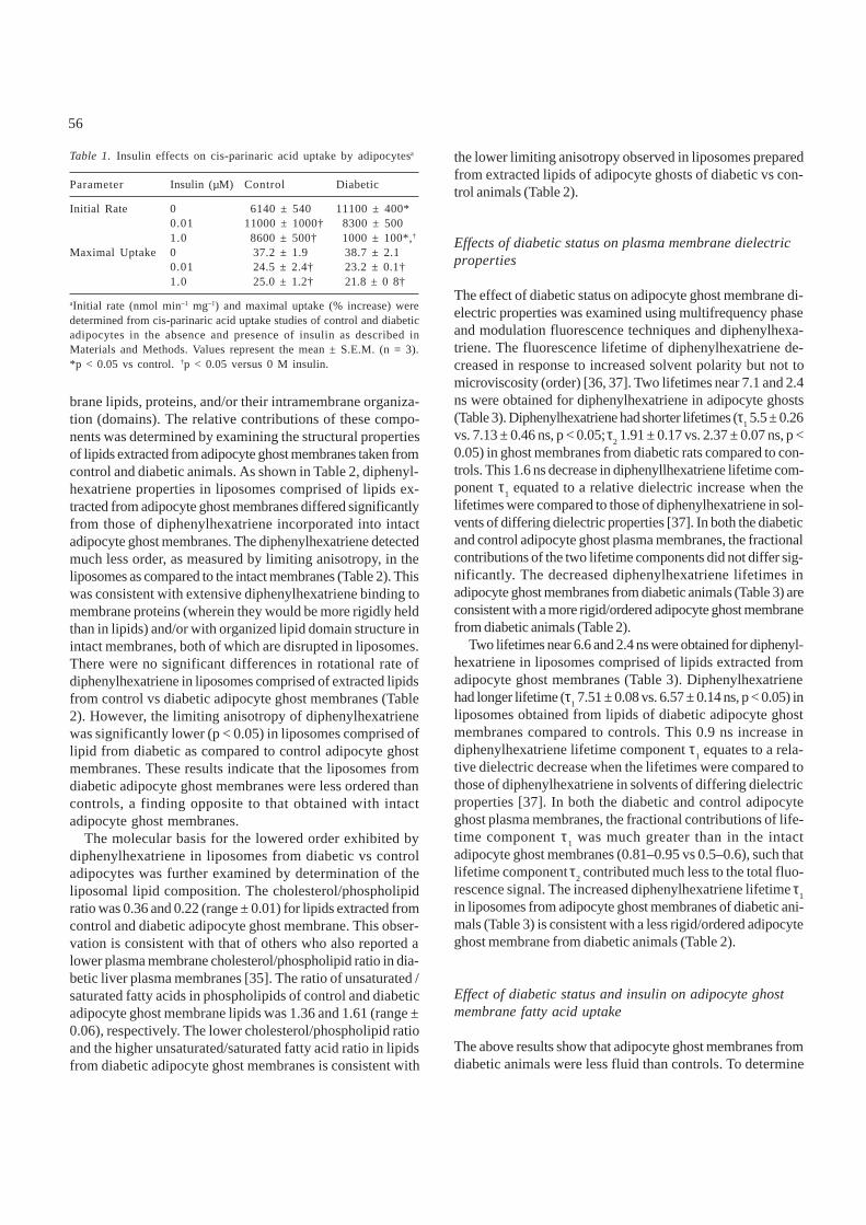
adipocytes from diabetic versus control rats (p < 0.05, Table1). The maximal uptake of cis-parinaric acid was unchangedin diabetic vs. control adipocytes (Table 1). Thus, diabeticstatus appeared to affect the K
m but not the V
max of cis-
parinaric acid uptake. These data indicate that diabetes stimu-lated the rate, but not extent of cis-parinaric acid uptake byadipocytes.
In-vitro insulin effects on cis-parinaric acid uptake inadipocytes
Cis-parinaric acid uptake by control rat adipocytes was meas-ured in the presence of two concentrations of insulin (Table1). At 0.01 µM insulin, the initial rate of cis-parinaric acid up-take was significantly increased from 2.15 ± 0.18 to 3.88 ± 0.35nmol min–1 mg–1 (p < 0.05). Insulin at the higher concentration,1 µM, appeared to have a diminished effect. The maximumuptake of cis-parinaric acid was lowered by approxi-mately 30% at both insulin concentrations (Table 1). Thus,although insulin in vitro stimulated the initial rate of cis-parinaric acid uptake by control adipocytes, the overallamount taken up was decreased.Diabetes markedly altered the effect of insulin in vitro on therate of cis-parinaric acid uptake by adipocytes. Instead ofstimulating the initial rate of cis-parinaric acid uptake as incontrol adipocytes, insulin inhibited cis-parinaric acid uptakein adipocytes from diabetic rats (Table 1). The initial rate de-crease at 0.01 µM insulin was from 3.89 ± 0.14 to 2.91 ± 0.18nmol min–1 mg–1 (p < 0.05) The rate at 1 µM insulin was furtherreduced to 0.35 ± 0.03 nmol min–1 mg–1 (p < 0.05). Insulin causeda statistically significant decrease in the rate of cis-parinaricacid uptake, and this effect appeared to be dose dependent.Insulin inhibited maximum uptake of cis-parinaric acid by thediabetic adipocytes to a similar degree as in controls. Thus,insulin affected the K
m, but not the V
max, for cis-parinaric acid
uptake differently in adipocytes from diabetic and control rats.
Effects of diabetic status on plasma membrane structure
To determine whether or not the above alterations were dueto differences in adipocyte ghost membrane structure, the flu-idity of the adipocyte ghost membranes was examined by de-termining both the dynamic (rotational relaxation time) andstatic/order (limiting anisotropy) parameters of diphenyl-hexatriene motion in the adipocyte ghost membranes. Adipo-cyte plasma membrane fluidity was measured with multi-frequency differential polarized phase fluorometry of DPH, aprobe for membrane lipid structure. There was a significantincrease (p < 0.05) in the rotational correlation time of DPH indiabetic ghost membranes, 1.55 ± 0.21 ns, vs. 0.88 ± 0.06 ns inDPH in control ghost membranes. This suggests that the dia-betic ghost membranes were less fluid than the controls. Fur-thermore, there was also a significant increase (p < 0.05) inthe limiting anisotropy as measured in diabetic ghosts (0.174± 0.005) compared to control ghosts (0.145 ± 0.002). The longerrotational relaxation time and higher limiting anisotropy ofdiphenylhexatriene in adipocyte ghost membranes from dia-betic animals was consistent with the diphenylhexatriene be-ing located in a more ordered, less fluid, environment.
The primary determinants of membrane fluidity are the mem-
56
brane lipids, proteins, and/or their intramembrane organiza-tion (domains). The relative contributions of these compo-nents was determined by examining the structural propertiesof lipids extracted from adipocyte ghost membranes taken fromcontrol and diabetic animals. As shown in Table 2, diphenyl-hexatriene properties in liposomes comprised of lipids ex-tracted from adipocyte ghost membranes differed significantlyfrom those of diphenylhexatriene incorporated into intactadipocyte ghost membranes. The diphenylhexatriene detectedmuch less order, as measured by limiting anisotropy, in theliposomes as compared to the intact membranes (Table 2). Thiswas consistent with extensive diphenylhexatriene binding tomembrane proteins (wherein they would be more rigidly heldthan in lipids) and/or with organized lipid domain structure inintact membranes, both of which are disrupted in liposomes.There were no significant differences in rotational rate ofdiphenylhexatriene in liposomes comprised of extracted lipidsfrom control vs diabetic adipocyte ghost membranes (Table2). However, the limiting anisotropy of diphenylhexatrienewas significantly lower (p < 0.05) in liposomes comprised oflipid from diabetic as compared to control adipocyte ghostmembranes. These results indicate that the liposomes fromdiabetic adipocyte ghost membranes were less ordered thancontrols, a finding opposite to that obtained with intactadipocyte ghost membranes.
The molecular basis for the lowered order exhibited bydiphenylhexatriene in liposomes from diabetic vs controladipocytes was further examined by determination of theliposomal lipid composition. The cholesterol/phospholipidratio was 0.36 and 0.22 (range ± 0.01) for lipids extracted fromcontrol and diabetic adipocyte ghost membrane. This obser-vation is consistent with that of others who also reported alower plasma membrane cholesterol/phospholipid ratio in dia-betic liver plasma membranes [35]. The ratio of unsaturated /saturated fatty acids in phospholipids of control and diabeticadipocyte ghost membrane lipids was 1.36 and 1.61 (range ±0.06), respectively. The lower cholesterol/phospholipid ratioand the higher unsaturated/saturated fatty acid ratio in lipidsfrom diabetic adipocyte ghost membranes is consistent with
the lower limiting anisotropy observed in liposomes preparedfrom extracted lipids of adipocyte ghosts of diabetic vs con-trol animals (Table 2).
Effects of diabetic status on plasma membrane dielectricproperties
The effect of diabetic status on adipocyte ghost membrane di-electric properties was examined using multifrequency phaseand modulation fluorescence techniques and diphenylhexa-triene. The fluorescence lifetime of diphenylhexatriene de-creased in response to increased solvent polarity but not tomicroviscosity (order) [36, 37]. Two lifetimes near 7.1 and 2.4ns were obtained for diphenylhexatriene in adipocyte ghosts(Table 3). Diphenylhexatriene had shorter lifetimes (τ
1 5.5 ± 0.26
vs. 7.13 ± 0.46 ns, p < 0.05; τ2 1.91 ± 0.17 vs. 2.37 ± 0.07 ns, p <
0.05) in ghost membranes from diabetic rats compared to con-trols. This 1.6 ns decrease in diphenyllhexatriene lifetime com-ponent τ
1 equated to a relative dielectric increase when the
lifetimes were compared to those of diphenylhexatriene in sol-vents of differing dielectric properties [37]. In both the diabeticand control adipocyte ghost plasma membranes, the fractionalcontributions of the two lifetime components did not differ sig-nificantly. The decreased diphenylhexatriene lifetimes inadipocyte ghost membranes from diabetic animals (Table 3) areconsistent with a more rigid/ordered adipocyte ghost membranefrom diabetic animals (Table 2).
Two lifetimes near 6.6 and 2.4 ns were obtained for diphenyl-hexatriene in liposomes comprised of lipids extracted fromadipocyte ghost membranes (Table 3). Diphenylhexatrienehad longer lifetime (τ
1 7.51 ± 0.08 vs. 6.57 ± 0.14 ns, p < 0.05) in
liposomes obtained from lipids of diabetic adipocyte ghostmembranes compared to controls. This 0.9 ns increase indiphenylhexatriene lifetime component τ
1 equates to a rela-
tive dielectric decrease when the lifetimes were compared tothose of diphenylhexatriene in solvents of differing dielectricproperties [37]. In both the diabetic and control adipocyteghost plasma membranes, the fractional contributions of life-time component τ
1 was much greater than in the intact
adipocyte ghost membranes (0.81–0.95 vs 0.5–0.6), such thatlifetime component τ
2 contributed much less to the total fluo-
rescence signal. The increased diphenylhexatriene lifetime τ1
in liposomes from adipocyte ghost membranes of diabetic ani-mals (Table 3) is consistent with a less rigid/ordered adipocyteghost membrane from diabetic animals (Table 2).
Effect of diabetic status and insulin on adipocyte ghostmembrane fatty acid uptake
The above results show that adipocyte ghost membranes fromdiabetic animals were less fluid than controls. To determine
Table 1. Insulin effects on cis-parinaric acid uptake by adipocytesa
Parameter Insulin (µM) Control Diabetic
Initial Rate 0 6140 ± 540 11100 ± 400*0.01 11000 ± 1000† 8300 ± 5001.0 8600 ± 500† 1000 ± 100*,†
Maximal Uptake 0 37.2 ± 1.9 38.7 ± 2.10.01 24.5 ± 2.4† 23.2 ± 0.1†1.0 25.0 ± 1.2† 21.8 ± 0 8†
aInitial rate (nmol min–1 mg–1) and maximal uptake (% increase) weredetermined from cis-parinaric acid uptake studies of control and diabeticadipocytes in the absence and presence of insulin as described inMaterials and Methods. Values represent the mean ± S.E.M. (n = 3).*p < 0.05 vs control. †p < 0.05 versus 0 M insulin.

57
whether this difference could account for the increased initialrate of cis-parinaric acid uptake in diabetic adipocytes (Table1), cis-parinaric acid uptake was measured with isolatedadipocyte ghost membranes. Adipocyte ghost membranes fromcontrol and diabetic rats had 2.8-fold and 6.8-fold lower initialrates (0.77 ± 0.05 and 0.57 ± 0.05 nmol min–1 mg–1, respectively)of cis-parinaric acid uptake as compared to intact cells (Table1). However, no significant differences were observed in eitherinitial rate or maximal uptake of cis-parinaric acid in adipocyteghost membranes obtained from control vs diabetic animals.These data indicate that diabetes did not alter the rate of cis-parinaric acid incorporation into the plasma membrane.
The presence of insulin in vitro had no significant effectson either the initial rate or maximal uptake of cis-parinaric acidin adipocyte ghosts. The initial rate of cis-parinaric acid up-take by control adipocyte ghosts treated in vitro with 0, 0.01,and 1.0 µM insulin was 0.25 ± 0.01, 0.23 ± 0.01, 0.25 ± 0.02nmol min–1 mg–2, respectively. Likewise, maximal fluorescencechanged less than 1 % upon addition of insulin in vitro. Simi-lar observations were obtained with diabetic adipocyte ghoststreated in vitro with insulin (data not shown). These obser-vations of a lack of direct effect of insulin in vitro on adipocyteghost membrane cisparinaric acid uptake confirm those of oth-ers showing that insulin in vitro did not affect basal radio-labeled fatty acid uptake in adipocytes [2]. Thus, the resultsindicate that the effect of insulin on adipocyte fatty acid up-take may be mediated more by intracellular factors rather thanby effects on the adipocyte plasma membrane fatty acid trans-port system.
Effect of diabetic status on adipocyte ALBP levels
As pointed out in the Introduction, the cytosolic fatty acidbinding proteins may contribute to the uptake/efflux of fattyacids in adipocytes. Since the relative function of the cyto-solic adipocyte lipid binding protein (ALBP) in fatty aciduptake/efflux is not known, two possibilities may account forthe enhanced cis-parinaric acid uptake in adipocytes of dia-betic animals shown in Table 1: (i) If ALBP enhances fattyacid uptake, then an increase in ALBP could give rise to highercis-parinaric acid uptake in diabetic adipocytestty acid. (ii) IfALBP enhances efflux of fatty acid, then decreased ALBPlevels could elicit an increased initial rate of cis-parinaric aciduptake in diabetic. Therefore, antisera against ALBP andWestern blotting were used to determine the relative amountsof ALBP expression in adipocytes from diabetic and controlanimals (Fig. 5). Densitometric analysis of Western blots ofcytosol from adipocytes of control versus diabetic animalsindicated a 2-fold decrease in ALBP in diabetic adipocytesvs. control adipocytes (p < 0.05). The ALBP level was 1.08 ±0.08 µg/100 µg cytosolic protein in control adipocytes and0.54 ± 0.10 µg/100 µg cytosolic protein in adipocytes fromdiabetic animals. This observation confirms an earlier reportshowing decreased expression of ALBP in adipocytes of STZ-diabetic rats [19]. Also, we have shown earlier that there is nomembrane association of ALBP and that ALBP is cytosolicin cultured murine adipocyte fat cells [38] and adipose tissue[39]. Thus, the Western blot analysis showing a decreased levelof ALBP in adipocytes from diabetic animals suggest that ALBPmay function more in fatty acid efflux than in fatty acid.
Discussion
These studies examined the uptake of cis-parinaric acid inadipocytes and the effects of insulin and diabetic status onadipocyte plasma membrane structure and membrane fattyacid uptake. Four observations can be made from the data.
First, the effects of protein inhibitors, ATP inhibitors, lowtemperature, competing ligands, and transport inhibitors suchas phloretin on adipocyte cis-parinaric acid uptake were allconsistent with a protein mediated process. The observations
Table 2. Static and dynamic parameters of diphenylhexatriene motion in adipocyte ghost membranes and liposomesa
Control Diabetic
Parameter Membranes Liposomes Membranes Liposomes
Rotational relaxation time 0.88 ± 0.07 1.60 ± 0.03 1.55 ± 0.21* 1.63 ± 0.10(η sec)Limiting anisotropy 0.145 ± 0.002 0.065 ± 0.002 0.174 ± 0.005* 0.014 ± 0.001*
aLimiting anisotropy and rotational relaxation time (n sec) of diphenylhexatriene in adipocyte ghosts and liposomes were measured as describedin Materials and Methods. Values represent the mean ± S.E.M. (n = 3–4). *p < 0.05 as compared to control.
Table 3. Lifetime of diphenylhexatriene in adipocyte ghostsa
Control Diabetic
Parameter Membranes Liposomes Membranes Liposomes
τ1 (η sec) 7.13 ± 0.46 6.57 ± 0.14 5.5 ± 0.26* 7.51 ± 0.08*
τ2 (η sec) 2.37 ± 0.07 2.44 ± 0.27 1.91 ± 0.17* 1.79 ± 0.001*
fl
0.52 ± 0.04 0.81 ± 0.01 0.62 ± 0.04 0.95 ± 0.05*f2
0.48 ± 0.04 0.19 ± 0.01 0.38 ± 0.04 0.05 ± 0.004*
aLifetimes of diphenylhexatriene in adipocyte ghosts were measuredas described in Materials and Methods. Values represent the mean ±S.E.M. (n = 3–4). *p < 0.05 as control.

58
with phloretin of cis-parinaric acid uptake were especially sig-nificant since they indicate that under the conditions of theassay used herein, the adipocyte plasma membrane fatty acidtransport system was primarily not diffusional. The roles ofthe plasma membrane and intracellular fatty acid binding sites(e.g. the cytosolic fatty acid binding protein, ALBP) in cis-parinaric acid uptake were compared. Adipocyte ghost plasmamembrane fatty acid uptake accounted for more than half ofcis-parinaric acid uptake. The remainder was apparently viaALBP and other intracellular binding sites. The specificity ofcis-parinaric acid uptake was similar to that of radiolabeledfatty acids. Cis-parinaric acid uptake was rapid and saturatedwithin 1 min incubation. The competition studies with stearicacid allow examination of the relative affinity of the adipocytetransport system for saturated and unsaturated fatty acids.The K
m based on initial rate of cis-parinaric acid uptake was
6.0 ± 0.9 µM. The Km was substantially higher than that for
other fatty acids such as oleic acid with Kms ranging from
0.06–0.30 µM [2]. Since oleate has only one double bond whilecis-parinaric acid has four double bonds, this suggests thatthe adipocyte fatty acid transport system may selectively takeup less unsaturated fatty acids. Consistent with this possi-bility, the data show stearic acid as an excellent competitor ofcis-parinaric acid initial rate of uptake. Radiolabeled saturatedfatty acids were also reported to be taken up with higher af-finity than unsaturated fatty acids in murine L-cells [32, 37].Finally, when the liver transport of cis-parinaric acid (a kinkedchain, unsaturated fatty acid analogue) and trans-parinaricacid (a straight chain, saturated fatty acid analogue) was com-pared in isolated plasma membranes, trans-parinaric acid wastaken up more rapidly and to a higher degree than cis-parinaricacid [13]. In summary, the adipocyte transport system appearsto exert specificity toward saturated and subsequently straightchain fatty acids similar to that observed for liver and L-celltransporters. Whether the transporter or the cytosolic ALBPprimarily affect specificity of fatty acid uptake is not known.However, the adipocyte lipid binding protein has little dis-crimination between ligands [40, 41]. Thus, the cytosolic ALBPis not likely to affect specificity of fatty acid uptake.
Second, insulin greatly affects and regulates fatty acid up-
take and liberation in many cell types, including adipocytes.Insulin administered in vitro to isolated adipocytes from con-trol animals stimulated the initial rate of cis-parinaric aciduptake, but decreased the extent of cis-parinaric acid uptake.Insulin thus stimulates basal fatty acid uptake in isolatedadipocytes and inhibits epinephrine-induced fatty acid mo-bilization [12]. Our results agree with those observed withmurine L-cell fibroblasts [7, 24, 25]. Therein it was shown thatinsulin stimulated the initial rate but not maximal uptake ofcis-parinaric acid. The enhanced initial rate of cis-parinaricacid uptake in adipocytes treated with insulin in vitro corre-lated with insulin increasing plasma membrane fluidity in vitroas observed by most laboratories [13, 42–45] with few excep-tions [46]. This observation is also consistent with effect ofhigher temperature (37°C vs 4°C), which fluidizes the adipo-cyte membrane, thereby increasing adipocyte initial rate ofcis-parinaric acid uptake.
Third, on the basis of the increased initial rate of cis-parinaric acid uptake observed upon insulin administrationto adipocytes in vitro it might be expected that lowering in-sulin, as in diabetes, would produce the opposite effect. How-ever, this was not the case. In the diabetic state the initial rateof cisparinaric acid uptake was stimulated while the maximaluptake of cis-parinaric acid was not affected. The increasedinitial rate of cis-parinaric acid uptake in diabetic adipocyteswas not, however, due to an effect of diabetes on the adipocyteplasma membrane. The initial rate of cis-parinaric acid uptakeby isolated adipocyte ghosts was similar in diabetes and con-trols. Adipocyte ghost membranes from diabetic animals weremore rigid than control adipocyte ghosts, as shown with thefluorescence probe diphenylhexatriene (Table 2). This obser-vation is consistent with that of others showing higher rigid-ity in membranes of diabetic erythrocytes [14,15, 47–50].However, other studies with erythrocyte [44, 45, 51] and liverhepatocyte [35] plasma membranes did not observe higherrigidity in diabetics. In any case, the present data demon-strated that the increased rigidity of the diabetic ghost mem-branes did not account for the decreased initial rate ofcis-parinaric acid uptake in adipocytes. The adipocyte ghostmembrane cis-parinaric acid uptake data were similar between
Fig. 5. Western blot of adipocyte lipid binding protein in adipocytes. Western blotting was performed as described in Materials and Methods. Sixdifferent samples were each loaded at 50 µg of cytosolic protein. Controls, lanes 1–8; diabetics, lanes 9–16.

59
control and diabetics despite differences in physical struc-ture. This would suggest that the alterations in adipocyte cis-parinaric acid uptake must be mediated through intracellularfactor(s) rather than through the plasma membrane.
Fourth, the possibility that changes in cytosolic factorssuch as ALBP may influence cis-parinaric acid uptake mustbe considered. This is based on earlier observations reportedfor cis-parinaric acid uptake in murine L-cells transfected withcDNA encoding liver cytosolic L-fatty acid binding protein(L-FABP) [7, 22, 24, 26, 34]. Expression of L-FABP stimulatedcis-parinaric acid uptake and oleic acid uptake. In contrast,expression of intestinal fatty acid binding protein (I-FABP) inmurine L-cells transfected with cDNA encoding I-FABP hadno effect on cis-parinaric acid uptake [22]. The data presentedherein show that ALBP levels decreased 50% rather than in-creased, confirming recent observations in diabetic rats [19].If ALBP either enhances fatty acid efflux or decreases fattyacid transfer between membranes, then a decreased ALBPlevel in diabetic adipocytes may account for the enhancedinitial rate of cis-parinaric acid uptake (Table 1). At least onereport in the literature shows that another member of the fattyacid binding protein superfamily, the retinol binding protein,decreases retinol transport between membranes [52].
Fifth, the sensitivity of adipocyte fatty acid uptake to in-sulin in vitro was opposite in control and diabetic adipocytes.Insulin stimulates basal fatty acid uptake in isolated adipo-cytes. In contrast, in the diabetic adipocytes the addition ofinsulin in vitro inhibited both the initial rate of cis-parinaricacid uptake as well as the maximal cis-parinaric acid uptake.
In conclusion, fatty acid uptake in rat adipocytes appearsto be comprised of at least two components: the plasma mem-brane transport system and the intracellular fatty acid bind-ing sites such as the ALBP. The plasma membrane transporterrather than the cytosolic ALBP appears to regulate thespecificity of fatty acid uptake. However, experiments withadipocytes treated with insulin in vitro and with adipocytesfrom diabetic animals suggest that intracellular factors ratherthan the transporter may be relatively more important in de-termining the effects of insulin. Which intracellular factor(s)is responsible is not known at this time. These results do,however, provide fundamental information regarding the prop-erties of the adipocyte plasma membrane fatty acid transportsystem. Work is underway to identify the molecular compo-nents of the transport system.
Acknowledgements
This work was supported in part by grants from the USPHS[NSF 9118685 to D.A.B. and DK 41402 to F.S.], and by theAmerican Diabetes Association, Ohio Affiliate (To V.G.).
References
1. Abumrad NA, Perkins RC, Park JH, Park CR: Mechanism of longchain fatty acid permeation in the isolated adipocyte. J BiolChem 256: 9183–9191, 1981
2. Abumrad NA, Park JH, Park CR: Permeation of long-chain fatty acidinto adipocytes. Kinetics, specificity, and evidence for involvement ofa membrane protein. J Biol Chem 259: 8945–8953, 1984
3. Abumrad NA, Melki SA, Harmon CM: Transport of fatty acid in theisolated rat adipocyte and in differentiating preadipose cells. BiochemSoc Trans 18: 1130–1132, 1990
4. Prows DR, Jefferson JR, Incerpi S, Heyliger CE, Hertelendy ZI, SchroederF: Arch Biochem Biophys, submitted 1995
5. Trigatti BL, Mangroo D, Gerber GE: Photoaffinity labelling and fatty acidpermeation in 3T3-L1 adipocytes. J Biol Chem 266: 22621–22625, 1991
6. Abumrad NA, Forest CC, Regen DM, Sanders S: Increase in mem-brane uptake of long-chain fatty acids early during preadipocyte dif-ferentiation. Proc Natl Acad Sci USA 88: 6008–6012, 1991
7. Schroeder F, Jefferson JR, Powell D, Incerpi S, Woodford JK, Colles S,Myers-Payne S, Emge T, Hubbell T, Moncecchi D, Prows D, HeyligerCE: Expression of rat L-FABP in mouse fibroblasts: role in fat absorp-tion. Mol Cell Biochem 123 : 73–83, 1993
8. Harmon CM, Luce P, Beth AH, Abumrad NA: Labeling of adipocytemembranes by sulfo-N-succinimidyl derivatives of long-chain fattyacids: inhibition of fatty acid transport. J Memb Biol 121: 261–268, 1991
9. Waggoner DA, Bernlohr DA: In situ labeling of the adipocyte lipidbinding protein with 3-[125]iodo-4-azido-N-hexadecylsalicylamide. Evi-dence for a role of fatty acid binding proteins in lipid uptake. JBiol Chem 265: 11417–11420, 1990
10. Buelt MK, Xu Z, Banaszak L, Bernlohr DA: Structural and func-tional characterization of the phosphorylated adipocyte lipid-binding protein (pp15). Biochemistry 31 : 3493–3499, 1992
11. Abumrad NA, Perry PR, Whitesell RR: Stimulation by epinephrine ofthe membrane transport of long chain fatty acid in the adipocyte. J BiolChem 260: 9969–9971, 1985
12. Abumrad NA, Perry PR, Whitesell RR: Insulin antagonizes epinephrineactivation of the membrane transport of fatty acids. J Biol Chem 261:2999–3001, 1985
13. Schroeder F: Hormonal effects on fatty acid binding and physical prop-erties of rat liver plasma membranes. J Memb Biol 68: 1–10, 1982
14. Ramsammy LS, Boos C, Josepovitz C, Kaloyanides GJ: Biophysical andbiochemical alteration of renal cortical membranes in diabetic rat.Biochim et Biophys Acta 1146: 1–8, 1993
15. Kamada T, Otsuji S: Lower levels of erythrocyte membrane flu-idity in diabetic patients. Diabetes 32: 585–591, 1983
16. Zhou SL, Stump D, Sorrentino D, Potter BJ, Berk PD: Adipocytedifferentiation of 3T3-L1 cells involves augmented expressionof a 43-kDa plasma membrane fatty acid binding protein. J BiolChem 267: 14456–14461, 1992
17. Sams GH, Hargis BM, Hargis PS: Isolation and characterization of afatty acid binding protein in adipose tissue of Gallus Domesticus. CompBiochem Physiol 96B: 585–590, 1990
18. Hresko RC, Bernier M, Hoffman RD, Flores-Riveros JR, Liao K, LairdDM, Lane MD: Identification of phosphorylated 422(aP2) protein aspp15, the 15-kilodalton target of the insulin receptor tyrosine kinase in3T3-4 adipocytes. Proc Natl Acad Sci USA 85: 8835–8839, 1988
19. Melki SA, Abumrad NA: Expression of adipocyte fatty acid bindingprotein in streptozotocin diabetes: effects of insulin deficiency andsupplementation. J Lipid Res 34: 1527– 1534, 1993
20. Rodbell M: Metabolism of isolated fat cells. Preparation of ‘ghosts’and their properties, adenyl cyclase and other enzymes. J BiolChem 242: 5744–5750, 1967

60
21. Grund VR, Goldberg ND, Hunninghake DB: Histamine receptorsin adipose tissue: involvement of cyclic adenosine monophos-phate and H2 receptors in the lipolytic response to histamine inthe isolated canine fat cell. J Pharmacol Exp Ther 195: 176–184, 1975
22. Prows DR, Murphy EJ, Schroeder F: Intestinal and liver fatty acid bind-ing proteins differentially affect fatty acid uptake and esterification inL-cells. Lipids 30: 907–910, 1995
23. Nemecz G, Schroeder F: Time-resolved fluorescence investigation ofmembrane cholesterol heterogeneity and exchange. Biochemistry 27:7740–7749, 1988
24. Prows DR, Jefferson JR, Incerpi S, Heyliger CE, Hertelendy ZI, MurphyE, Schroeder F: Cis-parinaric acid uptake in L-cell fibroblasts:Hormone effects. FASEB J 7: A385, 1993
25. Prows DR, Jefferson JR, Murphy EJ, Incerpi S, Hertelendy ZI, HeyligerCE, Schroeder F: Cis-parinaric acid uptake in L-cells. Arch BiochemBiophys, submitted 1996
26. Murphy EJ, Prows DR, Jefferson JR, Schroeder F: Liver fatty acid bind-ing protein expression in transfected fibroblasts stimulates fatty aciduptake and metabolism. Biochim Biophys Acta, in press, 1996
27. Schroeder F, Holland JF, Vagelos PR: Use of β-parinaric acid, a novelfluorimetric probe, to determine characteristic temperatures of mem-branes and membrane lipids from cultured animal cells. J Biol Chem251: 6739–6746, 1976
28. Nemecz G, Hubbell T, Jefferson JR, Lowe JB, Schroeder F: Interactionof fatty acids with recombinant rat intestinal and liver fatty acid-bind-ing proteins. Arch Biochem Biophys 286: 300–309, 1991
29. Nemecz G, Jefferson JR, Schroeder F: Polyene fatty acid interactionswith recombinant intestinal and liver fatty acid-binding proteins.Spectroscopic studies. J Biol Chem 266: 17112–17123, 1991
30. Stremmel W, Berk PD: Hepatocellular influx of [14C]oleate reflects mem-brane transport rather than intracellular metabolism or binding. ProcNatl Acad Sci USA 83: 3086–3090, 1986
31. Spector AA, Steinberg D, Tanaka A: Uptake of free fatty acids byEhrlich ascites tumor cells. J Biol Chem 240: 1032–1041, 1965
32. Heyliger CR, Khesghi T, Murphy EJ, Schroder F: Molec Cell Biochem inpress, 1996
33. Woodford JK, Jefferson JR, Wood WG, Hubbell T, Schroeder F: Expres-sion of liver fatty acid binding protein alters plasma membrane lipidcomposition and structure in transfected L-cell fibroblasts. BiochimBiophys Acta 1145: 257–265, 1993
34. Jefferson JR, Powell DP, Rymaszewski Z, Kukowska-Latallo J, Lowe JB,Schroeder F: Altered membrane structure in transfected mouse L-cellfibroblasts expressing rat liver fatty acid binding protein. J Biol Chem265: 11062–11068, 1990
35. Nasser K, Cheng S, Levy D: The effect of diabetes on hepatocyteplasma membrane fluidity and concanavalin A-induced agglutina-tion. Exptl Cell Res 132: 99–104, 1981
36. Zannoni C, Arcioni A, Cavortata P: Fluorescence depolarizationin liquid crystals and membrane bilayers. Chem Phys Lipid 32:
179–250, 198337. Schroeder F, Colles S, Kreishmann GP, Heyliger CE, Wood WG:
Synaptic plasma membrane structure and polarity of long sleepand short sleep mice. Arch Biochem Biophys 309: 369–376,1993
38. Matarese V, Bemlohr DA: Purification of murine adipocyte lipid bindingprotein. J Biol Chem 263: 14544–14551, 1988
39. Baxa CA, Sha RS, Buelt MK, Smith AJ, Matarese V, Chinander LL,Boundy KL, Bernlohr DA: Human adipocyte lipid binding protein: pu-rification of the protein and cloning of its cDNA. Biochemistry 28:8683–8690
40. Matarese V, Bernlohr DA: Purification of murine adipocyte li-pid-binding protein. Characterization as a fatty acid- and retinoicacid-binding protein. J Biol Chem 263: 14544–14551, 1988
41. Lalonde JM, Levenson MA, Roe JJ, Bernlohr DA, Banaszak LJ:Adipocyte lipid-binding protein complexed arachidonic acid: ti-tration calorimetry and x-ray crystallographic studies. J Biol Chem269: 25339–25347, 1994
42. Bailey IA, Garratt CJ, Wallace SM: An effect of fluorescent probes andof insulin on the structure of adipocyte membranes. Biochem Soc Trans6: 302–304, 1978
43. Dutta-Roy A, Ray TK, Sinha AK: Control of erythrocyte membranemicroviscosity by insulin. Biochim Biophys Acta 816: 187–190
44. Bryszewska M, Leyko W: Effect of insulin on human erythro-cyte membrane fluidity in diabetes mellitus. Diabetologia 24:311–313, 1983
45. Juhan-Vague I, Rahmani-Jourdheuil D, Mishal Z, Roul C, Mourayre Y,Aillaud MF, Vague P: Correction by insulin added in vitro of abnormalmembrane fluidity of erythrocytes from Type I (insulin dependent)diabetic patients. Diabetologia 29: 417–420, 1986
46. Luly P, Shinitzky M: Gross structural changes in isolated liver cell plasmamembranes upon binding of insulin. Biochemistry 18: 445–450, 1979
47. Kamada T, McMillan DE, Yamashita T, Otsuji S: Lowered membranefluidity of younger erythrocytes in diabetes. Diabetes Res And ClinPract 16: 1–6, 1992
48. Baba Y, Kai M, Kamada T, Setoyama S, Otsuji S: Higher levels of eryth-rocyte membrane microviscosity in diabetes. Diabetes 28: 1138–1140,1979
49. Bryszewska M, Watala C, Torzecka W: Changes in fluidity andcomposition of erythrocyte membranes and in composition ofplasma lipids in Type I diabetes. Brit J Haematol 62: 111–116,1986
50. Kamada T, Otsuji S: Lower levels of erythrocyte membrane fluidity indiabetic patients. A spin label study. Diabetes 32: 585–591, 1983
51. Hill MA, Court JM: Erythrocyte membrane fluidity in type 1 diabetesmellitus. Pathology 15: 449–551, 1983
52. Ho M-T, Massey JB, Pownall HJ, Anderson RE, Hollyfield JG: Mecha-nism of vitamin A movement between rod outer segment, interphoto-receptor retinoid binding protein, and liposomes. J Biol Chem264: 928–935