fast irregular sustained broad complex tachycardia in...
TRANSCRIPT

Fast irregular Sustained Broad Complex Tachycardia in young man
https://ekgvcg.wordpress.com/Raimundo Barbosa-Barros M.D1; Andrés Ricardo Pérez-Riera M.D.Ph.D.2; Luiz Carlos de Abreu P.h.D.3
1. Chief of the Coronary Center of the Hospital de Messejana Dr. Carlos Alberto Studart Gomes. Fortaleza–CE- Brazil
2. Post-Graduates Advisor at Design of Studies and Scientific Writing Laboratory in the ABC Faculty ofMedicine - ABC Foundation - Santo André – São Paulo – Brazil
3. Visiting Scientist at Program in Molecular and Integrative Physiological Sciences (MIPS), Department ofEnvironmental Health | Harvard T.H. Chan School of Public Health.

LAS 18-year-old Caucasian man presented to the emergence department (ED) with fast palpitations and breathlessness. He stated to have “fastpersistent heart beats” after alcoholic libation about 2 hours prior to his arrival at the ED. He had no fever, chest pain, nausea, vomiting, ordiaphoresis. He also denied any recent chest trauma. The patient reported a previous hospital admission for an episode of “fast heart beat”. At thattime, he was discharged from hospital against medical advice. Since then, he refers similar previous episodes until the current presentation. He wasnot taking any medications and denied any other medical problems. His social history was significant for frequent alcohol abuse, and no use of anylicit (smoking) or illicit drugs.On physical examination, the patient had good mental status, uncomfortable at tightening his chest. Pulse was irregular and faster than 180 bpmbut still palpable. Blood pressure was 100/58 mmHg and respiratory rate was 20 cycles per minute[. He was hemodynamically stable without signsof shock. On auscultation, his chest was clear with normal vesicular breath sounds, and air entry was equal on both sides. There was no murmur.The remaining physical examination was normal.We present a sequence of ECGs. The admission ECG shows irregular cardiac rhythm with wide QRS complex tachycardia.Normal transthoracic echocardiogram (TTE).
Questions1. What is the most appropriate initial approach? (Acute management of wide irregular QRS-complex tachycardia)2. What the electrocardiographic diagnosis of the admission ECG?3. What diagnostic resources should be employed in order to stratify the risk?4. Is this young man at risk of sudden death?
English: Case report

LAS homem caucasiano de 18 anos de idade apresentou-se na sala de emergência (SE), com palpitações rápidas e falta de ar. Ele afirmou quenotou o aparecimento de súbitos "rápidos batimentos cardíacos persistentes" depois de libação alcoólica cerca de duas horas antes de sua chegadaao SE. Não tinha febre, dor no peito, náuseas, vómitos, ou sudorese, e negava qualquer trauma de tórax recente. O paciente relatou uminternamento hospitalar prévio por episódio de "batimento cardíaco rápido“. Na oportunidade, recebeu alta a pedido contra o conselho médico.Desde então, refere episódios semelhantes até o presente evento. Ele não estava tomando qualquer medicação e nega qualquer outro problemamédico. Sua história social foi significativa para abuso de álcool frequente, e sem uso de outras drogas lícitas (tabaco) ou ilícitas.Ao exame físico, o paciente estava consciente, desconfortável com aperto no peito. Pulso irregular maior que 180 bpm, mas ainda palpável. Apressão arterial era 100/58 mm Hg e a frequência respiratória era de 20 ciclos/min. Hemodinamicamente estável, sem sinais clínicos de choque.Na ausculta, claro pulmonar com entrada de ar igual em ambos os lados e murmúrio vesicular presente. Não havia sopros. O restante do seu examefísico era normal. Apresentamos uma sequência de ECGs. O ECG da admissão mostra uma taquicardia irregular com complexos QRS largos.Ecocardiograma transtorácico normal.
Perguntas1. Qual a abordagem inicial mais adequada?2. Qual o diagnóstico eletrocardiográfico do ECG de admissão?3. Que recursos diagnósticos deveríamos empregar com o intuito de estratificar o risco?4. Este jovem corre o risco de morte súbita?
Português: Reporte de caso

May 15/2016 06:30 a.m. admission

May 15/2016 06:40 a.m.

Long continuous strip II lead May/15 /2016 06:44 a.m.

May 15/2016/ 07: 51 a.m.

May 15/2016/ 07: 51 a.m.

Long V5 continuous May 15/2016/ 08: 11 a.m.
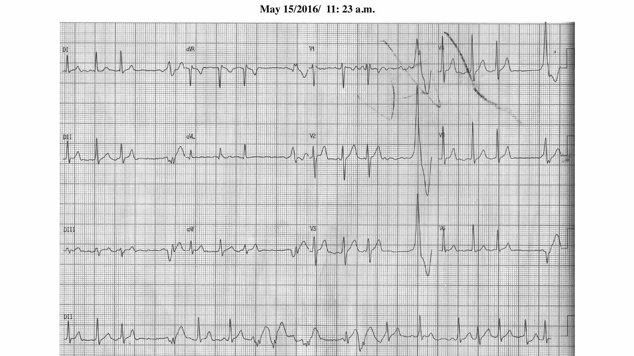
May 15/2016/ 11: 23 a.m.

May 15/2016/ 13: 53 p.m.

Colleagues opinions
Atrial Fibrillation in a patient with WPW. He needs EP and possible ablation.
Yochai Birnbaum, MDCardiology Houston, TX USAProfessor, Cardiology, Baylor College of Medicine

Spanish Hola amigos1. Por los antecedentes clínicos como la edad, la ausencia de cardiopatía estructural y “ por el golpe de primera
vista " de los ECGs 1 a 5 pienso que se trata de una FA pre-exitada de frecuencia cardiaca promedio de 230min. En este caso, se presenta el diagnóstico diferencial con TV por la presencia del patrón concordantepositivo en las precordiales. En principio el uso de drogas bloqueantes nódulo AV no son recomendables y esrazonable la cardioversión eléctrica (CVE) como abordaje inicial.
2. Por todo el comentario anterior existe riesgo de degenerar en fibrilación ventricular.3. El ECG 6 muestra ritmo sinusal (post cardioversion ?) con un patrón de WPW, por presencia de una vía
accesoria (o varias por presentar QRS de ancho variables durante la taquicardia)y localización probablementelateral izquierda.
4. Conducta definitiva: ablación por radiofrecuencia de la vía accesoria.-----------------------------------------------------\-----------------------------------------------------------------------------------
English Hello, friends,My opinion:1. Considering the setting of the clinical history, age of the patient, absence of structural heart disease and seeing
the ECGs 1 through 5 at first sight, it is PREEXCITED AF of 230 bpm in average, a rationale supported byBrugada algorithm (differential diagnosis of VT with PREEXCITED SVT) with matching positive pattern inprecordial leads.
2. So in principle, using AV node blocking drugs is not advisable and electrical cardioversion is reasonable fromthe start.
3. Considering all of the above, there is risk of VF.4. ECG6 shows sinus rhythm (post cardioversion?) with WPW pattern, by the presence of accessory pathway (or
several by presenting QRS with variable widths during tachycardia), with probably left lateral location.5. The management is RF ablation of the accessory pathway.Juan José Sirena MD Santiago del Estero Argentina

The ECGs show Atrial fibrillation with WPW pattern suggestive of a left posterior pathway. One ECG showing a QS pattern in V6 is probably a result of lead misplacement. Acute RX is IV Procaineamide or Ibutilide (in US) followed by ablation.
Department of Cardiac Electrophysiology, University of California San Francisco, San Francisco, California, USA. Professor of Medicine UCSF School of Medicine533 Parnassus Avenue San Francisco CA 94117 USAPhone: [email protected]

1. Tendo em vista a queixa de dispnéia e desconforto torácico, optaríamos por cardioversão elétrica sincronizada. 2. Fibrilação atrial com condução AV antidrômica, compatível com fibrilação atrial pré-excitada.
2. Estratificaríamos o risco com estudo eletrofisiológico que desempenharia papel terapêutico mediante a ablação da via anômala.3. Fibrilação atrial pré-excitada está associada a instabilidade elétrica com evolução para ritmos ventriculares acelerados, portanto o paciente corre
risco sim de morte súbita.Colaboradores dessa discussão os residentes da clínica médica: Larissa Barbosa-Talharo e Gabriel Afonso Dutra Kreling.
Dr. José GrindlerDiretor de Serviço: Eletrocardiologia HCFaculdade de Medicina USP
1. Taking into account the complaint of dyspnea and chest discomfort, we would choose synchronized electrical cardioversion.2. Atrial fibrillation with antidromic AV conduction, compatible with preexcited atrial fibrillation.3. We would stratify risk by electrophysiology study that would play a therapeutic role by ablation of the anomalous pathway.4. Preexcited atrial fibrillation is associated to electrical instability with evolution into accelerated ventricular rhythms, so the patient is indeed in risk of sudden cardiac death.Residents of clinical practice collaborated with this discussion:Larissa Barbosa-Talharo and Gabriel Afonso Dutra Kreling.
Dr. José Grindler

Dear Andrés Ricardo, regarding your Q, I think it is fascicular VT, that need in acute therapy
Adenosine or Verapamil, possibly transesophageal stimulation and RF ablation for final
treatment. This patient not have a risk of sudden cardiac death.
Best regards
Leonid Makarov M.D., Ph.D.
Prof. Dr. Leonid Makarov M.D., Ph.D. Head of Center for Syncope and Arrhythmias in Children
and Adolescents of Federal Medico-Biology Agency of Russia. President of the Russian Society
for Holter Monitoring (ROHMINE). Adress: Central Children Clinical Hospital FMBA of Russia.
115409 Moskvorechie str. 20. Tel. +7 (499)324-5756 E-mail: [email protected]
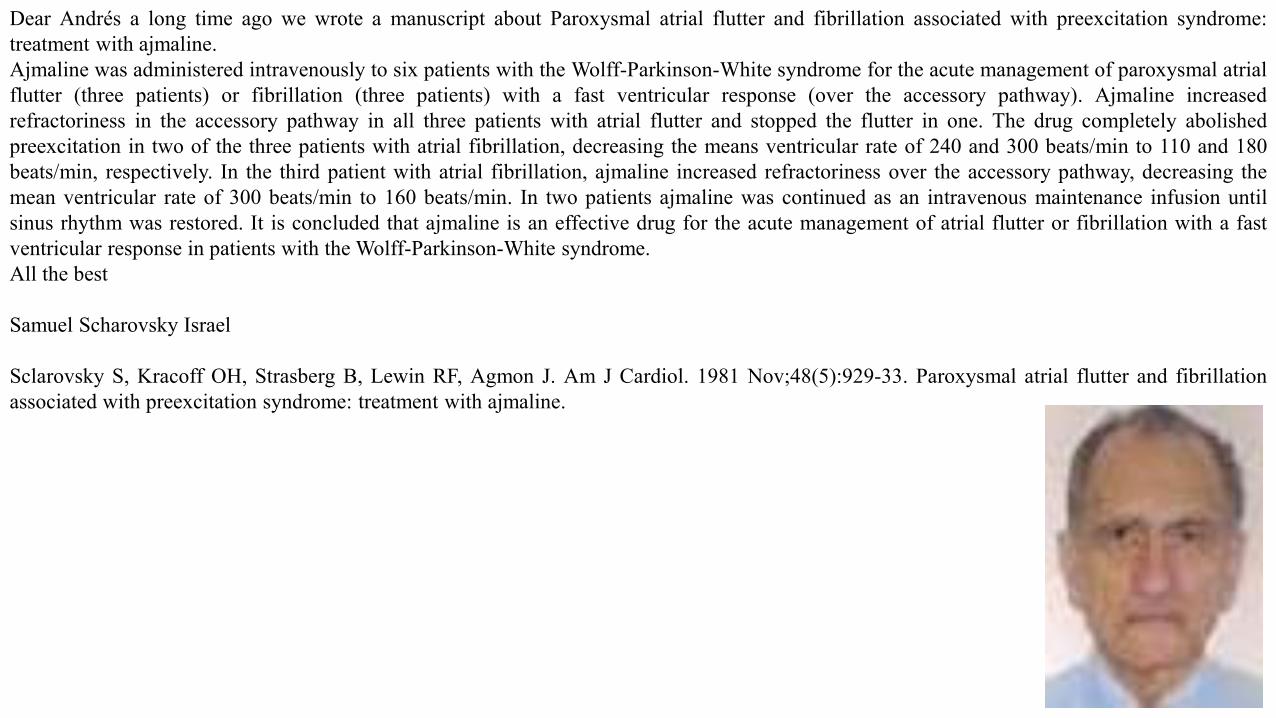
Dear Andrés a long time ago we wrote a manuscript about Paroxysmal atrial flutter and fibrillation associated with preexcitation syndrome:treatment with ajmaline.Ajmaline was administered intravenously to six patients with the Wolff-Parkinson-White syndrome for the acute management of paroxysmal atrialflutter (three patients) or fibrillation (three patients) with a fast ventricular response (over the accessory pathway). Ajmaline increasedrefractoriness in the accessory pathway in all three patients with atrial flutter and stopped the flutter in one. The drug completely abolishedpreexcitation in two of the three patients with atrial fibrillation, decreasing the means ventricular rate of 240 and 300 beats/min to 110 and 180beats/min, respectively. In the third patient with atrial fibrillation, ajmaline increased refractoriness over the accessory pathway, decreasing themean ventricular rate of 300 beats/min to 160 beats/min. In two patients ajmaline was continued as an intravenous maintenance infusion untilsinus rhythm was restored. It is concluded that ajmaline is an effective drug for the acute management of atrial flutter or fibrillation with a fastventricular response in patients with the Wolff-Parkinson-White syndrome.All the best
Samuel Scharovsky Israel
Sclarovsky S, Kracoff OH, Strasberg B, Lewin RF, Agmon J. Am J Cardiol. 1981 Nov;48(5):929-33. Paroxysmal atrial flutter and fibrillationassociated with preexcitation syndrome: treatment with ajmaline.

Final conclusions

May 15/2016 06:30 a.m. admission
ECG features: heart rate is faster than 180bpm, irregular broad complex rhythm, QRS complexes with change in thickness,positive concordance from V1 to V6: indicative of left posterolateral focus (VT) or left posterior accessory pathway (Fananapazir 1990).Additionally, closer inspection of the QRS complexes show evidence of pre-excitation (a slurred upstroke to the QRS complex). QRS axis locatedon right superior quadrant AKA “Northwest Axis” or no man's land and remains stable. Conclusion: preexcited atrial fibrillation. Observation:Paroxysmal atrial fibrillation (PAF) develops in up to one-third of patients with the WPW syndrome. The reason for this high incidence of PAF inthe WPW syndrome is not yet clearly understood. When PAF appears in patients with WPW syndrome who have anterograde conduction via theAP, it may be life-threatening if an extremely fast ventricular response develops degenerating into VF (Centurión 2008).
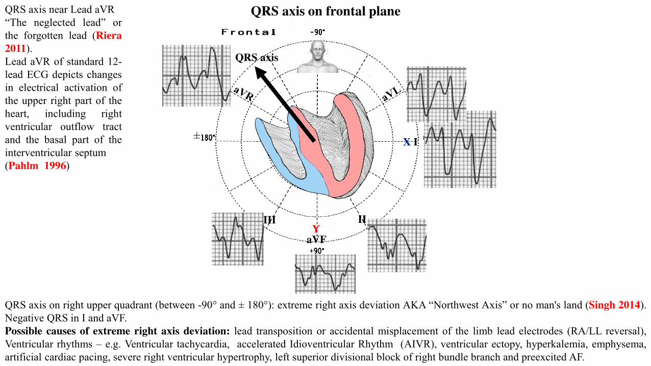
QRS axis on frontal plane
QRS axis on right upper quadrant (between -90° and ± 180°): extreme right axis deviation AKA “Northwest Axis” or no man's land (Singh 2014).Negative QRS in I and aVF.Possible causes of extreme right axis deviation: lead transposition or accidental misplacement of the limb lead electrodes (RA/LL reversal),Ventricular rhythms – e.g. Ventricular tachycardia, accelerated Idioventricular Rhythm (AIVR), ventricular ectopy, hyperkalemia, emphysema,artificial cardiac pacing, severe right ventricular hypertrophy, left superior divisional block of right bundle branch and preexcited AF.
IIIII
aVF
X I
Y
±
QRS axis near Lead aVR“The neglected lead” orthe forgotten lead (Riera2011).Lead aVR of standard 12-lead ECG depicts changesin electrical activation ofthe upper right part of theheart, including rightventricular outflow tractand the basal part of theinterventricular septum(Pahlm 1996)
QRS axis

Positive concordance indicates that the origin of the tachycardia lies on the posterior left ventricular wall; thewave of depolarization moves towards all the chest leads and produces positive complexes across precordial leadsfrom V1 to V6.
X V6
V1
V4
V5
V2V3
Z

May 15/2016/ 07: 51 a.m.
Precordial V6-V5 Electrodesare misplaced. Electrodes (low)
is similar to II pattern

Using the ACLS Tachycardia Algorithm for Managing Stable TachycardiaThe key to managing a patient with any tachycardia is to check if pulses are present, decide if the patient is stable or unstable, and then treat thepatient based on the patient's condition and rhythm. If the patient does not have a pulse, follow the ACLS Pulseless Arrest Algorithm. If the patienthas a pulse, manage the patient using the ACLS Tachycardia Algorithm.Definition of hemodynamically stable TachycardiaFor a diagnosis of stable tachycardia, the patient meets the following criteria: The patient's heart rate is >100 bpm, absence of shock signals: notchanges in mental status or behavior, such as anxiousness or agitation, ongoing chest pain, hypotension, or other signs of shock(cool, clammy skin,pale or ashen skin, rapid breathing, nausea or vomiting, enlarged pupils, weakness or fatigue, dizziness or fainting). The patient does not have anyserious signs or symptoms as a result of the increased heart rate.Overview: Find out if significant symptoms are present. Evaluate the symptoms and decide if they are caused by the tachycardia or other systemicconditions. Use these questions to guide your assessment:1. Does the patient have symptoms?2. Is the tachycardia causing the symptoms?3. Is the patient hemodynamically stable or unstable?4. Is the QRS complex narrow or wide?5. Is the rhythm regular or irregular?6. Is the rhythm sinus tachycardia?
GuidelinesSituation Assessment and Actions
Patient has significant signs or symptoms of tachycardia and they are being caused by the arrhythmia.
The tachycardia is unstable. Immediate cardioversion is indicated.
Patient has a pulseless ventricular tachycardia. Follow the Pulseless Arrest Algorithm. Deliver unsynchronized high-energy shocks.
Patient has polymorphic VT and the patient is unstable. Treat the rhythm as VF. Deliver unsynchronized high-energy shocks.

Does the patient have a pulse?Yes, the patient has a pulse. Complete the following: Assess the patient using the primary and secondary surveys, check the airway, breathing,and circulation, give oxygen and monitor oxygen saturation, get an ECG, identify rhythm, check blood pressure, identify and treat reversiblecauses.Is the patient stable or hemodynamically compromised? Look for altered mental status, ongoing chest pain, hypotension, or other signs ofshock.1. Prompt direct-current cardioversion is recommended for patients with AF, Wolff-Parkinson-White syndrome, and rapid ventricular response
who are hemodynamically compromised (Blomstrom-Lundqvist 2003). (Level of evidence: C)Yes, the patient is stable. Take the following actions:Start an IV.Obtain a 12-lead ECG or rhythm strip.2. Intravenous procainamide or ibutilide to restore sinus rhythm or slow the ventricular rate is recommended for patients with pre-excited AF and
rapid ventricular response who are not hemodynamically compromised (Blomstrom-Lundqvist 2003). (Level of evidence: C)Remember: Rate-related symptoms are uncommon if heart rate is < 150 bpm.Is the QRS complex wide or narrow?
Steps for Managing Stable Tachycardia
Patient Treatment
The patient's QRS is narrow and rhythm is regular. Try vagal maneuvers. Give adenosine 6 mg rapid IV push. If patient does not convert, give adenosine 12 mg rapid IV push. May repeat 12 mg dose of adenosine once.
Caution: If the tachycardia has a wide-complex QRS and is stable, consult with an expert. Management and treatment for a stable tachycardiawith a wide QRS complex and either a regular or irregular rhythm should be done in the hospital setting with expert consultation available.Management requires advanced knowledge of ECG and rhythm interpretation and anti-arrhythmic therapy.

If the rhythm pattern is irregular narrow-complex tachycardia, it is probably atrial fibrillation, possible atrial flutter, or multi-focal atrialtachycardia.
Patient Treatment
Patient's rhythm has wide (≥ 120 ms) QRS complex and patient's rhythm is regular.
Expert consultation is advised.
Patient is in ventricular tachycardia or uncertain rhythm. Amiodarone 150 mg IV over 10 min; repeat as needed to maximum dose of 2.2 g in 24 hours. Prepare for elective synchronized CV.
Patient is in supraventricular tachycardia with aberrancy. Adenosine 6 mg rapid IV push If no conversion, give adenosine 12 mg rapid IV push; may repeat 12 mg dose once.
Patient's rhythm has wide (> 0.12) QRS complex AND Patient's rhythm is irregular.
Seek expert consultation.
If pre-excited AF(AF in WPW Syndrome) Avoid AV nodal blocking agents such as adenosine, digoxin (Sellers 1977) , diltiazem, verapamil.
Consider amiodarone 150 mg IV over 10 min. Amiodarone is not superior to procainamide in rate control for WPW-AF, and may be dangerous (Simonian 2010)
Patient has recurrent polymorphic VT Seek expert consultation,
If patient has torsades de pointes rhythm on ECG Give magnesium (load with 1-2 g over 5-60 min; then infuse.

Considerations:You may not be able to distinguish between a supraventricular wide-complex rhythm and a ventricular wide-complex rhythm. Most wide-complextachycardias originate in the ventricles.If the patient becomes unstable, proceed immediately to treatment. Do not delay while you try to analyze the rhythm.
Wolff-Parkinson-White and Pre-Excitation Syndromes CLASS I1. Prompt direct-current cardioversion is recommended for patients with AF, WPW syndrome, and rapid ventricular response who are
hemodynamically compromised (Blomstrom-Lundqvist 2003) (Level of evidence: C).2. Intravenous procainamide or ibutilide to restore sinus rhythm or slow the ventricular rate is recommended for patients with pre-excited AF and
rapid ventricular response who are not hemodynamically compromised (Blomstrom-Lundqvist 2003) (Level of evidence: C).3. Radiofrequency catheter ablation (RFCA) of the AP is recommended in symptomatic patients with pre-excited AF, especially if the AP has a
short refractory period that allows rapid antegrade conduction (Blomstrom-Lundqvist 2003) (Level of evidence: C). The presence of J wavesafter RFCA was associated with a history of AF and shorter ventricular effective refractory period. The prevalence of J waves is high inpatients with WPW syndrome (≈N18%) and is influenced by manifest ventricular preexcitation (VPE) (Yagihara 2012).
4. CLASS III: HARM 1. Administration of intravenous amiodarone, adenosine, digoxin (Sellers 1977) (oral or intravenous), ornondihydropyridine calcium channel antagonists (oral or intravenous) in patients with WPW syndrome who have pre-excited AF is potentiallyharmful because these drugs accelerate the ventricular rate (Boriani1996; Kim2008; Simonian 2010) (Level of evidence: B).
Factors that determinate ventricular rate during AF:• Refractory period duration of the AP in the anterograde direction• Refractory period of the AV node• Refractory period of the ventricle• Concealed anterograde and retrograde penetration into the accessory pathway and AV node• Sympathetic stimulation shortens the refractory period of the AP and accelerates de rate. It is important to terminate this tachycardia promptly
and to reassure the patient in the meantime. A reflex sympathetic response to the fall in blood pressure is associated with the AF and the veryrapid ventricular rate, anxiety adds to the response.

VPE is asymptomatic in 65–90% of children and adolescents. The prevalence of VPE on baseline ECG is 0.1–0.3% either in the generalpopulation (Guize 1985; Rodday 2012) or in athletes (Furlanello1998). Children and adolescents with VPE are at increased risk for suddencardiac death (SCD). The most common manifestation of WPW syndrome is atrioventricular reentrant tachycardia (AVRT) or, more rarely, AF. Inyoung WPW children, AVRT is mostly orthodromic as a result of an excellent AV conduction and a long anterograde effective refractory period ofthe AV node (Cohen 1997). Inducible antidromic AVRT at EPS is reported in only 2.6% of children (Ceresnak 2012) as compared with 10–11% ofadult patients(Gillette 1979; Atié 1990).Rapid anterograde conduction in the setting of VPE is associated with an increased risk of SCD. In patients with a very short anterograde
refractory period of the AP, AF may degenerate into VF leading to SCD (Blomström-Lundqvist 2003; Timmermans 1995). Althoughantiarrhythmic therapy and RFCA are well established temporary or definitive treatments for patients with WPW syndrome, the optimalmanagement of asymptomatic children remains to be clearly defined. Children and adolescents with WPW syndrome have a higher rate of AVRTinducibility than asymptomatic patients. However, no differences between the two groups were found in atrial vulnerability and parameters relatedto the risk of SCD (Di Mambro 2015).An expert consensus document on the management of asymptomatic patients with persistent VPE (Cohen 2012) suggested that:1. Invasive measurement of the shortest pre-excited R–R interval (SPERRI) in AF is useful for risk stratification, and2. Patients with a SPERRI ≤ 250 ms at baseline are at increased risk for SCD.3. High-risk AP is defined as AP effective refractory period (APERP), block cycle length, or shortest preexcited RR interval during AF ≤ 250 ms.The 2005 European guidelines (Pelliccia2005) for competitive sports eligibility recognize the importance of performing an EPS in all patients withasymptomatic VPE either at rest or during adrenergic stimulation. According to these guidelines, athletes with multiple APs, easily inducible AF, aSPERRI ≤ 240 ms in AF, or a SPERRI ≤ 210 ms in AF during stress (exercise testing or isoproterenol infusion) should be considered at increasedrisk for SCD. In this group, RFCA is recommended before allowing participation in competitive sports. In anesthetized children with VPE, APsdisplay shorter conduction properties at younger ages and important adrenergic sensitivity at all ages. Use of low-dose isoproterenol resulted in asubstantial increase in the number of patients who would otherwise meet typical criteria for RFCA (Moore 2011).Comment: Special emphasis was given to the risk of SCD in children and adolescents with VPE, following degeneration of AF into VF with rapidAV conduction through the AP (Pappone 2004; Santinelli 2009). However, the risk for SCD seems to be relatively low in symptomatic patients.Munger et al (Munger 1993) reported an incidence of SCD of 0.0025 per patient-year, equal to 3% of patients during lifetime. In a 16-yearfollow-up, Timmermans et al. (Timmermans 1995) reported an incidence of SCD of 2.2%. These data were first outlined in a more limited studypopulation by Flensted-Jensen (Flensted-Jensen1969). The risk for SCD in the presence of WPW syndrome has also been demonstrated to have

a strong association with exercise (Wiedermann 1987).The WPW syndrome is associated with a small but finite risk of SCD that is attributed to rapid anterograde AP conduction in the setting of AF.Reports of potentially life-threatening arrhythmias have been described in seemingly asymptomatic children with VPE, suggesting that the risk ofSCD may be higher than previously suspected in this population (Pappone 2004; Santinelli 2009). It is speculated that increased sympatheticdischarge developing at the initiation of AF may enhance AP conduction and predispose certain individuals to VF (Wellens1982). Previous studiesin adults examining the effects of isoproterenol on pathway conduction have consistently demonstrated prominent adrenergic sensitivity, and forthis reason, some have favored the use of isoproterenol in the risk-stratification process (Przybylski1980; Brembilla-Perrot1988; Szabo1989). Isolated reports of cardiac-arrest survivors not meeting traditional high-risk criteria but who exhibited increased adrenergic sensitivityduring isoproterenol challenge lend support to this approach (Sharma 1987). SCD is even more uncommon in asymptomatic patients in theabsence of associated heart disease. In a large meta-analysis, Obeyesekere et al (Obeyesekere 2012) reported a risk of SCD of 1.93 per 1000patient-years in asymptomatic children compared with only 0.86 per 1000 patient-years in adults. Notwithstanding this, SCD may be the firstevidence of WPW syndrome in young patients with asymptomatic VPE. The low incidence of SCD and low risk of supraventricular tachycardiaargue against routine invasive management in most asymptomatic patients with the WPW ECG pattern. In a population of 25 young patients withWPW syndrome who manifested VF, 2 children (aged 8 and 9 years) and 1 adolescent (aged 16 years) were asymptomatic. In addition, life-threatening symptoms as first manifestation have recently been reported in 10–48% of asymptomatic children with VPE (Steinherz 2001).A SPERRI of 220–250 ms in AF has consistently been found to predict the risk of VF (van der Linden 2000). Although the occurrence of AF isextremely rare in healthy pediatric populations, this arrhythmia may be more frequent in young patients with VPE, being enabled by the presenceof APs and different trigger mechanisms (van der Linden 2000).In identifying symptomatic children and young adults with WPW syndrome at risk for VF , SPERRI has a high sensitivity (88–100%) and a highnegative predictive value, but a quite low specificity (<75%) and an extremely low positive predictive value. In a population of 60 pediatricpatients with WPW syndrome, a SPERRI < 220 ms was documented only in those with a history of aborted SCD. Conversely, APERP seems to beless predictive of life-threatening events. A short APERP was found to correlate significantly only with AVRT inducibility at EPS (positivepredictive value 47%, negative predictive value 97%). Inducibility and duration of induced AF were not significantly different between the twogroups both at baseline and during adrenergic stimulation. Notably, also the proportion of children with a high-risk SPERRI was comparablebetween groups both at baseline and during stress testing. This means that WPW and asymptomatic pediatric patients have the same potential riskof SCD in the presence of inducible AF, and AP location does not seem to be a predictive factor of an unfavorable course of VPE.
APs can conduct in the anterograde direction, retrograde direction, or both; and can be associated with several different supraventriculararrhythmias. Some anterograde pathways may place patients at risk of SCD.The most common tachycardia associated with an AP is orthodromic AVRT, with a circuit that uses the AV node and His Purkinje system in theanterograde direction, followed by conduction through the ventricle, retrograde conduction over the AP, and completion of the circuit byconduction through the atrium back into the AV node. Orthodromic AVRT accounts for approximately 90% to 95% of AVRT episodes in patientswith a manifest AP. Pre-excited AVRT, including antidromic AVRT, accounts for 5% of the AVRT episodes in patients with a manifest pathway andinvolves conduction from the atrium to the ventricle via the AP, causing a preexcited QRS complex. This is called antidromic AVRT tachycardiawhen the return reentrant conduction occurs retrogradely via the AV node. In rare cases of pre-excited AVRT, the return conduction occurs via asecond accessory AV pathway. AF can occur in patients with APs, which may result in extremely rapid conduction to the ventricle over a manifestpathway, which increases the risk of inducing VF and SCD.Rapid anterograde AP conduction during AF can result in SCD in patients with a manifest AP, with a 10-year risk ranging from 0.15% to 0.24%(Natale A 2000; Schumacher,1999). Unfortunately, SCD may be the first presentation of patients with undiagnosed WPW. Increased risk of SCDis associated with a history of symptomatic tachycardia, multiple accessory pathways, and a shortest pre-excited R-R interval of < 250 ms duringAF. The risk of SCD associated with WPW appears highest in the first 2 decades of life (Schumacher 1999).
Supraventricular tachycardia with atrioventricular conduction over an accessory pathway
Supraventricular tachycardia with AV conduction over an AP may occur during AT, atrial flutter, AF, AVNRT, or antidromic AVRT. The latter isdefined as anterograde conduction over the accessory pathway and retrograde conduction over the AV node or a second accessory AV pathway. Awide-QRS complex with left bundle-branch block (LBBB) morphology may be seen with anterograde conduction over other types of accessorypathways, such as atriofascicular, nodofascicular, or nodoventricular tracts.

Acute management of wide QRS-complex tachycardia
Immediate DC cardioversion is the treatment for hemodynamically unstable tachycardia's. If the tachycardia is hemodynamically stable anddefinitely supraventricular, then management is as described for narrow QRS tachycardias. For pharmacologic termination of a stable wide QRS-complex tachycardia, IV procainamide and/or sotalol are recommended on the basis of randomized but small studies. Amiodarone is alsoconsidered acceptable. Amiodarone is preferred compared with procainamide and sotalol for patients with impaired LV function or signs of HF.These recommendations are in accord with the current Advanced Cardiovascular Life Support guidelines. Special circumstances may requirealternative therapy (ie, pre-excited tachycardias and VT caused by digitalis toxicity). For termination of an irregular wide QRS-complextachycardia (ie, pre-excited AF), DC cardioversion is recommended. Or, if the patient is hemodynamically stable, then pharmacologic conversionusing IV ibutilide or flecainide is appropriate. The incidence of SCD in patients with the WPW syndrome has been estimated to range from 0.15%to 0.39% over 3- to 10-year follow-up. It is unusual for cardiac arrest to be the first symptomatic manifestation of WPW syndrome. Conversely, inabout half of the cardiac arrest cases in WPW patients, it is the first manifestation of WPW. Given the potential for AF among patients with WPWsyndrome and the concern about SCD resulting from rapid pre-excited AF, even the low annual incidence of SCD among patients with the WPWsyndrome is of note and supports the concept of liberal indications for RFCA. Studies of WPW syndrome patients who have experienced a cardiacarrest have retrospectively identified a number of markers that identify patients at increased risk. These include:• A shortest pre-excited R-R interval <250 ms during spontaneous or induced AF;• A history of symptomatic tachycardia;• Multiple accessory pathways, and• Ebstein’s anomaly. A high incidence of SCD has been reported in familial WPW. This familial presentation is, however, exceedingly rare
(Gollob 2001). Several noninvasive and invasive tests have been proposed as useful in risk-stratifying patients for SCD risk. The detection ofintermittent pre-excitation, which is characterized by an abrupt loss of the delta wave and normalization of the QRS complex, is evidence thatan accessory pathway has a relatively long refractory period and is unlikely to precipitate VF. The loss of pre-excitation after administration ofthe antiarrhythmic drug procainamide has also been used to indicate a low-risk subgroup. Noninvasive tests are considered inferior to invasiveEPS assessment for risk of SCD. For this reason, noninvasive tests currently play little role in patient management.
Acute treatmentThe approach to acute evaluation and management during a sustained regular tachycardia is covered in Sections IV. A and IV. B. The approach toacute termination of these arrhythmias generally differs from that used for long-term suppression and prevention of further episodes of SVT.

Special considerations for patients with wide-complex (Pre-Excited) tachycardias
In patients with antidromic tachycardia, drug treatment may be directed at the AP or at the AV node because both are critical components of thetachycardia circuit. Atrioventricular nodal-blocking drugs would, however, be ineffective in patients who have anterograde conduction over onepathway and retrograde conduction over a separate AP because the AV node is not involved in the circuit. Adenosine should be used with cautionbecause it may produce AF with a rapid ventricular rate in pre-excited tachycardias. Adenosine may cause VF when administeredduring preexcited AF. This phenomenon is seen in patients having APs with short refractory periods (Gupta 2002). Ibutilide, procainamide, orflecainide, which are capable of slowing the conduction through the AP, are preferred.Pre-excited tachycardias occurring in patients with either AT or atrial flutter with a bystander AP may present with a one-to-one conduction overthe AP. Caution is advised against AV-nodal-blocking agents, which would obviously be ineffective in this situation. Antiarrhythmic drugs, whichprevent rapid conduction through the bystander pathway, are preferable, even if they may not convert the atrial arrhythmia. Similarly, it ispreferable to treat pre-excited AF by either IV ibutilide, flecainide, or procainamide.Long-term pharmacologic therapy: Antiarrhythmic drugs represent one therapeutic option for management of AP-mediated arrhythmias, butthey have been increasingly replaced by RFCA. Antiarrhythmic drugs that primarily modify conduction through the AV node include digoxin,verapamil, β-blockers, adenosine, and diltiazem. Antiarrhythmic drugs that depress conduction across the AP include class I drugs, such asprocainamide, disopyramide, propafenone, and flecainide, as well as class III antiarrhythmic drugs, such as ibutilide, sotalol, and amiodarone.Prophylactic pharmacologic therapy: There have been no controlled trials of drug prophylaxis involving patients with AVRT; however, anumber of small, nonrandomized trials have been performed (each involving less than 50 patients), and they have reported the safety and efficacyof drug therapy for maintenance of sinus rhythm in patients with supraventricular arrhythmias. A subset of the patients in these studies had AVRTas their underlying arrhythmia. Available data do not allow a comparison of the efficacy of these drugs relative to one another. The drugs availableto treat AVRT include any drug that alters either conduction through the AV node (eg, nondihydropyridine calcium-channel blockers, β-blockers,digoxin) or a drug that alters conduction through the atrium, ventricle, or accessory pathway (eg, class Ia, Ic, or III antiarrhythmic agents). Theavailable data are outlined below. Of note is that no studies have examined the efficacy of chronic oral β-blockers in the treatment of AVRT and/orWPW syndrome. The absence of studies specifically examining the role of β-blocker therapy in the treatment of WPW syndrome likely reflects thefact that RFCA is the therapy of choice for these patients. Despite the absence of data from clinical trials, chronic oral β-blocker therapy may beused for treatment of patients with WPW syndrome, particularly if their AP has been demonstrated during EPS to be incapable of rapidanterograde conduction.

PropafenonePropafenone A class Ic antiarrhythmic agent, has potent inhibitory effects on APs and has additional significant effects on atrial, AV nodal andventricular refractoriness (Sethi1991).The largest published study that reported the efficacy of propafenone in adult patients involved 11 individuals. Propafenone resulted in anterogradeconduction block in the AP in 4 of 9 patients and retrograde block in 3 of 11 patients. AVRT was rendered noninducible in 6 of 11 patients. During9 plus or minus 6 months of follow-up, none of the 10 patients discharged on a combination of propafenone and a β-blocker experienced arecurrence. No major side effects were reported. Other small trials have evaluated the efficacy of propafenone in the treatment of AVRT inchildren. The largest of these involved 41 children. Chronic administration of propafenone was effective in 69%. Side effects occurred in 25% ofthese patients. A reversible protective effect against fast ventricular rate as the result of an episode of AF exists during exercise in patients with asymptomatic AP who are treated with propafenone (Auricchio1992).FlecainideA number of studies have examined the acute and long-term efficacy of oral and IV flecainide in the treatment of patients with AVRT. The largestof these studies involved 20 patients with AVRT. The oral administration of flecainide (200 to 300 mg/d) resulted in an inability to inducesustained tachycardia in 17 of the 20 patients. The electrophysiological effects of flecainide were partially reversed by administration ofisoproterenol. During 15 plus or minus 7 months of follow-up on oral flecainide treatment, 3 patients developed a recurrence of tachycardia. Otherstudies have reported similar findings. The addition of a β-blocker results in greater efficacy, with more than 90% of patients achieving abolition ofsymptomatic tachycardia. In addition to studies that specifically focused on patients with a known AVRT, several randomized trials have evaluatedthe efficacy of flecainide in the treatment of patients with PSVT of undetermined tachycardia mechanism. One study enrolled 34 patients withPSVT into a double-blinded, placebo-controlled trial with an 8-week crossover trial design. Flecainide was shown to be superior to placebo; 8 ofthe 34 patients had a recurrence during flecainide therapy, as compared with 29 of 34 patients having a recurrence on placebo. Treatment withflecainide also increases the time to first symptomatic event and time to subsequent events.AmiodaroneSeveral studies have evaluated the efficacy of amiodarone in the treatment of patients with AP-mediated tachycardias. These studies, however, donot demonstrate that amiodarone is superior to class Ic antiarrhythmic agents or sotalol. As a result of these findings, combined with the well-recognized organ toxicity associated with amiodarone and the high rate of discontinuation of this drug, amiodarone generally is not warranted fortreatment of patients with APs. Exceptions are for patients with structural heart disease who are not thought to be candidates for RFCA.

VerapamilThe efficacy of verapamil in the prevention of AVRT has been reported in a single study, which involved seven patients. Four of these 17 patientscontinued to have inducible AVRT during EPS despite treatment with oral verapamil. Adequate follow-up data in these patients were not providedin this manuscript. Intravenous verapamil can precipitate hemodynamic deterioration during AF. Verapamil and diltiazem should not be used as thesole therapy for patients with APs that might be capable of rapid conduction during AF. This concern also applies to digoxin, which also should notbe used in this situation (Sellers 1977).Other drugsNo studies have been performed to determine the short- or long-term efficacy of procainamide or quinidine in the treatment of AVRT.Single-dose oral therapy (pill-in-the-pocket)Some patients with infrequent episodes of tachycardia may be managed with the single-dose “pill-in-the-pocket” approach: taking anantiarrhythmic drug only at the onset of a tachycardia episode. This approach to treatment is reserved for patients without pre-excitation and withinfrequent and hemodynamically tolerated tachycardia. A recent study reported that 94% of induced PSVT episodes were terminated in theelectrophysiology laboratory within 32 minutes plus or minus 22 minutes by administration of a combination of diltiazem (120 mg) pluspropranolol (80 mg). This treatment was successful in terminating PSVT within 2 hours during outpatient follow-up in 81% of patients. Anotherfinding of this study was that flecainide, when given as a single dose for acute termination of PSVT, was significantly less effective than thecombination of diltiazem and propranolol.Radiofrequency Catheter ablationRFCA of APs is performed in conjunction with a diagnostic EPS. The purposes of the EPS are to confirm the presence of an AP, determine itsconduction characteristics, Identify the location of the AP, determine if the AP has dangerous properties and define its role in the patient’s clinicalarrhythmia. Once the arrhythmia is localized, RFCA is performed using a steerable RFCA catheter. There have been no prospective, randomizedclinical trials that have evaluated the safety and efficacy of RFCA of APs; however, the results of RFCA of APs have been reported in a largenumber of single-center trials, one multicenter trial, and several prospective registries. The initial efficacy of RFCA of APs approximately 95% inmost series. The success rate for RFCA of left free-wall accessory pathways is slightly higher than for RFCA of APs in other locations. After aninitially successful procedure, resolution of the inflammation or edema associated with the initial injury allows recurrence of AP conduction inapproximately 5% of patients. APs that recur can usually be successfully ablated during a second session. The occurrence of AF in the presence ofan AP that conducts rapidly is potentially lethal because the rapid ventricular response may lead to VF. In significant proportion of WPW patients,VF was the first clinical manifestation of WPW syndrome, often precipitated by physical activity, emotional stress or inappropriate drug

administration. Successful elimination of AP by RFCA is highly effective in secondary prevention of life-threatening tachyarrhythmias in patientswith VPE (Mujović 2010)Complications associated with RFCA of APs result from radiation exposure, vascular access (eg, hematomas, deep venous thrombosis, arterialperforation, arteriovenous fistula, pneumothorax), catheter manipulation (eg, valvular damage, microemboli, perforation of the coronary sinus ormyocardial wall, coronary artery dissection, thrombosis), or delivery of RF energy (eg, AV block, myocardial perforation, coronary artery spasm orocclusion, transient ischemic attacks, or cerebrovascular accidents). The procedure-related mortality reported for RFCA of APs ranges from 0% to0.2%. The voluntary Multicenter European Radiofrequency Survey (MERFS) reported data from 2222 patients who underwent RFCA of an AP.The overall complication rate was 4.4%, including 3 deaths (0.13%). The 1995 NASPE survey of 5427 patients who underwent RFCAs of anaccessory AP who underwent RDCA of an AP as part of a prospective, multicenter clinical trial, there was 1 death (0.2%). This patient died ofdissection of the left main coronary artery during an attempt at RFCA of a left free-wall AP. The most common major complications are completeAV block and cardiac tamponade. The incidence of inadvertent complete AV block ranges from 0.17% to 1.0%. Most occur in the setting ofattempted RFCA of septal APs located close to the AV junction. The frequency of cardiac tamponade varies between 0.13% and 1.1%.
Management of patients with asymptomatic accessory pathwaysAn ECG pattern of VPE is occasionally encountered in a subject who has no symptoms of arrhythmia. The role of EPS and RFCA in asymptomaticpatients with VPE is controversial. One-third of asymptomatic individuals younger than 40 years of age when VPE was identified eventuallydeveloped symptoms, whereas no patients in whom VPE was first uncovered after the age of 40 years developed symptoms. Most patients withasymptomatic VPE have a good prognosis; cardiac arrest is rarely the first manifestation of the disease. Studies have reported that approximately20% of asymptomatic patients will demonstrate a rapid ventricular rate during AF induced during EPS. During follow-up, however, very fewpatients developed symptomatic arrhythmias, and none of these individuals experienced a cardiac arrest. The positive predictive value of invasiveEPSs is considered to be too low to justify routine use in asymptomatic patients. The decision to ablate APs in individuals with high-riskoccupations, such as school bus drivers, pilots, and scuba divers, is made on the basis of individual clinical considerations. Theserecommendations are likely to remain unchanged despite the results of a study that identified the results of EPS as an important predictor ofarrhythmic events in patients with asymptomatic VPE. In young patients with WPW syndrome, occurrence of AF with a rapid ventricular responseduring EPS correlated well with a history of syncope and may be the cause of syncope in most patients. EPS may be helpful in identification ofyoung patients with WPW at risk for syncope (Paul 1990).

Propensity for AF in patients with an AP is strongly related to VPE, larger atria, male gender, and older age. Reflection and microreentry at the APmay be important for AF initiation in patients with manifest (preexcited) WPW syndrome. Similar mechanisms also may trigger AF in patientswithout an AP (Schwieler 2008).With the objective of evaluate the electrophysiological properties of children with WPW syndrome and to develop an algorithm for themanagement of these patients with limited access to EPS.Yıldırım et al studied one hundred nine patients underwent EPS at a single tertiary center (the median age 11 years). The induction of VF in 2 of109 patients study suggests that the prognosis of WPW in children is not as benign as once thought. All patients with a WPW pattern on the ECGshould be assessed EPS and risk-stratified. Ablation of patients with risk factors can prevent SCD in this population (Yıldırım 2015).
AF often coexists with WPW syndrome. Kawvata et al compared the efficacy of Kent bundle ablation alone and additional AF ablation onaccompanying AF, and examined which patients would still have a risk of AF after successful Kent bundle ablation. A univariate analysis showedthat age ≥ 60 years, left atrial dimension ≥38mm, B-type natriuretic peptide (BNP) ≥40pg/ml, and concomitant hypertension were predictivefactors for residual AF. However, in the multivariate analysis, only BNP ≥40pg/ml remained as an independent predictive factor. Screening theBNP level would help decide the strategy to manage those patients (Kawabata 2016).The presence of multiple accessory pathways was also identified as a predictor of future arrhythmic events. Of the 115 noninducible patients, only3.4% developed a symptomatic supraventricular arrhythmia during follow-up. In contrast, 62% of the 47 inducible patients developed asymptomatic arrhythmia during follow-up (including the 3 patients who experienced VF).Patients with asymptomatic VPE should be encouraged to seek medical expertise whenever arrhythmia-related symptoms occur. The potentialvalue of EPS in identifying high-risk patients who may benefit from RFCA must be balanced against the approximately 2% risk of a majorcomplication associated with RFCA.
Summary of managementIn general, patients who have WPW syndrome (ie, VPE and symptoms), and particularly those with hemodynamic instability during theirarrhythmia, should undergo RFCA as first-line therapy. Patients who experience infrequent minimally symptomatic episodes of SVT who do nothave evidence of VPE can be treated with a variety of approaches. These patients with concealed APs can be managed as patients with AVNRT.Patient preference is always an important consideration. RFCA has sufficient efficacy and low risk to be used for symptomatic patients, either asinitial therapy or for patients experiencing side effects or arrhythmia recurrence during drug therapy.

Atrial fibrillation (AF) and VPE may be deadly companions. The occurrence of AF in the presence of an AP that conducts rapidly is potentiallylethal because the rapid ventricular response may lead to VF and SCD in otherwise healthy and mostly young patients. Although rare, efforts haveto be undertaken to identify patients with AP at risk of AF occurrence. This may be especially true when considering the additional implications ofAF in patients at risk for thromboembolic events.Up to 38% of patients with APs may have episodes of AF. In some patients, recurrent AF after AP ablation may occur (Sharma 1985; Della Bella1991). This implies that AP-dependent and AP-independent (intrinsic) mechanisms are involved. Pulmonary vein (PV) triggers have beendocumented to be the initiators of paroxysmal AF(PAF). In addition, an atrial substrate appears to be mandatory for ongoing AF. So, why is theincidence of AF relatively higher in patients with the WPW syndrome when compared with non-WPW patients? Do the mechanisms of trigger andsubstrate differ in these two populations? In this issue of the Journal, Derejko et al (Derejko 2012), for the first time, give a conclusive analysis ofthe electrophysiology of the PVs in patients with APs in relation to a history of AF.Electrophysiology of the pulmonary veinsIn patients with idiopathic PAF, the PVs play a crucial role in the initiation of AF episodes and electrical disconnection of the PVs may lead tofreedom from AF. PV isolation is a well established method of definite treatment of AF. Derejkoet al (Derejko 2012) investigated the role of thePVs in patients with WPW syndrome comparing patients with and without a history of AF. In 16 patients with WPW and a history of AF, theeffective refractory periods (ERPs) of the PVs were significantly shorter than in 15 control patients without a history of AF. This finding isconsistent over all 4 PVs and is similar to observations made in patients with PAF who do not have a WPW.Jais et al (Jais 2002) documented mean PV-ERPs in patients with PAF to be significantly shorter than in control patients (185 ms compared to 282ms). Derejko et al (Derejko 2012) documented even shorter PV-ERPs in patients who had a recurrence of AF after effective AP ablation, althoughthe number of patients is limited. It is of interest that the PV-ERPs in patients with idiopathic PAF are shorter than the presented data by Derejko etal in WPW patients. In addition, Jais et al documented PV-ERPs as short as 60 ms contrary to the data presented for WPW patients (shortest PV-ERP 130 ms). Derejkoet al. (Derejko 2012 ) were able to evaluate only 3 patients with AF recurrences and these differences may be explained bythe low number of patients. In addition, Derejko et al (Derejko 2012) documented that the conduction delay when pacing from within the leftsuperior or right superior PV to the proximal coronary sinus is significantly longer in patients with a history of AF.Periprocedural onset of AF usually terminates spontaneously within minutes, but not in all cases. Antazoline is an antihistaminic agent withantiarrhythmic properties. This drug seems to be safe agent in termination of AF in patients undergoing PV isolation. Additionally, has satisfyingefficacy, which needs to be proved in a randomized clinical trial (Balsam 2015).
Atrial Fibrillation and Wolff–Parkinson–White Syndrome

These findings may imply that the electrophysiological properties of WPW patients with a history of AF are different from those without a historyof AF, even though it may only be speculated whether this is a consequence of electrical remodeling or an intrinsic factor. It would have beeninteresting to know whether differences in electrophysiology are detected when comparing studies before and after AP ablation. The shorter PV-ERPs in patients with recurrences of AF appear to indicate that the PVs play a more enhanced role in the perpetuation of AF after effective APablation.Jais et al (Jais 2002) identified a striking difference in patients with AF without WPW. The ERPs of the PVs were significantly shorter than thoseof the left atrial appendage. This gradient in refractory periods is a consistent and exclusive finding in patients with AF. A trend toward the sameERP gradient was seen in WPW patients with a history of AF but not in those without prior AF episodes. The gradient in both patient groups withAF is because of the shorter ERPs of the PVs. It would have been interesting to learn more about the subgroup of WPW patients with recurrence ofAF after AP ablation. It may be speculated, that in these patients PV-ERPs would be shorter indicating the arrhythmogeneity of the PVs.Potentially, the documented short ERPs of the PVs in patients with a history of AF may be a consequence rather than an arrhythmia-independentfinding. In the presented study by Derejko et al (Derejko 2012), only patients without a history of tachycardia within 7 days before theelectrophysiological study were included. A second phenomenon consistent with findings in idiopathic AF patients is the documented longer veno-atrial conduction delay. In the presented study by Derejko et al (Derejko 2012), no basket catheters were used to identify the exact site ofconduction delay. So, no analysis of intra-PV conduction disturbances and the occurrence of fractionated and long-duration potentials werepossible. Changed circumferential activation sequences and PV breakthroughs have been documented in patients with AF when performing PVprogrammed stimulation.The presented electrophysiological properties of patients with WPW and AF compared with those without AF are similar to the findings in patientswithout WPW. The data favor an AF mechanism including a potential trigger area (indicated by the short ERPs in the PV region) and a changedelectrophysiological atrial substrate (indicated by differences in conduction times). Specifically, shorter PV-ERPs than in the rest of the left atriummay lead to dispersion of refractoriness. It would have been interesting to see whether, at least in some patients with a history of AF, changes inERPs comparing pre- to postablation data were identified.There are discrepant findings on atrial electrophysiological parameters when comparing before and after AP ablation data. Whereas atrial ERPappears to be prolonged after surgical cryo-ablation of APs, the ERP remained unchanged after RFCA. Atrial vulnerability defined mostly bymaximum atrial conduction delay decreased after AP ablation in patients without a history of AF and remained unchanged in patients with ahistory of AF. Further studies are needed on the predictive value of electrophysiological parameters assessed in WPW patients in regard to AFrecurrence after AP ablation.

Determinants of AF Occurrence in WPW-patientsThe high incidence of AF in 10–38% of patients with APs is well known but mostly unexplained. Many studies have tried to evaluate factorspredictive of AF in patients with APs and data is somewhat conflicting. Patients with multiple APs compared with single APs, VPE versusconcealed APs, and patients with AP-dependent circus movement tachycardia (AVRT) versus no AVRT have a higher incidence of AF. Atrial wavefront collision during AF with retrograde AP conduction may perpetuate AF Shorter anterograde effective AP refractory periods have beendocumented in AF patients whereas the retrograde conduction properties of a single AP appear to be not a critical determinant of AF. AVRT maydegenerate into AF in between 16% and 26%. In addition, ongoing AVRT increases atrial stretch, sympathetic tone, and atrial vulnerabilitypredisposing for AF occurrence. Also, one could speculate that Aps — as developmental pathology — may involve developmental differences inatrial tissue close to the AP and therefore lead to enhanced structural atrial abnormalities. Histopathological data on this hypothesis are missing.Most of these determinants change after successful AP ablation but AF inducibility seems to change after AP ablation — at least in some patients.Hamada et al identified 2 distinct patient groups studying atrial vulnerability parameters. In one group, these parameters normalized after APablation (AP-dependent, reversible mechanism of AF) and in the other group atrial vulnerability remained high (AP-independent, intrinsic, andnonreversible mechanism). As a limitation, reversibility of electrophysiological changes may need more time and may not be properly studieddirectly after AP ablation.AP-independent Mechanism of AF in Patients With ApsAlthough in most cases effective AP ablation prevents recurrence of AF, 6–24% of patients may have AF in the absence of prior ablated APs. Inthese patients, an AP-independent mechanism — comparable to mechanisms identified in patients with idiopathic AF — is proposed to generateepisodes of AF. Derejko et al identified electrophysiological differences in WPW patients comparable to the changes documented in idiopathic AFpatients. A conclusive finding is that only patients with a prior history of AF or inducible AF during the procedure had recurrent AF episodes afterAP ablation. Also, Dagres et al have documented a clear age-related component in AF recurrences after AP ablation. Patients older than 50 yearshad a significantly higher recurrence rate (35%) compared with younger patients (12%) in a large cohort of WPW-patients. Another strikingdifference, although not statistically significant in a multivariate model, was the higher prevalence of coronary artery disease in patients withrecurrent AF compared with no AF recurrence. An interesting finding was documented by Szumowski et al identifying 2 peaks in timing of AFoccurrence in patients with APs but without documented AVRT, one at an early stage (<35) years of patient age and one in older patients (>50years). Whether these two peaks represent two different mechanisms has not been evaluated. In their study, though, male gender was alsoassociated with a higher incidence of AF, again comparable to the gender differences present in the general population.

In the presented study by Derejko et al, ablation of the AP prevented recurrence from AF in 13 of 16 patients (81%). In 3 patients, AF recurreddespite the absence of the ablated AP (in 2) and PV isolation in all 3 patients lead to freedom from recurrent AF. PV-triggered AF appeared to bethe mechanism in these patients. The presented clinical data allow the conclusion that — comparable to patients with idiopathic AF— themechanism of AF may be the same PV-focus triggered mechanism. Of interest, recurrence of AF after ablation of the AP occurred in 38% ofpatients with inducible AF after successful AP ablation. Therefore, patients inducible after AP ablation (especially those with a history of AF)should undergo close follow-up for recurrent AF especially if anticoagulation may be indicated in these patients.Recurrences of AF after AP ablation appear to be nearly exclusive to patients with prior history of AF. In addition, recurrent AF is more commonin older patients, male gender, and those with AF inducible after AP ablation. This subgroup of WPW patients may have comparable distribution ofpredictors as identified in the general population. Symptomatic patients with WPW syndrome generally have a good outcome, and predictors ofmalignant arrhythmias are similar to those reported for asymptomatic patients with VPE (Pappone 2012).The study by Derejko et al allows further insights into the mechanisms involved in the occurrence of AF in patients with APs. The PVelectrophysiology in these patients differs from patients without a history of AF and changes are comparable to those identified in idiopathic AFpatients. Short PV-ERPs, increased dispersion of refractoriness, and delayed left atrial conduction times appear to be involved in theelectrophysiological conditions initiating and perpetuating AF in WPW patients. It remains unclear, though, whether these changes inelectrophysiology are at least in part remodeling of the atria because of prior episodes of AF or may be the primary cause of AF. The authors are tobe commended on their decisive analysis of left atrial electrophysiology and it would have been very interesting to also have insights into the atrialstructural substrate of these patients. Performing delayed enhancement cardiac magnetic resonance imaging to identify the percentage of atrialfibrosis would have helped to further stage the AF disease. Recurrence of AF after AP ablation may also be related to the amount of atrial substratealterations. Recurrent AF after AP ablation appears to be related to primary AF mechanisms with specific electrophysiological properties of the PVarea. Parameters associated with the occurrence of AF in the general population appear to be related to recurrence of AF. Although, recurrent AFappears to be nearly exclusive to patients with a history of AF, and AF induced during an AP ablation procedure. These patients need close follow-up for recurrent arrhythmia especially when considering potential implications for interventional AF treatment and anticoagulation regimens.


1. Atié J, Brugada P, Brugada J, et al. Clinical and electrophysiologic characteristics of patients with antidromic circus movement tachycardia inthe Wolff-Parkinson-White syndrome. Am J Cardiol. 1990;66(15):1082–91.
2. Auricchio A. Reversible protective effect of propafenone or flecainide during atrial fibrillation in patients with an accessory atrioventricularconnection. Am Heart J. 1992;124(4):932-7.
3. Balsam P, Koźluk E, Peller M, et al. Antazoline for termination of atrial fibrillation during the procedure of pulmonary veins isolation. AdvMed Sci. 2015;60(2):231-5.
4. Blomström-Lundqvist C, Scheinman MM, Aliot EM, et al. ACC/AHA/ESC guidelines for the management of patients with supraventriculararrhythmias—executive summary: a report of the American College of Cardiology/American Heart Association Task Force on PracticeGuidelines and the European Society of Cardiology Committee for Practice Guidelines (Writing Committee to develop Guidelines for theManagement of Patients with Supraventricular Arrhythmias). Circulation. 2003;108(15):1871–909.
5. Boriani G, Biffi M, Frabetti L, et al. Ventricular fibrillation after intravenous amiodarone in WolffParkinson-White syndrome with atrialfibrillation. Am Heart J. 1996;131(6):1214–6.
6. Brembilla-Perrot B, Terrier de la Chaise A, Marçon F, Cherrier F, Pernot C. Should the Isuprel test be performed systematically in Wolff-Parkinson-White syndrome? Arch Mal Coeur Vaiss. 1988;81(10):1227–33.
7. Centurión OA, Shimizu A, Isomoto S, Konoe A. Mechanisms for the genesis of paroxysmal atrial fibrillation in the Wolff Parkinson-Whitesyndrome: intrinsic atrial muscle vulnerability vs. electrophysiological properties of the accessory pathway. Europace. 2008;10(3):294-302.
8. Ceresnak SR, Tanel RE, Pass RH, et al. Clinical and electrophysiologic characteristics of antidromic tachycardia in children with Wolff-Parkinson-White syndrome. Pacing Clin Electrophysiol. 2012;35(4):480–8.
9. Chen YJ, Chen SA, Tai CT, et al. Role of atrial electrophysiology and autonomic nervous system in patients with supraventricular tachycardiaand paroxysmal atrial fibrillation. J Am Coll Cardiol. 1998;32(3):732–8.
10. Cohen MI, Wieand TS, Rhodes LA, Vetter VL. Electrophysiologic properties of the atrioventricular node in pediatric patients. J Am CollCardiol. 1997;29(2):403–7.
11. Cohen MI, Triedman JK, Cannon BC, et al. PACES/HRS expert consensus statement on the management of the asymptomatic young patientwith a Wolff-Parkinson-White (WPW, ventricular preexcitation) electrocardiographic pattern: developed in partnership between the Pediatricand Congenital Electrophysiology Society (PACES) and the Heart Rhythm Society (HRS). Heart Rhythm. 2012;9(6):1006–24.
References

12. Dagres N, Clague JR, Kottkamp H, Hindricks G, Breithardt G, Borggrefe M. Impact of radiofrequency catheter ablation of accessorypathways on the frequency of atrial fibrillation during long-term follow-up. High recurrence rate of atrial fibrillation in patients older than 50years of age. Eur Heart J. 2001;22(5):423–7.
13. Della Bella P, Brugada P, Talajic M, et al. Atrial fibrillation in patients with an accessory pathway: Importance of the conduction properties ofthe accessory pathway. J Am Coll Cardiol. 1991;17(6):1352–6.
14. Derejko P, Szumowski LJ, Sanders P, et al. Atrial fibrillation in patients with Wolff-Parkinson-White syndrome: Role of the pulmonary veins. JCardiovasc Electrophysiol. 2012;23(3):280-6.
15. Di Mambro C, Russo MS, Righi D, et al. Ventricular pre-excitation: symptomatic and asymptomatic children have the same potential risk ofsudden cardiac death. Europace. 2015;17(4):617-21.
16. Fananapazir L, German LD, Gallagher JJ, Lowe JE, Prystowsky EN. Importance of preexcited QRS morphology during induced atrialfibrillation to the diagnosis and localization of multiple accessory pathways. Circulation. 1990;81(2):578-85.
17. Flensted-Jensen E. Wolff-Parkinson-White syndrome. A long-term follow-up of 47 cases. Acta Med Scand. 1969;186(1-2):65–74.18. Furlanello F, Bertoldi A, Dallago M, et al. Atrial fibrillation in elite athletes. J Cardiovasc Electrophysiol. 1998;9(8 Suppl):S63–8.19. Gillette PC, Garson A Jr., Kugler JD. Wolff-Parkinson-White syndrome in children: electrophysiologic and pharmacologic characteristics.
Circulation. 1979;60(7):1487–95.20. Gollob MH, Green MS, Tang AS, et al. Identification of a gene responsible for familial Wolff-Parkinson-White syndrome. N Engl J Med.
2001;344(24):1823–31.21. Guize L, Soria R, Chaouat JC, Chrétien JM, Houe D, Le Heuzey JY. Prevalence and course of Wolff-Parkinson-White syndrome in a
population of 138,048 subjects. Ann Med Interne (Paris). 1985;136(6):474–8.22. Gupta AK, Shah CP, Maheshwari A, Thakur RK, Hayes OW, Lokhandwala YY. Adenosine induced ventricular fibrillation in Wolff-Parkinson-
White syndrome. Pacing Clin Electrophysiol. 2002;25(4 Pt 1):477-80.23. Haissaguerre M, Fischer B, Labbe T, et al. Frequency of recurrent atrial fibrillation after catheter ablation of overt accessory pathways. Am J
Cardiol 1992;69(5):493–7.24. Hamada T, Hiraki T, Ikeda H, et al. Mechanisms for atrial fibrillation in patients with Wolff-Parkinson-White syndrome. J Cardiovasc
Electrophysiol. 2002;13(3):223–9.25. Iesaka Y, Yamane T, Takahashi A, et al. Retrograde multiple and multifiber accessory pathway conduction in the Wolff-Parkinson-White
syndrome: Potential precipitating factor of atrial fibrillation. J Cardiovasc Electrophysiol. 1998;9(2):141–51.

26. Jais P, Hocini M, Macle L, Choi KJ, et al. Distinctive electrophysiological properties of pulmonary veins in patients with atrial fibrillation.Circulation. 2002;106(19):2479–85.
27. Kawabata M, Goya M, Takagi T, Y.et al. The impact of B-type natriuretic peptide levels on the suppression of accompanying atrial fibrillationin Wolff-Parkinson-White syndrome patients after accessory pathway ablation. J Cardiol. 2016. pii: S0914-5087(16)00018-6.
28. Kim RJ, Gerling BR, Kono AT, Greenberg ML. Precipitation of ventricular fibrillation by intravenous diltiazem and metoprolol in a youngpatient with occult Wolff Parkinson-White syndrome. Pacing Clin Electrophysiol. 2008;31(6):776–9.
29. Ma L, Li Y, Wang Y, Wang X, Kong J, Wang L. Relationship between accessory pathway location and occurrence of atrial fibrillation inpatients with atrioventricular re-entrant tachycardia. Exp Clin Cardiol. 2004;9(3):196–9.
30. Mahnkopf C, Badger TJ, Burgon NS, et al. Evaluation of the left atrial substrate in patients with lone atrial fibrillation using delayed-enhancedMRI: Implications for disease progression and response to catheter ablation. Heart Rhythm 2010;7(10):1475–81.
31. Mujović N, Grujić M, Mrda S, et al. The role of the accessory pathway radiofrequency catheter ablation in the secondary prevention of themalignant tachyarrhythmias in patients with Wolff-Parkinson-White syndrome. Vojnosanit Pregl. 2010;67(1):48-54.
32. Moore JP, Kannankeril PJ, Fish FA. Isoproterenol administration during general anesthesia for the evaluation of children with ventricularpreexcitation. Circ Arrhythm Electrophysiol. 2011;4(1):73-8.
33. Munger TM, Packer DL, Hammill SC, et al. A population study of the natural history of Wolff-Parkinson-White syndrome in Olmsted County,Minnesota, 1953–1989. Circulation 1993;87(3):866–73.
34. Muraoko Y, Karakawa S, Yamagat T, Matsura H, Kajiyama G. Dependency on atrial electrophysiological properties of appearance ofparoxysmal atrial fibrillation in patients with Wolff-Parkinson-White syndrome: Evidence from atrial vulnerability before and after catheterablation and surgical cryoablation. Pacing Clin Electrophysiol. 1998;21(2):438–46.
35. Natale A, Newby KH, Pisanó E, et al. Prospective randomized comparison of antiarrhythmic therapy versus first-line radiofrequency ablationin patients with atrial flutter. J Am Coll Cardiol. 2000;35(7):1898-904.
36. Obeyesekere MN, Leong-Sit P, Massel D, et al. Risk of arrhythmia and sudden death in patients with asymptomatic preexcitation: a meta-analysis. Circulation. 2012;125(19):2308-15.
37. Oddsson H, Edvardsson N, Walfridsson H. Episodes of atrial fibrillation and atrial vulnerability after successful radiofrequency ablation inpatients with Wolff-Parkinson-White syndrome. Europace. 2002;4(2):201–6.
38. Ong JJC, Kriett JM, Feld GK, Chen PS. Prevalence of retrograde accessory pathway conduction during atrial fibrillation. J CardiovascElectrophysiol. 1997;8(4):377-87.

39. Pahlm US, Pahlm O, Wagner GS. The standard 11-lead ECG; neglect of lead aVR in the classical limb lead display. J Electrocardiol. 1996;29Suppl:270–4.
40. Pelliccia A, Fagard R, Bjørnstad HH, et al. Study Group of Sports Cardiology of the Working Group of Cardiac Rehabilitation and ExercisePhysiology; Working Group of Myocardial and Pericardial Diseases of the European Society of Cardiology. Recommendations for competitivesports participation in athletes with cardiovascular disease: a consensus document from the Study Group of Sports Cardiology of the WorkingGroup of Cardiac Rehabilitation and Exercise Physiology and the Working Group of Myocardial and Pericardial Diseases of the EuropeanSociety of Cardiology. Eur Heart J. 2005;26(14):1422–45.
41. Pappone C, Manguso F, Santinelli R, et al. Radiofrequency ablation in children with asymptomatic Wolff-Parkinson-White syndrome. N EnglJ Med. 2004;351(12):1197–205.
42. Pappone C, Vicedomini G, Manguso F, et al. Risk of malignant arrhythmias in initially symptomatic patients with Wolff-Parkinson-Whitesyndrome: results of a prospective long-term electrophysiological follow-up study. Circulation. 2012;125(5):661-8.
43. Paul T, Guccione P, Garson A Jr. Relation of syncope in young patients with Wolff-Parkinson-White syndrome to rapid ventricular responseduring atrial fibrillation. Am J Cardiol. 1990;65(5):318-21.
44. Przybylski J, Chiale PA, Halpern MS, Nau GJ, Elizari MV, Rosenbaum MB. Unmasking of ventricular preexcitation by vagal stimulation orisoproterenol administration. Circulation. 1980;61(5):1030–7.
45. Riera AR, Ferreira C, Ferreira Filho C, et al. Clinical value of lead aVR. Ann Noninvasive Electrocardiol. 2011;16(3):295-302.46. Rodday AM, Triedman JK, Alexander ME, et al. Electrocardiogram screening for disorders that cause sudden cardiac death in asymptomatic
children: a meta-analysis. Pediatrics. 2012;129(4):e999–1010.47. Santinelli V, Radinovic A, Manguso F, et al. The natural history of asymptomatic ventricular preexcitation. J Am Coll Cardiol.
2009;53(3):275–80.48. Schwieler JH, Zlochiver S, Pandit SV, Berenfeld O, Jalife J, Bergfeldt L. Reentry in an accessory atrioventricular pathway as a trigger for
atrial fibrillation initiation in manifest Wolff-Parkinson-White syndrome: a matter of reflection? Heart Rhythm. 2008;5(9):1238-47.49. Sharma AD, Klein GJ, Guiraudon GM, Milstein S. Atrial fibrillation in patients with Wolff-Parkinson-White syndrome: Incidence after
surgical ablation of the accessory pathway. Circulation. 1985;72(1):161–9.50. Sharma AD, Yee R, Guiraudon G, Klein GJ. Sensitivity and specificity of invasive and noninvasive testing for risk of sudden death in Wolff-
Parkinson-White syndrome. J Am Coll Cardiol. 1987;10(2):373–81.

51. Schumacher B, Jung W, Lewalter T, Vahlhaus C, Wolpert C, Lüderitz B. Radiofrequency ablation of atrial flutter due to administration of classIC antiarrhythmic drugs for atrial fibrillation. Am J Cardiol. 1999;83(5):710–3.
52. Sellers TD Jr, Bashore TM, Gallagher JJ. Digitalis in the pre-excitation syndrome. Analysis during atrial fibrillation. Circulation.1977;56(2):260-7.
53. Sethi KK, Prasad GS, Mohan JC, Arora R, Khalilullah M. Electrophysiologic effects of oral propafenone in Wolff-Parkinson-White syndromestudied by programmed electrical stimulationIndian Heart J. 1991;43(1):5-10.
54. Simonian SM, Lotfipour S, Wall C, et al. Challenging the superiority of amiodarone for rate control in Wolff-Parkinson-White and atrialfibrillation. Intern Emerg Med. 2010;5(5):421-6.
55. Singh B, Ali A, Singla V, Gowda SK. Lead aVR: beyond 'No man's land'. BMJ Case Rep. 2014;2014. pii: bcr2013202697.56. Steinherz L, Yalahom J. Cardiac toxicity. V.T. DeVita, S. Hellman, S.A. Rosenberg (Eds.). In: Cancer Principles and Practice of Oncology.
Lippincott Williams and Wilkins, Philadelphia, USA; 2001. P. 2904–21.57. Szabo TS, Klein GJ, Sharma AD, Yee R, Milstein S. Usefulness of isoproterenol during atrial fibrillation in evaluation of asymptomatic Wolff-
Parkinson-White pattern. Am J Cardiol. 1989;63(3):187–92.58. Szumowski L, Orczykowski M, Derejko P, et al. Predictors of the atrial fibrillation occurrence in patients with Wolff-Parkinson-White
syndrome. Kardiol Pol. 2009;67(9):973–8.59. Timmermans C, Smeets JL, Rodriguez LM, Vrouchos G, van den Dool A, Wellens HJ. Aborted sudden death in the Wolff-Parkinson-White
syndrome. Am J Cardiol. 1995;76(7):492–4.60. van der Linden GL, Stein DJ, van Balkom AJ. The efficacy of the selective serotonin reuptake inhibitors for social anxiety disorder (social
phobia): a meta-analysis of randomized controlled trials. Int Clin Psychopharmacol. 2000;15 Suppl 2:S15–23.61. Wathen M, Natale A, Wolfe K, Yee R, Klein G. Initiation of atrial fibrillation in the Wolff-Parkinson-White syndrome: The importance of the
accessory pathway. Am Heart J. 1993;125(3):753–9.62. Wellens HJ, Brugada P, Roy D, Weiss J, Bär FW. Effect of isoproterenol on the anterograde refractory period of the accessory pathway in
patients with the Wolff-Parkinson-White syndrome. Am J Cardiol. 1982;50(1):180–4.63. Wiedermann CJ, Becker AE, Hopferwieser T, Mühlberger V, Knapp E. Sudden death in a young competitive athlete with Wolff-Parkinson-
White syndrome. Eur Heart J. 1987;8(6):651–5.64. Yagihara N, Sato A, Iijima K, et al. The prevalence of early repolarization in Wolff-Parkinson-White syndrome with a special reference to J
waves and the effects of catheter ablation. J Electrocardiol. 2012;45(1):36-42.
.

65. Yıldırım I, Özer S, Karagöz T, et al. Clinical and electrophysiological evaluation of pediatric Wolff-Parkinson-White patients. Anatol JCardiol. 2015;15(6):485-90.