farfalla education llc radiology...
TRANSCRIPT

RADIOLOGY
OVERVIEW
Theresa M Campo DNP, APRN, NP-C, CEN
Farfalla Education LLC

Disclaimer
• Farfalla Education, LLC
• Owner and Primary Lecturer
• ALL patient information has been protected to the best of
my ability.
Farfalla Education LLC

3
3

The Basics
• Anatomy and Physiology is key
• Remember the body is 3-dimensional BUT…
• Radiographs are 2-dimensional and shades of grey
• Depicting densities
Farfalla Education LLC

Difference between
diagnosis and reading
• Pneumonia = Diagnosis
• Right Middle Lobe Infiltrate = FINDING
***Use the diagnostic triangle***
Farfalla Education LLC

What do the shades of gray mean?
Radiopague
Doesn’t allow the passage
of rays through – high
absorbency/only a little bit
gets through – Resulting
in white color
Radiolucent
Permits the passage of
rays through – low
absorbency/most of the
rays get through –
resulting in dark image
Farfalla Education LLC

Shades of Gray
Tissue absorbs x-ray beam to differing degrees ABSORPTION TISSUE
EFFECTS IN RADIOGRAPH
Least
Air Black
Fat Dark Gray
Soft tissue Grey
Bone/Calcium White
Most
Farfalla Education LLC

8
8

9
9

10
10

In order to know that you are holding a pineapple
and a banana, your friend would have to see your
shadow in both positions and form a complete
mental image
Farfalla Education LLC

THE RULES
• Treat the Patient not the radiograph
• History and physical examination before ordering
• Order radiographs only when necessary
• Look at the patient and the radiograph
• Look at the whole radiograph
• Re-examine the patient if incongruity exists
• Remember the rule of 2’s • Views, abnormalities, occasions, opinion, visits/procedures
• Failsafe measures in place
Farfalla Education LLC

Farfalla Education LLC
• Radiation exposure has been researched since the
atomic bomb exposure
• Increased use of plain radiographs, nuclear medicine
and CT scans has increased population exposure rates
Farfalla Education, LLC

The Basics
• X-rays are a type of high energy ionizing radiation
• Upon contact with a material causes loss of electrons and become
charged (ionized)
• Can cause damage to genetic material through
diminished cell division
• Risks:
fetus gonadal tissue
children rapid cell division
Farfalla Education LLC

Farfalla Education LLC
• Three measures to describe radiation dose
• Absorbed
• Amount of energy absorbed/unit mass
• Effective
• All irradiated tissue and organ risk of exposure
• Organ
• Organ risk of exposure
Farfalla Education, LLC

Farfalla Education LLC
• Plain Radiographs
• 0.02 – 6.4 (Chest x-ray – Lower GI)
• CT scan
• 2.0 – 20-40 (Head – Pulmonary A-gram)
• Nuclear Medicine
• 9 – 10-25 (sestamibi scan – dual isotope scanning)
Richardson, L. (2010). Radiation exposure and diagnostic imaging. Journal of
the American Academy of Nurse Practitioners 22, 178 – 85.
Farfalla Education, LLC

Farfalla Education LLC
• Use of CT and Nuclear Medicine has
significantly risen in past 25 years • CT increased >2000%
• Nuclear Medicine >285%
• Radiographs have recently been classified as
carcinogenic
• 1/4 - 1/3 of patients get multiple scans/tests
• Statistically significant increases in cancer with
doses over 50mSv • 10 – 25 mSv for single CT or nuclear medicine study
Farfalla Education, LLC

Farfalla Education LLC
• Interrupt cell DNA causing mutations
• Organs and tissue have varying sensitivities
• Gender, pediatric and child bearing women
• Genetic component
Farfalla Education, LLC

Farfalla Education LLC
• Studies suggest approximately 1% of cancer in the
United States is from radiation exposure
• Expanded evidence not available
• Can take 1 – 2 decades for radiation induced cancer to develop
after exposure
Farfalla Education, LLC

Chest Anatomy
Farfalla Education LLC
http://medinfo.ufl.edu/year1/rad6190/images/pa_chest_xray.gif
http://medinfo.ufl.edu/year1/rad6190/images/LatChest3%5B1%5D.gif

Lobes of the lung
Farfalla Education LLC
http://www.medcyclopaedia.com/library/r
adiology/chapter18/3.aspx

Chest Anatomy
Farfalla Education LLC
http://www.medcyclopaedia.com/upload/book%20of%20radiology/chapter18/nic_k18_915.jpg

Anatomy
Farfalla Education LLC
Left upper lobe
Lingula
Left lower lobe Right lower lobe
Right upper lobe

Anatomy
Farfalla Education LLC
Middle Lobe
Right diaphragm
Left diaphragm
Lower lobe
Upper lobe

Positioning
• Posterior Anterior (PA)
• Facing the cartridge
• Supine Anterior Posterior (AP)
• Only in the critical patient
• Lateral Position
• Lateral Decubitus
Farfalla Education LLC

Normal PA and Lateral
Farfalla Education LLC
Heart and vasculature more prominent
Heart no bigger than ½ the width
of the space within the cage

PA vs AP
Farfalla Education LLC

Lateral Decubitus Position
• Assess volume,
mobility or loculation of
pleural effusion
• Dependent lung should
have increased density
d/t atelectasis from
mediastinal pressure
• Airtrapping if not present
Farfalla Education LLC

Normal Inspiration
Farfalla Education LLC
Diaphragm at the level of the 8-10th posterior rib or 5-6th anterior rib

poor inspiration
Farfalla Education LLC

Penetration
PA
• Thoracic disc spaces
should be barely
visible through the
heart with vertebral
bodies not visible
• Over-penetration =
Dark
• Under-penetration =
Light/White
Lateral
• Should see 2 sets of
ribs
• Sternal edge may be
visible
• Vertebrae appear
darker as you move
caudally
Farfalla Education LLC

Under and Over Penetration
Farfalla Education LLC

Rotation
Farfalla Education LLC
Clavicle heads and spinal processes
should be symmetrical

Lungs
Farfalla Education LLC

Lungs
Farfalla Education LLC

ABC’s of Interpretation
• Adequacy, Airway
• Breathing
• Circulation
• Diaphragm
• Edges
• Skeleton, Soft Tissue
Farfalla Education LLC

Interpretation
• Trachea
• midline or deviated, caliber, mass
• Lungs
• abnormal shadowing or lucency
• Pulmonary vessels
• artery or vein enlargement
• Hila
• masses, lymphadenopathy
• Heart
• thorax: heart width > 2:1 ? Cardiac configuration?
• Mediastinal contour
• width? mass?
• Pleura
• effusion, thickening, calcification
• Bones
• lesions or fractures
• Soft tissues
• don’t miss a mastectomy
• ICU Films
• identify tubes first and look for pneumothorax
Farfalla Education LLC

What do you see????
Farfalla Education LLC

Okay now for the Lateral
Farfalla Education LLC

Look for a Bronchogram
• Outline of airway that is made visible by surrounding alveoli with fluid or exudate
• 6 causes • normal expiration
• lung consolidation
• pulmonary edema
• nonobstructive pulmonary atelectasis
• severe interstitial disease
• Neoplasm
Farfalla Education LLC

Farfalla Education LLC

Farfalla Education LLC

88 year old Female
• Presents with complaints of shortness of breath
• PMH – arthritis, hypercholesterolemia, HTN, CAD,
pulmonary HTN,
• PSH – CABG, Cataracts, Aortic Valve Repair
• Allergy – codeine, PCN
• PE – lungs decreased with bibasilar crackles
• Vital signs – BP 146/85, HR 85, RR 16, T 97F, pulse Ox
95% RA
Farfalla Education LLC


What’s wrong with this picture????

Atelectasis
• increased density usually linear
• collapse or incomplete expansion of the lung or part of the lung
• Segmental and subsegmental collapse may show linear, curvilinear, wedge shaped opacities
• Causes • endobronchial lesion (i.e.
mucus plug or tumor)
• extrinsic compression centrally • mass such as lymph nodes or
peripheral compression by pleural effusion
Farfalla Education LLC

78 year old Male
• Presents with shortness of breath for 1 day progressively
getting worse.
• PMH – CAD, HTN, Hypercholesterolemia
• PSH – CABG, pacemaker or AICD pt and family not sure
• Allergies – none
• PE – pale, diaphoretic, in mild respiratory distress. Mild
JVD. Lungs with course diffuse rhonchi. S1 S2 no M/G/C
• Vital signs – BP148/90, HR 102, RR 28, T 97.9F pulse Ox
94% RA
Farfalla Education LLC

Farfalla Education LLC
Pulmonary Edema
• Two basic types • Cardiogenic
• increased hydrostatic pulmonary capillary pressure
• Non-cardiogenic
• altered capillary membrane permeability or decreased plasma oncotic pressure
• NOT CARDIAC (Pneumonic)
• Near-drowning, Oxygen therapy, Transfusion or Trauma, CNS disorder, ARDS, Aspiration, or Altitude sickness, Renal disorder or Resuscitation, Drugs, Inhaled toxins, Allergic Alveolitis, Contrast or Contusion
Farfalla Education LLC

Cardiogenic
• Cephalization of the pulmonary vessels
• Kerley A lines • thin linear opacities in mid and upper zones radiating to hila
• Kerley B lines • linear opacities 1-2cm long and 1-2mm thick perpendicular to
pleural surface caused by intertsitial fluid (septal lines)
• Peribronchial cuffing
• "bat wing" pattern
• Patchy shadowing with air bronchograms
• Heart enlargement
• Pleural effusions
• Perihilar consolidation
Farfalla Education LLC

Pulmonary Edema Cephalization of Vessels Bat Wing
Farfalla Education LLC

Pulmonary Edema
Farfalla Education LLC

Diffuse Pulmonary Edema
Farfalla Education LLC

Congestive Heart Failure
Farfalla Education LLC

3 year old female
• 1-day onset fever 102F, sinus congestion and drainage,
cough
• PMH/PSH negative
• Medications – None
• Allergy – whole milk
• PE – only abnormal finding erythema
• Vital signs – HR 99; RR 24; T 102.8F, pulse Ox 98% RA
Farfalla Education LLC


Pneumonia
• Airspace disease and consolidation
• Air spaces are filled with bacteria or other
microorganisms and pus
Farfalla Education LLC

Types of Pneumonia
• Lobar • classically Pneumococcal pneumonia
• entire lobe consolidated and air bronchograms
• Lobular • often Staphlococcus
• multifocal, patchy, sometimes without air bronchograms
• Interstitial • Viral or Mycoplasma
• latter starts perihilar and can become confluent and/or patchy as disease progresses, no air bronchograms
• Aspiration pneumonia • follows gravitational flow of aspirated
contents
• anaerobic • Bacteroides
• Fusobacterium
• Diffuse pulmonary infections • community acquired
• Mycoplasma • resolves spontaneoulsy nosocomial
• Pseudomonas • high mortality rate
• patchy opacities, cavitation, ill-defined nodular
• immunocompromised host
• bacterial, fungal, PCP
Farfalla Education LLC

Where is the Consolidation?
• Are both the heart borders and domes of the diaphragm
easily visible?
Right Heart Border = Middle Lobe
Left Heart Border = Upper Lobe
Right Diaphragm = Right Lower Lobe
Left Diaphragm = Left Lower Lobe
Farfalla Education LLC

Right Upper Lobe
Farfalla Education LLC

Right Middle Lobe
Farfalla Education LLC

Right Lower Lobe and Middle lobe
Farfalla Education LLC

Left Upper Lobe
Farfalla Education LLC

Left Lower Lobe
Farfalla Education LLC

52 Year Old Male
• Complaints of not feeling well, chest tightness, and racing
heart. Denied SOB or fever
• PMH – alcohol abuse, depression
• PSH – none
• Allergies – none
• Social – alcohol use daily 1 bottle of scotch; tobacco 1-2
PPD
• PE – unremarkable
• Vital signs – BP 154/103, HR 138, RR 27, T 100.8, pulse
Ox 97% RA
Farfalla Education LLC


Pleural Effusion
• Causes
• CHF
• Infection (parapneumonic)
• Trauma
• PE
• Tumor
• Autoimmune disease
• Renal failure
Farfalla Education LLC

Pleural Effusion
Farfalla Education LLC

32 Year Old Male
• Aortic Valve Replacement Post-op 5 days thoracostomy
tube removed.
• Mild SOB
Farfalla Education LLC

Day 5 Post-op
Farfalla Education LLC

Pneumomediastinum
• Streaky lucencies over the mediastinum that extend into the neck, and elevation of the parietal pleura along the mediastinal borders
• Causes • asthma
• surgery
• traumatic tracheobronchial rupture
• abrupt changes in intrathoracic pressure (vomiting, coughing, exercise, parturition)
• ruptured esophagus
• Barotrauma
• smoking crack cocaine
Farfalla Education LLC

Pneumomediastinum
Farfalla Education LLC

Day 8 Post-op
Farfalla Education LLC

Farfalla Education LLC

Pneumothorax
Air inside the thoracic cavity but outside the lung
• Spontaneous
Pneumothorax -
Causes
• idiopathic
• asthma
• COPD
• pulmonary infection
• neoplasm
• Marfan syndrome
• smoking cocaine
• Pneumothorax
• most are iatrogenic
• caused by a provider
during surgery or central
line placement
• Trauma
• MVA, Blunt force trauma
Farfalla Education LLC

Pneumothorax on X-Ray
• Air without lung markings in the least dependant part of
the chest – pleural edge visible
• Best demonstrated by an expiration film
Farfalla Education LLC

Pneumothorax
Farfalla Education LLC

Tension Pneumothorax
Farfalla Education LLC
Should this patient had a
chest x-ray in the first place?

Emphysema
• Loss of elastic recoil of the lung with destruction of pulmonary capillary bed and alveolar septa
• Diffuse hyperinflation with flattening of diaphragms, increased retrosternal space, bullae (lucent, air-containing spaces that have no vessels that are not perfused) and enlargement of PA/RV (secondary to chronic hypoxia) an entity also known as cor pulmonale.
• Hyperinflation and bullae are the best radiographic predictors of emphysema
Farfalla Education LLC

Emphysema
Farfalla Education LLC

49 year old female
• Arrived to ED via ambulance – swallowed foreign body.
Attempted to vomit at home but unsuccessful.
• PMH – hypertension takes no medications
• No complaints offered
• PE – unremarkable
• Vital signs – BP 156/96; HR 85, RR 18, T 96.7F, pulse Ox
98% RA
Farfalla Education LLC


Indications for Ordering
Abdominal Studies • Radiographs
• Perforation
• Obstruction
• Renal Colic
• Ultrasound • Biliary disease
• AAA
• CT of Abdomen and Pelvis • In some facilities replacing all other studies
Farfalla Education LLC

Radiograph Studies
Perforation
• Well penetrated erect Chest X-ray
• Left Decubitus position if unable to sit-up or stand-up
• 1.0 ML of free air can be detected with erect film
Farfalla Education LLC

Obstruction
• Erect Chest X-ray and Supine
• Intrathoracic Disease may be revealed on Chest X-ray
• Pneumonia may present as abdominal pain
Farfalla Education LLC

Renal Colic
• Supine
• Part of limited IVP series
• Most facilities are replacing plain films with CT to rule out
stones
Farfalla Education LLC

ABC’s of Interpretation
• Adequacy, Air
• Bowel
• Calcification
• Densities
• Edges
• Fat planes
• Skeleton, Solid organs
Farfalla Education LLC

Normal
Farfalla Education LLC

Air
• Free intraperitoneal are rises to the front of the abdomen
while supine
• Best viewed on erect films if perforation suspected
Farfalla Education LLC

Free intra-peritoneal air
Farfalla Education LLC

Bowel Gas Patterns
• Gas rises anteriorly while supine
• Stomach, transverse and sigmoid colon
• Small vs Large bowel
Small Large
Lies centre of abdomen Lies peripherally
Smaller calibre ~3cm No definite measurement
Contains air and fluid Contains feces
Farfalla Education LLC

Calcifications
• Calcification can be seen and be normal or abnormal
• Phleboliths (small calcified vessels in pelvis) often
confused with stones
• More than 90% of renal stones visualized on plain films
• 10-15% of gall stones can be visualized
Farfalla Education LLC

Densities
• Foreign bodies
• Ingested or inserted
• Tablets
• Tampons
• Tubular structures
Farfalla Education LLC

Fat Planes
• Psoas
• Perirenal fat plane
• Perivesical fat plane
• Properitoneal fat plane
• Other solid organs
Farfalla Education LLC

Skeleton
• Look for occult and obvious fractures
• There may be underlying organ and/or soft tissue damage
Farfalla Education LLC

Dilated Small Bowel
• Most common cause is mechanical obstruction
and paralytic ileus
• Mechanical obstruction - causes
–Adhesions
–Strangulated hernia
–Masses
–Volulus (twisting of bowel loop) or intussuseption
–Gall stones
–Crohn’s disease
–Appendiceal abscess
Farfalla Education LLC

Volvulus
Farfalla Education LLC

Dilated Small Bowel
• Cardinal sign is dilated loops (usually > 3 cm) of small bowel containing variable air and fluid levels with collapse of large bowel
• String of beads appearance • Air becomes trapped between valulae connivates
• CT should be done whenever small bowel obstruction is suspected
Farfalla Education LLC

Dilated Small Bowel
Farfalla Education LLC

Farfalla Education LLC

Dilated Large Bowel
• Obstruction or Pseudo-Obstruction
• Large bowel dilitation with or without obstruction
• Causes
• Carcinoma
• Volvulus
• Inflammation
• Diverticular disease
• Colitis
Farfalla Education LLC

Dilated Large Bowel
• High risk of perforation especially when lumen
exceeds 9cm
• Volulus most often occurs in cecum and sigmoid
colon
• Dilation from colitis also associated with wall
thickening and mucosal edema (thumbprinting) –
known as toxic megacolon
• CT scan should be done on any patient
suspected
Farfalla Education LLC

Dilated Large Bowel
Farfalla Education LLC

Dilated Large Bowel
Farfalla Education LLC
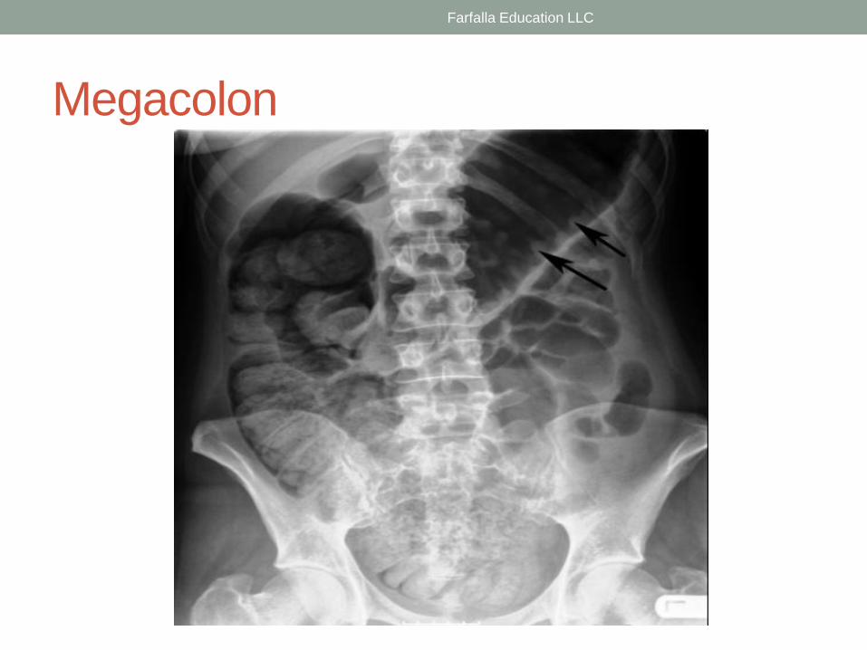
Megacolon
Farfalla Education LLC

Ulcerative Colitis
Farfalla Education LLC

Injuries to Bones and Joints
• Indication
• When to order x-rays
• Minor or no trauma
• Fractures can occur simply in terms of the physiologic process
involved
• Stress fractures
Farfalla Education LLC

Injuries to Bones and Joints
• Orthopedic emergencies
• Open fracture
• Dislocation
• Subluxation
Farfalla Education LLC

Describing Radiographs
• Open or closed
• Location of the fracture
• Orientation of the fracture line
• Displacement, separation, shortening, and angulation
• Epiphyseal fracture
• Salter Harris Classification
Farfalla Education LLC

Pediatric Considerations
• Bones more fibrous and less crystalline
• Enclosed in sheath of strong fibrous periosteum
• Buckle fractures and greenstick fractures more
common
• Bone ends do not separate as in adults with
complete fractures due to periosteal sleeve
• Epiphyseal growth plate is a zone of weakness
making fracture, separation, slipping more
common
Farfalla Education LLC

Pediatric Considerations
• Growth disturbances can be expected from epiphyseal injuries
• Union of fractures occurs much faster than adults
• Re-modeling of fractures after union means that less than perfect reduction can sometimes be accepted since nature will correct mistakes
• Loss of length after fracture of a long bone tends to be made up in the 1-2 years after injury by overgrowth
Farfalla Education LLC

Pediatric Considerations
• Dislocation occurs mainly at the elbow, hip and fingers
• Callus produced in larger quantities than in adults
• Pathological fracture occurs in children as result of local bone disease, simple bony cysts, osteitis, neoplasm, etc (example: osteogensis imperfecta – remember blue eyes)
Farfalla Education LLC

Common types of bone injuries in children
• Elastic deformation: a momentary, non-permanent deformation
• Bowing deformation: a deformity of bone that may or may not be completely resolved with bone remodeling
• Torus (Buckle) Fracture: involves the buckling of one cortex
• Greenstick fracture: an incomplete transverse fracture with fracture and periosteal rupture on the convex side
• Salter-Harris fracture: involves epiphyseal plate
• Stress Injury: a fracture caused by repetitive trauma
• Avulsion Injury: a bony structural defect at a tendinous or aponeurotic insertion resulting from excess stress
Farfalla Education LLC

Salter Harris Classifications
• Salter I – separation of the growth plate without involvement of metaphysis or epiphysis
• Salter II – fracture across the growth plate but with a small fragment of metaphysis remaining attached to the epiphysis
• Salter III – fracture across growth plate with extension of fracture across epiphysis
• Salter IV – combination of Salter II and Salter III fracture with the fracture line traversing the epiphysis and part of the diaphysis
• Salter V – crush injury of part of the epiphysis
Farfalla Education LLC

Farfalla Education LLC

Salter Harris I Fracture
Farfalla Education LLC

Salter Harris II Fracture
Farfalla Education LLC

Salter Harris III Fracture
Farfalla Education LLC

Salter Harris IV Fracture
Farfalla Education LLC

Salter Harris V Fracture
Farfalla Education LLC

Shoulder
Farfalla Education LLC

Shoulder - Approximate timings of
formation and fusion of secondary
ossification centres of the shoulder Seah, Elias & Chan. Chapter 5
BONE SECONDARY
OSSIFICATION
CENTRES
TIME OF
FORMATION
(YEARS)
Time of fusion
(years)
Humerus Head
Greater tuberosity
Lesser tuberosity
0-6 months
1
5
15-18 (with shaft)
4-6
7
Scapula Inferior angle
Coracoid
Acromium
15
1
15
20
15
20-25
Clavicle Medial Margin 18 25
Farfalla Education LLC

Shoulder – Radiographs
• Anterioposterior (AP) view • Contour of each bone should be traced systematically
• Cortices should be smooth
• Don’t overlook the ribs
• Axial view • ID coracoid process 1st
• Useful in assessing glenohumeral alignment, avulsion of glenoid rim and Hill-Sachs defects of humeral head
• Y view • Confirms normal alignment of glenohumeral joint
• Valuable if posterior dislocation suspected
Farfalla Education LLC

Extremities
• Adequacy
• Alignment
• Bone
• Cartilage and joints
• Soft tissues
Farfalla Education LLC

Injuries
• Dislocations
• Anterior
• Posterior
• Suspect in seizure patient with pain
• Acromio-clavicular subluxation and dislocation
• Key Points
• Always order a post-reduction film
• Look for subtle fractures
• Inferior aspects of acromion and clavicle should be a
straight line
Farfalla Education LLC

Direction of Dislocation
Farfalla Education LLC

Dislocations - Anterior
Farfalla Education LLC

Posterior Dislocation
Farfalla Education LLC

Farfalla Education LLC

Anatomy of Elbow
Farfalla Education LLC

Pediatric Elbow
Farfalla Education LLC

Pediatric Elbow
• Ossification Centres • There are 6 ossification centres around the elbow joint
• They appear and fuse to the adjacent bones at different ages
• It is important to know the sequence of appearance since the ossification centers always appear in a strict order
• Order of appearance
• Mnemonic C-R-I-T-O-E
• Capitellum - Radius - Internal or medial epicondyle - Trochlea - Olecranon - External or lateral epicondyle
• The ages at which these ossification centres appear are highly variable and differ between individuals
• It is not important to know these ages, but as a general guide you could remember 1-3-5-7-9-11 years
Farfalla Education LLC

Pediatric Alignment
Radiocapitellar line
• A line drawn through the long axis of the radius should always point toward the centre of the capitellum whatever the positioning of the patient, since the radius articulates with the capitellum
• In dislocation of the radius this line will not pass through the centre of the capitellum.
Anterior Humeral line
• A line drawn on a lateral view along the anterior surface of the humerus should pass through the middle third of the capitellum
• In cases of a supracondylar fracture the Anterior Humeral line usually passes through the anterior third of the capitellum or in front of the capitellum due to posterior bending of the distal humeral fragment
Farfalla Education LLC

Radiocapitellar line
Farfalla Education LLC

Anterior Humeral line
Farfalla Education LLC

Distal Humeral Fracture
Farfalla Education LLC

Fat Pad
Farfalla Education LLC

Elbow Fractures
• Essex-Lopresti’s Fracture – fracture radial head with
asociated dislocation of distalulnar joint
• Monteggia’s fracture – fractured proximal third ulna with
associated dislocation of radial head
Farfalla Education LLC

Radial Head Fracture
Farfalla Education LLC

Elbow Dislocation
Farfalla Education LLC

Midshaft Fracture
Radius and Ulna
Farfalla Education LLC

Distal Radius Fracture
• Barton’s Fracture – displaced articular lip fracture of
distal radius, may be associated with carpal subluxation
• Chauffeur’s Fracture (Hutchinson Fracture) – oblique
fracture of radial styloid. Originated from crank automobile
• Colles fracture – general fracture of distal radius with
dorsal displacement
Farfalla Education LLC

Chauffeur’s Fracture
Hutchinson Fracture
Farfalla Education LLC

Colles Fracture
Farfalla Education LLC

Distal Radius Fracture
Farfalla Education LLC

Distal Radius & Ulna Fracture
Farfalla Education LLC

Hand Fractures
• Boxer’s fracture – fracture 5th metacarpal neck with volar
displacement head of the metacarpal
• Bennett’s fracture – oblique fracture 1st metacarpal base
separating triangular fragment of volar lip from proximally
displaced metacarpal shaft
Farfalla Education LLC

Boxer’s Fracture
Farfalla Education LLC

Metacarpal Fractures
Farfalla Education LLC

Pelvis
• Comprised of three bone rings
• Main ring
• Sacroiliac joints and symphysis pubis
• Smaller rings
• Pubic and ischial bones
Farfalla Education LLC

Shenton’s Line
• A radiographic, curved line formed by the top of the obturator foramen and the inner side of the neck of the femur, used to determine the relationship of the head of the femur to the acetabulum
• In fractures or congenital luxation this line is broken.
Farfalla Education LLC

Shenton’s Line
Normal Fracture
Farfalla Education LLC

Pelvis X-ray
Farfalla Education LLC

Pelvis X-ray
Farfalla Education LLC

Pelvis x-rays
• One view only – AP view
• If you seen one fracture always look for two
• Prostate and breast cancer love the pelvis
• Osteoblastic lesion – light colored
• Osteolitic lesion – dark colored
Farfalla Education LLC

Pelvis x-rays
• Systematic approach • Main ring
• Inner and outer cortices
• 2 small rings • Form obturator foramina
• Sacroiliac joints • Equal widths
• Symphysis pubis • Alignment of superior surfaces of the pubic bone body
• Max width should be no more than 5mm
• Sacral foramina • Any distruption of smooth arcuate lines indicates sacral fracture
• Compare the arcs on the injured and uninjured side
• Acetabular region • Compare injured and uninjured sides (easy to miss fracture due to complexity)
Farfalla Education LLC

Pelvic Fractures
• Main ring
• Widening at symphysis pubis or sacroiliac joint indicates a fracture
in the ring
• Fracture at 1 site associated with second fracture
• Double break indicated unstable fracture
Farfalla Education LLC

Pelvic Fracture
• Acetabulum
• Frequently comminuted
• Bone fragments may be trapped within the joint
• Sacral
• Difficult to detect
• Arcuate lines need to be carefully assessed
Farfalla Education LLC

Pelvic Fracture
• Coccygeal • Radiograph not always necessary to diagnose fracture
• Care unchanged whether fractured or not
• Normal coccyx may appear angulated and abnormal
• Avulsion of apophysis • Most commonly caused by repeated sudden or violent contraction
of muscles in young patients
Farfalla Education LLC

Hip X-rays
• Adequacy and quality
of radiograph
• Check bone margins
and density
• Cartilage and joints
• Soft Tissue
• If X-ray for hip and
pelvis are negative,
make sure you walk
the patient. Subtle
fractures can be
missed on x-ray and
CT scan/MRI should
be ordered if unable to
ambulate.
Farfalla Education LLC

Proximal Femur Fracture
Farfalla Education LLC

Avascular Necrosis
(Osteonecrosis) • Painful condition involving weight-bearing portion
of femoral head
• Bone death results from lack of blood supply
• Trauma
• Prolonged corticocosteroid use
• Alcohol abuse
• Chemotherapy
• H/O diving and nitrogen toxicity
• Sickle cell disease
Farfalla Education LLC

Pediatric Considerations
• Slipped Capital Femoral
Epiphysis
• Condition of femoral neck
• Moves proximally and
externally rotates on unfused
epiphysis
• 20% of cases are bilateral
• Occurs in overweight,
hypogonadal or tall but thin
adolescents
• Pain sometimes referred to
the knee
• Legg-Calve-Perthes
Disease
• Osteonecrosis of proximal
femoral epiphysis
• Children ages 4-8 years
• Four times more common in
boys than girls
• 10% occur bilaterally
• Genetic basis
• Imaging esential
• Most present with painful limp
Farfalla Education LLC

• Shenton's curve: smooth, curved line connecting medial border of femoral metaphysis with the superior border of the obturator foramen
• Hilgenreiner's line: a horizontal line through the triradiate cartilage of the acetabulum
• Perkin's line: a vertical line (perpendicular to Hilgenreiner's line) from the lateral margin of the ossified acetabular roof that is normally tangential to the lateral margin of the ossification center of the femoral head
• Acetabular angle: angle that the acetabular line makes with Hilgenreiner's line
Farfalla Education LLC

Slipped Capital Femoral Epiphysis
Anatomy
Farfalla Education LLC

Slipped Capital Epiphysis
• A – Mild
• B – Moderate
• C - Severe
Farfalla Education LLC

Slipped Capital Femoral Epiphysis
Farfalla Education LLC

Legg-Calve-Perthes Disease
Farfalla Education LLC

Legg-Calve-Perthes Disease
Farfalla Education LLC

Knee Anatomy
Farfalla Education LLC

Tibial Plateau
• AP view
• Perpendicular line drawn at the most lateral margin of the femoral
condyle
• Should not be more than 5mm of lateral margin of tibial condyle
• Similar line can be drawn medially
Farfalla Education LLC

Tibial Plateau Fracture
• 80% involve the lateral plateau
• Causes
• Blow to the outside of the knee
• Pedestrian VS Automobile accident
• Fender Fracture
Farfalla Education LLC

Pediatric Consideration
Osgood Schlatter
• Traction apophysitis of tibial tubercle
• Typically seen in children ages 10-15 years
• Self-limited course of several months – 2 years
• Tenderness ocer tibial tubercle onpalpation
Farfalla Education LLC

Normal Patella X-ray
Farfalla Education LLC

Skyline or Sunrise View
Farfalla Education LLC

Ottawa Rules: Knee
• Order x-ray of the knee in the following:
• age 55 or over
• isolated tenderness of the patella
• no bone tenderness of the knee other than the patella
• tenderness at the head of the fibula
• inability to flex to 90 degrees
• inability to weight bear
• Immediately after injury or during evaluation
• 4 steps - unable to transfer weight twice onto each lower limb
regardless of limping
Farfalla Education LLC

Knee Fractures
• Direct blow
• Usual cause of fracture
• Vertical, horizontal and comminuted
• Violent contraction of Quadricep muscle
• Transverse fracture in athletes
• Bipartite patella
• May mimic fracture
Farfalla Education LLC

Patella Dislcoation
• Considered to be rare
• When occurs usually anterior translation of Tibia on the
femur
• Consider popliteal artery and nerve injury
• True dislocation associated with multiple ligamentous
tears
Farfalla Education LLC

Proximal Fibula Fracture
• Fibula head fracture
• May be isolated or associated with Tibial plateau fractures
• Fibula neck or proximal shaft fracture
• May be associated with ankle injury
• Maisonneuve fracture
• Proximal Fibula fracture may be associated with peroneal
nerve injury
Farfalla Education LLC

Ottawa Rules: Ankle
• Bony tenderness at tip or posterior edge of distal 6cm of
tibia or fibula
• Bony tenderness medial malleolus
• Tenderness base of 5th metatarsal
• Unable to bear weight immediately after injury or upon
evaluation
Farfalla Education LLC

Ankle X-ray
Lateral AP
Farfalla Education LLC

Ankle X-ray
Mortise Boehler’s Angle
Farfalla Education LLC

Calcaneal fracture
• Most commonly fractured bone of the hindfoot
• Occurs most commonly – fall from height
• Suspect spinal fracture
Farfalla Education LLC

Base of Fifth Metatarsal Fracture
• Avulsion fracture • Commonly caused by inversion injury
• Usually occurs at the metatarsal tuberosity at the insertion of the peronous brevis tendon
• Clinical evaluation will determine need for ankle and/or foot x-rays
• Don’t confuse with unfused apophysis
• Bone fragment from fracture usually lies transverse to long axis of metatarsal as opposed to apophysis lying oin the long axis of the metatarsal
Farfalla Education LLC

Tarsals, Metatarsals and Phalanges – Oh
My!
Farfalla Education LLC

THANK YOU!!!!
Theresa M Campo DNP, APRN, NP-C, CEN
Farfalla Education LLC
609-602-3034
Farfalla Education LLC

• Introduced in the 1970’s
• Three dimensional views
• Body organs
• bones
• Benefits usually outweigh the risks
• Radiation exposure
Farfalla Education, LLC
Farfalla Education LLC

• Specialized X-rays generate images with computer
• Quickly identify traumatic injuries to the lungs, heart,
vessels, liver, spleen, kidneys, bowel and other organs
• Patient lies on a table that slides in and out of a tube
Farfalla Education, LLC
Farfalla Education LLC

• The tube rotates around the patient
• Opposite side of the patient is x-ray detector
• Detector receives the beam going through the patient
• Measurements are made about 1000 times per second
• Scan rotations are usually 1-2 seconds long
• Each scan is compared to calibration data of air, water and polyethelyne (soft plastic) previously acquired in the same relative location • Comparisons allow for the image
• More samples, or views, the better the picture
Farfalla Education, LLC
Farfalla Education LLC

• Iodine based
• Absorbed by abnormal tissue
• Tumors, vascular malformations
• Risks
• Renal failure
• Metformin
Farfalla Education, LLC
Farfalla Education LLC

• Procedural guidance
• Biopsy
• Incision and drainage
• Assess post-surgical procedures
• Organ transplant
• Gastric bypass
• Radiation therapy
• Stage
• Plan and administer radiation
• Bone mineral density
Farfalla Education, LLC
Farfalla Education LLC
• Readily detects inflammation of the mesentery (fat streaking) • May be non-specific and should be correlated with the History and
Physical Examination
• Abscesses or Tumors • Intra-abdominal
• Intra-hepatic
• Intra-splenic
• Blunt Trauma – must be performed quickly • Spleen
• Liver
• Pancreas
Farfalla Education, LLC
Farfalla Education LLC

• Painless, noninvasive and accurate
• Bone, soft tissue and vessels visualized same time
• Fast and simple
• Cost-effective
• Less sensitive to movement
• Real time images
Farfalla Education, LLC
Farfalla Education LLC

• Not recommended in pregnancy
• Allergic reaction to contrast
• Large amount of radiation
Farfalla Education, LLC
Farfalla Education LLC

• Powerful magnet and radio waves generate images
• Detection coils in scanner read the energy produced by
water molecules as they mis-align themselves after each
radio frequency alignment pulse
• Reconstruction of the collected data into 2-dimensional
illustration through any axis of the body
• Best suited for soft tissue
Farfalla Education, LLC
Farfalla Education LLC

• Organs and blood vessels of chest, abdomen and pelvis
• Tumors of chest, abdomen, and pelvis
• Vascular and heart abnormalities
• Liver disease
• Cyst and tumors of genitourinary tract
• Breast cancer and implants
• Pelvic pain, congenital malformations leading to infertility
Farfalla Education, LLC
Farfalla Education LLC

• Non-invasive
• No radiation
• Early detection focal lesions and tumors
• View soft tissue obscured by bone
• Biliary system without contrast
• Can be used in pregnancy
Farfalla Education, LLC
Farfalla Education LLC

• Almost no risk
• Clostrophobia
• Metallic, pacemaker
• Nephrogenic systemic fibrosis from contrast material
Farfalla Education, LLC
Farfalla Education LLC

CT Scan MRI
Radiation Exposure Moderate to High None
Generated Image X-Ray Magnet and Radio
Waves
Duration of Test Very Quick < 5
minutes
Moderate approx. 30
minutes
Cost of Test >$1000 $1000 – 4000
Bony Structure Detailed Less Detail
Soft Tissue Less Detail Great Detail
Image Plane Can not be changed
without moving the
patient
Image produced in
any plane without
moving patient
Farfalla Education, LLC
Farfalla Education LLC

MRI CT Scan
Magnetic Field – No radiation exposure Great bony detail
Great soft-tissue detail Can be performed on patient with metal
Pacemaker, metallic FB, Clips, etc
Ability to change contrast of image
different types of tissue can be
contrasted with different contrast
settings
Test performed in less time
Image plane can be changed without
moving patient
Food for claustrophobic patients
Lower rate of reaction with non-iodine
contrast
Paramagnetic preparation –
Gadolinium
Cheaper
Superior tumor detection and
identification
Farfalla Education, LLC
Farfalla Education LLC

CT MRI
Bone Tendons
Lungs Ligaments
Organs in chest and
abdomen
Spinal cord
Cancer Ischemia (stroke)
Pneumonia Cartilage
Brain - Bleed Brain - Tumor
Acute organ injury
Vascular (AAA, PE)
Farfalla Education, LLC
Farfalla Education LLC

THANK YOU!!!!
Theresa M Campo DNP, APRN, NP-C, CEN
Farfalla Education LLC
609-602-3034
Farfalla Education LLC