fancj suppresses microsatellite instability and...
TRANSCRIPT

FANCJ suppresses microsatelliteinstability and lymphomagenesisindependent of the Fanconi anemiapathwayKenichiro Matsuzaki,1 Valerie Borel,1 Carrie A. Adelman,1 Detlev Schindler,2
and Simon J. Boulton1
1DNA Damage Response Laboratory, Clare Hall Laboratories, The Francis Crick Institute, South Mimms EN6 3LD, UnitedKingdom; 2Department of Human Genetics, Biozentrum, University of Wurzburg, 97074 Wurzburg, Germany
Microsatellites are short tandem repeat sequences that are highly prone to expansion/contraction due to theirpropensity to form non-B-form DNA structures, which hinder DNA polymerases and provoke template slippage.Although error correction bymismatch repair plays a key role in preventingmicrosatellite instability (MSI), which isa hallmark of Lynch syndrome, activities must also exist that unwind secondary structures to facilitate replicationfidelity. Here, we report that Fancj helicase-deficient mice, while phenotypically resembling Fanconi anemia (FA),are also hypersensitive to replication inhibitors and predisposed to lymphoma. Whereas metabolism of G4-DNAstructures is largely unaffected in Fancj−/− mice, high levels of spontaneous MSI occur, which is exacerbated byreplication inhibition. In contrast, MSI is not observed in Fancd2−/− mice but is prevalent in human FA-J patients.Together, these data implicate FANCJ as a key factor required to counteractMSI, which is functionally distinct fromits role in the FA pathway.
[Keywords: DNA repair; FANCJ; Fanconi anemia; genome stability; microsatellite instability]
Supplemental material is available for this article.
Received September 30, 2015; revised version accepted November 13, 2015.
Maintenance of genome integrity during DNA replicationis of vital importance to ensure that daughter cells inheritan intact copy of the genetic code. Repetitive DNA se-quences are a particular challenge to genome stabilitydue to their propensity to form secondary structures with-in template or nascentDNA strands that hinder replisomeprogression and promote template slippage. Microsatel-lites are repetitive sequences of 1–10 base pairs (bp) ofDNA (Boyer et al. 2013). The expansion or contractionof microsatellites during DNA replication has been impli-cated in a wide range of genetic disorders, including neu-romuscular or neurological diseases and cancer. A linkbetween human disease and repetitive sequence instabil-ity is most clearly illustrated for trinucleotide repeats(Mirkin 2007), whose expansion is a recurrent cause ofFriedreich’s ataxia (GAA/TTC repeats) and Huntington’sdisease (CAG/CTG repeats). Microsatellite instability(MSI) is also a hallmark of Lynch syndrome-associatedcancers (Aaltonen et al. 1993; Boland and Goel 2010),
which are caused by mutations in DNA mismatch repair(MMR) genes. Although MMR deficiency is associatedwith hereditary and sporadic colorectal cancers in hu-mans, MMR-deficient mice are primarily predisposed tolymphoma (Li 2008). Current evidence suggests thatMMR corrects slipped strand mispairing resulting fromadditions or deletions in the newly synthesized strand,which arise during secondary structure-triggered tem-plate slippage or when the replication of the repeats is im-paired. Given the strong association with human disease,understanding the mechanisms that maintain the integri-ty of repetitive sequences is of great clinical importance.
Whilemechanisms exist to directly detect structural al-terations in DNA, including helix-distorting lesions andbase–base mismatches, other lesions may go undetecteduntil encountered by the DNA replication machinery.When this occurs, repair must be orchestrated in the con-text of the replication fork, necessitating coordination ofcheckpoint, repair, and replication factors. In response toreplication fork blockages such as interstrand cross-links(ICLs), the ATR-dependent replication stress checkpoint,
Corresponding author: [email protected] published online ahead of print. Article and publication date areonline at http://www.genesdev.org/cgi/doi/10.1101/gad.272740.115. Free-ly available online through the Genes & Development Open Accessoption.
© 2015Matsuzaki et al. This article, published inGenes&Development,is available under a Creative Commons License (Attribution 4.0 Interna-tional), as described at http://creativecommons.org/licenses/by/4.0/.
2532 GENES & DEVELOPMENT 29:2532–2546 Published by Cold Spring Harbor Laboratory Press; ISSN 0890-9369/15; www.genesdev.org
Cold Spring Harbor Laboratory Press on April 27, 2019 - Published by genesdev.cshlp.orgDownloaded from
Fanconi anemia (FA), and homologous recombination(HR) pathways are essential for replication fork repairand restart (Clauson et al. 2013). Repair of ICLs requiresnucleolytic processing, translesion DNA synthesis, andHR (Deans andWest 2011). The requirement forHR stemsfrom the generation of DNA double-strand breaks (DSBs)that arise from nucleolytic processing events. The FApathway is comprised of at least 16 gene products defec-tive in FA patients (Clauson et al. 2013), which presentwith progressive bone marrow failure, developmental ab-normalities, subfertility, and tumor predisposition (Alterand Kupfer 2002; Alter 2003; Crossan and Patel 2012).At the molecular level, the primary function of the FApathway appears to be to induce monoubiquitylation ofthe heterodimeric FANCD2/FANCI complex, which co-ordinates ICL incision and recruitment of downstream re-pair factors (for review, see Kim and D’Andrea 2012).One of the most enigmatic FA proteins is FANCJ, a
DEAH superfamily 2 helicase and part of the subfamilyof Fe-S cluster-containing helicases, which also includesXPD, RTEL1, and CHL1 (Rudolf et al. 2006; Gari et al.2012; Brosh and Cantor 2014). Biallelic mutations inFANCJ give rise to FA complementation group J (Levituset al. 2005; Levran et al. 2005; Litman et al. 2005), whereasmonoallelic mutations predispose to ovarian and breastcancers (Seal et al. 2006; Rafnar et al. 2011). More recent-ly, a significant association with pancreatic and colorectalcancer was found (Rafnar et al. 2011). In line with its rolein the FA pathway, defects in FANCJ give rise to exquisiteICL sensitivity in a range of different species (Bridge et al.2005; Levitus et al. 2005; Litman et al. 2005; Youds et al.2008). However, attempts to place FANCJ within the FApathway have provided little insight into its precise func-tion in ICL repair. FANCJ is dispensable for the activationof the FA core complex and hence the monoubiquityla-tion of FANCD2/FANCI and its recruitment to ICLlesions (Levitus et al. 2004; Litman et al. 2005). Further-more, an interaction between FANCJ and BRCA1 is notrequired for classical ICL repair (Xie et al. 2010b), and arole for FANCJ in HR downstream from ICL incision re-mains ambiguous. Currently, the function of FANCJ inICL repair remains poorly defined.Biochemical studies have shown that FANCJ unwinds a
variety of DNA substrates, including 5′ flaps, forked du-plexes, D loops, 5′ tailed triplexes, and G4-DNA struc-tures in a 5′–3′ direction in vitro (Gupta et al. 2005;London et al. 2008; Wu et al. 2008; Sommers et al.2009). Of these DNA structures, a clear picture hasemerged linking FANCJ to the metabolism of G4-DNAsecondary structures in vivo. Such a role was first suggest-ed from the observation of increased G/C tract deletionsin dog-1 (deletion of G tracts; Caenorhabditis elegansFANCJ) mutant worms, which was proposed to reflect adefect in unwinding G4-DNA structures formed withinG/C tracts (Cheung et al. 2002; Youds et al. 2008). Subse-quent studies in human Fancj-deficient cells found thatgenomic deletions tend to accumulate in the vicinity ofpotential G4-DNA-forming sequences (London et al.2008). Genome-wide transcription profiling of FANCJknockout chicken DT40 cells has also revealed that dys-
regulated genes are significantly associated with G4sequences. It was proposed that FANCJ maintains epige-netic stability near G4-DNA motifs through two inde-pendent mechanisms dependent on either the Y familypolymerase REV1 or WRN/BLM helicases (Sarkies et al.2012). Recently, it was reported that FANCJ depletionfrom Xenopus laevis egg extract leads to persistent repli-cation stalling at G4 sequences (Castillo Bosch et al.2014). Despite these observations, it is currently unclearwhether FANCJ functions exclusively to maintain ge-nome stability associated with G4-DNA-forming se-quences or also participates in the metabolism of otherDNA secondary structures.In this study, we report that Fancj-null mice exhibit
subfertility, germ cell attrition, epithelial tumor predispo-sition, and exquisite sensitivity to ICL-inducing agents,which phenocopy other mouse models of FA (Bakkeret al. 2012). Unexpectedly, Fancj-deficient mice alsopresent with enhanced predisposition to lymphoma,and cells derived from these mice are hypersensitive toreplication inhibitors. Furthermore, Fancj−/−Fancd2−/−
double-knockout mice display heightened germ cell attri-tion and are considerably more sensitive to ICL-inducingagents than single knockouts. Since Fancj-deficient cellsare insensitive to G4-stabilizing drugs and are devoidof telomere fragility, we considered the possibility thatFANCJ performs additional functions in genome sta-bility independent of its role in the FA pathway and dis-tinct from a role in G4-DNA metabolism. Strikingly, weshow that Fancj-deficient, but not Fancd2-deficient,mice accumulate spontaneous MSI corresponding toboth expansions and contractions of repeat sequences.Similarly, FA-J patient cells and human Fancj knockoutscells also present withMSI, which is exacerbated by repli-cation inhibition. Thus, we propose that FANCJ counter-acts the formation of secondary structures that ariseduring replication of microsatellite sequences, whichminimizes the potential for strand slippage during DNApolymerization. Our findings can potentially explain thewidespread involvement of FANCJ in human cancers.
Results
Generation of Fancj-deficient mice
To investigate the function of FANCJ in vivo, we generat-ed a Fancj mutant mouse from an existing gene trap em-bryonic stem cell line (RRI409). Insertion site mappingby splinkerette PCR revealed the gene trap cassette to beintegrated into the fifth intron of the Fancj mouse gene,which is predicted to truncate the gene within the criticalhelicase motifs (Fig. 1A,B; Supplemental Fig. S1A; Devonet al. 1995). This facilitated development of a genotypingstrategy, which was used to identify wild-type, heterozy-gous, andmutantmouse embryonic fibroblasts (MEFs) de-rived from heterozygous mice (Fig. 1C). Quantitative RT–PCR analysis failed to detect Fancj mRNA expression(exon 5–6 junction) or expression of a fusion transcript be-tween exon 5 and the β-geo cassette (Supplemental Fig.S1B). Western blotting of whole-cell extracts with an
FANCJ prevents microsatellite instability
GENES & DEVELOPMENT 2533
Cold Spring Harbor Laboratory Press on April 27, 2019 - Published by genesdev.cshlp.orgDownloaded from

antibody raised against the conserved N terminus ofFANCJ protein also confirmed the absence of a truncatedor fusion protein (∼170 kDa) (Fig. 1D; SupplementalFig. S5A). These data suggest that the gene trap cassetteeliminates expression of the FANCJ ORF, resulting in anull allele.
Fancj-deficient mice are subfertile and presentwith germ cell attrition
Characterization of Fancj−/− mice indicated that they areviable and born at expectedMendelian ratios (Supplemen-tal Fig. S1C), as has also been reported for other FA andFA-like knockout mice, including Fancd2- andHelq-defi-cient mice (Houghtaling et al. 2003; Adelman et al. 2013).
Fancj−/− mice displayed normal growth and weight gainwhen compared with wild-type littermates up to theage of 19 wk and were devoid of any noticeable develop-mental abnormalities (Supplemental Fig. S1D). However,comparison ofmating ability between six Fancj+/− hetero-zygous and five Fancj−/− homozygous pairs mated contin-uously over a 5-mo period revealed reduced litter numbersand size, consistentwith subfertility. Indeed, Fancj+/−het-erozygousmatings resulted in 31 litters with 166 pups (0.9litter per 21-d gestation interval and 4.7 pups per litter),whereas Fancj−/− homozygous matings produced only 10litters and 66 pups (0.3 litter per 21-d gestation intervaland 2.2 pups per litter) (Fig. 1E). This observation raisedthe possibility that, like other FA mouse models, FANCJdeficiency is also associated with subfertility.
Figure 1. Germcell attrition and subfertil-ity in Fancj knockout mice. (A) Schematicrepresentation of the mouse Fancj genomiclocus. The pGT0Lxf gene trap vector is in-serted at position 11600. The whole geno-mic locus is 143.06 kb, and introns (lines)and exons (bars) are represented to scale.(B) FANCJ protein organization. Note thatthe Fancj mutant protein is truncated at212 amino acids and fused with the β-geocassette of the gene trap vector. (C ) FancjPCR genotyping strategy. (D) Western bloton Fancj+/+ and Fancj−/− MEF lysates. Thearrow indicates the FANCJ protein. Notethat FANCJ is not expressed in Fancj−/−
cells. Tubulin was used as loading control.(E) Fertility comparison between Fancj het-erozygous and mutant breedings. Each pairbred during 5–6 mo, and the number oflitters and pups per litter was quantifiedper 21-d gestation. Significance: t-test,litter per 21 d gestation, P = 0.01; pups per21 d gestation, P = 0.08. (F ) Fancj testisweights. Each testis weight was normalizedagainst mice body weight. Significance: t-test, P < 0.0001. (G, left) Abnormal seminif-erous tubule quantification on Fancj testissections. (Right) Representative Fancj−/−
seminiferous tubule images are shown.The Fancj−/− testis contains both normaltubules and also different levels of atrophictubules. The bottom picture shows a se-verely atrophic tubule with almost no sper-matogenic cells remaining. Asterisks showtubule degeneration. Bars, 50 µm. Signifi-cance: t-test, P = 0.01. (H, top) Five-day-oldneonate and adult seminiferous tubulesstained with PLZF (germ cells; brown) andSox9 (sertoli; brown) markers. DNA iscounterstained with hematoxylin (blue).Bars, 50 µm. (Bottom) Spermatogonia andsertoli cell quantification per tubule. Errorbars represent ±standard error of the mean(SEM) of at least 50 tubules. Significance:with t-test, P < 0.0001. (I, left) Dysgeneic
ovary frequency. n = 43 Fancj−/−; n = 18 Fancj+/+. (Right) Representative images of dysgeneic ovaries. Bars, 500 µm. (f) Follicles; (cl) corpusluteum. Significance: Fisher’s exact test, P = 0.1.
Matsuzaki et al.
2534 GENES & DEVELOPMENT
Cold Spring Harbor Laboratory Press on April 27, 2019 - Published by genesdev.cshlp.orgDownloaded from

To identify the possible cause of this fertility defect, wefirst examined testes from Fancj−/− mutant and wild-typelittermates. Similar to other FA mice, Fancj−/− mice havereduced testis size and weight relative to wild-type con-trols (Fig. 1F), which are not significantly altered by age(Supplemental Fig. S1E). Histological analysis of Fancj−/−
adult testes revealed many normal tubules populatedwith the full complement of spermatogonia; however, re-gions of atrophy were also evident. Thirty-four percent ofseminiferous tubules were found to be atrophic, comparedwith 11% in controls (Fig. 1G). The source of subfertilityof Fancj−/− mice was not limited to the male gonad, as23%of female gonads analyzed at 35wk exhibited ovariandysgenesis, compared with 5.6% in controls. Histologicalanalysis confirmed that only a few primary or no develop-ing follicles were present in Fancj−/− ovaries (Fig. 1I).Closer scrutinyof adultFancj−/− testis sections revealed
that the levels of atrophy in seminiferous tubuleswere sto-chastic, ranging from mild to severe atrophy, with sometubules devoid of all spermatogenic layers (Fig. 1G). Sincefurther analysis of themale gonad failed to reveal a specificspermatogenesis defect in Fancj−/−mice, it wasmost like-ly that atrophy arose from a stem cell problem, which wesought to assess by staining adult testis sections with thespermatogonial stem cell marker PLZF. A marked reduc-tion in the number of PLZF-positive cells per tubule wasobserved in the Fancj−/− mutant testes compared withcontrols (5.5 ± 0.4 vs. 8.0 ± 0.5, respectively). Correspond-ingly, the number of sertoli cells stained by the Sox9sertoli-specific marker was significantly increased inthe Fancj−/− mutant mice relative to controls (28 ± 0.8vs. 18 ± 0.4, respectively) (Fig. 1H, right). As the levels oftestis atrophy in the Fancj−/−mutant mice did not notice-ably worsen with age, we considered that the germ celldefect likely arises duringdevelopment.To assess this pos-sibility, we examined spermatogonia numbers in 5-d-oldneonate testes. Fancj−/− mutant testes presented with a2.5-fold decrease in the number of PLZF-positive sperma-togonia when compared with control testes. Similarly,we also observed a corresponding increase in the numberof sertoli cells in themutant testes (Fig. 1H, left). These ob-servations suggest that loss of FANCJ leads to subfertilityas a result of germ cell attrition during development.
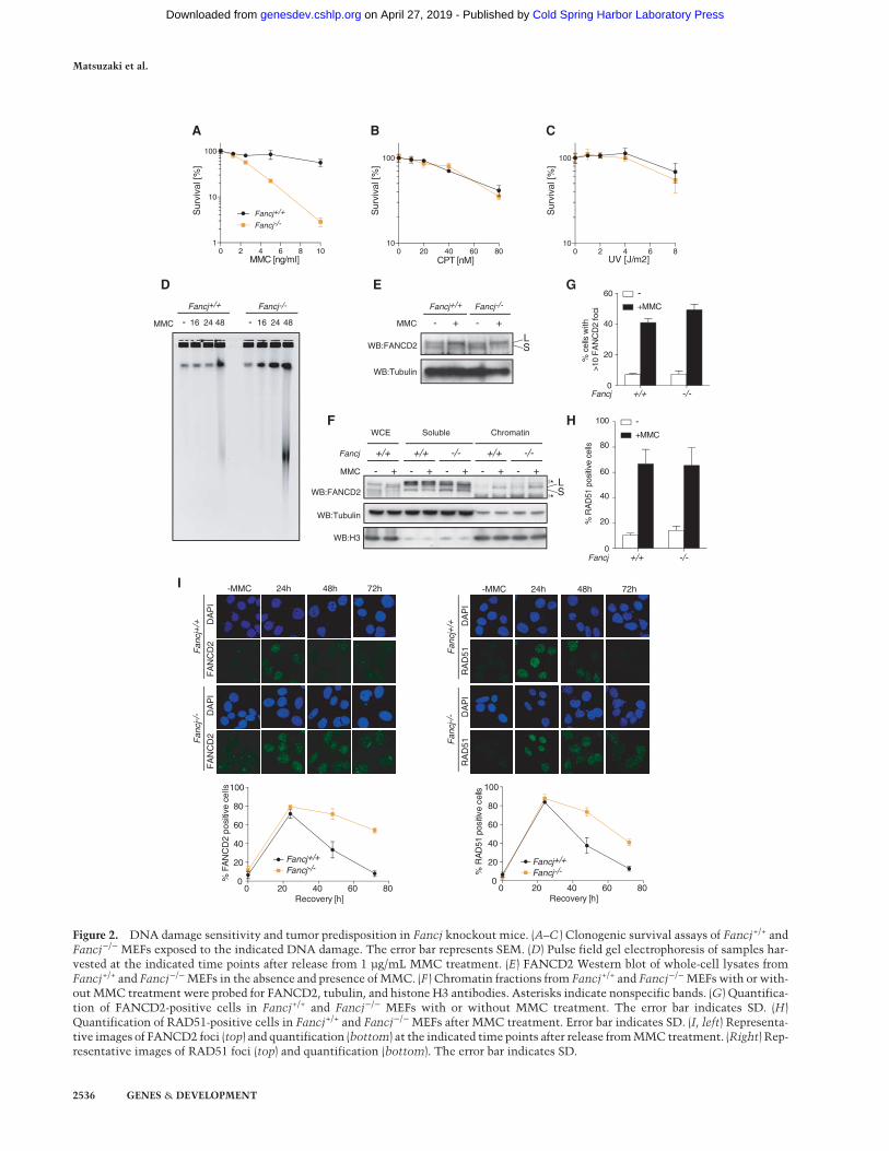
Fancj−/− MEFs are exquisitely sensitive to ICLs
Since subfertility and germ cell attrition are common fea-tures of FA, we next assessed whether Fancj−/− miceexhibit other FA phenotypes, including cellular sensitiv-ity to ICL agents. Fancj−/− MEFs exhibited exquisite sen-sitivity to the ICL-inducing agent mitomycin C (MMC)but not camptothecin or UV (Fig. 2A–C) and accumulatedradial chromosomes, a hallmark of FA (Supplemental Fig.S5B). Analysis of ICL incision at MMC lesions was as-sessed bymonitoring the accumulation of DSB intermedi-ates by pulse field gels following treatment of cells withMMC. Heightened levels of DSBs accumulated inFancj−/− MEFs with accelerated kinetics when comparedwith wild-type controls (Fig. 2D). These results suggestthat MMC-induced lesions are incised, but the damage
persists in Fancj−/− MEFs, indicating that lesion repair iscompromised downstream from ICL incision.To further examine the nature of the ICL repair defect in
Fancj-deficient cells, we monitored the integrity of twokey events in ICL repair; namely, the monoubiquitylationof FANCD2 by the FA core complex and its subsequent as-sembly into chromatin-associated repair foci. Monoubi-quitylated FANCD2 was induced to similar levels inboth wild-type and Fancj−/− MEFs in response to MMCtreatment (Fig. 2E). Furthermore, FANCD2 was readilydetected in the chromatin fraction and in repair foci inboth wild-type and Fancj−/− MEFs following MMC (Fig.2F,G). In the canonical ICL repair pathway, DSBs are pro-cessed by Rad51-mediated HR. To assess the effect ofFANCJ on early steps of HR, we monitored Rad51 andFANCD2 focus formation. Similar levels of RAD51 fociwere observed in Fancj+/+ and Fancj−/− MEFs afterMMC (Fig. 2H). However, in Fancj−/− MEFs, FANCD2and RAD51 foci persist much longer than in Fancj+/+
MEFs (Fig. 2I), suggesting that FANCJ is required for ICLrepair and is important for the timely resolution ofFANCD2- and RAD51-marked repair intermediates.
Fancj−/− mice are tumor-prone and predisposedto epithelial cancers
To study the impact of FANCJ deletion in aging mice, wemonitored a cohort of 21 Fancj+/+ and 44 Fancj−/− miceover the course of their lifetime. Aged Fancj−/− mutantmice presented with lipid accumulation in the liver (Fig.3A), and their tumor-free survival was found to be signifi-cantly reduced compared with control mice; 50% of themutantmice presentedwith tumors by559d (Fig. 3B). Sev-enty-four percent of Fancj−/− mutants developed tumors,with a greater incidence in females (∼74% vs. ∼48% onlyin males) (Fig. 3C; Supplemental Fig. S2A), and 60% ofthe Fancj−/− mutant mice presented with more than oneprimary tumor (Fig. 3D). Similar to other FA mouse mod-els, one of the most common tumors in Fancj−/− mice isof epithelial origin (Houghtaling et al. 2003). Indeed, 21 ep-ithelial tumors developed in 44 Fancj−/− mutant animals,which represent an incidence of 49% (Fig. 3E; Supplemen-tal Fig. S2B). Mutant females presented with more epithe-lial tumors when compared with males in the cohort,which corresponds to a frequency of ∼52% in femalesand 39% in males (Fig. 3F). Mutant females were parti-cularly prone to cystic ovaries (four tumors out of 23 ani-mals), endometrial hyperplasia (three tumors out of23 animals), and pituitary gland adenomas (three tumorsout of 23 animals), whereas Harderian gland adenomaswere the most prominent epithelial tumors in males(five tumors out of 21 animals) (Fig. 3G; SupplementalFig. S2C). Thus, loss of FANCJ leads to an increased predis-position to epithelial tumors in bothmales and females, ashas been shown in other FA mouse models.
Fancj−/− mice exhibit predisposition to lymphomas
Despite the tumor spectrum of Fancj−/− mice being simi-lar to that of other mouse models of FA, we also observed
FANCJ prevents microsatellite instability
GENES & DEVELOPMENT 2535
Cold Spring Harbor Laboratory Press on April 27, 2019 - Published by genesdev.cshlp.orgDownloaded from
0 20 40 60 8010
100
CPT [nM]0 2 4 6 8
10
100
UV [J/m2]0 2 4 6 8 10
1
10
100
MMC [ng/ml]
Fancj+/+
Fancj-/-
Sur
viva
l [%
]
Sur
viva
l [%
]
Sur
viva
l [%
]
A B C
MMC - 16 24 48 - 16 24 48
Fancj+/+ Fancj-/-
WB:FANCD2
MMC
Fancj+/+ Fancj-/-
- ++ -
WB:Tubulin
D
+/+ -/-0
20
40
60
% c
ells
with
>1
0 F
AN
CD
2 fo
ci
-+MMC
Fancj
E
LSWB:FANCD2
MMC
Fancj
WCE Soluble Chromatin
WB:Tubulin
WB:H3
- + - + - + - + - +
+/+ +/+ -/- +/+ -/-
F
G
H
0
20
40
60
80
100
% R
AD
51 p
ositi
ve c
ells
+/+ -/-Fancj
-+MMC
-MMC
DA
PI
FA
NC
D2
Fan
cj+
/+F
ancj
-/- D
AP
IF
AN
CD
2
24h 48h 72h
DA
PI
RA
D51
DA
PI
RA
D51
-MMC 24h 48h 72h
Fan
cj+
/+F
ancj
-/-
I
0 20 40 60 800
20
40
60
80
100
% R
AD
51 p
ositi
ve c
ells
Recovery [h]0 20 40 60 80
0
20
40
60
80
100
Recovery [h]
% F
AN
CD
2 po
sitiv
e ce
lls
Fancj+/+
Fancj-/-Fancj+/+
Fancj-/-
*
*
LS
Figure 2. DNA damage sensitivity and tumor predisposition in Fancj knockout mice. (A–C ) Clonogenic survival assays of Fancj+/+ andFancj−/− MEFs exposed to the indicated DNA damage. The error bar represents SEM. (D) Pulse field gel electrophoresis of samples har-vested at the indicated time points after release from 1 µg/mL MMC treatment. (E) FANCD2 Western blot of whole-cell lysates fromFancj+/+ and Fancj−/−MEFs in the absence and presence of MMC. (F ) Chromatin fractions from Fancj+/+ and Fancj−/−MEFswith or with-out MMC treatment were probed for FANCD2, tubulin, and histone H3 antibodies. Asterisks indicate nonspecific bands. (G) Quantifica-tion of FANCD2-positive cells in Fancj+/+ and Fancj−/− MEFs with or without MMC treatment. The error bar indicates SD. (H)Quantification of RAD51-positive cells in Fancj+/+ and Fancj−/− MEFs after MMC treatment. Error bar indicates SD. (I, left) Representa-tive images of FANCD2 foci (top) and quantification (bottom) at the indicated time points after release fromMMC treatment. (Right) Rep-resentative images of RAD51 foci (top) and quantification (bottom). The error bar indicates SD.
Matsuzaki et al.
2536 GENES & DEVELOPMENT
Cold Spring Harbor Laboratory Press on April 27, 2019 - Published by genesdev.cshlp.orgDownloaded from
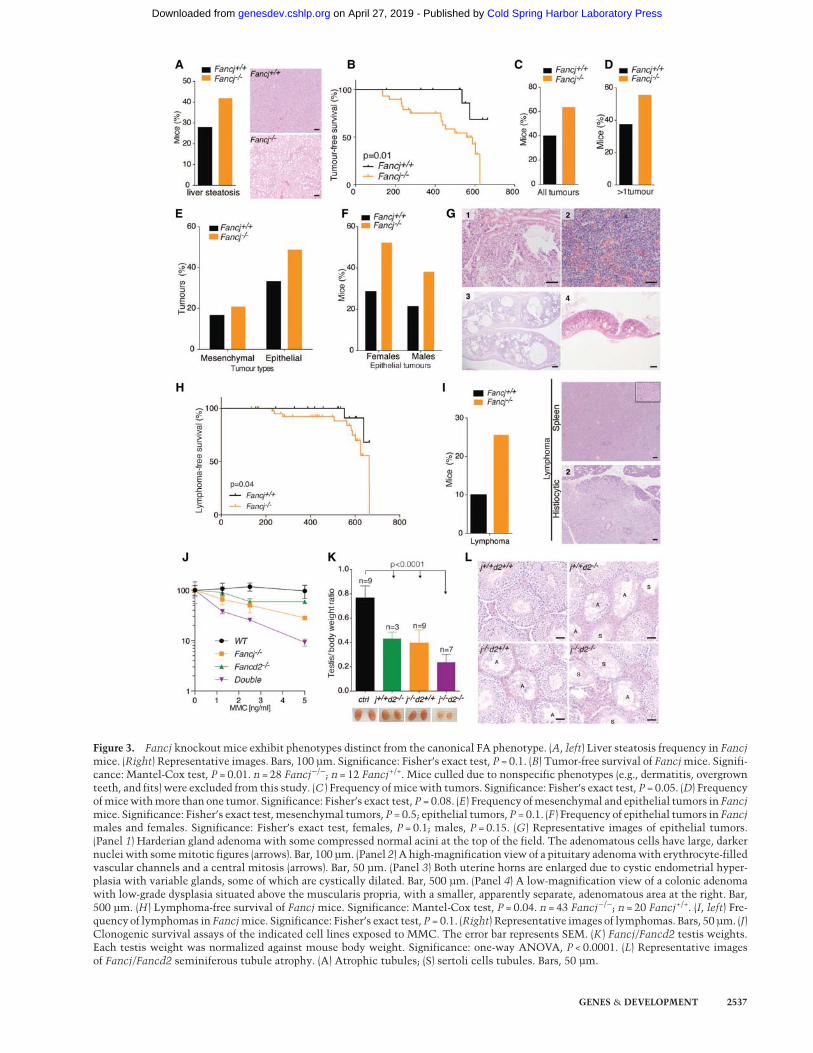
Figure 3. Fancj knockout mice exhibit phenotypes distinct from the canonical FA phenotype. (A, left) Liver steatosis frequency in Fancjmice. (Right) Representative images. Bars, 100 µm. Significance: Fisher’s exact test, P = 0.1. (B) Tumor-free survival of Fancjmice. Signifi-cance: Mantel-Cox test, P = 0.01. n = 28 Fancj−/−; n = 12 Fancj+/+. Mice culled due to nonspecific phenotypes (e.g., dermatitis, overgrownteeth, and fits) were excluded from this study. (C ) Frequency of micewith tumors. Significance: Fisher’s exact test, P = 0.05. (D) Frequencyofmicewithmore than one tumor. Significance: Fisher’s exact test, P = 0.08. (E) Frequency ofmesenchymal and epithelial tumors in Fancjmice. Significance: Fisher’s exact test, mesenchymal tumors, P = 0.5; epithelial tumors, P = 0.1. (F ) Frequency of epithelial tumors in Fancjmales and females. Significance: Fisher’s exact test, females, P = 0.1; males, P = 0.15. (G) Representative images of epithelial tumors.(Panel 1) Harderian gland adenoma with some compressed normal acini at the top of the field. The adenomatous cells have large, darkernuclei with somemitotic figures (arrows). Bar, 100 µm. (Panel 2) A high-magnification view of a pituitary adenomawith erythrocyte-filledvascular channels and a central mitosis (arrows). Bar, 50 µm. (Panel 3) Both uterine horns are enlarged due to cystic endometrial hyper-plasia with variable glands, some of which are cystically dilated. Bar, 500 µm. (Panel 4) A low-magnification view of a colonic adenomawith low-grade dysplasia situated above the muscularis propria, with a smaller, apparently separate, adenomatous area at the right. Bar,500 µm. (H) Lymphoma-free survival of Fancj mice. Significance: Mantel-Cox test, P = 0.04. n = 43 Fancj−/−; n = 20 Fancj+/+. (I, left) Fre-quency of lymphomas in Fancjmice. Significance: Fisher’s exact test, P = 0.1. (Right) Representative images of lymphomas. Bars, 50 µm. (J)Clonogenic survival assays of the indicated cell lines exposed to MMC. The error bar represents SEM. (K ) Fancj/Fancd2 testis weights.Each testis weight was normalized against mouse body weight. Significance: one-way ANOVA, P < 0.0001. (L) Representative imagesof Fancj/Fancd2 seminiferous tubule atrophy. (A) Atrophic tubules; (S) sertoli cells tubules. Bars, 50 µm.
GENES & DEVELOPMENT 2537
Cold Spring Harbor Laboratory Press on April 27, 2019 - Published by genesdev.cshlp.orgDownloaded from

that epithelial tumors are not the only prominent tumortype in Fancj−/− mice. Surprisingly, the lymphoma-free median survival age, although late in life, was signifi-cantly decreased in Fancj−/− mice (Fig. 3H), with 26% ofFancj−/− mutants developing lymphomas comparedwith ∼10% in littermate controls (Fig. 3I). This was incontrast to Fancd2−/− andHelQ-deficient FAmousemod-els, which do not presentwith heighted lymphoma predis-position (Supplemental Fig. S3A,B; Houghtaling et al.2003). Further interrogation of the Fancj−/− lymphoma-free median survival data revealed that mutant femalesare more susceptible than males to lymphomas, with anincidence of ∼40% compared with 9.5% in mutant males(Supplemental Fig. S3C). Histological analysis revealedthat lymphomas were widely spread in most animals,with an increased frequency in the spleen, B cells, andmesenteric and salivary lymph nodes (Supplemental Fig.S3D). This unexpected difference in the tumor spectrumof Fancj-deficient mice compared with other FA mousemodels raised the possibility that FANCJ performs rolesthat are independent of the canonical FA pathway, at leastwith respect to preventing lymphoma development.
Loss of Fancd2 exacerbates the Fancj mutantphenotype
Given the sensitivity to MMC, germ cell attrition, andpredisposition to epithelial tumors associated withFANCJ deficiency overlap with the phenotype of othermouse models of FA, yet lymphoma predisposition isunique to FANCJ, wewere compelled to examinewhetherFANCJ actually functions genetically in the FA pathway.To this end, we crossed Fancj−/− and Fancd2−/− mutantmice and subjected the resulting Fancj−/−Fancd2−/−
double-mutant mice to genetic epistasis analysis. Unex-pectedly, MEFs derived from Fancj−/−Fancd2−/− double-mutant mice were significantly more sensitive to MMCthan either single mutant (Fig. 3J). The increased sensitiv-ity in the double mutant implies that FANCJ may func-tion in parallel to or performs roles that are distinctfrom the canonical FA pathway in ICL repair.
We also examined the genetic relationship betweenFANCJ and FANCD2 in mice. Fancj−/−Fancd2−/− dou-ble-mutant mice appeared grossly normal and healthy,similar to their single-mutant counterparts, and wereborn at near to expectedMendelian ratios (χ2, 9.8) (Supple-mental Fig. S3E). However, when testes were examined,double mutants possessed significantly smaller testesthan the controls. There is also a subtle additive butnot significant decrease of testis size between Fancj−/−
or Fancd2−/− and Fancj−/−Fancd2−/− (Fig. 3K). Histologyanalysis revealed the presence of significantly more atro-phic tubules in the double mutant when compared withsingle mutants and controls, which were also mostlydevoid of spermatogenic cells and composed of sertolicells only (Fig. 3L; Supplemental Fig. S3F). Togetherwith the increased MMC sensitivity and lymphomapredisposition, these data suggest that FANCJ performscellular and organismal roles that are distinct and inde-pendent of the FA pathway.
Fancj−/− MEFs exhibit spontaneous DNA damageand aphidicolin sensitivity
To gain further insight into the functions of FANCJ inmaintaining genome integrity, we examined Fancj−/−
MEFs for markers of spontaneous DNA damage. In con-trast to wild-type controls, Fancj−/− MEFs exhibit sponta-neous γH2AX foci even in the absence of DNA-damagingagents (Fig. 4A). To examine whether the elevated levelsof DNA damage influence cell growth, cellular senes-cence was monitored in primary cells (Fig. 4B). PrimaryFancj−/− MEFs were found to undergo spontaneous senes-cence at later passages, which was not observed in wild-type controls. These observations suggest that Fancj−/−
MEFs undergo senescence due to increased levels of spon-taneous DNA damage. Consistent with the DNA damagearising duringDNA replication,weobserved that Fancj−/−
MEFs exhibit hypersensitivity to low doses of the replica-tion inhibitor aphidicolin (Fig. 4C). In addition, the levelsof γH2AX foci were significantly increased in Fancj−/−
MEFs after aphidicolin treatment (Fig. 4A). These resultssupport that FANCJ also has a noncanonical FA function.
FANCJ is required for normal replication fork progression
The spontaneous DNA damage and aphidicolin sensitiv-ity of Fancj−/− MEFs raised the possibility that FANCJ isrequired to facilitate normal DNA replication. To exam-ine this possibility, we assessed replication fork dynamicsby monitoring iodo-deoxyuridine and chloro-deoxyuri-dine (IdU/CldU) incorporation by DNA combing. Asshown in Figure 4D, loss of FANCJ results in significantlyslower replication fork extension rates in unchallengedcells relative to control cells (1.56 and 2.51 kb/min, re-spectively), and this is further exacerbated by aphidicolintreatment (1.24 kb/min). By measuring the length of theleft and right moving fork, we also observed elevated lev-els of asymmetric replication forks in Fancj−/−MEFs com-paredwithwild-type controls (22.9%vs. 3.1%) under bothunchallenged and aphidicolin-treated conditions (Fig. 4E).These data suggest that loss of FANCJ results in increasedreplication fork stalling and/or collapse. Consistent withthe reduced fork extension rates, interorigin distances un-der both unchallenged and aphidicolin-treated conditionswere significantly shorter in Fancj−/− MEFs relative towild-type controls (145.3 and 203.7 kb in untreated, and107.7 and 136.3 kb in treated, respectively). These resultsestablish that FANCJ facilitates normal DNA replicationand plays a crucial role in DNA replication when replica-tion forks are perturbed.
Fancj−/− MEFs are not sensitive to G4-stabilizing drugs
Biochemical studies have shown that FANCJ is proficientat unwinding G4-DNA structures in vitro (London et al.2008) and can promoteDNA replication throughG4 struc-tures in aX. laevis cell-free system (Peng et al. 2014). A rolefor FANCJ inunwindingG4-DNAstructures invivo is alsosuggested by the accumulation of large genomic deletionsin the vicinity of G-rich sequences in dog-1 (C. elegans
Matsuzaki et al.
2538 GENES & DEVELOPMENT
Cold Spring Harbor Laboratory Press on April 27, 2019 - Published by genesdev.cshlp.orgDownloaded from

IdU replication extension rate [kb/min]
Fancj-/-
Fancj+/+
Fancj-/-+A
ph
Fancj+/+
0 2 4 6
Fancj-/-
Fancj+/+
Fancj-/-+A
ph
Fancj+/+
Fancj-/- Fancj-/-Fancj+/+ Fancj+/++Aph+Aph
Fancj-/-
Fancj+/+
Fancj-/-+A
ph
Fancj+/+
010
020
030
040
0
Inter-origin distance [kb]
Left-
mov
ing
fork
[kb]
Right-moving fork [kb] Right-moving fork [kb] Right-moving fork [kb]0 50 100 150
0
50
100
150
0 50 100 1500
50
100
150
0 50 100 1500
50
100
150
0 50 100 1500
50
100
150
Right-moving fork [kb]
3.1% 22.9% 21.5% 37.7%
SA
--g
al p
ositi
ve c
ells
P4 P8P12 P15
0
20
40
60
80
p<0.001
p<0.001
p<0.001
p=0.003
p=0.005
p<0.001
p<0.001
p<0.001
A
D
E
F
Fancj+/+ Fancj-/-0
10
20
30
40
% c
ells
with
>10
H
2AX
foci
-+ 5nM Aph
B
0 200 400 600 8000.01
0.10
1
10
100
Aph [nM]
Fancj+/+
Fancj-/-
C
Fancj+/+
Fancj-/-
IdU - CldU 20’ - 20’
IdU - CldU 20’ - 20’
Figure 4. Spontaneous DNA damage, senescence, and replication stress in Fancj knockout cells. (A) Quantification of γH2AX-positivecells in Fancj+/+ and Fancj−/− MEFs in the absence or presence of 5 nM aphidicolin (Aph). The error bar indicates SD. (B) Quantification ofSA-β-gal-positive cells in Fancj+/+ and Fancj−/− primary MEFs at the indicated passage numbers. The error bar indicates SD. (C ) Clono-genic survival assays of Fancj+/+ and Fancj−/− MEFs exposed to aphidicolin. The error bar represents SEM. (D) Replication rates inFancj+/+ and Fancj−/− MEFs in the absence or presence of aphidicolin. Nascent DNA strands were pulse-labeled with iodo-deoxyuridine(IdU) and chloro-deoxyuridine (CldU) for 20min, individually.More than 300 fibers in the indicated cell linesweremeasured. Arrowheadsindicate the potential position of replication origin. The error bar indicates SD. (E) Quantification of asymmetry between sister replicationforks in the indicated MEFs with or without aphidicolin. The areas between the black lines contain sister forks with <25% length differ-ence. (F ) Interorigin distances in Fancj+/+ and Fancj−/− MEFs with or without aphidicolin. The error bar shows SD.
FANCJ prevents microsatellite instability
GENES & DEVELOPMENT 2539
Cold Spring Harbor Laboratory Press on April 27, 2019 - Published by genesdev.cshlp.orgDownloaded from

FANCJ) mutant worms (Cheung et al. 2002) and humancells lacking FANCJ (London et al. 2008). These observa-tionspromptedus to testwhether the replicationproblemsdetected in Fancj-deficient cells reflect a function in coun-teracting G4-DNA structures. To this end, we firstanalyzed the sensitivities of Fancj−/− MEFs to a range ofG4-stabilizing agents. Unexpectedly, Fancj−/− MEFs ex-hibit comparable sensitivity to TMPyP4, telomestatin,andpyridostatincomparedwithwild-typecontrols. Incon-trast,Rtel1-deficientMEFs exhibit sensitivity to TMPyP4and telomestatin, as previously reported (Fig. 5A–C).
Telomeric G4-DNA sequences are stablein Fancj−/− MEFs
Next, we examined a possible role for FANCJ in unwind-ing G4-DNA sequences that form within the TTAGGG
repeats at vertebrate telomeres. Defects in counteractingtelomeric G4-DNA structures during telomere replica-tion give rise to telomere fragility, which is observed asmultiple spatially distinct telomere FISH signals at chro-mosomes ends (Sfeir et al. 2009). Consistent with thelack of sensitivity to G4-stabilizing agents, the levels oftelomere fragility and telomere loss in Fancj−/− MEFswere indistinguishable from wild-type controls (Fig.5E,F). This is in contrast to Rtel1-deficient MEFs, whichexhibit extensive telomere fragility (Vannier et al. 2012).Prompted by our previous observation that the rtel-1dog-1 (C. elegans RTEL1 and FANCJ) double mutant issynthetic-lethal due to catastrophic genome instability(Barber et al. 2008), we generated conditional Rtel1F/F
Fancj−/− double-mutant MEFs to investigate a potentialredundant role for FANCJ in unwinding G4-DNA struc-tures in the absence of RTEL1. Consistent with our
Fancj+/+ Fancj-/-0
5
10
15
20
Frag
ile te
lom
eres
/Met
apha
se
0 10 20 30 40 501
10
100
TMPyP4[µM]0 1 2 3 4
10
100
Telomestatin[µM]0.0 0.5 1.0 1.5 2.0
10
100
Pyridostatin[µM]
Sur
viva
l [%
]
WT
Fancj-/-
Rtel1-/-
Wild type
Telomere fragility
Telomere loss
A
D
Fra
gile
telo
mer
es/ M
etap
hase
0
10
20
30
40
Cre - + - +
p<0.001
p<0.001
p=0.
01
p<0.
001
p<0.001
H
0
20
40
60
80
% S
A-
-gal
pos
itive
cel
ls
Rtel1F/F,Fancj+/-
Rtel1F/F,Fancj+/- +CreRtel1F/F,Fancj-/-
Rtel1F/F,Fancj-/- +Cre
P4 P8 P12
0
20
40
60
%
H2A
X p
ositi
ve c
ells
-Cre+Cre
Rtel1F/FFancj+/- Fancj-/-
Rtel1F/F
G I
E
0
5
10
15
20
Telo
mer
e lo
ss/M
etap
hase
F
B C
Fancj+/+ Fancj-/-
Rtel1F/FFancj+/- Fancj-/-
Rtel1F/F
p=0.
002
p<0.
001
Figure 5. Fancj knockout cells are not sensi-tive to G4-DNA-stabilizing drugs and do notpresent with telomere fragility. (A–C ) Clono-genic survival assays of Fancj+/+, Fancj−/−,and Rtel1−/− MEFs exposed to TMPyP4 (A),telomestatin (B), and pyridostatin (C ). The er-ror bar represents SEM. (D) Representative im-age of fragile telomeres and telomere loss. (E,F )Quantification of fragile telomeres (E) andtelomere loss (F ) per metaphase in Fancj+/+
and Fancj−/− MEFs. The error bar indicatesSD. Significance: t-test, P = 0.08 in E; P = 0.3in F. (G) Quantification of SA-β-gal-positivecells in the indicated primary MEFs with orwithout Cre treatment at passages 4, 8, and12. The error bar indicates SD. (H) Quantifica-tion of γH2AX-positive cells in the indicatedMEFs with or without Cre treatment. The er-ror bar indicates SD. (I ) Quantification of frag-ile telomeres per metaphase in the indicatedMEFs with or without Cre treatment. The er-ror bar represents SD. Significance betweenRtel1f/f Fancj+/− + Cre and Rtel1F/F Fancj−/− +Cre: t-test, P = 0.5.
Matsuzaki et al.
2540 GENES & DEVELOPMENT
Cold Spring Harbor Laboratory Press on April 27, 2019 - Published by genesdev.cshlp.orgDownloaded from

previous findings in worms, Rtel1−/−Fancj−/− double-knockout MEFs display accelerated senescence (Fig. 5G)and elevated levels of spontaneous γH2AX foci relativeto single-knockout MEFs (Fig. 5H). However, the levelsof telomere fragility observed in Rtel1F/FFancj−/− dou-ble-knockout MEFs was not significantly different fromthe levels present in the Rtel1−/− single-knockout cells(Fig. 5I). These data suggest that mouse FANCJ is dispen-sable for the maintenance of telomeric G4 sequences andpotentially other G4-DNA sequences.
MSI in Fancj−/− MEFs
In the absence of any overt phenotype associated withG4-DNA metabolism in Fancj-deficient cells, we consid-ered the possibility that problems associated with othertypes of DNA sequences/secondary structures are thecause of the replication defects in Fancj−/− MEFs. Previ-ous studies reported that FANCJ physically interactswith the MMR protein MLH1 (Cannavo et al. 2007;Peng et al. 2007), but the functional relevance of this as-sociation remains unknown. Intriguingly, MMR defi-ciency is associated with MSI, which reflects a failureto correct base–base mismatches and short deletionsthat arise following template slippage at these short tan-dem repeated sequences. Since MMR-deficient miceare also associated with increased risk of lymphoma(Li 2008), we considered the possibility that the replica-tion defects in Fancj−/− cells might arise from problemsat microsatellite sequences. To test this hypothesis, wefirst derived wild-type and two Fancj−/− (#1 and #2) prima-ryMEF lines from timedmatings of Fancj+/− heterozygousmice and examined the integrity of nine distinctmicrosat-ellites, which were chosen based on their reported insta-bility in MMR-deficient cells. PCR was used to assessMSI and was defined as a deviation from the wild-typebanding pattern, with higher molecular bands and lowermolecular bands classified as microsatellite expansionand microsatellite contraction, respectively. Strikingly,both Fancj−/− MEF lines (#1 and #2) exhibited instabilityassociated with eight out of nine (#1) and four out ofnine (#2) microsatellites, which included both expansionand contraction (Fig. 6B). To confirm this result, we inde-pendently derived a wild-type and two further Fancj−/−
MEF lines from a second timedmating of Fancj+/− hetero-zygous mice. Unlike the wild-type control cells, bothFancj−/− MEF lines exhibited instability associated withfour out of nine (#1) and four out of nine (#2) microsatel-lites (Fig. 6B).To investigate whether MSI is also observed in other
mouse models of FA, we analyzed the integrity of 18 mi-crosatellite sequences in two Fancd2−/− primary MEFlines (#1 and #2) and compared these with wild-type andFancj−/− primary MEF lines derived from a third indepen-dentmating. FANCD2was analyzed, as it is a key compo-nent of the FA pathway and is regulated by the FA corecomplex. In contrast to theMSI evident in Fancj−/− prima-ry cells (12 of 20), the two Fancd2−/− primaryMEFs (1: twoof 18; #2: two of 18) were largely devoid of unstablemicro-satellite sequences, similar to wild-type cells (one of 18;
one of 20) (Fig. 6C). These data suggest that MSI is likelyunique to FANCJ-deficient cells among the defined FAgenes.To further analyze the sequence changes associated
with MSI in the absence of FANCJ, we cloned and se-quenced the microsatellite D1Mit36 from wild-type andFancj−/−MEFs (Fig. 6D; Supplemental Fig. S4A). Sequenc-ing of the PCR products from the wild type showed onlytwo sequences, which likely correspond to themicrosatel-lites oneachhomologous chromosome. In contrast, sixdif-ferent sequence changes were detectable from Fancj−/−
cells, which corresponded to a single-repeat contraction,four- to six-repeat expansions, and more complex alter-ations. These observations are consistent with increasedtemplate slippage at microsatellites, which could occur ifextruded secondary structures formed from the tandem re-peats persist in the absence of FANCJ.
Human FA-J patient cells and CRISPR-derivedFancj−/− U2OS cells exhibit MSI, which is exacerbatedby aphidicolin treatment
To determine whether disruption of human FANCJ givesrise to MSI, we generated a FANCJ knockout U2OS cellline by genome editing with the CRISPR–Cas9 system.Guide RNAs targeting exon 5 of Fancj resulted in a knock-out line devoid of detectable FANCJ by Western blotting(Fig. 7A). The integrity of 10 different microsatelliteswas assessed in the isogenic U2OS Fancj+/+ and Fancj−/−
cell lines. Consistent with the MSI observed in Fancj−/−
MEFs, U2OS Fancj−/− cells, but not isogenic wild-typecontrols, exhibited instability associated with six out of10 microsatellites (Fig. 7B,C).Prompted by our findings that the replication inhibitor
aphidicolin induces DNA damage, replication fork stall-ing/collapse, and sensitivity to Fancj-deficient MEFs,we considered the possibility that this phenotype mayreflect increased problems at microsatellite sequences.To this end, we assessed the integrity of 10microsatellitesin isogenic U2OS Fancj+/+and Fancj−/− cell lines fol-lowing treatment with 0.2 µM aphidicolin for 24 h.Consistently, we observed that the extent of MSI inU2OS Fancj−/− cells was exacerbated by aphidicolin treat-ment, but no such changes occurred in the controls(Fig. 7C).To determinewhether theMSI is also evident in human
FA patients harboring mutations in FANCJ, we obtainedprimary lymphoblastoid cell lines (LCLs) from affectedand unaffected members of a second-degree consanguine-ous FA-J family (two unaffected parents and three affectedchildren), with affected individuals carrying a large auto-zygous region on chromosome 17, including FANCJ/BRIP1, and homozygous for the missense mutationc.1878A>T (exon 13 of FANCJ) with the effect p.E626D,which destabilizes the protein. Of the 12 microsatellitesexamined, no evidence of MSI was observed in the unaf-fected parents, whereas all affected children presentedwith MSI (Fig. 7D). Hence, loss of FANCJ in mouse or hu-man cells gives rise to spontaneous MSI, which is exacer-bated under conditions of replication stress.
FANCJ prevents microsatellite instability
GENES & DEVELOPMENT 2541
Cold Spring Harbor Laboratory Press on April 27, 2019 - Published by genesdev.cshlp.orgDownloaded from

Discussion
Copy number changes within specific microsatelliteshave been implicated in >30 human CNS disorders. MSIis also a hallmark of certain cancers and is used as a diag-nostic marker of Lynch syndrome, which is caused bygermline mutations in MMR genes (Li 2008; Boland andGoel 2010). While MMR suppresses MSI by correcting
base–base mismatches arising from strand slippages thatoccur at high frequency within microsatellite repeats,activities that prevent or dismantleDNA secondary struc-tures that stabilize strand slippage events were unknown.Here, we establish a crucial role for the helicase FANCJ inpreventing MSI in vertebrate cells, which is distinct fromits role in the FA pathway. In the absence of murineFANCJ single-repeat contractions, four- to six-repeat
expansioncontraction
both
Fancj
D1Mit36
+/+ -/-
1e2.3
+/+ -/-
D6Mit59
+/+ -/-
D6Mit236
+/+ -/-
D3Mit278
+/+ -/-
Control
+/+ -/-
0-1 repeat
+6 repeat
+5 repeat
+4 repeat
other
10
20
30
40
50
5
15
25
35
45
D1Mit36
% M
utat
ion
even
t
Fancj+/+ Fancj-/-
Locus
D1Mit36
D3Mit22
D7Mit91
D3Mit278
D6Mit59
D6Mit236
D7Nds1
1e2.3
Control
8b3.3
CA/TG
CAG/CTG
CA/TG
CA/TG
CA/TG
TC/GA
TC/GA
TTC/GAA
Repeat unit
TTC/GAA
+
+
+
+
+
+
-
+
-
+
Fancj-/- Fancj+/+
#1(1st)
-
-
-
+
+
-
-
+
-
+
#2
% unstable sites 88.9 44.4
-
-
-
-
+
-
-
-
-
-
11.1
-
-
-
-
-
-
-
-
-
+
11.1
+
-
-
-
+
+
-
-
-
+
Fancj-/-
#1
-
-
-
-
+
+
-
+
+
-
#2
44.4 44.4
Fancj+/+
(2nd)
CA/TG
CAG/CTG
CA/TG
CA/TG
CA/TG
TA/AT
TC/GA
TC/GA
TTC/GAA
Repeat unitLocus
CA/TG
CA/TG
CA/TG
CA/TG
CA/TG
AC+AG
CA/TG
CA/TG
TTC/GAA
TTC/GAA
TC/GA
D1Mit36
D3Mit22
D7Mit91
D3Mit278
D6Mit59
D5Mit48
D6Mit236
D7Nds1
1e2.3
Control
D10Mit266
D11Mit258
D12Nds2
DXMit172
D14Mit102
D13Mit13
D10Mit2
D14Mit15
8b3.3
8b3.3b
D17Nds3
% unstable sites
NA
NA
-
-
-
-
-
-
-
-
-
-
-
+
-
-
-
+
-
-
-
#1Fancd2-/-Fancd2+/+
#2
11.1
NA
NA
-
-
-
-
-
-
-
-
-
+
-
+
-
-
-
-
-
-
-
11.1
NA
NA
-
-
-
-
-
-
-
-
-
-
-
+
-
-
-
-
-
-
-
5.6
-
-
-
+
-
-
-
-
-
-
-
-
-
-
-
-
-
-
-
-
-
5.0
+
+
+
+
+
+
+
+
+
+
+
-
-
-
-
-
-
-
+
-
-
Fancj-/-
#1
60.0
Fancj+/+
(3rd)
(3rd)
A
B
C
D
Figure 6. Fancj knockout mice exhibit MSI independent of the canonical FA pathway. (A) Representative images of MSI at the indicatedloci in MEFs. (B) Microsatellite analysis in two independent sets of Fancj+/+ and Fancj−/− cell lines. MSI status is indicated as unstable (+)and stable (−). Types of instability are indicated as expansion (magenta), contraction (green), and both (blue). (C ) MSI in Fancd2−/− andFancj−/− cell lines. MSI status and types of instability are indicated as in B. (D) Distribution of mutation types at the D1Mit36 locus.D1Mit36 PCR products were cloned into empty vector and sequenced. The percentage of mutation type is calculated. DNA sequencesare shown in Supplemental Figure S4A.
Matsuzaki et al.
2542 GENES & DEVELOPMENT
Cold Spring Harbor Laboratory Press on April 27, 2019 - Published by genesdev.cshlp.orgDownloaded from

A/T
A/T
A/T
A/T
Repeat unitLocus
CA/TG
CA/TG
CA/TG
CTG/CAG
A/T
CTG/CAG
BAT-25
NR-21
NR-24
BAT-26
D2S123
D5S346
D17S250
SCA10
DMPK
MONO-27
SCA1
% unstable sites
-
+
-
-
-
-
-
-
+
-
-
FA-J patients LCL
25.0
-
-
-
+
+
-
-
+
-
+
+
50.0
-
-
-
+
+
+
-
-
-
CTG/CAGERDA1 +++
Control ---
-
-
33.3
KIGJKIDAKIVY
ATTCT/AGAAT
(vs unaffected parents)
FANCJ
U2OS
Tubulin
Fancj -/-+/+
A/T
A/T
A/T
Repeat unitLocus
CA/TG
CA/TG
CA/TG
CTG/CAG
CTG/CAG
BAT-25
NR-21
BAT-26
D2S123
D5S346
D17S250
SCA10
DMPK
SCA1
- -
- -
+ +
+
-
+
+
+ ++
++
++
++
++
++
+
-
-
-
-
-
-
-
-
-
-
-
-
-
-
-
-
CTG/CAGERDA1 - ---
Control - ---
-
-
+Aph +Aph
ATTCT/AGAAT
Fancj+/+ Fancj-/-
SCA1SCA10
Aph
Fancj -/-+/+
+
*
*
- +-
-/-+/+
+- +-
-/-+/+
+- +-
Control
Fancj-/-Fancj+/+
Stable Unstable
Triplex
Hairpin
Z-DNA
FANCJ
expansioncontraction
both
A B
C D
E
*
Figure 7. MSI in human FANCJ-deficient patient and knockout cells. (A) Western blot with FANCJ antibody confirmed the loss ofdetectable FANCJ protein in control (+/+) and CRISPR knockout (−/−) U2OS cells. (B) Representative images of MSI at the indicatedloci in U2OS cells. Asterisks indicate PCR products specifically found in Fancj knockout cells. (C ) Microsatellite analysis in FANCJCRISPRknockoutU2OS cells.MSI in FANCJ knockoutU2OS cells were analyzed after aphidicolin (Aph) treatment.Microsatellite statusat the indicated loci is indicated as stable (−) or unstable (+). Compared with untreated samples, changes in the banding pattern by aphi-dicolin treatment are shown as two plus signs (++). (D) MSI in FA-J patient lymphoblastoid cell lines (LCLs). Twelve microsatellites inthree FA-J patient LCLs and two unaffected parent LCLs were analyzed by PCR-based MSI assay using primers labeled with a fluorescentdye. Unstablemicrosatellites in FA-J LCLs relative to unaffected LCLs are indicated as plus signs (+), and stablemicrosatellite are shown asminus signs (−). (E) Model of the maintenance of microsatellite stability by the FANCJ helicase.
FANCJ prevents microsatellite instability
GENES & DEVELOPMENT 2543
Cold Spring Harbor Laboratory Press on April 27, 2019 - Published by genesdev.cshlp.orgDownloaded from
expansions and more complex alterations arise, which isconsistent with increased template slippage events at mi-crosatellites (Boyer et al. 2013).
Our analysis of Fancj-deficient mice uncovered strikingphenotypic similarities to other FA mouse models, in-cluding gonadal atrophy and subfertility, epithelial tumorpredisposition, and cellular hypersensitivity to ICL-inducingagents (Parmaretal. 2009).Consistentwithprevi-ous observations (Levitus et al. 2004; Litman et al. 2005),murine FANCJ is dispensable for the monoubiquitylationof FANCD2 and its subsequent recruitment into chroma-tin-associated repair foci. FANCJ also appears to bedispen-sable for the initial processing of the ICL lesion as DSBsaccumulate in Fancj−/− MEFs following treatment withMMC. Nevertheless, this observation does not excludethe possibility that ICL lesions are inappropriately pro-cessed in the absence of FANCJ, which could potentiallygive rise to erroneous repairand radial chromosome forma-tion. While the strand exchange protein Rad51 accumu-lates in repair foci, DSBs persist in the absence of FANCJ,suggestive of a defect after ICL incision and downstreamfrom Rad51 loading onto damaged chromatin.
Several unexpected phenotypes of Fancj−/−mice absentfrom other mouse models of FA prompted us to investi-gate potential functions for FANCJ in the metabolism ofG4-DNA and other DNA secondary structures. Thesephenotypic differences included (1) the absence of growthretardation or microphthalmia; (2) Fancj−/−Fancd2−/−
double-mutant mice and cell lines exhibit a more severephenotype than either single mutant in some assays, in-cluding gonadal atrophy and cellular sensitivity toMMC; and (3) Fancj−/− mice display increased predisposi-tion to lymphomas, and cells derived from these mice areacutely sensitive to aphidicolin. Since FANCJ has beenimplicated in the metabolism of G4-DNA structures inchicken DT40 cells (Sarkies et al. 2012; Schwab et al.2013) and X. laevis cell-free egg extracts (Castillo Boschet al. 2014), it is perhaps surprising that Fancj−/− MEFsare insensitive to G4-stabilizing drugs and do not presentwith telomere fragility. In contrast, cells deficient for therelated helicase,Rtel1, which is also capable of unwindingG4-DNA structures in vitro, are hypersensitive toTMPyP4 and telomestatin and present with telomere fra-gility (Uringa et al. 2012; Vannier et al. 2012). In addition,Rtel1−/−Fancj−/− double-knockout cells do not showheightened telomere fragility, yet, analogous to the phe-notype of dog-1; rtel-1 double mutants in C. elegans (Bar-ber et al. 2008), double-knockout MEFs display enhancedlevels of genome instability and accelerated senescencewhen compared with single mutants. These data suggestthat, in mice, RTEL1 plays a prominent role in the metab-olism of G4-DNA structures, while FANCJ plays no sig-nificant role in the metabolism of G4-DNA structures.
Our finding that loss of either murine or human FANCJresults in MSI is intriguing in light of previous observa-tions. Loss of MMR in mouse models is also associatedwith increased predisposition to lymphoma (Li 2008), al-beit with an earlier age of onset relative to Fancj−/−
mice. Several studies have reported that FANCJ interactswith the MMR protein MLH1 (Cannavo et al. 2007; Peng
et al. 2007), yet the biological importance of this interac-tion and its importance for maintenance of microsatel-lites remain uncertain. Notably, Mlh1 deficiency alsogives rise toMSI, which is exacerbated by replication inhi-bition, and its mutation in humans results in Lynch syn-drome and associated cancers (Lindblom et al. 1993;Bronner et al. 1994; Papadopoulos et al. 1994). Strikingly,the L607Hmutation inMLH1 observed in hereditary non-polyposis colon cancer (HNPCC) disrupts the interactionwith FANCJ (Xie et al. 2010a), which raises the possibilitythat the FANCJ–MLH1 interaction is important forpreventing colorectal cancers. Consistent with this sup-position, mutations in FANCJ have been previously asso-ciated with rectal cancers (Rafnar et al. 2011). Despitethese observations, FANCJ is not known to be mutatedin Lynch syndrome, which is associated with MMR defi-ciency and strikingly elevated mutation rates, including,but not limited to, MSI. A potential explanation for thisis that FANCJ, while playing a role in the maintenanceof microsatellites, is not required for MMR per se, as itsloss does not give rise to a spectrum of mutagenesis sim-ilar to that seen in the absence of MMR, which reflectsa defect in the correction of base–base mismatches andother discontinuities in the double helix. Nevertheless,the MSI associated with loss of FANCJ could explain itsimpact in colorectal and pancreatic cancers. In addition,as human Fancjheterozygous and homozygousmutationsare associated with some types of cancers, the co-occur-rence of MSI might suggest that these tumors are ICL-sensitive.
Current models propose that expansions or contrac-tions of repetitive sequences arise as a result of strand slip-page during DNA polymerization, which is favored by thepropensity of microsatellites to form DNA secondarystructures within ssDNA that stabilize strand misalign-ments (Wang and Vasquez 2014). Indeed, the enhancedMSI observed following treatment with replication inhib-itors in Fancj-deficient cells could be explained by the factthat this increases the presence of ssDNA at the replica-tion fork, which favors DNA secondary structure forma-tion. The impact of replication inhibitors on MSIprovides a plausible explanation for the hypersensitivityof Fancj−/− MEFs to aphidicolin, which is associatedwith increased fork asymmetry/stalling, reduced fork ex-tension rates, and reduced interorigin distance. The latterpresumably reflects the consequences of fork stalling atmicrosatellite sequences. Importantly, of the microsatel-lites that are unstable in Fancj−/− MEFs, CAG/CTG,TTC/GAA, CA/TG, and CT/AG repeats are potentialDNA slippage sites, and CAG/CTG, TTC/GAA, CT/AG, and CA/TG are believed to form hairpin-like struc-tures, triplex structures, triplex structures, and Z-DNAstructures, respectively (Zhao et al. 2010). Since FANCJhas been shown to unwind a variety of DNA secondarystructures in vitro (Brosh and Cantor 2014), including,but not limited to, hairpins and triplex structures, we pro-pose that FANCJ acts to suppress MSI by dismantlingDNA secondary structures that favor repeat sequence ex-pansion or contraction via a strand slippage mechanism(Fig. 7E), which is distinct from error correction by MMR.
Matsuzaki et al.
2544 GENES & DEVELOPMENT
Cold Spring Harbor Laboratory Press on April 27, 2019 - Published by genesdev.cshlp.orgDownloaded from

Materials and methods
Fancj knockout mice were generated by a gene trap strategy. Pri-mary MEFs were derived at 13.5 d post-coitum. For immortaliza-tion, primary MEFs were transfected with large T SV40 plasmid.For histology and post-mortem tissues, samples were fixed, paraf-fin-embedded, sectioned, and stained with hematoxylin andeosin. For immunohistochemistry, tissue sections were stainedby avidin–biotin complex methods. For FANCJ expression andFANCD2 ubiquitylation, cell lysates were prepared fromFancj+/+ and Fancj−/− MEFs and U2OS cells and subjected toWestern blot with the indicated antibodies. For clonogenic sur-vival assays, MEFs were seeded in 10-cm dishes and stainedwith crystal violet 7–13 d after treatment with DNA-damagingagents. For immunofluorescence staining experiments, MEFswere permeabilized, fixed, and stained with the indicated anti-bodies. For chromatin fractionation, insoluble chromatin frac-tions and soluble fractions were separated by centrifugation. ForPFGE experiments, cells were embedded in agarose plugs andtreated with proteinase K overnight. Genomic DNAwas separat-ed by PFGE apparatus and stained with ethidium bromide. ForSA-β-gal assay, primary MEFs were stained using senescencecell histochemical staining kit (Sigma). For DNA combing, pri-mary MEFs were pulse-labelled with IdU for 20 min and subse-quently labelled with CldU for 20 min. Extracted DNA wasstretched on silanized coverslips and stained with two differentanti-BrdU antibodies. For telomere FISH experiments, swollencells were fixed by methanol and acetate solution and droppedon a glass slide. Slides were hybridized with PNA telomericprobe. To generate human FANCJ knockout U2OS cells by theCRISPR/Cas9 system, FANCJ guide oligos were cloned intopX462 plasmid and used for transfection. Transfected cloneswere isolated by limiting dilution and subjected to Western blotand MSI analysis. For MSI analysis, microsatellites were ampli-fied by PCR with fluorescent-labelled primers or nonlabelledprimers. Labelled PCR products were analyzed by ABI3130XLsystems. Nonlabelled PCR products were analyzed by polyacryl-amide gel electrophoresis. For sequencing microsatellites, PCRproducts were cloned into empty vector and used for transforma-tion. Plasmids were prepared and sequenced from >20 bacterialclones. See also the expanded Materials and Methods section inthe Supplemental Material for further details.
Acknowledgments
We thank Gordon Stamp and the Histopathology laboratory fortheir help in analyzing mice post-mortem. We also thank the ex-perimental Biological Resource Unit, notably Danielle Cox andGary Martin, for their support and advice in animal welfare. Weare also grateful to members of the Boulton laboratory for com-ments and discussion throughout the project. We acknowledgeMichael Leffak for discussion on our respective findings linkingFANCJ andMSI. The Boulton laboratory is funded by The FrancisCrick Institute, a EuropeanResearch Council (ERC) Advanced In-vestigator Grant (RecMitMei), and a Wellcome Trust Senior In-vestigator Grant. S.J.B. is a recipient of a Royal Society WolfsonResearch Merit Award.
References
Aaltonen LA, Peltomaki P, Leach FS, Sistonen P, Pylkkanen L,Mecklin JP, Jarvinen H, Powell SM, Jen J, Hamilton SR,et al. 1993. Clues to the pathogenesis of familial colorectalcancer. Science 260: 812–816.
Adelman CA, Lolo RL, Birkbak NJ, Murina O, Matsuzaki K,Horejsi Z, Parmar K, Borel V, Skehel JM, Stamp G, et al.2013. HELQ promotes RAD51 paralogue-dependent repair toavert germ cell loss and tumorigenesis. Nature 502: 381–384.
Alter BP. 2003. Cancer in Fanconi anemia, 1927–2001.Cancer 97:425–440.
Alter BP, Kupfer G. 2002. Fanconi anemia. In GeneReviews (In-ternet) (ed. Pagon RA, et al.), University of Washington, Seat-tle, Seattle, WA. http://www.ncbi.nlm.nih.gov/books/NBK1401.
Bakker ST, van de Vrugt HJ, Visser JA, Delzenne-Goette E, vander Wal A, Berns MA, van de Ven M, Oostra AB, de Vries S,Kramer P, et al. 2012. Fancf-deficient mice are prone todevelop ovarian tumours. J Pathol 226: 28–39.
Barber LJ, Youds JL, Ward JD, McIlwraith MJ, O’Neil NJ, Petal-corin MI, Martin JS, Collis SJ, Cantor SB, Auclair M, et al.2008. RTEL1 maintains genomic stability by suppressing ho-mologous recombination. Cell 135: 261–271.
Boland CR, Goel A. 2010. Microsatellite instability in colorectalcancer. Gastroenterology 138: 2073–2087.e3.
Boyer AS, Grgurevic S, CazauxC,Hoffmann JS. 2013. The humanspecialized DNA polymerases and non-B DNA: vital relation-ships to preserve genome integrity. J Mol Biol 425: 4767–4781.
BridgeWL, VandenbergCJ, Franklin RJ, HiomK. 2005. The BRIP1helicase functions independently of BRCA1 in the Fanconianemia pathway for DNA crosslink repair. Nat Genet 37:953–957.
BronnerCE, Baker SM,Morrison PT,WarrenG, Smith LG, LescoeMK, Kane M, Earabino C, Lipford J, Lindblom A, et al. 1994.Mutation in the DNA mismatch repair gene homologuehMLH1 is associated with hereditary non-polyposis coloncancer. Nature 368: 258–261.
Brosh RM Jr, Cantor SB. 2014.Molecular and cellular functions ofthe FANCJ DNA helicase defective in cancer and in Fanconianemia. Front Genet 5: 372.
Cannavo E, Gerrits B, Marra G, Schlapbach R, Jiricny J. 2007.Characterization of the interactome of the human MutL ho-mologues MLH1, PMS1, and PMS2. J Bioll Chem 282:2976–2986.
Castillo Bosch P, Segura-Bayona S, Koole W, van Heteren JT,Dewar JM, Tijsterman M, Knipscheer P. 2014. FANCJ pro-motes DNA synthesis through G-quadruplex structures.EMBO J 33: 2521–2533.
Cheung I, SchertzerM, Rose A, Lansdorp PM. 2002. Disruption ofdog-1 in Caenorhabditis elegans triggers deletions upstreamof guanine-rich DNA. Nat Genet 31: 405–409.
Clauson C, Scharer OD, Niedernhofer L. 2013. Advances in un-derstanding the complex mechanisms of DNA interstrandcross-link repair. Cold Spring Harb Perspect Med 3: a012732.
Crossan GP, Patel KJ. 2012. The Fanconi anaemia pathway or-chestrates incisions at sites of crosslinked DNA. J Pathol226: 326–337.
Deans AJ, West SC. 2011. DNA interstrand crosslink repair andcancer. Nat Rev Cancer 11: 467–480.
Devon RS, Porteous DJ, Brookes AJ. 1995. Splinkerettes—im-proved vectorettes for greater efficiency in PCR walking. Nu-cleic Acids Res 23: 1644–1645.
Gari K, Leon Ortiz AM, Borel V, Flynn H, Skehel JM, Boulton SJ.2012. MMS19 links cytoplasmic iron–sulfur cluster assemblyto DNA metabolism. Science 337: 243–245.
Gupta R, Sharma S, Sommers JA, Jin Z, Cantor SB, Brosh RM Jr.2005. Analysis of the DNA substrate specificity of the humanBACH1 helicase associated with breast cancer. J Biol Chem280: 25450–25460.
FANCJ prevents microsatellite instability
GENES & DEVELOPMENT 2545
Cold Spring Harbor Laboratory Press on April 27, 2019 - Published by genesdev.cshlp.orgDownloaded from

Houghtaling S, Timmers C, Noll M, Finegold MJ, Jones SN,MeynMS, GrompeM. 2003. Epithelial cancer in Fanconi ane-mia complementation group D2 (Fancd2) knockout mice.Genes Dev 17: 2021–2035.
Kim H, D’Andrea AD. 2012. Regulation of DNA cross-link repairby the Fanconi anemia/BRCA pathway. Genes Dev 26:1393–1408.
Levitus M, Rooimans MA, Steltenpool J, Cool NF, Oostra AB,Mathew CG, Hoatlin ME, Waisfisz Q, Arwert F, de WinterJP, et al. 2004. Heterogeneity in Fanconi anemia: evidencefor two new genetic subtypes. Blood 103: 2498–2503.
Levitus M, Waisfisz Q, Godthelp BC, de Vries Y, Hussain S, Wie-gant WW, Elghalbzouri-Maghrani E, Steltenpool J, RooimansMA, Pals G, et al. 2005. The DNA helicase BRIP1 is defectivein Fanconi anemia complementation group J. Nat Genet 37:934–935.
Levran O, Attwooll C, Henry RT, Milton KL, Neveling K, Rio P,Batish SD, Kalb R, Velleuer E, Barral S, et al. 2005. TheBRCA1-interacting helicase BRIP1 is deficient in Fanconi ane-mia. Nat Genet 37: 931–933.
Li GM. 2008. Mechanisms and functions of DNA mismatch re-pair. Cell Res 18: 85–98.
Lindblom A, Tannergard P, Werelius B, Nordenskjold M. 1993.Genetic mapping of a second locus predisposing to hereditarynon-polyposis colon cancer. Nat Genet 5: 279–282.
Litman R, Peng M, Jin Z, Zhang F, Zhang J, Powell S, AndreassenPR, Cantor SB. 2005. BACH1 is critical for homologous re-combination and appears to be the Fanconi anemia gene prod-uct FANCJ. Cancer Cell 8: 255–265.
London TB, Barber LJ, Mosedale G, Kelly GP, Balasubramanian S,Hickson ID, Boulton SJ, Hiom K. 2008. FANCJ is a structure-specific DNA helicase associated with themaintenance of ge-nomic G/C tracts. J Biol Chem 283: 36132–36139.
Mirkin SM. 2007. Expandable DNA repeats and human disease.Nature 447: 932–940.
Papadopoulos N, Nicolaides NC, Wei YF, Ruben SM, Carter KC,Rosen CA, Haseltine WA, Fleischmann RD, Fraser CM, Ad-ams MD, et al. 1994. Mutation of a mutL homolog in heredi-tary colon cancer. Science 263: 1625–1629.
Parmar K, D’Andrea A, Niedernhofer LJ. 2009. Mouse models ofFanconi anemia. Mutat Res 668: 133–140.
PengM, Litman R, Xie J, Sharma S, Brosh RM Jr, Cantor SB. 2007.The FANCJ/MutLα interaction is required for correction ofthe cross-link response in FA-J cells. EMBO J 26: 3238–3249.
Peng M, Xie J, Ucher A, Stavnezer J, Cantor SB. 2014. Crosstalkbetween BRCA–Fanconi anemia and mismatch repair path-ways prevents MSH2-dependent aberrant DNA damage re-sponses. EMBO J 33: 1698–1712.
Rafnar T, GudbjartssonDF, Sulem P, Jonasdottir A, SigurdssonA,Jonasdottir A, Besenbacher S, Lundin P, Stacey SN, Gud-mundsson J, et al. 2011. Mutations in BRIP1 confer high riskof ovarian cancer. Nat Genet 43: 1104–1107.
Rudolf J, Makrantoni V, IngledewWJ, Stark MJ, White MF. 2006.The DNA repair helicases XPD and FancJ have essential iron–sulfur domains. Mol Cell 23: 801–808.
Sarkies P, Murat P, Phillips LG, Patel KJ, Balasubramanian S, SaleJE. 2012. FANCJ coordinates two pathways that maintain epi-genetic stability at G-quadruplex DNA.Nucleic Acids Res 40:1485–1498.
Schwab RA, Nieminuszczy J, Shin-ya K, Niedzwiedz W. 2013.FANCJ couples replication past natural fork barriers withmaintenance of chromatin structure. J Cell Biol 201: 33–48.
Seal S, Thompson D, Renwick A, Elliott A, Kelly P, Barfoot R,Chagtai T, Jayatilake H, Ahmed M, Spanova K, et al. 2006.Truncating mutations in the Fanconi anemia J gene BRIP1are low-penetrance breast cancer susceptibility alleles. Nat.Genet 38: 1239–1241.
Sfeir A, Kosiyatrakul ST, Hockemeyer D,MacRae SL, Karlseder J,Schildkraut CL, de Lange T. 2009. Mammalian telomeres re-semble fragile sites and require TRF1 for efficient replication.Cell 138: 90–103.
Sommers JA, Rawtani N, Gupta R, Bugreev DV, Mazin AV, Can-tor SB, Brosh RM Jr. 2009. FANCJ uses its motor ATPase todestabilize protein–DNA complexes, unwind triplexes, andinhibit RAD51 strand exchange. J Biol Chem 284: 7505–7517.
Uringa EJ, Lisaingo K, Pickett HA, Brind’Amour J, Rohde JH,Zelensky A, Essers J, Lansdorp PM. 2012. RTEL1 contributesto DNA replication and repair and telomere maintenance.MolrBiol Cell 23: 2782–2792.
Vannier JB, Pavicic-Kaltenbrunner V, Petalcorin MI, Ding H,Boulton SJ. 2012. RTEL1 dismantles T loops and counteractstelomeric G4-DNA to maintain telomere integrity. Cell 149:795–806.
Wang G, Vasquez KM. 2014. Impact of alternative DNA struc-tures on DNA damage, DNA repair, and genetic instability.DNA Repair (Amst) 19: 143–151.
Wu Y, Shin-ya K, Brosh RM Jr. 2008. FANCJ helicase defective inFanconia anemia and breast cancer unwinds G-quadruplexDNA to defend genomic stability. Mol Cell Biol 28:4116–4128.
Xie J, Guillemette S, PengM, Gilbert C, Buermeyer A, Cantor SB.2010a. An MLH1 mutation links BACH1/FANCJ to coloncancer, signaling, and insight toward directed therapy.CancerPrev Res 3: 1409–1416.
Xie J, Litman R, Wang S, Peng M, Guillemette S, Rooney T, Can-tor SB. 2010b. Targeting the FANCJ–BRCA1 interaction pro-motes a switch from recombination to poleta-dependentbypass. Oncogene 29: 2499–2508.
Youds JL, Barber LJ, Ward JD, Collis SJ, O’Neil NJ, Boulton SJ,Rose AM. 2008. DOG-1 is the Caenorhabditis elegansBRIP1/FANCJ homologue and functions in interstrandcross-link repair. Mol Cell Biol 28: 1470–1479.
Zhao J, Bacolla A,WangG, VasquezKM. 2010.Non-BDNAstruc-ture-induced genetic instability and evolution. Cell Mol LifeSci 67: 43–62.
Matsuzaki et al.
2546 GENES & DEVELOPMENT
Cold Spring Harbor Laboratory Press on April 27, 2019 - Published by genesdev.cshlp.orgDownloaded from

10.1101/gad.272740.115Access the most recent version at doi: originally published online December 4, 201529:2015, Genes Dev.
Kenichiro Matsuzaki, Valerie Borel, Carrie A. Adelman, et al. independent of the Fanconi anemia pathwayFANCJ suppresses microsatellite instability and lymphomagenesis
Material
Supplemental
http://genesdev.cshlp.org/content/suppl/2015/12/04/gad.272740.115.DC1
References
http://genesdev.cshlp.org/content/29/24/2532.full.html#ref-list-1
This article cites 48 articles, 17 of which can be accessed free at:
License
Commons Creative
.http://creativecommons.org/licenses/by/4.0/License (Attribution 4.0 International), as described at
, is available under a Creative CommonsGenes & DevelopmentThis article, published in
ServiceEmail Alerting
click here.right corner of the article or
Receive free email alerts when new articles cite this article - sign up in the box at the top
© 2015 Matsuzaki et al.; Published by Cold Spring Harbor Laboratory Press
Cold Spring Harbor Laboratory Press on April 27, 2019 - Published by genesdev.cshlp.orgDownloaded from