familial calcification of the basal ganglia with response to
TRANSCRIPT
J. Neurol. Neurosurg. Psychiat., 1957, 20, 172.
FAMILIAL CALCIFICATION OF THE BASAL GANGLIA WITHRESPONSE TO PARATHORMONE
BY
W. B. MATTHEWSFrom the Derbyshire Royal Infirmary
Massive symmetrical calcification of the basalganglia and dentate nuclei is now a well recognizedradiological finding and great emphasis has beenlaid on its association with hypoparathyroidism(Lowenthal, 1948). While such an association isundeniable the connexion between the two phe-nomena is ill understood and cannot at present beaccepted as invariable. It is the purpose of this paperto describe a family in which certain members wereafflicted in middle life by a progressive neurologicaldisorder accompanied by calcification of the basalganglia. No clinical or chemical evidence ofhypoparathyroidism was present but, in the memberinvestigated in detail, an abnormal response toparathyroid hormone was observed.
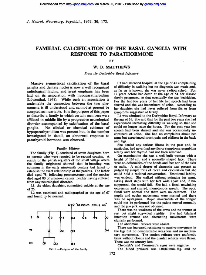
Family HistoryThe family (Fig. 1) consisted of seven daughters born
to parents who were reputed to be second cousins. Asearch of the parish registers of the small village wherethe family originated showed that in-breeding wascommon in the early nineteenth century but failed toestablish the exact relationship of the parents. The fatherdied aged 78, following prostatectomy, and the motherdied aged 80 of unknown causes, neither having sufferedfrom any neurological disorder.
L.1, the eldest daughter, committed suicide at the ageof 25.
1.2 was examined and radiographed at the age of 67and found to be normal.
DOO 'SECOND
I
m
2 3
62
COUSINS
4 'I5 6
3 04
3
Fio. 1.-Pedigree of the family.
1.3 had attended hospital at the age of 45 complainingof difficulty in walking but no diagnosis was made and,as far as is known, she was never radiographed. For12 years before her death at the age of 54 her diseaseslowly progressed so that eventually she was bedridden.For the last few years of her life her speech had beenslurred and she was incontinent of urine. According toher daughter she had never suffered from fits or fromsymptoms suggestive of tetany.
I.4 was admitted to the Derbyshire Royal Infirmary atthe age of 61. She said that for the past two years she hadexperienced increasing difficulty in walking so that shecould no longer leave the house. For the past year herspeech had been slurred and she was occasionally in-continent of urine. She had no complaints about herarms but experienced much pain and stiffness in the backand legs.She denied any serious illness in the past and, in
particular, had never had any fits or symptoms resemblingtetany and her thyroid had not been removed.On examination she was a well built woman with a
height of 165 cm. and a normally shaped face. Therewere no deformities of the hands and feet nor of the skinor nails. A mild degree of dementia was present asjudged by simple tests of recall and calculation but shecould hold a rational conversation. Emotional labilitywas evident. She walked without swinging her arms,taking short steps with her feet wide apart and, if un-supported, she would fall. She had a fixed, unwinkingexpression and slurred, monotonous speech. The opticfundi were normal and there were no cataracts. Thepupils and ocular movements were normal and therewas no nystagmus. Rapid movements of the tonguecould not be performed but the palate moved normallyand the jaw jerk was not obtained.There was no weakness of the arms and no tremor at
rest but slight cog-wheel rigidity. She had bilateralintention tremor and alternating movements wereclumsily performed.The abdominal reflexes were absent.There was increased resistance to passive movement in
the legs but no demonstrable weakness and no involun-tary movements. The tendon reflexes were uniformlybrisk without clonus and the plantar reflexes were flexor.There was no sensory loss.
Chvostek's and Trousseau's signs were negative.The blood pressure was 140/80 mm. Hg. and no
172
group.bmj.com on March 30, 2018 - Published by http://jnnp.bmj.com/Downloaded from group.bmj.com on March 30, 2018 - Published by http://jnnp.bmj.com/Downloaded from group.bmj.com on March 30, 2018 - Published by http://jnnp.bmj.com/Downloaded from

FAMILIAL CALCIFICATION OF BASAL GANGLIAabnormality was found in the chest or abdomen.
Radiographs of the skull showed massive symmetricalcalcification of the basal ganglia and dentate nuclei(Figs. 2 and 3).
In the course of the next year her condition showed aslow decline so that even walking with the aid of thefurniture became difficult and for most of the time shewas confined to a chair.
1.5 died in infancy.
1.6 was examined when aged 59. She had no complaintsbut it was possible to predict that she would show similarcalcification as she resembled an early case of paralysisagitans with a fixed expression and a stooping stance.She did not swing her arms when walking but there wasno tremor or ataxia. She had never suffered from fitsor tetany and had no cataracts. Radiographs of the skullshowed calcification perhaps even denser than in hersister (Figs. 4 and 5).
FIG. 4FIG. 2
:;F*-.i~~~~~~~~~~~~~~~~~~~~~~~~~~~~~~~~~~~~~~~~~~~~~~~~~~~~~~~~~~~~~~~~~~~~~~~~----
FIG. 5FIG. 3
FIGS. 2 and 3.-Radiographs of the skull of Case 1.4 showing FIGs. 4 and 5.-Radiographs of the skull of Case 1.6 showing similarcalcification. calcification.
173
group.bmj.com on March 30, 2018 - Published by http://jnnp.bmj.com/Downloaded from

W. B. MATTHEWS
1.7 was examined and radiographed at the age of 57and found to be normal.
II.1 was killed in the war.11.2, aged 33, 11.3, aged 28, and 11.4, aged 23, were
examined and radiographed with negative results.111.1, 2, and 3 were small children stated to be normal
but were not examined.
Case 1.4Case 1.4 was investigated in detail, but the other
surviving member of the family with neurological disease1.6, was not aware that she was affected and it was notthought permissible to draw attention to what appearedto be a progressive and incurable disease by admittingher to hospital.The patient's urine contained no glucose or albumin.
Dr. J. M. Walshe kindly examined the urine for amino-acids and reported a normal pattern of excretion.The cerebrospinal fluid pressure was 100 mm. and it
contained 30 mg. of protein per 100 ml. and 1 white cellper c.mm. The Wassermann reaction was negative inblood and C.S.F.The complement-fixation test for toxoplasmosis was
negative.The serum alkaline phophatase was 18 King-Armstrong
units per 100 ml., the serum flocculation test was + +.Total serum proteins were 6 5 g. per 100 ml. (albumin4.7 g. and globulin 1 8 g). There was no anaemia and thewhite cell count was normal.The electroencephalograph was normal and consisted
of low amplitude alpha with a little fast activity.Calcium and phosphorus metabolism were investi-
gated in greater detail. The serum calcium and phos-phorus were estimated in all available members of the
0 80.I
g70
t 60
o 50
0~3O0.t 30-> 20
z10l
PATIENTuJ
z .
0>
z
a:O
<0<Of
I
TABLE ISERUM CALCIUM, PHOSPHORUS, AND CHOLESTEROL
LEVELS IN THE AFFECTED FAMILY
Patient Serum Calcium Serum Phosphorus CholesterolPatient (mg./100 ml.) (mg./100 ml.) (mg./100 ml.)1.2 9 6 2-9 2611.4 100-11 3 26-43 3001.6 10 8 4-1 2381.7 110 3-6 22311.2 104 3 5 21511.3 110 29 20411.4 10 2 3-3 219
family and were all within normal limits. The serumcholesterol was also estimated as it was a little high inCase 1.4 and had been raised in the cases reported byBowman (1954). The results are shown in Table 1.
Radiographs showed no subcutaneous calcification,normal bone density and no deformities of the hands orfeet.The C.S.F. calcium level was 5 0 mg. per 100 ml. and
phosphorus 1 4 mg., which are normal figures.On a normal diet the urinary phosphorus excretion
ranged from 0 48 to 0 67 g. per day and the calciumfrom 0-13 to 0-16 g. A calcium balance was carried outon an intake of 1 0 g. and also on a low intake of 0-2 g.per day. On the former diet the output in the faeces was2 24 g. and in the urine 0 69 g. over a four-day period,giving a positive balance of 0 25 g. per day. On the lowintake the faeces contained 0 29 g. and the urine 0 45 g.in four days, approximately equivalent to the intake.These results were interpreted as normal.The effects of parathyroid hormone in producing an
increased excretion of phosphorus in the urine and inelevating the serum calcium level were assessed. The
CONTROL
TIME IN HOURS
FIG. 6-The effect of intravenous parathormone on phosphorus excretion in Case 1.4 and one control subject.
174
A^
group.bmj.com on March 30, 2018 - Published by http://jnnp.bmj.com/Downloaded from
FAMILIAL CALCIFICATION OF THE BASAL GANGLIA
WV
--o
,-#0
CONTROL
PAT IENT
TIME IN DAYS
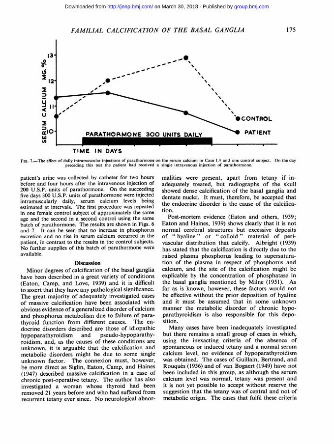
FIG. 7.-The effect of daily intramuscular injections of parathormone on the serum calcium in Case 1.4 and one control subject. On the daypreceding this test the patient had received a single intravenous injection of parathormone.
patient's urine was collected by catheter for two hoursbefore and four hours after the intravenous injection of200 U.S.P. units of parathormone. On the succeedingfive days 300 U.S.P. units of parathormone were injectedintramuscularly daily, serum calcium levels beingestimated at intervals. The first procedure was repeatedin one female control subject of approximately the same
age and the second in a second control using the same
batch of parathormone. The results are shown in Figs. 6and 7. It can be seen that no increase in phosphorusexcretion and no rise in serum calcium occurred in thepatient, in contrast to the results in the control subjects.No further supplies of this batch of parathormone were
available.
DiscussionMinor degrees of calcification of the basal ganglia
have been described in a great variety of conditions(Eaton, Camp, and Love, 1939) and it is difficultto assert that they have any pathological significance.The great majority of adequately investigated cases
of massive calcification have been associated withobvious evidence of a generalized disorder of calciumand phosphorus metabolism due to failure of para-thyroid function from different causes. The en-
docrine disorders described are those of idiopathichypoparathyroidism and pseudo-hypoparathy-roidism, and, as the causes of these conditions are
unknown, it is arguable that the calcification andmetabolic disorders might be due to some singleunknown factor. The connexion must, however,be more direct as Siglin, Eaton, Camp, and Haines(1947) described massive calcification in a case ofchronic post-operative tetany. The author has alsoinvestigated a woman whose thyroid had beenremoved 21 years before and who had suffered fromrecurrent tetany ever since. No neurological abnor-
malities were present, apart from tetany if in-adequately treated, but radiographs of the skullshowed dense calcification of the basal ganglia anddentate nuclei. It must, therefore, be accepted thatthe endocrine disorder is the cause of the calcifica-tion.
Post-mortem evidence (Eaton and others, 1939;Eaton and Haines, 1939) shows clearly that it is notnormal cerebral structures but excessive depositsof " hyaline " or " colloid " material of peri-vascular distribution that calcify. Albright (1939)has stated that the calcification is directly due to theraised plasma phosphorus leading to supersatura-tion of the plasma in respect of phosphorus andcalcium, and the site of the calcification might beexplicable by the concentration of phosphatase inthe basal ganglia mentioned by Milne (1951). Asfar as is known, however, these factors would notbe effective without the prior deposition of hyalineand it must be assumed that in some unknownmanner the metabolic disorder of chronic hypo-parathyroidism is also responsible for this depo-sition.Many cases have been inadequately investigated
but there remains a small group of cases in which,using the inexacting criteria of the absence ofspontaneous or induced tetany and a normal serum
calcium level, no evidence of hypoparathyroidismwas obtained. The cases of Guillain, Bertrand, andRouques (1936) and of van Bogaert (1949) have notbeen included in this group, as although the serum
calcium level was normal, tetany was present andit is not yet possible to accept without reserve thesuggestion that the tetany was of central and not ofmetabolic origin. The cases that fulfil these criteria
00
D6
'I
175
_0 .-
group.bmj.com on March 30, 2018 - Published by http://jnnp.bmj.com/Downloaded from
W. B. MATTHEWS
are those of Foley (1951), Fritzsche (1935), Bowman(1954), Eaton and others (1939, Cases 4 and 5),Sammet and Bucy (1951), and Rand, Olsen, andCourville (1943, Case 1). The present family alsofalls into this group, which then comprises 15 cases.Of this number, 11 were familial or hereditary,while in the much larger group of those with knownhypoparathyroidism the only familial cases ofcerebral calcification discovered are those of Vasiliu(1940) and Camp (1947).
This strong familial tendency increases the temp-tation to place these cases in a separate categoryof a specific hereditary disease but a study of thecase reports shows that they form very unpromisingmaterial for a clinical syndrome. The patientdescribed by Foley (1951) closely resembled those inthe present family and a minor degree of calcificationwas present in her two daughters who had no symp-toms. In this family, therefore, inheritance appearsto have been dominant. The three siblings describedby Fritzsche (1935) were born of consanguineousparents, suggestive of recessive inheritance, anddeveloped symptoms in early childhood. The twobrothers described by Bowman (1954) were infants,both of whom probably had a nephrotic syndromeand who died before the age of 3. Sammet andBucy's (1951) case was a man of 43 without symp-toms. Eaton and others' (1939) Case 4 developedrecurrent hemiplegia at the age of 31 and theirCase 5 was a defective woman of 40 without abnor-mal neurological signs. Rand and others' Case 1suffered from epilepsy and hypertension but had noabnormal neurological signs. The great diversityof the clinical state must make it unlikely that thereis any single cause of the calcification in this groupof patients without evidence of hypoparathyroidism.The investigation of the propositus in the present
family, however, suggests that defects of parathyroidfunction may exist without producing any dis-turbance of serum calcium or phosphorus levels, atleast at the time of the investigation. The conditionof pseudo-hypoparathyroidism was described byAlbright, Burnett, Parson, and Sulkowitch (1941)as an example of failure of the target organs torespond to normally produced hormone. As far asis known the target organs of the parathyroidhormone are the bones and the renal tubules.Albright and his colleagues established the lack ofresponse by the failure of injected hormone toproduce an increased urinary excretion of phos-phorus, using the Ellsworth-Howard test (Ellsworthand Howard, 1934). Certain physical stigmatafrequently accompany this metabolic disorder: around face, short stature, shortening of the meta-carpal and metatarsal bones, and subcutaneouscalcification. The condition is extremely rare and
MacGregor and Whitehead (1954) found only27 cases in the literature that satisfied their criteria,10 of which were known to have cerebral calcifica-tion. It must be emphasized that these patients havechanges in the blood chemistry typical of hypo-parathyroidism and suffer from chronic tetany. Theconcept of failure of target organ response has notpassed unchallenged and certainly the Ellsworth-Howard test of response to parathyroid hormone isunreliable as it may be negative in normal subjects(MacGregor and Whitehead, 1954; Milne, 1951;Dent, 1953). This has been attributed to lack of thisparticular activity in the commercial gland extracts.The batch of parathormone used in this investigationwas, however, shown to be active in producing anincrease in phosphorus excretion in a normal control.The extract was also effective in raising the serumcalcium level in a second control but produced norise in the patient. This latter effect is probablydue to the action of the hormone on bone (Milne,1951), and failure of this response is consideredmore significant than the failure to increase phos-phorus excretion (Dent, 1953; MacGregor andWhitehead, 1954).
This patient therefore showed two features ofpseudo-hypoparathyroidism, namely, cerebral calci-fication and a failure of response to parathyroidhormone, but without changes in blood chemistryor any of the physical stigmata commonly associatedwith the condition. The latter are known to occurindependently of the chronic tetany and the con-dition ofpseudo-pseudohypoparathyroidism has beendescribed by Albright, Forbes, and Henneman(1952) and by Miles and Elrick (1955). This un-wieldy name implies that the physical habitus andbone deformities, and in one case subcutaneouscalcification, and in the other, cataract, may occurwithout changes in the blood chemistry or tetany.In the case of Albright et al. phosphorus excretionwas not increased after parathormone but theextract was also inactive in a normal subject. Inneither case was cerebral calcification present. Thatthis condition bears some relationship to pseudo-hypoparathyroidism is shown by the family reportedby Selye (1949) where mother and daughter showedthe characteristic deformities but hypocalcaemiawas present only in the daughter. If the tests can beaccepted as showing lack of response to parathyroidhormone the patient reported here seems to repre-sent a different fraction of the pseudo-hypopara-thyroid syndrome. It is, of course, exceedinglydifficult to reconcile a lack of response to injectedhormone with an apparently normal response tothe patient's own hormone as reflected in theabsence of changes in blood chemistry. The answermay be a matter of the degree of abnormality. The
176
group.bmj.com on March 30, 2018 - Published by http://jnnp.bmj.com/Downloaded from

FAMILIAL CALCIFICATION OF THE BASAL GANGLIA
family history is strongly suggestive of a recessivelyinherited metabolic disorder and the late age ofonset of symptoms, presumably due to this disorder,may indicate that only a slight disturbance offunction is present. Some response to parathyroidhormone must exist in order to maintain the serumcalcium and phosphorus at normal levels but thismight only be possible by a maximum response thatcould not be increased by injected hormone. Suchan uneasy equilibrium would be unlikely to beperfectly maintained throughout life. It must notbe forgotten that the lack of parathyroid functionor of a normal response to the hormone appears tofavour the excessive deposition of hyaline materialin the basal ganglia, a change not obviously relatedto the known functions of the hormone nornecessarily related to measurable disturbances ofblood chemistry.
SummaryA family is described in which three members
developed a progressive neurological disorder inmiddle life, accompanied by massive symmetricalcalcification of the basal ganglia and dentate nucleiin the two members who were examined.No abnornality of blood chemistry was found in
any member of the family but in the patient in-vestigated in detail no response to parathyroidhormone could be obtained.
The possible relationships of this condition withthose of pseudo-hypoparathyroidism and pseudo-pseudohypoparathyroidism are discussed.
The investigation of this family was greatly helped bythe laboratory work of Mr. D. Watson, M.Sc., andMiss A. Matthews, B.Sc.
REFERENCESAlbright, F. (1939). In discussion of Eaton and Haines (1939).
Burnett, C. H., Smith, P. H., and Parson, W. (1942).Endocrinology, 30, 922.Forbes, A. P., and Henneman, P. H. (1952). Trans. Ass. Amer.Phys., 65, 337.
Bogaert, L. van (1949). Mschr. Psychiat. Neurol., 118, 30.Bowman, M. S. (1954). Amer. J. Path., 30, 87.Camp, J. D. (1947). Radiology, 49, 568.Dent, C. E. (1953). Proc. roy. Soc. Med., 46, 291.Eaton, L. M., Camp, J. D., and Love, J. G. (1939). Arch. Neurol.
Psychiat. (Chicago), 41, 921.and Haines, S. F. (1939). J. Amer. med. Ass., 113, 749.
Ellsworth, R., and Howard, J. E. (1934). Bull. Johns Hopk. Hosp.,55, 296.
Foley, J.(1951). Journal of Neurology, Neurosurgery and Psychiatry,14, 253.
Fritzsche, R. (1935). Schweiz. Arch. Neurol., 35, 1.Guillain, G., Bertrand, I., and Rouques, L. (1936). Rev. neurol.
(Paris), 65, 737.Lowenthal, A. (1948). Acta neurol. psychiat. belge, 48, 613.MacGregor, M. E., and Whitehead, T. P. (1954). Arch. Dis. Childh.,
29, 398.Miles, J., and Elrick, H. (1955). J. clin. Endocr., 15, 576.Milne, M. D. (1951). Clin. Sci., 10, 471.Rand, C. W., Olsen, C. W., and Courville, C. B. (1943). Bull. Los.
Angeles neurol. Soc., 8, 118.Sammet, J. F., and Bucy, P. C. (1951). Amer. J. Roentgenol.,
66, 880.Selye, H. (1949). Textbook of Endocrinology, 2nd ed. Acta Endo-
crinologica, Montreal.Siglin, I. S., Eaton, L. M., Camp, J. D., and Haines, F. (1947).
J. clin. Endocr., 7, 433.Vasiliu, 0. (1940). Wien. med. Wschr., 90, 153.
177
group.bmj.com on March 30, 2018 - Published by http://jnnp.bmj.com/Downloaded from
PARATHORMONERESPONSE TOTHE BASAL GANGLIA WITH FAMILIAL CALCIFICATION OF
W. B. Matthews
doi: 10.1136/jnnp.20.3.1721957 20: 172-177 J Neurol Neurosurg Psychiatry
http://jnnp.bmj.com/content/20/3/172.citationUpdated information and services can be found at:
These include:
serviceEmail alerting
online article. article. Sign up in the box at the top right corner of the Receive free email alerts when new articles cite this
Errata
/content/20/4/317.1.full.pdf or: next pagePlease see
An erratum has been published regarding this article.
Notes
http://group.bmj.com/group/rights-licensing/permissionsTo request permissions go to:
http://journals.bmj.com/cgi/reprintformTo order reprints go to:
http://group.bmj.com/subscribe/To subscribe to BMJ go to:
group.bmj.com on March 30, 2018 - Published by http://jnnp.bmj.com/Downloaded from

BOOK REVIEWS
an excellent account of existing knowledge, and can berecommended with every confidence to all concernedwith the subject.
An Introduction to Electromyography. By FritzBuchthal. (Pp. 43; 8 figures.) Copenhagen: GyldendalsPresseafdeling. 1957.
This simple introduction to electromyography is auseful guide to those who wish to take up the subject.
Proceedings of the Second International Congress ofNeuropathology, London, 1955
At the request of the Organizing Committee of theSecond International Congress of Neuropathology heldin London, 1955, all reports, communications, anddiscussions presented to this congress have been publishedby the Excerpta Medica Foundation.These Proceedings (edited by Dr. W. H. McMenemey)
consist of three parts, Parts I and II containing 650 pagesof text, and Part III containing 156 plates.The Proceedings can be ordered from EXCERPTA
MEDICA FOUNDATION, 111 Kalverstraat, Amsterdam,The Netherlands, at the price of £4 10s., fl. 50.-, or$12 50.
International Association of Applied PsychologyThe XIIIth Congress will be held in Rome from
April 9-14, 1958.Those who wish to attend should apply to:
Segreteria del XIII Congresso,Internazionale di Psicologia Applicata,
Instituto Nazionale di Psicologia del C.N.R.,Piazzale delle Scienze 7,
RomA (Italia).
Sherrington Memorial LectureThe first Sherrington Memorial Lecture, organized by
The Royal Society of Medicine, will be delivered byLord Adrian, O.M., M.D., F.R.S., in the Society's Houseat 8 p.m., on Wednesday, November 27, 1957.The subject of the lecture is " The Analysis of the
Nervous System ".Admission will be by ticket only and applications for
tickets may be made to the Assistant Secretary of theSociety.
Correction.-The title of the paper by W. B. Mathewsin the last issue of the Journal (20, 172) should read"' Familial Calcification of the Basal Ganglia withAbsence of Response to Parathormone."
BOOKS RECEIVED(Review in a later issue is not precluded by notice here of books
recently received.)
Practical Clinical Psychiatry, 8th ed. By Jack R.Ewalt, Edward A. Strecker, and Franklin G. Ebaugh.(Pp. xiv + 457. 60s.) London: McGraw-Hill. 1957.
The Family in Psychotherapy. By C. F. Midelfort.(Pp. ix + 203. 49s.) London: McGraw-Hill. 1957.
Peripheral Nerve Regeneration: A Follow-up Study of3,656 World War II Injuries. Edited by Barnes Woodhalland Gilbert W. Beebe. (Pp. xvii + 671; monograph,illustrated.) Washington, D.C.: Veterans Administration.1956.
Der Suicid. By F. Dubitscher. (Pp. viii + 224;7 figures. DM 16.50.) Stuttgart: Georg Thieme. 1957.
Psychosomatic Medicine: A Clinical Study of Psycho-physiologic Reactions, 3rd ed. By E. Weiss and 0. S.English. (Pp. xix + 557; illustrated. 73s. 7d.) Londonand Philadelphia: W. B. Saunders. 1957.
Die Vaskulfiren Erkrankungen im Gebiet der Arteriavertebralis und Arteria basialis. By H. Krayenbiuhl andG. Yasargil. (Pp. viii + 170; 125 figures on 205 plates.DM 69.30.) Stuttgart: Georg Thieme. 1957.
Cerebral Lipidoses: A Symposium. Chairman: L. vanBogaert; Editor: J. N. Cumings; Assoc. Ed.: Lowenthal.(Pp. x + 212; illustrated. 42s.) Oxford: Blackwell. 1957.
New Research Techniques of Neuroanatomy. A Sym-posium sponsored by the National Multiple SclerosisSociety. Edited by William F. Winde; Foreword byFrederick L. Stone. (Pp. ix + 98; 25 figures. 36s.)Oxford: Blackwell; Springfield (Illinois): Charles C.Thomas. 1957.
Collection Testut. Precis de Neurologie, 6th editionentirely revised. By L. Rimbaud in collaboration withP. Passouant and Cl. Gros. (Pp. 1186; 331 figures inblack-and-white and colour. Fr.fr. 8,700.-.) Paris:G. Doin. 1957.
The Clinical Examination of Patients with OrganicCerebral Disease. By R. Klein and W. Mayer-Gross.(Pp. xiii + 96. 15s.) London: Cassell. 1957.
The Human Brain: From Primitive to Modem. ByA. M. Lassek. (Pp. viii + 242. 36s.) Oxford: Blackwell;Springfield (Illinois): Charles C. Thomas. 1957.
Pneumoencephalography. By E. Graeme Robertson.(Pp. xxi + 482; 34 plates, 209 figures. £5 10s.) Oxford:Blackwell; Springfield (Illinois): Charles C. Thomas.1957.
Mental Deficiency. By L. T. Hilhiard and Brian H.Kirman. (Pp. xvi + 517; 90 figures. 60s.) London:J. & A. Churchill. 1957.
317