fall/winter 2013 news from the wu center … 2013 news from the wu center for molecular cardiology...
TRANSCRIPT
FALL/WINTER 2013 NEWS FROM THE WU CENTER FOR MOLECULAR CARDIOLOGY
HEART HORIZONSThe Clyde and Helen Wu Center For Molecular Cardiology
C O L U M B I A U N I V E R S I T Y M E D I C A L C E N T E R
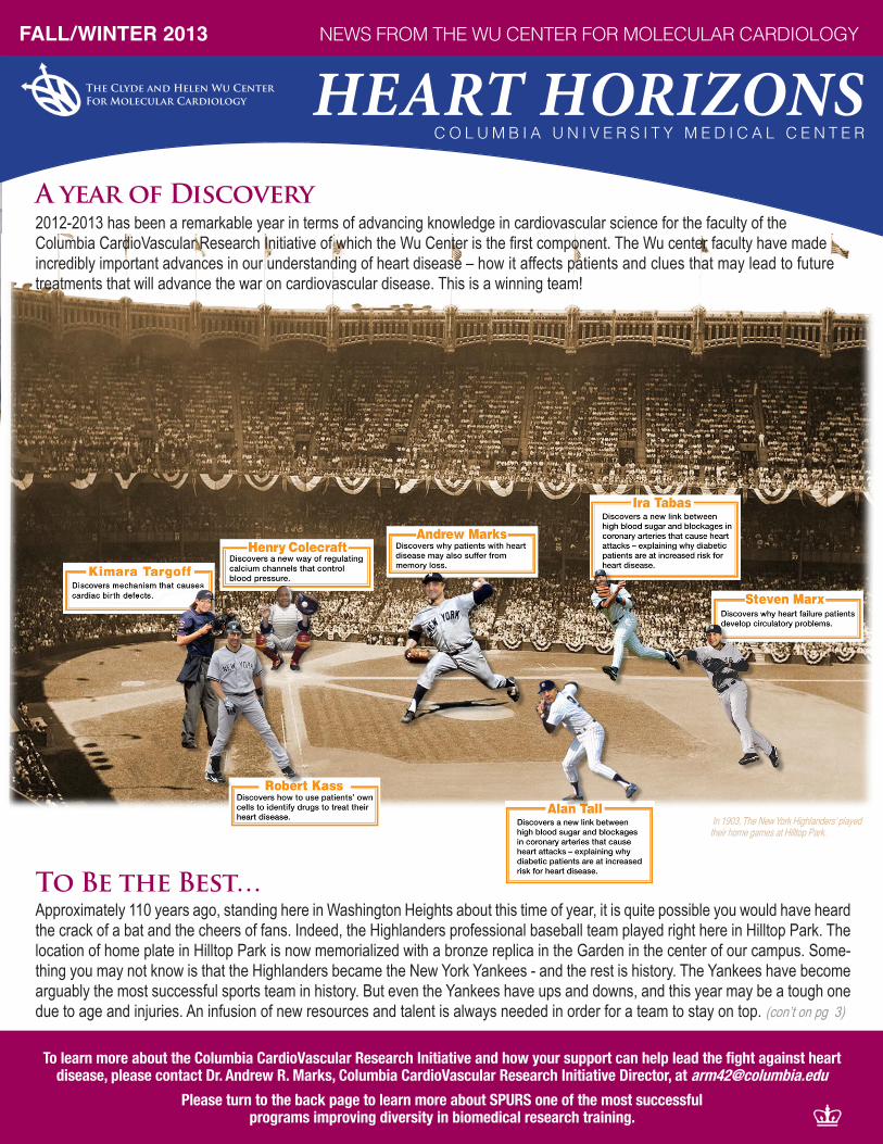
A year of Discovery
To Be the Best…
2012-2013 has been a remarkable year in terms of advancing knowledge in cardiovascular science for the faculty of the Columbia CardioVascular Research Initiative of which the Wu Center is the first component. The Wu center faculty have made incredibly important advances in our understanding of heart disease – how it affects patients and clues that may lead to future treatments that will advance the war on cardiovascular disease. This is a winning team!
Approximately 110 years ago, standing here in Washington Heights about this time of year, it is quite possible you would have heard the crack of a bat and the cheers of fans. Indeed, the Highlanders professional baseball team played right here in Hilltop Park. The location of home plate in Hilltop Park is now memorialized with a bronze replica in the Garden in the center of our campus. Some-thing you may not know is that the Highlanders became the New York Yankees - and the rest is history. The Yankees have become arguably the most successful sports team in history. But even the Yankees have ups and downs, and this year may be a tough one due to age and injuries. An infusion of new resources and talent is always needed in order for a team to stay on top. (con’t on pg 3)
To learn more about the Columbia CardioVascular Research Initiative and how your support can help lead the fight against heart disease, please contact Dr. Andrew R. Marks, Columbia CardioVascular Research Initiative Director, at [email protected]
Please turn to the back page to learn more about SPURS one of the most successful programs improving diversity in biomedical research training.
In 1903, The New York Highlanders’ played their home games at Hilltop Park.
2 HEART HORIZONS | FALL/WINTER 2013
The Future of Cardiovascular Research The Columbia Cardiovascular Research Initiative (CVRI) is the conceptualization of a comprehensive long-term approach to cardiovascular research de-signed by Dr. Lee Goldman Executive Vice President for Health and Biomedical Sciences, and Dean of the Faculties of Health Sciences and Medicine. Dr. Goldman established the CVRI as part of the CUMC strategic plan for the future. The focus is on continuing to build in areas of strength and identifying areas ripe for development. How will the CVRI affect the Wu Center? If we are
successful in raising philanthropic funds, the CVRI can be transformative! Columbia has great strengths in many areas of molecular cardiology, including heart failure, cardiac arrhythmias, and atherosclerosis. Many of these important advances have been made by the Wu Center’s faculty and are outlined in this issue of Heart Horizons. But we need to expand our reach in other areas, including genetics and hypertension. In order to maintain excellence in our areas of strength and to fill the gaps in areas of need, we must recruit new faculty and build programs. These are tough times for biomedical researchers in all fields. The National Institutes of Health (NIH), by far the most important source of funding for molecular cardiology, is in the doldrums. Success rates for NIH grants are in single digits and researchers, as well as those thinking of going into the field, are losing hope. Much great science has been put on hold due to lack of funding. This translates into lost opportunities for patients with heart disease; discovery has been slowed and new therapies have been delayed. It has never been more important to find sources of funding other than the NIH. The CVRI is our big hope for the future. Will Columbia be able to reach its full potential and continue to produce paradigm-changing discoveries that bring new hope to patients with heart disease? We don’t know the answer to that question yet, but we are ever hopeful. In this issue of Heart Horizons, you will find brief articles describing the incredible advances made by seven of the Wu Center faculty. These run the gamut from fighting ath-erosclerosis, still the leading cause of heart attacks, to using zebrafish to understand how and why some babies are born with developmental defects in their hearts. Please feel free to contact Dr. Andrew Marks ([email protected]) with any questions you have about our research and how it may affect you and your family. We look forward to partnering with you to fight heart disease and to improve the lives of our patients!
Table of Contents
The Future of Cardiovascular Research
To Be The Best
Laboratory of Henry Colecraft Lessons Learned from Natural Genetically- Encoded Calcium Channel Blockers
Laboratory of Robert S. Kass Patient Specific Induced Pluripotent Stems Cells Provide A Novel Platform For Drug Screening In Heritable Cardiac Arrhythmias
Laboratory of Andrew R. Marks Fixing the Leak that Causes Diseases of Heart, Muscle & Brain for Drug Screening In Heritable Cardiac Arrhythmias
Laboratory of Steven Marx Patients with Diabetes, Hypertension and Heart Failure Have Increased Vascular ToneFor Drug Screening In Heritable Cardiac Arrhythmias
Laboratory of Ira Tabas Heart Disease and Type 2 Diabetes
Laboratory of Allan Tall Therapeutic Target for Cardiovascular Disease
Laboratory of Kimara Targoff Understanding Cardiac Morphogenesis
Annual Clyde and Helen Wu Distinguished Lecture Series Professors Christine and Jonathan Seidman Deliver the Annual Clyde and Helen Wu Distinguished Lecture
273
84
95
10
116

To Be The Best
Robert Loeb. Cournand and Richards used cardiac catheterization to study the hemodynamics of traumatic shock during World War II, to diagnose congenital heart disease, and to characterize the physiology of heart failure and the action of cardiac drugs. In 1956 Cournand, Richards, and Werner O.T. Forssman were awarded the Nobel Prize in Physiology or Medicine for the development of cardiac catheterization To be the best and to continue this tradition of excellence on the Heights, Columbia must substantially expand its cardiovascular research program. With the opening of the new Manhattanville campus, in several years there will be an unprecedented availability of prime laboratory space on the Washington Heights campus. This will provide the opportunity to build on our strengths in research in the areas of heart failure, arrhythmia, atherosclerosis, and interventional cardiology and to establish vibrant new programs in the areas of hypertension, cardiovascular genetics, imaging, and vascular biology. To make this new vision of cardiovascular research a reality, we need to recruit 5-10 new faculty over the next five years. Ideally, some of these new faculty will be established, internationally-renowned scientists with well-developed research programs; others will be just starting their careers but will have the promise to become the star scientists of the future.
A recruitment program like this will require a substantial investment in order to attract and retain the best of the best. Why is this effort so important? Heart disease is unquestionably the lead-ing cause of mortality in the United States - far greater than the combined total of all other causes of death. Yet we can make a difference in the battle against heart disease. For example, you may have a relative or a friend, or possibly even yourself, with a drug-eluting stent in place to keep the blood flowing to your heart to prevent heart attacks. The drug on this stent is most likely the one identified by Columbia scientists. Putting this drug (known as rapamycin)
on the stents reduced the incidence of stent failure from 30-50% to less than 10% and benefitted thousands of heart disease patients. This is just one example of many paradigm-changing advances made by Columbia scientists that have already been translated into improved care for heart patients. But much more is needed. Hypertension remains a silent killer. We need scientific breakthroughs to better understand the causes of high blood pressure in order to identify new, more effective, less toxic forms of treatment that will prevent the strokes, heart failure, kidney problems, and vascular disease caused by uncontrolled hypertension. Columbia is a world leader in heart failure and arrhythmias research, but effective therapies for these disorders have been elusive. We have to really focus in and discover the basic causes of heart failure and arrhythmias in order to develop more targeted and beneficial therapies. This will change people’s lives! Columbia is one of the few places in the world where the kinds of advances that will defeat heart disease can and do occur. We need help to continue to be the best and to make sure the promise of better lives for patients with heart disease is realized. Please contact Dr. Andrew Marks ([email protected]) if you have any questions about the content of this magazine and if you want information on how you can support cardiovascular research at Columbia University.
3 HEART HORIZONS | FALL/WINTER 2013
> continued from page 1
4 HEART HORIZONS | FALL/WINTER 2013
Laboratory of Henry Colecraft Lessons Learned from Natural Genetically-Encoded
Calcium Channel Blockers
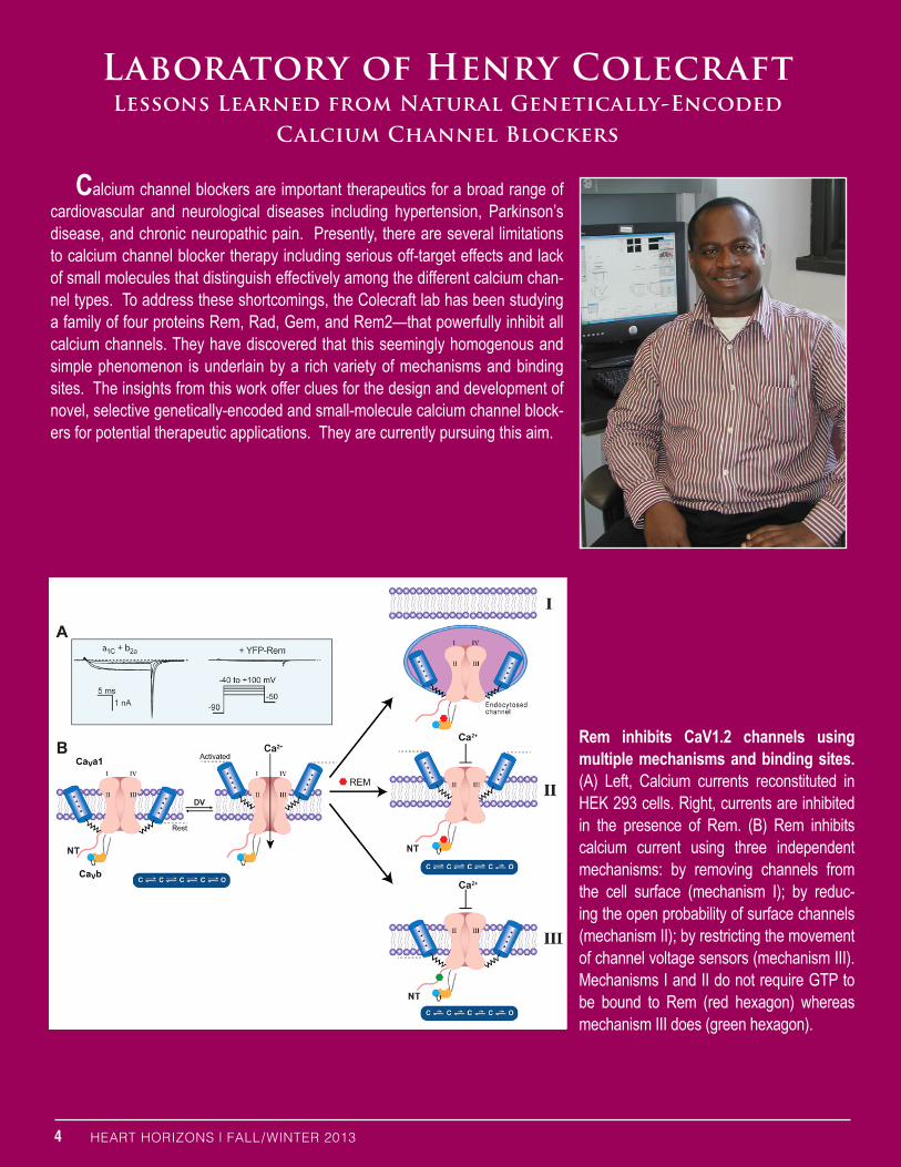
Calcium channel blockers are important therapeutics for a broad range of cardiovascular and neurological diseases including hypertension, Parkinson’s disease, and chronic neuropathic pain. Presently, there are several limitations to calcium channel blocker therapy including serious off-target effects and lack of small molecules that distinguish effectively among the different calcium chan-nel types. To address these shortcomings, the Colecraft lab has been studying a family of four proteins Rem, Rad, Gem, and Rem2—that powerfully inhibit all calcium channels. They have discovered that this seemingly homogenous and simple phenomenon is underlain by a rich variety of mechanisms and binding sites. The insights from this work offer clues for the design and development of novel, selective genetically-encoded and small-molecule calcium channel block-ers for potential therapeutic applications. They are currently pursuing this aim.
Rem inhibits CaV1.2 channels using multiple mechanisms and binding sites. (A) Left, Calcium currents reconstituted in HEK 293 cells. Right, currents are inhibited in the presence of Rem. (B) Rem inhibits calcium current using three independent mechanisms: by removing channels from the cell surface (mechanism I); by reduc-ing the open probability of surface channels (mechanism II); by restricting the movement of channel voltage sensors (mechanism III). Mechanisms I and II do not require GTP to be bound to Rem (red hexagon) whereas mechanism III does (green hexagon).

Laboratory of Robert S. Kass Patient Specific Induced Pluripotent Stems Cells Provide A Novel Platform For Drug Screening In Heritable Cardiac Arrhythmias
The Kass laboratory has had a long standing interest in investigating the mechanistic basis of cardiac rhythm disturbances (arrhythmia) that can be caused by dysfunction of proteins called ion channels (channelopathies), which normally regulate the heart’s electrical activity. Mutations in genes coding for ion channels, or ion channel associated proteins, place mutation carriers at risk of sudden cardiac death in these disorders.The Long QT Syndrome (LQTS) is now the most thoroughly studied cardiac channelopathy. In individuals affected by LQTS, the electrocardiogram, which monitors the heart’s electrical activity, is altered such that a select section of the recorded signal, called the QT interval, is prolonged compared to healthy individuals. This condition is known to increase the risk of developing arrhythmia in a gene mutation-dependent manner, and risk of arrhythmia and sudden death as well as therapeutic approaches to manage the disorder are mutation dependent. Studies of individual mutated ion channels, using expres-sion systems in non-cardiac cells, have provided great insight into mechanisms of and specific therapeutic strategies to manage LQTS, but - particularly in cases in which patient genetic backgrounds are complex- these approaches are not always effective. Moving this field forward has required new approaches that will allow studying ion dysfunctional channel activities in a cellular background that dupli-cates that of the cardiac cells of patients being treated. A novel approach that is very promising is to derive cardiac lineage from a special type of stem cells, induced pluripotent stem cells (iPSCs), that are generated after molecular manipulation of adult somatic cells, such as skin cells from the very patients presenting with the disease. This technology presents many advantages: i) the surgical procedure required to collect somatic cells is simple and minimally invasive (skin biopsy); ii) human cardiac cells derived from skin cells can be produced in large amount; iii) the genetic background of these cardiac cells is specific to the individual whose skin was biopsied, therefore allowing their use for personalized medicine. The Kass laboratory has begun using this approach as a novel in vitro platform to investigate mechanisms underlying LQTS in a pediatric patient and therapeutic strategies to treat the problem. This work is truly translational in that in vitro experiments carried out in the Kass lab have been used not only to determine how the arrhythmias are initiated but also to understand therapeutic regimens used to prevent them. This approach is novel and has been car-ried out as a “proof of concept” approach to determine whether information gained from the iPSC-derived cardiac cells can be used to understand then ultimately change clinical management of an inherited disorder in a patient-specific manner. In multi-investigator collaboration, the Kass lab generated cardiac cells derived from skin cells from family members of a LQTS patient whose heart was found to express two different types of abnormal ion channels: i) a mutant sodium channel that can underlie the disease phenotype and ii) a mutant potassium channel that may complicate the disease phenotype and also alter the patient’s response to potential therapeutic agents. The in vitro work revealed that the LQT phenotype observed in the patient was likely to be caused by the mutant sodium channel without any additional contribution of the mutant potassium channel to the disease. But, in addition, the in vitro studies revealed that the arrhythmic activity caused by the inherited mutation was extremely sensi-tive to heart rate such that if it were possible to increase the patient’s heart rate while administering a drug identified in the in vitro studies, control of arrhythmic activity was predicted to be greatly improved. These predictions were shown to correlate very closely with an altered clinical regimen in which arrhythmic activity in the patient was markedly reduced using a combination of increased heart pacing via a previously implanted pacemaker and administration of a single drug, mexiletine, which had been profiled in the iPSC-cardiac cells. This study in the Kass lab constitutes a proof of concept that human cardiac cells produced by iPSCs technology can be used for direct investigation of the cellular physiology and pharmacology of mutations that cause LQTS within a background that reproduces the complex genetic background of a patient and, as such, is the first time patient specific iPS cells have been used in this manner. The lab is continuing to build upon this technology to reduce turnaround times both for the derivation of patient specific myocytes from other LQTS patients and to reduce turnaround time for drug screening such that this platform may be used to improve patient care in cases in which complex patient genetics contributes to resistance to other approaches.
5
6 HEART HORIZONS | FALL/WINTER 2013
Laboratory of Andrew R. Marks Fixing the Leak that Causes Diseases of Heart, Muscle and Brain
Dr. Marks’ work on the mechanisms of action of drugs that inhibit vascular smooth muscle proliferation and migration has been translated into novel ther-apeutics including drug-eluting stents for treatment of coronary artery disease that have substantially reduced the incidence of in-stent restenosis, as well as effective therapy to reduce accelerated arteriopathy following cardiac transplan-tation. Dr. Marks has defined how macromolecular signaling complexes regu-late ion channel function in muscle and non-muscle systems. His work has con-tributed new understandings of fundamental mechanisms that regulate muscle contraction. He discovered that “leaky” intracellular calcium release channels (ryanodine receptors, RyR) contribute to heart failure, fatal cardiac arrhythmias, and impaired exercise capacity particularly in muscular dystrophy. Dr. Marks discovered a new class of small molecules (rycals), developed in his laboratory, that effectively treat cardiac arrhythmias, heart failure, and muscular dystrophy in pre-clinical studies. His new approach, based on fixing the “leak” in the ryanodine receptor/calcium release channels, is in Phase II clinical trials for the treatment of heart failure and cardiac arrhythmias, and is being developed for the treatment of muscular dys-trophy. Dr. Marks found that RyR channels develop a stress-induced diastolic SR calcium leak that contributes to heart failure progression and sudden cardiac death. To address this problem, he developed a novel class of small molecule compounds called “rycals” that prevent the calcium leak by stabilizing the closed state of the channel, without impairing its normal function. He has shown that rycals reduce heart failure progression, prevent arrhythmias, and improve exercise capacity in animal models. A rycal is now in Phase II clinical trials in patients for prevention of heart failure progression and sudden cardiac death. Muscle fatigue is one of the most fundamental physiological processes. Fatigue of skeletal muscle determines the outer limits of physical activity and explains, for example, why an elite athlete who can run 100 meters in under 10 seconds cannot run 1,000 meters in 100 seconds. Severe muscle fatigue is a life threatening problem for millions of patients suffering from chronic diseases, including heart failure, cancer, kidney failure, AIDS, as well as rarer genetic abnormalities including muscular dystrophies. Moreover, millions of individuals over the age of 70 suffer from unexplained muscle weakness that limits their quality of life. Muscle fatigue has been described and studied in enormous detail for centuries but mechanistic understand-ings remain obscure. Indeed, the prevailing theory of the past 100 years, that build up of lactic acid causes muscle fatigue, has only recently been refuted with convincing data. Attention has now turned to potential abnormalities in calcium handling as the most likely cause of skeletal muscle fatigue. In particular, several groups have observed defective intracellular calcium release from the sarcoplasmic reticulum in fatigued skeletal muscle, although no mechanisms have been established. Dr. Marks has tackled the new problem of the molecular basis of skeletal muscle fatigue, particularly as it relates to loss of muscle function with aging. These new understandings may lead to a novel therapeutic approach that improves muscle function in older individuals and in those with chronic diseases. There is no effective therapy for muscle fatigue, and the same type of Rycal drugs that Dr. Marks is testing for heart failure also improve exercise capacity in aged mice. Thus, one outcome could be a completely novel and effective treatment that improves muscle performance. This would be revolutionary in the field of muscle fatigue. Dr. Marks has also discovered that intracellular Ca2+ leak represents a key pathology that impairs memory storage and alters signaling in the brain required for normal behavior (e.g. threat avoidance) and contributes to post-traumatic stress dis-order (PTSD), Alzheimer’s Disease (AD), and possible Parkinson’s Disease (PD). A novel drug called a Rycal that we have developed that prevents intracellular Ca2+ leak prevents symptoms associated with PTSD and AD.

Laboratory of Steven Marx
Patients with diabetes, hypertension, and heart failure have increased vas-cular tone, which adversely impacts their cardiovascular outcomes. Increased vascular tone in peripheral vessels increases the workload of the heart, impairing cardiac output and ventricular remodeling, and accelerating the progression to decompensated heart failure. Decreasing vascular tone, thereby unloading the heart and improving coronary perfusion, is an important therapeutic approach in patients with diseases associated with increased vasoconstriction. The diameter of resistance vessels, which fundamentally regulates blood flow within an organ as well as the systemic vascular resistance and arterial pressure, is primarily determined by the contraction of vascular smooth muscle cells. Depolarization of the smooth muscle cell membrane potential and elevated cytosolic calcium increase contractility causing vasoconstriction, whereas hy-perpolarization and decreased calcium reduce contractility causing vasodilation. Strong hyperpolarizing currents are required both to prevent excessive smooth muscle cell depolarization and vascular contractility in response to intraluminal pressure and vasoconstrictors as well as to enable smooth muscle cell hyperpo-larization and vascular relaxation in response to vasodilators. We found that six weeks after sham or permanent ligation of the left anterior descending artery, myogenic constriction, in the absence of systemic factors such as short-acting circulating neurohormones and neural inputs, is markedly increased in resistance vessels isolated from mice with heart failure. We demonstrate that reduced expression and activity of the large conductance calcium-activated potassium channel (BK channels) sensitize the vascular smooth muscle cells to depolariza-tion, causing increased cytosolic calcium and myogenic tone in mice with heart failure. We are now exploring whether phar-macological activation of BK channels, which we have identified, can specifically blunt the abnormal vasoconstriction and consequently slow or prevent the frequently inevitable clinical decompensation of patients with heart failure.
7 HEART HORIZONS | FALL/WINTER 2013
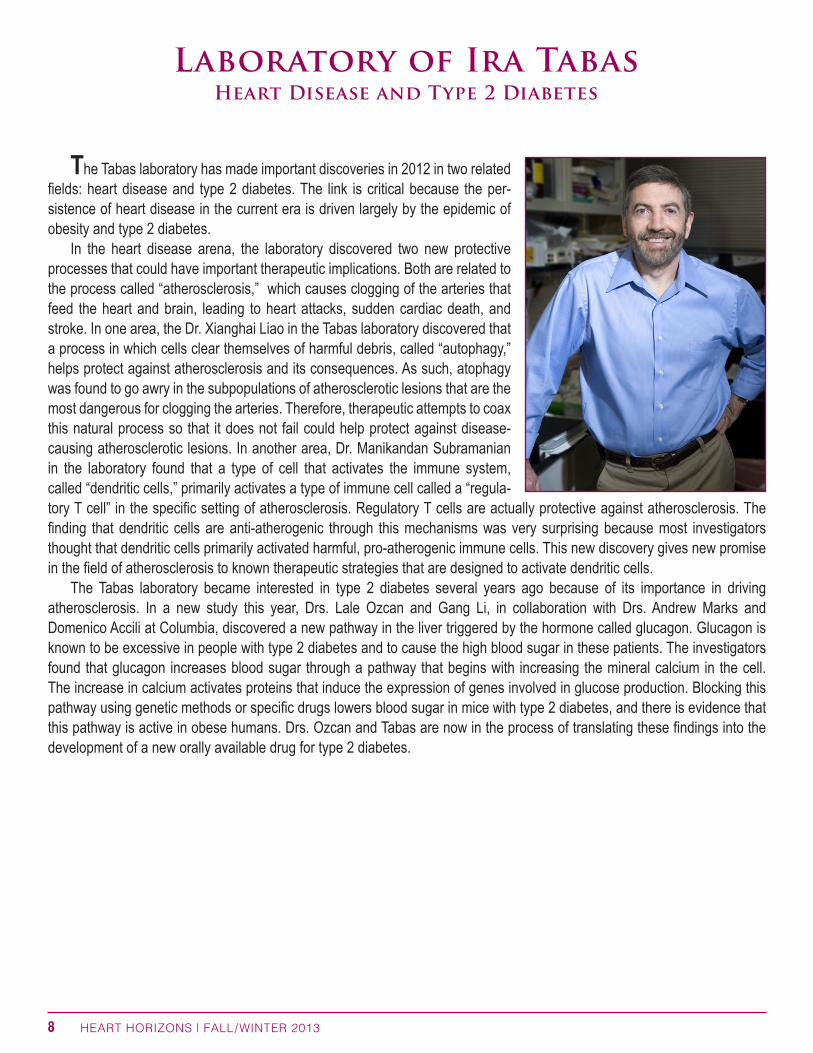
Laboratory of Ira Tabas Heart Disease and Type 2 Diabetes
The Tabas laboratory has made important discoveries in 2012 in two related fields: heart disease and type 2 diabetes. The link is critical because the per-sistence of heart disease in the current era is driven largely by the epidemic of obesity and type 2 diabetes. In the heart disease arena, the laboratory discovered two new protective processes that could have important therapeutic implications. Both are related to the process called “atherosclerosis,” which causes clogging of the arteries that feed the heart and brain, leading to heart attacks, sudden cardiac death, and stroke. In one area, the Dr. Xianghai Liao in the Tabas laboratory discovered that a process in which cells clear themselves of harmful debris, called “autophagy,” helps protect against atherosclerosis and its consequences. As such, atophagy was found to go awry in the subpopulations of atherosclerotic lesions that are the most dangerous for clogging the arteries. Therefore, therapeutic attempts to coax this natural process so that it does not fail could help protect against disease-causing atherosclerotic lesions. In another area, Dr. Manikandan Subramanian in the laboratory found that a type of cell that activates the immune system, called “dendritic cells,” primarily activates a type of immune cell called a “regula-tory T cell” in the specific setting of atherosclerosis. Regulatory T cells are actually protective against atherosclerosis. The finding that dendritic cells are anti-atherogenic through this mechanisms was very surprising because most investigators thought that dendritic cells primarily activated harmful, pro-atherogenic immune cells. This new discovery gives new promise in the field of atherosclerosis to known therapeutic strategies that are designed to activate dendritic cells. The Tabas laboratory became interested in type 2 diabetes several years ago because of its importance in driving atherosclerosis. In a new study this year, Drs. Lale Ozcan and Gang Li, in collaboration with Drs. Andrew Marks and Domenico Accili at Columbia, discovered a new pathway in the liver triggered by the hormone called glucagon. Glucagon is known to be excessive in people with type 2 diabetes and to cause the high blood sugar in these patients. The investigators found that glucagon increases blood sugar through a pathway that begins with increasing the mineral calcium in the cell. The increase in calcium activates proteins that induce the expression of genes involved in glucose production. Blocking this pathway using genetic methods or specific drugs lowers blood sugar in mice with type 2 diabetes, and there is evidence that this pathway is active in obese humans. Drs. Ozcan and Tabas are now in the process of translating these findings into the development of a new orally available drug for type 2 diabetes.
8 HEART HORIZONS | FALL/WINTER 2013
9 HEART HORIZONS | FALL/WINTER 2013
Laboratory of Alan Tall Therapeutic Target for Cardiovascular Disease
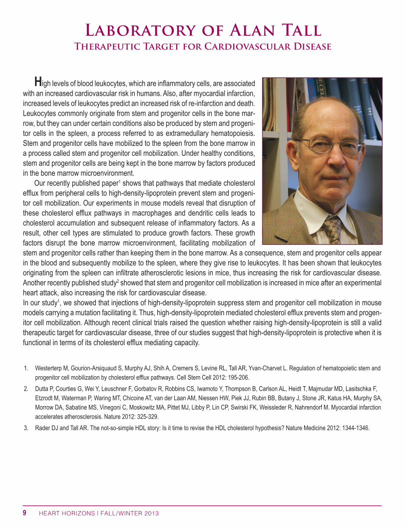
High levels of blood leukocytes, which are inflammatory cells, are associated with an increased cardiovascular risk in humans. Also, after myocardial infarction, increased levels of leukocytes predict an increased risk of re-infarction and death.Leukocytes commonly originate from stem and progenitor cells in the bone mar-row, but they can under certain conditions also be produced by stem and progeni-tor cells in the spleen, a process referred to as extramedullary hematopoiesis. Stem and progenitor cells have mobilized to the spleen from the bone marrow in a process called stem and progenitor cell mobilization. Under healthy conditions, stem and progenitor cells are being kept in the bone marrow by factors produced in the bone marrow microenvironment. Our recently published paper1 shows that pathways that mediate cholesterol efflux from peripheral cells to high-density-lipoprotein prevent stem and progeni-tor cell mobilization. Our experiments in mouse models reveal that disruption of these cholesterol efflux pathways in macrophages and dendritic cells leads to cholesterol accumulation and subsequent release of inflammatory factors. As a result, other cell types are stimulated to produce growth factors. These growth factors disrupt the bone marrow microenvironment, facilitating mobilization of stem and progenitor cells rather than keeping them in the bone marrow. As a consequence, stem and progenitor cells appear in the blood and subsequently mobilize to the spleen, where they give rise to leukocytes. It has been shown that leukocytes originating from the spleen can infiltrate atherosclerotic lesions in mice, thus increasing the risk for cardiovascular disease. Another recently published study2 showed that stem and progenitor cell mobilization is increased in mice after an experimental heart attack, also increasing the risk for cardiovascular disease. In our study1, we showed that injections of high-density-lipoprotein suppress stem and progenitor cell mobilization in mouse models carrying a mutation facilitating it. Thus, high-density-lipoprotein mediated cholesterol efflux prevents stem and progen-itor cell mobilization. Although recent clinical trials raised the question whether raising high-density-lipoprotein is still a valid therapeutic target for cardiovascular disease, three of our studies suggest that high-density-lipoprotein is protective when it is functional in terms of its cholesterol efflux mediating capacity.
1. Westerterp M, Gourion-Arsiquaud S, Murphy AJ, Shih A, Cremers S, Levine RL, Tall AR, Yvan-Charvet L. Regulation of hematopoietic stem and progenitor cell mobilization by cholesterol efflux pathways. Cell Stem Cell 2012: 195-206.
2. Dutta P, Courties G, Wei Y, Leuschner F, Gorbatov R, Robbins CS, Iwamoto Y, Thompson B, Carlson AL, Heidt T, Majmudar MD, Lasitschka F, Etzrodt M, Waterman P, Waring MT, Chicoine AT, van der Laan AM, Niessen HW, Piek JJ, Rubin BB, Butany J, Stone JR, Katus HA, Murphy SA, Morrow DA, Sabatine MS, Vinegoni C, Moskowitz MA, Pittet MJ, Libby P, Lin CP, Swirski FK, Weissleder R, Nahrendorf M. Myocardial infarction accelerates atherosclerosis. Nature 2012: 325-329.
3. Rader DJ and Tall AR. The not-so-simple HDL story: Is it time to revise the HDL cholesterol hypothesis? Nature Medicine 2012: 1344-1346.
Laboratory of Kimara Targoff Understanding Cardiac Morphogenesis
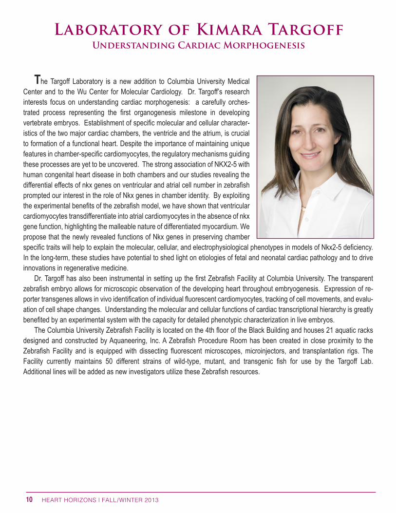
The Targoff Laboratory is a new addition to Columbia University Medical Center and to the Wu Center for Molecular Cardiology. Dr. Targoff’s research interests focus on understanding cardiac morphogenesis: a carefully orches-trated process representing the first organogenesis milestone in developing vertebrate embryos. Establishment of specific molecular and cellular character-istics of the two major cardiac chambers, the ventricle and the atrium, is crucial to formation of a functional heart. Despite the importance of maintaining unique features in chamber-specific cardiomyocytes, the regulatory mechanisms guiding these processes are yet to be uncovered. The strong association of NKX2-5 with human congenital heart disease in both chambers and our studies revealing the differential effects of nkx genes on ventricular and atrial cell number in zebrafish prompted our interest in the role of Nkx genes in chamber identity. By exploiting the experimental benefits of the zebrafish model, we have shown that ventricular cardiomyocytes transdifferentiate into atrial cardiomyocytes in the absence of nkx gene function, highlighting the malleable nature of differentiated myocardium. We propose that the newly revealed functions of Nkx genes in preserving chamber specific traits will help to explain the molecular, cellular, and electrophysiological phenotypes in models of Nkx2-5 deficiency. In the long-term, these studies have potential to shed light on etiologies of fetal and neonatal cardiac pathology and to drive innovations in regenerative medicine. Dr. Targoff has also been instrumental in setting up the first Zebrafish Facility at Columbia University. The transparent zebrafish embryo allows for microscopic observation of the developing heart throughout embryogenesis. Expression of re-porter transgenes allows in vivo identification of individual fluorescent cardiomyocytes, tracking of cell movements, and evalu-ation of cell shape changes. Understanding the molecular and cellular functions of cardiac transcriptional hierarchy is greatly benefited by an experimental system with the capacity for detailed phenotypic characterization in live embryos. The Columbia University Zebrafish Facility is located on the 4th floor of the Black Building and houses 21 aquatic racks designed and constructed by Aquaneering, Inc. A Zebrafish Procedure Room has been created in close proximity to the Zebrafish Facility and is equipped with dissecting fluorescent microscopes, microinjectors, and transplantation rigs. The Facility currently maintains 50 different strains of wild-type, mutant, and transgenic fish for use by the Targoff Lab. Additional lines will be added as new investigators utilize these Zebrafish resources.
10 HEART HORIZONS | FALL/WINTER 2013

Though no one in her family was a physician, Christine Seidman always wanted to be a doctor. But the word had a slightly different meaning for her than it does for most. “To me, that was a person who was medically trained and took care of sick people, but who also really understood why they got sick…Some peo-ple think you’re a physician or a scientist. To me, they’re synonymous. I still think that.” Seidman—who goes by Kricket (thanks to a young cousin who couldn’t pronounce “Christine”)—met her husband and research partner, Jon, when they were both undergraduates at Harvard. “We had a lab research project that we had to design and have approved. My group’s project was not approved. So they split up our group and reassigned us to other projects, and I got assigned to Jon’s group.” The two were married during Seidman’s junior year. After graduation and medical school, Seidman headed to Johns Hopkins for her residency and intern-ship “because it spoke science to me.” She was there for three years before mov-ing to Boston, where she did a cardiology fellowship at Massachusetts General Hospital before finishing her training in Baltimore. At MGH, Seidman worked with a group led by the late Edgar Haber, trying to isolate and clone the genes for adrener-gic receptors, which are important in cardiovascular physiology. She then became interested in atrial natriuretic peptide, or ANP. Released by the heart, ANP regulates salt and water in the bloodstream to reduce blood pressure. A partial amino acid sequence of this natriuretic peptide had just been published, and Seidman was intrigued; studying ANP had broad implications for treatment of high blood pressure. “As a cardiologist, you think this might cure hypertension.”She moved to her husband’s lab at Harvard Medical School, where she cloned the ANP cDNA and gene. The two have worked together ever since, studying the effects of genetic variation in heart disease. In 1998, she began studying disorders of heart muscle. Seidman’s work began with familial hypertrophic cardiomyopathy (HCM), which increases heart thickness and predisposes to the development of heart failure and sudden death. HCM is the most common cause of sudden death on the athletic field; it also affects many more people than originally thought. Seidman used genetic approaches to discover mutations that altered proteins involved in heart muscle contraction. This work enabled the development of models that can help researchers understand the mechanisms by which mutations cause disease. The work also allowed for gene-based diagnosis of HCM. Seidman’s group also has identified gene mutations that cause dilated cardiomyopathy and congenital heart malformations. To understand how gene mutations affect heart structure and function, Seidman’s laboratory does much of their work in mouse models. “If you know that a gene abnormality causes disease, you ought to be able to stick that gene into a cell and figure out the pathways it affects and what it does. But we don’t have any cell lines in cardiology. So we put the genes into mice and let them get heart disease and then study the heart.”
Professors Christine and Jonathan Seidman Deliver the Annual Clyde
and Helen Wu Distinguished Professor Lecture
11 HEART HORIZONS | FALL/WINTER 2013

NEWS FROM THE WU CENTER FOR MOLECULAR CARDIOLOGY