faculty of resource science and technology … parameters and... · 2.5 api 20e kit test ... api...
TRANSCRIPT
Physicochemical Parameters and Microbiological Quality of Water from Local Fish
Farm
FOO TUN XIAN
Bachelor of Science with Honours
(Resource Biotechnology)
2013
Faculty of Resource Science and Technology

Physicochemical Parameters and Microbiological Quality of Water from Local Fish
Farm
Foo Tun Xian (26331)
This project is submitted in partial fulfillment of the requirements for the degree of
Bachelor of Science with Honours (Resource Biotechnology)
Supervisor: Dr. Lee Kui Soon
Co-supervisor: Dr. Samuel Lihan
Resource Biotechnology
Department of Molecular Biology
Faculty of Resource Science and Technology
University Malaysia Sarawak
12/06/13

i
ACKNOWLEDGEMENTS
I would like to offer my appreciation to my project supervisor, Dr Lee Kui Soon and my
Co-supervisor, Dr Samuel Lihan on giving me lots of guidance and encouragements upon
completing this research project.
Besides, I would love to thank all the seniors who had been assisting me during the
research projects, special thanks to Kathleen Michelle, Felecia anak Collick (Master
students) and Leong Sui Sien (PhD student) who guided me in the laboratory. Additional
thanks also to the lab assistants and science officers who provided guidance and supplied
laboratory apparatus and materials.
Also, I would like to offer my deepest appreciation to my colleagues, especially to
Wong Gwo Rong, Tang Ping Sia, Ng Kok Hua and Chai Siong Kiat for their support and
encouragements. Last but not the least, my deepest appreciation also goes to my parents
who trusted me always.

ii
DECLARATION
I hereby declare that this thesis is based on my original work except for quotation and
citation, which have been acknowledged. I also declare that it has not been previously or
concurrently submitted for any other degree at UNIMAS or other institutions.
Foo Tun Xian
Resource Biotechnology Programme
Department of Molecular Biology
Faculty of Resource Science and Technology
University Malaysia Sarawak

TABLES OF CONTENTS
ACKNOWLEDGEMENTS ............................................................................................... I
DECLARATION .............................................................................................................. II
LIST OF ABBREVIATIONS ......................................................................................... IV
LIST OF TABLES ............................................................................................................V
LIST OF FIGURES ........................................................................................................ VI
ABSTRACT .................................................................................................................. VII
1.0 INTRODUCTION .................................................................................................. 1
2.0 LITERATURE REVIEW........................................................................................ 3 2.1 Tor tambroides ............................................................................................................... 3 2.1 Enterobacteriaceae ......................................................................................................... 3 2.2 Vibrionaceae ................................................................................................................... 4 2.3 Physicochemical Factors .................................................................................................. 5
2.3.1 pH ............................................................................................................................ 5 2.3.2 Temperature ............................................................................................................. 6 2.3.3 Turbidity .................................................................................................................. 6 2.3.4 Dissolved Oxygen (DO) ........................................................................................... 7 2.3.5 Biochemical Oxygen Demand (BOD) ....................................................................... 7
2.4 (GTG)5 PCR FINGERPRINTING .................................................................................... 8 2.5 API 20E KIT TEST ......................................................................................................... 9
3.0 MATERIALS & METHODOLOGY .................................................................... 10 3.1 SAMPLE COLLECTION AND PROCESSING ............................................................ 10 3.2 TOTAL PLATE COUNT............................................................................................... 11 3.3 BACTERIAL ISOLATION ........................................................................................... 11 3.4 DNA EXTRACTION .................................................................................................... 12 3.5 (GTG)5 PCR FINGERPRINTING .................................................................................. 13 3.7 AGAROSE GEL ELECTROPHORESIS (AGE) ............................................................ 14 3.8 API 20E Test ................................................................................................................. 15
4.0 RESULTS ............................................................................................................ 16 4.1 Sample Processing ........................................................................................................ 16
4.1.1 Sample Analysis .................................................................................................... 16 4.1.2 Biochemical Oxygen Demand (BOD) .................................................................... 18
4.2 Isolation of Bacteria ....................................................................................................... 19 4.2.1 Total Plate Count ................................................................................................... 19 4.2.2 Isolation of Enterobacteriaceae ............................................................................. 23
4.3 (GTG)5 PCR Fingerprinting .......................................................................................... 24 4.4 Genetic Diversity Evaluation ......................................................................................... 25 4.5 API 20E Identification System ...................................................................................... 27
5.0 DISCUSSION....................................................................................................... 30
6.0 CONCLUSION AND RECOMMENDATION ..................................................... 36
7.0 REFERENCES ..................................................................................................... 37
8.0 APPENDIX .......................................................................................................... 41

iv
LIST OF ABBREVIATIONS
µl microliter
ml milliliter
API Analytical Profile Index
DO Dissolved Oxygen
PPM Parts Per Million
BOD Biochemical Oxygen Demand
TAN Total Ammonia Nitrogen
ddH2O Sterilize Double Distilled Water
PFGE Pulse-field gel electrophoresis
LAB Lactic acid bacteria
RAPD Randomly amplified polymorphic DNA
Rep-PCR PCR amplification of repetitive bacterial DNA element
AGE Agarose Gel Electrophoresis
LB Luria Bertani
EtBr Ethidium Bromide
TBE Tris-Borate-EDTA
v
LIST OF TABLES
Table 1 PCR reaction set-up for (GTG)5
Table 2 (GTG)5 PCR amplification conditions
Table 3 Reading of pH for six random Mahseer ponds.
Table 4 Reading of temperature for six random Mahseer ponds.
Table 5 Reading of dissolved oxygen for six random Mahseer ponds.
Table 6 Biochemical Oxygen Demand reading.
Table 7 CFU of aquaculture water sample on dilution 10-1
on Nutrient agar.
Table 8 CFU of original water sample on MacConkey agar.
Table 9 CFU of gill and mouth swabs of Empurau fish on Nutrient agar with
dilution 10-1
.
Table 10 CFU of gill and mouth swabs of Empurau fish on MacConkey agar
with dilution 10-1
.
Table 11 Code for isolates and results of Sting-Test.
Table 12 Numbers represented the isolates in the gel.
Table 13 API identification result of 25 sample isolates.
vi
LIST OF FIGURES
Figure 1 NaOH String Test
Figure 2 API 20E Identification Kit
Figure 3 Plate spreading result of water sample from pond Sale A on Nutrient
Agar with dilution 10-1
.
Figure 4 Plate spreading result of water sample on MacConkey agar from
pond Culture A.
Figure 5 String-Test. A: AC-A2 showed positive result as presence of a DNA
string; B: AB-A1 showed negative result with formation of cell
suspension.
Figure 6 Banding patterns of GTG-PCR products. Lane M: 1kb ladder. Numbers in
figure represented each isolates was shown in Table 4.9.
Figure 7 The dendrogram showing the genetic diversity among all
Enterobacteriaceae isolates using (GTG)5 primer.
Figure 8 API Identification strips of sample isolates AB-C2, AB-A6 and AB-
A7.

vii
Physicochemical Parameters and Microbiological Quality of Water from Local Fish
Farm
Foo Tun Xian
Resource Biotechnology Program
Department of Molecular Biology
Faculty of Resource Science and Technology
University Malaysia Sarawak
ABSTRACT
The outbreak of fish disease has threatened the development of aquaculture industry in
years. Enterobacteriaceae is a member of gram-negative bacilli and contain more than 30
genera. They act as the main indicator for the bacteriological water quality. The study was
conducted at Malaysian Mahseer breeding farm near Asajaya to determine the
physicochemical parameter and occurrence of Enterobacteriaceae family in fishes in the
ponds which this could reflect the healthiness of water. Water sample were collected from
the ponds and analyzed for the presence of Enterobacteriaceae. Samples were plated on
MacConkey agar and colonies formed were further testes by ‘String Test’. Gram-negative
colonies were analyzed with (GTG)5 PCR. The phylogenetic tree was constructed based on
the DNA banding pattern from the (GTG)5 PCR. Suspicious species were identified using
API 20 E Diagnostic Kit. Four major clusters were shown in the dendrogram indicate the
diversity of the bacteria strains. Twelve strains of Escherichia coli, four strains Serratia
odorifera, one strain of Serratia liquefaciens and eight strains of Aeromonas
hydrophila/caviae/sobria were detected.
Key words: Enterobacteriaceae, (GTG)5 PCR, API 20E Diagnostic Kit
ABSTRAK
Penyakit ikan telah melambatkan pembangunan industri akuakultur. Enterobacteriaceae
merupakan bakteria “gram-negatif, mempunyai lebih daripda 30 genera. Mereka
merupakan salah satu penanda utama untuk mengaji kuality air dari sudut mikrobiologi.
Kajian tersebut telah dijalankan di ladang pembiakan Mahseer Malaysian berhampiran
Asajaya untuk menentukan parameter fizikokimia dan Enterobacteriaceae dalam kolam
yang dapat terus mencerminkan kesihatan air. Sampel air dibawa ke makmal dan diuji atas
agar MacConkey, seterusnya koloni bakteria yang tumbuh atas agar dianalisis dengan ujian
“String-Test”. Koloni gram-negatif di analisis dengan PCR (GTG)5 and pokok
“phylogeny” dihasilkan. Spesies yang mencurigakan telah dikenal pasti menggunakan API
20 E Diagnostik Kit. Dua belas Escherichia coli, empat Serratia odorifera, satu Serratia
liquefaciens and lapan Aeromonas hydrophila/caviae/sobria telah dikenalpastikan.
Kata kunci: Enterobacteriaceae, (GTG)5 PCR, kit API 20E.

1
1.0 INTRODUCTION
Freshwater aquaculture industry is highly important for the development of Malaysia. It
provides tremendous and steady export revenue and increases the local security of food in
the country (National Aquaculture Sector Overview, 2012). Common freshwater fish
species that are normally bred for sale are tilapia, eel, catfish, tuna, rainbow trout, carp and
a number of species. In order to consistently supply healthy aquatic organisms, water
quality management is crucial in aquaculture industry.
Fish diseases emerged as a popular and formidable issue for most fish farmers. Threat of
the fish diseases has slowed down the progress of aquaculture (National Aquaculture
Sector Overview, 2012). These diseases have originated from a few agents such as
parasites, fungus, viruses, bacteria or improper physical environment, which lead to the
unnecessary loss of the industry. According to Chuah et al (2010), Edwardsiellosis tarda
outbreak has led to a heavy financial loss of the fish farmers where the Edwardsiellosis
tarda infected fishes are founded.
Bacteria are one of the causative agents that result in several significant diseases that might
create a massive loss to the Malaysian aquaculture industry. Hazardous bacteria strains that
are commonly found in the freshwater are mostly gram-negative such as Aeromonas
hydrophila, Pseudomonas fluorescens, Vibrio vulnificus, Vibrio parahaemolyticus,
Edwardsiella ictaluri and Edwardsiella tarda. The degree of existence of these strains in
the source correlates with the likeliness of disease found on the fishes within the source.
For instance, Genus Vibrio commonly causes necrosis and septicemia. Disease prevention
and control is steps necessary to be taken in order to hamper the massive damage to the

2
industry. Early conformation of the presence and characterization of the hazardous strains
are crucial to suppress the disease outbreak.
Diverse physicochemical factors such as pH, temperature, turbidity, dissolved oxygen
(DO), Biochemical Oxygen Demand (BOD), Total Ammonia Nitrogen (TAN) and total
suspended solids (TSS) have a large impact on the fish health, growth and their hostility to
the disease triggered by the causative agents. Most of the massive fish death’ cases that
occur around the world are mainly due to the changes of environment factor. For example,
10,000 of fishes were killed in Butterfly Lake in South Knoxville due to depletion of
water’s oxygen level after the fluctuation of atmosphere temperature (Hickman, 2012).
Similar case happened in Lake Taal, Philippine where 750 tons of fishes was found death
in 2011 after climate changed over the weekend (Li, 2011).
The objectives of this research project were as below;
i) to detect the level of physicochemical factors in selected local fish farm
ii) to detect the potential disastrous microbiological factors that might cause the
transmission of infectious diseases that may eventually lead to zoonosis.
iii) to determine the genetic diversity distance between isolates.

3
2.0 LITERATURE REVIEW
2.1 Tor tambroides
Tor tambroides and Tor douronensis or its common name is mahseer that are most
valuable and overpriced fish in Malaysia (Litis et al., 2007). Their high market price is
largely due to the fine and tasty taste of the flesh. Also, the species population is
decreasing sharply due to the environment degradation by human activities, for example,
logging, deforestation and overfishing has endangered the Tor sp.. Combination of these
events has exaggerated the price of the fish. According to Ingram et al. (2005), the price of
T. douronensis has reached RM100/kg while the price of T. tambroides has reached
rm400/kg in the Kapit, Sarawak. Many entrepreneurs have also started to breed this
overpriced fish, therefore generating a high demand in the fresh water fish industry.
2.1 Enterobacteriaceae
Members of Enterobacteriaceae comprised of more than 170 recognized species today.
They possess of few easily distinguishable and unique characteristics, which are Gram-
negative bacteria rod, some of them are motile, facultatively anaerobic, absent of oxidase
and presence of catalase. Their optimum growing temperature is at 37˚C (some prefer 25-
30˚C). Common habitats of them are in living organisms (plants and animal), soil and
water. Members that are included in the family are Citrobacter species, Enterobacter
species, Escherichia species, Hafnia alvei, Klebsiella species, Morganella morganii,
Proteus species, Providencia species, Salmonella species, Serratia species, Shigella
species and Yersinia species (UK Standards for Microbiology Investigations, 2011). Most
of the bacteria species under family Enterobacteriaceae are pathogenic to living
organisms, therefore their presence are the main indicator of disease outbreak most of the
4
time. According to Kamble et al. (2012), genus Aeromonas causes the furunculosis and
hemorrhagic Septicemia diseases to the fishes.
2.2 Vibrionaceae
The family Vibrionaceae is a very significant bacterial group that is found all around the
world in different area that affects all types of marine and freshwater fishes (Kumiko et al.,
1993; Gwedelynne et al., 2005; Safinaz et al., 2011). Vibrios are Gram-negative, motile
rods, have a facultatively fermentation metabolism, and presence of oxidase. Most of the
strains are pathogenic to human and animals. Moreover, Vibrio species are normally
inhabited in the intestinal flora or marine and freshwater fishes (Simidu et al., 1977).
Vibrio species like V.splendidus, V. tubiashii, V. fluvialis, V. neonatus, V. ezurae, and V.
ponticus, V. harveyi, V. splendidus, and V. tubiashii are often connected with the diseases
occurring in different types of fishes (Macian et al., 2004; Sawabe et al., 2004; Thompson
et al., 2005). According to Safinaz et al. (2011), Vibrio species affects fishes with
Vibriosis, which produces symptoms like “severe congestion at the base of the fins,
erosion of the fins, excessive mucous secretion of gills, severe congestion of gills,
hemorrhagic ulcerations, linear hemorrhages over different parts of the body and severe
congestion or hemorrhagic protrusion of the anal opening”.

5
2.3 Physicochemical Factors
Based on Rheinheimer (1991), the physical and chemical properties of the source have a
major influence of the microbial growth and the morphological and physiological of the
bacterial population. Moreover, the changes in pH, temperature and salt concentration may
also affect the metabolism, and reproduction system of certain microorganism. The aquatic
life becomes stressful when one or more of these physicochemical factors have altered.
The stressed organisms are more prone to the infection of bacteria, fungus, and other
pathogens.
2.3.1 pH
A stable physiological hydrogen ion concentration (pH) is crucial to maintain the aquatic
life. pH of most freshwater site apart from estuary that containing fishes ranges from 6 to 9
(Robertson - Bryan, Inc, 2004). The basic process of fishes such as respiration is closely
influenced by pH changes. Sub-lethal effect such as declining of the growth rates can occur
if the pH is fluctuated. In more serious cases, if the fluctuation of pH were out of the
toleration level of the freshwater organism, death would occur. Bacteria can be
differentiated into acidophile (pH 1 - 5.9), neutralphile (pH 6 – 9) or alkaliphile (pH 9 –
11) and the adaptation of bacteria vary accordingly to pH (Todar, n. d.). Similarly, changes
in pH have a discernible effect on the bacterial enzymes; therefore, exposure to extreme
pH escalates the mortality rates of bacteria in pond.
6
2.3.2 Temperature
Temperature is significant in term of maintaining the life process of microorganism.
Temperature changes can affect growth rate, nutritional requirement, enzymatic and
chemical component of the cell (Rheinheimer, 1991). Basically, microorganism can be
categorized as the psychrophiles (able to grow at 0˚C), mesophiles (optimum
temperature~37˚C), thermophiles (optimum temperature 45˚C to 70˚C), and lastly
hyperthermophiles (optimum temperature 80˚C to 115˚C). Increasing in temperature would
lead to an increasing of metabolic rate of microorganism due to the activation enzymatic
reaction. As a result, the Biochemical Oxygen Demand (BOD) level is raised when the
temperature is increased to a certain extent. Furthermore, the oxygen solubility drops when
the water temperature is raised. The concentration of CO2 in the water is increased, as the
O2 is decreased, in fact, the aggregation of CO2 limit the O2 level in the water. Lastly, the
level of toxicity is associated with the changes in temperature. As the temperature
increases, more toxic would be emitted from the poisonous substances.
2.3.3 Turbidity
Turbidity is one of the main factors that affect the life of microorganism. The presence of
silt, clay, sextons, organic and inorganic matter, and tiny organisms and plankton have
greatly influence the level of turbidity. Sexton is the accumulation of living and dead
materials in the water that led to the formation of sediment. Most of the microorganisms
treat the sexton as the food source. Therefore, increasing organic suspended materials can
lead to accretion of turbidity and finally accompanied by an aggressive growth of the
bacterial population (Rheinheimer, 1991). Besides affecting the growth of bacteria, the
turbidity causes light to be scattered rather than penetrating directly through the
7
photosynthetic organisms (APHA, 1992). Thus, the amount of light absorbed by the
photosynthetic organisms might be reduced.
2.3.4 Dissolved Oxygen (DO)
Dissolved oxygen (DO) is the most fundamental criterion in water. The carbon dioxide gas
from photosynthesis of land and aquatic vegetation is the major source of DO. Aquatic
organisms required dissolved oxygen to sustain life. The volume of oxygen needed is
varied among species and phase of life. Dissolved oxygen level between 5 to 6 ppm is
normally needed for fish’s activities and development. Level 3 ppm and below are
insufficient for most aquatic living organisms (LaMotte, n.d.). However, alteration of
temperature affects the solubility of oxygen in the water (Canadian Council of Ministers of
the Environment, 1999). During summer, with the temperature increases, the DO
concentration and solubility on the warm upper water layer decreases. On the other hand,
when the temperature drops due to the mixing warm water layer and cold lower water
layer, oxygen concentration and level of saturation are increasing. Oxygen lost may be due
to several factors, for example, respiration of bacterial, plant and animal, direct chemical
oxidation of dissolved organic matter (Canadian Council of Ministers of the Environment,
1999).
2.3.5 Biochemical Oxygen Demand (BOD)
BOD is a measurement of the total oxygen required for the aerobic bacteria when digesting
the organic matter. Besides, BOD calculates the chemical oxidation of inorganic materials
as well. Temperature, pH, types of microorganisms and kind of organic and inorganic
8
matter are able to affect the speed of consumption of oxygen in the water (EPA, 2012).
Plant detritus, plants and animals carcasses and animal wastes are sources of BOD (EPA,
2012). BOD is mainly to indicate the strength of organic in wastewater and heavily
polluted water source, because these types of water are normally containing high amounts
of organic matter (National Water Quality and Availability Management, 2003).
2.4 (GTG)5 PCR Fingerprinting
The commonly used method such as the 16S rRNA sequencing, ribotyping, and pulsed-
field gel electrophoresis (PFGE) are too arduous to carry out from the classification and
identification of Lactic acid bacteria (LAB) (Gevers et al., 2001). Even though randomly
amplified polymorphic DNA (RAPD) is the most used PCR-based genomic technique for
LAB identification, however, according to Gevers et al. (2001), the downsides of
randomly amplified polymorphic DNA (RAPD) are the high discriminatory power and the
large applicability within a large group of LAB species that have not been described.
Moreover, it has displayed the weak reducibility.
As an alternative to provide a better identification of bacteria species, PCR amplification
of repetitive bacterial DNA elements (rep-PCR) is used. It is a straightforward PCR-based
technique with several advantages which are high discriminatory power, inexpensive,
applicable for huge number of strains and able to identify and type a huge range of bacteria
(Gevers et al., 2007; Olive & Bean, 1999; Versalovic et al., 1994). Oligonucleotide primer
that designed to be used in the rep-PCR is (GTG)5. By using this technique, the genetic
diversity of different isolates can be identified through the DNA fingerprinting. AGE is
used to observe the multiple banding patterns after PCR amplification. The banding shows
9
the profile of various isolates. A dendrogram is plotted to determine the genetic diversity
distance between isolates.
2.5 API 20E Kit Test
Rapid and reliable diagnostic tests are required to provide the faultless detection of the
bacterial strains in the water source (Coz-Rakovac et al, 2007). This is to effectively
control and cure the aquatic animal diseases before a disease outbreak that might be
leading to zoonosis. API 20E system’s role is to identify the Enterobacteriaceae and other
non-fastidious Gram-negative bacteria. It contains 21 standardized and small-scale
biochemical tests and a database. Also, the test kit consists of 21 microtubes comprised of
dehydrated substances (bioMérieux, 2002). API 20E ratings are based on three factors. The
three factors include “the likelihood of a match between the unknown organism’s profile
and computer profile, the relative value between the likelihood of the first and the
likelihood of the second choice, and the number of tests against the first choice” (Coz-
Rakovac et al, 2007). There are several pathogenic bacterial species, which is in the
current identification database of API 20E. The bacteria referred are Acinetobacter spp.,
Aeromonas hydrophila, A. salmonicida subsp., Salmonicida, Citrobacter freundii,
Edwadsiella tarda, Escherichia vulneris, Hafnia alvei, Klebsiella pneumonia, Moraxella
spp., Pantoea spp., Photobacterium damsel, Plesiomonas shigelloides, Providencia
rettgeri, Pseudomonas aeruginosa, P. fluorescens/putida, Salmonella arizonae, Serratia
liquefaciens, Serratia plymuthica, Shewanella putrefaciens, Vibrio alginolyticus, V.
cholera, V. vulnificus.

10
3.0 MATERIALS & METHODOLOGY
3.1 Sample Collection and Processing
Sample collection was carried out at Malaysian Mahseer breeding farm near Asajaya,
Sarawak on 1st of February 2013. This farm mainly breeds Tor tambraides
(Kelah/Empurau), Tor dourenensis (Semah), and Barbonymus schwanenfeldii (Tengadak).
Water samples were obtained at three different sites, which are the breeding sites, culture
site and sale sites. Overall there were six samples collected where three samples taken
from breeding site, one sample from culture site and two samples from the sale site. Water
samples were collected by using 50 ml Falcon tube, respectively at three different points;
inlet, random spot in the tank, and outlet. Gill and fin swabs were taken from one adult Tor
tambraides in the breeding tank. The water samples collected from three points were then
poured into a new sterile 50 ml Falcon tube and homogenized. All tubes were labeled with
the date and source of the samples. All samples were kept at 4ºC in an insulated double
walled container and were transported to the laboratory for further analysis. The
temperature, dissolved oxygen (DO), biochemical oxygen demand (BOD) and pH of
samples were also recorded.

11
3.2 Total Plate Count
1 mL of the homogenized water was pipetted into 9 mL of saline solution to make up the
dilution of 10-1
. The process was continued by pipetting 1 mL of solution from dilution 10-
1 into 9 mL of saline solution and labeled as dilution 10
-2. Tail and Fin swabs were mixed
with the distilled water and diluted into dilution of 10-1
and 10-2
. 100 µL from original,
dilution 10-1
and 10-2
solution were then spread onto non-selective Nutrient agar and
MacConkey agar and incubated at 27ºC for 24 hours. Single colonies formed were counted
after the overnight incubation.
3.3 Bacterial Isolation
Following the total plate count on MacConkey agar, presumptive Enterobacterioceae
appeared as colourless, transparent colonies were isolated and streaked on new nutrient
agar and incubated at 27ºC for 24 hours. Double confirmation was done by using NaOH
String Test to ensure bacteria colonies isolated are gram-negative. The colonies were
emulsified onto a glass slide that contained 10 µL of 0.5M of NaOH. The suspension was
mixed and next observed whether there is the formation of a string (Figure 1). The colonies
that failed the test were discarded. The positive strands were grown in the nutrient broth at
room temperature for 24 hours on the shaker (New Bunswick Scientific Exccela E10
Platform Shaker) at 1200rpm prior DNA extraction.
Figure 1: NaOH String Test

12
3.4 DNA Extraction
Bacteria were grown overnight in 50 mL nutrient broth at room temperature on the shaker
at 1200 rpm. 1.5ml of overnight culture was poured into the 1.5 mL Eppendorf (Eppy)
tube. The tube was centrifuged under 10,000 rpm for 5 minutes. After centrifugation, the
supernatant was discarded but cell pellet remained. Another 1.5 mL of the overnight
culture was poured into the same Eppy tube. The tube was centrifuged under previous
conditions. The supernatant was discarded following by addition of 500 µL dH2O then
vortex to mix well. The Eppy tube immersed in boiling water and boiled for 10 minutes.
After boiling, the Eppy tube was immediately placed into a box containing ice for 5
minutes. Next, the tube was centrifuged for 10 minutes to suspend the cell debris. The
supernatant contained the DNA of the bacteria was transferred to the new Eppy tube for
PCR purpose.

13
3.5 (GTG)5 PCR Fingerprinting
(GTG)5 PCR was conducted according to Gevers et al. (2001) . The primer used is (GTG)5
with the sequence of 5’-GTGGTGGTGGTGGTG-3’.
The PCR reaction set-up used for this study is shown in Table 1.
Table 1: PCR reaction set-up for (GTG)5
PCR Compound Volume
Taq Polymerase (2U) 0.5 µl
(GTG)5 primer 1 µl
MgCl2 3 µl
dNTP 1 µl
Distilled H2O 9.5 µl
Buffer (10x) 5 µl
Template DNA 5 µl
Total 25 µl
The amplification of (GTG)5 region was carried out in SenseQuest thermal cycler and the
amplification conditions are listed in Table 2.
14
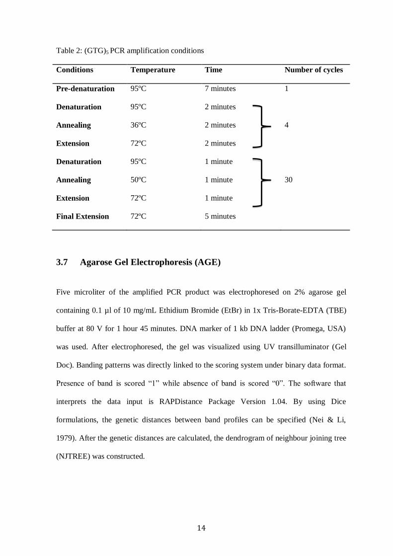
Table 2: (GTG)5 PCR amplification conditions
Conditions Temperature Time Number of cycles
Pre-denaturation 95ºC 7 minutes 1
Denaturation 95ºC 2 minutes
Annealing 36ºC 2 minutes 4
Extension 72ºC 2 minutes
Denaturation 95ºC 1 minute
Annealing 50ºC 1 minute 30
Extension 72ºC 1 minute
Final Extension 72ºC 5 minutes
3.7 Agarose Gel Electrophoresis (AGE)
Five microliter of the amplified PCR product was electrophoresed on 2% agarose gel
containing 0.1 µl of 10 mg/mL Ethidium Bromide (EtBr) in 1x Tris-Borate-EDTA (TBE)
buffer at 80 V for 1 hour 45 minutes. DNA marker of 1 kb DNA ladder (Promega, USA)
was used. After electrophoresed, the gel was visualized using UV transilluminator (Gel
Doc). Banding patterns was directly linked to the scoring system under binary data format.
Presence of band is scored “1” while absence of band is scored “0”. The software that
interprets the data input is RAPDistance Package Version 1.04. By using Dice
formulations, the genetic distances between band profiles can be specified (Nei & Li,
1979). After the genetic distances are calculated, the dendrogram of neighbour joining tree
(NJTREE) was constructed.
15
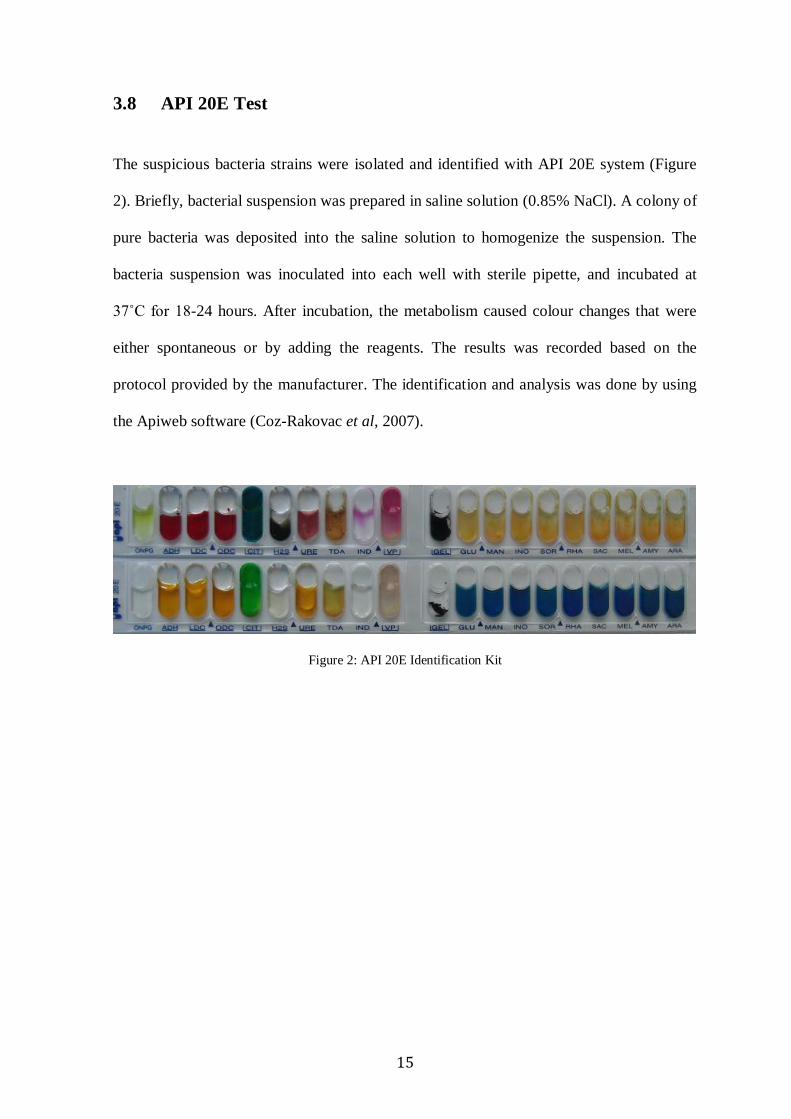
3.8 API 20E Test
The suspicious bacteria strains were isolated and identified with API 20E system (Figure
2). Briefly, bacterial suspension was prepared in saline solution (0.85% NaCl). A colony of
pure bacteria was deposited into the saline solution to homogenize the suspension. The
bacteria suspension was inoculated into each well with sterile pipette, and incubated at
37˚C for 18-24 hours. After incubation, the metabolism caused colour changes that were
either spontaneous or by adding the reagents. The results was recorded based on the
protocol provided by the manufacturer. The identification and analysis was done by using
the Apiweb software (Coz-Rakovac et al, 2007).
Figure 2: API 20E Identification Kit