factors involved in the atrophy of the organs of the larval frog
TRANSCRIPT
Factors Involved in the Atrophy of the Organs of the Larval FrogAuthor(s): Withrow MorseSource: Biological Bulletin, Vol. 34, No. 3 (Mar., 1918), pp. 149-166Published by: Marine Biological LaboratoryStable URL: http://www.jstor.org/stable/1536263 .
Accessed: 14/05/2014 11:16
Your use of the JSTOR archive indicates your acceptance of the Terms & Conditions of Use, available at .http://www.jstor.org/page/info/about/policies/terms.jsp
.JSTOR is a not-for-profit service that helps scholars, researchers, and students discover, use, and build upon a wide range ofcontent in a trusted digital archive. We use information technology and tools to increase productivity and facilitate new formsof scholarship. For more information about JSTOR, please contact [email protected].
.
Marine Biological Laboratory is collaborating with JSTOR to digitize, preserve and extend access toBiological Bulletin.
http://www.jstor.org
This content downloaded from 194.29.185.170 on Wed, 14 May 2014 11:16:27 AMAll use subject to JSTOR Terms and Conditions
FACTORS INVOLVED IN THE ATROPHY OF THE ORGANS OF THE LARVAL FROG.
WITHROW MORSE.1
From the Nelson Morris Memorial Institute for Medical Research of the Michael Reese Hospital, Chicago.
PRELIMINARY.
Throughout the two great branches of the organic realm, in- stances are presented where persons, organs or tissues or cells themselves undergo retrogressive changes.2 In the plant king- dom, we have the familiar elimination of stems, leaves and other parts of the individual, upon the approach of untoward condi- tions. In the animal series, we are enabled to identify an anal- ogous process in the protozoa where, in Amoeba the ephemeral pseudopodia are constantly being absorbed into the protoplasmic mass proper; in the species of Actinophrys, where this process of absorption is delayed permanently or at infrequent intervals; in the genus Trypanosoma, where the undulating membrane be- comes absorbed under certain conditions. Similar phenomena are observed in the sponges where Maas's studies have shown that hitherto highly differentiated cells become reduced to a more typical and fundamental cell-form, the amoebocyte. In the Ccelenterata we find the hydranths of Tubularta (i) and of other species either absorbed or eliminated ,n toto, while in Renilla Wilson has described a degeneration of the polyp. The "brown bodies" of the Bryozoa represent degenerated individuals. In brachiopods, during the stages of fixation there is present an extensive degeneration of parts resembling in a superficial way the various changes undergone by the larvae of the ascidians. In Sacculina we have, facile princeps, this property of involution. Even amongst the vertebrates we find instances of important degenerations of a natural sort, as in the fish Fierasfer where Bykowski and Nusbaum discovered extensive degeneration proc- esses when the fish became parasitic.
1In order to avoid confusion, the author's name will be used henceforth as in the title of this paper.
2 Compare Child, C. M., "Senescence and Rejuvenescence." Chicago, I915. 149
This content downloaded from 194.29.185.170 on Wed, 14 May 2014 11:16:27 AMAll use subject to JSTOR Terms and Conditions
WITHROW MORSE.
Interesting as these instances are, relatively meagre investi-
gation has been made as to the causes and factors involved in the physiology of the process. On the other hand, such studies as have been made have borne good fruit. It was in the studies of
ctenophore larvae that Metchnikoff conceived the idea of what later became the theory of phagocytosis. Later this investiga- tor studied the cytology of the larval frog's tail during the stages of metamorphosis. Extensive morphological studies have been made upon the stages of metamorphosis in insects. The changes which the salmon undergoes in breeding season have likewise received considerable attention.
Passing from the natural atrophy found in such instances as these, to what is termed pathological atrophy, we encounter the processes of progressive muscle atrophy, acute yellow atrophy of the liver and other states which bear many features in common with certain of the so-called natural atrophy processes, and this is true to such an extent in regard to the atrophy of the muscles of the larval frog at metamorphosis and the conditions observed in progressive muscle atrophy or Zenker's degeneration that it is difficult to believe that we are dealing with fundamentally different physiological processes.
The purpose of the following communication is to attempt to show that there are certain factors involved in the atrophy of the tadpole's tail which are to be considered as causal or of primary importance as conditions; and the similarity in cytological pic- tures and in certain known conditions in this case and in that of pathological atrophy makes it fairly probable that we are dealing with the same physiological factors, so that we encounter here an instance where the comparative physiology of a lower form of organism sheds light upon the economy of the higher organisms of especial interest to the pathologist.
HISTORICAL.
Although Metchnikoff had witnessed the activity of phagocytic cells earlier, in the larvae of errantioe, his first report involving the conception of cells wandering in the body fluids of one organism and serving as scavengers was made in I883 (2). Early in the history of the problem, this investigator recognized the practical
I50
This content downloaded from 194.29.185.170 on Wed, 14 May 2014 11:16:27 AMAll use subject to JSTOR Terms and Conditions
ATROPHY OF THE ORGANS OF THE LARVAL FROG.
value of such a study when he says "L'atrophie des muscles des batraciens presente un grand interet parce qu'elle peut servir de type de phenomenes pathologiques."
Following Metchnikoff, several investigators concerned them- selves with the problem. Thus Barfurth (3), Looss (4), Noetzel
(5), Anglas (6), Guieweisse (7), Mercier (8) are among the more important names in this connection. Of these, Looss and Mer- cier are perhaps of the greater importance. Grund (9) has more recently studied the problem of muscle degeneration from the chemical standpoint. In the field of fish physiology, Miescher's (Io) classic researches and those of Noel Paton (ii) and his co- workers demand especial mention. Even mere mention of the investigations which have been made in insect physiology and morphology during metamorphosing periods io impracticable in the piresent paper, a review of which being available in the paper of Mercier (8).
THE PROBLEM STATED.
Two quite diverse theories concerning the factors involved in muscle atrophy exist, namely, phagocytosis and autolysis. The former theory can best be stated in the words of Metchnikoff: "Von den ersten Stadien seiner Atrophie an, kann man in ihm eine gross Anzahl amoboider Zellen finden, in deren Innerin ganze Stiicke von Nervenfasern und Muskelprimitivbiindeln enthalten sind" (I883, p. 56I). Looss adheres to the latter view in the following words: " Wir es hier nicht mit einer Degenera- tion, einer Entartung der Gewebe und ihrer histologischen Bestandtheil zu thun haben, sondern mit einer reiner Auflo6sung, mit einer Resorption im strengen Sinne des Wortes" (p. 9I).
In the larval frog, no one has attempted to verify the belief of Looss save upon histological grounds. Anglas, Noetzel, Guie- weisse and others subscribe freely to Metchnikoff's theory of phagocytosis. The burden of the present paper is to show that Looss is correct in believing that fundamentally and primarily, a change is initiated interpretable as autolysis and that phagocy- tosis, which unquestionably is present at a later stage, is of secondary importance.
151
This content downloaded from 194.29.185.170 on Wed, 14 May 2014 11:16:27 AMAll use subject to JSTOR Terms and Conditions
WITHROW MORSE.
HISTOLOGICAL EVIDENCE.
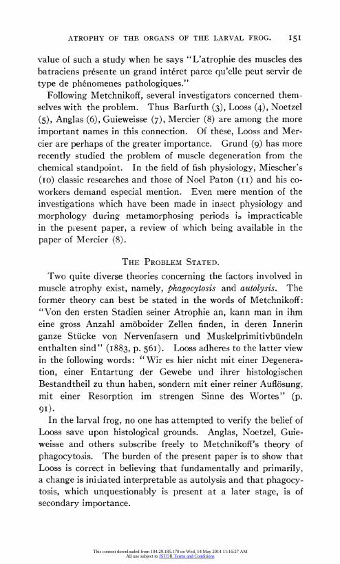
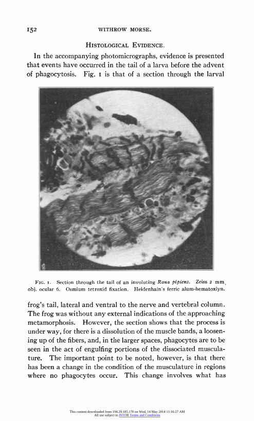
In the accompanying photomicrographs, evidence is presented that events have occurred in the tail of a larva before the advent of phagocytosis. Fig. I is that of a section through the larval
FIG. i. Section through the tail of an involuting Rana pipiens. Zeiss 2 mm. obj. ocular 6. Osmium tetroxid fixation. Heidenhain's ferric alum-hematoxlyn.
frog's tail, lateral and ventral to the nerve and vertebral column. The frog was without any external indications of the approaching metamorphosis. However, the section shows that the process is under way, for there is a dissolution of the muscle bands, a loosen- ing up of the fibers, and, in the larger spaces, phagocytes are to be seen in the act of engulfing portions of the dissociated muscula- ture. The important point to be noted, however, is that there has been a change in the condition of the musculature in regions where no phagocytes occur. This change involves what has
I52
This content downloaded from 194.29.185.170 on Wed, 14 May 2014 11:16:27 AMAll use subject to JSTOR Terms and Conditions

ATROPHY OF THE ORGANS OF THE LARVAL FROG. 153
been described as a "chromatolysis," but the dark masses running transversely over the muscle bands are depositions of fats bearing unsaturated fatty acids (oleic) which stain with the osmium tetroxid used in fixation. Willard and Guenther in unpublished investigations upon the micro-chemistry of degenerating muscle
^^ff~~~~~~~~1 %. *1
^^~~~~~~ .^?-a
^ ? ̂ ^?^~~.11 - 1 * . . "
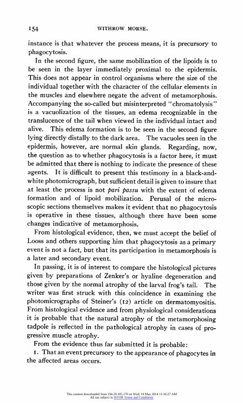
FIG. 2. Section of similar stage, same species, showing peripheral tissues. Not all of the intensely black areas are those of chromatolysis, for the osmium stains those neutral fats containing unsaturated fatty acids, such as oleic.
fibers find similar conditions in mammalian muscles permitted to degenerate after severing the innervation; a change in the distri- bution of neutral fats occurs involving the grouping of these com- pounds into masses, or droplets.' The significance in the present
1 There is no evidence of an infiltration of fat, but rather of a simple process of accumulation of fats already present in diffused form.
This content downloaded from 194.29.185.170 on Wed, 14 May 2014 11:16:27 AMAll use subject to JSTOR Terms and Conditions
WITHROW MORSE.
instance is that whatever the process means, it is precursory to phagocytosis.
In the second figure, the same mobilization of the lipoids is to be seen in the layer immediately proximal to the epidermis. This does not appear in control organisms where the size of the individual together with the character of the cellular elements in the muscles and elsewhere negate the advent of metamorphosis. Accompanying the so-called but misinterpreted "chromatolysis" is a vacuolization of the tissues, an edema recognizable in the translucence of the tail when viewed in the individual intact and alive. This edema formation is to be seen in the second figure lying directly distally to the dark area. The vacuoles seen in the epidermis, however, are normal skin glands. Regarding, now, the question as to whether phagocytosis is a factor here, it must be admitted that there is nothing to indicate the presence of these agents. It is difficult to present this testimony in a black-and- white photomicrograph, but sufficient detail is given to insure that at least the process is not pari passu with the extent of edema formation and of lipoid mobilization. Perusal of the micro- scopic sections themselves makes it evident that no phagocytosis is operative in these tissues, although there have been some changes indicative of metamorphosis.
From histological evidence, then, we must accept the belief of Looss and others supporting him that phagocytosis as a primary event is not a fact, but that its participation in metamorphosis is a later and secondary event.
In passing, it is of interest to compare the histological pictures given by preparations of Zenker's or hyaline degeneration and those given by the normal atrophy of the larval frog's tail. The writer was first struck with this coincidence in examining the photomicrographs of Steiner's (I2) article on dermatomyositis. From histological evidence and from physiological considerations it is probable that the natural atrophy of the metamorphosing tadpole is reflected in the pathological atrophy in cases of pro- gressive muscle atrophy.
From the evidence thus far submitted it is probable: i. That an event precursory to the appearance of phagocytes in
the affected areas occurs.
154
This content downloaded from 194.29.185.170 on Wed, 14 May 2014 11:16:27 AMAll use subject to JSTOR Terms and Conditions

ATROPHY OF THE ORGANS OF THE LARVAL FROG.
2. That phagocytosis contrary to being a primary agent in effecting the dissolution of the tissues of the tail is of secondary importance.
BIOCHEMICAL EVIDENCE.
During the last few years, a more complete understanding of the softening of tissues in the living and in the dead organism without the participation of microorganisms has been obtained through the work of Bradley (I3) and others. There is a more or less definite relation between reaction of medium and the proc- ess known variously as self-digestion, maceration, auto-diges- tion, autolysis, etc., wherein tissue enzymes resembling in a general way the enzymes of the alimentary tract operate in the tissues themselves, causing autolytic processes. This relation of reaction is such that when the average normal alkalinity of the tissues falls below a certain point, autolysis begins. In the ter- minology of physical chemistry, when the number of hydroxyl groups or ions (OH) approach (molecular weight in hydrogen equivalent)/(io,ooo,ooo) or N/(io)7, which is neutrality for the ions involved, then either the proteolytic enzymes of autolysis are activated, or the reaction causes a change in the proteins rendering them digestible by the enzymes (I4). A distinctly acid reaction, that is, where the concentration of OH ions is less than I/Io,ooo,ooo normal, or, in the terminology of Sorensen which is being universally adopted, where Ph < 71, autolysis proceeds rapidly.
Up until the present time, the hydrogen ion concentration of the blood of the larval frog has not been accurately determined, a point to which the writer means to direct attention when material is available.2 It is desirable to determine whether there
1 The expression Ph is borrowed from the principles of electromotive force, where the "potential" difference is measured on a potentiometer between a known concentration of hydrogen or hydroxyl ions and the unknown solution whose concentration in hydrogen or hydroxyl ions is required. The expression Ph sig- nifies that the potential refers to hydrogen (h). Its numerical value is the same as the expression N/(Io)a, where a is a number greater than 7 if the solution in question is alkaline, or less than 7 if the solution is acid. Inasmuch as N/(io)a is the same as N(Io)-a, the expression Ph means "minus the logarithm of con- centration."
2 Only the larger species of frogs can be utilized for these determinations. The arva of Rana catesbiana is of sufficient size to give 2 c.c. of blood for experimental
I55
This content downloaded from 194.29.185.170 on Wed, 14 May 2014 11:16:27 AMAll use subject to JSTOR Terms and Conditions

WITHROW MORSE.
is any change in alkalinity coordinate with atrophy. In the case of atrophy of the kidney tissue of the mammal where autol- ysis can be demonstrated, the writer and colaborator (I5) have found that there is a rapid loss of alkalinity when autolysis sets in, while a true acidity develops later. If, in the larval frog, autolysis is vera causa for the process of atrophy, we should expect to find a low alkalinity or actual acidity of the body fluids of the tail during atrophy.
If autolysis actually produces atrophy of the larval frog's organs, we should expect to find a difference in the quantities of end products of protein digestion between the normal, non-meta-
morphosing tail and that which is atrophying. However, the practical demonstration could not be considered especially easy since the work of Van Slyke and others upon mammals has shown that these end products, the lower polypeptids and amino-acids, are rapidly withdrawn from the blood by the various tissues. Even after a full meal, until the micro-methods of Folin, Van Slyke and others were brought into play, amino-acids in the blood could not be satisfactorily demonstrated. A fortiori, in the atrophying frog, large differences in amino-nitrogen in the two cases in question are not to be expected. Another point should be recalled in this connection: Muscle, of the various tissues, undergoes autolysis slowest, with the exception of nervous tissue.
The following experiment was designed to show whether, qualitatively, a difference in concentration of amino-acids could be observed between normal and atrophying tissues.'
Experiment.-The tail of a larva of the common frog, Rana areolata, was used. The larva selected had small posterior legs, anterior ones still beneath the integument and histological sections of similar stages of Rana catesbianalarvae failed to show evidence of metamorphosis. The tissue was weighed to 0.5 gram wet weight, chipped fine and transferred to a Schleicher and Schtill No. 579 purposes, which is practically the lower limit for work such as the determination of acidity where at least a half cubic centimeter of serum is desirable.
1 The writer reported, earlier (i6) upon the concentration of amino-acids in the tissues of the larval frog, where direct determination by the method of Van Slyke was attempted; from further work it is evident that this evidence is of little value since the calculated amounts of amino-acid would be extremely small.
I56
This content downloaded from 194.29.185.170 on Wed, 14 May 2014 11:16:27 AMAll use subject to JSTOR Terms and Conditions
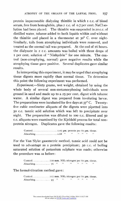
ATROPHY OF THE ORGANS OF THE LARVAL FROG. 157
protein impermeable dialyzing thimble in which I c.c. of blood serum, free from hemoglobin, plus 2 c.c. of 0.7 per cent. NaCl so- lution had been placed. The thimble was suspended in 20 c.c. of distilled water, toluene added to both liquids within and without the thimble and placed in a thermostat at 30? C. over night. Similarly, tails from atrophying individuals were removed, and treated as the normal tail was prepared. At the end of I6 hours, the dialyzate in 2 c.c. amounts was boiled with three drops of I per cent. solution of "Ninhydrin" for one minute. The con- trol (non-atrophying, normal) gave negative results while the atrophying tissue gave positive. Several duplicates gave similar results.
In interpreting this experiment, it maybe urged that atrophying tissue digests more rapidly than normal tissue. To determine this point the following experiment was performed.
Experiment.-Sixty grams, wet weight, obtained by using the whole body of several non-metamorphosing individuals were ground in sand and made up to a 25 per cent. digest with toluene water. A similar digest was prepared from involuting larvae. The preparations were incubated for five days at 37? C. Twenty- five cubic centimeter aliquots of the digests were pipetted into 50 c.c. tannic acid solution which was left to precipitate over night. The preparation was diluted to 100 c.c. filtered and 50 c.c. aliquots were examined by the Kjeldahl process for total non- protein nitrogen. Duplicates gave the following results:
Control ................... 07 gm. protein per 60 gm. tissue. Absorbing ................. .68 " " " " " "
For the Van Slyke gasometric method, tannic acid could not be used to advantage as a protein precipitant; 50 c.c. of boiling saturated solution of potassium sulphate was made; otherwise the procedure was as before:
Control ............. I mm. NH2 nitrogen per 60 gm. tissue. Absorbing .......... 76 " " " " " " "
The formol-titration method gave:
Control .............117 mm. NH2 nitrogen per 60 gm. tissue. Absorbing .......... 95 " " " " " " "
This content downloaded from 194.29.185.170 on Wed, 14 May 2014 11:16:27 AMAll use subject to JSTOR Terms and Conditions
WITHROW MORSE.
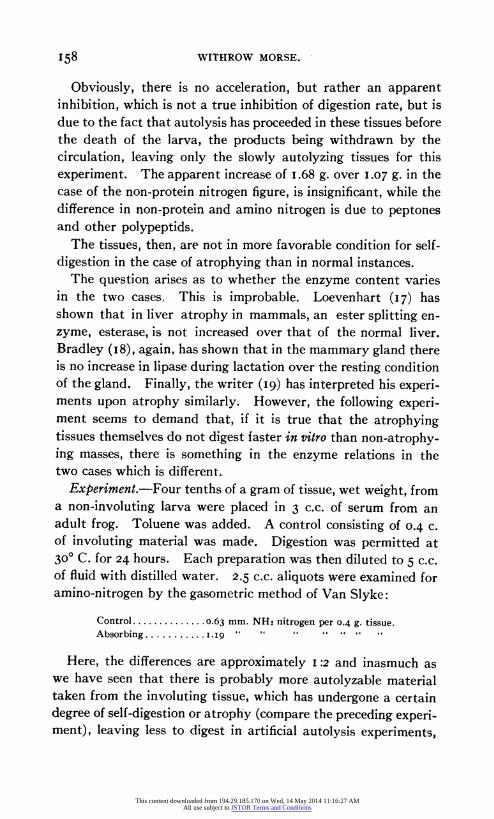
Obviously, there is no acceleration, but rather an apparent inhibition, which is not a true inhibition of digestion rate, but is due to the fact that autolysis has proceeded in these tissues before the death of the larva, the products being withdrawn by the circulation, leaving only the slowly autolyzing tissues for this experiment. The apparent increase of I.68 g. over 1.07 g. in the case of the non-protein nitrogen figure, is insignificant, while the difference in non-protein and amino nitrogen is due to peptones and other polypeptids.
The tissues, then, are not in more favorable condition for self- digestion in the case of atrophying than in normal instances.
The question arises as to whether the enzyme content varies in the two cases. This is improbable. Loevenhart (I7) has shown that in liver atrophy in mammals, an ester splitting en- zyme, esterase, is not increased over that of the normal liver. Bradley (i8), again, has shown that in the mammary gland there is no increase in lipase during lactation over the resting condition of the gland. Finally, the writer (I9) has interpreted his experi- ments upon atrophy similarly. However, the following experi- ment seems to demand that, if it is true that the atrophying tissues themselves do not digest faster in vitro than non-atrophy- ing masses, there is something in the enzyme relations in the two cases which is different.
Experiment.-Four tenths of a gram of tissue, wet weight, from a non-involuting larva were placed in 3 c.c. of serum from an adult frog. Toluene was added. A control consisting of 0.4 c. of involuting material was made. Digestion was permitted at 30? C. for 24 hours. Each preparation was then diluted to 5 c.c. of fluid with distilled water. 2.5 c.c. aliquots were examined for amino-nitrogen by the gasometric method of Van Slyke:
Control ............ .0.63 mm. NH2 nitrogen per 0.4 g. tissue. Absorbing ........ 9 .i.9.
" o " " " " .
Here, the differences are approximately I :2 and inasmuch as we have seen that there is probably more autolyzable material taken from the involuting tissue, which has undergone a certain degree of self-digestion or atrophy (compare the preceding experi- ment), leaving less to digest in artificial autolysis experiments,
158
This content downloaded from 194.29.185.170 on Wed, 14 May 2014 11:16:27 AMAll use subject to JSTOR Terms and Conditions
ATROPHY OF THE ORGANS OF THE LARVAL FROG. 159
the difference is significant. There are two substrates present in this system, namely, blood-serum proteins and secondly the tissue from the tail. The conclusion is warranted that there is a dif- ference in enzyme content between the atrophying and the normal tissue. Granting that the serum proteins are more readily digested by the enzymes of the tail issue, we are still driven to assume that the enzymes are different in the two cases.
We have seen, then, that correlative with histological dif- ferences, there are enzyme differences in the case of atrophying and non-atrophying material.
It is possible that the experiments described in the preceding paragraphs do not bear critically upon the problem as to what induces atrophy of the metamorphosing larva. It may be urged that phagocytes could be operative in each of these cases. To assume this, it would be necessary to demonstrate that within the relatively short period of 24 hours, phagocytes, working in vitro, could perform the task of digesting the tissue in the amounts given. We should at once seek to answer the question as to whether a marked increase of phagocytes is to be observed in atrophying material over the normal. The writer (20) has made differential counts of the blood cells of normal and involuting individuals with the result that no increase commensurate with the difference in digesting power in the two cases exists. That there is a concentration of phagocytes (polynuclear leucocytes) in the affected areas is evident from Fig. I, given above, and also from the work of Mercier; the actual number, however, remains the same.
THE CAUSES INDUCING ATROPHY.
Mammalian experiments at the hands of Martin Jacoby and many other investigators have shown that atrophy involving autolysis is induced when the blood supply is interfered with. It is not necessary to occlude the supply directly; interruption of the blood supply will serve to cause atrophy. Bataillon (2I) found that in the frog, the development of the pygostyle caused a change in the distribution of the blood supply throughout the tail. This does not involve complete occlusion, for Mercier found that, comparatively late in metamorphosis, phagocytes bearing carmin granules picked up from the dorsal lymph sac into which
This content downloaded from 194.29.185.170 on Wed, 14 May 2014 11:16:27 AMAll use subject to JSTOR Terms and Conditions

WITHROW MORSE.
Mercier had injected the grains, appeared in the muscle masses undergoing atrophy.
Granting that this explanation is adequate, we may seek the more immediate factor involved. The first suggestion may be that oxygen inhibits the activity of the autolyzing enzymes and that when the blood supply is interfered with, the inhibiting action of oxygen is thrown off, permitting autolysis. The experi- ments of the writer (22) speak decidedly against this theory of inhibition of autolysis by oxygen.1
The known relation between reaction of medium and autolysis, of which we have spoken earlier in the present paper is sufficient to afford an adequate explanation of what takes place at the inception of autolysis. Partial interruption of the blood supply everywhere results in an accumulation of carbon dioxid. The "buffer" value of the blood in alkalinity is soon neutralized and an actual acidity of the blood results (Ph<7). Perhaps, like- wise, as in starvation, acids other than carbonic acid enter the blood stream, such. as those of incomplete oxidation-the so- called acids of acidosis, ketonic acids, beta-hydroxybutyric acid, aceto-acetic acid and the keton aceton. Even in the total exclu- sion of respiratory oxygen, intramolecular oxidation occurs, giving rise to carbon dioxid and to the acids of acidosis. This the writer takes to be the modus operandi of atrophy in the larval frog.
With regard to oxygen, the following experiment of the writer bears:
Experiment.-Several tall glass cylinders, 45 cm. high and 5 cm. in diameter were filled with water and into each was intro- duced a single larva which gave no evidence of metamorphosis. The larva were fed algae, but owing to the darkened space in which the experiment was conducted, photosynthesis, giving rise to oxygen, did not occur. The jars were left for several weeks, during which time the larvae grew, but none of them exhibited any tendency towards metamorphosis. Controls in finger- bowls with oxygenated water metamorphosed.
1 The recent papers by Burge in the American Journal of Physiology contain the assumption of oxygen inhibition, but there is no experimental data to substantiate the theory.
I6o
This content downloaded from 194.29.185.170 on Wed, 14 May 2014 11:16:27 AMAll use subject to JSTOR Terms and Conditions

ATROPHY OF THE ORGANS OF THE LARVAL FROG. 161
Experiment.-Tap water was boiled and cooled, in hermetically sealed jars. Finger-bowls were filled with the water and larvae introduced. There was a high mortality amongst the larvae, but a few were kept for two weeks, during which time no meta- morphosis occurred.
Experiment.-The dorsal nerve cord together with the aorta was ligated with silk thread, at the base of the tail. Again, the mortality was high, the open cut being attacked by Saprolegnia and water moulds, wild yeasts, etc. No acceleration in meta- morphosis was observed.
Lack of oxygen, or a marked reduction in its amount does not induce autolysis in the larvae. It must be admitted, however, that the negative results of the last experiment do not bear out the theory that interference in the blood supply induces atrophy. Lateral circulation, however, may have been a factor.
Wintrebert (23), seeking the cause of atrophy in the larval frog, suggested that an internal secretion induces metamorphosis. The following experiment was directed towards the obtaining of evidence upon this point:
Experiment.-Five normal larvae were isolated in large petri dishes. Serum was injected into the dorsal lymph sacs of these larvae, the serum having been obtained either from the blood of involuting larvae or from the expressed juices of the tail. No attempt to keep the preparation sterile was made owing to the obviously impossible nature of the task. The results were negative as far as inducing metamorphosis.
Attention has been directed to the glands of internal secretion. Thus Babak (24) has studied the effects of injections of hypo- physis and of other portions of the brain. Positive results were obtained which this writer interpreted as due to direct effects upon the nervous system rather than upon the tissues. Babak considers that there is a hormone-action involved in the sense of Bayliss and Starling. The thyroid has received a relative large amount of attention. Gudernatsch originally observed a pre- cocious metamorphosis in the case of larvae fed upon thyroid preparations and since his work, others have studied the problem from the biochemical aspect. David Marine (25), Lenhart, the writer (26) and recently, from a more general physiological
This content downloaded from 194.29.185.170 on Wed, 14 May 2014 11:16:27 AMAll use subject to JSTOR Terms and Conditions
WITHROW MORSE.
aspect, Allen (27) have examined the relations of the thyroid and thyroid components upon metamorphosis. The writer found that an iodized amino-acid (3-5-di-iodo-tyrosin, C6H50HI2CH2.CH. NH2.COOH), induces metamorphosis in the frog's larvae, this amino-acid being derived from the thyroid tissue by means of acid hydrolysis and hence being a normal component of the thyroid. These observations taken in connection with the interesting morphological studies of Allen give us a rationale of the r61e of the thyroid in inducing metamorphosis. When thy- roid tissue or its components is fed to an organism, the general metabolism is increased as evidenced by the increased output of the end products of protein, carbohydrate and fat metabolism. When thyroid is removed from the economy of the organism, as in Allen's thyroidectomv experiments, metabolism is decreased; this is indicated by his further observations that reduced nourish- ment obtains the same results, that is, in both cases metamor- phosis is suppressed or delayed. We may conclude, then, that metamorphosis involves, as one of its principal factors, heightened metabolism.
It is necessary to distinguish between heightened metabolism, which involves only catabolism, in Gudernatsch's experiments and the accelerated anabolism present in Allen's cases. In both instances, metabolism is heightened, but the current is down stream in the one case, leading to loss of storage materials and a piling up of materials in the form of fat, etc., in the second case. Now it is of interest to recall that according to Voegtlin and Strouse, the iodized amino-acid which was found to operate as whole thyroid tissue in inducing precocious metamorphosis, fails to replace the function of thyroid in those pathological cases where there is a deficiency in thyroid function. It would be of considerable interest to determine whether 3-5-di-iodo-tyrosin, thyreoglobulin or any of the chemical components of the intact thyroid tissue replaces the absent thyroid in cases of thyroid- ectomy in inducing metamorphosis, for wrapped up with the question is the important and fundamental one as to what rela- tion the iodine has in the thryoid. If the iodized amino-acid should in itself, in the absence of the thyroid, induce metamor- phosis, it would show that the theory that iodine stimulates an
I62
This content downloaded from 194.29.185.170 on Wed, 14 May 2014 11:16:27 AMAll use subject to JSTOR Terms and Conditions

ATROPHY OF THE ORGANS OF THE LARVAL FROG.
internal secretion in the thyroid gland, rather than functioning as hormone itself, is untenable. In either case it would clear up the apparent discrepancy in the findings of Voegtlin and Strouse and in those of the students of amphibian metamorphosis.
THE ECONOMY OF THE PROCESS.
In the investigations upon the salmon conducted by Miescher and later by Noel Paton and others, it is evident that there is a high degree of economy involved in the process of metamorphosis of the muscles into gonad material. The question arises, is this true for the metamorohosis of the larval frog?
In the foregoing paragraphs, we have spoken of this process of metamorphosis as if it were confined to the regions of the tail, but the difficulty of studying the participation of the alimentary tract, gills, etc., in the process is not a small one and these inves- tigations have been confined to the changes in the tail. One primary difficulty regarding the study of the alimentary tract is that at metamorphosis, preceding the shortening of that sys- tem, a large accumulation of fecal matter is expelled, containing the same chemical compounds which are utilized in studying the transformations of tissues. It is for this reason that the simple expedient of weighing the organism before and after metamor- phosis to determine the loss, if any, of material, is not feasible, the fecal matter far overbalancing any amount of product of tissue change thrown to the outside.
A study was made of the excretions of the larvae immediately after the expulsion of the fecal matter. Larvae were isolated in distilled water in large petri dishes. After twelve hours, the water was filtered and estimations were made of the total nit- rogen, amino-nitrogen, ammonia and urea. While the method was crude, owing to the impossibility of inhibiting the transfor- mation of urea into ammonia and of the probable deaminization of the amino-acids by bacteria, there was no constant difference in the figures for non-metamorphosing and metamorphosing lavr2e. Unless, then, there were extensive excretion of the products of the decomposition of the larval tissues, it is safe to conclude that the process is an economic one, the tissues of the larval organs contributing to the formation of those of the adult.
I63
This content downloaded from 194.29.185.170 on Wed, 14 May 2014 11:16:27 AMAll use subject to JSTOR Terms and Conditions
WITHROW MORSE.
Pointing to the correctness of this interpretation is the fact that during the development of the adult organs (legs, etc.) no food is taken by the larva. Storage of fat, etc., has taken place during the earlier period of larval existence, but the impor- tant thing is to remember that protein is never stored as such in the animal organism. The nitrogenous material for the legs, etc., developing during metamorphosis must be supplied from some source other than storage. The simplest explanation is to assume that it is derived from the larval organs.
VON BAER'S LAW AND LARVAL METAMORPHOSIS.
The frog has been taken as a classic example of the operations of the so-called Law of Von Baer, termed sometimes the Law of Recapitulation and at others that of Repetition. The question is, can the metamorphosis be explained wholly upon the assump- tion that physico-chemical events other than those concerned with heredity initiate the process?
Those who have experimented with the agents accelerating metamorphosis have found that these compounds are impotent except in the case of larvae which have reached a certain period of their existence. In other words, we know of no agent which is operative regardless of the stage of development of the larvae. We may suppress metamorphosis; we can not, or have not in- duced it in stages far removed from those in which it would nor- mally occur. This indicates that a certain cycle of events, probably determined by heredity, are necessary before any stimu- lating agent is effective. The growth of the pygostyle according to Barfurth, initiates the process of atrophy in the tail, but what events stimulate the growth of the pygostyle are unknown. In some species, which do not metamorphose during the first sum- mer, the larvae are subjected to relatively the same stimuli from the environment during the second summer as during the first. It is improbable that external conditions determine the time of metamorphosis. Again, to offer the explanation that hormones or enzymes initiate metamorphosis leaves open the more potent question as to what determines the development of these com- pounds.
i64
This content downloaded from 194.29.185.170 on Wed, 14 May 2014 11:16:27 AMAll use subject to JSTOR Terms and Conditions

ATROPHY OF THE ORGANS OF THE LARVAL FROG. 165
CONCLUSIONS.
I. Autolysis is the primary factor in the physiology of atrophy in metamorphosis.
2. This process is probably induced by an acidosis in the tissues. 3. Adopting Barfurth's suggestion of the occlusion of the
blood vessels at the base of the tail by the growth of the pygos- tyle, it is probable that carbon dioxid and acids of incomplete combustion accumulate and render the reaction of the blood less alkaline or more acid.
4. Phagocytosis is not a primary factor, as Metchnikoff be-
lieved, but a secondary one, the phagocytes being attracted by chemotaxis to the atrophying organs.
5. The process of metamorphosis is economic, the substances derived from the atrophying organs being utilized by the growing adult organs.
REFERENCES. I. Morse, Max.
1i2 The Autotomy of the Hydranth of Tubularia. BIOL. BULL. 2. Metchnikoff, Elie.
'83 Untersuchungen fiber die Mesodermalen Phagocyten einiger Wirbel- tiere. Biol. Centralb., III., 560.
3. Barfurth, D. '87 Die Riickbildung des Froschlarvenschwanzes und die sogenannten
Sarcoplasten. Arch. f. mikr. Anat., XXIX., 55. 4. Looss, A.
'89 Ueber Degenerations-Erscheinungen im Thierreich, usw. Presi- schriften von der Ftirstlich Jablowoniski'schen Gesellschaft in Leipzig, XXVIII., Verlag von S. Hirzel.
5. Noetzel, W. '95 Die Riickbildung der Gewebe im Schwanzeder Froschlarve. Arch. f.
Mikr. Anat., XLV., 475. 6. Anglas, J.
'04 Observationes sur les metamorphoses internes des batracienc anoures. Assoc. franc pour l'avancement des sciences, 33e session, Grenoble, p. 855.
7. Guieweysse, A. 'o5 Etude de la r6gression de la queue chez les tetards des amphibiens
anoures. Arch. de l'Anatomie microsc., VII., 429. 8. Mercier, L.
'o6 Les processus phagocytaires pendent la m6tamorphose des batraciens anoures et des insectes. Arch. d. zool. exper. et g6ner., IV., Ser. v, I.
9. Grund, G. 'i2 The Chemical Changes of Muscle under the Influence of Degeneration.
Zentralb. f. inn. Med., XXXIII., I73. 'i2a Chemical Pathology of Muscle. II. Nitrogen and Phosphorus Dis.
This content downloaded from 194.29.185.170 on Wed, 14 May 2014 11:16:27 AMAll use subject to JSTOR Terms and Conditions

i66 WITHROW MORSE.
tribution in Degenerated Muscle. Arch. f. exper. Pathol. u. Pharmak., LXVII., 39I.
10. Miescher, F. Histochemisches und physiologisches Untersuchungen. Heft IV.
II. Paton, Noel. '98 Report of the Investigations on the Life-history of the Salmon in
Fresh Waters. Glasgow: James Hedderwick and Sons. I2. Steiner, W. R.
'o04 Dermatomyositis. Journ. Exp. Med., VI. I3. Bradley, H. C. et al.
'15ff Papers in the Journal of Biological Chemistry. 14. Bradley, H. C. and Taylor, J.
'i6 Studies of Autolysis. III. The Effect of Reaction on Liver Autolysis. Journ. Biol. Chem., XXV., 26I.
15. Straus, D. C. and Morse, M.
'17 The R61le of Autolysis in Infarction. Proc. Soc. Exp. Biol. and Med. I6. Morse, M.
'I5 The Amino-acid Content of Involuting Frog Larvae. Proc. Soc. Exp Biol. and Med.
17. Loevenhart, A. S. '02 Lipase and Enzyme Reaction. Amer. Journ. Physiol., VI., 33I.
18. Bradley, H. C. 1I3 The Problem of Enzyme Synthesis. Journ. Biol. Chem., XIII., 407.
19. Morse, M.
14 Atrophy Does not Involve Acceleration of Tissue Enzyme Action. Proc, Soc. Exp. Biol. and Med., XII., 46.
20. Morse, M. 'I2 The R6ole of Phagocytosis in Involuting Organs. Proc. Soc. Exp. Biol.
and Med., X., 3I. 21. Bataillon, E.
'91 Recherches anatomiques et experimentales sur la m6tamorphose des Amphibiens anoures. Ann. de l'Universit6 de Lyon, T. 2.
22. Morse, M. '16 Oxygen Relations in Autolysis. Biochem. Bull., V., I43.
23. Wintrebert, P. 'o8 Une demi-metamorphose experimentale. Comptes rendus, Paris,
LX., 415, I908 and LII., 52I, 1907. 24. Babik, E.
'o5 Ueber die Beziehung des zentralen Nervensystems zu den Gestaltungs- vorgangender Metamorphoses des Frosches. Pfluger's Arch., CII., 78.
25. Marine, David. 'i6 Journ. Pharmacology and Exper. Ther., IX., 57.
26. Lenhart, C. H. 'I5 Journ. Exp. Med., XXII., 739.
27. Morse, M. '14 The Effective Principle in Thyroid Accelerating Involution in Frog
Larvae. Journ. Biol. Chem., XIX., 42I. 28. Allen, B. M.
'17 The Effects of Thyroid Removal upon the Development of the Gonads in the Larvae of Rana pipiens. BIOL. BULL., XLVI., 216.
This content downloaded from 194.29.185.170 on Wed, 14 May 2014 11:16:27 AMAll use subject to JSTOR Terms and Conditions