factors affecting the growth and survival of didymosphenia … · 2017-02-14 · factors affecting...
TRANSCRIPT

Factors affecting the growth and survival of Didymosphenia geminata in rivers and their spring fed tributaries: update of experimental investigations to June 2010
NIWA Client Report: CHC2010-094 June 2010 NIWA Projects: DOC10504 CF105047 FGC10502 MEL10510


© All rights reserved. This publication may not be reproduced or copied in any form without the permission of the client. Such permission is to be given only in accordance with the terms of the client's contract with NIWA. This copyright extends to all forms of copying and any storage of material in any kind of information retrieval system.
Factors affecting the growth and survival of Didymosphenia geminata in rivers and their spring fed tributaries: update of experimental investigations to June 2010 Cathy Kilroy Max Bothwell NIWA contact/Corresponding author
Cathy Kilroy
Prepared for
Department of Conservation NZ Fish & Game Meridian Energy Internal use NIWA Client Report: CHC2010-0 June 2010 NIWA Projects: DOC10504, CF105047, FGC10502, MEL10510 National Institute of Water & Atmospheric Research Ltd 10 Kyle Street, Riccarton, Christchurch 8011 P O Box 8602, Christchurch 8440, New Zealand Phone +64-3-348 8987, Fax +64-3-348 5548 www.niwa.co.nz


Contents Executive Summary i
1. Introduction 1
2. Phases 1 and 2: Research up until June 2009 2 2.1. Experimental channels 2 2.2. Phase 1: Why doesn’t didymo grow in spring-fed creeks? 2 2.3. Phase 2: Factors affecting didymo growth and blooms 3 2.4. Outputs 5
3. Phase 3: Research from July 2009 to June 2010 5 3.1. Effects of light and nutrients on cell division rates and stalk
lengths 5 3.2. Validating a method for determining cell division rates in
didymo 5 3.2.1. Introduction 5 3.2.2. Methods 6 3.2.3. Results 8 3.2.4. Conclusion 10 3.3. Colonisation patterns 10 3.3.1. Methods 12 3.3.2. Data analysis 13 3.3.3. Results 14 3.3.4. Discussion 17 3.4. The role of organic phosphorus 19 3.4.1. Methods 19 3.4.2. Results 20 3.4.3. Discussion 22 3.5. Response of didymo to micro-nutrient additions in spring-fed
creeks 23 3.5.1. Methods 24 3.5.2. Results 26 3.5.3. Discussion 28 3.6. Long-term N, P and N+P enrichment trial 29 3.6.1. Results 30 3.6.2. Discussion 32
4. Recommendations 32
5. Acknowledgements 33

6. References 34 Appendices: Draft papers
Reviewed by: Approved for release by:
JoAnna Lessard Ton Snelder

Factors affecting the growth and survival of Didymosphenia geminata in rivers and their spring fed tributaries: update of experimental investigations to June 2010
i
Summary
• Streamside experimental channels set up at the confluence of the Otiake Spring Creek and the Waitaki River in late 2008 have been used to investigate (1) the observed absence of the non-indigenous stalked diatom Didymosphenia geminata (didymo) from spring-fed creeks; and (2) the conditions under which didymo forms blooms.
• Experiments carried out in 2008 – 2009 (described in full in previous reports) are summarised. Two papers largely based on these experiments have been submitted to international journals, and drafts are appended to this report.
• In the first paper, we demonstrated that didymo cell division rates in the Waitaki River water are phosphorus (P)-limited, despite the fact that the river supports large blooms. In the second paper we present experimental results consistent with the hypothesis that extensive stalk production in didymo occurs when cell division rates are nutrient-limited and photosynthetic rates are high. In other words, in high light didymo cells photosynthesise fast, but with insufficient P, most of the photosynthate is diverted into stalk production rather than new cells. These stalks make up almost all the material in the blooms.
• The 2008 research also included nutrient diffusing substrate trials, the results of which suggested that didymo could be absent from the Otiake Spring Creek because a required micro-nutrient was absent from the water.
• The experiments carried out in 2009 -2010 are described in six sections.
o Stalk length and cell division rates under a range of environmental conditions (continuation). Experiments tracking the response of didymo to high and low levels of inorganic phosphorus confirmed earlier conclusions that cell division rates in Waitaki River water are P-limited.
o A method for determining the cellular-level response of didymo to environmental changes. In 24- and 48 hour experiments, it was demonstrated that the frequency of dividing cells (FDC) in didymo is constant regardless of time of day and can therefore be readily used as a measure of population growth and community “health”.
o Exploratory investigations on colonisation patterns under different levels of light and nutrient availability. Experiments under UV-blocking screens showed that colonisation rates were enhanced by both light and nutrients, but there was no light effect if nutrients were very low or very high, and there was no nutrient effect in the dark. In the presence of UV (full ambient light) there were also no nutrient effects.

Factors affecting the growth and survival of Didymosphenia geminata in rivers and their spring fed tributaries: update of experimental investigations to June 2010
ii
This highlighted the general question of whether levels of UV exposure are linked to didymo growth and blooms in other ways.
o Can the “success” (i.e., the blooms) of didymo in oligotrophic rivers be attributed to its ability to preferentially utilise organic sources of phosphorus? This hypothesis may be relevant to didymo absence in the North Island, where rivers tend to have high inorganic and low organic P, in contrast to the opposite pattern on the South Island. Experimental additions of an organic form of P did not stimulate rates of didymo colonisation or cell division more than additions of the same concentration of inorganic P. Furthermore, our earlier experiments indicated that blooms of didymo occur under P-limitation. We conclude that the inorganic/organic phosphorus hypothesis does not explain the occurrence of didymo blooms, or why didymo is not yet present in the North Island.
o Nutrient-diffusing substrate trials to determine the responses of didymo to micro-nutrient additions in the spring-fed creek. More detailed nutrient diffusing substrate trials were repeated in the Otiake Spring Creek, Waitaki River and the Ohau Spring Creek, which flows into the Lower Ohau River. In autumn 2010 we were unable to replicate the strong responses with trace metal and vitamin mixtures which were observed in spring 2008. In the Otiake Spring Creek didymo cell densities showed a moderate response to vitamin B additions, suggesting that this micronutrient might be limiting in the Spring Creek. However repeat trials in the Ohau Spring Creek and a second trial in the Otiake Spring Creek, were negative.
o Complete nutrient addition experiment to investigate longer-term effects of nutrients on didymo FDC and cell densities. In April 2010, we ran a 10-day experiment to compare the effects of added N, P and N+P on didymo growth. The results confirmed P-limitation in cell division rates in unamended Waitaki River water.
• The research in 2009–10 raised several further questions. Based on these questions we suggest continuation of the research into: (a) the range of didymo FDC in natural populations to aid prediction of blooms in relation to nutrient availability; (b) the question of whether high UV promotes didymo dominance; (c) the role of other potentially limiting nutrients with respect to FDC and stalk length; (d) the possibility that vitamin B12 may limit didymo growth in some waterways, and the possible role of other taxa (e.g., the cyanobacterium Phormidium) in didymo colonisation and growth.

Factors affecting the growth and survival of Didymosphenia geminata in rivers and their spring fed tributaries: update of experimental investigations to June 2010 1
1. Introduction
The pattern of the invasive alga Didymosphenia geminata (didymo) growing prolifically in rivers, but not at all in their spring-fed tributaries, has been observed all over the South Island (e.g., Sutherland et al. 2007). This pattern was first noticed in the Mararoa River in 2005 before didymo was discovered in other rivers, and still persists despite many opportunities for colonisation of these tributaries. As didymo has spread to other South Island rivers, it has also become clear that some systems are resistant to colonisation. For example, to date no didymo growth has been sighted in the Taieri River, Otago, despite heavy infestation of the surrounding catchments. Similarly, Fraser Stream, a tributary of the didymo-affected Twizel River has consistently tested negative for didymo in an ongoing monitoring programme, even though there are obvious colonisation pathways from the Twizel. These disjunct patterns of didymo distribution in the South Island led us to ask whether the continued absence of didymo from the North Island could be caused by environmental factors, rather than a lack of vectors from the South Island.
Research specifically to investigate the absence of didymo from spring-fed creeks was initiated in 2008. Experimental channels were established at the confluence of the Waitaki River and the Otiake Spring Creek, near Kurow, South Canterbury. Over the course of the experiments, thick mats of didymo were present in the river almost constantly, but didymo was completely absent from the spring creek, except during a brief period in November 2009 when water from the didymo-infested Otiake River overflowed into the creek. Shortly after the overflow ceased, didymo disappeared from the creek.
Experiments were conducted to determine the response of didymo to spring water and to river water augmented to the higher levels of nutrients (dissolved nitrogen and phosphorus) found in the spring water. Nutrient addition experiments were also conducted in the spring creek (Bothwell et al. 2009; Bothwell and Kilroy 2009). In 2009, the scope of the research was expanded to investigate the conditions under which didymo blooms (Bothwell and Kilroy 2009). This has been a puzzle because didymo blooms in rivers with very low nutrient loads. Most algal blooms are caused by high nutrients.
In this report we first provide a summary of the results of the research up until June 2009, described in detail in Bothwell and Kilroy (2009). We then report on the experiments conducted in 2009 – 2010, the aims of which were five-fold, as follows.
1. Continue experiments to determine stalk length and cell division rates under a range of environmental conditions;

Factors affecting the growth and survival of Didymosphenia geminata in rivers and their spring fed tributaries: update of experimental investigations to June 2010 2
2. Validate a method for determining the cellular-level response of didymo to environmental changes;
3. Conduct exploratory investigations on colonisation patterns under different levels of light and nutrient availability;
4. Experimentally investigate a theory which proposes that the “success” (i.e., the blooms) of didymo in oligotrophic rivers can be attributed to its ability to utilise organic sources of phosphorus;
5. Repeat in-situ nutrient-diffusing substrate trials to determine the responses of didymo to micro-nutrient additions in the spring-fed creek.
2. Phases 1 and 2: Research up until June 2009
2.1. Experimental channels
The channels set up at the confluence of the Waitaki River and the Otiake Spring Creek were constructed to the design used by Bothwell et al. 1988. The system is described in detail in Bothwell and Kilroy (2009a, b). Briefly, the channels were designed so that water could be pumped from two separate sources (in this case the Otiake Spring Creek and the Waitaki River) through to up to twelve channels. The water was set to flow at approximately 30 litres per minute in each channel, which is equivalent to a water depth of 1.2 mm, and a water velocity of 0.2 m/s. A 10-litre mixing tank at the head of each channel permitted thorough mixing of dropwise nutrient additions before entering the channels. For all the experiments, we allowed didymo to colonise a substrate of 6-mm thick Styrofoam by running river water through the system for up to four weeks. Before starting each experiment, we brushed the Styrofoam to clear accumulated deposited algae and other debris.
2.2. Phase 1: Why doesn’t didymo grow in spring-fed creeks?
For these experiments, our alternative hypotheses were: (a) there is something in spring water which kills didymo; (b) there is something missing from spring water which didymo requires; (c) the physical conditions in spring creeks (smooth non-turbulent flow, frequent shading, etc.) are unsuitable for didymo growth.
(a) Does spring creek water kill didymo?
After colonising 6 channels with Waitaki River water, we switched three channels to spring water. In experiments running for up to 100 days we found that spring water

Factors affecting the growth and survival of Didymosphenia geminata in rivers and their spring fed tributaries: update of experimental investigations to June 2010 3
stimulated didymo growth (measured as cell division rates – see Section 3.2), rather than killing it. Further trials showed that this was due to high nutrient (nitrogen N and phosphorus P) levels in the spring water compared to extremely low levels in the Waitaki River. Measured NO3-N and total dissolved P in the spring and river were ~500 and 6 mg/m3, and 4 and <1 mg/m3, respectively.
(b) Are required micronutrients missing from spring water?
We carried out in situ trials in the spring creek using precolonised Styrofoam circles inserted over nutrient reservoirs so that nutrient solutions diffused over the substrate. We found that didymo died off after 2 weeks, unless it was supplied with a mixture of micronutrients (metals, vitamins, etc.).
(c) Are physical conditions unsuitable?
In April 2009, we investigated the effect of shading on didymo growth in the experimental channels and found that cell division rates are depressed under shaded conditions (see Section 2.3 below). However, this cannot explain the absence of didymo from shaded creeks, especially in view of the fact that in October 2009 we observed didymo growing in the Otiake Spring Creek, when the spring water was diluted by a river water overflow. The unsuitable physical conditions hypothesis can also be largely discounted by other observations: there are examples of didymo absence in spring-fed creeks that are completely unshaded and have fast, turbulent flows and stable cobble substrates.
Conclusion: The higher levels of nutrients in the spring creek stimulate didymo growth in the short term, but in the longer term, growth declines. We suspect that one or more required micronutrients may be missing from the spring waters. Physical factors (light) could also be unfavourable, but cannot explain absence. The combined results indicated that further nutrient diffusing substrate (NDS) trials would be needed to determine which micronutrient(s) might stimulate didymo growth in spring-fed creeks. Parallel NDS trials in the river would also be necessary.
2.3. Phase 2: Factors affecting didymo growth and blooms
The experiments using spring water and river water enriched with inorganic nitrogen and phosphorus showed that cell division rates increased with nutrient additions (see Section 2.2). Furthermore, samples of didymo from mats within the river confirmed a gradient of cell division rates across the mixing zone of the spring creek with the river. Cell division rates were low in the zone of 100% river water, and increased as the proportion of spring water increased. At the same time, there was a gradient of growth form from continuous thick mats in 100% river water, to small scattered colonies in

Factors affecting the growth and survival of Didymosphenia geminata in rivers and their spring fed tributaries: update of experimental investigations to June 2010 4
approx. 60% spring water, and no visible colonies in 100% spring water. This raised the question of the nutrient status of didymo in the Waitaki River. We therefore asked: how do nutrients affect (a) cell division rates and (b) stalk development in didymo? Because light levels also appear to be important in determining the extent of didymo growth, we also investigated the effects of shading.
(a) Effect of nutrients and shading on rates of cell division
Using 12 channels we examined the effects on didymo cell division of adding inorganic nitrogen (N) and inorganic nitrogen + phosphorus (N+P). We found that additions of N temporarily increased cell division rates, but rates returned to ambient after 4 days. The concentration of N addition (15, 50 and 500 mg/m3) had no effect. In contrast addition of N+P (one concentration only) resulted in increased cell division that was sustained at 4 days. In both experiments, shading led to reduced cell division rates in both the control and nutrient enriched treatments.
These experiments, combined with the data from the river transect, suggested that in ambient river water, cell division rates were limited by low phosphorus levels. This presents a paradox: how could didymo grow so prolifically in the river, when its cell division rates were very low due to low levels of phosphorus? This prompted us to look at the main constituent of didymo blooms – the stalks.
(b) Effect of nutrients and light on stalk length
Didymo stalks are primarily composed of polysaccharide, and are exuded from a field of tiny pores located at the narrow apex of the cell. They provide the cell with an attachment to the substratum, but in blooms form the bulk of the biomass with cells concentrated at the surface of the mats. When cells divide each new cell starts exuding its own stalk, creating a bifurcation in the stalk. To examine differences in stalk production we measured stalk lengths, focusing on the lengths from the cell back to the nearest stalk bifurcation (i.e., the most recent growth).
We found that the amount of stalk material is closely linked to cell division. The faster the cells divide, the shorter the stalks; the slower the cell division, the longer the stalks. Because high nutrients stimulate cell division, stalk production is limited where nutrient levels (especially phosphorus) are high. In contrast, when levels of P are low enough to limit cell division, the cells continue to produce long stalks.
Conclusion: Where there is low phosphorus, but high light (as in the P-limited conditions of the Waitaki River), we think that didymo cells photosynthesise fast, but can’t translate all the photosynthate into new cells without more phosphorus. So the cells produce lots of stalk material. These stalks make up almost all the material in the blooms. This process at least partially explains the ability of didymo to develop blooms in low nutrient rivers.

Factors affecting the growth and survival of Didymosphenia geminata in rivers and their spring fed tributaries: update of experimental investigations to June 2010 5
2.4. Outputs
The results of the research in 2008 – 09 have been written up as two papers, which have been submitted for publication in international journals. Some data obtained during experiments carried out in 2009 – 10 was used to reinforce the conclusions from the first round of experiments (see notes in Section 3.1 below). The two draft papers are included as Appendices 1 and 2 in this report.
3. Phase 3: Research from July 2009 to June 2010
3.1. Effects of light and nutrients on cell division rates and stalk lengths
This was a continuation of Phase 2. In the experiments outlined in Section 2.3 above we looked at the effects of additions of N (three levels) and N+P (one level) on FDC and stalk length. In November 2009, we ran a further experiment, using exactly the same experimental set-up but this time adding P (two levels). This experiment confirmed the result of the N+P addition, and the combination of the three experiments indicated P-limitation for didymo in Waitaki River water. For more details refer to the papers in Appendix 1.
3.2. Validating a method for determining cell division rates in didymo
3.2.1. Introduction
In the first phase of the present series of experiments, the following were considered as possible ways to measure didymo responses to our experimental treatments.
1. Chlorophyll a and ash-free dry mass (AFDM) (as weight per unit area) are routinely used as surrogates for the amount of live algal material in a periphyton sample, and the total amount of organic material present, respectively. These methods are appropriate if we are interested in the response of a mixed algal community, but do not necessarily apply to single species in that community, which was what we were considering in the present experiments
2. In previous research, the “health” of didymo in samples has been assessed by determining the proportion of viable cells present, using a staining method that distinguishes live cells from non-viable cells (Kilroy et al. 2007). This method is suitable for assessing the effect of biocides on didymo cells (e.g., Jellyman et al. in press). However, it is not suitable for measuring responses to environmental changes because: (a) proportions of non-viable cells may be affected by a range of factor, for example influxes as a result of flow changes;

Factors affecting the growth and survival of Didymosphenia geminata in rivers and their spring fed tributaries: update of experimental investigations to June 2010 6
(b) both viable and non-viable cells can be lost due to release from the stalk. As a result, simple proportions of live cells may be meaningless unless they can be converted to a more quantitative measure. For example, in Sutherland et al. (2007) a combination of % viable cells, proportional biovolume of cells in samples, and AFDM was used to estimate the biomass of live cells in a sample, per unit area.
3. Quantitative cell counts are a standard and defensible way of determining species responses to environmental changes. While straightforward, they require rigorous collection of quantitative samples, and can also be highly variable because of community patchiness. Therefore many replicates may be required.
Because of the various drawbacks of the above methods, we started using a technique that is well-established in marine plankton ecology: estimation of cell division rates from counts of the frequency of dividing cells (FDC). FDC was used as a response variable in the experiments summarised in Sections 2.3 above. An issue with FDC is that cell division is a light-driven process (i.e., photosynthetically driven). Therefore it is possible that there could be diel patterns of FDC. In other words, under otherwise uniform environmental conditions the proportions of dividing cells could vary throughout the day. The implication is that, to be comparable, samples must always be collected at the same time of day. This was first investigated in a 24-hour experiment on 11-12 May 2009 and later followed by a 48-hour experiment on 13-15 November 2009.
3.2.2. Methods
24-hour Diel study
At the end of the inorganic N+P enrichment FDC trial (6-10 May 2009), a section of one of the enriched channels (target concentrations of 100 and 10 mg/m3, respectively) recieving 100% light exposure was selected for a 24 diel study of FDC. Beginning at 0600h on 11 May, six replicate cores were taken every four hours, fixed in Lugol’s solution, and cells in different stages of division were enumerated.
48-hour Diel study
In November two experimental channels were allowed to colonise with a didymo-dominated community by running Waitaki River water through them for ~5 weeks. Two days before starting the experiment the water supply to one channel was enriched with inorganic N and P with target concentrations of 100 and 10 mg/m3, respectively. Additions were made using a peristaltic pump. In addition, a section of the enriched

Factors affecting the growth and survival of Didymosphenia geminata in rivers and their spring fed tributaries: update of experimental investigations to June 2010 7
channel was shaded by covering the top and sides with 8 layers of shade-cloth so that approximately 1% of ambient light reached the substrate. The initial purpose of the enrichment was to ensure that there were sufficient cells to be able to accurately determine percentages of dividing cells. The control treatment was included to check the effect of different cell division rates. The shaded treatment was to verify previous observations that cell division rates stall at a certain stage if deprived of light.
Starting at 1800 h on 13 November 2009, six replicate samples were collected from each of the three treatments (control – light, enriched – light, enriched – dark), and preserved in Lugol’s iodine. Samples were collected in the same way at 4 hourly intervals until 1800 h on 15 November 2009.
For each sample, the frequency of dividing cells was determined by enumerating at least 150 cells, and placing each into one of the following categories:
• Non-dividing cell.
• Stage 1. The chloroplast has divided and the frustule has begun to enlarge (widened). Incipient indentation between daughter cells is only visible at the apical cell ends in girdle view. No partitioning cell wall is apparent in the dividing cell and the stalks do not differ in appearance from non-dividing cells. In valve (top) view, Stage 1 is easily missed. Visualization of Stage 1 is enhanced with Lugol’s fixative.
• Stage 2. A cell wall separating the newly forming daughter cells appears. This stage is unmistakeable in girdle (side) view and even oblique valve views. The greater depth of the cells in Stage 2 usually makes them lie in girdle view.
• Stage 3. The development of daughter cells is complete and they are now full-sized cells with separate chloroplasts and separate frustule walls, held together by an outer membrane. The stalks are also much wider. In this stage didymo always rests in girdle view.
• Stage 4. The outer membrane has broken and two fully formed daughter cells with basal ends close together, and distal ends widely separated, sit at the end of a single stalk. Eventually, stalk production pushes the basal ends apart. Under very low light (low photosynthetic rate) didymo cells can accumulate at this stage.

Factors affecting the growth and survival of Didymosphenia geminata in rivers and their spring fed tributaries: update of experimental investigations to June 2010 8
3.2.3. Results
In the 48 hour trial, Stages 1 and 2 tended to be higher in relative abundance during the day while Stages 3 and 4 tended to be higher at night (Figure 1). This pattern suggests that mitosis occurs during the night-early morning hours before sunrise and that cytokinesis begins during the morning and progresses from Stage 1 to Stage 2 during the daylight hours. During hours of darkness, either at night or very low light during the day, Stage 4 (cell pairs without stalks) cells accumulate under both enriched and non-enriched conditions (Figures 2, 4). Under both enriched and non-enriched conditions the sum of doublets (Stage 2 and Stage 3) used to compute the FDC does not change over the diel cycle indicating that the FDC is not biased by the time of day the samples are taken (Figures 3, 4 ).
Figure 1: Diel cycle of didymo dividing cells at stages 1+2 and Stages 3+4 in the Control (unenriched) channel during 48 h experiment 13-15 November 2009. Histograms are hourly light intensity. Darkened areas indicate night time.

Factors affecting the growth and survival of Didymosphenia geminata in rivers and their spring fed tributaries: update of experimental investigations to June 2010 9
Figure 2: Diel cycle of didymo dividing cells at Stage 4 in sections of the enriched channel exposed to 100% and 1% ambient light during 48 h experiment 13-15 November 2009. Histograms are hourly light intensity. Darkened areas indicate night time. This illustrates the accumulation of cell pairs without stalks (Stage 4) during the night or under reduced light levels during the day.
Figure 3: Diel cycle of didymo dividing cells at Stage 2+3 in the Control (unenriched) channel during 48h trial 13-15 November 2009. Histograms are hourly light intensity. Darkened areas indicate night time. The summation of doublets (Stages 2+3) fluctuates but shows no systematic day-night trend.

Factors affecting the growth and survival of Didymosphenia geminata in rivers and their spring fed tributaries: update of experimental investigations to June 2010 10
Figure 4: Diel cycle of Stage 2+3 in the enriched channel during 24h trial 11-12 May 2009. Histograms are hourly light intensity. Darkened areas indicate night time. The summation of doublets (Stages 2+3) shows no obvious day-night trend. Also shown is the decline of Stage 4s during the daylight period and the increase at night.
3.2.4. Conclusion
The frequency of dividing cells (FDC) should be counted as the proportion of Stage 2 + Stage 3 dividing cells. These are cells that are obviously dividing (doublets), but are not yet split into two cells. We found that the number of doublets fluctuated over time but not in a systematic way with time of day. On the other hand Stage 4 cells showed strong diel periodicity, and larger fluctuations than seen in counts of doublets.
Consequently, FDC expressed as a percentage of total cell numbers is calculated by:
% FDC = (#doublets / (#doublets + #non-dividing cells)) * 100
where: doublets = Stage 2 + Stage 3.
Stage 4 cells are considered non-dividing cells in the computation of the FDC.
3.3. Colonisation patterns
The ability of didymo to bloom in nutrient poor rivers can be explained at least partially by excessive stalk production under phosphorus limitation. However, this

Factors affecting the growth and survival of Didymosphenia geminata in rivers and their spring fed tributaries: update of experimental investigations to June 2010 11
process does not explain mass colonisation by didymo cells, which may be a precursor to large blooms. For example, if a mat is peeled back from a stone, multiple attachments are evident (Figure 5). This implies that many cells colonised the stone at around the same time. Alternatively, initial colonisation by just a few cells could result in small colonies which shed cells that colonise the bare surfaces in their immediate surroundings, as suggested by Whitton et al (2009).
Figure 5: Didymo mat peeled away from a river stone showing multiple stalk attachments.
As a first step to understanding the conditions under which colonisation occurs in didymo, we asked whether colonisation rates were influenced by light and/or nutrient levels. By colonisation we refer to the formation by individual cells of attachments to the substratum. During direct observations of didymo stalks in situ on Styrofoam substrate, aided by staining with methylene blue, we observed that the attachments of stalks to the substrate stain slightly differently from the stalks. The attachment is a relatively large “blob” of material that stains a violet colour, rather than the blue of the stalk (Figure 6). This suggested that it might be possible to enumerate attachments per unit surface area, provided that counts are made at an early enough stage.
Figure 6: Typical appearance of the blob of “adhesive” which is formed when didymo cells first start attaching to a substratum, and which takes on a more violet colout than the stalks when stained with methylene blue.

Factors affecting the growth and survival of Didymosphenia geminata in rivers and their spring fed tributaries: update of experimental investigations to June 2010 12
3.3.1. Methods
We carried out a series of trials in October 2009 by running Waitaki River water over sections of clean Styrofoam substrate for various periods to determine the optimal time for enumerating cell attachments. This showed that didymo cells started to colonise Styrofoam almost immediately, especially if the Styrofoam was soaked in pre-filtered river water (no didymo propagules present) prior to installation in the treatment. After three days, debris build-up on the substrate started to obscure attachments, making accurate counts more difficult. The method adopted for all trials was therefore as follows. Sections of new Styrofoam were soaked for 4 – 7 days prior to starting the experiment, with one water replacement. After starting the treatments, the prepared Styrofoam sections were inserted into the channels. Samples for enumeration of attachments were collected after 72 – 84 hours in each case. Because densities could be quite low, six to ten Styrofoam cores were collected from each replicate. Cores were preserved in Lugol’s iodine.
For counting, single cores were placed in an Utermöhl chamber containing about 1 ml of ~200 ppm Methylene Blue, colonised side downwards. Complete cores were systematically scanned under an inverted microscope, and all attachments counted. The presence and lengths of stalks were also recorded. For each replicate channel we counted up to six cores, or until about 100 attachments had been enumerated.
The three experiments carried out are summarised in Table 1. The light treatments in Experiment 1 are described in Bothwell and Kilroy (2009). Briefly these were shading to 19% and 4% of ambient light by covering sections of channel (top and sides) with two and four layers of shadecloth, respectively. In Experiment 2, to create a dark treatment, we covered the top and sides of sections of channel with thick black plastic sheeting. The ends of the section were blocked off to the level of the water surface, to exclude as much light as possible. In all cases the channels were covered with UV-opaque polycarbonate sheets. In Experiment 3, we looked for effects of UV exposure for the first time, by comparing colonisation of substrates covered by UV opaque screens, versus substrates left exposed to full light.
Rates of colonisation depend on the density of propagules in the water column as well as potentially on light levels and nutrient concentrations. In experiments involving comparisons of colonisation rates in different nutrient conditions, it was possible that the nutrient treatments were themselves influencing the densities of cells in the water column. Alternatively, there might be bias in different channels because of consistent differences in mixing and /or settling of the water as it passed through the delivery pipes and tanks. Therefore we collected sets of samples from the channels periodically, including six sets of samples during application of three nutrient treatments March – April 2010: added N, P and N+P, plus unamended controls.

Factors affecting the growth and survival of Didymosphenia geminata in rivers and their spring fed tributaries: update of experimental investigations to June 2010 13
Table 1: Treatments tested in three experiments investigating didymo colonisation rates in the Otiake experimental channels. Each replicate was a clean section of substrate inserted into a channel receiving a nutrient treatment. Multiple samples were taken from each substrate section.
Expt Date Nutrient treatment Light treatment
(% ambient) Replicates
1 Nov-09 11-13 Nov Control, no additions 100, 19, 4 (-UV) 3
P-addition 5 ppb 100, 19, 4 (-UV) 3
P-addition 50 ppb 100, 19, 4 (-UV) 2
2 Mar-10 05-08 Mar Control, no additions 100, 0 (-UV) 3
Pi-addition 3 ppb 100, 0 (-UV) 3
Po- addition 3 ppb 100, 0 (-UV) 3
3 Apr-10 04-07 Apr Control, no additions 100 + UV, 100 - UV 4
P-addition 5 ppb 100 + UV, 100 - UV 3
N-addition 100 ppb 100 + UV, 100 - UV 3
N+P-addition 5 + 100 ppb 100 + UV, 100 - UV 2
Ten or 20 litre samples were collected from the outlet to each flume (after adjusting the flows to 30 litres per minute, so that velocities and depths were uniform). These samples were then passed through a 40 μm filter, which is fine enough to trap didymo cells, while allowing large volumes to be filtered. Each filter + trapped cells was thoroughly rinsed in pre-filtered river water, made up to a known volume, and processed within 1 hour of collection. Aliquots of the cell suspension were pipetted into an Utermöhl chamber. If cell densities were sparse, the entire sample was scanned; for denser samples a known number of fields were scanned. In all cases we counted live, stalk-free cells (i.e., cells with intact chloroplasts available to form an attachment to the substratum). Densities of cells per litre of river water were then calculated.
3.3.2. Data analysis
Differences in mean suspended cell density between different treatments were identified using ANOVA. For the series of six samples, a repeated measures ANOVA was used to determine whether there were consistent differences among treatments or channels.
Mixed models were run using SYSTAT v. 12, to look for differences in colonisation rates under different light and nutrient treatments (fixed effects) and including channel as a random effect. Mixed models account for the fact that the treatments were not all independent from one another, because different light treatments were applied to single channels. Before analysis, data were checked for normality and homoscedasticity, and transformed as appropriate.

Factors affecting the growth and survival of Didymosphenia geminata in rivers and their spring fed tributaries: update of experimental investigations to June 2010 14
3.3.3. Results
Suspended cell densities measured in the water column varied by more than an order of magnitude at different times of the year in 2009 – 10. Suspended cell densities were very high in early spring (October), then declined in November. No measurements were taken in January to March, but we recorded consistently much lower suspended cell densities in April (Table 2). Although at first glance there seems to be a seasonal effect, suspended cell densities (log-transformed) were strongly correlated with mean flow in the previous week (R2 = 0.844, P = 0.000).
Table 2: Suspended cell densities in water pumped from the Waitaki River on different dates and in different flow conditions. Densities were calculated from counts of cells filtered from the outlets of 6 – 12 experimental channels.
Cells/litre Mean flows, Waitaki River (m3/s) Date sampled Replicates Mean s.d Sampling day Previous week
21-Oct-09 12 698 69 348 293
2-Nov-09 6 289 44 246 258
13-Nov-09 6 244 42 209 294
18-Nov-09 6 179 37 282 263
05-Apr-10 9 34 7 172 202
08-Apr-10 11 83 31 293 204
09-Apr-10 12 59 15 273 218
17-Apr-10 11 37 9 186 203
18-Apr-10 12 33 6 183 200
19-Apr-10 12 21 7 196 199
We found no differences in suspended cell densities between the different nutrient treatments, regardless of absolute cell density (ANOVA, P > 0.3 on all dates). The repeated measures ANOVA on data from 5 – 19 April when the channels were receiving various nutrients also showed no bias across channels or treatments. Therefore we assumed that bias according to the different treatments was not contributing to differences in colonisation rates.
In Experiment 1, colonisation rates were generally higher in the presence of added phosphorus (Figure 7, Table 3a). There was no overall response to light (Table 3a). The interaction between the light and nutrient treatments was very close to significant, caused by nominally higher colonisation in higher light in the 5 mg/m3 added P treatment (One-way ANOVA, with light as factor, P < 0.05). Added P at 50 mg/m3 appeared to override the effect of light, and there was no light effect in the control treatments.

Factors affecting the growth and survival of Didymosphenia geminata in rivers and their spring fed tributaries: update of experimental investigations to June 2010 15
low med full.
10
20
30
40
50
60A
ttach
men
ts p
er c
ore
Low light (4%)
Medium light (19%)
High light (100%)
Control5 mg/m3 P50 mg/m3 P
b
aca
aac
ab
a
bc
bc
Figure 7: Mean colonisation by didymo, as number of cell attachments per core (n = 3), in two level of phosphorus enrichment and three levels of shading. The channels were covered with UV-opaque polycarbonate for this experiment. Therefore the light treatments were minus UV. Different letters indicate significant differences between treatments (all nine treatments compared). Error bars are standard deviations.
Table 3: Results of mixed models (light and nutrients as fixed effects and channel as a random effect) testing for differences in mean rates of colonisation of didymo cells in various nutrient and light treatments, as shown in Table 1
F-ratio p-value
(a) Experiment 1
Light treatment (100, 19, 4% ambient light, -UV) 0.863 0.446
Nutrient treatment (P additions) 24.550 0.000
Light x nutrient interaction 3.215 0.052
(b) Experiment 2
Light treatment (light vs. dark, - UV) 60.335 0.001
Nutrient treatment (P additions) 8.145 0.046
Light x nutrient interaction 3.401 0.139
(c) Experiment 3
Light treatment (UV vs. no. UV) 0.111 0.749
Nutrient treatment (N, P and N+P additions) 12.325 0.004
Light x nutrient interaction 14.519 0.002
In Experiment 2, there was a strong effect of light, and also a nutrient effect (Table 3b, Figure 8). The latter was caused by significantly higher colonisation in 100% PAR and in the presence of (nominal) 3.2 mg/m3 inorganic P, compared to the control.

Factors affecting the growth and survival of Didymosphenia geminata in rivers and their spring fed tributaries: update of experimental investigations to June 2010 16
Colonisation rates were very low in this experiment compared to that in Experiments 1 and 3.
0
5
10
15
Atta
chm
ents
per
cor
e
a
b
~0% ambient light 100% ambient light
ControlInorganic P
c c
Figure 8: Mean colonisation by didymo, as number of cell attachments per core (n = 3), with enrichment with approximately 3.2 mg/m3 of inorganic P, vs. an unenriched control. Light treatments were dark (~0% ambient light) and full light, but channels were covered with UV-opaque polycarbonate. Different letters indicate significant differences between treatments. Error bars are standard deviations.
In Experiment 3, when all the data were analysed together, there was no difference between the UV exposed and UV-shaded treatments (Table 3c). However there was a significant nutrient effect caused mainly by higher colonisation in the N+P-enriched treatments, compared to the controls (Figure 9). There was also a significant interaction effect, resulting from different nutrient effects in the UV exposed and UV-shaded treatments. In the UV-shaded treatments, colonisation with N+P-enrichment was higher than in all other treatments. Also colonisation under P-enrichment was significantly higher than that under N-enrichment. In contrast, when exposed to UV there was no effect of nutrient enrichment on colonisation. The only difference for an individual nutrient treatment was for N-enrichment: we found significantly higher colonisation rates in the presence of UV (Figure 5).
For the UV-exposed dataset, there was a statistically significant difference between colonisation in the control and N+P treatments only when the data were analysed separately from the UV-shaded samples.
Factors affecting the growth and survival of Didymosphenia geminata in rivers and their spring fed tributaries: update of experimental investigations to June 2010 17
0
10
20
30
40
50
60
Atta
chm
ents
per
cor
e
Control100 mg/m3 N100 mg/m3 N + 5 mg/m3 P5 mg/m3 P
ambient light, no UV ambient light, UV
a
bc
ab
c
abb
bcab
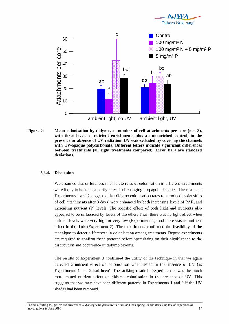
Figure 9: Mean colonisation by didymo, as number of cell attachments per core (n = 3), with three levels of nutrient enrichments plus an unenriched control, in the presence or absence of UV radiation. UV was excluded by covering the channels with UV-opaque polycarbonate. Different letters indicate significant differences between treatments (all eight treatments compared). Error bars are standard deviations.
3.3.4. Discussion
We assumed that differences in absolute rates of colonisation in different experiments were likely to be at least partly a result of changing propagule densities. The results of Experiments 1 and 2 suggested that didymo colonisation rates (determined as densities of cell attachments after 3 days) were enhanced by both increasing levels of PAR, and increasing nutrient (P) levels. The specific effect of both light and nutrients also appeared to be influenced by levels of the other. Thus, there was no light effect when nutrient levels were very high or very low (Experiment 1), and there was no nutrient effect in the dark (Experiment 2). The experiments confirmed the feasibility of the technique to detect differences in colonisation among treatments. Repeat experiments are required to confirm these patterns before speculating on their significance to the distribution and occurrence of didymo blooms.
The results of Experiment 3 confirmed the utility of the technique in that we again detected a nutrient effect on colonisation when tested in the absence of UV (as Experiments 1 and 2 had been). The striking result in Experiment 3 was the much more muted nutrient effect on didymo colonisation in the presence of UV. This suggests that we may have seen different patterns in Experiments 1 and 2 if the UV shades had been removed.
Factors affecting the growth and survival of Didymosphenia geminata in rivers and their spring fed tributaries: update of experimental investigations to June 2010 18
At this stage we cannot explain the contrasting colonisation patterns detected in Experiment 3 in the presence and absence of UV. However, it does highlight the general question of whether levels of UV exposure are linked to didymo growth and blooms in other ways. The negative effects of enhanced UVB exposure on primary producers are well known (e.g., see review in Bothwell et al. 1993). Bothwell et al. (1993) showed that larger-sized diatom cells may be more resistant to damage from UV than smaller-sized cells; furthermore, they suggested that UV light may contribute to the dominance of the stalked diatom taxa Gomphoneis and Cymbella in shallow water. These observations are very relevant to didymo, as it is a large-sized, stalked diatom. The possibility of some influence of UVB on didymo blooms was considered following the appearance of didymo blooms in rivers on Vancouver Island, Canada, for the first time in the mid-1980s. However, initial experiments indicated that there was no UV effect (M. Bothwell, unpublished data). We ran Experiment 3 as a trial because our work in the Waitaki River has included development of new techniques for measuring didymo responses to environmental changes, and raised the possibility that the cell densities and chlorophyll a measures in the initial experiments on Vancouver Island might have been inadequate to detect a response.
Thus our results serve to revive the question of possible involvement of global changes in UV exposure over the past few decades (e.g., McKenzie et al. 1999; Gao et al. 2010) with the expansion of didymo blooms worldwide. The effect would have been exacerbated by the parallel increase in global air travel facilitating the transfer of organisms across continents (Tatem 2009) and, for freshwater organisms such as didymo, the rapid uptake of felt-soled waders as the footwear of choice for anglers (Bothwell et al. 2009). In addition, very high spring and summertime levels of UV in New Zealand compared to those in the Northern Hemisphere (McKenzie et al. 1999) could explain the anecdotal severity of didymo blooms in New Zealand, compared to those elsewhere.
We therefore suggest that further trials on UV effects would be worthwhile. Future experiments would ideally include measures of other growth parameters in didymo, such as FDC, cell densities and stalk production, as well as colonisation rates.
With regard to the technique of measuring colonisation rates, if repeats prove to be consistent with the current findings, then the technique may also be suitable for further experiments investigating, for example, the effects of micronutrients, temperature, etc. on colonisation. If it proves possible to maintain cultures of didymo, there may be scope to trial bulk methods for determining colonisation rates, such as analyses to quantify adhesive material (taking advantage of compositional differences between the stalks and adhesive pads). A drawback of the direct method used in the experiments described in this section is that it is only possible to measure short-term responses.

Factors affecting the growth and survival of Didymosphenia geminata in rivers and their spring fed tributaries: update of experimental investigations to June 2010 19
3.4. The role of organic phosphorus
The experiment described in this section was prompted by the proposal by Ellwood and Whitton (2008) and Whitton et al. (2009) that the recent spread of didymo globally may be attributed to increases in sources of organic phosphorus to freshwaters, because didymo is able to utilise organic sources more efficiently than other algae. This hypothesis is relevant to the current apparent absence of didymo from the North Island because North Island rivers tend to have higher levels of inorganic P than South Island rivers. The implication is that high levels of phosphorus are so disadvantageous to didymo that they might lead to complete absence of the species. The purpose of the present experiment was to compare the response of didymo cells to additions of phosphorus in inorganic and organic form. The question is: does phosphorus in organic form have the same effect on didymo growth as the same concentration of phosphorus in inorganic form? If didymo growth is favoured in the presence of organic phosphorus sources (as suggested by Ellwood and Whitton, 2008), then we might expect a growth response to organic P similar to or greater than that in inorganic P.
3.4.1. Methods
In our trial we used glucose-6-P, a labile form of organic phosphorus, which is readily broken down to inorganic P (subsequently referred to as Pi). Detection of a different response in glucose-6-P compared to inorganic P would imply that the less labile forms of organic P (and more likely to occur in nature) would show even larger differences. (Organic P is subsequently referred to as Po.)
Glucose-6-P and an inorganic form of P (NaH2PO4.2H2O) were each metred into three experimental channels using a peristaltic pump. The target concentration in each case was 3.2 mg/m3 P. Glucose-6-P, as a sugar, is extremely susceptible to bacterial breakdown. If this happened in the reservoir, the phosphorus could be consumed by bacteria before reaching the channels. To minimise this scenario, glucose-6-P was weighed out into amounts sufficient for 24 hours, and the reservoirs were renewed each day. All glucose-6-P was stored at -20 ºC. Each 24-hour dose was dissolved in distilled water in a new, clean reservoir bottle immediately before the daily bottle change. The line from the reservoir was sterilised in boiling water for about 30 seconds before replacing in the new reservoir. A water sample was collected from the outflow end of each experimental channel, as well as from three control channels, on the first and last day of the experiment. Water samples were analysed for dissolved reactive phosphorus and total dissolved phosphorus. Thus we could check whether bacterial consumption of Po had occurred.

Factors affecting the growth and survival of Didymosphenia geminata in rivers and their spring fed tributaries: update of experimental investigations to June 2010 20
The experiment commenced on 5 March 2010, and ran for 6 days. Just before starting the treatments, three cores were collected from each channel for FDC analysis (day 0 samples) and sections of pre-soaked clean substrate were inserted to measure colonisation (see Section 3.3 above). On days 2, 4 and 6, further samples were collected for FDC determination. On day 3, nine replicate cores were taken from the colonisation plate in each channel.
Samples were analysed for FDC as described in Section 3.2, and for cell attachments as described in Section 3.3.
A single-factor ANOVA (with nutrient treatment as factor) was used to identify differences in colonisation, and a repeated measures ANOVA to identify differences in mean FDC among the treatments and through time.
3.4.2. Results
Although the nutrient analyses indicated that the target concentration of 3.2 mg/m3 P was not consistently achieved, the differences between the controls, Pi and Po were largely as expected (Table 4). Unexpectedly, measurements of TDP reflected the treatment differences better than DRP: thus the Pi additions to channels 3, 6 and 9 were picked up more strongly as TDP when compared to the controls. On day 6, TDP measurements indicated that only one of the Po replicate channels (channel 1) was affected by bacterial consumption of glucose-6-P, probably within the delivery line.
Table 4: DRP and TDP measured in each channel at the start and end of the organic phosphorus trial.
Dissolved reactive P (mg/m3) Total dissolved P (mg/m3)
Channel Treatment day 1 day 6 day 1 day 6
4 Control <1 1 1 1
5 <1 2 1 2
7 <1 <1 2 1
3 Inorganic P 2 3 5 5
6 2 3 11 4
9 2 2 4 4
1 Organic P 2 2 4 <1
2 1 3 4 5
8 1 1 4 4

Factors affecting the growth and survival of Didymosphenia geminata in rivers and their spring fed tributaries: update of experimental investigations to June 2010 21
Colonisation after 3 days was higher in the Pi -enriched channels than in the controls. Colonisation in the Po enriched channels did not differ from either of the other treatments (Figure 10).
0
5
10
15A
ttach
men
ts p
er c
ore
a
b
ab
Control
Inorganic P
Organic P
Figure 10: Mean didymo attachments per core after 3 days in the three treatments. Different letters above the bars indicate a significant difference. Error bars are standard deviations. n = 3.
By day 4, mean FDC in both the Pi and Po channels was higher than that in the control channels. On day 6 mean FDC in the Pi treatments remained higher than that in the control, but mean FDC in the Po channels had returned to the level of the controls (Figure 11). The repeated measures ANOVA showed an almost significant nutrient effect (P = 0.053), but a significant interaction between nutrient treatment and day (P = 0.000), indicating that the responses of the nutrient treatments over time were different.
0 2 4 60
10
20
30
Freq
uenc
y of
div
idin
g ce
lls (%
)
Days after start of treatments
ControlInorganic POrganic P
Figure 11: Frequency of dividing cells in didymo over 6 days of enrichment with inorganic
and organic phosphorus (nominal 3.2 mg/m3 P in each case). Error bars are standard deviations. n = 3.

Factors affecting the growth and survival of Didymosphenia geminata in rivers and their spring fed tributaries: update of experimental investigations to June 2010 22
3.4.3. Discussion
If didymo is particularly favoured by Po as Whitton et al. (2009) suggested, then we would expect at least the same or an enhanced response to Po compared to Pi in terms of cell division rates and colonisation. We found a similar response in terms of colonisation in didymo, except that colonisation after 3 days enrichment with a labile organic form of phosphorus did not differ significantly from the colonisation under unenriched conditions, whereas enrichment with Pi increased colonisation. This intermediate response with Po could be due to bacterial consumption of glucose-6-P. However given that two of the three replicates had retained their expected Po concentration after 6 days, we suspect that there would have been enrichment in all three replicate channels after 3 days (when the colonisation samples were collected). This was confirmed by comparing attachment numbers on individual cores (n = 5) in the three Po replicate channels: there was no difference between the three channels (ANOVA, P = 0.413).
Retention of Po (measured as TDP) to the end of the experiment in two of the three replicate channels means that it is difficult to explain the decline in FDC in the Po-enriched treatments between days 4 and 6. FDC in all three channels had returned to around their Day 0 values or lower. This suggests that perhaps sustained utilisation of Po is not as efficient as Pi in terms of cell division rates. This might be expected in view of the greater metabolic cost of breaking down Po compared to direct uptake of Pi (Cembella et al. 1984). At the same time the nutrient analyses highlighted that low concentrations of both Po and Pi seem to be more successfully detected through TDP analysis. TDP includes all organic and inorganic P that passes through a GFF filter, and presumably covers a wide range of substances. Therefore it is not clear what we were actually measuring: glucose-6-P, inorganic P or some organic product that was less available for uptake by didymo? To understand the processes at work, a potential line of enquiry might be the linkages between autotrophic (algal) and heterotrophic (bacterial) uptake of carbon, N and P in periphyton and bacterial regeneration of nutrients (e.g., see Rier and Stevenson 2002; Scott et al. 2008 for recent brief reviews).
Regardless of the outcome of the present experiment, the results of our other experiments with Pi indicate that blooms of didymo (i.e., excessive stalk production) do occur under conditions of phosphorus limitation (see Section 2.3, and Appendices 1 and 2). This experiment showed that additions of both Pi and (to a lesser extent) Po stimulate cell division, probably at the expense of stalk production (see Section 2.3, and Kilroy and Bothwell, Appendix). Therefore the question of whether didymo can utilize Po more efficiently than other algae may be irrelevant to bloom formation. In this experiment, the fact that didymo responded to Po no more strongly than to Pi reinforces our beliefs that (a) organic phosphorus sources do not promote didymo blooms, and (b) the inorganic/organic phosphorus hypothesis does not explain why didymo is not yet present in the North Island.

Factors affecting the growth and survival of Didymosphenia geminata in rivers and their spring fed tributaries: update of experimental investigations to June 2010 23
3.5. Response of didymo to micro-nutrient additions in spring-fed creeks
In Phase 1 of this series of experiments, the results of nutrient-diffusing substrate (NDS) trials in the Otiake Spring Creek in November-December 2008 suggested that didymo might be absent from the spring creek because one or more required micro-nutrients were missing from the spring waters (see Section 2.2 above). The purposes of repeat experiments in 2010 were: (1) to verify the 2008 results; (2) to try to identify specific micronutrients driving the didymo response seen in 2008, when the tested additions were mixtures; (3) to extend the experiment to at least one other spring-fed creek.
The standard technique for NDS experiments investigating the effects of enhanced nitrate-N or phosphate-P, is to set the nutrient within a matrix of agar, so that nutrients can diffuse out slowly to a test substrate overlaying the agar (e.g., see methods described in Biggs and Kilroy 2000). The micronutrient additions in the 2008 trials were either cations, or high molecular weight (HMW) compounds (vitamins, organic substances). Because of the likelihood that metals and HMW compounds would adsorb to the carbohydrate complex of agar, or otherwise diffuse poorly, aqueous-based NDS were used. Existing apparatus used for agar NDS was adapted for this purpose, but was not ideal. Therefore for the 2009–10 experiments we designed a new NDS system to facilitate the use of aqueous media, but which would also be suitable for agar-based media. This new system (see below) was trialled in November 2009 and subsequently used for detailed experiments in March – April 2010. Thus a fourth aim of the experiments was to establish whether aqueous media (which require daily maintenance; see Methods, below) were necessary. If it could be shown that agar media gave the same results, then this would make future experiments easier.
Three experiments were conducted:
1. Otiake Spring Creek. A comparison of the effects of a range of micronutrients (in both aqueous and agar media) on didymo FDC (short-term exposure – 3 days) and didymo cell densities (long-term exposure – 14 days).
2. Ohau Spring-fed Creek. Effect of vitamin B12 augmentation on didymo FDC and cell densities (aqueous media, 17 day exposure, with collections every 4 days). The Ohau Spring Creek is a completely didymo-free tributary of the didymo-infested Lower Ohau River. Our experiment site was situated at approx. 1373380E, 5089732N, about 50 metres upstream from the confluence with the Ohau.
3. Otiake Spring Creek and Waitaki River. Effect of vitamin B12 augmentation on didymo FDC and cell densities in aqueous media, with additional N + P

Factors affecting the growth and survival of Didymosphenia geminata in rivers and their spring fed tributaries: update of experimental investigations to June 2010 24
additions to the Waitaki River samples to the levels in the spring-fed creek (aqueous media, 15 day exposure).
3.5.1. Methods
The same procedure for preparing the NDS and media was used in all experiments. The starting point was pre-colonised Styrofoam substrate, prepared by running Waitaki River water through the experimental channels for about 4 weeks. The substratum was brushed at intervals to clear away accumulated debris caught on the Styrofoam and to allow maximum colonisation by didymo cells.
The holders for the NDS were specially made concrete blocks set in moulds around pairs of 130 ml jars (90 mm tall, ~45 mm diameter) placed side by side 3–4 cm apart (Figure 12a). Thus two jars fitted exactly into each block, with the friction between the jar and the concrete holding the jar in place once deployed in running water. Blocks were placed in the spring creek for about 4 weeks to allow any soluble materials to leach from the concrete. The exteriors were then sealed with white paving paint. A clear Perspex cover fitted around the jar openings on each block helped to make a flat surface, and also provided an adhesion surface for colour-coded tape to distinguish the different treatments. The covers were secured with rubber bands (Figure 12b).
(a) (b)
Figure 12: (a) Concrete NDS jar holder; (b) blocks in situ in the spring creek. Red and white strips on the underside of Perspex covers indicate treatments.

Factors affecting the growth and survival of Didymosphenia geminata in rivers and their spring fed tributaries: update of experimental investigations to June 2010 25
For agar media, agar was prepared as described in Biggs and Kilroy (2000). N and P were added at molar concentrations, and micronutrients at micromolar levels. The mixed agar was poured into the jars, leaving sufficient room for insertion of the Styrofoam substrate. For aqueous media, the jars were first filled with plain river or spring water (depending on where the experiment was to be run).
In all cases, to insert the colonised substrate, jars were topped up to the brim with spring or river water. A section of precolonised Styrofoam was then placed over the jar opening, colonised side up, and a Perspex sheet placed over the top. Even pressure applied to the Perspex sheet forced the Styrofoam into the jar opening, making a watertight seal. Jars were inserted into the appropriate concrete holder and the Perspex, appropriately colour-coded cover secured over the top. A series of pinholes made along the upstream perimeter of each Styrofoam round allowed the solution in the jar to stream out over the colonised surface. Blocks were immediately placed into the river or creek.
Treatments with agar media required no further attention until sample collection. For treatments in aqueous solution, we used a hypodermic needle (no. 2 gauge) to inject 2 ml of (4 nanomolar) solution into each jar. Injections were repeated daily until sample collection.
For sample collection, entire Styrofoam rounds were popped out of the jars, cut in half, and inserted into 35 ml vials. All samples were preserved with Lugols Iodine. FDC was determined as described in Section 3.2. For quantitative counts, rounds were homogenized for 10 sec in distilled water with a hand blender and aliquots counted in an Utermöhl chamber at 125× on an inverted microscope (Leitz Diavert).
The experiments are summarised in Table 5. In Experiment 1, the aim was to repeat the responses to trace metals and vitamins obtained in 2008. The N+P treatment was included to demonstrate that elevated N+P in the spring waters was responsible for the initial increase in cell division rates observed when didymo was exposed to spring water in the 2008 experiments. Thus the response to N+P was not expected to differ from that in the control.
The purpose of Experiment 2 was to determine whether the most promising micronutrient (vitamin B12) in terms of a positive response by didymo showed the same response in another didymo-free spring fed creek.
In Experiment 3, we compared the didymo response to vitamin B12 in the Otiake Spring Creek and in the Waitaki River, in aqueous media. NDS in the Waitaki River were supplemented with N+P to ensure that any micronutrient response was not obscured because cell division rates were already limited by another nutrient.
Factors affecting the growth and survival of Didymosphenia geminata in rivers and their spring fed tributaries: update of experimental investigations to June 2010 26
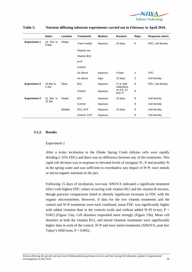
Table 5: Nutrient diffusing substrate experiments carried out in February to April 2010.
Dates Location Treatments Medium Duration Reps Response metric
Experiment 1 22 Feb to 9 Mar
Otiake Trace metals Aqueous 15 days 6 FDC, cell density
Vitamin mix
Vitamin B12
N+P
Control
As above Aqueous 4 days 4 FDC
As above Agar 15 days 2 Cell density
Experiment 2 Ohau B12 Aqueous 8 FDC, cell density
15 Mar to 2 Apr
Control Aqueous
17 d, with collections on d 6, 12 and 17 8
Experiment 3 Otiake B12 Aqueous 15 days 8 Cell density
31 Mar to 15 Apr
Control Aqueous 8 Cell density
Waitaki B12, N+P Aqueous 15 days 8 Cell density
Control, N+P Aqueous 8 Cell density
3.5.2. Results
Experiment 1
After a 4-day incubation in the Otiake Spring Creek didymo cells were rapidly dividing (~35% FDC) and there was no difference between any of the treatments. This rapid cell division was in response to elevated levels of inorganic N , P and possibly Si in the spring water and was sufficient to overshadow any impact of N+P, trace metals or micro-organic nutrients in the jars.
Following 15 days of incubation, two-way ANOVA indicated a significant treatment effect with highest FDC values occurring with vitamin B12 and the vitamin B mixture, though pairwise comparisons failed to identify significant increases in FDC with the organic micronutrients. However, if data for the two vitamin treatments and the control and N+P treatments were each combined, mean FDC was significantly higher with added vitamins than in the controls (with and without added N+P) (t-test, P < 0.001) (Figure 13a). Cell densities responded more strongly (Figure 13b). Mean cell densities in both the vitamin B12, and mixed vitamins treatments were significantly higher than in each of the control, N+P and trace metal treatments (ANOVA, post-hoc Tukey’s HSD tests, P < 0.005).

Factors affecting the growth and survival of Didymosphenia geminata in rivers and their spring fed tributaries: update of experimental investigations to June 2010 27
0.0
0.1
0.2
0.3
0.4
0.5
0.6
FDC
Control N+P B12 vitB TrMet0
50
100
150
200
Cel
ls /
cm2
of c
ore
FDC
(pro
porti
on)
(a)
(b)
Figure 13: FDC (a) and cell densities (b) in five nutrient diffusing substrate treatments (aqueous medium), after 14 days deployment in the Otiake Spring Creek. n = 6.
Nutrient concentrations in the Otiake and Ohau spring creeks differed (Table 6). In contrast to the high nutrients in the Otiake spring creek, the Ohau creek had concentrations of both NO3-N and DRP almost as low as in the Waitaki River, although TDP was higher than in the Waitaki.
Table 6: Summary of water chemistry data available for the Otiake and Ohau Spring Creeks. Data are means of at least three measurements between November 2009 and April 2010. Data for the Waitaki River and Ohau River are included for comparison.
Variable Unit Otiake Spring
Creek Ohau Spring
Creek Waitaki R. Ohau R. DRP mg/m3 4.1 0.8 0.5 1.5 NO3-N mg/m3 601.8 6.1 4.4 24.3 Total dissolved N (TDN) mg/m3 641.2 60.3 70.7 Total dissolved P (TDP) mg/m3 9.2 3.7 1.7 2.0 Dissolved organic P mg/m3 5.6 2.8 0.8 Dissolved organic N mg/m3 90.2 55.0 46.3 Dissolved organic carbon g/m3 1.1 0.5 0.4 Si g/m3 4.3 4.1 1.6 Ca g/m3 14.4 7.7 8.2 Mg g/m3 4.9 1.9 0.7 K g/m3 1.2 0.6 0.6 Na g/m3 9.1 3.3 2.4

Factors affecting the growth and survival of Didymosphenia geminata in rivers and their spring fed tributaries: update of experimental investigations to June 2010 28
FDC in both the control and vitamin B12-enriched NDS increased sharply by Day 6 (Figure 14). On Day 12 mean FDC was nominally higher in the B12 treatments but the difference was not significant. By Day 17 there were so few live cells remaining in the samples that it was not possible to estimate FDC. Cell densities were determined on days 0 and 17. On Day 0, mean density was ~225 live cells/cm2. By day 17 this had declined to ~40 live cells/cm2, with no difference between the B12-enriched and control treatments. However 400 – 500 empty (dead) frustules per cm2 were present in both treatments, indicating that population numbers had increased, but that the cells had subsequently died. Almost no empty cells were noted on Day 0.
0 6 12 17DAY
0.0
0.1
0.2
0.3
0.4
FDC
(pro
porti
on)
ControlB12
Figure 14: FDC on days 0, 6 and 12 following deployment of vitamin B12 enriched and control nutrient diffusing substrates in the Ohau Spring Creek. n = 8. Error bars are standard deviations.
Experiment 3.
Cell counts after a 15 day incubation in the Otiake Spring Creek and the Waitaki River were not significantly different between controls and those with added B12.
3.5.3. Discussion
A comparison of the in situ amendment trials with NDS conducted in 2008 and 2010 showed varying and sometimes conflicting results both within the same system and between systems tested. The transplanting of nutrient-limited didymo cells into the Otiake Spring Creek always results in greatly elevated FDC (cell division rates of didymo). The same was true in 2010 in the Ohau Spring creek even though nitrate and DRP levels in that spring were the same as in the Waitaki River. TDP measurements were higher in the Ohau Spring and appear to be a more reliable indicator of P availability for didymo than DRP in low P waters (see Section 3.4). The elevated didymo FDC following the 2010 transplant into the Ohau Spring Creek likely resulted from P-enrichment.
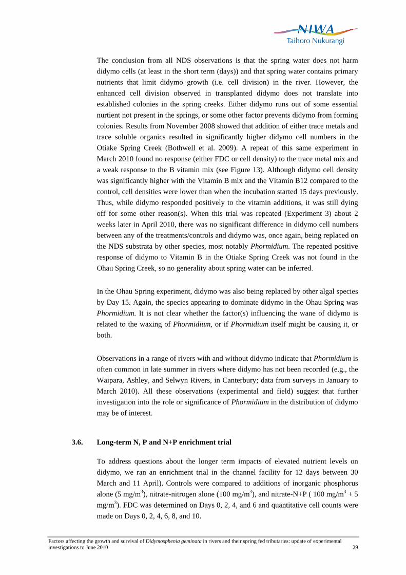
Factors affecting the growth and survival of Didymosphenia geminata in rivers and their spring fed tributaries: update of experimental investigations to June 2010 29
The conclusion from all NDS observations is that the spring water does not harm didymo cells (at least in the short term (days)) and that spring water contains primary nutrients that limit didymo growth (i.e. cell division) in the river. However, the enhanced cell division observed in transplanted didymo does not translate into established colonies in the spring creeks. Either didymo runs out of some essential nurtient not present in the springs, or some other factor prevents didymo from forming colonies. Results from November 2008 showed that addition of either trace metals and trace soluble organics resulted in significantly higher didymo cell numbers in the Otiake Spring Creek (Bothwell et al. 2009). A repeat of this same experiment in March 2010 found no response (either FDC or cell density) to the trace metal mix and a weak response to the B vitamin mix (see Figure 13). Although didymo cell density was significantly higher with the Vitamin B mix and the Vitamin B12 compared to the control, cell densities were lower than when the incubation started 15 days previously. Thus, while didymo responded positively to the vitamin additions, it was still dying off for some other reason(s). When this trial was repeated (Experiment 3) about 2 weeks later in April 2010, there was no significant difference in didymo cell numbers between any of the treatments/controls and didymo was, once again, being replaced on the NDS substrata by other species, most notably Phormidium. The repeated positive response of didymo to Vitamin B in the Otiake Spring Creek was not found in the Ohau Spring Creek, so no generality about spring water can be inferred.
In the Ohau Spring experiment, didymo was also being replaced by other algal species by Day 15. Again, the species appearing to dominate didymo in the Ohau Spring was Phormidium. It is not clear whether the factor(s) influencing the wane of didymo is related to the waxing of Phormidium, or if Phormidium itself might be causing it, or both.
Observations in a range of rivers with and without didymo indicate that Phormidium is often common in late summer in rivers where didymo has not been recorded (e.g., the Waipara, Ashley, and Selwyn Rivers, in Canterbury; data from surveys in January to March 2010). All these observations (experimental and field) suggest that further investigation into the role or significance of Phormidium in the distribution of didymo may be of interest.
3.6. Long-term N, P and N+P enrichment trial
To address questions about the longer term impacts of elevated nutrient levels on didymo, we ran an enrichment trial in the channel facility for 12 days between 30 March and 11 April). Controls were compared to additions of inorganic phosphorus alone (5 mg/m3), nitrate-nitrogen alone (100 mg/m3), and nitrate-N+P ( 100 mg/m3 + 5 mg/m3). FDC was determined on Days 0, 2, 4, and 6 and quantitative cell counts were made on Days 0, 2, 4, 6, 8, and 10.

Factors affecting the growth and survival of Didymosphenia geminata in rivers and their spring fed tributaries: update of experimental investigations to June 2010 30
3.6.1. Results
Between Day 0 and Day 2, FDC increased in all channels (Figure 15) following brushing of the Styrofoam to remove unattached cells and detritus. This is a phenomenon we have repeatedly observed and is likely attributable to improved nutrient supply to cells following removal of the overlying detrital material. Between Day 2 and Day 4, FDC returned to Day 0 levels. To correct for this “brushing stimulation” we subtracted the control FDCs from each of the treatment values. FDC values corrected in this manner more closely reflect the stimulation of FDC resulting from the experimental nutrient addition. Corrected FDCs confirmed that addition of nitrate alone resulted in an increase in cell division which was not sustained (Figure 16). Additions of phosphorus alone or phosphorus together with nitrate resulted in elevated FDC that we followed for 12 days in this experiment.
Figure 15: The time course of the FDC of D. geminata during a long-term enrichment trial (31 March-11 April 2010) with N alone, P alone, and N+P additions. Additions of nitrate alone did not result in sustained elevation in FDC but additions of P alone or in combination with nitrate did.
The sustained increases in FDC under elevated P and N+P additions, translated into higher didymo cell densities that showed the same relative response of nutrient amendments found with the FDC (Figure 17). Cell densities increased progressively from Day 2 through Day 10 with the highest densities found under N+P stimulation, followed by P alone. Didymo cell densities under nitrate alone were not significantly different from the control after 10 days.

Factors affecting the growth and survival of Didymosphenia geminata in rivers and their spring fed tributaries: update of experimental investigations to June 2010 31
Figure 16: The time course of the FDC of D. geminata during a long-term enrichment trial
(31 March-11 April 2010) with N alone, P alone, and N+P additions. The FDC values have been corrected for fluctuations in the control by subtracting control FDC from each of the treatments.
Figure 17: The time course of D. geminata cell density during a long-term enrichment trial (31 March-11 April 2010) with N alone, P alone, and N+P additions.
N 100 ppb P 5 ppb N+P 100, 5 ppb

Factors affecting the growth and survival of Didymosphenia geminata in rivers and their spring fed tributaries: update of experimental investigations to June 2010 32
3.6.2. Discussion
We have shown that the growth and cell division of didymo in the Waitaki River is controlled primarily by the availability of phosphorus (Bothwell and Kilroy 2009; Bothwell and Kilroy in press, and see Appendix 1). However, the concentrations of nitrate in the Waitaki River are also very low and addition of phosphorus easily shifts didymo into N limitation. Hence the greatest response in didymo growth in the Waitaki is always found when N and P are added together. Silicate concentrations in the Waitaki are also low and amendments of Si coupled with N+P would likely elicit an additional incremental growth response. These experiments confirm that blooms of didymo occur while the cells are growing under P-limited conditions in situ and coroborrate the broad-scale observations in New Zealand that didymo blooms favour low phosphorus rivers.
4. Recommendations
Various questions arising from the research and suggestions for further experiments have been raised in the discussions in Sections 3.2 to 3.6. These are summarised below along with other suggestions.
Natural populations of didymo in rivers
1. Our conclusions about P-limitation of didymo in blooms are based on results of FDC determined in the experimental channels and from limited observations in the Waitaki River. This can be confirmed as a broad pattern by determining didymo FDC in natural populations, ranging from those in P-limited rivers with established didymo mats, and in other rivers in which didymo occurs more intermittently over space and time. Accompanying data on nutrient levels would be needed.
Experimental channels
1. There has been some speculation in the international literature about environmental or genetic changes that could be driving the appearance of blooms of didymo as a new phenomenon on a world-wide scale. Our preliminary observations indicate that UV deserves further investigation. How does UV affect didymo colonization and growth? Is globally elevated UV a factor in didymo dominance? The first step to answering these questions would be to run controlled experiments in the channels with different combinations of UV levels and nutrient additions, and measuring colonisation rates, FDC, cell densities, stalk lengths over short- and longer-term exposures.

Factors affecting the growth and survival of Didymosphenia geminata in rivers and their spring fed tributaries: update of experimental investigations to June 2010 33
2. Although phosphorus limitation in didymo FDC has been established, consistently enhanced responses to N+P indicate that nitrogen can be limiting in the Waitaki River. This raises the possibility that silicon (which is low in the Waitaki River) could also become limiting. We suggest that it would be informative to confirm the role of silicon by running a 4-6 day experiment. Treatments would be: a control, +N, +P, +Si, N+P and N+P+Si, and measurements could include FDC, cell densities, stalk lengths and possibly colonisation rates. The question would be: does low Si also contribute to excessive stalk production (i.e., blooms).
Nutrient diffusing substrate experiments
1. The very strong response to Vitamin B12 in November 2008 leads us to suspect that the effect of B12 as a limiting micro-nutrient is seasonal. This can only be confirmed by repeating the replicated B12 NDS experiment in the Otiake Spring Creek in early summer (November). It would also be desirable to look for B12 response in other ground water fed spring creeks (i.e., repeat experiments at other sites).
2. Because Phormidium eventually dominated growth on our NDS in the experiments in both the Otiake and Ohau spring creek, and we have observed dominance of Phormidium in several rivers where didymo has never been recorded, investigation of the role or significance of Phormidium would be of interest. A potential preliminary experiment would be to test the effect of Phormidium extract on didymo growth in an NDS deployment in the Waitaki River. Alternatively, if sufficient Phormidium material can be collected, a 4-day addition experiment in the experimental channels (3 controls and 3 treatments run for 4 days) may be feasible.
All the suggested projects have the aim of improving our understanding of the factors that influence both the distribution of and bloom formation in didymo. Such understanding could contribute to more accurate prediction of rivers at risk (including in the North Island), and may eventually lead to suggestions for mitigation of blooms.
5. Acknowledgements
The research in 2008 – 2010 has been funded by: NIWA (FRST Capability Funds); Environment Canada (support for Max Bothwell); NZ Fish & Game (including local logistical support); Department of Conservation; Meridian Energy Ltd. Resource consents have been granted by Environment Canterbury for taking and discharging water. Local iwi, landowners and lessees have assisted with and supported the research. We thank Neil Blair, Graeme Hughes (NZ Fish & Game), Chris
Factors affecting the growth and survival of Didymosphenia geminata in rivers and their spring fed tributaries: update of experimental investigations to June 2010 34
Cunningham and Helen Brider for their assistance in the laboratory and field. Water chemistry analyses were carried out in the NIWA Hamilton Water Quality Laboratory. JoAnna Lessard and Ton Snelder are thanked for review comments on an earlier version of this report..
6. References
Biggs, B.J.F.; Kilroy, C. (2000). Stream Periphyton Monitoring Manual. Published by NIWA for MfE. 226 p.
Bothwell, M.; Sherbot, D.; Roberge, A.; Daley, R. (1993). Influence of natural ultraviolet radiation on lotic periphyton community growth, biomass accrual, and species composition: short-term versus long-term effects. Journal of Phycology 29: 24-35.
Bothwell, M.; Kilroy, C. (2009). Experimental investigations into factors influencing the poor survival of Didymosphenia geminata in spring-fed creeks 2: progress at 30 June 2009. NIWA Client Report CHC2009-101. 36 p. For: For Department of Conservation / Internal Use.
Bothwell, M.; Kilroy, C.; Blair, N.; Hughes, G. (2009). Experimental investigations into factors influencing the poor survival of Didymosphenia geminata in spring-fed creeks: progress at 30 December 2008. NIWA Client Report CHC2009-002. 20 p. For: F&G New Zealand / Internal Use.
Bothwell, M.L.; Lynch, D.R.; Wright, H.; Deniseger, J. (2009). On the boots of fishermen: the history of didymo blooms on Vancouver Island, British Columbia. Fisheries 34: 382-388.
Cembella, A.D.; Antia, N.J.; Harrison, P.J. (1984). The utilization of inorganic and organic phosphorus compounds as nutrients by eukaryotic microalgae: a multidisciplinary perspective. Critical Reviews in Microbiology 10: 317-391.
Ellwood, N.T.W.; Whitton, B.A. (2007). Importance of organic phosphate hydrolyzed in stalks of the lotic diatom Didymosphenia geminata and the possible impact of atmospheric and climatic changes. Hydrobiologia 592: 121-133.
Gao, Z.; Gao, W.; Chang, N. (2010). Detection of multidecadal changes in UVB and total ozone concentrations over the continental US with NASA TOMS data and USDA ground-based measurements. Remote Sensing 2: 262-277.
Factors affecting the growth and survival of Didymosphenia geminata in rivers and their spring fed tributaries: update of experimental investigations to June 2010 35
Jellyman, P.G.; Clearwater, S.J.; Clayton, J.S.; Kilroy, C.; Hickey, C.W.; Blair, N.; Biggs, B.J.F. (2010). Rapid screening of multiple compounds for control of the invasive diatom Didymosphenia geminata. Journal of Aquatic Plant Management in press.
Kilroy, C.; Lagerstedt, A.; Davey, A.; Robinson, K. (2007). Studies on the survivability of the invasive diatom Didymosphenia geminata under a range of environmental and chemical conditions. NIWA Client Report CHC2006-116. For Biosecurity New Zealand. 110 p.
McKenzie, R.; Connor, B.; Bodecker, G. (1999). Increased summertime UV radiation in New Zealand in response to ozone loss. Science 285: 1709-1711.
Sutherland, S.; Rodway, M.; Kilroy, C.; Jarvie, B.; Hughes, G. (2007). The survival of Didymosphenia geminata in three rivers and associated spring-fed tributaries in the South Island of New Zealand. Southland Fish & Game report to MAF Biosecurity New Zealand.
Tatem, A. (2009). The worldwide airline network and the dispersal of exotic species. Ecography 32: 94-102.
Whitton, B.A.; Ellwood, N.T.W.; Kawecka, B. (2009). Biology of the freshwater diatom Didymosphenia: a review. Hydrobiologia 630: 1-37.

Factors affecting the growth and survival of Didymosphenia geminata in rivers and their spring fed tributaries: update of experimental investigations to June 2010 36
Appendices
Draft papers.
Bothwell, M.R.; Kilroy, C. Phosphorus limitation of the freshwater benthic diatom Didymosphenia geminata determined from the frequency of dividing cells. Submitted to Freshwater Biology (accepted subject to revision)
Kilroy, C.; Bothwell, M.R. Environmental control of stalk length in the bloom-forming, freshwater benthic diatom Didymosphenia geminata. Submitted to Journal of Phycology