fabrication of porous titanium with directional pores for ... of porous titanium with directional...
TRANSCRIPT
Fabrication of Porous Titanium with Directional Poresfor Biomedical Applications
Ji-Woon Lee1,+1, Ji-Sun Lee1,+1, Myoung-Gyun Kim2 and Soong-Keun Hyun1,+2
1Division of Materials Science and Engineering, Inha University, Incheon 402-751, Korea2New Materials Research Department, Research Institute of Industrial Science and Technology (RIST), Pohang 790-330, Korea
A new fabrication method was developed for porous Ti with directional pores. Large sized-pores were generated by the evaporation of Mgwires, and small sized-pores were generated by sintering process. The porosity of the material was increased with increasing the number of wiresand decreased with increasing sintering temperature and compact pressure. The diameter of large-sized pores was accurately same with that ofMg wire, and the diameter of small-sized pores was varied with the fabrication parameters showing similar trend to the porosity variation. Themost advantageous point of the novel fabrication process is that the both porosity of the material and the diameter of the pores can be easilycontrolled by fabrication parameters, such as the number of wires, the sintering temperature and the compact pressure. The material can be usedas possible bone implants possessing not only closer modulus to human bone, but also superior osteogenesis properties.[doi:10.2320/matertrans.MC201211]
(Received July 4, 2012; Accepted August 28, 2012; Published November 23, 2012)
Keywords: biomaterials, porous materials, sintering, titanium
1. Introduction
Titanium has been widely used as materials for orthopedicand dental implants because of their advantageous mechan-ical properties, corrosion resistance and biocompatibility.1,2)
In the materials currently used for surgical implants, Ni, Coand Cr are released from stainless steel and CoCr alloys, andV from Ti6Al4V.3) The toxic effects of these elements tothe human body have been reported by Wapner.4) Therefore,in the view point of biocompatibility, titanium is the mostpromising material for biomedical applications.
When an implant is much stiffer than the bone, a reductionin the bone mass adjacent to the implant occurs because aninsufficient stress is transferred to the bone, resulting boneresorption adjacent to the implant. This phenomenon isknown as “stress shielding effect” and leads to death of thebone cells.5) A material with closer modulus to bone has tobe used for implants to prevent this negative effect. Themodulus of CP Ti (100GPa) is much closer to that of bone(30GPa) than other metallic biomaterials but still higher.Therefore, porous materials have been proposed for implantapplications because they can reduce their modulus comparedto inherent values.6)
Optimal pore size that can facilitate bone ingrowth is in therange of 150 to 500 µm.79) There are several manufacturingprocesses for porous Ti-based materials.1014) These conven-tional methods, however, could not control the pore shapeand its size. These pore characteristics are controllable fora manufacturing method using gas solubility gap betweenliquid and solid phase.15) Although various porous metalswith uniform pore shape and pore size can be fabricated bythe above method, the pore shape and the pore size of porousTi are not uniform because of high dissolving concentrationof hydrogen. In this study, a new method for fabrication ofporous Ti with directional pores is developed. We especially
focus on the uniformity of pore shape and its size. To achievethis, we used the vacuum sintering of Ti powders and theevaporation of Mg wires.
2. Experimental Procedure
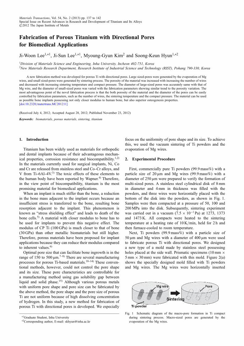
First, commercially pure Ti powders (99.9mass%) with aparticle size of 20 µm and Mg wires (99.9mass%) with adiameter of 250 µm were prepared to verify the formation ofmulti-sized pores. A stainless steel cylindrical disk of 8mmin diameter and 4mm in thickness was filled with thepowders, and three wires were horizontally placed with thebottom of the disk into the powders, as shown in Fig. 1.Samples were then compacted at a pressure of 50, 100 and200MPa into the disk. Subsequently, sintering experimentwas carried out in a vacuum (7.5 © 10¹1 Pa) at 1273, 1373and 1473K. All compacts were heated to the sinteringtemperature at a heating rate of 10K/min, held for 2 h andthen furnace-cooled to room temperature.
Next, Ti powders (99.9mass%) with a particle size of50 µm and Mg wires with a diameter of 400 µm were usedto fabricate porous Ti with directional pores. We designeda new type of a mold made by stainless steel possessingholes placed at the side wall. Prismatic specimens (10mm ©5mm © 30mm) were fabricated with this mold. Figure 2(a)shows the specially designed mold filled with Ti powdersand Mg wires. The Mg wires were horizontally inserted
Fig. 1 Schematic diagram of the macro-pore formation in Ti compactduring sintering process. Macro-sized pores are generated by theevaporation of the Mg wires.
+1Graduate Student, Inha University+2Corresponding author, E-mail: [email protected]
Materials Transactions, Vol. 54, No. 2 (2013) pp. 137 to 142Special Issue on Recent Advances in Research and Development of Titanium and Its Alloys©2012 The Japan Institute of Metals

into holes on the side wall of the mold. Ti powders and Mgwires in the mold were compacted with a pressure of 100and 200MPa. Subsequently, sintering experiment was carriedout in a vacuum (7.5 © 10¹1 Pa) at 1473K. All compactswere heated to the sintering temperature at a heating rateof 10K/min, held for 2 h and then furnace-cooled to roomtemperature.
Particle size distribution of Ti powders was measured bythe particle size analyzer (Hydro 2000s, Malvern Instru-ments, Ltd., UK). The density values of the green compactswere determined by measuring the dimensions of thesamples. The porosity of the sintered samples was measuredusing the relative density method. This relative densityvalue was calculated by taking the theoretical value to be4500 kg/m3. The pore size was determined with the imageanalysis software (Image-pro plus, Media Cybernetics Inc.,USA). The microstructure and the pore morphology of theporous samples were observed by scanning electron micro-scope (S-4300SE, Hitachi Ltd., Japan). The directional porestructure was observed by Micro-CT system (SMX-100CT-SV, Shimadzu Corp., Japan). Each porous sample waspolished and etched with Kroll’s reagent. Atomic absorptionspectroscopy (Aomalyst 400, PerkinElmer Inc., USA) wasused to analyze the composition of the samples, especiallywhether Mg residuals were present or not.
3. Results and Discussions
3.1 Verification of multi-sized pore formationFigure 3 shows the microstructure of porous Ti fabricated
by vacuum sintering in a stainless steel disk. Two types of thepores were found in the specimens after sintering. Smallsized-pores (hereafter micro-pore) were found in the entire ofthe specimens, and large sized-pores (hereafter macro-pore)were partially found in the specimens. The diameter of themacro-pore was 250 µm same with that of Mg wire, and itsmorphology was cylindrical. The diameter of the micro-porevaried with the fabrication conditions, such as sinteringtemperature and compact pressure.
Figure 4 shows the microstructure of the micro-poresdispersed in porous Ti after sintering. The micro-pores werenot clearly formed after sintering at 1273K. Above 1373K,the micro-pores were clearly formed at grain boundaries, andtheir shape was mostly irregular. Figure 5 shows the size ofthe micro-pores in porous Ti depending on sintering temper-ature and compact pressure, and the values are listed inTable 1. After sintering, the size of the micro-pores wasdecreased with increasing sintering temperature and compactpressure. At 1373K, the size was greatly decreased, and thenits decrease was reduced up to the sintering temperature of1473K.
Multi-sized pores were also found in the porous Tispecimens fabricated by vacuum sintering in the prismaticmold, as shown in Fig. 6. Macro-pores were partially locatedat where the Mg wires were placed before sintering process.The diameter of the macro-pore was 400 µm. Micro-pores,which were shown in the disk-type specimen, were found inthe entire of the prismatic specimen. The size of the micro-pores was 12.2 « 6.0 and 10.1 « 4.8 µm with a compactpressure of 100 and 200MPa, respectively.
Figure 7 shows micro-CT results of the prismatic porousspecimen after sintering process. Pores were shown nearly atthe same position in the images taken from the top to one-third of the specimen. This indicated that cylindrical poreswere directionally aligned in the specimen.
Fig. 2 (a) Schematic diagram of a specially designed mold for thefabrication of porous Ti and photograph of (b) a green body of prismaticTi compact with inserted Mg wires and (c) porous Ti with macro-sizedpores after sintering process. Macro-pores are generated by theevaporation of the Mg wires.
Fig. 3 Scanning electron micrograph of the pore structure for disk-typeporous Ti fabricated by vacuum sintering. The white arrows indicatemacro- and micro-pores. The diameter of the macro-pore is 250µm.
Table 1 Porosity and micro-pore size of porous Ti fabricated by thevacuum sintering depending on the sintering temperature and the compactpressure.
Sinteringtemperature,
T/K
Compactpressure,p/MPa
Porosity,P/%
Micro-poresize,D/µm
1273 50 42.7 19.4 « 8.4
100 38.7 18.9 « 7.8
200 33.3 18.8 « 9.6
1373 50 18.3 15.5 « 8.7
100 17.3 15.0 « 6.9
200 14.0 14.3 « 5.8
1473 50 17.6 15.0 « 7.5
100 14.0 13.3 « 5.1
200 10.7 13.3 « 5.4
J.-W. Lee, J.-S. Lee, M.-G. Kim and S.-K. Hyun138

In this study, the porous Ti possesses macro- and micro-pores. The macro-pores were formed at where the Mg wireswere placed before sintering, and the diameter of the macro-pores in both disk-type and prismatic specimen wasaccurately same with that of the Mg wires, as shown inFigs. 3 and 6. From these results, it would be expected thatthe macro-pores are formed by the evaporation of the Mgwires during sintering.
Mg contents in the specimens are analyzed by atomicabsorption spectroscopy (AAS), as shown in Table 2. AASresults showed that Mg contents in the specimen are less thanthat in Ti powders (0.022mass%). Mg wire was entirelyevaporated after sintering, owing to its equilibrium vaporpressure. The equilibrium vapor pressure of Mg at therelevant temperature can be calculated with the followingequation:16)
Fig. 5 Micro-pore size of porous Ti fabricated by the vacuum sinteringdepending on the sintering temperature and the compact pressure.
Fig. 4 Scanning electron micrographs of the microstructure for micro-pores dispersed in porous Ti after sintering at the sinteringtemperature of (a), (d), (g) 1273K, (b), (e), (h) 1373K and (c), (f ), (i) 1473K. The compact pressure was (a), (b), (c) 50MPa, (d), (e), (f )100MPa and (g), (h), (i) 200MPa.
Fig. 6 Scanning electron micrographs of porous Ti fabricated by vacuumsintering with a sintering temperature of 1473K and a compact pressureof (a), (c) 100 and (b), (d) 200MPa in a specially designed mold.
Fig. 7 Micro-CT images taken at the position of (a) top, (b) half and (c) one-third of porous Ti fabricated by vacuum sintering in aspecially designed mold. Pore morphology is long and cylindrical as same as Mg wire.
Fabrication of Porous Titanium with Directional Pores for Biomedical Applications 139

logp ¼ � 7636
Tþ 2:5 log T � 2:949� 10�3T
þ 0:874� 10�6T 2 þ 3:26 ð1Þwhere p is vapor pressure and T is temperature. At thetesting pressure (7.5 © 10¹1 Pa), the equilibrium temperatureis 694K. The testing temperature was higher than 694K.Therefore, Mg is completely evaporated after sintering.Schematic diagram of pore formation in the sintering processis illustrated in Fig. 1. Although Mg might be still presentin the implant, Mg will not generate the harmful effect onhuman body because of its biodegradable characteristics.17)
Micro-pores of the porous Ti in this study were formed atthe entire of the specimens by sintering. Solid state sinteringis usually divided into three stages® initial, intermediateand final. The initial stage follows the formation of necksbetween particles. During the intermediate stage, consid-erable densification occurs before isolation of the pores. Thefinal stage involves densification from the isolated pore stateto the final densification.18) In this study, the micro-pores areformed in the following of three stages above. The diameterof the pores is influenced by the sintering variables, such aspowder size, temperature, compact pressure, and so on. Inthis study, the diameter of the micro-pores is decreased withincreasing sintering temperature and compact pressure.
3.2 Porosity controlFigure 8 shows the porosity of the porous Ti depending on
compact pressure and sintering temperature, and the valuesare listed in Table 1. After sintering, the porosity of the Ticompacts was decreased with increasing compact pressureand sintering temperature. The porosity was 42.733.3% aftersintering at 1273K. At the sintering temperature of 1373K,the porosity of the specimens was greatly decreased to 18.314.0%. The porosity was slightly decreased to 17.610.7%after sintering at 1473K.
Figure 9 shows the porosity of disk-type and prismaticporous Ti specimen with the compact pressure of 100 and200MPa. Measured porosity of the prismatic specimenwas 19.1 and 15.9% for the compact pressure of 100 and200MPa, respectively. Furthermore, each porosity value ofthe prismatic specimen (eighteen wires) was compared withthat of the disk-type specimen (three wires) at the samecompact pressure. The porosity of the prismatic specimenwas higher than that of disk-type specimen, and thedifference in the porosity between the disk-type and prismaticspecimen was 5 and 4.8% each at the same compact pressure.
One of the most attractive properties of porous metal as abiomaterial is that the modulus of the material can be reducedwith increasing porosity. To avoid stress shielding of bone, animplant for human body should possess closer modulus tonatural bone. The porosity dependencies of the porousmaterials to their modulus can be expressed by the followingequation:15)
E ¼ E0ð1� pÞm; ð2Þwhere E and E0 are the moduli of porous and densematerials, respectively, and m is the empirical constant.Boccaccini et al.19) suggested that the empirical constant mis related to the stress concentration around the pores in theporous material. When a porous material possesses cylin-drical pores, it exhibits the mechanical anisotropy causedby the orientation of the pore axis. The reason for this wasidentified with the stress concentration differences dependingon the direction of the pore axis.20) When the direction ofthe pore axis is parallel to the applied stress, the empiricalconstant m is usually known as 1 for the porous materialwith directional pores. Also, m value is 3 for the materialin this study with the pore axis perpendicular to the appliedstress.21) When the material is implanted to the human body
Table 2 Mg contents (mass%) in the specimens analyzed by atomicabsorption spectroscopy depending on sintering temperature and compactpressure. Mg contents (mass%) in the starting material (Ti powders) was0.022mass%.
Compact pressure,p/MPa
Sintering temperature,T/K
1273 1373 1473
50 0.0220 0.0008 0.0010
100 0.0014 0.0008 0.0009
200 0.0019 0.0014 0.0007
Fig. 8 Porosity of porous Ti fabricated by the vacuum sintering dependingon the sintering temperature and the compact pressure.
Fig. 9 Porosity of disk-type (3 wires) and prismatic (18 wires) porous Tiwith directional pores fabricated by vacuum sintering with a compactpressure of 100 and 200MPa.
J.-W. Lee, J.-S. Lee, M.-G. Kim and S.-K. Hyun140
to facilitate bone ingrowth, the modulus of the porousmaterial is decreased with increasing porosity according toeq. (2).
The porosity of the porous Ti in this study can be mainlycontrolled by the number of the macro-pores. This type ofpore is formed by the evaporation of Mg wires. Thus, theporosity will be increased with increasing the number ofwires. The number of wires can be set as the target porosityis decided. Therefore, the modulus of the material can becontrolled to reduce its mismatch between the human boneand the implant.
Total porosity of the porous Ti can also be controlled bythe fabrication condition, such as compact pressure, sinteringtemperature and particle size of the initial material. At thesame compact pressure, the porosity of the specimens isdecreased with increasing sintering temperature. The amountof porosity decrease of the specimens sintered at between1273 and 1373K is higher than that of the specimenssintered at between 1373 and 1473K. At the same sinteringtemperature, the porosity of the specimens was decreasedwith increasing compact pressure. However, the porositydecrease caused by increasing compact pressure is not muchhigher than that caused by increasing sintering temperature.These indicate that the sintering temperature exhibitssignificant effect rather than compact pressure on the porosityof the specimens. Therefore, the porosity of the material canbe controlled with these parameters.
The total porosity, P, of the porous Ti with directionalpores in this study can be calculated with the followingequation:
P ¼ Pw þ Ps; ð3Þwhere Pw and Ps are the porosities caused by the evaporationof wires and by the sintering, respectively, and followed as
Pw ¼ 1� Vw � n
V0
� �� 100ð%Þ; ð4Þ
Ps � 5{10% ðexperimentally determinedÞ; ð5Þwhere Vw and V0 are the volume of wire and solid,respectively, and n is the quantities of wires. The porositycaused by the sintering process is varied with the fabricationconditions, and followed by the previous studies.11,2224)
3.3 Availability of the porous Ti with directional poresas a biomaterial
An ideal implant should possess similar mechanicalproperties to natural bone and should bond well with humantissue. As mentioned earlier, the optimal size of the pore forbone ingrowth is ranged between 150500 µm. In this study,the diameter of the macro-pores in porous Ti is 250 and400 µm for disk-type and prismatic specimens, respectively.Therefore, the diameter of the pores fulfills for the boneingrowth in this study. Furthermore, Higuchi et al.25)
suggested that pores with the diameter of 1020 µm allowonly fibrous tissue to come into the pores, thus adding bufferto the stress. The fibrous connection is applied at the site ofcommunication between implants and the external environ-ment, enhancing physical binding strength. In this study,the diameter of the micro-pores is varied to 13.319.4 µmwith the fabrication condition. This will be advantageous
for the bonding between the implant and the human tissueadjacent to the implant.
In the process we developed, the material possesses macro-and micro-pores. The macro- and micro-pores will be utilizedfor bone ingrowth and fibrous tissue regeneration to addbuffer to the stress, respectively. Especially, the macro-pore islong and cylindrical, thus, the bone ingrowth will be easierfor the material in this study than other porous metals. In theanimal experiment of Ref. 25), the osteogenesis was moreactive for cylinder type porous stainless steel than cubic type.This result indicates that the depth of the pores is alsoimportant criteria for pore shape affecting bone ingrowth.
Furthermore, the most advantageous point of the novelprocess is that the porosity of the material can be easilycontrolled by the number of wires, the sintering temperatureand the compact pressure. The porosity of the material can becontrolled to reduce the modulus mismatch between theimplant and the human bone by adjusting the portion ofmacro-pores in the material, which is by controlling thenumber of Mg wires to be evaporated in this study. Also,the formation of the micro-pores can affect the porosity ofthe material by controlling the fabrication condition. Thisporosity controllability attributes that the modulus of thematerial can be set as closer to that of human bone from thereduction of the modulus mismatch. Therefore, the stressshielding phenomena in the human body can be minimized.Consequently, the osteocompatibility and the minimizednegative effect of the implant (Stress shielding effect) can beachieved at the same time with the material fabricated by thenovel process in this study.
4. Conclusion
In conclusion, we developed a new fabrication method ofporous Ti with directional pores for biomedical applications.The conclusions in this study are presented in the followings.(1) The porous Ti possessed macro- and micro-pores.
The macro-pores with the diameter of 250 and 400 µmwere generated by the evaporation of Mg wires for disk-type and prismatic specimen, respectively. The micro-pores with the size of 1020 µm were generated bysintering.
(2) The porosity of the material was decreased withincreasing sintering temperature and compact pressure.The size decrease of the micro-pores showed a similartrend to the porosity decrease of the material. Withthe novel process, the porosity of the material can besimply controlled by changing fabrication parameters,such as the number of wires, the sintering temperatureand the compact pressure and, thus, the modulus of thematerial can be obtained as closer to human bone.
We expect that the material fabricated by a new methodcan achieve not only active bone ingrowth from directionalpores, but also active fibrous tissue regeneration to add bufferto the stress from small-sized pores. Therefore, this materialcan be used as promising bone implants in the viewpointof similar modulus to the natural bone. Furthermore, thenovel process contributes to widen the concept for thefabrication of the porous metals that can be used asbiomedical applications.
Fabrication of Porous Titanium with Directional Pores for Biomedical Applications 141

Acknowledgments
This work was supported by the Basic Science ResearchProgram through the National Research Foundation of Korea(NRF) funded by the Ministry of Education, Science andTechnology (2012007739), Inha University Research Grantand the Fundamental R&D Programs for Core Technologyof Materials funded by Ministry of Knowledge Economy,Republic of Korea (M-2009-01-0020).
REFERENCES
1) M. Geetha, A. K. Singh, R. Asokamani and A. K. Gogia: Prog. Mater.Sci. 54 (2009) 397425.
2) O. E. M. Pohler: Injury 31 (2000) D7D13.3) Y. Okazaki and E. Gotoh: Biomaterials 26 (2005) 1121.4) K. L. Wapner: Clin. Orthop. Relat. Res. 271 (1991) 1220.5) D. R. Summer, T. M. Turner, R. Igloria, R. M. Urban and J. O. Galante:
J. Biomech. 31 (1998) 909917.6) S. Kashef, W. Yan, J. Lin and P. D. Hodgson: Mod. Phys. Lett. B 22
(2008) 61556160.7) S. F. Hulbert, F. W. Cooke, J. J. Klawitter, R. B. Leonard, B. W. Sauer
and D. D. Moyle: J. Biomed. Mater. Res. 4 (1973) 123.8) B. S. Chang, C. K. Lee, K. S. Hong, H. J. Youn, H. S. Ryu, S. S. Chung
and K. W. Park: Biomaterials 21 (2000) 12911298.9) A. I. Itälä, H. O. Ylänen, C. Ekholm, K. H. Karlsson and H. T. Aro:
J. Biomed. Mater. Res. 58 (2001) 679683.
10) I. H. Oh, N. Nomura, N. Masahashi and S. Hanada: Scr. Mater. 49(2003) 11971202.
11) M. Bram, C. Stiller, H. P. Buchkremer, D. Stöver and H. Baur: Adv.Eng. Mater. 2 (2000) 196199.
12) Y. Chino and D. C. Dunand: Acta Mater. 56 (2008) 105113.13) B. P. Lee, M. G. Kim, B. J. Choi, Y. J. Kim and S. K. Hyun: J. Korean
Powder Metall. Inst. 18 (2011) 95104.14) I. H. Oh, H. T. Son, S. H. Chang, H. M. Kim, K. Y. Lee, S. S. Park and
H. Y. Song: J. Kor. Inst. Met. Mater. 44 (2006) 441445.15) H. Nakajima: Prog. Mater. Sci. 52 (2007) 10911173.16) F. F. Coleman and A. Egerton: Phil. Trans. R. Soc. A 234 (1935) 177
204.17) M. P. Staiger, A. M. Pietak, J. Huadmai and G. Dias: Biomaterials 27
(2006) 17281734.18) S. J. L. Kang: Sintering-Densification, Grain Growth, and Micro-
structure, 1st ed., (Elsevier Butterworth-Heinemann, Oxford, 2005).19) A. R. Boccaccini, G. Ondracek and E. Mombello: J. Mater. Sci. Lett. 15
(1996) 534536.20) S. K. Hyun, K. Murakami and H. Nakajima: Mater. Sci. Eng. A 299
(2001) 241248.21) S. K. Hyun and H. Nakajima: Mater. Sci. Eng. A 340 (2003) 258
264.22) O. M. Ferry, T. Ebel and T. Bormann: Mater. Sci. Eng. A 527 (2010)
18001805.23) Y. H. Li, R. B. Chen, G. X. Qi, Z. T. Wang and Z. Y. Deng: J. Alloy.
Compd. 485 (2009) 215218.24) Y. Torres, J. J. Pavon, I. Nieto and J. A. Rodriguez: Metall. Mater.
Trans. B 42 (2011) 891900.25) Y. Higuchi, Y. Ohashi and H. Nakajima: Adv. Eng. Mater. 8 (2006)
907912.
J.-W. Lee, J.-S. Lee, M.-G. Kim and S.-K. Hyun142