fabrication of hydrogel particles of defined shapes using
TRANSCRIPT
1
DOI: 10.1002/((please add manuscript number))
Article type: Communication
Fabrication of Hydrogel Particles of Defined Shapes using Hydrophilic-Superhydrophobic Micropatterns
Ana I. Neto, Konstantin Demir, Anna A. Popova, Mariana B. Oliveira, João F. Mano and Pavel
A. Levkin*
MSc. Ana I. Neto, Dr. Mariana M. Oliveira and Prof. João F. Mano
3B´s Research Group, University of Minho, Avepark – Parque de Ciência e Tecnologia, Zona
Industrial da Gandra, 4805-017 Barco GMR, Portugal
MSc. Ana I. Neto, Dr. Mariana M. Oliveira and Prof. João F. Mano
ICVS/3B´s PT Government Associate Laboratory, Braga/Guimarães, Portugal.
MSc. Ana I. Neto, Konstantin Demir, Dr. Anna A. Popova and Dr. Pavel A. Levkin*
Karlsruhe Institute of Technology
Institute of Toxicology and Genetics
Hermann-von-Helmholtz-Platz 1
2
76344 Eggenstein-Leopoldshafen, Germany
*E-mail: [email protected]
Keywords: 3D cell culture; Microarrays; Tissue engineering; Hydrogels; Superhydrophobic
Abstract
We report a method to rapidly fabricate alginate hydrogel particles of specific sizes and shapes.
Our method is based on the formation of arrays of droplets of pre-hydrogel solutions on
superhydrophobic-hydrophilic patterns using the process of discontinuous dewetting, followed
by their gelation via the parallel addition of CaCl2 to the individual droplets via the sandwiching
method. We demonstrate that viability of living cells incorporated within the hydrogel particles is
higher during the long-term cultivation than in the case of cells cultured in the bulk three-
dimensional hydrogel matrix. Incorporation of magnetic particles into the free-standing hydrogel
particles containing living cells enabled ease manipulation of the particles using an external
magnetic field.
Introduction
Hydrogels are hydrated three-dimensional (3D) cross-linked polymers resembling natural
extracellular matrix (ECM) that provide soft 3D support for cellular growth and tissue formation.1
Due to their unique properties such as high porosity, permeability for gases, nutrients and
metabolites, as well as their compatibility with physiological conditions, hydrogels have been
extensively studied as material support for immobilizing cells, cultivating cells in 3D,2 biomedical
applications, drug delivery and tissue engineering.3

3
Two distinct approaches namely “top-down” or “bottom-up” have recently emerged in hydrogel
engineering. Top-down approaches control the microscale features (i.e. size and shape) of
relatively large pieces of bulk hydrogels,4 whereas bottom-up approaches aim to generate larger
tissue constructs via the assembly of smaller building blocks (usually cell-laden hydrogels)
which mimic the in vivo tissue structure of repeating functional units.5
The limitations of bulk hydrogels are that they usually lack the hierarchical architecture of in vivo
tissues and suffer from the slow diffusion of nutrients and other biological signaling molecules
(e.g., growth factors) from the surrounding medium into the entrapped cells, leading to higher
toxicity and incompatibility with long-term cell cultivation. These drawbacks undermine the
concept of employing hydrogels for 3D cell cultivation.6, 7
Several attempts have been proposed to solve this problem.8-12 The Doyle group developed
methods to generate hydrogel microparticles with specific shapes using continuous flow or stop-
flow lithography.13 A droplet-based microfluidic system was proposed to construct alginate gel
beads encapsulating cells.14 Cell-encapsulating hydrogel particles have recently been used in
several fields such as 3D cell culture and in vitro micro-physiological models.15, 16
However, there are still too few methods allowing for the fabrication of hydrogel particles with
different geometries and dimensions and compatible with the cell encapsulation.17 New
approaches combining the encapsulation of cells into structures with complex geometries and
long-term cell analysis are also required.
We recently demonstrated a versatile platform for creating thousands of isolated microdroplets
of specific geometry and volume, based on the use of superhydrophobic (SH) surfaces
patterned with wettable superhydrophilic (SL) domains.18-22 The extreme wettability contrast of
the SH-SL patterns allows the spontaneous separation of an aqueous solution into high-density
arrays of microdroplets using the effect of discontinuous dewetting. This rapid and facile droplet

4
formation does not require multiple pipetting or a liquid handling device. The handling of small
volumes of droplets requires fewer reagents than with conventional microplates.
Microarray platforms can be used to study cellular behavior by creating distinct combinations of
chemical/biochemical microenvironments.23, 24 Micropatterning and microfluidic systems have
been used to create hydrogels with spatially controlled organization as well as numerous cell-
material or cell-cell combinations to analyze distinct biological issues.25, 26 Micro-patterned
arrays have been generated by immobilizing hydrogels on different micro-domains within a
plane surface, and they have been used for disease diagnosis, prognosis, biochemical analysis,
and therapeutic regimes, and have become an alternative approach for high-throughput
multiplexed assays.27-30 Strategies avoiding such 2D fixation of various hydrogels have
employed shape-coded hydrogel particles as a suspension microarray format for multiplexed
bioassays.31, 32
Here we describe a method for the rapid fabrication of alginate hydrogel particles of defined
sizes and shapes using the effect of discontinuous dewetting on an SH-SL microarray combined
with the sandwiching method to achieve the simultaneous gelation of the pre-hydrogel droplets.
We applied this method in three demonstrations: (1) preparing an array of hydrogel particles
and free-standing hydrogel particles with distinct geometries and sizes defined by the
photomask features; (2) examining the cells' in situ viability encapsulated into free-standing
hydrogel particles, and (3) constructing magnetic responsive hydrogel particles for modular
tissue engineering and shape-coded free-standing hydrogels of distinct cell types.
Results and discussion
To produce the SH-SL array, a nanoporous poly(2-hydroxylethyl methacrylate-co-ethylene
dimethacrylate) (HEMA-EDMA) polymer layer was formed on a microscope glass slide followed

5
by modifying the polymer surface with alkyne groups via esterification.20 Afterwards, an SH
pattern with specific geometry was formed by functionalizing the surface with 1H,1H,2H,2H-
perfluorodecanethiol using the thiol-yne photo-click reaction20 and by applying a corresponding
quartz photomask. The remaining alkyne groups were reacted with 2-mercaptoethanol under
UV light to form the desired pattern of superhydrophilic (SL) areas separated by
superhydrophobic (SH) borders. The porous SH regions possessed advancing (θadv), static (θst)
and receding (θrec) WCAs of 173°, 170° and 164°, respectively, while the SL areas showed
WCAs close to zero.20
Figure 1. Schematic representation of the DMA platform and workflow for the high-throughput fabrication of hydrogel particles via the sandwiching method. Step 1: Formation of an array of droplets of a pre-hydrogel solution on a superhydrophobic-superhydrophilic array. Step 2:

6
Crosslinking of alginate droplets by performing parallel addition of CaCl2 solutions into the individual droplets via the sandwiching method. By changing the position of the slide 1 (bottom vs top) containing CaCl2 droplets, it is possible to form either an array of fixed hydrogel particles (Step 2a) or detach hydrogel particles to form free-floating hydrogel particles (Step 2b). Scale bar: 2 mm.
The extreme difference in wettability between the SL spots and SH background creates a
means of forming arrays of microdroplets using the effect of discontinuous dewetting33 (Figure
1, Step 1) (Movie S1 and Movie S2). The size and volume of the droplets depend on the
geometry and surface area of the hydrophilic regions and is controllable from 700 pL up to 3
µL.18 This allows for the dispensing of aqueous solutions into thousands of droplets without the
need for manual pipetting or robotic devices. As soon as the nanoporous SL areas become wet,
the porous polymer becomes transparent due to reduced light scattering caused by matched
refractive indexes, thereby making SL spots visible and enabling light and fluorescent
microscopy analysis. In this project we prepared arrays of droplets of different shapes and sizes
(Figure 2, S2) with droplet height of 14.2 ± 1.3 µL (Figure S1).22
The workflow of the high-throughput fabrication of hydrogel particles is illustrated in Figure 1. In
the first step, two separated arrays of droplets were formed. Slide 1 was prepared using CaCl2
solution (1 mol/L) as a crosslinker. The second slide (Slide 2) contained cells suspended in 2
mg/mL of alginate solution. Afterwards, the two slides were aligned to come briefly into contact
(Slide 1 – bottom, Slide 2 – top) to mix and form separated hydrogel droplets (Figure 1). The
droplets' gelation occurred within seconds (~10 sec) after the addition of CaCl2 solution. Then,
the two slides were separated and an array of adhered hydrogel particles formed on the slide,
which was located at the bottom during sandwiching (Figure 1, step 2a). In this case, our
hypothesis is that CaCl2 trapped in the porous polymer continues to diffuse into the droplets
after the sandwiching step, leading to the formation of a stronger hydrogel layer at the surface,
which anchors hydrogel to the polymer substrate even when the slide is immersed into solution

7
(Movie S3). The stability of hydrogels-on-chip was tested for 7 days of culturing fully immersed
in medium, which did not result in detachment. By changing the position of the two slides (Slide
1 – top, Slide 2 - bottom) an array of hydrogel particles formed again (Figure 1, step 2b). During
the sandwiching step, the DMA slide containing alginate droplets is placed at the bottom. In this
case, hydrogel particles could be easily detached from the DMA slide by immersing in culture
medium or buffer. Free-standing hydrogel particles containing cells could be also formed using
this method (Movie S4). We hypothesize that the reason for the different gelation and adhesion
strengths of the hydrogel particles is caused by fast consumption of CaCl2 during the
crosslinking process in the droplets resulting in a weaker hydrogel layer at the bottom of
droplets and their easy detachment.
To demonstrate the broad applicability of this method, we used the DMA platform to fabricate a
variety of hydrogel particles with different geometries (Figure 2).
Figure 2. Microscopic images of free-standing hydrogel particles (bright-field images) containing magnetic beads and arrays of fluorescent hydrogel particles anchored to the patterned surfaces (insets). Fluorescent hydrogel particles contain 0.25 wt % of rhodamine 6G as additive. The following geometries were used: squares with 3 mm (a) and 1 mm (b) side length, circle with 3

8
mm (c) and 1 mm (d) diameter, hexagon of 3 mm (e) and 2 mm (f) side length, triangle with 3 mm side length (g), hearts with width of 3 mm (h) and stripes of 1 mm width. Scale bar 1 mm.
Both free-standing and on-array hydrogel particles of different sizes and geometries could be
prepared (Movies S5-8). The dimension and shape of the hydrogel particles can be controlled
by the photomask design. Comparison of the fluorescence intensity of hydrogels formed on
distinct SL spots, as well as the dimensions of the free-standing hydrogels proved the
homogeneous size distribution of hydrogel particles of different shapes (Figure S2).
Figure 2 demonstrates that the droplet array method is not perfectly suited for the formation of
hydrogel particles with sharp corners. This is a result of the surface tension of water solutions
that tries to round off the corners of liquid droplets at the sharp edges of the hydrophilic-
hydrophobic patterns. However, the shapes of hydrogel particles are easily recognizable and
could be distinguished automatically by an image-recognition software. It is also evident from
the images in Figure 2 that the geometry of edges of the pattern (straight lines – triangles and
hexagons, round, or even concave in the case of hearts) is transferred to the hydrogel particles.
The shape of hydrogel particles can be applied in various ways and can also have an effect of
cells or cell encapsulation. For example, the shape and size of particles will determine the
number of cells per particle. The shape of particles will be important for constructing macro
assemblies with diverse architectures, inter-particulate distances or particle-to-particle
interactions. This method can be used as a toolbox of cell bearing hydrogel bricks of different
shapes to enable diversity of multi-particle assemblies for modular tissue engineering29. In
addition, shape of such building blocks can be used to encode information about the
composition of the individual blocks without the need for separate fluorescent labeling (vide
infra).

9
The mechanical properties of hydrogels are important for mimicking the in-vivo cellular
environment and for achieving higher cell viability during long-term cell culture. The stiffness of
hydrogels depends on the concentration of CaCl2 used for gelation. Dynamic mechanical
analysis (see supplementary information and Figures S1e-f for details) was used as a non-
destructive technique to characterize the viscoelasticity of the hydrogels fabricated using
different concentrations of CaCl2. To check the long-term stability of free-standing hydrogels, we
assessed the mechanical and viscoelastic properties on hydrogels immersed in cell culture
medium with or without the supplementation of 1% (v/v) of CaCl2 solution for 1, 3, 5 and 7 days
(see Figure S3 for details). Our results revealed that after immersion of crosslinked alginate
hydrogels into cell culture medium the hydrogels’ storage modulus decreased from 95 kPa (day
1) to approximately 19 kPa on day 7 (Figure S3a).34 This could be explained by ionic exchange
between calcium and monovalent ions (such as sodium ions). However, this tendency seemed
to be reversed by a possible continuation of the crosslinking, by adding calcium ions to the
medium. The hydrogels' storage modulus increased with the duration of immersion in the
calcium-supplemented medium, to approximately 200 kPa, after 7 days of immersion (Figure
S3b). Moreover, in hydrogels immersed in calcium-containing medium, the loss-factor values
did not vary significantly with time (Figures S3c-d).
In the next step we also fabricated free-standing hydrogel particles incorporating living cells
(Figure 3a). We first demonstrated that alginate and CaCl2 exerted no cytotoxic effect on live
human cervical tumor cell line expressing GFP (HeLa-GFP) (Figure S4). Then, cells were
seeded onto the SH-SL patterned surface containing round SL spots of 3 mm diameter by
applying cell suspension containing 2 mg/mL of alginate to form an array of separated droplets.
CaCl2 solution was spread on a separate DMA slide. Hydrogels were formed by sandwiching
both slides using the sandwiching device (Figure 1). This led to the formation of an array of
round, free-standing hydrogel particles particles of 3 mm diameter at the base. Each hydrogel

10
particle incorporated on average 588 ± 62 cells (Figure S5). The hydrogel array slide was
immersed in cell culture medium supplemented with 1% of CaCl2 for a few seconds, leading to
the immediate detachment of hydrogel particles (Movies S4-S10). CaCl2 was added to the
medium to keep the hydrogel particles from dissociating. The medium supplemented with
calcium was exchanged every 2 days. Square hydrogel particles whose sides measured 1 mm
were also fabricated using this method (Figure S6). The viability of cells cultured in free-floating
hydrogel particles was monitored for up to 7 days via propidium iodide staining and MTT assay
(Figure 3b,c). These results were compared with the viability of HeLa-GFP cells cultured in a
standard petri dish (2D positive control) and cells cultured in bulk 2 mm thick alginate hydrogel
layers prepared in a 24-well microtiter plate. Already 24h after the onset of culturing, the viability
of cells in free-standing hydrogels was more than 20% higher than that of the bulk hydrogels.
The toxicity of cells cultured in the bulk hydrogel rose to 60% after 5 days and 80-90% after 7
days of culturing, while that of cells in the hydrogel particles rose to just about 20% and 30%,
respectively. This is attributable to the cells' superior accessibility to oxygen and nutrients when
cultured in the thin free-standing hydrogel particles compared with the limited nutrient and gas
exchange in the bulk hydrogel system. The difference in cell viability can be explained by much
higher surface area-to-volume ratio of free-standing hydrogels (app.11) compared to bulk
hydrogel (app. 0.9), which reflects in exchange rate of nutrients and oxygen. One property of
ionically cross-linked alginate hydrogel particles is that they dissolve when cultured in standard
Ca-negative culture medium. This happens due to the release of calcium ions into the
surrounding media caused by the exchange with monovalent cations (such as sodium ions
present in the medium).34 This could be either an advantage (enabling cell release from the
hydrogel) or a disadvantage (when long-term cultivation is required). To address this problem,
we supplemented the culture medium with 1% (v/v) solution of CaCl2 (Figure S7). Those results
revealed no significant differences in the cells' viability, whereas the viability of cells cultured in
cell culture medium (without calcium addition) dropped to 70% of viability on day 7. Thus, the

11
addition of calcium ions to physiological media promotes mechanical integrity without affecting
cellular viability.
Figure 3. a) Representative brightfield and corresponding fluorescence images of free-standing hydrogels encapsulating HeLa-GFP cells for up to 7 days of culturing time. Dead cells are visualized using PI staining. Scale bar: 1 mm. b) Percentage of live cells obtained using image-based analysis and c) MTT colorimetric assay. Statistical differences by time point analysis were marked with (*), (**), (***), which stand for p-values <0.05; p<0.01; p<0.001, respectively. Statistical differences related to the previous time points are indicated by ## (p<0.01) or ### (p<0.001). All results are presented as ± standard deviation.
Magnetic field has been used in various cellular applications, cell sorting, 3D cell cultures, tissue
engineering, local hyperthermia therapies, and clinical imaging applications.35-38 Using the
12
droplet microarray method, free-standing hydrogels can be loaded with functional magnetic or
other nanoparticles to enable the use of magnetic field for the remote manipulation of the
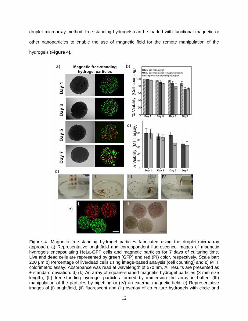
hydrogels (Figure 4).
Figure 4. Magnetic free-standing hydrogel particles fabricated using the droplet-microarray approach. a) Representative brightfield and correspondent fluorescence images of magnetic hydrogels encapsulating HeLa-GFP cells and magnetic particles for 7 days of culturing time. Live and dead cells are represented by green (GFP) and red (PI) color, respectively. Scale bar: 200 μm b) Percentage of live/dead cells using image-based analysis (cell counting) and c) MTT colorimetric assay. Absorbance was read at wavelength of 570 nm. All results are presented as ± standard deviation. d) (I.) An array of square-shaped magnetic hydrogel particles (3 mm size length), (II) free-standing hydrogel particles formed by immersion the array in buffer, (III) manipulation of the particles by pipetting or (IV) an external magnetic field. e) Representative images of (i) brightfield, (ii) fluorescent and (iii) overlay of co-culture hydrogels with circle and

13
square shapes. HeLa cells expressing GFP cells (green) were immobilized in circle-shaped free-standing hydrogels and MLIy-mCherry cells (expressing fluorescent red) were immobilized in squared-shaped free-standing hydrogels. Scale bars: 1 mm.
Magnetic beads measuring 2 μm were added to alginate solution containing living Hela-GFP
cells. The viability of cells cultured in free-floating magnetic hydrogel particles was evaluated for
up to 7 days using propidium iodide staining, followed by fluorescence microscopy (Figure 4b).
MTT colorimetric assay was performed separately (Figure 4c). We observed no significant
differences in the viability of the cells cultured in the presence of magnetic beads in comparison
to 2D cell culture (Figure S8). After 1 day of cell culturing in a 2D and 3D environment, viability
remained at 100%, dropping to 70% on day 7.
Magnetic hydrogel particles can be useful for the Modular Tissue Engineering39 due to the
possibility to manipulate both single particles and particle assemblies using external magnetic
field. Thus, Movie S11 shows the possibility to remotely control exchange of the medium,
collection of hydrogel particles and active movement of particles across culture medium. On the
other hand, application of the external magnetic field to a suspension of free-standing magnetic
hydrogel particles permits rapid assembly of the particles into stable macroscopic 3D
architectures (Movie S12, S13 and Figure S11). The ability to remotely modulate the density
and interparticulate volume of 3D hydrogel architectures using external magnetic field has been
also demonstrated in a proof-of-concept experiment (Figure S11, Movie S13). We showed that
this method could be used to remotely compress or expand 3D hydrogel particle assemblies (3
mm diameter, n=250), which is important, for example, for achieving active perfusion of large
3D hydrogel particle assemblies with medium for long-term cell culturing (Figure S11, Movie
S13). The remotely controlled compression-expansion cycles could be repeated at least 50
times (Figure S11). Finally, the ability to remotely compress hydrogel particle assemblies was
14
applied to demonstrate the stimuli-responsive release of a small molecule drug incorporated
inside the hydrogel using an external magnetic field as the stimulus (Figure S9).
In order to demonstrate the ability to use hydrogel particles of diverse geometries for shape-
coding to distinguish between hydrogels´ different compositions, we prepared two types of free-
standing hydrogels: round 3 mm hydrogel particles encapsulating HeLa-GFP cells and square 3
mm hydrogel particles incorporating MLTy-CMV-mCherry-neo cells expressing cherry
fluorescent protein (Figure 4e). The density of both cell types inside the distinctly shaped
hydrogel particles was set to 6 x 10⁵ cells per mL of alginate solution. The same amounts of
free-standing hydrogel particles were mixed in a Petri dish. The fluorescence images show that
no cross-contamination occurred between hydrogels, and that the cells remained immobile
inside the particles (Figure 4). It is also evident that the shape difference opens the way to
differentiate between particles´ various compositions and encapsulated cell types without
special fluorescence labeling.
Conclusions
We have demonstrated a new method to fabricate free-standing hydrogel particles with defined
geometries and sizes, while maintaining control of the elastic modulus and the composition of
the hydrogel. This platform offers several advantages: (i) thousands of hydrogel particles can be
rapidly formed without the need for multiple pipetting; (ii) their size and composition can be
controlled using the geometry of the hydrophilic areas; (iii) cells can be encapsulated inside
hydrogel particles; (iv) the volume of the hydrogel particles can be in the nanoliter range scale,
which is one order of magnitude less than that of a standard well in a 96-well plate; (v) sample
cross-contamination is prevented by the use of superhydrophobic barriers; (vi) the hydrogel
particles can be used either in the form of an array for screening applications or as free-standing
particles. The presented technology was also used to form magnetic hydrogel particles

15
incorporating live cells. These particles can be employed for the modular tissue engineering due
to the possibility to manipulate both single particles and particles assemblies using external
magnetic field. We believe that the ability to easily create and manipulate thousands of hydrogel
particles of controlled size and geometry will be essential for 3D cell studies and modular tissue
engineering. Finally, such a platform can potentially be applied to different types of biomaterials,
hydrogels and cells.
Acknowledgments
The authors acknowledge financial support from the FCT- Fundação para a Ciência e para a
Tecnologia through a Ph.D. grant with the reference SFRH/BD/73119/2010. This work was
funded by the Helmholtz Association’s Initiative and Networking Fund (grant VH-NG-621) and
European Research Council Starting Grant (ERC-2013-StG 337077 DropCellArray). We are
grateful to Dr. Christian Greiner’s group of (KIT) for their help with optical profilometry
measurements and to Dr. Gary Davidson and Dr. Olivier Kassel (Institute of Toxicology and
Genetics, KIT) for providing the cell lines.
References
[1] O. Smidsrød, G. Skja˚k-Br˦k, Trends in Biotechnology 1990, 8, 71.
[2] K. Y. Lee, D. J. Mooney, Chemical Reviews 2001, 101, 1869.
[3] J. L. Drury, D. J. Mooney, Biomaterials 2003, 24, 4337.
[4] V. Chan, P. Zorlutuna, J. H. Jeong, H. Kong, R. Bashir, Lab on a Chip 2010, 10, 2062.
[5] R. Gauvin, R. Parenteau-Bareil, M. R. Dokmeci, W. D. Merryman, A. Khademhosseini, Wiley Interdisciplinary Reviews-Nanomedicine and Nanobiotechnology 2012, 4, 235.
[6] T. R. Hoare, D. S. Kohane, Polymer 2008, 49, 1993.
[7] T. Billiet, M. Vandenhaute, J. Schelfhout, S. Van Vlierberghe, P. Dubruel, Biomaterials 2012, 33, 6020.
[8] Y. Du, E. Lo, S. Ali, A. Khademhosseini, Proceedings of the National Academy of Sciences of the United States of America 2008, 105, 9522.
[9] P. Panda, S. Ali, E. Lo, B. G. Chung, T. A. Hatton, A. Khademhosseini, P. S. Doyle, Lab on a Chip 2008, 8, 1056.

16
[10] M. E. Helgeson, S. C. Chapin, P. S. Doyle, Current Opinion in Colloid & Interface Science 2011, 16, 106.
[11] W. Lee, D. Choi, J.-H. Kim, W.-G. Koh, Biomedical Microdevices 2008, 10, 813.
[12] J. Y. Sim, J.-H. Choi, J.-M. Lim, S. Cho, S.-H. Kim, S.-M. Yang, Small 2014, 10, 3979.
[13] D. Dendukuri, D. C. Pregibon, J. Collins, T. A. Hatton, P. S. Doyle, Nature Materials 2006, 5, 365.
[14] L. Yu, M. C. W. Chen, K. C. Cheung, Lab on a Chip 2010, 10, 2424.
[15] D. Wei, W. Xiao, J. Sun, M. Zhong, L. Guo, H. Fan, X. Zhang, Journal of Materials Chemistry B 2015, 3, 2753.
[16] A. Kumachev, J. Greener, E. Tumarkin, E. Eiser, P. W. Zandstra, E. Kumacheva, Biomaterials 2011, 32, 1477.
[17] S. P. R. Kobaku, G. Kwon, A. K. Kota, R. G. Karunakaran, P. Wong, D. H. Lee, A. Tuteja, Acs Applied Materials & Interfaces 2015, 7, 4075.
[18] E. Ueda, F. L. Geyer, V. Nedashkivska, P. A. Levkin, Lab on a Chip 2012, 12, 5218.
[19] F. L. Geyer, E. Ueda, U. Liebel, N. Grau, P. A. Levkin, Angewandte Chemie-International Edition 2011, 50, 8424.
[20] W. Feng, L. Li, E. Ueda, J. Li, S. Heissler, A. Welle, O. Trapp, P. A. Levkin, Advanced Materials Interfaces 2014, 1.
[21] A. N. Efremov, E. Stanganello, A. Welle, S. Scholpp, P. A. Levkin, Biomaterials 2013, 34, 1757.
[22] A. A. Popova, S. M. Schillo, K. Demir, E. Ueda, A. Nesterov-Mueller, P. A. Levkin, Advanced Materials 2015, 27, 5217.
[23] M. B. Oliveira, J. F. Mano, Trends in Biotechnology 2014, 32, 627.
[24] F. Xu, J. H. Wu, S. Q. Wang, N. G. Durmus, U. A. Gurkan, U. Demirci, Biofabrication 2011, 3.
[25] L. Wang, M. Qiu, Q. Yang, Y. Li, G. Huang, M. Lin, T. J. Lu, F. Xu, Acs Applied Materials & Interfaces 2015, 7, 11134.
[26] G. T. Vladisavljevic, I. Kobayashi, M. Nakajima, Microfluidics and Nanofluidics 2012, 13, 151.
[27] A. Peters, D. M. Brey, J. A. Burdick, Tissue Engineering Part B-Reviews 2009, 15, 225.
[28] H. Moch, P. Schraml, L. Bubendorf, M. Mirlacher, J. Kononen, T. Gasser, M. J. Mihatsch, O. P. Kallioniemi, G. Sauter, American Journal of Pathology 1999, 154, 981.
[29] F. Deiss, A. Mazzeo, E. Hong, D. E. Ingber, R. Derda, G. M. Whitesides, Analytical Chemistry 2013, 85, 8085.
[30] C. L. Salgado, M. B. Oliveira, J. F. Mano, Integrative Biology 2012, 4, 318.
[31] C. L. Lewis, C.-H. Choi, Y. Lin, C.-S. Lee, H. Yi, Analytical Chemistry 2010, 82, 5851.
[32] D. C. Pregibon, M. Toner, P. S. Doyle, Science 2007, 315, 1393.
[33] R. J. Jackman, D. C. Duffy, E. Ostuni, N. D. Willmore, G. M. Whitesides, Analytical Chemistry 1998, 70, 2280.
[34] K. Y. Lee, D. J. Mooney, Progress in Polymer Science 2012, 37, 106.
[35] J. Dobson, Nature Nanotechnology 2008, 3, 139.
[36] A. Ito, M. Shinkai, H. Honda, T. Kobayashi, Journal of Bioscience and Bioengineering 2005, 100, 1.
[37] F. Xu, C.-a. M. Wu, V. Rengarajan, T. D. Finley, H. O. Keles, Y. Sung, B. Li, U. A. Gurkan, U. Demirci, Advanced Materials 2011, 23, 4254.
17
[38] S. Gil, J. F. Mano, Biomaterials Science 2014, 2, 812.
[39] J. W. Nichol, A. Khademhosseini, Soft Matter, 2009, 5, 1312
[40] S. Guven, P. Chen, F. Inci, S. Tasoglu, B. Erkmen, U. Demirci, Trends in Biotechnology 2015, 33, 269.