f etal m embranes dr rania gabr. o bjectives by the end of this lecture, the student should be able...
TRANSCRIPT
FETAL MEMBRANES
Dr Rania Gabr
OBJECTIVES
By the end of this lecture , the student should be able to:
Define the fetal membranes Define and enumerate types of decidua Describe the development , types ,functions
and anomalies of chorion Describe the development , types ,functions
and anomalies of amnion Describe the development , types ,functions
and anomalies of yolk sac Describe the development , types ,functions
and anomalies of allantois
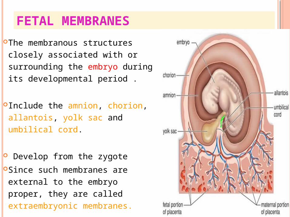
FETAL MEMBRANES The membranous structures
closely associated with or surrounding the embryo during its developmental period .
Include the amnion, chorion, allantois, yolk sac and umbilical cord.
Develop from the zygote Since such membranes are
external to the embryo proper, they are called extraembryonic membranes.
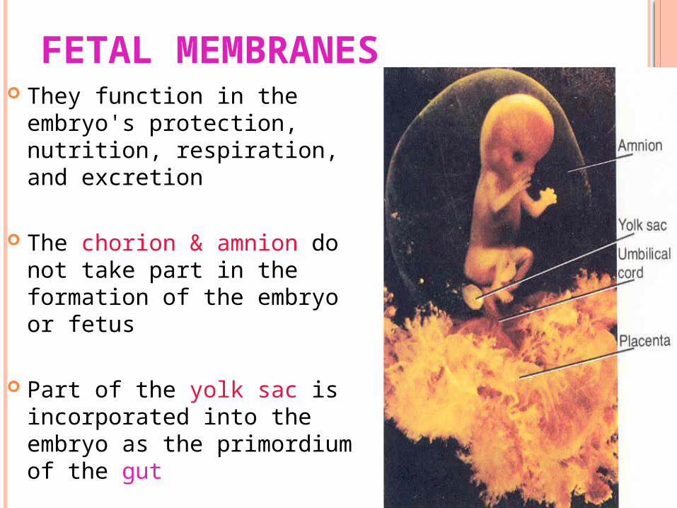
FETAL MEMBRANES They function in the embryo's
protection, nutrition, respiration, and excretion
The chorion & amnion do not take part in the formation of the embryo or fetus
Part of the yolk sac is incorporated into the embryo as the primordium of the gut
The allantois forms a fibrous cord called urachus
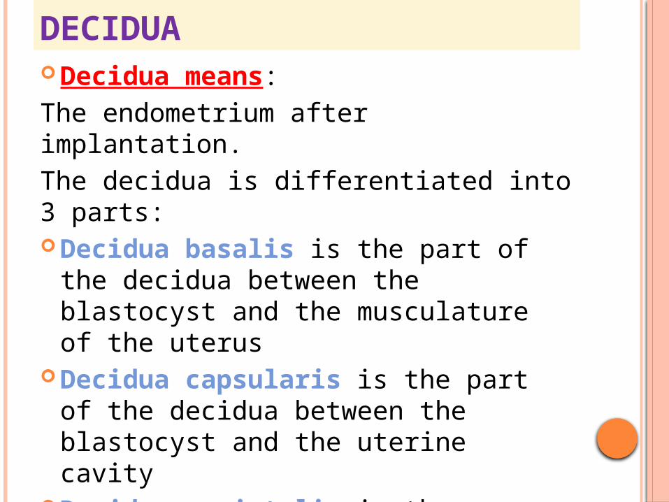
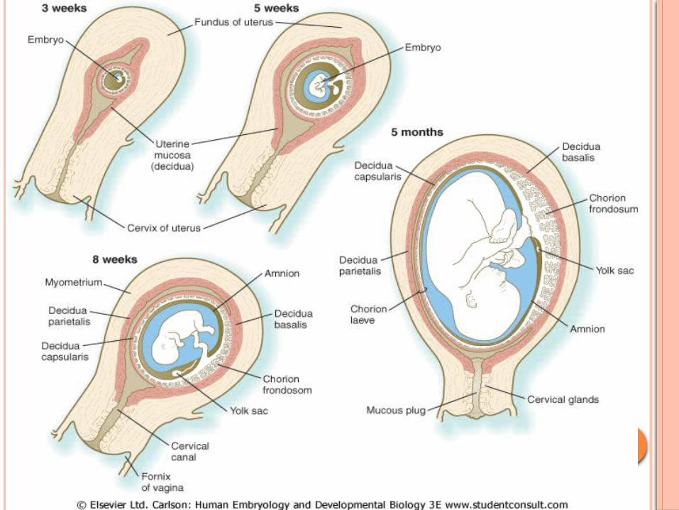
DECIDUADecidua means:The endometrium after implantation.The decidua is differentiated into 3 parts:Decidua basalis is the part of the
decidua between the blastocyst and the musculature of the uterus
Decidua capsularis is the part of the decidua between the blastocyst and the uterine cavity
Decidua parietalis is the uninvolved uterine mucosa i.e the rest of the decidua lining the cavity of the uterus

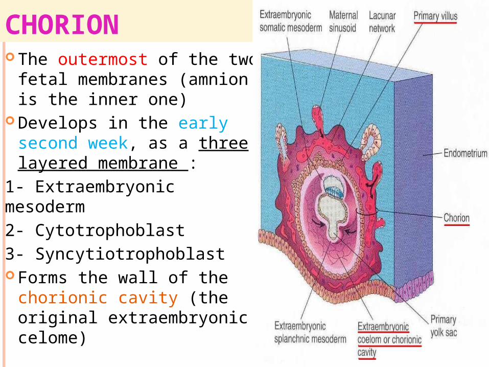
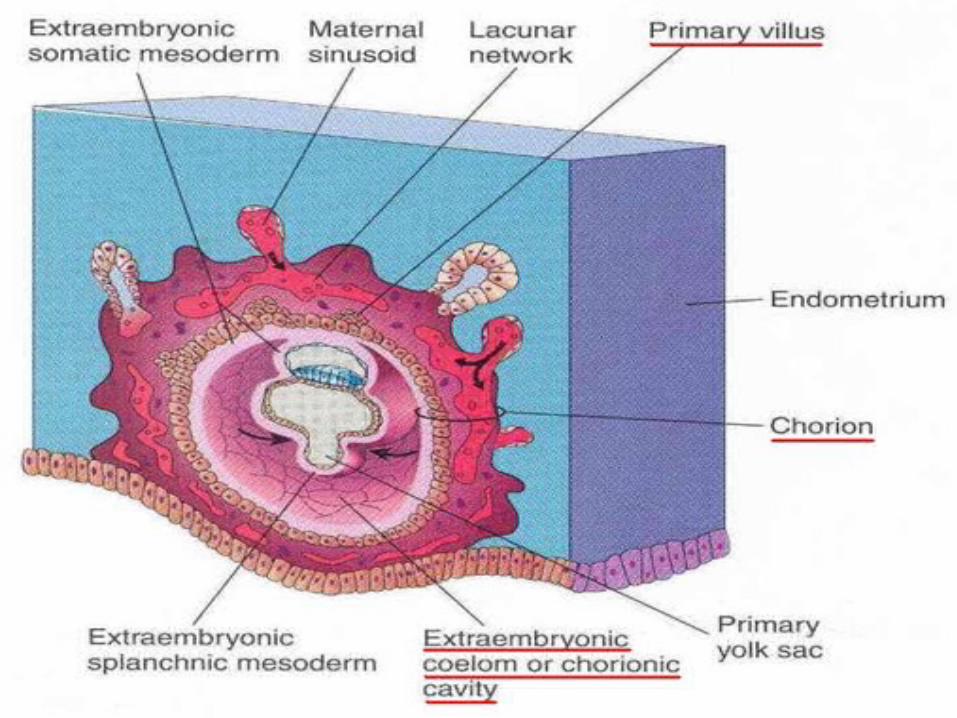
CHORION The outermost of the two
fetal membranes (amnion is the inner one)
Develops in the early second week, as a three layered membrane :
1- Extraembryonic mesoderm 2- Cytotrophoblast3- Syncytiotrophoblast Forms the wall of the
chorionic cavity (the original extraembryonic celome)

CHORIONIC VILLIOn day 13-14 the
primary villi appear as cellular extensions from the cytotrophoblast that grow into the syncytio-trophoblast.
Shortly after their apperance, the primary villi begin to branch
A mesodermal core appears in the cytotrophoblast, this is the secondary villi

CHORIONIC VILLI Blood vessels appear in
the mesodermal core of the villi that are now called the tertiary villi.
These blood vessels connect up with vessels that develop in the chorion and connecting stalk and begin to circulate embryonic blood about the third week of development.
primary villus
secondary villus
tertiary villus
FUNCTIONS OF THE VILLI
1. Nutrition of the embryo (free villi).
2. Fixation of the embryo (anchoring villi).
3. Respiration of the embryo.4. Excretion of the embryo.
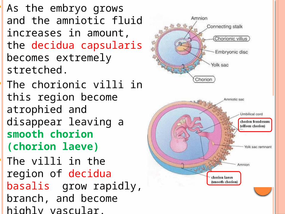
As the embryo grows and the amniotic fluid increases in amount, the decidua capsularis becomes extremely stretched.
The chorionic villi in this region become atrophied and disappear leaving a smooth chorion (chorion laeve)
The villi in the region of decidua basalis grow rapidly, branch, and become highly vascular.
This region of chorion is called chorion frondosum (villous chorion)

Chorionic villi
embryo
Chorionic cavity
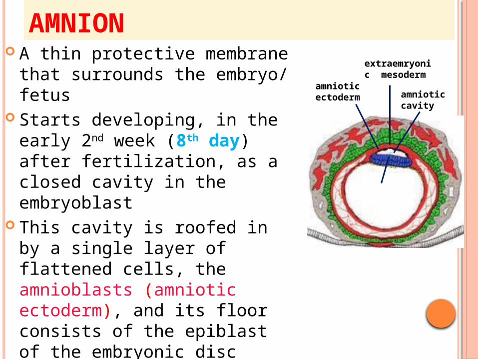
AMNION A thin protective membrane
that surrounds the embryo/ fetus
Starts developing, in the early 2nd week (8th day) after fertilization, as a closed cavity in the embryoblast
This cavity is roofed in by a single layer of flattened cells, the amnioblasts (amniotic ectoderm), and its floor consists of the epiblast of the embryonic disc
Outside the amniotic ectoderm is a thin layer of extraembryonic mesoderm
amniotic cavity
amniotic ectoderm
extraemryonic mesoderm
epiblast
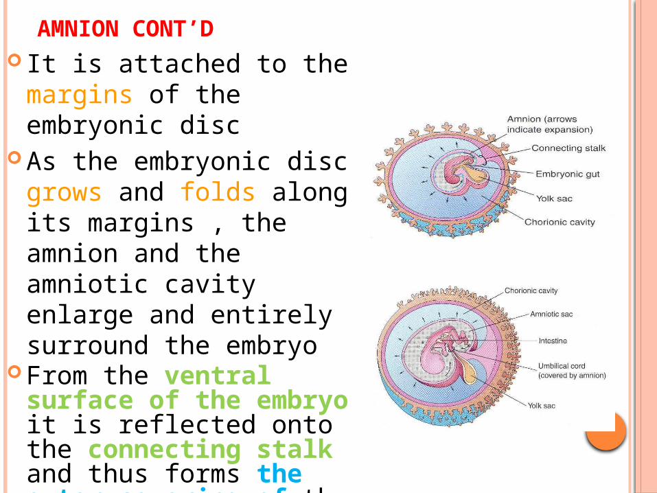
AMNION CONT’D It is attached to the
margins of the embryonic disc
As the embryonic disc grows and folds along its margins , the amnion and the amniotic cavity enlarge and entirely surround the embryo
From the ventral surface of the embryo it is reflected onto the connecting stalk and thus forms the outer covering of the future umbilical cord
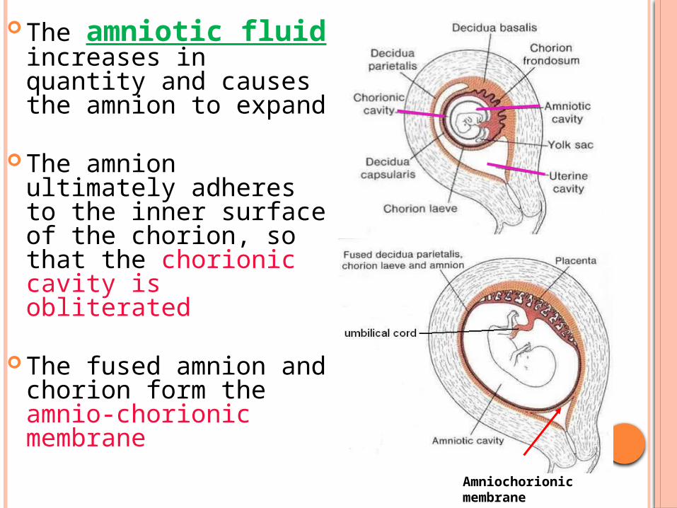
The amniotic fluid increases in quantity and causes the amnion to expand
The amnion ultimately adheres to the inner surface of the chorion, so that the chorionic cavity is obliterated
The fused amnion and chorion form the amnio-chorionic membrane
Amniochorionic membrane
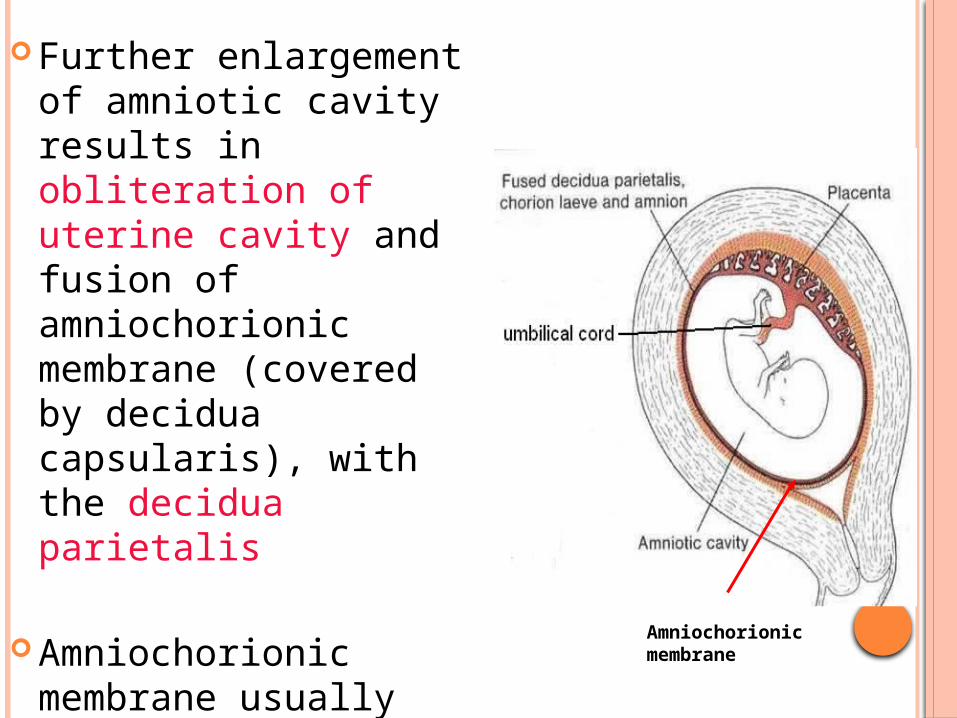
Further enlargement of amniotic cavity results in obliteration of uterine cavity and fusion of amniochorionic membrane (covered by decidua capsularis), with the decidua parietalis
Amniochorionic membrane usually ruptures just before birth
Amniochorionic membrane
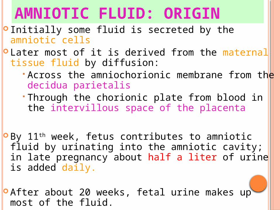
AMNIOTIC FLUID: ORIGIN Initially some fluid is secreted by the amniotic cells Later most of it is derived from the maternal tissue
fluid by diffusion: Across the amniochorionic membrane from the decidua parietalis
Through the chorionic plate from blood in the intervillous space of the placenta
By 11th week, fetus contributes to amniotic fluid by urinating into the amniotic cavity; in late pregnancy about half a liter of urine is added daily.
After about 20 weeks, fetal urine makes up most of the fluid.
AMNIOTIC FLUID: COMPOSITION
Amniotic fluid is a clear, slightly yellowish liquid
99% of fluid in the amniotic cavity is water
Suspended in this fluid are undissolved substances e.g. desquamated fetal epithelial cells, proteins, carbohydrates, fats, enzymes, hormones and pigments
As pregnancy advances the composition of amniotic fluid changes as fetal waste products (meconium & urine) are added
AMNIOTIC FLUID: CIRCULATION The water content of the amniotic fluid changes
every three hours
Large volume moves in both directions between the fetal & maternal circulations mainly through the placental membrane
It is swallowed by the fetus, is absorbed by respiratory & GIT and enters fetal circulation. It then passes to maternal circulation through placental membrane. During final stages of pregnancy fetus swallows about 400ml of amniotic fluid per day
Excess water in the fetal blood is excreted by the fetal kidneys and returned to the amniotic sac through the fetal urinary tract
AMNIOTIC FLUID: VOLUME By the beginning of the second trimester the
amniotic sac contains 50 ml of the amniotic fluid
The volume of amniotic fluid increases gradually, reaching about 1000ml by 37th week.
High volume of amniotic fluid i.e. more than 2000 ml is called Polyhydramnios. It results when the fetus does not swallow the usual amount of amniotic fluid e.g. in esophageal atresia
Low volume of amniotic fluid i.e. less than 400 ml is called Oligohydramnios. Renal agenesis (failure of kidney formation) is the main cause of oligohydramnios
AMNIOTIC FLUID: FUNCTIONS• The fetus floats in the amniotic fluid. It allows
fetus to move freely, aiding development of muscles and bones.
• Prevents adherence of the amnion to the embryo
• Acts as a cushion to protect embryo from injuries
• Acts as a barrier to infection• Permits normal lung development• Permits symmetrical external growth of the
embryo• Regulates fetal water/electrolyte balance• Assists in regulation of fetal body temperature
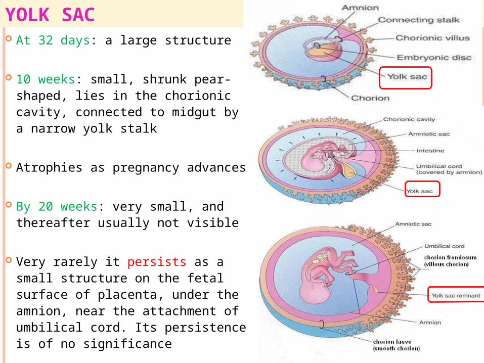
YOLK SAC At 32 days: a large structure
10 weeks: small, shrunk pear-shaped, lies in the chorionic cavity, connected to midgut by a narrow yolk stalk
Atrophies as pregnancy advances
By 20 weeks: very small, and thereafter usually not visible
Very rarely it persists as a small structure on the fetal surface of placenta, under the amnion, near the attachment of umbilical cord. Its persistence is of no significance
YOLK SAC: SIGNIFICANCE Source of nutrition for the embryo during 2-3
weeks
Blood development first occurs in the mesodermal layer of the yolk sac (early 3rd week) and continues until hemopoietic activity begins in the liver (6th week)
Primordial germ cells appear in the endodermal lining of the wall of the yolk sac (3rd week) and then migrate to the developing gonads
Part of yolk sac is incorporated into the embryo as the primitive gut (4th week)

YOLK STALK (VITELLINE DUCT) A tubular connection
between the midgut and the yolk sac
Initially wide, becomes narrow with the folding of the embryo
Becomes one of the contents of the developing umbilical cord
Attached to the tip of the midgut loop
Usually detaches from midgut loop by the end of the 6th week
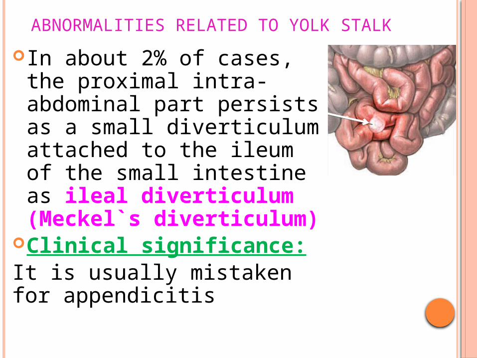
ABNORMALITIES RELATED TO YOLK STALK
In about 2% of cases, the proximal intra-abdominal part persists as a small diverticulum attached to the ileum of the small intestine as ileal diverticulum (Meckel`s diverticulum)
Clinical significance:It is usually mistaken for appendicitis
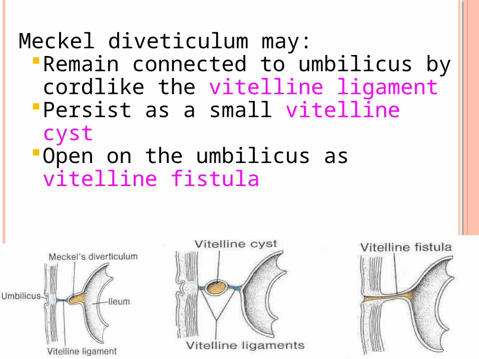
Meckel diveticulum may:Remain connected to umbilicus by cordlike the vitelline ligament
Persist as a small vitelline cystOpen on the umbilicus as vitelline fistula

ALLANTOIS Appears in 3rd week as a
diverticulum from the caudal wall of the yolk sac, that extends into the connecting stalk
During folding of the embryo, a part of allantois is incorporated into the hindgut
During the 2nd month, the extra-embryonic part of allantois degenerates
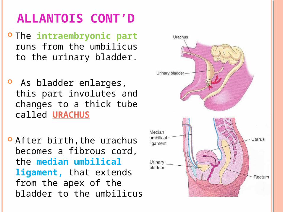
ALLANTOIS CONT’D The intraembryonic part
runs from the umbilicus to the urinary bladder.
As bladder enlarges, this part involutes and changes to a thick tube called URACHUS
After birth,the urachus becomes a fibrous cord, the median umbilical ligament, that extends from the apex of the bladder to the umbilicus

Blood formation occurs in its walls during the 3rd week
Its blood vessels persist as umbilical vesselsAllantois: Anomalies Allantois may not involute properly and give rise to:
Urachal fistula Urachal cyst Urachal sinus
Allantois: Significance