eye as a camera - mcgill university · pdf filephotopic vs scotopic vision . ... magno...
TRANSCRIPT
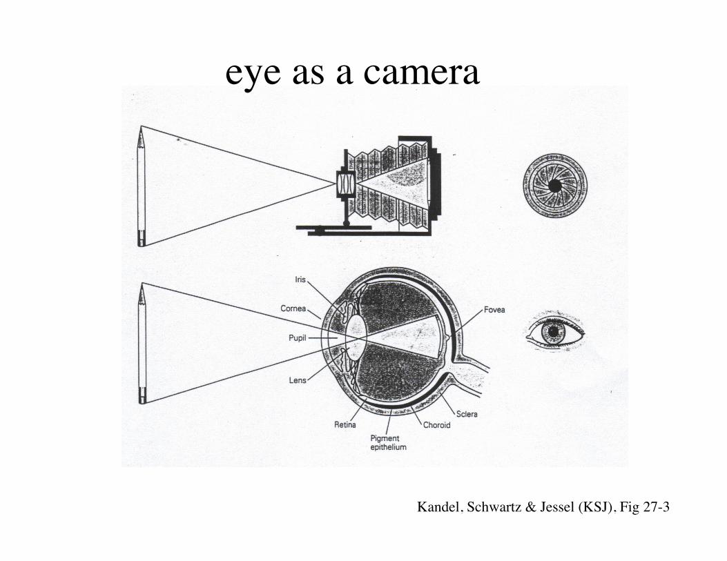
eye as a camera
Kandel, Schwartz & Jessel (KSJ), Fig 27-3
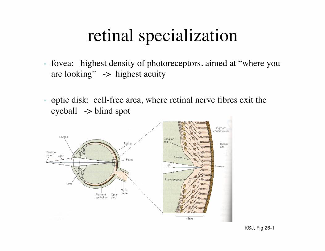
retinal specialization • fovea: highest density of photoreceptors, aimed at “where you
are looking” -> highest acuity
• optic disk: cell-free area, where retinal nerve fibres exit the eyeball -> blind spot
KSJ, Fig 26-1

demonstration of blind spot
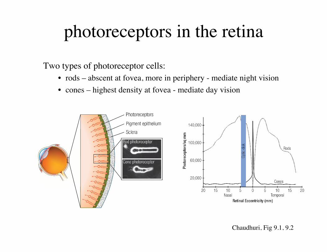
photoreceptors in the retina
Two types of photoreceptor cells: • rods – abscent at fovea, more in periphery - mediate night vision • cones – highest density at fovea - mediate day vision
Chaudhuri, Fig 9.1, 9.2
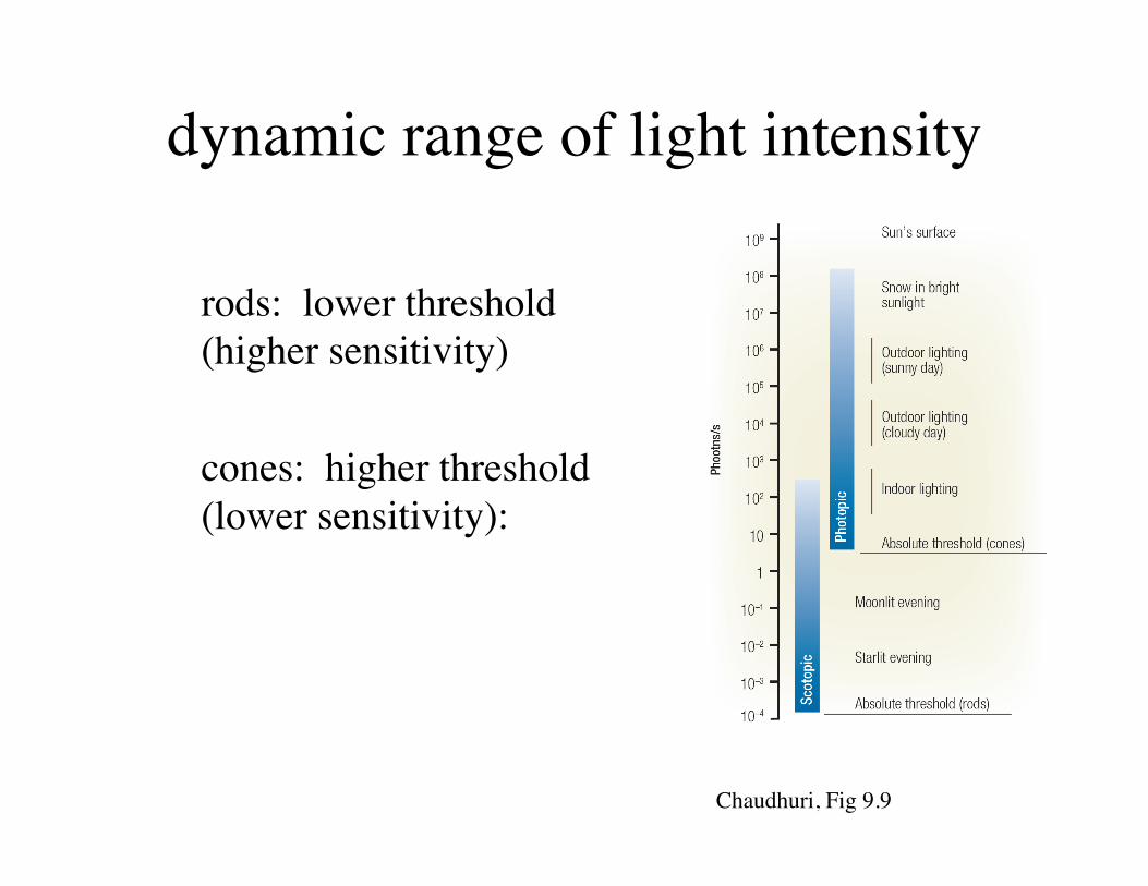
dynamic range of light intensity
rods: lower threshold (higher sensitivity) cones: higher threshold (lower sensitivity):
Chaudhuri, Fig 9.9
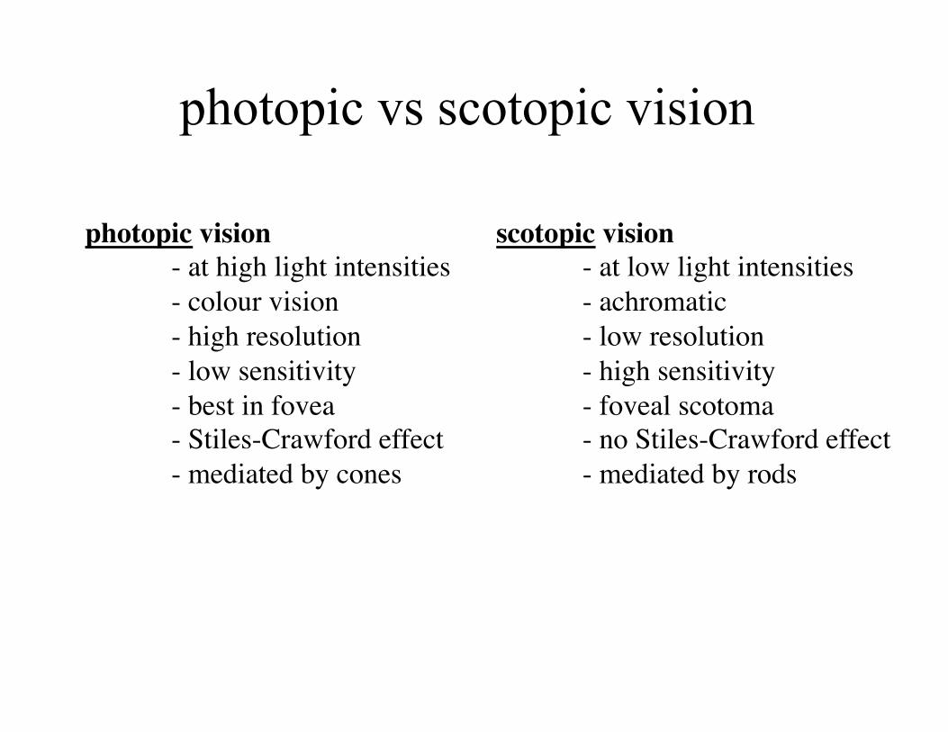
photopic vision - at high light intensities - colour vision - high resolution - low sensitivity - best in fovea - Stiles-Crawford effect - mediated by cones
scotopic vision - at low light intensities - achromatic - low resolution - high sensitivity - foveal scotoma - no Stiles-Crawford effect - mediated by rods
photopic vs scotopic vision
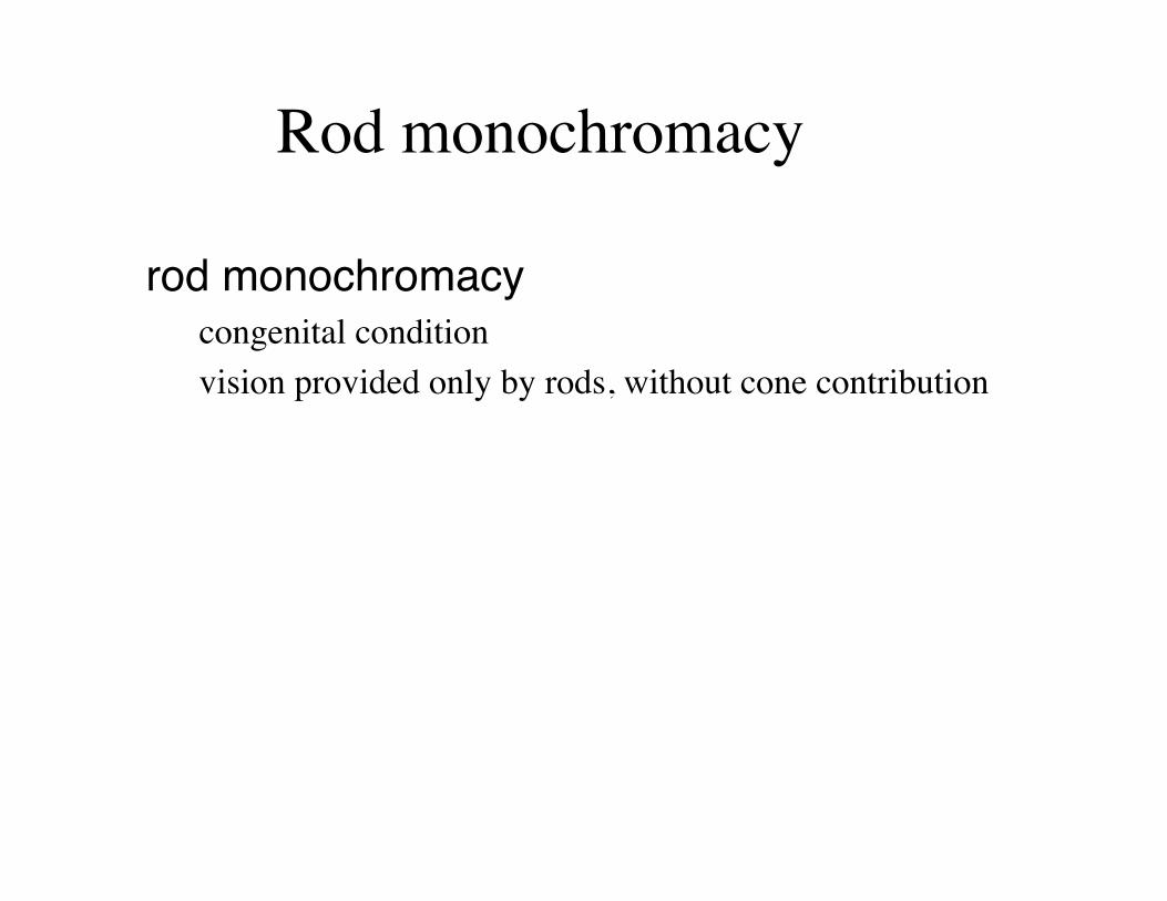
rod monochromacy" congenital condition vision provided only by rods, without cone contribution
Rod monochromacy
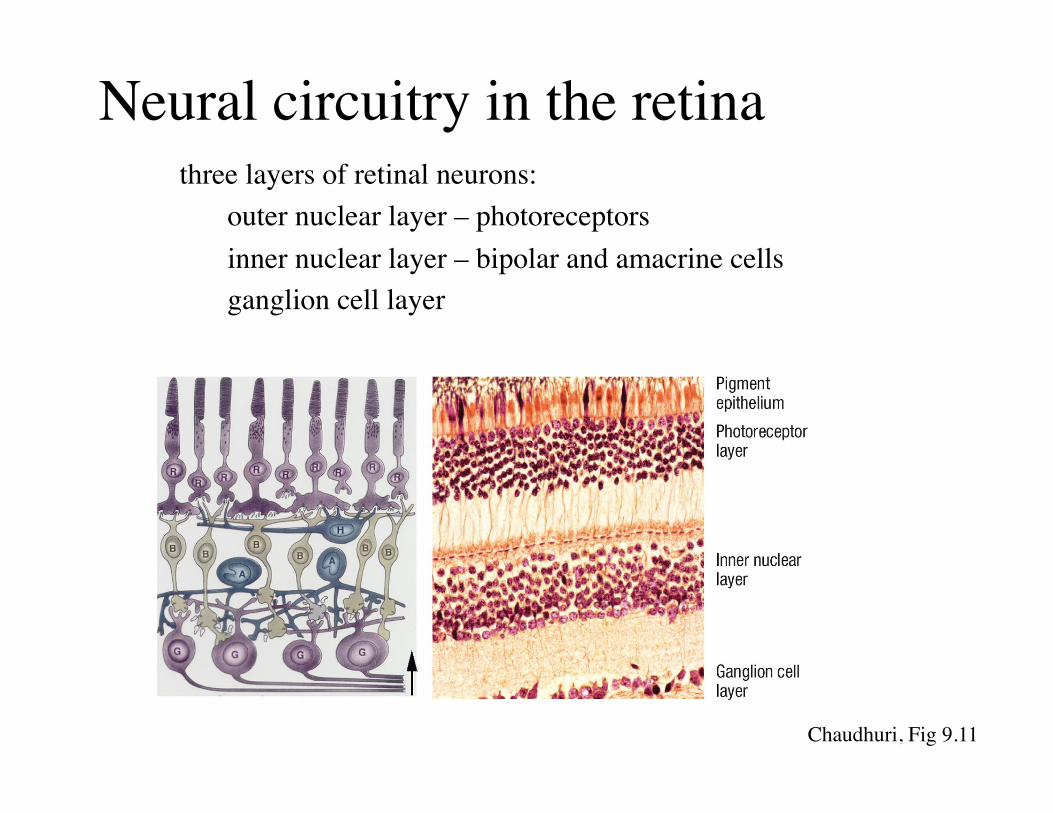
Neural circuitry in the retina three layers of retinal neurons:
outer nuclear layer – photoreceptors inner nuclear layer – bipolar and amacrine cells ganglion cell layer
Chaudhuri, Fig 9.11
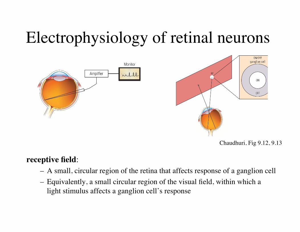
Electrophysiology of retinal neurons
receptive field: – A small, circular region of the retina that affects response of a ganglion cell – Equivalently, a small circular region of the visual field, within which a
light stimulus affects a ganglion cell’s response
Chaudhuri, Fig 9.12, 9.13

Receptive fields of retinal ganglion cells Two kinds:
• ON-center/OFF-surround cell: – Centre circular region of receptive field is excited by light, surrounding
zone is inhibited by light. • OFF-center/ON-surround cell:
– Centre circular region of receptive field is inhibited, surrounding zone is excited by light.
Chaudhuri, Fig 9.13

Receptive fields of retinal ganglion cells Retinal ganglion cells are optimized for detecting contrast:
• Centre-surround antagonism: – results from the concentric
spatial arrangement of the ON and OFF subregions
• Consequence is that retinal output sent to the brain by ganglion cells is driven by light contrast, i.e. differences in luminance
Chaudhuri, Fig 9.14
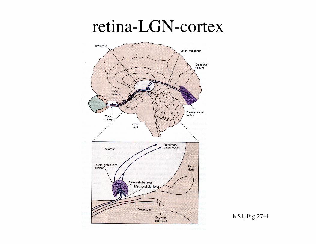
retina-LGN-cortex
KSJ, Fig 27-4
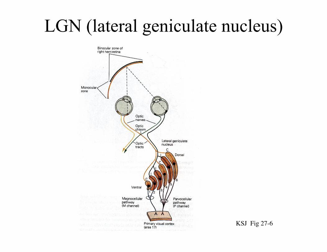
LGN (lateral geniculate nucleus)
KSJ Fig 27-6

LGN receptive fields
KSJ, Fig 29-11
achromatic
colour-opponent
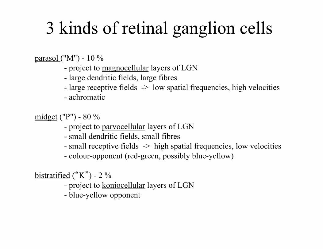
3 kinds of retinal ganglion cells parasol ("M") - 10 %
- project to magnocellular layers of LGN - large dendritic fields, large fibres - large receptive fields -> low spatial frequencies, high velocities - achromatic
midget ("P") - 80 %
- project to parvocellular layers of LGN - small dendritic fields, small fibres - small receptive fields -> high spatial frequencies, low velocities - colour-opponent (red-green, possibly blue-yellow)
bistratified (“K”) - 2 %
- project to koniocellular layers of LGN - blue-yellow opponent

Visual angle • Resolution:
– Often express acuity in terms of visual angle – Visual angle = angle subtended by image on the retina – An object at a greater distance subtends a smaller visual angle
http://en.wikipedia.org/wiki/Visual_angle

Sinewave gratings: spatial frequency spatial frequency: cycles per degree of visual angle
Chaudhuri, Fig 9.26

contrast = (Lmax - Lmin) / (Lmax + Lmin) x 100%
100 % 50 % 25 % 12.5 %
Sinewave gratings: contrast
contrast sensitivity = 1 / contrast threshold

Contrast sensitivity function
Measure minimum contrast to make a grating of a particular spatial frequency just visible. Plot threshold data in terms of sensitivity = 1 / threshold.
Chaudhuri, Fig 9.27
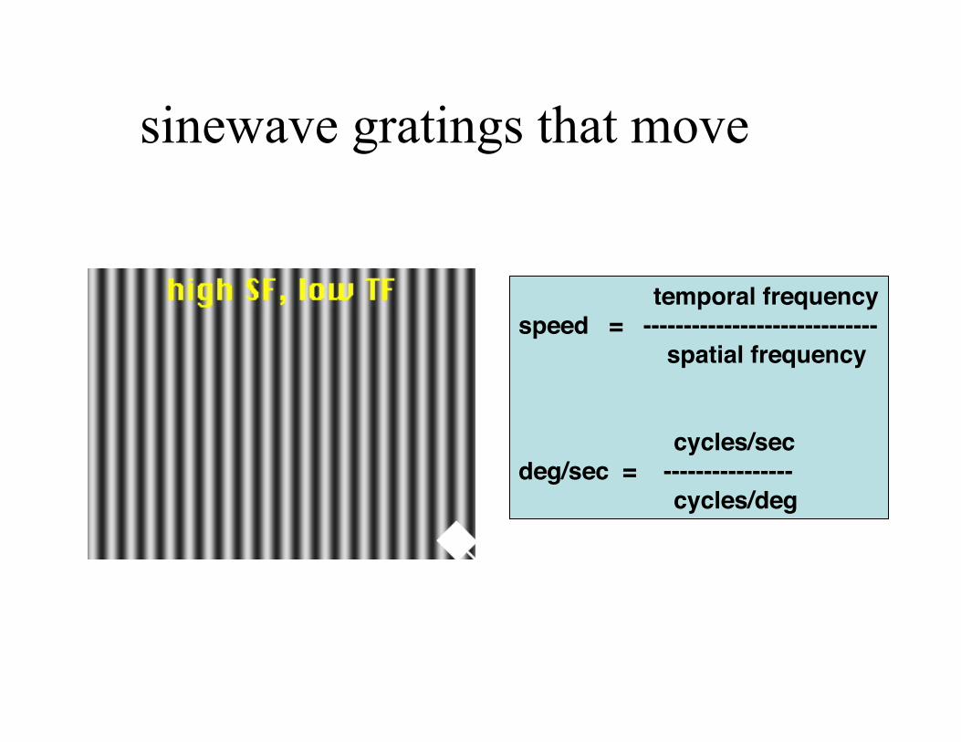
sinewave gratings that move
temporal frequency!speed = -----------------------------! spatial frequency!! ! cycles/sec!deg/sec = ----------------! cycles/deg!

contrast sensitivity after M-lesions
Merigan et al, Fig 2&3

effects of M vs P lesions: summary
parvo lesion: - lower acuity - abolishes colour discrimination - reduced contrast sensitivity to gratings, at low temporal / high spatial frequencies (low velocities)
magno lesion:
- no effect on acuity - no effect on colour discrimination - reduced contrast sensitivity to gratings, at high temporal / low spatial frequencies (high velocities)
- does not support idea of magno for motion, parvo for form vision
central problem: need for early detection
"at risk": ocular hypertension (OHT)
perceptual "filling in" - example is failure to see your "blind spot"
conventional (static) perimetry - detects problem only later
human psychophysics, as approach for early detection: why you would not expect a deficit on many tasks:
earliest lesions in peripheral vision, but many tasks use foveal vision
-> need to do perimetry (automated) using the task task may be mediated by unaffected neurons, e.g. color-discrimination (P-cells)
glaucoma: early detection
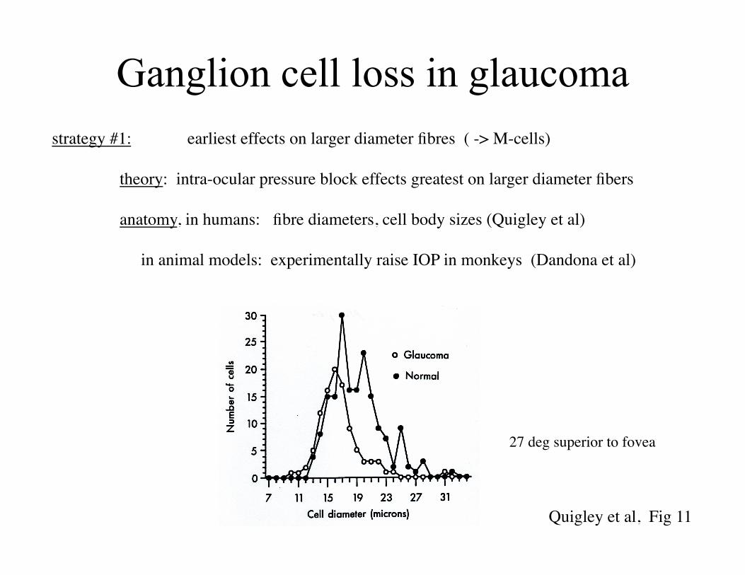
Ganglion cell loss in glaucoma
Quigley et al, Fig 11
27 deg superior to fovea
strategy #1: earliest effects on larger diameter fibres ( -> M-cells) theory: intra-ocular pressure block effects greatest on larger diameter fibers
anatomy, in humans: fibre diameters, cell body sizes (Quigley et al) in animal models: experimentally raise IOP in monkeys (Dandona et al)

motion coherence: stimulus
task: report direction of motion noisy random dots: prevent using change-of-position
a demanding task, requiring: combining responses of multiple neurons correct timing relations between neurons vary signal-to-noise (% coherence): best performance requires all the neurons
see Adler’s, Fig 20-12, 22-11
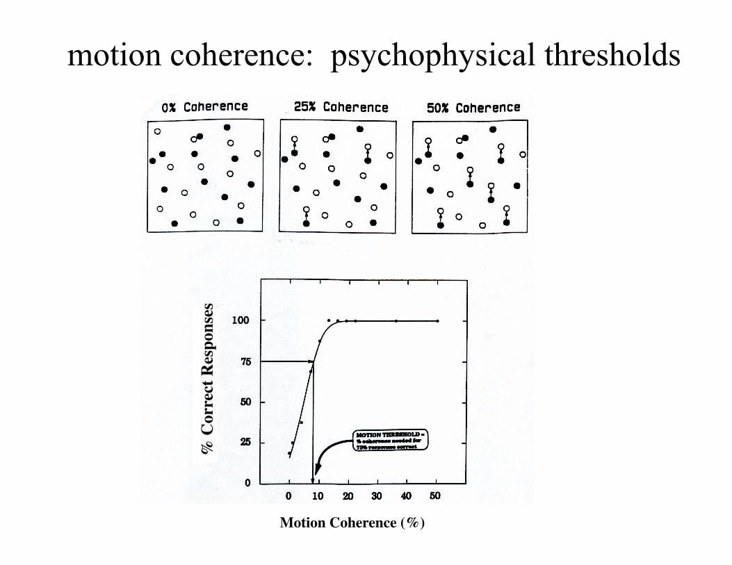
motion coherence: psychophysical thresholds
% C
orre
ct R
espo
nses
Motion Coherence (%)
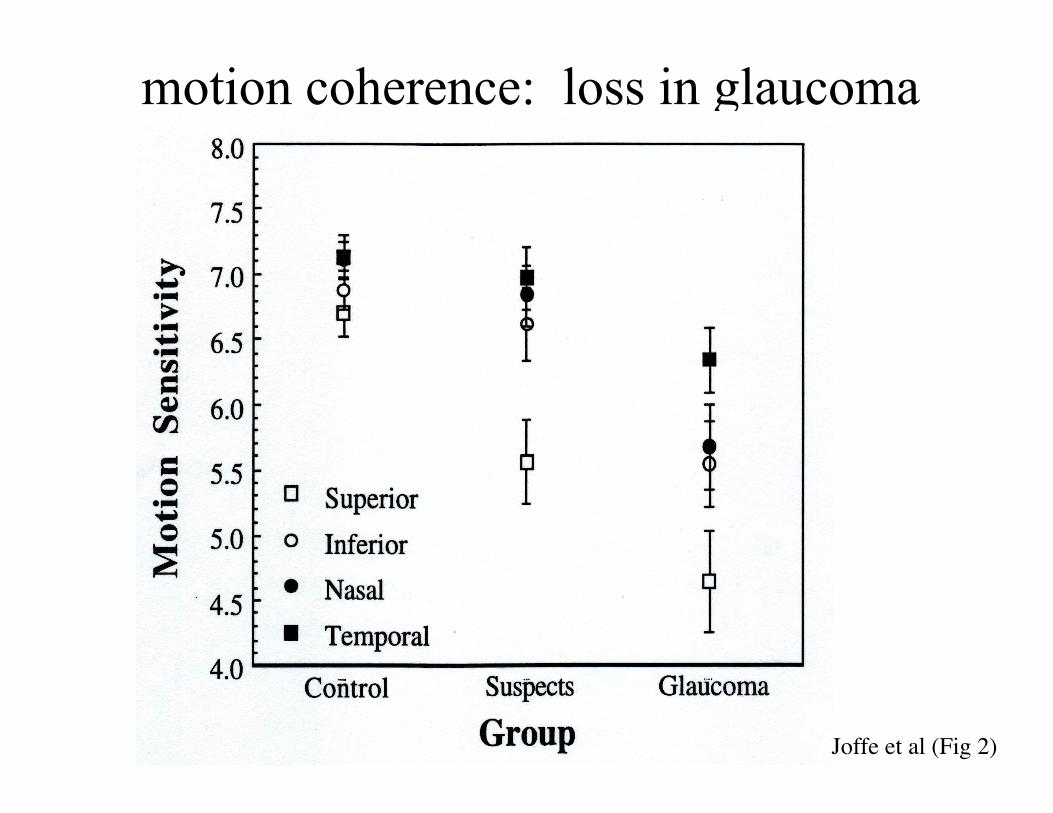
motion coherence: loss in glaucoma
Joffe et al (Fig 2)
apparent loss of large cells/fibres might be artifact of cell shrinkage also find losses of P-cell dependent psychophysics
selective M-cell loss hypothesis: criticisms

strategy #2: most sensitive tests for capricious loss are those for sparse cell types:
(explains loss of abilities that depend on M-cells)
-> S-cones, blue/yellow (bistratified ganglion cells)
color: detection of blue spot on yellow background
rationale: blue-yellow ganglion cells (bistratified) are relatively sparse (ca 5%)
results: Sample et al, Johnson et al: perimetry, longitudinal study
testing for loss of sparse cell types
general textbooks: Carpenter RHS (2003) Neurophysiology, (4th Ed) London: Arnold. Chaudhuri A (2011) Sensory Perception. Oxford: Oxford Press. Kaufman PL, Alm A (Ed) (2003) Adler's Physiology of the Eye, 10th ed. St.Louis: Mosby. Kandel, Schwartz, and Jessell , Principles of Neural Science (4th Ed.) journal articles: Ansari EA, Morgan JE, Snowden RJ (2002) “Glaucoma: squaring the psychophysics and neurobiology” British Journal of Ophthalmology 86:823-826. http://bjo.bmjjournals.com/cgi/content/full/86/7/823 Joffe KM, Raymond JE, Chrichton A (1997) "Motion coherence perimetry in glaucoma and suspected glaucoma" Vision Research 37:955-964. Johnson CA, Adams AJ, Casson EJ (1993) "Blue-on-yellow perimetry can predict the development of glaucomatous visual field loss" Arch. Ophthalmol. 111: 645-650. Maddess, T., Goldberg, I., Dobinson, J., Wine, S., Welsh, A.H., and James, A.C., “Testing for glaucoma with the spatial frequency doubling illusion”, Vision Research 39: 4258-4273 (1999). Merigan WH, Byrne CE, Maunsell HR (1991) "Does primate motion perception depend on the magnocellular pathway ?" J. Neuroscience 11: 3422-4329. Quigley HA, Dunkelberger GR, Green WR (1989) "Retinal ganglion cell atrophy correlated with automated perimetry in human eyes with glaucoma", Am. J. Ophthal. 107: 453-464. Sample, P.A., Taylor, J.D.N., Martinez, G.A., Lusky, M., and Weinreb, R.N., "Short-wavelength color visual fields in glaucoma suspects at risk", Am. J. Ophthal. 115: 225-233 (1993). Shapley R, Perry VH (1986) "Cat and monkey retinal ganglion cells and their visual functional roles", Trends in Neurosciences 9:229-235.
References