extrinsic inhibition of the inferior colliculus during
TRANSCRIPT

EXTRINSIC INHIBITION OF THE INFERIOR COLLICULUS DURING AUDIOGENIC SEIZURES: EFFECTS OF UNILATERAL DORSAL NUCLEUS OF THE LATERAL
LEMNISCUS LESIONS IN YOUNG RATS
A Thesis By
AMY M. MORGAN
Submitted to the Graduate School Appalachian State University
In partial fulfillment of the requirements for the degree MASTER OF ARTS
August 2010 Major Department: Psychology

EXTRINSIC INHIBITION OF THE INFERIOR COLLICULUS DURING AUDIOGENIC SEIZURES: EFFECTS OF UNILATERAL DORSAL NUCLEUS OF THE LATERAL
LEMNISCUS LESIONS IN YOUNG RATS
A Thesis
By AMY M. MORGAN
August 2010
APPROVED BY: __________________________ Mark C. Zrull Chairperson, Thesis Committee __________________________ James C. Denniston Member, Thesis Committee __________________________ Kurt D. Michael Member, Thesis Committee __________________________ James C. Denniston Chair, Department of Psychology __________________________ Edelma D. Huntley Dean, Research and Graduate Studies
Copyright by Amy M. Morgan All Rights Reserved
Permission is hereby granted to the Appalachian State University Belk Library and the Department of Psychology to display and provide access to this thesis for appropriate
academic and research purposes

FOREWORD
This thesis is written in accordance with the style of the
Publication Manual of the American Psychological Association (5th Edition) as required by the Department of Psychology at Appalachian State University
Extrinsic inhibition of 1
Running head: EXTRINSIC INHIBITION DURING AUDIOGENIC SEIZURES IN RATS
Extrinsic Inhibition of the Inferior Colliculus during Audiogenic Seizures: Effects of
Unilateral Dorsal Nucleus of the Lateral Lemniscus Lesions in Young Rats
Amy M. Morgan
Appalachian State University
Extrinsic inhibition of 2
Abstract
In this study, a model of generalized epilepsy was used to investigate the role of extrinsic
inhibition of the inferior colliculus (IC) during audiogenic seizures (AGS). There is evidence
to suggest that a deficiency of inhibition within the IC plays a major role in AGS; however,
the importance of extrinsic inhibition of the IC is not as well understood. To investigate this,
neurotoxin lesions of dorsal nucleus of the lateral lemniscus (DNLL) were made, therefore
eliminating the main source of extrinsic inhibition to the IC. Long-Evans rats (N = 18) were
primed for AGS susceptibility via acoustic insult on post-natal day (pnd) 18, tested for
susceptibility on pnd 32, and then subjected to a series of inductions on seven occasions over
14 days beginning on pnd 35. Seizure-susceptible rats were divided into three groups (n = 6).
One group received a neurotoxic injection in the DNLL, a second group was prepared for
surgery, but did not receive the injection, and a third group experienced no aspects of the
surgical procedure. Subjects then followed a post-surgery induction schedule to evaluate
differences across three measures: latency to enter a wild-running phase, latency to enter
clonus, and duration of the seizure. The hypotheses that subjects in the lesion group would
have a shorter latency to enter a wild running phase in post-surgery inductions (p = .5289)
and would exhibit a shorter latency to enter clonus in post inductions (p = .1713) were not
supported by the data. The hypothesis that subjects in the lesion group would exhibit longer
AGS post-surgery approached statistical significance (p = 0.0584); however, rats in the lesion
group exhibited shorter AGS than controls. While a lack of intrinsic inhibition of the IC plays
an important role in AGS, the data from this study suggest that a lack of extrinsic inhibition
to the IC does not have a significant role in inhibiting the initiation and propagation of AGS
activity in rats, but may aid in decreasing the duration of clonic seizures.

Extrinsic inhibition of 3
Extrinsic Inhibition of the Inferior Colliculus during Audiogenic Seizures: Effects of
Unilateral Dorsal Nucleus of the Lateral Lemniscus Lesions in Young Rats
Epilepsy is a neurological disorder characterized by temporary abnormal neuronal
activity which causes seizures ranging in severity from temporary lapses in consciousness to
muscular convulsions. Over three million Americans of all ages are affected by epilepsy or
seizures, with approximately 200,000 new cases occurring each year (Epilepsy Foundation,
2010). While there are many seizure disorders, the seizure itself always involves abnormal
excitatory and inhibitory activity in the brain. Seizure disorders can be characterized as either
focal (also referred to as partial) or generalized. In focal seizures, the abnormal neuronal
activity is confined to one area of the brain. Symptoms of this type of seizure are categorized
as either simple or complex. Simple focal seizures do not result in unconsciousness, but
rather an altered state of consciousness including exaggerated emotions or hallucinations.
Complex focal seizures can result in either consciousness or unconsciousness (Epilepsy
Foundation; Freeman, Vining, & Pillas, 2002). Abnormal neuronal activity that begins in one
structure and spreads throughout the entire brain is known as a generalized seizure (Ross &
Coleman, 2000). Like focal seizures, generalized seizures are also categorized by symptoms
exhibited. Individuals experiencing absence seizures (referred to as petit mal seizures) may
exhibit blank stares and mild muscle twitches. Tonic seizures result in stiff muscles and
immobility. Conversely, atonic seizures exhibit loss of muscle tone. During a clonic seizure,
the muscles contract and relax, resulting in full body jerks. Myoclonic seizures are similar to
clonic seizures, with the exception that muscle jerks are confined to the limbs and upper
body. Tonic-clonic seizures, also known as grand mal seizures, display stiffening and
repeated jerking of the limbs (Epilepsy Foundation; Freeman et al.).

Extrinsic inhibition of 4
Epilepsy, particularly with generalized seizures, is a heritable disorder; however,
seizures can also be acquired in response to an environmental insult such as an intense or
unexpected acoustic, visual, or tactile stimulus, a condition referred to as reflex epilepsy.
Most reflex epilepsy in humans is the result of exposure to a visual stimulus such as sunlight,
intense bright or flashing light, or even some video games (Epilepsy Foundation, 2010;
Freeman et al., 2002). Rodent models can be used to study seizure disorders, and generally
rodents will express reflex seizure activity in response to intense acoustic stimulation
(Pierson & Swann, 1991). In this thesis, a rodent model of acquired reflex epilepsy was used
to investigate neural mechanisms underlying reflex seizures.
While the role of runaway excitation within the auditory midbrain during audiogenic,
or sound-induced seizures has been investigated extensively (Garcia-Cairasco,Terra, &
Doretto, 1993; Pierson & Swann, 1991; Pollack, Burger, & Klug, 2003; Ross & Coleman,
2000), the importance of normal inhibition, or lack thereof, during seizures is not as well
understood. This thesis examined the relationship between over-excitation of the inferior
colliculus (IC), the initiation site of audiogenic seizure (AGS) activity, and the lack of
extrinsic inhibition, which is supplied by the dorsal nucleus of the lateral lemniscus (DNLL;
Garcia-Cairasco et al.; Pierson & Swann; Pollack et al.; Raisinghani & Faingold, 2002; Ross
& Coleman).
Audiogenic Seizure Model of Epilepsy
There are multiple models used to investigate generalized seizures in rats, including
electric and chemical kindling models, genetically epilepsy-prone rat models (GEPRs), and
developmentally-primed audiogenic seizure models (Pierson & Swann, 1991; Ross &
Coleman, 2000). In the present study, a developmentally-primed model in which rats
Extrinsic inhibition of 5
acquired susceptibility to AGS through a procedure in which the animal is subjected to
intense acoustic insult at specific times during development was used (Ross & Coleman,
1999).
Priming and testing. According to the developmentally primed model, seizure-
resistant strains of rats can be made seizure prone at a young age through exposure to intense
acoustic stimulation around the time the ear canal opens, a procedure referred to as priming.
Priming causes a disruption of nerve connections of the normal auditory pathway, damage
that the brain will attempt to repair by reorganizing nerve connections which allow for over-
excitation of neurons if acoustic insult is presented later (Pierson & Swann, 1991; Ross &
Coleman, 1999). Subjects must then be tested via exposure to loud noise approximately two
weeks after priming to determine seizure susceptibility. The parameters for both priming and
testing, such as intensity of noise and days to perform the procedures, are fairly specific and
strain-dependent. For the Long-Evan strain, the rat stain used in this study, the optimum day
to prime is postnatal day (pnd) 18, and the optimum day to test is pnd 32 (Ross & Coleman,
1999; Ross & Coleman, 2000).
Inductions. A series of seizure inductions are necessary after priming and testing in
order to evaluate AGS severity. The number of inductions varies depending upon the
investigation and research design. There is a reliable progression of AGS behavior exhibited
during each trial (Garcia-Cairasco, 2001; Ross & Coleman, 1999). In the first phase of AGS
activity, referred to as the wild running phase, the subject runs uncontrollably at a high rate
of speed around the walls of the cylindrical testing chamber some time after acoustic
stimulation begins. Wild running gives way to one of four possible outcomes: a period of
inactivity, a second run, a clonic seizure, or tonic-clonic seizure (Pierson & Swann, 1991;
Extrinsic inhibition of 6
Ross & Coleman). In the Audiogenic Response Score (ARS), a scale for measuring AGS
response to sound stimulation in rats, AGS inductions with only one phase of wild running
before entering a seizure state are given a higher rating on the scale than episodes with two
phases of wild running. It is suggested that a temporary pause between runs is a
demonstration of control provided by inhibition within the brain, indicating that a subject
may have more control over the runaway excitation than subjects who are unable to stop
running (Jobe, Picchioni, & Chin, 1973).
A wild running phase is often followed by either a tonic, clonic, or tonic-clonic
seizure, the only types of seizures exhibited by the many rat strains, including Long-Evans.
Tonic seizures, the most severe seizure displayed in the rat, are given the highest ARS (Jobe,
et al., 1973). These seizures are characterized by muscle rigidity of the limbs, neck, and back,
and can sometimes lead to death. Clonic seizures in rats are characterized by an arched back
and neck, stiffness of the limbs, and full-body muscle spasms accompanied by rocking
motions (Ross & Coleman, 2000). Because it is the least severe type of seizure exhibited by
the rat, it is given the lowest ARS of the seizures. If the clonic convulsions are separated by
moments of muscle rigidity, the seizure is characterized as tonic-clonic. While latency to
enter a seizure state is strain-dependent, the average latency for the Long-Evans rat is
approximately 30 s, and the duration of clonus averages approximately 20 s. The final phase
of AGS activity is referred to as the post-ictal period, the period after the seizure when the
subject exhibits behaviors such as immobility and vocalization, and is typically unresponsive
to acoustic stimulation (Ross & Coleman, 1999; Ross & Coleman, 2000).
The Auditory Midbrain and the DNLL
Auditory processing begins within the cochlea of the inner ear, which tranduces
Extrinsic inhibition of 7
acoustic information into neural signals. Potentials from hair cells of the cochlea activate the
auditory nerve, which is the source of innervation for the cochlear nucleus (CN). The CN
projects to multiple ascending pathways. The major projection from the CN is to the
contralateral IC, a major processing, integration, and relay center for all auditory input
(Coleman & Clerici, 1987; Pollack et al., 2003). CN also projects to the medial and lateral
superior olive (MSO, LSO), which are responsible for the detection of interaural time
differences, aiding in sound localization. Once excitatory signals leave MSO or LSO, they
can project either directly to the IC, or a second route starting in the LSO, which goes then to
the ventral nucleus of the lateral lemniscus (VNLL), and finally to the IC (Faye-Lund &
Osen, 1985).
The DNLL, located just ventral to the IC, lies within the fibers of the lateral
lemniscus (LL) and is a part of the ascending pathway (Bajo, Merchán, Lopez, & Rouiller,
1993; Pollack, et al., 2003; Schneiderman, Chase, Rockwood, Benson, & Potashner, 1993;
Wu & Kelly, 1996), and is the primary source of extrinsic inhibition to the IC (Kelly, Li, &
van Adel, 1996; Wu & Kelly; Zhang, Li, Kelly, & Wu, 1998). The main source of excitation
to the DNLL ipsilateral to a sound source comes from the contralateral CN and superior
olivary complex (SOC). This DNLL in turn projects to the contralateral DNLL, which then
projects to the IC (Bajo et al., 1993; Pollack et al.). Thus, hearing a sound involves excitation
of the IC contralateral to the sound source and inhibition of the IC ipsilateral to the source via
the DNLL.
Most excitatory neurotransmission within the auditory system, including the DNLL,
is modulated by neurons with glutamate receptors. There are three types of receptors that
may be present on glutamate neurons, N-methyl-D-aspartate (NMDA), α-amino-3-hydroxy-

Extrinsic inhibition of 8
5-methyl-4-isoxazolepropionic acid (AMPA), and kainic acid (KA). Furthermore, each
receptor is broken down into four or five protein subunits. NMDA receptor subunits are
labeled NR1 and NR2A-D. AMPA receptors contain four subunits, GluR1-4, and KA
receptors are labeled GluR5-7 and KA1 and 2, dependent upon gene family (Hermit,
Greenwood, & Brauner-Osborne, 2004; Johansen, Greenwood, Frydenvang, Madsen, &
Krogsgaard-Larson, 2003; Parks, 2000). Most if not all DNLL neurons also contain the
enzyme glutamate decarboxylase (GAD67), a catalyst that aids in the production of γ-
aminobutyric acid (GABA), the most common inhibitory neurotransmitter in the nervous
system (Garcia-Cairasco et al., 1993). Therefore, GABA-producing neurons are activated
through glutamate synapses, which is why much of the DNLL’s projection to the IC is
inhibitory in nature (Merchán, Saldaña, & Plaza, 1994; Schneiderman et al., 1993; Wu &
Kelly, 1996). Because AMPA receptors are essential to the activation of glutamate neurons,
an AMPA agonist, such as quisqualic acid, can target and kill GluR1 and GluR4 subunits,
ultimately preventing activation of glutamate neurons within the DNLL (Hermit et al.;
Johansen, et al.). If glutamate neurons cannot be activated, GABA cannot be released to
provide extrinsic inhibition to the IC.
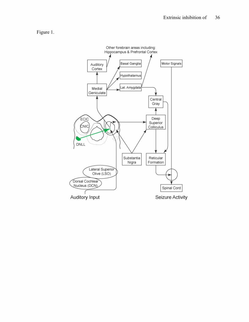
Neural network for AGS. The neural network is identical for all models of AGS,
including the developmentally primed model used in this thesis as well as in GEPRs, and
electrical and chemical kindling models (Ross & Coleman, 2000; see Figure 1). Once altered
by acoustic insult, the complex connections within the auditory pathway reorganize,
beginning with those from hair cells to the auditory nerve. If very intense sound is presented
later, neurons within the pathway become over-excited due to the new organization of
excitatory and inhibitory connections, which culminates in a seizure. Specifically, AGS
Extrinsic inhibition of 9
induction begins with the over-stimulation of the cochlea within the inner ear. From the
cochlea, acoustic information is sent to the SOC, which in turn relays the information via the
LL. The LL is responsible for sending acoustic information to the IC where abnormal
excitation becomes sufficient to produce a seizure (Garcia-Cairasco et al., 1993; Raisinghani
& Faingold, 2002).
The IC relays information to higher areas of the forebrain for interpretation, and is
also the initiation sight for AGS activity (Garcia-Cairasco et al.1993; Pierson, & Swann
1991; Pollack et al., 2003; Raisinghani & Faingold, 2002; Ross & Coleman, 2000). There is
ample evidence to suggest that when the efficacy of GABAergic cells is compromised within
this structure, an animal is more susceptible to AGS (Faingold & Anderson, 1991). The
central nucleus of the IC (CNIC) relays information from the LL to the medial geniculate
body (MG), which in turn sends information to the acoustic cortex, as well as areas of the
forebrain including the lateral amygdala and the hippocampus (Garcia-Cairasco et al.;
Raisinghani & Faingold). The amygdala projects to the frontal motor cortex via the perirhinal
cortex, which could explain some of the motor activity involved in AGS (Hirsch et al 1997).
The external cortex of the IC (ECIC), which research implicates in the sensorimotor aspect of
AGS activity, including wild running, is the source of efferent projections to multiple areas
of the reticular formation (RF) as well as to the superior colliculus (SC; Faingold, 2002;
Garcia-Cairasco et al.; Raisinghani & Faingold; Ribak & Morin, 1995; Ross & Coleman;
Zrull, Mengle, Hall, Hairston, & Sedivec, 1999). Data from lesion studies suggest that while
the SC can propagate AGS activity, its role is not obligatory like the IC (Ribak, Khurana, &
Lien, 1994; Ross & Coleman, 2000). Parts of the substantia nigra (SN) project to both the IC
and the SC, regulating efferent seizure pathways. Each structure in the neural network plays a
Extrinsic inhibition of 10
role in the propagation of AGS activity; however, the purpose of this thesis was to investigate
the relationship between two particular structures within the network, the DNLL and the IC.
Although there is already substantial evidence to suggest that the IC is the initiation site of
AGS, and that abnormalities associated with the inhibition within this structure contributes to
this activity, further investigation is necessary to determine the importance of extrinsic
inhibition within the DNLL.
Statement of Purpose
In this study, the developmentally primed model was used to investigate sound-
induced seizures in the Long-Evans rat. Ample research has been conducted to determine the
role of the IC in AGS activity. There is evidence to suggest that the IC is the initiation site of
AGS, and that runaway excitation initiated within this structure is the cause of generalized
seizures exhibited when loud sound is presented (Garcia-Cairasco et al., 1993; Garcia-
Cairasco, 2001; Pollack et al., 2003; Raisinghani & Faingold, 2002; Ross & Coleman, 2000).
The role of normal inhibition in AGS, however, is not as clear as the role of excitation. A
review of the literature failed to find any prior investigation specifically of the role of
extrinsic inhibition of the IC during these seizures. The purpose of this experiment was to
determine the importance of this inhibition during sound-induced seizures, an investigation
which required a chemical lesion of the DNLL to eliminate GABAergic neurons within the
structure. To conduct this experiment, three groups of animals were used. An experimental
group received a neurotoxin that created a chemical lesion of the area. Two control groups,
one that was prepared for surgery and received identical post-operative care, but did not
receive any injection, and one that did not experience any aspects of the surgery were used
for comparison. All groups were made seizure-prone prior to surgery by following the
Extrinsic inhibition of 11
procedures of the developmentally primed model. Because the chosen neurotoxin
purportedly eliminated the IC’s main source of extrinsic inhibition in the brains of
experimental animals, it was hypothesized that there would be a shorter latency to enter the
wild running phase, a shorter latency to enter clonus, and longer seizures in the group who
received the chemical lesion than either control group. This hypothesis was tested with both
pre- and post-surgery series of AGS inductions identical in procedure.
Method
Subjects and Experimental Groups Three litters of Long-Evans hooded rats (N = 36), born in the Arts and Sciences
Animal Facility at Appalachian State University, were bred for this study. Rat pups were
weaned from their dams between pnd 21 and 24, and thereafter were housed in same-sex
groups of three or four in plastic shoebox cages. Subjects were given free access to food and
water. The housing room within the facility maintains a 14 h light/10 h dark illumination
schedule with a constant temperature of 22 oC and 50% relative humidity. All animal care
and use procedures were approved by the Institutional Animal Care and Use Committee at
Appalachian State University prior to being performed (protocol #09-07, approved on
5/13/2009, see Appendix).
All subjects were primed on pnd 18 and tested for AGS susceptibility on pnd 32. To
be included in this study, subjects must have exhibited an audiogenic response during testing
on pnd 32. Pups in one litter (n = 11) did not meet this requirement, and therefore were
eliminated from the experiment. Prior to group assignment, all remaining subjects (N = 25)
were subjected to seven AGS inductions across 12 days beginning on pnd 35. Subjects must
have responded appropriately to hearing tests, indicating no signs of hearing loss, prior to
Extrinsic inhibition of 12
each induction. Group membership was determined based upon matching similar audiogenic
responses in pre-surgery inductions across all dependent measures. One group (n = 9)
received a neurotoxin injection of quisqualic acid into the DNLL (lesion group). This
procedure was expected to result in a chemical lesion of the DNLL, thereby eliminating the
IC’s primary source of extrinsic inhibition. The number of subjects in this group was reduced
(n = 6) during histological procedures either due to the inability to verify lesion sites, or the
inability to assess damage due to the nature of the tissue. A second group (n = 7) served as a
sham operated control (sham control), undergoing all steps of the surgical procedure with the
exception of the chemical injection. One brain was damaged during tissue processing and
could not be used thus necessitating removal of the rat from the study, bringing the number
of subjects in this group to six as well (n = 6). The final group (n = 9), which also served as a
control group (unoperated control), did not experience any aspect of the surgical procedure.
Three subjects were removed from this group either due to injuries sustained during seizure
inductions that prevented the completion of behavioral testing, or because the brain was
damaged, and therefore histological data could not be provided, which left six in this group
as well (n = 6). After surgery was performed on assigned subjects, all rats were again
subjected to a series of AGS inductions identical to the pre-surgery schedule, beginning on
pnd 63 and lasting 14 days.
Priming
All pups were primed for AGS on pnd 18. Using a standard protocol for the
laboratory (e.g., Adams, Glenn, Richardson, & Zrull, 2008; Hodgin, 2006), subjects were
placed individually into ½-in. hardware cloth cages, 9.5 cm x 9.5 cm x 9.5 cm, and exposed
to high intensity (120 dB re 0.0002 dyne/cm2, A scale) 10 kHz tone pips (8/s, 75 ms on, 50

Extrinsic inhibition of 13
ms off, 5 ms rise and fall times) for 8 min. Cages were equidistant from a paper cone speaker
(Realistic 40-1270D) and situated on a wooden platform. A digital wave form generator
(TDT, Inc WG-1) created, shaped, and gated a 10 kHz sine wave at the desired rate, and then
the signal was amplified (Realistic MPA-101) before it was switched to the tweeter. Stimulus
production and presentation was controlled by an Apple 2E computer interfaced with the
auditory signal generating apparatus (A-D/D-A board, Applied Engineering). After the
priming procedure, pups were monitored for 20 min and then returned to the appropriate
cages with their dams.
Audiogenic Response Induction and Testing
Following the priming procedure, all pups were assessed for susceptibility to AGS on
pnd 32, and subsequently subjected to inductions on seven occasions over 12 days beginning
on pnd 35. After surgery, rats were subjected to eight AGS inductions beginning on pnd 63
and that were continued every other day until pnd 77. Testing and inductions were identical
in procedure, which is standard for the lab (e.g., Hodgin, 2006). To begin, subjects were
placed individually into a cylindrical ¼-in. hardware cloth chamber (36 cm tall with a
diameter of 30 cm) with a wooden roof and floor. White noise (100 Hz to 20 kHz) generated
by a Coulbourn Noise Generator (S81-02) was used to elicit AGS activity. This noise was
amplified to at least 120 dB (re 0.0002 dyne/cm2, A scale) using a Realistic amplifier (MPA-
101) and delivered through broad range speakers (Realistic 40-1354A). Each trial lasted for 2
min, or until a clonic seizure began. All trials were immediately evaluated by multiple raters,
and videotaped for a second analysis by one individual in order to increase reliability of
audiogenic response measures. Severity of audiogenic response was evaluated based on three

Extrinsic inhibition of 14
measures, latency to enter the first wild running phase, latency to enter clonus, and the
duration of the seizure.
Chemical Lesion Surgery
After pnd 50, each rat in the lesion or sham control group was anesthetized (ketamine
70 mg/kg b.w., ip and xylazine, 8 mg/kg b.w., ip) and prepared for surgery. Once
anesthetized, the rat’s head was positioned in a stereotaxic instrument, the scalp was cleaned
and incised, and a burr hole was drilled at AP 0.0 to 0.5 mm, relative to interaural zero,
lateral 2.8 to 3.0 mm, relative to midsagittal suture, and DV -5.0 to -5.4, relative to dura
mater. Animals in the sham operated control group experienced all steps of the surgical
procedure with the exception of receiving the neurotoxic injection that produced the chemical
lesion. Lesion group rats received a 0.5 µL pressure injection of 100 mM quisqualic acid into
one DNLL over 1 min, and 5 min was allowed for diffusion. After the injection was complete,
the burr hole was filled with bone wax and the skin was sutured. Prior to removal from the
stereotaxic instrument, each rat receives a prophylactic injection of diazepam (5 mg/kg b.w.,
sc) to prevent an excitotoxin-induced seizure. Each animal was kept in a warm place and
monitored closely for the first 24 h after surgery. They were returned to their home cages
once awake and monitored for an additional 72 h after surgery. After a recovery period of 10
to 13 days, the post-surgery induction schedule began.
Histology
After post-surgical AGS inductions, subjects in all groups were sacrificed via an
overdose of sodium pentobarbital (100 mg/kg b.w., ip) and perfused intracardially with 100
mL of pH 7.2, 10 mM phosphate buffered saline (PBS) solution followed by 500 mL of 4%
paraformaldehyde in phosphate buffer (PB). Brains were removed and postfixed for at least
Extrinsic inhibition of 15
24 h in 100 mL of a 10% sucrose-4% paraformaldehyde solution at 4 oC. Brain tissue was cut
into 50 μm sections using a Vibrotome®. Tissue sections which included the DNLL were
placed into individual wells of 24-well Falcon tissue culture plates with approximately 1 mL
of PB (7.2 pH, 10mM). These sections were used to quantify the GABAergic neurons within
the DNLL by staining for GAD67 positive cells. The procedure was performed using a
standard laboratory protocol (Hodgin, 2006).
Immunohistochemistry (IHC) sections were exposed to a number of solutions while
in the wells on a rotator operating 45-50 rpm. Sections were incubated in 0.1% hydrogen
peroxidase for 30 min then rinsed 3 times in PBS. After rinsing, 10% goat serum in PBS was
applied for 1 to 1.5 h, followed by the application of the primary antibody to GAD67
(Millipore, made in mouse). After 12 to 18 h incubation at room temperature, the tissue was
again rinsed three times for 10 min and then exposed to a biotinylated secondary antibody to
mouse made in goat (Vector Laboratories) for 1.5 h. The tissue was again rinsed three times
for 10 min in PBS before being exposed to a solution containing a peroxidase labeled avidin-
biotin complex (ABC, Vector Laboratories) that binds to the secondary antibody. After 1 h,
tissue sections were rinsed (3 x 10 min) with PBS. Sections were then exposed to VIP
(Vector laboratories), a peroxidase substrate which forms a purple precipitate following an
enzymatic reaction, in order to visualize immunoreacted GAD67-containing neurons. Next,
tissue was again rinsed in PBS for 10 min before being mounted onto gelatin-coated slides
from distilled water, where they were allowed to air dry. Each section was then dehydrated in
graded ethanols, cleared with toluene, and cover-slipped with Permount (Fisher Scientific).
This process allowed for the visualization of GABAergic neurons, which in turn yielded cell
densities necessary for comparison. The remaining tissue sections containing DNLL were

Extrinsic inhibition of 16
also mounted, air dried, dehydrated in graded ethanols, and then rehydrated, stained with
Thionin, differentiated in acid-alcohol, again dehydrated, cleared in toluene cover-slipped
with Permount. Thionin stained sections were used to visualize the cytoarchitecture of the
midbrain, and verify the location of the DNLL, as well as to compute neuron densities.
Microscopy
To locate the DNLL and assess mechanical damage, slides were viewed using a
Nikon Eclipse light microscope and Plan achromatic 4 and 10 objectives. The Plan 10
objective and a 1.2 megapixel digital firewire camera (PixeLink) were used to project images
onto a computer screen (Dell). A transparency with eight 200 x 200 μm scale boxes was
placed over the computer screen, and cells within the both the right and left DNLL of each
tissue section were counted on separate sides of the same transparency. In Nissl sections,
only large cell bodies with visible nuclei were counted as neurons (Zrull & Coleman, 1991).
Because the GAD67 enzyme is only present in neurons, all cell bodies were counted in the
IHC tissue sections. Cells were marked using three different colored markers, each
representing a different plane through the depth of each tissue section. The first plane of each
section was excluded from the final count due to the possibility of these cells being in that
are the adjacent section. For each brain, four to six sections of GAD67, Nissl, or a
combination of both, were used.
Data Analysis
The lesion study fits a 3 x 2 mixed design, and an analysis of variance (ANOVA) was
used to partition the variance. The hypothesis was tested with a series of contrasts. For each
dependent measure, it was expected that all three groups would exhibit similar AGS
responses prior to surgery. After this procedure, however, it was expected that subjects in the
Extrinsic inhibition of 17
lesion group would have a shorter latency to enter the first wild running phase, a shorter
latency to enter clonus, and longer seizures than subjects in either of the control groups.
Because this experiment fits a mixed design, and measures of AGS severity were tested prior
to and after surgery, each rat essentially served as its own control. The average of each
dependent measure across the last four inductions prior to surgery served as a baseline for
comparison to the average times for each measure across the last four inductions after
surgery. Because the means and standard deviations of the latency to enter clonus and the
duration of clonus dependent measures were somewhat proportional and the variances were
heterogeneous, a logarithmic transformation was used to equalize the variability across
groups as much as possible. This transformation was deemed most appropriate based upon
criteria for transformations found in Kirk (1982).
The histology part of this thesis fits a somewhat different 3 x 2 mixed design (Group
x Brain Side), and an ANOVA was again used to partition the variance. The density of Nissl
or GAD67 positive cells was computed for both sides of all brains to evaluate differences
between DNLL of individual subjects as well as across groups. A gain-loss ratio of neurons
in Nissl sections, and GABAergic neurons in IHC sections, as indicated by GAD67 densities,
was computed for each animal, and then average ratios among groups were compared.
Finally, a one-factor ANOVA was used to compare gain-loss ratios of GABAergic neurons
among the groups. Individual differences, as indicated by GAD67 positive neurons, were also
taken into account with this design.

Extrinsic inhibition of 18
Results
Behavior
To test the hypothesis that unilateral DNLL lesions would have an effect on the first
dependent measure, latency to enter a sound-induced wild running phase, an a priori contrast
on the group by surgery interaction was assessed, and was not statistically significant F(1,15)
= 0.42, p > .05, η² = .01. The contrast compared the combined unoperated and sham controls
to DNLL lesion rats across pre- and post-surgery AGS inductions for this measure. The
lesion group exhibited a 31% decrease in latency to enter a wild run from pre- to post surgery
inductions. The sham control group exhibited a 7% decrease on the same measure, and the
unoperated control group exhibited a 33% decrease in the latency (see Figure 2).
Because the assumption of homogeneity of variance was violated for the second
dependent measure, latency to enter clonus, a logarithmic transformation was used prior to
inferential data analysis. To test the hypothesis that unilateral DNLL lesions would have an
effect on the latency to enter clonus, an a priori contrast was tested on the group by surgery
interaction effect for this measure, F(1,15) = 2.08, p > .05, η² = .06. The contrast assessed
differences between the combined unoperated and sham control groups and the lesion group
across pre- and post-surgery AGS inductions for this measure. The lesion group exhibited an
8% decrease in latency to enter clonus from pre- to post surgery inductions. The sham control
group exhibited a 20% decrease on the same measure, and the unoperated control group
exhibited a 46% decrease in latency to enter clonus (see Figure 3).
The between-group variability for the third dependent measure, duration of the clonic
seizure, again warranted the use of a logarithmic transformation (see Figure 4). To test the
hypothesis that unilateral DNLL lesions would have an effect on the duration of clonic

Extrinsic inhibition of 19
seizures, an a priori contrast was tested on the group by surgery interaction effect for this
measure F(1,15) = 4.20, p = .0584, η² = .10. The contrast again compared the two control
groups and the lesion group across pre- and post-surgery AGS inductions for the duration of
clonus measure. Contrary to the hypothesis, the lesion group exhibited only a 4%, rather than
a greater, increase in clonic seizure duration from pre- to post surgery inductions. In contrast,
the sham control group exhibited a 76% increase on the same measure, and the unoperated
control group exhibited a 21% increase in duration.
Histology
In order to assess the extent of the damage created by lesions, tissue was processed to
visualize DNLL neurons and cell densities were calculated. An a priori contrast was tested to
compare the lesion group and the sham control and unoperated control groups in neuron
density changes in the DNLL of subjects, which was marginally statistically significant
F(1,15) = 4.48, p = .0514, η ²= 0.23. Another contrast was tested to determine if there was a
difference between the sham control and unoperated control groups in neuron density
changes, which was not statistically significant F(1,15) = 0.15, p > .05, η² = .01. Overall, the
sham and control groups exhibited minimal or no change in neural density, while the lesion
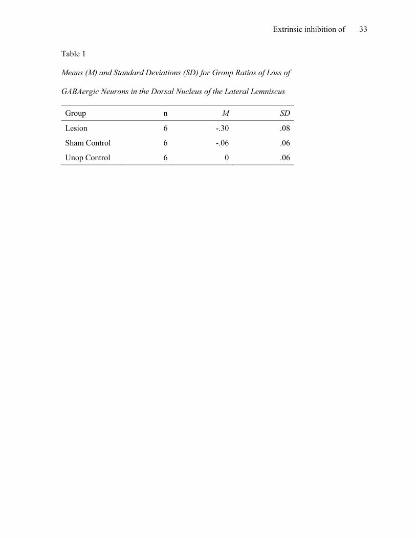
group exhibited substantial difference in cell density of DNLL within subjects (see Table 1).
Discussion
The purpose of this study was to investigate the importance of the role of extrinsic
inhibition during sound-induced seizures, specifically the role of the DNLL in the neural
network that subserves audiogenic responses. While the IC is a key processing, integration,
and relay center of all auditory input (Coleman & Clerici, 1987; Faingold, 2002; Faingold &
Anderson, 1991; Garcia-Cairasco, 2001; Garcia-Cairasco et al 1993; Pierson & Swann, 1991;

Extrinsic inhibition of 20
Pollack et al., 2003), the main source of extrinsic inhibition to this structure is supplied by
the DNLL (Bajo et al., 1993; Kelly et al 1996; Merchán et al., 1994; Schneiderman et al.,
1993; Zhang et al., 1998). There is ample evidence to implicate the IC as the initiation site of
the abnormal neural excitation implicated in AGS activity (i.e., Faingold; Faingold &
Anderson; Garcia-Cairasco; Garcia-Cairasco et al.; Raisinghani & Faingold, 2002; Ross &
Coleman, 2000). While the lack of intrinsic inhibition of the IC, in conjunction with
abnormal excitation, may play a role in the propagation of AGS (Faingold; Faingold &
Anderson; Garcia-Cairasco; Ross & Coleman), the role of extrinsic inhibition of the IC,
specifically the role of the DNLL, during AGS had not been investigated prior to this study.
It was expected that a reduction of extrinsic inhibition of the IC via unilateral DNLL
lesions would negatively impact the severity of AGS seizures due to a reduction of the
inhibition available to control runaway excitation originating in the IC. This hypothesis was
only marginally supported by data for one of the three dependent measures, the duration of
sound-induced seizures. The other dependent measures of seizure activity, latency to enter a
wild running phase and latency to enter clonus, were not affected by a lack of extrinsic
inhibition.
The hypotheses that unilateral DNLL lesions would have an effect on both the latency
to enter a wild running phase and the latency to enter clonus measures were not supported by
the data of this study, suggesting that a lack of extrinsic inhibition of the IC does not play a
significant role in the initiation or propagation of audiogenic responses. The data implies that
extrinsic inhibition of the IC does not provide significant inhibition to aid in slowing or
abolishing the abnormal excitation initiated in the IC that leads to a clonic seizure (Garcia-
Cairasco, 2001; Garcia-Cairasco et al., 1993; Raisinghani & Faingold, 2002; Ribak et al.,

Extrinsic inhibition of 21
1994; Ribak, & Morin, 1995; Ross & Coleman, 2000). Thus, seizure-prone rats with or
without the normal inhibitory connections of the auditory midbrain were equally likely to
exhibit audiogenic responses (see Figure 2 and Figure 3).
In contrast, the converse of the third hypothesis of this study, that DNLL lesions
would negatively impact clonic duration, was marginally supported by the data of this study.
While the group by surgery interaction contrast for this measure accounted for a relatively
small proportion of variability in seizure duration (η² = .10), rats with DNLL lesions
exhibited shorter or similar duration clonic AGS than those in control groups (see Figure
4) suggesting that a lack of the integrity of one DNLL in the ascending auditory pathway
may prevent the gradual increase in duration often seen with repeated seizure activity (e.g.,
Ross & Coleman, 2000). Perhaps alterations in the circuitry of the auditory midbrain in the
AGS-prone rat may provide some control to the animal, shortening the duration of the
seizure (cf. Pierson & Swann, 1991; Ribak et al., 1994; Zrull et al., 1999).
The behavioral data from this experiment seem to suggest that lack of extrinsic
inhibition of the IC does not play as significant a role in the either the initiation or
propagation of audiogenic seizure activity as does the lack of intrinsic inhibition of the
structure (Faingold & Anderson, 1991). The histology data of this thesis provides further
support for this conclusion because the average loss of neurons in the lesion group was
dramatically higher than in either control group (see Table 1 and Figure 5), indicating that the
quisqualic acid injection did cause significant cell loss in the DNLL of lesioned animals.
However, results also yielded an approaching-significant effect of the group by surgery
interaction contrast for the third dependent measure, duration of clonus. These results
indicate that, while reduced extrinsic inhibition of the IC is not a key factor in seizure
Extrinsic inhibition of 22
initiation and propagation, it does decrease the duration of clonus. This suggests that this
inhibition can aid the animal in regaining control after an AGS has begun, a proposal in
contrast to that of previous AGS studies where a temporary pause between wild runs is
presumed to be a demonstration of control provided by inhibition within the brain (Jobe et al.,
1973; Ross & Coleman, 1999).
Evidence from previous AGS research implicates the external cortex of the IC
(ECIC), not the central nucleus of the IC (CNIC), as the area responsible for the sensorimotor
aspect of AGS activity, including wild running (i.e., Garcia-Cairasco et al., 1993; Ribak &
Morin, 1995; Zrull et al., 1999). Furthermore, bilateral lesions of the IC that damage the
ECIC have effectively eliminated sensorimotor activity exhibited during AGS (Ribak et al.,
1994; Ross & Coleman, 2000). Therefore, it is possible that lesions of the DNLL, a structure
which projects to the CNIC, do not actually eliminate any extrinsic inhibition to the area
critical for sensorimotor activity (Garcia-Cairasco et al.; Zrull et al.). Another important
aspect to consider is the binaural nature of the auditory pathway. Only bilateral lesions of the
IC have effectively abolished AGS activity (Faingold, 2002; Ribak et al; Ross & Coleman).
It is possible that reducing projection from one DNLL did not effectively reduce available
extrinsic inhibition during AGS. In this study, unilateral DNLL lesions did not abolish AGS
activity; subjects in the lesion group did not exhibit shorter latencies to enter a wild running
phase or shorter latencies to enter clonus, as was expected from previous studies (e.g.
Faingold, 2002; Ribak et al; Ross & Coleman). Similarly, lesions did not increase seizure
duration, suggesting that disruption of midbrain circuitry or reduction of extrinsic inhibition
may aid in controlling a seizure once it has begun.
Extrinsic inhibition of 23
Although interesting conclusions can be drawn from the results of this study, further
investigation is necessary to make proper implications about the importance of the
relationship between the role of the DNLL and the IC during sound-induced seizures. Only
bilateral lesions of the IC can effectively eliminate AGS activity (Faingold, 2002; Ribak et al.,
1995; Ribak & Morin, 1994; Ross & Coleman, 2000), suggesting that due to the binaural
nature of the auditory pathway, damage to one structure could have little impact if the
contralateral structure remains intact. Subjects with bilateral DNLL lesions should be
examined to evaluate the significance of projection from both DNLL. Furthermore, all three
groups in this experiment were primed and tested for AGS susceptibility, and all followed an
identical pre- and post-surgery induction schedule. An unoperated control group (one that is
not exposed to any aspect of the AGS process) should be included in further studies to
understand the relationship between the DNLL and the IC in normal, properly functioning
brains. It is also possible that younger rats have the ability to cope with detrimental cells loss
within this structure better than adult rats, thus lesions of the DNLL may have less of an
impact on the age group of animals used for this study (i.e., Adams et al., 2008; Minton &
Zrull, 2010). Further investigation across various age groups is necessary to determine if cell
loss within this structure has more of an impact at particular stages in life.
Additionally, although quisqualic acid was an adequate choice to create lesions in the
DNLL, creating a larger lesion of the structure in further studies may provide more insight
into the importance of extrinsic inhibition in AGS. Combining quisqualic acid and AMPA
would enhance binding affinity at the receptors, creating more cell death (Johansen et al.,
2003; Parks, 2000). While a 30% neuron loss in this structure in the lesion group subjects is

Extrinsic inhibition of 24
substantial, it is possible that more cell death within the structure, and therefore even less
projection to the IC, could produce different results than those of this study.
One final limitation of this study could be the number of subjects used. Due to
unavoidable circumstances such as injuries sustained during seizure inductions, loss of
sections during tissue processing, and inability to assess tissue due to staining anomalies, the
sample size for this study was ultimately relatively small. Just as there is variation in
audiogenic responses across strains (Ross & Coleman, 1999), there are also individual
differences within strains. Thus, there was an extensive amount of variability within groups
for all three dependent measures, which could have significantly impacted the results.
Individual differences across subjects within groups used in this study may also explain why
the severity of unoperated control subjects increased so dramatically in post-surgery
inductions. However, it is possible that a larger sample size would simply allow room for
more individual differences within groups.
Summary and Conclusion
The results of this thesis provided interesting information about the importance, or
perhaps lack thereof, of the role of the DNLL during AGS. The data suggest that significant
cell loss within one DNLL does not impact the latency to enter a wild running phase or the
latency to enter clonus components of AGS activity; therefore, lack of extrinsic inhibition of
the IC, the initiation site of AGS (Faingold, 2002; Faingold & Anderson, 1991; Garcia-
Cairasco, 2001; Raisinghani & Faingold, 2002; Ross & Coleman, 2000), as provided by the
DNLL (Bajo et al., 2003; Coleman & Clerici, 1987; Merchán et al., 1994; Schneiderman et
al., 1993; Zhang et al., 1998), may not be a key component in the initiation and propagation
of sound-induced seizures. However, the data also suggest that a lack of extrinsic inhibition

Extrinsic inhibition of 25
to the IC may play a somewhat important role in AGS duration, by aiding in decreasing
duration of clonic seizures. It is possible that creating bilateral lesions of this structure,
therefore reducing extrinsic inhibition from both structures could have a significant effect on
AGS activity.

Extrinsic inhibition of 26
References
Adams, M. R., Glenn, E. A., Richardson, D. A., & Zrull, M. C. (2008, November). Brief
episodes of seizure inducing noise exposure alter neuron densities in the dorsal
nucleus of the lateral lemniscus. Poster presented at annual meeting for Society for
Neuroscience, Washington, D.C. Abstract available online at:
http://www.abstractsonline.com/plan/ViewAbstract
Bajo, V. M., Merchán, M. A., Lopez, D. E., & Rouiller, E. M. (1993). Neuronal morphology
and efferent projections of the dorsal nucleus of the lateral lemniscus in the rat. The
Journal of Comparative Neurology, 334, 241-262.
Coleman, J. R., & Clerici, W. J. (1987). Sources of projections to subdivisions of the inferior
colliculus in the rat. Journal of Comparative Neurology, 262, 215-226.
Epilepsy Foundation of America. (2010). [Online]. General information. Retrieved April 10,
2010, from the Epilepsy Foundation of America website:
http://www.epilepsyfoundation.org/about/statistics.cfm
Faingold, C. L. (2002). Role of GABA abnormalities in the inferior colliculus
pathophysiology-audiogenic seizures. Hearing Research, 168, 223-237.
Faingold, C. L., & Anderson, C. A. (1991). Loss of intensity-induced inhibition in inferior
colliculus neurons leads to audiogenic seizure susceptibility in behaving genetically
epilepsy-prone rats. Experimental Neurology, 113, 354-363.
Faye-Lund, H., & Osen, K. K., (1985). Anatomy of the inferior colliculus in rat. Anatomy
and Embryology, 171, 1-20.

Extrinsic inhibition of 27
Freeman, J. M., Vining, E. P., & Pillas, D. J. (2002). Seizures and epilepsy in childhood: A
guide. Maryland: Johns Hopkins University Press.
Garcia-Cairasco, N. (2001). A critical review on the participation of inferior colliculus on
acoustic-motor and acoustic-limbic networks involved in the expression of acute and
kindled audiogenic seizures. Hearing Research, 168, 208-222.
Garcia-Cairasco, N., Terra, V. C., & Doretto, M.C. (1993). Midbrain substrates of audiogenic
seizures in rats. Behavioral Brain Research, 58, 57-67.
Hermit, M. B., Greenwood, J. R., & Brauner-Osborne, H. (2004). Mutation-induced
quisqualic acid and ibotenic acid affinity at the metabotropic glutamate receptor
subtype 4. Ligand selectivity results from a synergy of several amino acid residues.
Journal of Biological Chemistry, 279, 34811-34817.
Hirsch, E., Danober, L., Simler, S., Pereira de Vasconcelos, A., Maton, B., Nehlig, A., et al.
(1997). The amygdala is critical for seizure propagation from brainstem to forebrain.
Neuroscience, 77, 975-984.
Hodgin, E. L. (2006). The effects of frequent generalized audiogenic seizures on some
aspects of memory and exploratory behavior. Unpublished M.A. thesis, Appalachian
State University, Boone, N.C.
Jobe, P. C., Picchioni, A. L., & Chin, L. (1973). Role of brain norepinephrine in audiogenic
seizure in the rat. The Journal of Pharmacology and Experimental Therapeutics, 184,
1-10.
Johansen, T. N., Greenwood, J. R., Frydenvang, K., Madsen, U., & Krogsgaard-Larsen, P.
(2003). Stereostructure–Activity studies on agonists at the AMPA and kainate
subtypes of ionotropic glutamate receptors. Chirality, 15, 167–179.

Extrinsic inhibition of 28
Kirk, R. E. (1982). Experimental design: Procedures for the behavioral sciences. Pacific
Grove, California: Brooks-Cole.
Kelly, J. B., Li, L., & van Adel, B. (1996). Sound localization after kainic acid lesions of the
dorsal nucleus of the lateral lemniscus in the albino rat. Behavioral Neuroscience, 110,
1445-1455.
Merchán, M. A., Saldaña, E., & Plaza, I. (1994). Dorsal nucleus of the lateral lemniscus in
the rat: Concentric organization and tonotopic projection to the inferior colliculus.
The Journal of Comparative Neurology, 342, 259-278.
Minton, B. R., & Zrull, M. C. (2010, November). Age dependent neuron loss in dorsal
nucleus of the lateral lemniscus after bouts of sound induced seizures. Poster to be
presented at annual meeting for Society for Neuroscience, San Diego, CA. Abstract
available online at: http://www.abstractsonline.com/plan/ViewAbstract
Parks, T. N. (2000). The AMPA receptors of auditory neurons. Hearing Research, 147, 77-
91.
Pierson, M. G., & Swann, J. (1991). Ontogenetic features of audiogenic seizure susceptibility
induced in immature rats by noise. Epilepsia, 32, 1-9.
Pollack, G. D., Burger, R. M., & Klug A. (2003). Dissecting the circuitry of the auditory
system. Trends in Neuroscience, 26, 33-39.
Raisinghani, M., & Faingold, C. L. (2002). Identification of the requisite brain sites in the
neuronal network subserving generalized clonic audiogenic seizures. Brain Research,
967, 113-122.

Extrinsic inhibition of 29
Ribak, C. E., Khurana, V., & Lien, N. T. (1994). The effect of midbrain collicular knife cuts
on audiogenic seizure activity in the genetically epilepsy-prone rat. Journal of Brain
Research, 35, 303-311.
Ribak, C. E., & Morin, C. L. (1995). The role of the inferior colliculus in a genetic model of
audiogenic seizures. Anatomy & Embryology, 191, 279-295.
Ross, K. C., & Coleman, J. R. (1999). Audiogenic seizures in the developmentally primed
Long-Evans rat. Developmental Psychobiology, 34, 303-313.
Ross, K. C., & Coleman, J. R. (2000). Developmental and genetic audiogenic seizure models:
Behavioral and biological substrates. Neuroscience and Biobehavioral Reviews, 24,
639-653.
Schneiderman, A., Chase, M. B., Rockwood, J. M, Benson, C. G., & Potashner, S. J. (1993).
Evidence of GABAergic projection from the dorsal nucleus of the lateral lemniscus to
the inferior colliculus. Journal of Neurochemistry, 60, 72-82.
Wu, S. W., & Kelly, J. B. (1996). In vitro brain slice studies of the rat’s dorsal nucleus of the
lateral lemniscus. III. Synaptic pharmacology. Journal of Neurophysiology, 75, 1271-
1282.
Zhang, D. X., Li, L., Kelly, J. B., & Wu, S. H. (1998). GABAergic projections from the
lateral lemniscus to the inferior colliculus of the rat. Hearing Research, 117, 1-12.
Zrull, M. C., & Coleman, J. R. (1991). Structural features of neurons in whole grafts of the
rat inferior colliculus, Hearing Research, 55, 117-132.
Zrull, M. C., Mengle, K. E., Hall, M. M., Hairston, W. D., & Sedivec, M. J. (1999). The
external cortex of the inferior colliculus in primed, audiogenic seizure-prone rats.

Extrinsic inhibition of 30
Society for Neuroscience Abstracts, 25, 1113. Poster presented at the 29th Annual
Meeting of the Society for Neuroscience.

Extrinsic inhibition of 31
Appendix

Extrinsic inhibition of 32
Author Note
First and foremost I would like to thank my thesis chair, Dr. Mark Zrull, whose extraordinary
guidance, patience, and advice made this thesis possible. I would also like to thank the
undergraduate research assistants who dedicated a great deal of their time to assist with this
project, particularly Benjamin Minton, Chase Francis, Joshua Smith, Alexandra Fuller,
Alexandria Squires, Jamie Milton, and Zachary Riemenschneider. Additional thanks are
warranted to my committee members, Dr. James Denniston and Dr. Kurt Michael for their
advice and assistance with this project.
Finally, I wish to dedicate this thesis to my parents, Robert and Sharon Morgan, for their love
and support throughout my graduate experience.
Extrinsic inhibition of 33
Table 1
Means (M) and Standard Deviations (SD) for Group Ratios of Loss of GABAergic Neurons in the Dorsal Nucleus of the Lateral Lemniscus Group n M SD
Lesion 6 -.30 .08
Sham Control 6 -.06 .06
Unop Control 6 0 .06

Extrinsic inhibition of 34
Figure Captions
Figure 1. The neural network for audiogenic seizures in rat. This figure shows the
propagation of the abnormal excitation, which culminates in a generalized seizure, as well as
the projection from the dorsal nucleus of the lateral lemniscus (DNLL) to the inferior
colliculus (IC; adapted from Garcia-Cairasco, 2002).
Figure 2. The group by surgery interaction effect on latency to enter wild running measure is
shown. The graph shows the mean latency to wild running with standard deviations from the
last four pre- and post-surgery inductions for the lesion (n = 6), sham (n = 6), and unoperated
control (n = 6) groups.
Figure 3. The group by surgery interaction effect on latency to enter clonus measure is
shown. The graph shows the mean latency to clonus with standard deviations from the last
four pre- and post-surgery inductions for the lesion (n = 6), sham (n = 6), and unoperated
control (n = 6) groups.
Figure 4. The group by surgery interaction effect on latency to enter wild running measure is
shown. The graph shows the mean duration of clonus with standard deviations from the last
four pre- and post-surgery inductions for the lesion (n = 6), sham (n = 6), and unoperated
control (n = 6) groups.
Figure 5. Example photomicrographs of GABAergic neurons in the dorsal nucleus of the
lateral lemniscus (DNLL) containing GAD67. Images (800 x 600 pixel) were made using a
Pixelink camera (1.3 MB) attached to a Nikon Eclipse light microscope (Plan 10 infinity
objective). The images show far more neurons in the DNLL of control rats (A and B) than
with neurotoxin injections. A. Section 22 from the brain of unoperated control Rat 0920. B.
Extrinsic inhibition of 35
Section 17 from the brain of sham operated control Rat 0909. C. Section 14 from the brain of
Rat 0914 with a quisqualic acid lesion of the DNLL.
Extrinsic inhibition of 36
Figure 1.

Extrinsic inhibition of 37
Figure 2.

Extrinsic inhibition of 38
Figure 3.

Extrinsic inhibition of 39
Figure 4.

Extrinsic inhibition of 40
Figure 5.

Extrinsic inhibition of 41
Vita
Amy Marie Morgan was born in Ruston, Louisiana on February 12, 1986. She attended
elementary school in that city, and graduated from Choudrant High School in May 2004. In
the fall of 2004 she entered Louisiana Tech University, where she majored in psychology. In
May 2008, she was awarded the Bachelor of Arts degree. In August 2008, she accepted a
research assistantship in experimental psychology at Appalachian State University and began
study toward a Master of Arts degree, which was awarded in August 2010. Ms. Morgan is a
member of Phi Gamma Mu, the Society for Neuroscience, and was the president of the
Psychology Graduate Student Organization of Appalachian State University from 2009-2010.
Her home address is 1947 Florida St, Arcadia, LA, 71001. Her parents are Robert and Sharon
Morgan of Arcadia and Choudrant Louisiana.