extracorporeal membrane oxygenation (ecmo) · extracorporeal membrane oxygenation (ecmo) is a form...
TRANSCRIPT

Extracorporeal Membrane Oxygenation (ECMO)
Final Evidence Report
February 12, 2016
20, 2012
Health Technology Assessment Program (HTA) Washington State Health Care Authority
PO Box 42712 Olympia, WA 98504-2712
(360) 725-5126 www.hca.wa.gov/hta
Health Technology Assessment

FINAL APPRAISAL DOCUMENT
Extracorporeal Membrane Oxygenation (ECMO)
February 12, 2016
Karin U. Travers, DSc Research Director
Elizabeth Russo, MD Research Scientist Patricia Synnott, MALD, MS Research Associate Rick Chapman, PhD, MS Director of Health Economics Steven D. Pearson, MD, MSc President
Daniel A. Ollendorf, PhD Chief Scientific Officer

WA – Health Technology Assessment February 12, 2016
Extracorporeal Membrane Oxygenation: Final Evidence Report Page i
Contents List of Acronyms........................................................................................................................................ ii
About ICER ............................................................................................................................................... iii
Acknowledgements ................................................................................................................................. iv
Executive Summary ............................................................................................................................. ES-1
ICER Integrated Evidence Ratings ...................................................................................................... ES-38
1. Background ........................................................................................................................................... 1
2. Washington State Agency Utilization Data ........................................................................................... 3
3. Treatment Strategies: Interventional and Conventional ...................................................................... 4
4. Clinical Guidelines and Training Standards ........................................................................................... 8
5. Medicare and Representative Private Insurer Coverage Policies ....................................................... 12
6. Previous Health Technology Assessments and Systematic Reviews .................................................. 14
7. Ongoing Comparative Studies ............................................................................................................ 17
8. Methods .............................................................................................................................................. 20
9. Results ................................................................................................................................................. 25
ICER Integrated Evidence Ratings ........................................................................................................... 52
Appendix A: Milestones in the Development of ECMO .......................................................................... 63
Appendix B: Literature Search Strategy .................................................................................................. 64
Appendix C: Summary Evidence Tables .................................................................................................. 66
Appendix D: ICER Integrated Evidence Ratings ...................................................................................... 91

WA – Health Technology Assessment February 12, 2016
Extracorporeal Membrane Oxygenation: Final Evidence Report Page ii
List of Acronyms AED Automated external defibrillators ARDS Acute respiratory distress syndrome AV Arteriovenous avECCO2-R Arteriovenous extracorporeal carbon dioxide removal CADTH Canadian Agency for Drugs and Technology in Health CI Confidence interval CMS Centers for Medicare and Medicaid COPD Chronic obstructive pulmonary disease CPB Cardiopulmonary bypass CPR Cardiopulmonary resuscitation ECCO2-R Extracorporeal carbon dioxide removal ECMO Extracorporeal membrane oxygenation ECLS Extracorporeal life support ED Emergency department ELSO Extracorporeal Life Support Organization FiO2 Fraction of inspired oxygen HR Hazard ratio ICU Intensive care unit iLA Interventional lung assist IQR Interquartile range LOS Length of stay LV Left ventricle NICE National Institute for Health and Care Excellence NIV Noninvasive ventilation NS Not significant OR Odds ratio PaC O2 Partial pressure of arterial CO2 PaO2 Arterial oxygen pressure pECLA Pumpless extracorporeal lung assist PEEP Positive end-expiratory pressure PICO Population, Intervention, Comparators, Outcomes PRISMA Preferred Reporting Items for Systematic Reviews and Meta-Analyses RCT Randomized controlled trial RR Relative risk SD Standard deviation SLR Systematic literature review SPO2 Oxygen saturation USPSTF united States Preventive Services Task Force VA Veno-arterial VAD Ventricular assist device VA-ECMO Veno-arterial ECMO VV Veno-venous VV-ECMO Veno-venous ECMO
WA – Health Technology Assessment February 12, 2016
Extracorporeal Membrane Oxygenation: Final Evidence Report Page iii
About ICER The Institute for Clinical and Economic Review (ICER) is an independent non-profit health care research organization dedicated to improving the interpretation and application of evidence in the health care system. ICER directs three core programs: the California Technology Assessment Forum (CTAF), the Midwest Comparative Effectiveness Public Advisory Council (Midwest CEPAC) and the New England Comparative Effectiveness Public Advisory Council (New England CEPAC). For more information about ICER, please visit ICER’s website, www.icer-review.org.

WA – Health Technology Assessment February 12, 2016
Extracorporeal Membrane Oxygenation: Final Evidence Report Page iv
Acknowledgements ICER would like to thank the following individuals for their expert opinions as well as peer review of draft documents:
Eileen Bulger, MD FACS Professor of Surgery Chief of Trauma University of Washington Harborview Medical Center Michael Mulligan, MD Professor of Cardiothoracic Surgery Director of Lung Transplant Program Director of Advanced Lung Disease Surgery Program University of Washington Medical Center
We would also like to thank Shanshan Liu of ICER for her contributions to this report.

WA – Health Technology Assessment February 12, 2016
Extracorporeal Membrane Oxygenation: Final Evidence Report Page ES-1
Executive Summary Background
Extracorporeal membrane oxygenation (ECMO) is a form of life support that provides cardiopulmonary assistance outside the body. ECMO may be used to support lung function for severe respiratory failure or heart function for severe cardiac failure. An ECMO circuit can be set up as veno-venous (VV) or veno-arterial (VA). VV-ECMO provides external gas exchange, bypassing the lungs and protecting them from high tidal volumes of ventilation that would otherwise be needed to oxygenate and ventilate the patient. VV-ECMO is indicated for patients with potentially reversible respiratory failure, including those with severe acute respiratory distress syndrome (ARDS), primary graft dysfunction following lung transplant, and trauma to the lungs. VA-ECMO provides the same external gas exchange as VV-ECMO, but also augments blood flow in settings of severe cardiac injury. VA-ECMO is indicated for patients with cardiac failure, including cardiogenic shock unresponsive to typical intensive care medicines and cardiac arrest that does not respond to cardiopulmonary resuscitation (CPR). VA-ECMO may also be used for patients following heart surgery or as a bridge to heart transplantation. Both VA- and VV-ECMO may be used intraoperatively as a planned alternative to traditional cardiopulmonary bypass in selected patient populations (e.g., lung or heart transplantation). Other external gas exchange systems provide similar functions without the pump component of VV- or VA-ECMO. These arteriovenous extracorporeal lung assist devices bypass the lungs, but not the heart, and use the patient’s blood pressure in order to sustain circulation of externally oxygenated blood.1-3 Because of the requirement for adequate cardiac function in candidate patients, these systems have more limited application. These devices are known by a variety of names, including pumpless extracorporeal lung assist (pECLA), arteriovenous extracorporeal membrane carbon dioxide removal (avECCO2-R), or interventional lung assist (iLA). In this report, we refer to these devices by the name used by their clinical investigators, although these devices are functionally equivalent. Over the past 30 years, ECMO has become a well-established treatment for infants with lung and heart failure and has become a standard of care in many pediatric care centers.4 A large multicenter randomized controlled trial published in 1996 demonstrated a clear survival benefit with ECMO as well as a reduction in risk of severe disability in neonatal patients with severe respiratory failure.5 In contrast, early studies of ECMO in adults showed poor survival rates, and its use was limited for many years to pediatric populations with life-limiting illness.6,7 The lack of demonstrated benefit from these studies, published in 1979 and 1994, halted enthusiasm for widespread ECMO use. However, several developments have prompted renewed interest and wider utilization of ECMO in recent years.8 First, technological advancements have improved the safety of the technique and broadened the application.9 These improvements include heparin-coated cannulae, new oxygenators, and more efficient pump technology.10 Second, more recent clinical trials have shown improved survival without severe disability with ECMO compared to conventional ventilator support.2,11 Finally, the 2009 H1N1 pandemic spurred increased demand for ECMO at rates higher than previously seen, resulting in additional evidence of a survival benefit.12,13 Appendix A depicts major advancements in the development and implementation of ECMO over time.

WA – Health Technology Assessment February 12, 2016
Extracorporeal Membrane Oxygenation: Final Evidence Report Page ES-2
In select cases, the use of ECMO in adults can clearly result in patients’ neurologically intact survival; however, the question remains as to whether this benefit is consistently observed in comparison to conventional care in the variety of settings in which it is used. Appropriate patient selection has been identified as key to such evaluation,14,15 and strategies at various stages of development have been proposed to do just that.16 Currently, these strategies are not incorporated into comparative evaluations of ECMO, as there exists no validated prognostic approach for identifying appropriate patients at ECMO initiation. Such entry criteria for ECMO have been described as a “moving target.”15 Our review therefore focuses on the current use of ECMO, differentiated by indication. In this way, we will be addressing the question of what patient populations, as defined by indication, might be best served by ECMO treatment. Still at issue will be more careful delineation of those patient populations in which ECMO remains an exercise in futility, or a “bridge to nowhere.”17
Policy Context
Due to the expense and intensity of critical care, guidelines regarding implementation of life-sustaining and life-saving technologies warrant careful attention. Although consensus around indications for ECMO is still developing, the use of ECMO has grown in recent years and continues to rise.18 Because the availability of ECMO is limited and requires specialized medical care, which diverts resources from other recipients, liberalizing its use in the intensive care or operating room settings has important policy implications and warrants consideration of the benefit-harm tradeoffs in each patient population of interest.19 The Washington State Health Care Authority has commissioned ICER to conduct a systematic review of the published literature on the use of extracorporeal membrane oxygenation in 1) critically ill adult patients with severe respiratory or cardiac failure, and 2) adult patients who receive ECMO as a planned intra-operative procedure. Evidence will be culled from randomized controlled trials (RCTs), systematic reviews, and high-quality observational studies. Specific details on the proposed scope (Population, Intervention, Comparators, and Outcomes [PICO]) are detailed in the following sections. Treatment Strategies: Interventional and Conventional
Interventional Treatments
Extracorporeal Membrane Oxygenation (ECMO) Extracorporeal membrane oxygenation (ECMO) is a temporary mechanical support system used to aid heart and lung function in patients with severe respiratory or cardiac failure.20 There are two types of ECMO: veno-arterial ECMO (VA-ECMO), which is connected to both a vein and an artery, and veno-venous ECMO (VV-ECMO), which is connected to one or more veins. These systems are illustrated further in Figure ES-1 on the following page. Being placed on ECMO requires surgical cannulation. The patient is sedated and given pain medication and an anti-coagulant to minimize blood clotting. A surgeon, assisted by an operating room team, inserts the ECMO catheters into either an artery or vein.21 With most approaches to ECMO for respiratory failure, a catheter is placed in a central vein, usually near the heart. A mechanical pump draws blood from the vein into the circuit, where the blood passes along a membrane (referred to as an "oxygenator" or "gas exchanger"), providing an interface between the blood and freshly delivered oxygen. The blood may be warmed or cooled as needed and is returned either to a central vein (VV-ECMO) or to an artery (VA-ECMO). VV-ECMO provides respiratory support alone, while VA-ECMO

WA – Health Technology Assessment February 12, 2016
Extracorporeal Membrane Oxygenation: Final Evidence Report Page ES-3
provides both respiratory and hemodynamic support.22 Usually a patient on ECMO is also on a mechanical ventilator at low settings, which assists in lung recovery.21
While on ECMO, the patient is monitored by specially trained nurses and respiratory therapists, as well as a surgical team. Supplemental nutrition is provided either intravenously or through a nasogastric tube. Certain medications may be given including heparin to prevent blood clots, antibiotics to prevent infections, sedatives to minimize movement and improve sleep, diuretics to help the kidney process fluids, electrolytes to maintain the proper balance of salts and sugars, and blood products to replace blood loss.21
Discontinuing ECMO requires decannulation. Multiple tests are usually done prior to the discontinuation to confirm that the heart and lungs are sufficiently recovered. Once the ECMO cannulae are removed, the vessels need to be repaired, which can be done at the bedside or in the operating room. The surgeon uses small stitches to suture closed the blood vessels. After discontinuation, patients may still require mechanical ventilation.21
Complications from ECMO include surgical and organ bleeding, renal and multi-organ failure, and central nervous system problems. Blood clots in the ECMO circuitry and mechanical problems may also cause complications. Because mortality rates increase with longer periods of ECMO duration, prompt weaning is recommended and should begin as soon as cardiorespiratory function can be maintained independently. The need for extended ECMO support may indicate irreversible cardiorespiratory dysfunction and poor prognosis. Patients who cannot be weaned off ECMO should undergo careful evaluation to justify continued support.20 Figure ES-1. Diagrammatic representation of peripheral veno-venous (VV-ECMO) and peripheral veno-arterial (VA-ECMO) extracorporeal membrane oxygenation.23
Pumpless Extracorporeal Lung Assist (pECLA, iLA, avECCO2-R) pECLA, also referred to as interventional lung assist (iLA) or arteriovenous extracorporeal carbon dioxide removal (avECCO2-R), is distinct from ECMO in that it requires normal left ventricular cardiac function to drive the blood across the extracorporeal membrane where carbon dioxide is removed. It is a pumpless arterio-venous shunt (femoral artery and vein) which eliminates carbon dioxide and slightly increases arterial oxygenation to normalize respiratory acidosis.24

WA – Health Technology Assessment February 12, 2016
Extracorporeal Membrane Oxygenation: Final Evidence Report Page ES-4
Conventional Treatments
Cardiopulmonary Bypass (CPB) Traditional CPB is a form of extracorporeal circulation in which the patient's blood is circulated, oxygenated, and ventilated without the heart and lungs using a bypass machine while surgeons operate on a non-beating heart devoid of blood. The bypass machine has pumps, tubing, artificial organs, and monitoring systems. Modern bypass machines also have continuous vascular pressure monitoring; blood gas, hemoglobin, and electrolyte monitoring; air detection systems; and blood filters. Unlike with ECMO, CPB circuits include a large reservoir for keeping blood outside the body. This non-endothelial surface triggers an intense inflammatory response which consumes blood products – platelets, coagulation factors – and contributes to challenges to postoperative recovery.25 Ventricular Assist Devices (VADs) Ventricular assist devices are a type of mechanical circulatory support used for managing cardiogenic shock, acute decompensated heart failure, or cardiopulmonary arrest. The inflow for the axial flow pump (e.g., Impella microaxial flow device) is placed retrograde across the aortic valve into the left ventricle. A high-speed pump draws blood out of the left ventricle and ejects it into the ascending aorta. These pumps can be placed surgically or percutaneously via the femoral artery. A left atrial to aorta assist device (e.g., TandemHeart) is placed in the left atrium by transseptal puncture and iliofemoral artery. In patients with very poor left ventricle (LV) function but adequate right ventricle (RV) function, blood is pumped from the left atrium to the ileofemoral system using a centrifugal pump that contains a spinning impeller.26 These devices provide circulatory support, but do not oxygenate the blood. The primary advantage of VA-ECMO over VAD devices is that it is easier to implant and can be used in a more diverse set of cardiopulmonary pathologies.27 Cardiopulmonary Resuscitation (CPR) High quality cardiopulmonary resuscitation (CPR) and early defibrillation are the critical life-saving components of basic and advanced cardiac life support. High quality CPR is defined by deep (2 inches) and brisk chest compressions (100-120/min) on the center of the chest with minimal interruption (<10 seconds at intervals >2 min). Defibrillation itself should interrupt the chest compressions for no more than 3-5 seconds. Early defibrillation to minimize “downtimes” is associated with better survival. Defibrillation can be administered by non-medical rescuers using automated external defibrillators (AED), which detect shockable rhythms and voice commands. Biphasic defibrillators are used by trained medical providers. Adding ventilation (mouth-to-mouth, bag valve mask, or advanced airway) is of secondary importance in administering high quality CPR. Excessive ventilations should be avoided; each breath should be given over no more than one second and provide enough tidal volume to see the chest rise.28,29 Extracorporeal CPR may induce return of spontaneous circulation for patients with cardiogenic shock from acute myocardial infarction who otherwise may not respond to conventional CPR. Mechanical Ventilation Mechanical ventilation, or positive pressure ventilation, uses a ventilator to push air into the lungs through an endotracheal tube or tracheostomy tube. Noninvasive ventilation can be delivered through a face mask for some patients who retain control of their airway (intact gag reflex). For intubated patients, the machine pushes in a mixture of oxygen and other gasses until a signal causes the ventilator to stop and allows passive expiration. The ventilator can replace or support spontaneous breathing. The ventilator can be set to coincide with the patient’s own breath (triggered) or set to deliver a targeted flow rate or volume of air. The tidal volume is the amount of air delivered with each breath.

WA – Health Technology Assessment February 12, 2016
Extracorporeal Membrane Oxygenation: Final Evidence Report Page ES-5
Low tidal volume ventilation (≤6mL/kg/predicted body weight) is associated with better outcomes for patients with ARDS. The low tidal volume requires a higher respiratory rate (~35 breaths/min) in order to support adequate tissue oxygenation. Positive end-expiratory pressure (PEEP) is added to prevent end-expiratory alveolar collapse; this is set at 5 cmH2O for most patients and 20 cmH2O for ARDS patients. Peak flow rates are usually set at 60 L/min. The fraction of inspired oxygen (FiO2) is the percent of oxygen mixed into the inspired gas. The lowest fraction necessary to sustain oxygenation should be used to prevent oxygen toxicity. FiO2 is titrated to maintain arterial oxygen pressure (PaO2) greater than 60 mmHg and oxygenation saturation (SpO2) above 90%. ARDS patients have PaO2 targets 55-80 mmHg and SpO2 targets of 88-95% to reduce plateau pressures and risk of lung injury.30 ECMO allows the lung to be ventilated at lower settings (while maintaining adequate oxygenation), which prevents barotrauma and allows the lungs to recover from their underlying insult. Key Questions The following key questions were felt to be of primary importance for this review: Key Question #1: What is the comparative clinical effectiveness of ECMO versus conventional treatment strategies in adults (age≥18 years)? Key Question #2: What are the rates of adverse events and other potential harms associated with ECMO compared to conventional treatment strategies? Key Question #3: What is the differential effectiveness and safety of ECMO according to sociodemographic factors (e.g., age, sex, race or ethnicity), severity of the condition for which ECMO is used (e.g., Murray score or Acute Physiology and Chronic Health Evaluation [APACHE] score), setting in which ECMO is implemented (e.g., specialized ECMO centers), time of ECMO initiation (early vs. late), and duration of time on ECMO? Key Question #4: What are the costs and potential cost-effectiveness of ECMO relative to conventional treatment strategies?

WA – Health Technology Assessment February 12, 2016
Extracorporeal Membrane Oxygenation: Final Evidence Report Page ES-6
Analytic Framework The analytic framework for this project is depicted below, including key comparators and outcomes of interest.
Figure ES-2. Analytical Framework: ECMO
Adults (age ≥ 18) with severe respiratory or cardiac failure
Extracorporeal membrane oxygenation (ECMO)
Survival Less disability Length of hospital stay Costs Adverse events
Conventional ventilator therapies
Adults (age ≥ 18) who require hemodynamic support during surgery
Cardiopulmonary bypass

WA – Health Technology Assessment February 12, 2016
Extracorporeal Membrane Oxygenation: Final Evidence Report Page ES-7
Results
Overall Evidence Quality
Our review identified only two RCTs, both of good quality. Among the 41 comparative cohort studies
dentified, only 16 were deemed to be of good quality. Eight comparative cohort studies were found to be of fair quality, as they included comparison groups with substantial variation in baseline demographic or clinical characteristics; attempts were made in the analysis of these studies to account for these differences, most often through the use of multivariate logistic regression or survival analysis. An additional 17 comparative cohort studies identified were of poor quality, based on a lack of presented information regarding baseline characteristics, or an analytic approach that did not appropriately account for substantial differences between groups. The dearth of RCTs of ECMO is perhaps unsurprising, as it is very difficult to implement a well-designed RCT in this area because of the ethical concerns and challenges to standardizing care across institutions for critically ill patients. In addition, conventional therapy itself is subject to change, so static comparisons between treatment arms become outdated relatively quickly.19 Most studies described as fair compared patient groups with disparate demographic or clinical characteristics. Those described as poor did not present enough information to make this determination or did not sufficiently attempt to control for confounding variables in some way. It is also challenging to pool information across comparative observational studies (cohort and case-control study designs) because these studies examined distinct patient populations with different disease entities and variable severities of illness. Another limitation of drawing conclusions across studies is that there is so much variability to the care given between treatment arms within studies and between treatment arms across studies. Standards of care, device technology, protocol development, clinical decision-making, and patient characteristics are variable within and across studies. For example, studies reported by both Peek et al. and Davies et al. centralized care of ECMO patients in a single medical center, whereas patients in the conventional/non-ECMO treatment groups remained in multiple outlying hospitals.11,12 There is no way to fully account for differences in patient care administered in one hospital versus handfuls of others. RCTs may overcome such a problem with techniques like cluster randomization; however, such a technique is not available for cohort studies. This and other variations precludes generalization of findings, and for this reason, we did not formally pool data to conduct quantitative synthesis.
A summary evidence table (Table ES-1) capturing the strength of evidence for each of the four key questions of interest can be found on the following page.

WA – Health Technology Assessment February 12, 2016
Extracorporeal Membrane Oxygenation: Final Evidence Report Page ES-8
Table ES-1: Summary evidence table for good quality studies of ECMO in comparison to alternative treatment strategies
Study Information Comparators Risk of Bias Consistency Directness Precision Strength of
Evidence
Direction of Effect of
ECMO Comments
Key Question #1: Effectiveness ECMO
Cardiac support N=79
RCT=0 Cohort studies=1
VAD Medium Consistency unknown (single study)
Direct Imprecise ++ Low
Comparable: No differences in in-hospital survival or successful bridging to active therapy
Single retrospective study
ECMO Pulmonary support
N=793 RCT=2
Cohort studies=6
Mechanical ventilation
Medium Inconsistent Direct Precise +++ Moderate
Comparable: No consistent differences in survival, length of stay, or disability
Variation in disease entities, disease severity, and in standards of care
ECMO Bridge to
transplant N=742 RCT=0
Cohort studies=3
Cardiopulmonary bypass
Medium Inconsistent Direct Imprecise ++ Low
Comparable: No survival benefit; shorter length of stay (1 study)
Two studies examined heart transplant; one studied heart-lung transplant
ECMO ECPR
N=1,543 RCT=0
Cohort studies=5
Conventional cardiopulmonary resuscitation
Medium Inconsistent Direct Imprecise ++ Low
Comparable: Short-term survival benefit is lost in longer-term. One study showed neurologic benefit
Only one study reported positive survival benefit in longer term.

WA – Health Technology Assessment February 12, 2016
Extracorporeal Membrane Oxygenation: Final Evidence Report Page ES-9
Study Information Comparators Risk of Bias Consistency Directness Precision Strength of
Evidence
Direction of Effect of
ECMO Comments
Key Question #2: Harms Bleeding Various Medium Consistent Direct Imprecise +++
Moderate 2.5-25% Heterogeneous
patient populations
Limb ischemia Various Medium Consistent Direct Precise ++++ High
2.5%-7.6% Heterogeneous patient populations
Cannulation site complications
Various Medium Consistent Direct Imprecise +++ Moderate
1-23.1% Heterogeneous patient populations
Key Question #3: Differential ECMO effects and risk factors Age Various Medium Inconsistent Direct Imprecise ++
Low Limited and conflicting evidence that older age predicts survival and positive neurologic outcomes
Gender Various Medium Inconsistent Direct Imprecise ++ Low
Limited evidence that male gender predicts survival
Dialysis Various Medium Inconsistent Direct Imprecise ++ Low
Limited evidence that dialysis is associated with ECMO survival
Associations only found in case series

WA – Health Technology Assessment February 12, 2016
Extracorporeal Membrane Oxygenation: Final Evidence Report Page ES-10
Study Information Comparators Risk of Bias Consistency Directness Precision Strength of
Evidence
Direction of Effect of
ECMO Comments
Key Question #4: Costs and Cost-Effectiveness ECMO for
pulmonary support Mechanical Ventilation
Medium Consistency unknown (one study)
Direct Imprecise ++ Low
Cost-effectiveness $7,000 - $35,000 per LY or QALY gained; incremental costs in US of $100,000 - $500,000
Two studies of cost-effectiveness in non-US settings

WA – Health Technology Assessment February 12, 2016
Extracorporeal Membrane Oxygenation: Final Evidence Report Page ES-11
Key Question #1: What is the comparative clinical effectiveness of ECMO versus conventional treatment strategies in adults (age≥18 years)?
Central to this question is whether ECMO preserves quantity and quality of life without ultimate futility. The evidence base for Key Question #1 can be categorized by the specific use of ECMO: Intensive Care Unit (ICU) cardiac support, ICU pulmonary support, surgical bridge to transplantation, or extracorporeal cardiopulmonary resuscitation (ECPR). ►ICU Cardiac Support This section summarizes the findings from the only good quality study to compare ECMO to a conventional alternative (miniaturized percutaneous VAD), in which no benefit from use of ECMO was found on in-hospital survival, successful weaning off mechanical support, or bridging to long-term support or transplant. Chamogeorgakis et al. conducted a retrospective chart review to compare outcomes associated with using a temporary miniaturized percutaneous ventricular assist device (mp-VAD) with ECMO in 79 patients with cardiogenic shock seen at a single academic medical center, the Cleveland Clinic.27 The patient population was mostly male adults who had had myocardial infarction documented during the same hospital admission. One patient crossed over to the ECMO group and was analyzed based on intention to treat. See Appendix C for more information about entry criteria and study design. As shown in Table ES-2 below, successful weaning off mechanical support, in-hospital survival, and successful bridging to long-term support or transplant did not differ between groups.
Table ES-2: Summary of evidence for ECMO used to provide cardiac support
Study (Setting and
Time) Population Intervention
Control (p values for comparison to
intervention group) Follow-up and Outcomes
Chamogeorgakis et al. 201327 (Cleveland, OH: single site; January 2006-September 2011)
Cardiogenic shock ECMO (n=61) Mean age: 58 years 72.2% male 77.8% postinfarction
mp-VAD (n=18) Mean age: 53 years (p=0.121) 80.3% male (p=0.519) 52.5% postinfarction (p=0.063)
Mean follow-up 14.3 months Successfully weaned: ECMO 33.3% mp-VAD 19.7% (p=0.336) In-hospital survival: ECMO 50.0% mp-VAD 49.2% (p>0.999) Bridge to long-term support or transplant: ECMO 27.8% mp-VAD 31.1% (p>0.999)
►ICU Pulmonary Support A larger body of good-quality evidence was found evaluating the use of ECMO for pulmonary support. Below we summarize findings from two randomized control trials and six observational studies that compared conventional mechanical ventilation with either pump-driven VV-ECMO/VA-ECMO or

WA – Health Technology Assessment February 12, 2016
Extracorporeal Membrane Oxygenation: Final Evidence Report Page ES-12
pumpless avECCO2-R. Similar to findings from other systematic reviews, we did not find consistent evidence for an in-hospital survival benefit from pECLA or ECMO for respiratory failure compared to conventional ventilator support.31 Some of the observational studies found an in-hospital survival benefit that was not detected in the RCTs. This suggests the potential for some selection bias playing a role, although one of the observational studies reporting ECMO survival benefit 32 utilized the same inclusion criteria as one of the RCTs 11. It’s also possible that publication bias plays a role in these inconsistent findings.
Resource use as measured by length of hospital and ICU stay appears to be comparable or more substantial for patients treated with pECLA or ECMO compared to conventional ventilation. Across studies, morbidity and disability was not consistently found to be better for patients treated with pECLA or ECMO compared to conventional ventilation. Quality of life and functional outcomes were only examined in a single RCT, and all of these measures were improved, but not statistically significantly so, in the ECMO treatment arm compared to conventional ventilation.11
Randomized Controlled Trials We identified two RCTs comparing extracorporeal lung assistance (pECLA and ECMO) with conventional ventilator management. Trial design and setting are described below; results are organized by type of outcome in the sections that follow. See Appendix C for more detail about entry criteria and study design. Bein et al. randomized 79 adult patients with established ARDS diagnoses into either a pumpless extracorporeal lung assist (avECCO2-R) treatment arm (n=40) or to a control arm with conventional ventilation maintaining low tidal volumes (n=39).2 Established ARDS was determined by monitoring patients initially screened into the study for a 24-hour stabilization period during which mechanical ventilation was maintained with high PEEP (≥12cmH2O), other supportive measures, and echocardiography. Both arms had similar mean age, BMI, and proportion of males, but more patents in the avECCO2-R group had secondary ARDS (22.5% vs. 5.1%, significance not reported). Patients were followed for 6 months. Both arms were treated with “best clinical evidence” recommendations with ventilation targets of maintaining PaO2 ≥60mmHg and arterial pH ≥7.2. Both groups experienced daily screening for spontaneous breathing trials and were extubated when no deterioration was detected over a one-hour period. No statistically-significant differences were observed for any outcome of interest, including mortality, organ failure, days without ventilation assistance, and length of stay in ICU or in the hospital overall.
WA – Health Technology Assessment February 12, 2016
Extracorporeal Membrane Oxygenation: Final Evidence Report Page ES-13
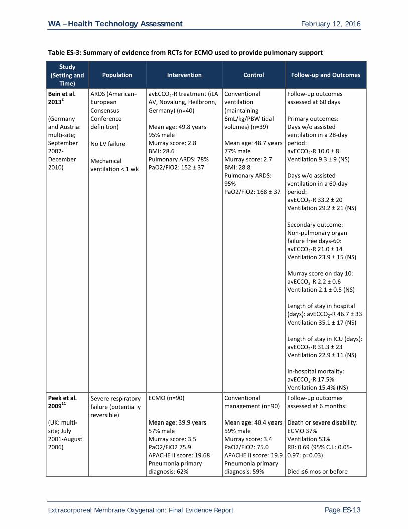
Table ES-3: Summary of evidence from RCTs for ECMO used to provide pulmonary support
Study (Setting and
Time) Population Intervention Control Follow-up and Outcomes
Bein et al. 20132 (Germany and Austria: multi-site; September 2007-December 2010)
ARDS (American-European Consensus Conference definition) No LV failure Mechanical ventilation < 1 wk
avECCO2-R treatment (iLA AV, Novalung, Heilbronn, Germany) (n=40) Mean age: 49.8 years 95% male Murray score: 2.8 BMI: 28.6 Pulmonary ARDS: 78% PaO2/FiO2: 152 ± 37
Conventional ventilation (maintaining 6mL/kg/PBW tidal volumes) (n=39) Mean age: 48.7 years 77% male Murray score: 2.7 BMI: 28.8 Pulmonary ARDS: 95% PaO2/FiO2: 168 ± 37
Follow-up outcomes assessed at 60 days Primary outcomes: Days w/o assisted ventilation in a 28-day period: avECCO2-R 10.0 ± 8 Ventilation 9.3 ± 9 (NS) Days w/o assisted ventilation in a 60-day period: avECCO2-R 33.2 ± 20 Ventilation 29.2 ± 21 (NS) Secondary outcome: Non-pulmonary organ failure free days-60: avECCO2-R 21.0 ± 14 Ventilation 23.9 ± 15 (NS) Murray score on day 10: avECCO2-R 2.2 ± 0.6 Ventilation 2.1 ± 0.5 (NS) Length of stay in hospital (days): avECCO2-R 46.7 ± 33 Ventilation 35.1 ± 17 (NS) Length of stay in ICU (days): avECCO2-R 31.3 ± 23 Ventilation 22.9 ± 11 (NS) In-hospital mortality: avECCO2-R 17.5% Ventilation 15.4% (NS)
Peek et al. 200911 (UK: multi-site; July 2001-August 2006)
Severe respiratory failure (potentially reversible)
ECMO (n=90) Mean age: 39.9 years 57% male Murray score: 3.5 PaO2/FiO2 75.9 APACHE II score: 19.68 Pneumonia primary diagnosis: 62%
Conventional management (n=90) Mean age: 40.4 years 59% male Murray score: 3.4 PaO2/FiO2: 75.0 APACHE II score: 19.9 Pneumonia primary diagnosis: 59%
Follow-up outcomes assessed at 6 months: Death or severe disability: ECMO 37% Ventilation 53% RR: 0.69 (95% C.I.: 0.05-0.97; p=0.03) Died ≤6 mos or before

WA – Health Technology Assessment February 12, 2016
Extracorporeal Membrane Oxygenation: Final Evidence Report Page ES-14
Study (Setting and
Time) Population Intervention Control Follow-up and Outcomes
discharge: ECMO 37% Ventilation 45% RR: 0.73 (95% CI: 0.52-1.03; p=0.07) Median days between randomization and death: ECMO 15 Ventilation 5 Median length of stay in hospital (days): ECMO 35.0 (IQR 15.6-74.) Ventilation 17.0 (IQR 4.8-45.3) Median length of stay in ICU (days): ECMO 24.0 IQR 13.0-40.5) Ventilation 13.0 (IQR 11.0-16.0) Overall health status (VAS; 0-100; higher score is better): ECMO 67.9 Ventilation 65.9 (NS)
NS=non-significant For the Conventional ventilation or ECMO for Severe Adult Respiratory failure (CESAR) trial, Peek et al. randomized 180 adults with severe but potentially reversible respiratory failure into two treatment arms: ECMO (n=90) and conventional management (n=90).11 Demographic characteristics and physiologic presentation were similar at baseline between the treatment and control groups (Table ES-3). Conventional management included low-volume low-pressure ventilation strategy, but there was no mandated management protocol. ECMO patients were transferred to one hospital where standard ARDS and institutional protocols were used to determine whether they still were candidates for VV-ECMO. Investigators used an intention to treat analysis, and 75% (n=68) of patients randomized to the treatment arm actually received ECMO support. An important caveat to interpreting results from the CESAR trial is that all of the ECMO patients, whether recipients of ECMO or not, were treated in a single referral center whereas the control patients received conventional management as determined by their diverse institutions. Six-month follow-ups were performed in the patients’ homes by researchers blinded to the treatment arm, and patients and their relatives were asked not to reveal their treatment to the researcher (including a neck scarf to hide cannulation status). ECMO was associated with a significantly lower rate of death or severe disability at 6 months (p=0.03); however, the 6-month disability status was unknown for several study participants, making interpretation of this composite

WA – Health Technology Assessment February 12, 2016
Extracorporeal Membrane Oxygenation: Final Evidence Report Page ES-15
outcome uncertain. There was a non-significant trend toward lower mortality at 6 months (p=0.07). Length of stay was also substantially longer in ECMO recipients, but no statistical significance testing was reported; the rate of severe disability at discharge was not reported. These studies are described in additional detail in Appendix C. Observational Studies There were six observational studies of good quality that addressed comparisons of interest. These included Del Sorbo 2015, a comparative cohort study of adults treated with noninvasive ventilation plus or minus extracorporeal CO2 removal;33 Kluge 2012, a matched case control study comparing patients treated with pECLA versus mechanical ventilation;34 Noah 2011, a matched case-control study of H1N1 adult patients treated with and without ECMO;32 Pham 2013, a propensity score matched analysis of H1N1 patients treated with and without ECMO;35 Tsai 2015, a case-control study of ARDS patients treated with and without ECMO.36 One retrospective cohort study by Guirand et al. addressed use of ECMO among adult trauma patients who had acute hypoxemic respiratory failure.37 The design of these studies is described below; results are organized by type of outcome in the sections that follow. Del Sorbo et al. sought to estimate the efficacy and safety of ECCO2-R in association with noninvasive ventilation to reduce the need for intubation in hypercapnic patients at risk of respiratory failure.33 They enrolled 25 adult patients (aged 18-90 years) who received ECCO2-R in addition to noninvasive ventilation for chronic obstructive pulmonary disease (COPD) exacerbations. Patients were removed from ECCO2-R when respiratory rate, pH, and partial pressure of arterial carbon dioxide (PaCO2) improved for at least 12 hours. A matched cohort of 21 patients who did not receive ECCO2-R was drawn from the same patient population; these populations did not differ by age or baseline illness severity. Kluge et al. compared the feasibility, effectiveness, and safety of pECLA with conventional mechanical ventilation in patients with acute hypercapnic respiratory failure unresponsive to noninvasive ventilation.34 The iLA pECLA device was used in 21 patients with respiratory acidosis (pH<7.35) and clinical signs of ventilator pump failure. Twenty-one matched controls were selected from a database of patients who had been admitted with acute hypercapnic respiratory failure and were intubated after failing noninvasive ventilation. Other than baseline PaCO2, these populations had no differences by reported demographic or physiologic baseline characteristics. The relative hypercapnia among the pECLA treatment group may suggest more advanced COPD despite the other matching variables reported. Noah et al. compared mortality for patients referred, accepted, and transferred to UK ECMO centers for H1N1-related ARDS with matched non-ECMO-referred patients drawn from a prospective cohort of patients with suspected or confirmed H1N1 requiring critical.32 At the point of referral to the ECMO centers, more of these patients were female (62.5%) than patient populations in other studies. The non-ECMO-referred patients were similar adult patients who were not referred, accepted, or transferred to one of the ECMO centers. As with the CESAR trial, there was no protocol for managing ventilation among the non-ECMO-referred patients. An additional limitation of this analysis is that some of the non-ECMO-referred patients may have seemed too sick for transfer. Of 80 patients transferred to referral ECMO centers, 69 (86.3%) received ECMO, but it is not clear how many of these were retained in the 75 patients included in the matched analysis. The investigators used several methods for matching patients in treatment groups. The GenMatch algorithm iteratively checks the balance and directs the search toward the best matches. Compared with propensity score matching, GenMatch matching reduces covariate imbalance and bias from confounding. Given the purported increase in

WA – Health Technology Assessment February 12, 2016
Extracorporeal Membrane Oxygenation: Final Evidence Report Page ES-16
rigor, GenMatched data are used for comparison in this assessment, none of which significantly differed at baseline. Pham et al. described role of ECMO on H1N1 patients with ARDS treated in French ICUs.35 They compared outcomes from 52 pairs of patients: those treated with ECMO in the first week of ARDS propensity-score matched with patients with severe H1N1-related ARDS not treated with ECMO. There were no demographic or physiologic differences between groups at baseline. There was minimal description of the treatment strategies used for the non-ECMO group. Tsai et al. compared the outcomes of 90 ARDS patients, half of whom did and half of whom did not receive ECMO matched by APACHE score.a 36 These patients received care in a single tertiary referral hospital in Taiwan. The non-ECMO group received low tidal volume ventilation. Most demographic and physiologic characteristics were matched between groups. However, more patients in the ECMO group needed to receive renal replacement therapy than the non-ECMO group (40.0% vs. 17.8%; p=0.020), but there was no difference in the number who needed chronic dialysis. In 2014, Guirand et al. described their retrospective cohort study of adults aged 16-55 years with acute hypoxemic respiratory failure in the setting of acute trauma.37 Patients were divided into those treated with VV-ECMO (n=26) and those with conventional ventilation (n=76). Patients in the conventional ventilation arm were managed with a range of ventilator modes, but the ARDSNet protocol was used as a general guide. Seventeen patients within each treatment arm were matched according to age and PaO2/FiO2. These results, presented in Table ES-4 on the following page, are limited by the small number of patients in the matched analysis and lack of long-term follow-up. There were no significant differences in demographic or physiologic characteristics between matched groups. These studies are described in additional detail in Table ES-4 on pages ES-17 – ES-20.
Table ES-4: Summary of evidence from observational studies for ECMO used to provide pulmonary support
Study (Setting and Time) Population Intervention
Control (p values for comparison
to intervention group) Follow-up and Outcomes
Del Sorbo et al. 2015 33 (Italy: two sites; May 2011-November 2013)
Hypercapnic (COPD) risk of respiratory failure
ECCO2-R + noninvasive ventilation (n=25) Mean age: 70.7 years FEV1: 30.80 Simplified Acute
Noninvasive ventilation (NIV) (matched n=21) Mean age: 70.4 years (p=0.8778) FEV1: 28.7 (p=0.6374) SAP II score: 36.14
28 days Endotracheal intubation during the 28 d after ICU admission (ref: NIV-only) HR=0.27 (95% CI: 0.07-0.98; p=0.047)
a The APACHE II score (Acute Physiology and Chronic Health Evaluation II) is a severity-of-disease classification system used in the ICU. The score considers patient age, alveolar-arterial oxygen difference or PaO2, temperature, mean arterial pressure, pH arterial, heart rate, respiratory rate, sodium, potassium, creatinine, hematocrit, white blood cell count, and Glasgow Coma Scale. A score can range from 0 to 71, with higher scores corresponding to more severe disease and a higher risk of death.38

WA – Health Technology Assessment February 12, 2016
Extracorporeal Membrane Oxygenation: Final Evidence Report Page ES-17
Study (Setting and Time) Population Intervention
Control (p values for comparison
to intervention group) Follow-up and Outcomes
Physiology (SAP) II score (0-163; increases with illness severity): 36.52
(p=0.6364) Intubation rate: ECCO2-R+NIV 12% NIV 33% (p=0.1495) In-hospital mortality: ECCO2-R+NIV 8% (95% CI: 1.0-26.0) NIV 35% (95% CI: 18.0-57.5) (p=0.0347) Median length of stay in hospital (days): ECCO2-R+NIV 24 (IQR 21-28) NIV 22 (IQR 13-36) (p=0.8007) Median length of stay in ICU (days): ECCO2-R+NIV 8 (IQR 7-10) NIV 12 (IQR 6-15) (p= 0.1943)
Kluge et al. 2012 34 (Germany: multi-site; January 2007-December 2010)
Acute hypercapnic respiratory failure unresponsive to noninvasive ventilation
iLA pECLA device (n=21) Median age: 58 years 48% male COPD diagnosis 66.7% Median SAPS II score: 39 Median PaO2/FiO2: 208 Median PaCO2: 84.0 mmHg
Ventilation (matched n=21) Median age: 58 years (NS) 43% male COPD 66.7% (NS) Median SAP II score: 40 (NS) Median PaO2/FiO2: 179 (NS) Median PaCO2: 65.0 mmHg (p=0.001)
6-month follow-up duration Endotracheal intubation during the 28 d after ICU admission (ref: NIV-only) HR=0.27 (95% CI: 0.07-0.98; p=0.047) Intubation rate: ECCO2-R+NIV 12% NIV 33% (p=0.1495) In-hospital mortality: ECCO2-R+NIV 8% (95% CI: 1.0-26.0) NIV 35% (95% CI: 18.0-57.5) (p=0.0347) Median length of stay in hospital (days): ECCO2-R+NIV 24 (IQR 21-28)

WA – Health Technology Assessment February 12, 2016
Extracorporeal Membrane Oxygenation: Final Evidence Report Page ES-18
Study (Setting and Time) Population Intervention
Control (p values for comparison
to intervention group) Follow-up and Outcomes
NIV 22 (IQR 13-36) (p=0.8007) Median length of stay in ICU (days): ECCO2-R+NIV 8 (IQR 7-10) NIV 12 (IQR 6-15) (p= 0.1943)
Noah et al. 2011 32 (UK: multi-site; September 2009- January 2010)
H1N1-related ARDS CESAR trial entry criteria11
ECMO-referred (n=75) Mean age: 36.5 Mean PaO2/FiO2: 54.9 mmHg Mean SOFA score: 9.1 Currently/recently pregnant: 26.7% BMI<18.6: 5.3% 18.6<BMI<40: 84.0% BMI≥40: 10.7%
Non-ECMO-referred (GenMatched n=75) Mean age: 37.1 (NS) Mean PaO2/FiO2: 55.2 mmHg Mean SOFA score: 8.9 (NS) Currently/recently pregnant: 26.7% (NS) BMI<18.6: 1.3% (NS) 18.6<BMI<40: 88.0% (NS) BMI≥40: 10.7% (NS)
Follow-up duration not reported Mortality: ECMO-referred 24% Non-ECMO-referred 50.7% GenMatched RR 0.47 (95% CI: 0.31-0.72; p=0.001)
Pham et al. 2013 35 (France: multi-site; July 2009 to March 2010)
H1N1-related ARDS
ECMO treatment in the first week of ARDS (n=52) Mean age: 45 years 58% male Mean BMI: 30 Mean PaO2/FiO2: 70 Mean PaCO2: 56 mmHg Murray score: 3.3
Non-ECMO treatment in severe H1N1-related ARDS (matched n=52) Mean age: 45 years (NS) 56% male (NS) Mean BMI: 31 (NS) Mean PaO2/FiO2: 60 (NS) Mean PaCO2: 55 mmHg (p=NS) Murray score: 3.3 (NS)
Follow-up duration not reported Median length of mechanical ventilation (days): ECMO 22 (IQR 11.7-35) Non-ECMO 13.5 (IQR 7-21) (p<0.01) Median length of stay in ICU (days): ECMO 27 (IQR 12-52) Non-ECMO 19.5 (9-26) (p=0.04) Mortality: ECMO 50% Non-ECMO 40% (p=0.44)
Tsai et al. 2015 36 (Taiwan: single site; January 2007 to December 2012
ARDS
ECMO (n=45) • VV-ECMO
(n=37) • VA-ECMO (n=8) Mean age: 56 years 71% male Mean PaO2/FiO2:
Low tidal volume ventilation (APACHE score-matched n=45) Mean age: 56 years (NS) 75% male (NS) Mean PaO2/FiO2:
6-month follow-up duration In-hospital mortality: ECMO 48.9% Ventilation 75.6% (p=0.009)

WA – Health Technology Assessment February 12, 2016
Extracorporeal Membrane Oxygenation: Final Evidence Report Page ES-19
Study (Setting and Time) Population Intervention
Control (p values for comparison
to intervention group) Follow-up and Outcomes
92.9 APACHE II score: 25 SOFA score: 11.9 RRT: 40% Chronic dialysis: 15.6%
123.5 (NS) APACHE II score: 25 (NS) SOFA score: 10.2 (NS) RRT: 17.8% (p=0.020) Chronic dialysis: 8.9% (NS)
Guirand et al. 2014 37 (California: two sites; January 2001-December 2009)
Acute hypoxemic respiratory failure in trauma patients Defined as PaO2/FiO2≤80 with FiO2>0.9 without evidence of cardiogenic pulmonary edema and Murray score ≥3.0
VV-ECMO (n=26) Included in age and PaO2/FiO2-matched analysis (n=17) Mean age: 30.9 years 71% male 88% Blunt trauma Mean PaO2/FiO2: 52.1 Murray score: 3.9 35% RRT
Conventional ventilation (n=76) Included in age and PaO2/FiO2-matched analysis (n=17) Mean age: 34.1 years (NS) 88% male (NS) 65% Blunt trauma (NS) Mean PaO2/FiO2: 51.1 (NS) Murray score: 3.8 (NS) 24% RRT (NS)
60-day follow-up duration Mean length of mechanical ventilation (days): ECMO 28.5 Ventilation 15.4 (p=0.105) Mean length of stay in hospital (days): ECMO 45.9 Ventilation 21.1 (0.040) Mean length of stay in ICU (days): ECMO 38.5 Ventilation 18.2 (p=0.064)
NS=non-significant Summary of Results Across Studies: Mortality The impact of ECMO on in-hospital or post-discharge mortality was mixed in the available evidence. Neither RCT showed an independent mortality benefit for ECMO. Bein et al. described low overall hospital mortality (16.5%), which was not statistically significantly different between groups.2 While Peek et al. described a composite outcome of death or severe disability at 6-months which was improved for ECMO patients versus controls (37% vs. 53%, RR 0.69, CI= 0.05-0.97, p=0.03), the study was not powered to detect differences in survival alone, and indeed did not. In contrast to the RCTs, four of the six observational studies found that use of ECMO resulted in statistically-significant reductions in in-hospital mortality. While populations and extracorporeal technology differed, mortality ranged from 8-49% in the ECMO arms and 35-76% in comparator groups. A single study examined mortality over the longer-term; Kluge et al. found no differences at 28 days or 6 months between patients receiving pECLA and those receiving invasive mechanical ventilation. This study was hampered by relatively low statistical power however, with only 21 patients in each treatment arm.34 Specific study findings are presented in Table ES-4 on pages ES-17 – ES-20.

WA – Health Technology Assessment February 12, 2016
Extracorporeal Membrane Oxygenation: Final Evidence Report Page ES-20
Length of Hospitalization The two RCTs showed no significant difference in length of hospital or ICU stay between treatment groups or did not formally present significance testing for the comparison. Bein et al. found no statistically significant differences between groups for either length of stay in ICU or total length of stay in hospital.2 Peek et al. included length of stay in the ICU and length of hospital stay as secondary outcomes, which were longer in the ECMO group (ICU median days: ECMO 24 vs. conventional management 13; hospital median days: ECMO 35 vs. conventional management 17), but did not present statistical testing. Of the four observational studies to include length of stay as outcomes, two described significantly longer hospital or ICU stays among patients treated with ECMO versus non-ECMO therapies. Pham et al. described significantly longer ICU stay among patients treated with ECMO versus non-ECMO (27 days vs. 19.5 days; p=0.04), and Guirand et al. described longer hospital and ICU stays among patients treated with ECMO compared to mechanical ventilation (hospital LOS 45.9 days vs. 21.1 days; p=0.040; ICU LOS 38.5 vs. 18.2; p=0.064). Del Sorbo et al. found no significant difference in hospital or ICU length of stay between patients treated with or without ECCO2-R in addition to noninvasive ventilation, and Kluge et al. found no significant difference in length of hospital or ICU stay between patients treated with pECLA versus mechanical ventilation. Morbidity and Disability Neither RCT found differences in measures of morbidity or disability between treatment arms. Bein et al. found no statistically significant differences between groups for the Murray Lung Injury Score on day 10.b 2 One of the primary outcomes of interest in the CESAR trial was severe disability at 6 months after randomization. Severe disability was defined as confinement to bed and inability to wash or dress independently. None of these patients had been severely disabled before their presenting illness, and all of them were severely disabled at the time of randomization. The proportion of severe disability among those alive at six months of follow-up and with disability data did not significantly differ between treatment arms (ECMO 0 vs. control 1%). Neither observational study, which compared measures of illness severity found significant differences between treatment arms. Tsai et al. found no differences in APACHE II score, SOFA score, or RIFLE score between treatment arms.c 36 Matched analysis results from Guirand et al. showed no difference in Murray Lung Injury Score between groups.37
bThe Murray Lung Injury Score (LIS) was proposed in 1988 by Murray et al.39 It has been commonly used as a measure of acute lung injury severity in clinical studies. The four component score was derived empirically by expert consensus to include 1) chest Xray; 2) hypoxemia score; 3) PEEP; and 4) static compliance of respiratory system. The final score is obtained by dividing the aggregate sum by the number of components. The LIS preceded the first American-European Consensus Committee definition of ARDS in 1994. Although it has not been validated as an accurate measure of lung injury severity, LIS has become a standard measure of ARDS severity. It is used both as a description of baseline lung injury characteristics and as a physiologic endpoint.40 c The APACHE II score (Acute Physiology and Chronic Health Evaluation II) is a severity-of-disease classification system used in the ICU. The score considers patient age, alveolar-arterial oxygen difference or PaO2, temperature, mean arterial pressure, pH arterial, heart rate, respiratory rate, sodium, potassium, creatinine, hematocrit, white

WA – Health Technology Assessment February 12, 2016
Extracorporeal Membrane Oxygenation: Final Evidence Report Page ES-21
Quality of Life and Functional Outcomes Although there was a trend toward higher health-related quality of life and functional outcome measures in one RCT evaluating such outcomes among those treated with ECMO compared to conventional management, these differences were not statistically significant. In the CESAR trial, quality of life and other functional indicators were collected using a number of psychometric instruments at 6-month follow-up.11 Of the patients to participate in follow-up data collection (63% ECMO sample, 51% conventional therapy sample), all assessments favored the ECMO group, but none differed significantly. The proportion of individuals in both arms lacking follow-up data diminishes the statistical power of the study to document differential trends in these longer term outcomes where in fact they might exist. • The EuroQol-5 dimensions (EQ-5D): none in the ECMO group were confined to bed compared to two
in the control group, and there were no differences between groups in the ability to wash or dress independently.
• The Visual Analogue Scale (VAS, scored 0-100): More of the patients in the ECMO group reported feeling better compared with a year ago than did the control group (10% vs. 2%); this difference was not statistically significant.
• The SF-36 (scored 0-100): Physical functioning, general health, vitality, and mental health scores were not significantly different between ECMO patients than those in the control group.
• St. George’s hospital respiratory questionnaire (SGRQ, scored 0-100): Patients in the ECMO group had lower (i.e., better) total scores than did those in the control group (22.4 vs. 27.6); this difference was not statistically significant.
• The mini mental state examination score (MMSE, 0-100): There were no differences on the MMSE between groups.
• Hospital Anxiety and Depression Scale (HADS, scored 0-21): The depression score was similar between groups. Fewer ECMO patients had clinically significant anxiety than did those in the control group (8% vs. 11%); this difference was not statistically significant
• Strain reported among patient caregivers was higher among the ECMO group than the control group (10% vs. 7%); this difference was not statistically significant.
Use of Mechanical Ventilation The evidence base provides conflicting evidence around the impact of ECMO on the duration of mechanical ventilation between treatment arms. For Bein et al., the primary outcome of interest was the number of days without assisted ventilation in 28-day and 60-day follow-up periods.2 These did not statistically differ across treatment groups (means of 9-10 days in a 28-day period, 29-33 days in a 60-day period). Peek et al found that the ECMO treatment arm received low-volume low-pressure ventilation for more days than patients in the control arm (93% vs. 70% at any time; p<0.0001).11 Of the four observational studies to report length of time on mechanical ventilation, two showed significant differences between treatment arms, but in opposite directions. For Del Sorbo et al.,
blood cell count, and Glasgow Coma Scale. A score can range from 0 to 71, with higher scores corresponding to more severe disease and a higher risk of death.38
WA – Health Technology Assessment February 12, 2016
Extracorporeal Membrane Oxygenation: Final Evidence Report Page ES-22
cumulative prevalence of endotracheal intubation during the 28 days after ICU admission was a primary outcome. The decision to intubate was made according to clinical signs by attending physicians uninvolved with the study.33 They reported a Hazard Ratio of 0.27 (95% CI: 0.07-0.98; p=0.047) for endotracheal intubation for ECCO2-R patients compared to those who received only noninvasive ventilation. (Of note, intubation rate itself did not significantly differ between groups.) Pham et al., on the other hand, reported longer time on mechanical ventilation within the ECMO versus non-ECMO group [median days 22 (Interquartile range [IQR] 11.7-35) vs. 13.5 (IQR 7-21); p<0.01]. Kluge et al. and Guirand et al. reported no significant differences in length of time using mechanical ventilation between groups.34,37 ►Surgical Bridge to Transplant In total, our review identified three comparative cohort studies that report perioperative use of ECMO as a bridge to transplantation; no clinical benefit was associated with ECMO other than a decrease in hospital stay. ECMO patients were compared to those who did not require ECMO or those who required conventional cardiopulmonary bypass (CPB). Study populations were lung transplant recipients in two studies and heart lung transplant recipients in one study. Evidence on ECMO’s benefits is inconsistent across these studies; for example, two of the three studies showed higher mortality rates in ECMO-treated patients. The only consistent effect demonstrated for ECMO in this population was shorter hospital length of stay. Detailed descriptions of major study findings can be found in the sections that follow. Bittner et al. reported on 27 lung transplant recipients (mean age=49, standard deviation [SD]=12) who required VA-ECMO preoperatively (n=9), intraoperatively (n=7), and postoperatively (n=11) with 81 recipients who did not require ECMO (mean age=53, SD=11) in Germany.41 Demographics and transplantation characteristics were balanced at baseline except that a higher proportion of ECMO patients underwent sternotomy than patients without ECMO (22.2% vs 6.2%, p=0.027). Ius compared 46 lung transplantation patients (mean age=42.8, SD=14.4) who required VA-ECMO intraoperatively with 46 (mean age=42.6, SD=16.7) who required conventional cardiopulmonary bypass (CPB) and 211 off-pump patients (age not reported) in terms of their survival during a follow-up of 18 (SD=11) months in Germany.42 Preoperative characteristics of ECMO patients and CPB patients were generally comparable but ECMO patients had a greater prevalence of pulmonary hypertension as the indication for transplantation (37% vs 11%, p=0.003) and preoperative ECMO/iLA support (17% vs 2%, p=0.03), both of which were cited as well-recognized risk factors for mortality in lung transplantation. The authors used propensity score matching and multivariate analyses to create more balanced comparisons between the technologies. Jayarajan et al reviewed 15 heart lung transplant patients (mean age=39.5 years, SD=9.8 years) who required ECMO and 505 who did not require either ECMO or mechanical ventilation (mean age=39.2 years, SD=11.1 years) in the United States and compared their survival at 30 days and 5 years.43 At baseline, the ECMO group had a greater number of total human leukocyte antigen mismatches (4.7) than the control group (4.6) and those requiring MV (4.0; p=0.041). Also, the ECMO group had the highest class I plasma-reactive antigen panel (25.5%) compared with control (9.7%) or the MV group (10.8; p=0.041). In addition, lung allocation scores at the time of match were higher in the ECMO group (45.6) and the MV group (40.2) compared with the control (35.7; p=0.019). But none of these imbalances were found to be significant covariates in Cox proportional regression analysis.

WA – Health Technology Assessment February 12, 2016
Extracorporeal Membrane Oxygenation: Final Evidence Report Page ES-23
Mortality All three studies evaluated short-term or long-term mortality, ranging from 1 month to 5 years. All three are comparative cohort studies based on retrospective database reviews. Overall, patients who received ECMO had higher mortality compared to those who did not require cardiopulmonary support; however, compared to those requiring cardiopulmonary bypass, those treated with ECMO had lower short-term mortality. However, the differences disappeared once the patients survived discharge or the first year post-operation. During a mean of 2.3 years of follow-up in Bittner et al., short-term and long-term survival was significantly reduced in ECMO patients. The 30-day, 90-day, 1-year, and 5-year survival was estimated to be 63%, 44%, 33%, and 21%, respectively, in ECMO patients, compared to 97%, 91%, 83%, and 58% in the patient group without ECMO (p=0.001, log-rank test). However, in patients who survived beyond one year, there was no difference in long-term survival between groups (no statistical test reported). In Ius et al., ECMO patients had lower in-hospital mortality than CPB patients (13% vs 39%, p=0.004).42 At 3, 9, and 12 months, overall survival was 87%, 81%, and 81%, respectively, in ECMO patients, compared to 70%, 59%, and 56% in CPB patients (p=0.004). However, among those discharged from the hospital, there was no difference in survival between the 2 groups (p=0.42) at 3, 9, and 12 months, implying that ECMO mainly improved short-term survival. Off-pump patients appeared to have better survival than ECMO patients, but these differences were not statistically significant. Jayarajan et al. found that the ECMO patients had significantly lower survival over the period of follow-up; using multivariate adjustment for demographic and clinical characteristics among both organ donors and recipients, the authors report a hazard ratio of 3.8 (95% C.I.: 1.6-9.1; p=0.003).43 Length of Hospitalization Only Jayarajan reported difference in postoperative length of stay between ECMO patients and controls.43 Length of stay was shorter in ECMO group (mean LOS= 12.4 days, SD= 10.3days) compared with controls (mean LOS= 39.4 days, SD= 46.1 days). The authors suspected that the shorter LOS in ECMO was likely skewed due to the high mortality in these patients. Morbidity and Disability None of the three available studies for this indication examined disability. Neither did the studies report health-related quality of life or functional outcomes. ►Cardiopulmonary Resuscitation (CPR) The evidence base presents an inconsistent picture regarding short- versus long-term outcomes in cardiac arrest patients treated with ECPR compared to conventional CPR, with one study reporting significant findings for ECMO-associated benefit on both mortality and neurologically intact survival, while others report short-term benefit that disappeared in the longer-term. Our review identified five studies evaluating the use of ECMO in patients requiring cardiopulmonary resuscitation. All were good quality comparative cohort studies conducted over a fairly constrained temporal period, and likely represent recent technologic advances in the area of ECPR. Several studies found a significant short-term mortality benefit conferred by ECPR; this disappeared over the longer term (up to three months). In contrast was one study which reported significant mortality benefit associated with ECPR in both the short- and long-term (up to 2 years). It is possible that this study had substantially greater statistical

WA – Health Technology Assessment February 12, 2016
Extracorporeal Membrane Oxygenation: Final Evidence Report Page ES-24
power to document such relative effect within propensity score-matched cohorts. Detailed descriptions of major study findings can be found organized by outcome, beginning on page ES-26. Limitations to the available evidence in this area include the fact that all studies were carried out in Southeast Asia, limiting the generalizability of the findings to other regions, and as well the bulk of the evidence is from retrospectively analyzed data. Our review identified five good quality comparative cohort studies comparing the use of extracorporeal cardiopulmonary resuscitation (ECPR) to conventional CPR; these studies were described in six publications.44-49 All five studies enrolled patients between 2003 and 2013, and all five studies were conducted in Southeast Asia, representing, therefore, a fairly homogenous temporal and geographic sample. Three studies44,46,48 evaluated the role of ECPR in cardiac arrest occurring in-hospital, while the remaining 2 evaluated its role in out-of-hospital cardiac arrests.45,47 Four of the five comparative cohort studies were retrospective in nature44-46,48,49 , and therefore subject to the implicit bias inherent in this design. Three of the four retrospective studies employed propensity score-matching to minimize the impact of hidden bias.45,46,48 Chou et al. described a retrospective comparative cohort study of 66 adult patients in Taiwan, with sudden in-hospital cardiac arrest due to a diagnosis of acute myocardial infarction, followed by CPR for more than 10 minutes, treated with ECPR (VA circuit, Centrifugal pump, Biomedicus Pump Console-560) and conventional CPR respectively, following them until discharge and evaluating survival using multivariate analyses accounting for multiple potentially confounding variables including age.44 Kim et al. described a retrospective comparative cohort study of 499 patients in Korea with out-of-hospital cardiac arrest.45 The study incorporated an analysis of propensity score-matched cohorts with 52 patients each treated with ECPR (T-PLS, or Capiox system) and CCPR respectively, and followed patients until 3 months post-cardiac arrest. Lin et al described a retrospective comparative cohort study of 118 patients in Taiwan, all responders to CPR treatment of in-hospital cardiac arrest of cardiac origin.46 Patients were aged 18-75 years with cardiac arrest of cardiac origin, undergoing CCPR for >10 minutes without sustained ROSC, defined as continuous maintenance of spontaneous circulation for >=20 minutes, subsequently treated to response with either CCPR or ECPR (Medtronic) with ROSC or ROSB. This study incorporated an analysis of propensity score-matched cohorts with 27 patients in each group, and evaluated mortality over a one-year period. Sakamoto et al. described a prospective comparative cohort study of 454 adult patients in Japan, with out-of-hospital cardiac arrest of cardiac origin, with no restoration of spontaneous circulation (ROSC) during the 15 minutes after hospital arrival.47 There were no significant differences in the treatment groups with respect to age, gender, time from emergency call to hospital arrival, or comorbidities present, and the authors evaluated both survival and neurologic outcomes at 6 months post-arrest. Shin et al. described a retrospective comparative cohort study of 406 patients in Korea, with in-hospital cardiac arrest.48,49 The study incorporated an analysis of propensity score-matched cohorts with 60 patients each, and evaluated both survival and neurologic outcomes over a 2-year period post-arrest. These studies are described in more detail in Table ES-5 below.

WA – Health Technology Assessment February 12, 2016
Extracorporeal Membrane Oxygenation: Final Evidence Report Page ES-25
Table ES-5: Summary of evidence for ECMO used as ECPR
Study (Setting and Time)
Patient Population ECPR Conventional CPR
Follow-up
Duration Chou et al., 201444 (Single center Taiwan: 2006-2010)
in-hospital cardiac arrest
n=43 Treated with ECPR Mean age 60.5
n=23 Mean age 69.6
Until discharge (NR)
Kim et al., 201445 (Single Center Korea: 2006-2013)
out-of-hospital cardiac arrest
n=52 in propensity matched group Mean age: 54 M/F: 40/12 Comorbidity score: 0
n=52 in propensity matched group Mean age: 54 (NS) M/F: 38/14 (NS) Comorbidity score: 0 (NS)
3 months post-cardiac arrest
Lin et al., 201046 (Single Center Taiwan: 2004-2006)
in-hospital cardiac arrest responders
n=27 in propensity-matched group Mean age 59 Male 77.8%
n=27 in propensity matched group Mean age 60 (NS) 85.2% (NS)
1 year
Sakamoto et al. 201447 (Multicenter Japan: 2008-2011)
out-of-hospital cardiac
n=260 Mean Age: 56.3 Male: 90.4%
n=194 Mean Age: 58.1 (NS) Male: 88.7% (NS)
6 months
Shin et al. (Shin 2011, Shin 2013)48,49 (Korea: 2003-2009)
Patients with witnessed in-hospital cardiac arrests at Samsung Medical Center; ages 18-80
n=60 in propensity-matched group Treated with ECPR (Capiox bypass system)
n=60 in propensity-matched group Treated with CCPR
2 years
Mortality All five identified studies examined mortality, although at varying timepoints and with disparate results. There was an inconsistent pattern of outcomes being relatively better in cardiac arrest patients treated with ECPR compared to conventional CPR, with short-term ECPR benefit diminishing over time being reported in several studies, in contrast to one study reporting maintenance of benefit over the longer term. Chou et al. found that survival for more than 3 days was significantly improved in in-hospital cardiac arrest patients treated with ECPR (p=0.009) in a univariate analysis.44 However, when survival to discharge was evaluated in a multivariate analysis, the effect of ECPR diminished to non-significance (OR 1.9, 95% C.I.: 0.60-6.23; p=0.40). Kim et al. described a higher rate of return of spontaneous beating (ROSB) or return of spontaneous circulation (ROSC)(p<0.001) and a higher rate of survival at 24 hours (p<0.01) within the ECPR group compared to the conventional CPR group (p<0.001) in a cohort of out-of-hospital cardiac arrest patients; however, survival at 3 months post-arrest was numerically superior in the ECPR group, but no longer statistically significant (p=0.358)45 The short-term benefit of ECPR is echoed by Sakamoto et al. finding that survival at 24 hours is substantially higher in in-hospital cardiac

WA – Health Technology Assessment February 12, 2016
Extracorporeal Membrane Oxygenation: Final Evidence Report Page ES-26
arrest patients treated with ECPR group (68.1%) rather than CCPR group (19.1%).47 In distinct contrast to the lack of long-term benefit evidence is a report by Shin et al., describing statistically significant short-term (28 day) and long term (2 year) benefit for in-hospital cardiac arrest patients treated with ECPR compared to CCPR on both survival and survival with minimal neurologic impairment. This paper (Shin et al.) has possibly higher statistical power conferred by greater sample size even after propensity score matching than does the other evaluation of in-hospital cardiac arrest 44, suggesting that there is higher relative benefit of ECPR over CCPR in this subgroup of cardiac arrest patients. Chou et al. found that survival for more than 3 days (35% vs. 22% for ECPR and CPR, respectively) was significantly improved in patients treated with ECPR (p=0.009) in a univariate analysis.44 However, when survival to discharge was evaluated in a multivariate survival analysis also incorporating VT/VF rhythms, STEMI, time to coronary intervention, as well as demographic factors, the effect of ECPR diminished to non-significance. Variables remaining significant in the model were STEMI as a cause (OR 7.5, 95% C.I.: 2.1-26.2; p=0.001) and time from collapse to coronary intervention <210 minutes (OR 4.0, 95% C.I.: 1.2-13.8; p=0.03). Kim et al. described a higher rate of return of spontaneous breathing or return of spontaneous circulation (ROSB/ROSC) within the ECPR group (81%) than the conventional CPR group (39%; p<0.001).45 Survival at 24 hours was also higher in ECPR group (57.7% vs 30.8% in for CPR, p<0.01). However, there were no differences in survival at three months post-arrest, suggesting that the short-term ECMO-associated survival benefit did not persist over a longer period. Lin et al found no significant difference in short-term or one-year survival when looking at responders to CPR, whether conventional or ECPR.46 These conclusions were derived from observation of both the original and propensity score-matched cohorts. Sakamoto et al. found survival at 24 hours to be substantially higher in the ECPR group than in the CCPR group, though the statistical significance of this was not reported; 177/260 (68.1%) of the ECPR treated group survived, compared to 37/194 (19.1%) of the CCPR-treated group.47 Shin et al. reported benefit of ECPR compared to CCPR on 28-day survival (p=0.011); 28-day survival with minimal neurologic impairment (OR 0.17, 95% C.I.: 0.04-0.68; p=0.012); 6-month survival (p=0.019); 6-month survival with minimal neurologic impairment (per Modified Glasgow Outcome Score [MGOS]>=4) (HR for ECPR adjusted with propensity score: 0.51 (95% C.I.: 0.34-0.77); 1-year survival (p=0.019), 1-year survival with minimal neurologic impairment (per Modified Glasgow Outcome Score [MGOS]>=4) (HR for ECPR : 0.52, 95% C.I.: 0.35-0.78); 2-year survival (p=0.019); 2-year survival with minimal neurologic impairment (per Modified Glasgow Outcome Score [MGOS]>=4): HR for ECPR : 0.53 (95% C.I.: 0.36-0.80); and death at 2 years with documented hypoxic brain damage (HR for ECPR : 0.42, 95% C.I.: 0.13-1.41).48,49 ECPR therefore significantly increased both overall 2-year survival, and 2-year survival with minimal neurologic impairment, compared to CCPR. Similarly, substantial and significant impacts on survival at one month, 6 months, and one year were reported. Length of Hospitalization The limited evidence base in this area suggests that ECPR provides no benefit on length of hospitalization. Only one study identified in this review evaluated days in the hospital associated with various CPR modalities. Kim et al. reported hospital length of stay (days) was not significantly different between the groups.

WA – Health Technology Assessment February 12, 2016
Extracorporeal Membrane Oxygenation: Final Evidence Report Page ES-27
Morbidity and Disability The evidence base provides conflicting information regarding the impact of ECPR on CPC outcomes, with one study reporting significant short-term benefit conferred by ECPR diminishing in the longer-term, and another study reporting maintenance of the ECPR benefit on this outcome. Lin et al. reported lower CPC scores (indicating better neurologic outcomes) in the ECPR group at discharge (p=0.011) but no difference by three months.46 However, the authors described a significantly beneficial effect of ECPR on CPC outcome at 3 months in subgroups of patients defined by length of CPR, indicating that ECPR in patients with CPR duration between 21-80 minutes provided a significant treatment benefit over CPR (p=0.026). It is unclear whether the range of categories defined by CPR duration were pre-planned subgroups for study; the five different categorization schemes evaluated evoke concern regarding multiple comparisons. There was no significant difference in CPC scores overall at 3 months (p=0.070). There was no significant difference in short-term or one-year survival when looking at responders to CPR, whether conventional or ECPR. Sakamoto et al. found that significantly higher proportions of patients treated with ECPR achieved favorable neurological outcomes that persisted at 6 months of observation, with 11.2% of the ECPR group maintaining a favorable CPC score of 1 or 2 at 6 months compared to 3.1% in the CCPR group (p=0.002).47
Long Term Outcomes of ECMO Long-term prognosis and outcomes in the years following ECMO use and hospital discharge have rarely been evaluated, irrespective of indication for use.50 There is no clear consensus about whether adult patients treated with ECMO have better or worse long-term outcomes, and there are studies indicating divergent trends. There is no consistent time period for assessing follow-up in this critically ill patient population, and few studies examine long-term outcomes. Of the two RCTs and 16 good-quality observational studies in our evidence base, only two reported outcomes beyond one year, and two provided data beyond two years of follow-up. From the transplant literature included in this review of the evidence, Bittner et al., Jayarajan et al., and Ius et al. examined outcomes greater than one year after ECMO use.41,51,52 Bittner and Jayarajan reported lower one-year and five-year survival compared to patients who did not receive ECMO, and Ius reported greater survival at one-year compared to patients who received CPB. Two ECPR studies examined outcomes at one-year and two-year follow-up points. Lin found comparable survival at one year following ECPR, and Shin et al., on the other hand, found significant improvement at both one and two years of follow-up.46,48,49 Although Peek et al. suggested comparable or better health-related quality of life scores compared with patients treated with conventional ventilation, the follow-up period was limited to 6 months.11 Other studies outside of our evidence provide information around longer term outcomes. Such studies include that of Hodgson and colleagues which found that only 26% of long-term survivors returned to their previous work at eight months follow-up, and health-related quality of life scores were lower than described in other ARDS patient populations.53 Another study reported relatively normal respiratory function but worsening self-reported pulmonary symptoms at follow-up assessments made at least 12 months following ECMO use among adult ARDS survivors.54 Because ECMO use is more well-established in the pediatric setting, there is a larger evidence base from which to examine long-term outcomes. However, this literature is similarly limited by diverse patient

WA – Health Technology Assessment February 12, 2016
Extracorporeal Membrane Oxygenation: Final Evidence Report Page ES-28
populations, variable follow-up duration across studies, and the challenge of attributing outcomes to ECMO as a treatment strategy versus the underlying disease process. In a study of children treated with ECMO as neonates compared to healthy controls, Hamutcu et al. reported greater incidence of lung injury among ECMO survivors (hyperinflated residual lung volume, greater airway obstruction, and lower oxygen saturation).55 Another study of survivors of neonatal ECMO found that exercise tolerance was reduced at 5, 8, and 12 years follow-up compared to healthy controls, irrespective of underlying diagnosis.56 Sensorineural hearing loss has been associated with ECMO use among children.57 One review of studies published between 1985 and 1996 found that 7.5% (range across study centers 3-21%) of ECMO survivors suffered from sensorineural hearing loss over follow-up durations of 1-10 years.58 Although a similar prevalence (12%) of sensorineural hearing loss was observed in a pediatric RCT, the rate did not differ among those who received conventional treatment.5,57 In contrast, a seven-year follow-up of this same RCT evaluated the cognitive ability of surviving patients; 76% of children achieved a cognitive level within the normal range and learning problems were similar between children treated with ECMO and conventional management.59 Authors of the study attributed long-term morbidity to underlying disease processes rather than the ECMO treatment protocols. Other studies have provided mixed results. Two studies reported normal intelligence levels at five years of follow-up,60,61 but three commonly cited studies have reported that 6-17% of neonatal ECMO survivors have demonstrated neurologic deficits that include epilepsy, cognitive delays, and motor difficulties.61-63
Key Question #2: What are the rates of adverse events and other potential harms associated with ECMO compared to conventional treatment strategies?
Our review identified nine comparative studies that reported harms related to extracorporeal life support. Commonly reported complications included bleeding, cannula site complications, and distal limb ischemia. There is substantial variation in the reported rates of such complications. Furthermore, there is little correlation between the rates and duration of follow-up, and most are peri-operative in nature. It is likely that the noted variations are due instead to the heterogeneous study populations and settings described in the reports. Thus, there is insufficient evidence to fully evaluate whether complications differ by indication or type of ECMO. These studies are described in more detail in Table ES-6 below, with outcomes described in the sections that follow.

WA – Health Technology Assessment February 12, 2016
Extracorporeal Membrane Oxygenation: Final Evidence Report Page ES-29
Table ES-6: Summary of evidence for complications associated with ECMO
Study & Indication Patients with Complications Bleeding Limb
Ischemia
Cannulation Site
Complications
Follow-up Period
Bein et al. 20132 40 patients with ARDS treated with avECCO2-R
3 (7.5%) - 1 (2.5%) 2 (5%) 60 days
Bittner et al. 201241 Perioperative VA-ECMO support for 27 patients undergoing lung transplantation
- 4 (14.8%) 0 - 5 years
Chamogeorgakis et al. 201327 61 patients treated with VA-ECMO for post-infarction- or decompensated cardiomyopathy-related cardiogenic shock
8 (13.1%) 2 (2.5%)β 6 (7.6%)β 8 (13.1%)π 14 months
Del Sorbo et al. 201533 25 patients with acute hypercapnic respiratory failure due to exacerbation of COPD treated with ECCO2-R
13 (52%) 4 (16%) - 1 (4%) 28 days
Guirand et al. 201437 26 trauma patients with life-threatening acute hypoxemic respiratory failure treated with VV-ECMO
23 (88%) 4 (15%) - 0 60 days
Ius et al. 201242 46 patients undergoing lung transplant were supported perioperatively with VA-ECMO
- - 2 (4.3%) 5 (11%) 18 months
Kim et al. 201464 52 patients with out-of-hospital cardiac arrest treated with ECPR
16 (30.8%) 13 (25%) 3 (6.8%) 12 (23.1%)µ 3 months
Peek et al. 200911 90 patients with ARDS randomized to receive VV-ECMO (68 treated)
2 (2%) - - 1 (1%)* 6 months
Pham et al. 201335 123 patients with H1N1-associated ARDS treated with VV- or VA-ECMO
65 (53%) - - - NR (In-ICU)
*Percent of 90 randomized to ECMO (68 patients [75%] actually treated with ECMO) βPercent of total patient population of 61 ECMO and 18 VAD πAll complications were limb complicates related to cannulation site µ12 bleeding events were at cannulation site ►ICU Cardiac Support We identified a single good-quality study that reported harms associated with ECMO in patients requiring cardiac support.65 The study retrospectively reviewed the charts of 79 patients (mean age 55.5; 76% male; 77.8/52.5% post-infarction for VAD, ECMO, respectively) who received VA-ECMO or a short-term VAD between 2006 and 2011 for either post-infarction or decompensated cardiomyopathy cardiogenic shock. The incidence of limb complications related to the arterial cannulation site for the overall study population (12) included limb ischemia (6), compartment syndrome (2), and hyperfusion

WA – Health Technology Assessment February 12, 2016
Extracorporeal Membrane Oxygenation: Final Evidence Report Page ES-30
syndrome (2). Limb complications occurred in 13.1% of ECMO patients, which was not statistically different from the VAD group.65 ►ICU Pulmonary Support Several good-quality studies assessed the harms associated with ECMO or avECCO2-R in patients who required pulmonary support. One RCT of avECCO2-R (described previously on page ES-12) reported low incidence of avECCO2-R-related adverse events.2 In total, three of 40 patients (7.5%) in the treatment arm experienced a complication, which consisted of one transient lower limb ischemia and two false aneurysms due to arterial cannulation.2 A second RCT, the CESAR trial (described on page ES-14) reported similar incidence of complications in 90 ARDS patients randomized to receive VV-ECMO support: two serious adverse events occurred, one related to mechanical failure of the oxygen supply during transport to the ECMO center, and a second vessel perforation during cannulation.11 Another good quality retrospective comparative cohort study of patients with ARDS evaluated 123 patients who received ECMO support for H1N1-associated ARDS. Sixty-five patients (53%) experienced at least one complication. Among the most common complications were bleeding events, such as epistaxis (15 [12%]] and cannulation-site bleeding (10 [8%]), and complications related to cannulation or the ECMO device, such as cannula-site infection and/or septicemia 14 [11%], deep vein thrombosis (8 [7%]), or hemolysis (8 [7%]).35 The incidence of adverse events reported in this study are similar to those reported by Del Sorbo and colleagues (2015) in a retrospective cohort analysis of 46 patients who required support with avECCO2-R or conventional ventilation for acute hypercapnic respiratory failure due to exacerbation of COPD.33 Del Sorbo and colleagues reported that 13 (52%) patients experienced adverse events related to avECCO2-R, which consisted of bleeding episodes (3: 1 hematuria, 1 retroperitoneal hematoma, 1 bleeding at groin), vein perforation at cannula insertion (1), and system malfunctioning (9: 6 clots in circuit, 2 pump malfunctions, 1 membrane lung failure). The incidence of adverse events among patients supported with conventional ventilation was not reported in the study publication. A final retrospective study evaluated ECMO in trauma patients with life-threatening acute hypoxemic respiratory failure treated between 2001 and 2009. Guirand and colleagues found that the overall rate of complications did not statistically differ between patients supported with VV-ECMO and conventional ventilation, however ECMO patients were transfused more packed red blood cells units than patients treated with conventional ventilation (8.4 U vs. 0.6; p<0.001) and experienced more hemorrhagic complications (4 [15%] vs. 1 [1%]; p=0.014). Whereas patients supported with ECMO did not experience pulmonary complications (pneumothorax, pulmonary hemorrhage, or pneumonia), 21 (28%) patients supported with conventional ventilation experienced such complications. Statistical differences disappeared in a matched cohort analysis for all complication types.37 ►Surgical Bridge to Transplant We identified two good-quality comparative cohort studies that evaluated perioperative use of ECMO in patients undergoing lung transplantation.41,42 The first study, from Bittner and colleagues, evaluated 108 patients (63% male; mean age 51.4) who underwent 50 bilateral sequential and 58 single lung transplants for various end-stage lung diseases including idiopathic pulmonary fibrosis (n=49) and chronic obstructive pulmonary disease (n=35). Twenty-seven patients were supported with VA-ECMO (9 preoperatively, 7 intraoperatively, and 11 postoperatively); these patients were compared to eighty-one patients who did not receive perioperative ECMO support. Four patients experienced bleeding complications (the severity of which was not described) in the ECMO group (one with pre-transplant support and three with post-operative support); distal limb ischemia did not occur in any of the ECMO-

WA – Health Technology Assessment February 12, 2016
Extracorporeal Membrane Oxygenation: Final Evidence Report Page ES-31
supported patients. Complications experienced by patients who did not receive perioperative ECMO support were not described.41 A second study from Ius and colleagues evaluated 46 patients who underwent lung transplant with cardiopulmonary bypass support and 46 patients who were supported with ECMO (n=92; 52.2% male; mean age 42.7).42 Post-transplant, CPB patients experienced greater morbidity than ECMO patients: (12 [26%] vs. 2 [4%]; p<0.01) required secondary ECMO/iLA implantation for acute rejection or primary graft dysfunction 18 ± 32 days after lung transplantation. There were no statistical differences between groups in vascular complications, the number of patients with grade 3 primary graft dysfunction, atrial fibrillation, rejection, stroke, or superficial secondary wound infection. Of the ECMO patients, five (1%) experienced complications related to cannulation of the femoral vessels (2 arteriovenous fistulas, 1 type B dissection, and 2 lower limb ischemias). ►Cardiopulmonary Resuscitation (CPR) Our review identified two good-quality retrospective studies that assessed harms related to ECPR compared to conventional CPR in patients who experienced cardiac arrest. In the first study, sixteen patients experienced complications during ECPR, which included bleeding at access site (12/55), lower limb ischemia (3/55), and one intracranial hemorrhage. Patients who experienced fewer ECPR-related complications had better neurologic outcomes; the relationship between complications and neurologic outcomes was not evaluated among those treated with conventional CPR in this study.45 Another study of ECPR reported that non-life-threatening bleeding and hematoma of insertion sites were relatively common complications but did not provide the rates with which these events occurred; rarer complications included vascular injury, catheter infection, limb ischemia, gastrointestinal bleeding, hemolysis, and stroke.49 We also identified a single systematic review (described on page 13) from Cheng and colleagues, which evaluated twenty studies that reported complication rates for ECMO in 1,866 adult patients who experienced cardiogenic shock or cardiac arrest. Pooled estimate rates of complications included: lower extremity ischemia, 16.9% (95% C.I.: 12.5-22.6); lower extremity amputation, 4.7% (95% C.I.: 2.3-9.3); stroke, 5.9% (95% C.I.:4.2-8.3); neurologic complications, 13.3% (95% C.I.: 9.9-17.7); acute kidney injury, 55.6% (95% C.I.: 35.5-74.0); major or significant bleeding, 40.8% (95% C.I.: 26.8-56.6); and significant infection, 30.4% (95% C.I.: 19.5-44.0).66 Case Series We identified ten case series that met predefined quality criteria and reported ECMO-related harms. Several of these studies accessed the ELSO database for mechanical and patient-related complications.67-70 Two studies looked specifically at the prevalence of infection during extracorporeal life support. Vogel and colleagues examined data from the ELSO database, comparing 2,996 adult patients who experienced infectious complications with those who did not have infectious complications; an infectious complication was defined as the presence of a new organism during ECMO or a white blood cell count below 1500. Adult patients with infectious complications experienced significantly more mechanical (59.2% vs. 34.4%), hemorrhagic (48.8% vs. 39.5%), neurologic (12.4% vs. 15.1%), renal (77.2% vs. 54.6%), cardiovascular (87.6% vs. 72.5%), pulmonary (22.5% vs. 10.7%), and metabolic complications (53.5% vs. 29.1%) than those patients who did not have infections.67 A second study of

WA – Health Technology Assessment February 12, 2016
Extracorporeal Membrane Oxygenation: Final Evidence Report Page ES-32
the ELSO database reported that of the patients recorded as having fungal infections, 34/59 acquired the infection while on VA-ECMO and 16/47 acquired an infection while supported with VV-ECMO.68 Two studies of the ELSO database from Paden and colleagues found cannula site bleeding, surgical site bleeding, oxygenator failure, and cannula problems to be among the most common complications from ECMO.69 Although statistical comparisons were not made, patients who were received ECMO for cardiac support appear to have more bleeding complications than patients who received ECMO for respiratory support.70
Key Question #3: What is the differential effectiveness and safety of ECMO according to sociodemographic factors (e.g., age, sex, race or ethnicity), severity of the condition for which ECMO is used (e.g., Murray score or APACHE score), setting in which ECMO is implemented (e.g., specialized ECMO centers), time of ECMO initiation (early vs. late), and duration of time on ECMO?
There is little evidence describing factors impacting the differential effectiveness of ECMO, with one RCT reporting no interaction between the effect of age and the ECMO treatment effect. There is inconsistent evidence suggesting that age is a predictor of short-term (in hospital) survival, and limited data suggest its association with neurologic outcome at 3 months post-cardiac arrest. More consistent findings suggest that gender is not associated with ECMO outcome, in either the short-term (prior to discharge), or medium-term (3 months post-admission). Limited but consistent evidence suggests that renal replacement therapy (dialysis) is associated with negative outcomes related to ECMO. These findings suggest that it will be difficult to use the described factors to define subgroups of patients with need for cardiopulmonary support for whom ECMO would be preferentially indicated or contraindicated. There are scant and often conflicting data addressing intervention-associated and patient-based factors that influence outcomes following treatment with ECMO. Several factors (e.g., age, gender, need for renal replacement therapy, and other comorbidities) are often adjusted for in analyses of the effect of ECMO treatment; however, there are few data available to describe differential impact of such factors among those treated with ECMO versus those treated with conventional therapy. While there is a dearth of formal subgroup analyses in this area, there are data describing various factors as independent risk factors for ECMO-related outcomes. These data are described in the sections that follow. We gave priority to evidence from RCTs and comparative cohort studies where available but also augment our analyses with data from case series describing ECMO use in US populations. The lack of evidence evaluating the effect of ECMO setting, time of ECMO initiation, and duration of ECMO treatment precluded its synthesis here. Age Our review identified one RCT11 and four comparative cohort studies35,36,41,45 which evaluated the role of age as an independent predictor of ECMO-related outcomes. In the area of ECMO for pulmonary support, one RCT11 and two comparative cohort studies35,36 described the effect of age on ECMO outcomes. Peek et al. is described earlier; in brief, it is a report on the Conventional ventilation or ECMO for Severe Adult Respiratory failure (CESAR) trial, in which adults
WA – Health Technology Assessment February 12, 2016
Extracorporeal Membrane Oxygenation: Final Evidence Report Page ES-33
with severe but potentially reversible respiratory failure were randomized into two treatment arms: ECMO and conventional management.11 Demographic characteristics and physiologic presentation were similar at baseline between the treatment and control groups., and investigators used an intention to treat analysis. This study reports no significant interaction between the treatment group and age category with respect to the outcome of severe disability or death (p=0.20), suggesting no differential effect of age on treatment with ECMO versus treatment with conventional therapy. While age does not appear to differentially impact the effect of ECMO treatment compared to conventional treatment of patients requiring pulmonary support, there are inconsistent suggestions from comparative cohort studies indicate that it is an independent predictor of treatment outcomes. Pham et al. described the use of ECMO in H1N1 patients with ARDS treated in French ICUs from July 2009 to March 2010, comparing outcomes from 52 pairs of patients: those treated with ECMO in the first week of ARDS matched with patients with severe H1N1-related ARDS not treated with ECMO.35 In this study, younger age was not a significant independent predictor of survival to discharge in patients treated with ECMO (p=0.06). In contrast, Tsai et al. compared the outcomes of 90 ARDS patients, half of whom did and half of whom did not receive ECMO matched with APACHE score.36 In this Japanese study, younger age was a significant independent predictor of survival (p=0.008). Kim and colleagues describe results from a retrospective comparative cohort study of 499 patients in Korea, with out-of-hospital cardiac arrest treated with ECPR or CPR.45 The study incorporated an analysis of propensity score-matched cohorts with 52 patients each in the ECPR treated group and CPR treated groups. In this study, Kim et al. reported that younger age was an independent predictor of better neurologic outcome (CPC score 1, 2) at 3 months post-arrest in those treated with ECPR (p=0.014). In contrast, Bittner et al. reported on 27 lung transplant recipients (mean age=49) who required VA-ECMO compared with 81 recipients who did not require ECMO (mean age=53) in Germany, finding that there was no significant effect of age on survival.41 We used evidence from several case series with drawing data from US patients to augment the findings around the effect of age on ECMO outcomes. Several such case series evaluated age as an independent risk factor for ECMO outcomes. Reflecting some of the findings from the comparative studies above, analysis of a case series of 405 adult patients in the US treated for severe ARDS with ECMO over the period of 1989 through 2003 identified age as an independent predictor of survival to discharge (p=0.01).71 Another case series describing the use ECMO in mixed cardiopulmonary support settings also found age to be an independent predictor of outcomes. Guttendorf et al. described a case series of 212 patients receiving ECMO for cardiac (n=126), or respiratory (n=86) failure during the time period 2005 through 2009 in the US.72 Overall survival to hospital discharge was 33%, with a higher rate of survival in those with a respiratory indication (50%) than with a cardiac indication (33%); older age was an independent risk factor for mortality, with survivors having a mean age of 48 and non-survivors a mean age of 53 (p=0.01). Analysis of data derived from the ELSO registry, which collects data on ECMO used to support cardiopulmonary function from 116 US and international centers, documents a 27% rate of survival to discharge over the period of 1992 to 2007 in 297 adult patients receiving ECPR. In this group, age was not independently associated with survival (p value not reported).73 Another analysis of data derived from the ELSO registry documented survival to discharge in 3846 patients treated with ECMO for cardiogenic shock over the period 2003 through 2013.50 Age less than 38 years was an independent predictor of survival (OR 2.6, 95% C.I.: 2.1-3.2; p<0.0001), as was age between 39 and 52 years (OR 1.7, 95% C.I.: 1.4-2.0; p<0.001).

WA – Health Technology Assessment February 12, 2016
Extracorporeal Membrane Oxygenation: Final Evidence Report Page ES-34
Gender No RCTs evaluated the role of gender on ECMO related outcomes; however, our review identified four comparative cohort studies which did so.35,36,41,45 In the area of ECMO for pulmonary support, gender was not a significant predictor of outcome in the comparative cohort studies from Pham, Tsai, or Bittner. The finding that gender is not an independent predictor of ECMO outcome is reflected in Kim et al., which describes results from a retrospective comparative cohort study of 499 patients in Korea, with out-of-hospital cardiac arrest treated with ECPR or CCPR.45 The study incorporated an analysis of propensity score-matched cohorts with 52 patients each in the ECPR treated group and CCPR treated groups. In this study, Kim et al. reported that male gender was not a significant independent predictor of better neurologic outcome (CPC score 1, 2) in those treated with ECPR (NS). In contrast to the findings from the comparative studies above, analysis of a case series of 405 adult patients in the US treated for severe ARDS with ECMO over the period of 1989 through 2003 identified male gender as an independent predictor of survival (p=0.048).71 Renal Replacement Therapy/Dialysis We identified no RCTs describing the effect of renal replacement therapy on outcomes related to cardiopulmonary support provided by ECMO or other means. We did identify a comparative cohort study reporting that neither renal replacement therapy nor chronic dialysis was a significant predictor of survival to discharge in 90 ARDS patients matched on APACHE score, half of whom did and half of whom did not receive ECMO.36 In contrast to the findings above, several analyses of data derived from the ELSO registry documented a significant association of renal dysfunction on ECMO outcomes. Thiagarajan et al. reported a 27% rate of survival to discharge over the period of 1992 to 2007 in 297 adult patients in the ELSO registry receiving ECPR.73 In this group, the need for dialysis was independently associated with mortality (OR 2.41, 95% C.I. 1.34-4.34; p=0.003). Another analysis of data derived from the ELSO registry documented survival to discharge in 3846 patients treated with ECMO for cardiogenic shock over the period 2003 through 2013.50 In this study, chronic renal failure was an independent predictor of reduced survival (OR 0.42, 95% C.I.: 0.26-0.68; p=0.0001).
Key Question #4: What are the costs and potential cost-effectiveness of ECMO relative to conventional treatment strategies?
Prior Published Evidence on Costs and Cost-Effectiveness As clinical evidence has accumulated on ECMO, data on the costs and potential cost-effectiveness of ECMO in certain populations has been more sparse. Below we summarize the findings of a review of published studies available since 2000. The current review identified the following literature describing costs and cost-effectiveness related to ECMO. Findings from two studies suggest that ECMO meets commonly-accepted thresholds for cost-effectiveness, but both used data from non-US settings. Studies of the budgetary impact of ECMO in the US suggest substantial incremental costs, ranging from $100,000 to nearly $600,000 depending on setting, indication, and timing of analysis.

WA – Health Technology Assessment February 12, 2016
Extracorporeal Membrane Oxygenation: Final Evidence Report Page ES-35
Peek et al. (2009, 2010) The CESAR randomized controlled trial of 180 UK adults with severe but potentially reversible respiratory failure included a concurrent economic evaluation of the cost-effectiveness of ECMO provided at a specialist center compared to conventional ventilator support, as described by Peek and colleagues.11 The analysis used both NHS and societal perspectives in the UK to evaluate the cost-effectiveness of ECMO at 6 months post-randomization and modeled to a lifetime horizon. The societal perspective analysis included costs borne by family and friends visiting or caring for patients. Health care resource utilization was collected for each patient both during hospitalization (within the trial) and after 6 months (via questionnaire), with unit costs applied to calculate total costs. Quality of life utility scores were measured using the EQ-5D at 6 months post-randomization, with an assumption that all patients had quality of life scores of 0 at randomization. Mean costs per patient (in 2005 USD) were $65,519 higher for patients allocated to ECMO than for patients allocated to conventional ventilator support (more than double the cost of conventional treatment), with 0.03 additional QALYs gained at 6 months; the resulting cost-effectiveness estimate at 6 months exceeded $2 million. When extrapolated over a lifetime horizon, cost-effectiveness was calculated as $31,112 per QALY gained (95% C.I.: $12,317-$95,507), with costs and QALYs discounted at 3.5%. The authors also noted that the budget impact of ECMO would likely be small, due to the relatively small number of patients with severe respiratory failure. As an economic evaluation conducted alongside a RCT, this study provides the best evidence to date on the cost-effectiveness of ECMO. However, it should be noted that ECMO was provided in only one experienced specialist center with clinical expertise on ECMO in the UK, and no standardized treatment protocol was used for the conventional treatment arm, so the results of this analysis may not be generalizable to other settings. St-Onge et al. (2015) St-Onge and colleagues estimated the cost-effectiveness of VA-ECMO in adults with cardiac arrest or cardiotoxicant-induced shock, compared with standard care. This analysis used a societal perspective (including medical and nonmedical costs) and lifetime horizon. Intervention effectiveness (survival) and probabilities used in the model were taken from the Masson et al. observational study of 62 patients (Masson et al. Resuscitation 2012).74 The incremental cost per life-year (LY) gained was estimated to be $7,185/LY in 2013 Canadian dollars, using estimates of 100% survival for cardiac arrest patients and 83% for severe shock patients from the Masson study. However, using survival estimates from other cohort studies in a sensitivity analysis (of 27% survival in cardiac arrest and 39% for severe shock), the incremental cost per LY gained increased to $34,311/LY. The authors noted that the survival estimates and some of the costs used in their analysis were based on a nonrandomized study of a small sample of selected European patients, and so should be confirmed in future studies. In addition, quality of life was not measured, so cost-per-QALY gained could not be calculated. Gregory et al. (2013) Gregory and colleagues developed a budget impact model from the payer perspective of percutaneous cardiac assist devices (pVADs), using data from a commercial claims database from 2009-2011.75 Patients experiencing cardiogenic shock who received surgical support using ECMO or extracorporeal LVADs, in comparison to those receiving non-surgical support using pVAD were included. Their model estimated the per-patient and overall cost of increasing use of pVADs vs. other surgical hemodynamic support, including ECMO and extracorporeal LVAD, from hospitalization to one year. The model

WA – Health Technology Assessment February 12, 2016
Extracorporeal Membrane Oxygenation: Final Evidence Report Page ES-36
estimated mean total allowed costs per case of $457,730 for surgical hemodynamic support during the index hospitalization and up to 30 days following; this was $170,000 (or 59%) higher than the mean cost per case for pVAD. When these patients were tracked for one year following hospitalization, the mean cost per surgical hemodynamic support case increased to $533,284 ($192,244, or 56%, higher than mean pVAD costs). In both cases, most of the difference was due to inpatient costs for the index admission, associated with longer mean length of stay for ECMO patients (30.9 days) that for pVAD patients (20.4 days, p=0.053). Aplin et al. (2015) Aplin and colleagues examined the variables affecting hospital costs from 2008 to 2010, using the AHRQ Nationwide Inpatient Sample database. In a ranking of DRGs by average hospital charge, ECMO or tracheostomy with 96+ hours of mechanical ventilation (DRG 3) was one of the top 10 costliest DRGs, with average charge per admission of $411,061.76 Maxwell et al. (2014) Maxwell and colleagues examined resource use trends in the use of ECMO in critically ill adults using the Nationwide Inpatient Sample database for the years 1998 through 2009. They found an average charge per admission of $344,009 (in 2009 US$). Total national hospital charges for these patients increased from $109.0 million in 1998 to $764.7 million in 2009 (p=0.0016), with mean total charges per admission increasing from less than $200,000 per patient to almost $500,000 per patient over this period (test for trend, p=0.0032). Total charges were highest for patients with heart transplant ($722,123 per patient) and lung transplant ($702,973), intermediate for respiratory failure ($421,037) and cardiogenic shock ($352,559) and lowest for patients post-cardiotomy ($273,429 per patient). Sauer et al. (2015) Sauer and colleagues also examined trends in the use of ECMO in adults using the Nationwide Inpatient Sample database, but for the years 2006 through 2011. Using simple linear regression analyses, they found no significant differences in trend in median cost per day or median cost per patient, with a median cost per patient of approximately $120,000 in 2011. Differences between the Maxwell and Sauer studies included the use of different ICD-9 codes to identify ECMO (Maxwell used code 39.65 and 39.66, while Sauer used only 39.65), the use of reported charges in Maxwell and HCUP cost-to-charge ratios in Sauer, and the use of regression analyses in Sauer. Higgins et al. (2011) Higgins and colleagues investigated critical care and hospital costs for patients with influenza A/H1N1 who were admitted to ICU in Australia and New Zealand in 2009 (n=762), in a multicenter cohort study.77 ECMO costs were included as one component of overall costs of care for these patients. They calculated the costs of ECMO using a “ground-up” costing method including supplies, labor and capital costs, in 2009 Australian dollars (AU$). For the 7% of patients who required ECMO, median ICU and median total hospital costs were found to be AU$160,735 and AU$177,158 respectively, compared to median ICU and hospital costs of AU$30,807 and AU$47,366, respectively, for the patients who did not receive ECMO (p<0.001 for both comparisons). The mean additional cost for providing ECMO was calculated as AU$13,646 per patient. Hsu et al. (2015) This study examined ECMO expenditures in Taiwan from 2000 to 2010, using retrospective claims data.78 Hsu et al. found that median expenditure per patient was $604,317 in 2000, increasing to $673,888 in 2010 (New Taiwan dollars). The authors also reported that median expenditures for newborns was

WA – Health Technology Assessment February 12, 2016
Extracorporeal Membrane Oxygenation: Final Evidence Report Page ES-37
significantly higher than that for adults, and significantly higher for males than for females, although exact amounts were not provided. In addition, patients receiving ECMO for trauma had significantly lower median expenditures than those receiving ECMO for cardiovascular, respiratory, or other indications. Other studies Mishra et al. (2010) examined the cost of ECMO in a single academic hospital in Norway in 2007. Costs were obtained for 14 consecutive ECMO patients (9 adults and 5 patients <18 years old), with mean estimated total hospital costs (in 2007 US dollars) of $213,246 (SD=$12,265) and estimated median costs of $191,436. Tseng and colleagues (2011) conducted a single-center study of costs associated with extra-corporeal life support in 72 consecutive adult patients treated for postcardiotomy cardiogenic shock, non-postcardiotomy cardiogenic shock or arrest, and ARDS in 2008 and 2009. They found mean and median total hospital costs of $39,845 (SD=$18,911) and $39,262, respectively (in 2010 US dollars). As single-center studies conducted in other countries, these results would be difficult to generalize to U.S. settings.

WA – Health Technology Assessment February 12, 2016
Extracorporeal Membrane Oxygenation: Final Evidence Report Page ES-38
ICER Integrated Evidence Ratings The ICER integrated evidence rating matrix is shown below; a detailed explanation of the methodology underpinning this rating system can be found in Appendix D to the full report. Separate ratings are provided for each of the indications of ECMO under consideration; the ratings and rationale are described on the following pages. Figure ES-3: ICER Integrated Evidence Ratings
Comparative Value
a
High
b
Reasonable/Comp
c
Low
Promising but Inconclusive: P/I
Comparative Clinical Effectiveness
Superior: A
Incremental: B+/B
Comparable: C+/C
Insufficient: I
Inferior: D

WA – Health Technology Assessment February 12, 2016
Extracorporeal Membrane Oxygenation: Final Evidence Report Page ES-39
Specific Intervention/Setting
1. ECMO versus VAD for cardiac support: Insufficient (I/Low Value) 2. ECMO versus mechanical ventilation for pulmonary support: Comparable or Better (C+c/Low
Value) 3. ECMO versus cardiopulmonary bypass as a bridge to heart and/or lung transplant: Insufficient
(I/Low Value) 4. ECMO versus conventional cardiopulmonary resuscitation for cardiac arrest: Comparable
(Cc/Low Value)
Rationale for ICER Ratings This review noted no consistent documentation of the benefit of ECMO on survival, days in the hospital, or disability across the comparisons present in a variety of settings. Randomized trials and other nonrandomized studies showed no distinct benefit for ECMO compared to ventricular assist devices, mechanical ventilation, cardiopulmonary bypass, or conventional resuscitation. Additionally, the use of ECMO in critically ill patients is associated with several complications and harms, although there is also no consistent evidence that rates of key harms differ from that of conventional management. In our view, the benefits and harms associated with ECMO yield a net health benefit rating of “Comparable” (C) when used for cardiopulmonary resuscitation, as the benefit-harm tradeoffs appear to be similar and relatively consistent across multiple available studies. However, despite challenges with the evidence base for pulmonary support, a majority of studies provide evidence of reduced mortality with ECMO, at least over the short term. We therefore consider the net health benefit in this instance to be “Comparable or Better” (C+), but the certainty in this rating to be moderate. Finally, in the case of ICU cardiac support and as a bridge to transplant, the presence of only one good-quality study with a relevant comparator in each indication was insufficient (I) to determine net health benefit. Two cost-effectiveness analyses evaluating the use of ECMO for pulmonary support and cardiac arrest/shock respectively estimated, over a lifetime horizon, cost-effectiveness ratios ranging from $7,000 - $35,000 per life year or QALY gained. However, these evaluations were based on data from single studies conducted in non-US settings with institutional cost structures that are vastly different from those in the US. Because ECMO appears to introduce substantial incremental hospital costs in the US in comparison to alternative means of cardiac or respiratory support (up to or exceeding $500,000 in some studies), we consider its use to represent a low value in all indications in the context of its general functional equivalence to alternative management.
WA – Health Technology Assessment February 12, 2016
Extracorporeal Membrane Oxygenation: Final Evidence Report Page 1
Full Report
1. Background
Extracorporeal membrane oxygenation (ECMO) is a form of life support that provides cardiopulmonary assistance outside the body. ECMO may be used to support lung function for severe respiratory failure or heart function for severe cardiac failure. An ECMO circuit can be set up as veno-venous (VV) or veno-arterial (VA). VV-ECMO provides external gas exchange, bypassing the lungs and protecting them from high tidal volumes of ventilation that would otherwise be needed to oxygenate and ventilate the patient. VV-ECMO is indicated for patients with potentially reversible respiratory failure, including those with severe acute respiratory distress syndrome (ARDS), primary graft dysfunction following lung transplant, and trauma to the lungs. VA-ECMO provides the same external gas exchange as VV-ECMO, but also augments blood flow in settings of severe cardiac injury. VA-ECMO is indicated for patients with cardiac failure, including cardiogenic shock unresponsive to typical intensive care medicines and cardiac arrest that does not respond to cardiopulmonary resuscitation (CPR). VA-ECMO may also be used for patients following heart surgery or as a bridge to heart transplantation. Both VA- and VV-ECMO may be used intraoperatively as a planned alternative to traditional cardiopulmonary bypass in selected patient populations (e.g., lung or heart transplantation). Other external gas exchange systems provide similar functions without the pump component of VV- or VA-ECMO. These arteriovenous extracorporeal lung assist devices bypass the lungs, but not the heart, and use the patient’s blood pressure in order to sustain circulation of externally oxygenated blood.1-3 Because of the requirement for adequate cardiac function in candidate patients, these systems have more limited application. These devices are known by a variety of names, including pumpless extracorporeal lung assist (pECLA), arteriovenous extracorporeal membrane carbon dioxide removal (avECCO2-R), or interventional lung assist (iLA). In this report, we refer to these devices by the name used by their clinical investigators, although these devices are functionally equivalent. Over the past 30 years, ECMO has become a well-established treatment for infants with lung and heart failure and has become a standard of care in many pediatric care centers.4 A large multicenter randomized controlled trial published in 1996 demonstrated a clear survival benefit with ECMO as well as a reduction in risk of severe disability in neonatal patients with severe respiratory failure.5 In contrast, early studies of ECMO in adults showed poor survival rates and its use was limited for many years to pediatric populations with life-limiting illness.6,7 The lack of demonstrated benefit from these studies, published in 1979 and 1994, halted enthusiasm for widespread ECMO use. However, several developments have prompted renewed interest and wider utilization of ECMO in recent years.8 First, technological advancements have improved the safety of the technique and broadened the application.9 These improvements include heparin-coated cannulae, new oxygenators, and more efficient pump technology.10 Second, more recent clinical trials have shown improved survival without severe disability with ECMO compared to conventional ventilator support.2,11 Finally, the 2009 H1N1 pandemic spurred increased demand for ECMO at rates higher than previously seen, resulting in additional evidence of a survival benefit.12,13 Appendix A depicts major advancements in the development and implementation of ECMO over time.

WA – Health Technology Assessment February 12, 2016
Extracorporeal Membrane Oxygenation: Final Evidence Report Page 2
In select cases, the use of ECMO in adults can clearly result in patients’ neurologically intact survival; however, the question remains as to whether this benefit is consistently observed in comparison to conventional care in the variety of settings in which it is used. Appropriate patient selection has been identified as key to such evaluation,14,15 and strategies at various stages of development have been proposed to do just that.16 Currently, these strategies are not incorporated into comparative evaluations of ECMO, as there exists no validated prognostic approach for identifying appropriate patients at ECMO initiation. Such entry criteria for ECMO have been described as a “moving target.”15 Our review therefore focuses on the current use of ECMO, differentiated by indication. In this way, we will be addressing the question of what patient populations, as defined by indication, might be best served by ECMO treatment. Still at issue will be more careful delineation of those patient populations in which ECMO remains an exercise in futility, or a “bridge to nowhere.”17
Policy Context Due to the expense and intensity of critical care, guidelines regarding implementation of life-sustaining and life-saving technologies warrant careful attention. Although consensus around indications for ECMO is still developing, the use of ECMO has grown in recent years and continues to rise.18 Because the availability of ECMO is limited and requires specialized medical care, which diverts resources from other recipients, liberalizing its use in the intensive care or operating room settings has important policy implications and warrants consideration of the benefit-harm tradeoffs in each patient population of interest.19 The Washington State Health Care Authority has commissioned ICER to conduct a systematic review of the published literature on the use of extracorporeal membrane oxygenation in 1) critically ill adult patients with severe respiratory or cardiac failure, and 2) adult patients who receive ECMO as a planned intra-operative procedure. Evidence will be culled from randomized controlled trials (RCTs), systematic reviews, and high-quality observational studies. Specific details on the proposed scope (Population, Intervention, Comparators, and Outcomes [PICO]) are detailed in the following sections.

WA – Health Technology Assessment February 12, 2016
Extracorporeal Membrane Oxygenation: Final Evidence Report Page 3
2. Washington State Agency Utilization Data Extracorporeal Membrane Oxygenation (ECMO) Between 2010 and 2014 utilization of Extracorporeal Membrane Oxygenation (ECMO) was relatively small N=34. Findings are, therefore, presented in aggregate across agencies.
Extracorporeal membrane oxygenation (ECMO) is a form of life support that provides cardiopulmonary assistance outside the body. ECMO may be used to support lung function for severe respiratory failure or heart function for severe cardiac failure.
PARAMETERS: The ECMO analysis includes utilization data from PEBB/UMP (Public Employees Benefit Board Uniform Medical Plan), PEBB Medicare, the Department of Labor and Industries (L&I) workers’ compensation plan, and the Medicaid Fee-for-Service and Managed Care programs. The analysis period for all populations is calendar years. Population primary inclusion criteria included: age greater than 17 years old at time of service AND one of the following CPT/HCPCS codes: 33960, 33961, 36822. Denied claims were excluded from the analysis. Unique patients averaged 4.2 days of ECMO treatment (Range 1 to 20 days). A total of 34 individuals across all agencies received ECMO procedures between 2010 and 2014 (5 years).
CHART 1
PEBB/UMP, MEDICARE PEBB, L & I, MEDICAID FEE-FOR-SERVICE, AND MEDICAID MANAGED CARE 2010 – 2014 UTILIZATION: EXTRACORPOREAL MEMBRANE OXYGENATION (ECMO)
UNIQUE PATIENTS GREATER THAN 17 YEARS OLD RECEIVING ECMO BY YEAR N = 34
0
2
4
6
8
10
12
14
16
18
2010 2011 2012 2013 2014
Num
ber o
f Uni
que
Indi
vidu
als
Rece
ivin
g EC
MO

WA – Health Technology Assessment February 12, 2016
Extracorporeal Membrane Oxygenation: Final Evidence Report Page 4
CHART 2 PEBB/UMP, MEDICARE PEBB, L & I, MEDICAID FEE-FOR-SERVICE, AND MEDICAID MANAGED CARE
UTILIZATION: EXTRACORPOREAL MEMBRANE OXYGENATION (ECMO) 2010 – 2014 DISTRIBUTION OF SUMMARY DIAGNOSES FOR ECMO PATIENTS GREATER THAN 17 YEARS OLD

WA – Health Technology Assessment February 12, 2016
Extracorporeal Membrane Oxygenation: Final Evidence Report Page 5
3. Treatment Strategies: Interventional and Conventional Interventional Treatments
Extracorporeal Membrane Oxygenation (ECMO)
Extracorporeal membrane oxygenation (ECMO) is a temporary mechanical support system used to aid heart and lung function in patients with severe respiratory or cardiac failure.20 There are two types of ECMO: veno-arterial ECMO (VA-ECMO), which is connected to both a vein and an artery, and veno-venous ECMO (VV-ECMO), which is connected to one or more veins. These systems are illustrated further in Figure 1 on the following page.
Being placed on ECMO requires surgical cannulation. The patient is sedated and given pain medication and an anti-coagulant to minimize blood clotting. A surgeon, assisted by an operating room team, inserts the ECMO catheters into either an artery or vein.21 With most approaches to ECMO for respiratory failure, a catheter is placed in a central vein, usually near the heart. A mechanical pump draws blood from the vein into the circuit, where the blood passes along a membrane (referred to as an "oxygenator" or "gas exchanger"), providing an interface between the blood and freshly delivered oxygen. The blood may be warmed or cooled as needed and is returned either to a central vein (VV-ECMO) or to an artery (VA-ECMO). VV-ECMO provides respiratory support alone, while VA-ECMO provides both respiratory and hemodynamic support.22 Usually a patient on ECMO is also on a mechanical ventilator at low settings, which assists in lung recovery.21
While on ECMO, the patient is monitored by specially trained nurses and respiratory therapists, as well as a surgical team. Supplemental nutrition is provided either intravenously or through a nasogastric tube. Certain medications may be given including heparin to prevent blood clots, antibiotics to prevent infections, sedatives to minimize movement and improve sleep, diuretics to help the kidney process fluids, electrolytes to maintain the proper balance of salts and sugars, and blood products to replace blood loss.21
Discontinuing ECMO requires decannulation. Multiple tests are usually done prior to the discontinuation to confirm that the heart and lungs are sufficiently recovered. Once the ECMO cannulae are removed, the vessels need to be repaired, which can be done at the bedside or in the operating room. The surgeon uses small stitches to suture closed the blood vessels. After discontinuation, patients may still require mechanical ventilation.21
Complications from ECMO include surgical and organ bleeding, renal and multi-organ failure, and central nervous system problems. Blood clots in the ECMO circuitry and mechanical problems may also cause complications. Because mortality rates increase with longer periods of ECMO duration, prompt weaning is recommended and should begin as soon as cardiorespiratory function can be maintained independently. The need for extended ECMO support may indicate irreversible cardiorespiratory dysfunction and poor prognosis. Patients who cannot be weaned off ECMO should undergo careful evaluation to justify continued support.20

WA – Health Technology Assessment February 12, 2016
Extracorporeal Membrane Oxygenation: Final Evidence Report Page 6
Figure 1. Diagrammatic representation of peripheral veno-venous (VV-ECMO) and peripheral veno-arterial (VA-ECMO) extracorporeal membrane oxygenation.23
Pumpless Extracorporeal Lung Assist (pECLA, iLA, avECCO2-R)
pECLA, also referred to as interventional lung assist (iLA) or arteriovenous extracorporeal carbon dioxide removal (avECCO2-R), is distinct from ECMO in that it requires normal left ventricular cardiac function to drive the blood across the extracorporeal membrane where carbon dioxide is removed. It is a pumpless arteriovenous shunt (femoral artery and vein) which eliminates carbon dioxide and slightly increases arterial oxygenation to normalize respiratory acidosis.24
Conventional Treatments
Cardiopulmonary Bypass (CPB)
Traditional CPB is a form of extracorporeal circulation in which the patient's blood is circulated, oxygenated, and ventilated without the heart and lungs using a bypass machine while surgeons operate on a non-beating heart devoid of blood. The bypass machine has pumps, tubing, artificial organs, and monitoring systems. Modern bypass machines also have continuous vascular pressure monitoring; blood gas, hemoglobin, and electrolyte monitoring; air detection systems; and blood filters. Unlike with ECMO, CPB circuits include a large reservoir for keeping blood outside the body. This non-endothelial surface triggers an intense inflammatory response which consumes blood products – platelets, coagulation factors – and contributes to challenges to postoperative recovery.25
Ventricular Assist Devices (VADs)
Ventricular assist devices are a type of mechanical circulatory support used for managing cardiogenic shock, acute decompensated heart failure, or cardiopulmonary arrest. The inflow for the axial flow pump (e.g., Impella microaxial flow device) is placed retrograde across the aortic valve into the left ventricle. A high-speed pump draws blood out of the left ventricle and ejects it into the ascending aorta. These pumps can be placed surgically or percutaneously via the femoral artery. A left atrial to aorta assist device (e.g., TandemHeart) is placed in the left atrium by transseptal puncture and iliofemoral

WA – Health Technology Assessment February 12, 2016
Extracorporeal Membrane Oxygenation: Final Evidence Report Page 7
artery. In patients with very poor left ventricle (LV) function but adequate right ventricle (RV) function, blood is pumped from the left atrium to the ileofemoral system using a centrifugal pump that contains a spinning impeller.26 These devices provide circulatory support, but do not oxygenate the blood. The primary advantage of VA-ECMO over VAD devices is that it is easier to implant and can be used in a more diverse set of cardiopulmonary pathologies.27
Cardiopulmonary Resuscitation (CPR)
High quality cardiopulmonary resuscitation (CPR) and early defibrillation are the critical life-saving components of basic and advanced cardiac life support. High quality CPR is defined by deep (2 inches) and brisk chest compressions (100-120/min) on the center of the chest with minimal interruption (<10 seconds at intervals >2 min). Defibrillation itself should interrupt the chest compressions for no more than 3-5 seconds. Early defibrillation to minimize “downtimes” is associated with better survival. Defibrillation can be administered by non-medical rescuers using automated external defibrillators (AED), which detect shockable rhythms and voice commands. Biphasic defibrillators are used by trained medical providers. Adding ventilation (mouth-to-mouth, bag valve mask, or advanced airway) is of secondary importance in administering high quality CPR. Excessive ventilations should be avoided; each breath should be given over no more than one second and provide enough tidal volume to see the chest rise.28,29 Extracorporeal CPR may induce return of spontaneous circulation for patients with cardiogenic shock from acute myocardial infarction who otherwise may not respond to conventional CPR.
Mechanical Ventilation
Mechanical ventilation, or positive pressure ventilation, uses a ventilator to push air into the lungs through an endotracheal tube or tracheostomy tube. Noninvasive ventilation can be delivered through a face mask for some patients who retain control of their airway (intact gag reflex). For intubated patients, the machine pushes in a mixture of oxygen and other gasses until a signal causes the ventilator to stop and allows passive expiration. The ventilator can replace or support spontaneous breathing. The ventilator can be set to coincide with the patient’s own breath (triggered) or set to deliver a targeted flow rate or volume of air. The tidal volume is the amount of air delivered with each breath. Low tidal volume ventilation (≤6mL/kg/predicted body weight) is associated with better outcomes for patients with ARDS. The low tidal volume requires a higher respiratory rate (~35 breaths/min) in order to support adequate tissue oxygenation. Positive end-expiratory pressure (PEEP) is added to prevent end-expiratory alveolar collapse; this is set at 5 cmH2O for most patients and 20 cmH2O for ARDS patients. Peak flow rates are usually set at 60 L/min. The fraction of inspired oxygen (FiO2) is the percent of oxygen mixed into the inspired gas. The lowest fraction necessary to sustain oxygenation should be used to prevent oxygen toxicity. FiO2 is titrated to maintain arterial oxygen pressure (PaO2) greater than 60 mmHg and oxygenation saturation (SpO2) above 90%. ARDS patients have PaO2 targets 55-80 mmHg and SpO2 targets of 88-95% to reduce plateau pressures and risk of lung injury.30 ECMO allows the lung to be ventilated at lower settings (while maintaining adequate oxygenation), which prevents barotrauma and allows the lungs to recover from their underlying insult.

WA – Health Technology Assessment February 12, 2016
Extracorporeal Membrane Oxygenation: Final Evidence Report Page 8
4. Clinical Guidelines and Training Standards Extracorporeal Life Support Organization (ELSO) (2010) http://www.elso.org/resources/Guidelines.aspx Indications for ECMO include acute severe heart or lung failure with high mortality risk despite optimal conventional therapy. ECMO is considered for use in patients at ≥50% mortality risk and indicated in most circumstances at ≥80% mortality risk. Specific indications include the following:
• Primary or secondary hypoxic respiratory failure o 50% mortality risk is associated with a PaO2/FiO2 < 150 on FiO2 > 90% and/or Murray
Lung Injury Score 2-3.
o 80% mortality risk is associated with a PaO2/FiO2 < 100 on FiO2> 90% and/or Murray Lung Injury Score 3-4 despite optimal care for 6 hours or more.
o H1N1 disease progression can be very fast (12-24 hours to arrest), so there is a low threshold for failure of optimal therapy.
• CO2 retention on mechanical ventilation despite high Pplat (>30 cm H2O)
• Severe air leak syndromes
• Bridge to lung transplant
• Immediate cardiac or respiratory collapse (PE, blocked airway, unresponsive to optimal care)
• Cardiogenic shock
o Inadequate tissue perfusion manifested as hypotension and low cardiac output despite adequate intravascular volume.
o Shock persists despite volume administration, inotropes and vasoconstrictors, and intraaortic balloon counterpulsation if appropriate.
o Acute myocardial infarction
o Myocarditis
o Peripartum cardiomyopathy
o Decompensated chronic heart failure
o Post cardiotomy shock
o Septic shock is an indication in some centers
o Bridge to cardiac transplant
• ECMO to aid cardiopulmonary resuscitation in patients who have an easily reversible event and have had excellent CPR
Contraindications are relative, balancing the risks of the procedure (including diversion of limited resources) vs. the potential benefits. Relative contraindications include the following:
• Conditions incompatible with normal life if the patient recovers (e.g., massive cranial or cerebral destruction, sustained lack of cardiac or pulmonary function in patients who are not transplant candidates, other circumstances that make temporary cardiopulmonary support clinically futile)
• Mechanical ventilation at high settings (FiO2 > .9, Pplat > 30) for ≥ 7 days

WA – Health Technology Assessment February 12, 2016
Extracorporeal Membrane Oxygenation: Final Evidence Report Page 9
• Major pharmacologic immunosuppression (absolute neutrophil count <400 /mm3)
• Preexisting conditions which affect the quality of life (CNS status, recent CNS hemorrhage, end stage malignancy, risk of systemic bleeding with anticoagulation)
• Age and size of patient (e.g., increasing risk with increasing age)
• Chronic organ dysfunction (emphysema, cirrhosis, renal failure)
• Compliance (financial, cognitive, psychiatric, or social limitations)
• Prolonged CPR without adequate tissue perfusion
• Contraindication for anticoagulation
• Obesity
• DNR orders
• Unsuccessful CPR (no return of spontaneous circulation) for 5-30 minutes. ECPR may be indicated on prolonged CPR if good perfusion and metabolic support is documented.
Settings for ECMO
• ECMO centers should be located in tertiary centers with a tertiary level Adult Intensive Care Unit
• ECMO Centers should be located in geographic areas that can support a minimum of 6 ECMO patients per center per year.
• The cost effectiveness of providing fewer than 6 cases per year combined with the loss, or lack of clinical expertise associated with treating fewer than this number of patients per year should be taken into account when developing a new program.
• ECMO Centers should be actively involved in ELSO including participation in the ELSO Registry.
ECMO Training
• ECMO nurses should have completed their programs at approved schools of nursing and have achieved passing scores on their state written exams
• ECMO respiratory therapists should have completed their programs at accredited schools of respiratory therapy and have successfully completed the registry examination for advanced level practitioners and be recognized as Registered Respiratory Therapists (RRT) by the National Board of Respiratory Care (NBRC).
• ECMO perfusionists should have completed their programs at accredited schools of perfusion and have national certification through the American Board of Cardiovascular Perfusion (ABCP).
• ECMO physicians should have successfully completed institutional training requirements for their clinical specialty.
• Other medical personnel such as biomedical engineers or technicians who received specific ECMO training and have practiced as ECMO specialists should complete the equivalent training in ECMO management as the other specialists and document skills as ECMO specialists. These personnel can be approved institutionally as ECMO specialists under the “grandfather” principle.

WA – Health Technology Assessment February 12, 2016
Extracorporeal Membrane Oxygenation: Final Evidence Report Page 10
American Heart Association Guidelines for Cardiopulmonary Resuscitation and Emergency Cardiovascular Care (AHA) (2010) 79 http://circ.ahajournals.org/content/122/18_suppl_3/S720.full.pdf+html Both CBP and ECMO are sophisticated techniques for circulating blood outside the body with or without extracorporeal oxygenation with the goal of supporting circulation without a functioning cardiac pump. Extracorporeal CPR (ECPR) requires highly trained personnel. Although limited by small sample sizes and unbalanced comparison groups, case series and observational studies support use of ECPR for cardiac arrest in patients <75 years old with reversible conditions. AHA considers the evidence base insufficient to recommend ECPR routinely for patients in cardiac arrest (Class IIb, level C recommendation), but concludes that ECPR may be considered when time without blood flow is brief and cardiac arrest is reversible or pending cardiac transplantation or revascularization. International Society of Heart and Lung Transplantation (ISHLT) (2010) http://www.guideline.gov/content.aspx?id=45068 For diseases or conditions requiring heart transplantation, the recommendation for using ECMO support in peri-operative management of mechanical circulatory support is to consider the risk of infection, immobility, and need for anticoagulation. This recommendation received a Class IIb consideration for usefulness and efficacy less well established by the evidence, which itself was based on a consensus of expert opinion and small studies.
The absence of objective evidence of myocardial recovery within 3-5 days should trigger consideration of mechanical circulatory support as a bridge to recovery or heart transplantation or withdrawal of life-sustaining therapy. This Class IIb recommendation is based on less well-established evidence and expert opinion.
American Thoracic Society (1997) http://www.thoracic.org/statements/resources/archive/acute1-5.pdf Extracorporeal membrane oxygenation (ECMO) and CO2 (ECC02-R) removal in the management of ARDS has enabled patients treated by experienced medical teams to continue extracorporeal support for weeks, with eventual successful discontinuation. However, the methodology remains extremely resource intensive and beset by complications, particularly intracranial hemorrhage. The technique should be applied selectively by experienced, well-supported centers to those patients with disease refractory to other therapies. Other simpler measures (e.g., prone positioning) have demonstrated improved oxygenation in many patients with ARDS. National Institute for Health and Care Excellence (NICE) (2014) https://www.nice.org.uk/guidance/ipg482 NICE found adequate evidence on the efficacy of using ECMO for adults with acute heart failure but described uncertainty about which patients would benefit from the procedure. There is also evidence of high incidence of serious complications. Therefore, the procedure is indicated only with special arrangements for clinical governance, consent and audit, or research.
ECMO for acute heart failure in adults should only be carried out by clinical teams with specific training and expertise in the procedure. NICE encourages further research into ECMO for acute heart failure including clear documentation of patient selection and indications for the use of ECMO. Outcome measures should include survival, quality of life, and neurological status.

WA – Health Technology Assessment February 12, 2016
Extracorporeal Membrane Oxygenation: Final Evidence Report Page 11
The Committee emphasized the importance of a strategy for management after ECMO before undertaking the procedure. Patient selection should include only patients whose conditions are refractory to other treatments and who have acute heart failure that is likely to recover spontaneously (e.g., myocarditis) or for whom there is a clear plan for subsequent intervention (e.g., heart transplant). ECMO may need to be withdrawn for patients whose heart failure will not recover or is not suitable for further treatment.

WA – Health Technology Assessment February 12, 2016
Extracorporeal Membrane Oxygenation: Final Evidence Report Page 12
5. Medicare and Representative Private Insurer Coverage Policies
5.1 Centers for Medicare and Medicaid Services (CMS)
We did not identify any national or local coverage determinations for ECMO in adults from the Centers for Medicare and Medicaid Services.
5.2 Representative National Private Insurer Policies
Aetna http://www.aetna.com/cpb/medical/data/500_599/0546.html Aetna covers ECMO for adults who have a high risk of death despite optimal conventional therapy and have any of the following diagnoses: ARDS, as a short-term bridge to heart, lung, or heart-lung transplantation; as a bridge to durable mechanical circulatory support; during a transition from cardiopulmonary bypass to ventilation; non-necrotizing pneumonias; primary graft failure after heart, lung, or heart-lung transplantation; pulmonary contusion; smoke inhalation injury; and other reversible causes of respiratory or cardiac failure that is unresponsive to other measures. Aetna considers ECMO to be experimental and investigational for all other indications because of insufficient evidence of its safety and effectiveness. We did not identify a medical coverage policy for ECMO in adults from CIGNA, Humana, UnitedHealthcare, or Anthem/Wellpoint.
5.3 Representative Regional Private Insurer Policies
Premera Blue Cross https://www.premera.com/medicalpolicies/8.01.60.pdf The use of ECMO in adults is considered medically necessary for the management of patients with acute respiratory failure when respiratory failure is severe and due to a potentially reversible etiology. To be considered for ECMO, patients must be free from any contradictions including high ventilator pressure or high FiO2 ventilation for more than 168 hours, signs of intracranial bleeding, multisystem organ bleeding, prior diagnosis of a terminal condition with expected survival less than 6 months, a do-not-resuscitate (DNR) directive, cardiac decompensation in a patient already declined for ventricular assist device or transplant, known neurologic devastation without potential to recover meaningful function, or determination of care futility. ECMO is also considered medically necessary as a bridge to heart, lung, or combined heart-lung transplantation for the management of adults with respiratory, cardiac, or combined cardiorespiratory failure refractory to optimal conventional therapy. ECMO is considered investigational and is not covered when the above criteria are not met, including but not limited to acute and refractory cardiogenic shock and as an adjunct to cardiopulmonary resuscitation. The Regence Group http://blue.regence.com/trgmedpol/medicine/med152.pdf ECMO is considered medically necessary in adult patients as a treatment of respiratory or cardiac failure that is potentially reversible, when patients have respiratory failure despite maximal lung-protective ventilation, severe leak syndromes, refractory cardiogenic shock or hypothermia. ECMO is also considered medically necessary in heart, lung, or heart-lung transplantation.
WA – Health Technology Assessment February 12, 2016
Extracorporeal Membrane Oxygenation: Final Evidence Report Page 13
To be considered for ECMO, patients must be free from any contradictions, including ventilation with high ventilator pressure or high FiO2 sustained throughout a 7 day period, sign of intracranial bleeding or other major CNS injury without the potential to recover, irreversible terminal illness, cardiac decompensation and not meeting medical necessity criteria for heart transplant or ventricular assist device, chronic organ failure without the potential to recover meaningful function, prolonged CPR without adequate tissue perfusion, or patient choice to decline extraordinary life support interventions. ECMO is considered not medically necessary if any or more of the following conditions are present for 5 or more days: neurologic devastation determined by at least 2 physicians agreeing after evaluation that the patient has sustained irreversible cessation of all functioning of the brain, end stage fibrotic lung disease confirmed by lung biopsy, hypotension and/or hypoxemia recalcitrant to all maneuvers causing inadequate aerobic metabolism demonstrated by evidence of profound tissue ischemia, or end-stage cardiac or lung failure without alternative long-term plan. Health Net Health Net does not have a plan-specific policy for ECMO in adult patients.

WA – Health Technology Assessment February 12, 2016
Extracorporeal Membrane Oxygenation: Final Evidence Report Page 14
6. Previous Health Technology Assessments and Systematic Reviews
6.1 Health Technology Assessments
We identified two rapid response reports from the Canadian Agency for Drugs and Technology in Health (CADTH), and one technology assessment from the Ontario Health Technology Advisory Committee. Several peer-reviewed systematic reviews have evaluated ECMO, the majority of which explicitly acknowledge the lack of randomized clinical data, as well as variation in care processes and device technology, to be key limitations on analysis.
Canadian Agency for Drugs and Technology in Health (CADTH) Extracorporeal Membrane Oxygenation for Acute Respiratory Failure: A Review of the Clinical Effectiveness and Guidelines (2014) https://www.cadth.ca/sites/default/files/pdf/htis/dec-2014/RC0616-ECMO-respiratory-Final.pdf There is no clear mortality benefit with ECMO compared with mechanical ventilation or standard care in adult and pediatric patients with acute respiratory failure. However, if evidence is limited to good-quality studies alone, of which CADTH identified three, VV-ECMO may offer a statistically significant mortality benefit over conventional mechanical ventilation. Bleeding was statistically higher with ECMO compared to mechanical ventilation, but little information was available on other adverse events. Extracorporeal Membrane Oxygenation for Cardiac Failure: A Review of the Clinical Effectiveness and Guidelines (2014) https://www.cadth.ca/sites/default/files/pdf/htis/dec-2014/RC0615-ECMO-cardiac-Final.pdf CADTH found limited data comparing conventional CPR to ECPR in adult patients with cardiac failure, particularly among patients with congestive heart failure. Although the results failed to reach statistical significance, available evidence suggested better survival with ECMO compared to conventional CPR. In contrast, evidence comparing ECMO with VAD was extremely limited and inconsistent. The authors noted considerable variation in study populations, settings, and conduct of procedures, making it difficult to compare outcomes across studies. Given these inconsistencies and lack of data more generally, it is not possible to make definitive conclusions about the effectiveness of ECPR in adult patients with cardiac failure.
Ontario Health Technology Advisory Committee Extracorporeal Lung Support Technologies – Bridge to Recovery and Bridge to Lung Transplantation in Adult Patients: An Evidence-Based Analysis (2010) http://www.hqontario.ca/english/providers/program/mas/tech/reviews/pdf/rev_lung_support_20100416.pdf The Medical Advisory Secretariat of the Ontario Ministry of Health and Long-Term Care conducted a systematic review for the Ontario Health Technology Advisory Committee to assess the effectiveness, safety, and cost-effectiveness of interventional lung assist (iLA) and ECMO in adult patients who require pulmonary support for acute pulmonary failure or as a bridge to lung transplantation. Among patients with acute pulmonary failure, there is a high level of evidence that referral of patient to an ECMO based center significantly improves patient survival without disability compared to conventional ventilation. The Secretariat did not identify any studies that assessed the use of ECMO as a bridge to lung transplant.

WA – Health Technology Assessment February 12, 2016
Extracorporeal Membrane Oxygenation: Final Evidence Report Page 15
6.2 Systematic Reviews
We identified several systematic reviews, which examined ECMO or pECLA by various clinical indications. We present the systematic reviews, which included RCTs and/or observational studies, summarized by indication. ►ICU Cardiac Support Cheng 201466 The authors of this systematic review considered twenty studies of 1,866 adult patients treated with ECMO for cardiogenic shock or cardiac arrest. Studies with more than 10 patients published since 2000 that reported complication rates for ECMO were included. The overall survival was 534 of 1,529 (range 20.8-65.4%). Pooled estimate rates of complications included: lower extremity ischemia, 16.9% (95% C.I.: 12.5-22.6); lower extremity amputation, 4.7% (95% C.I.: 2.3-9.3); stroke, 5.9% (95% C.I.:4.2-8.3); neurologic complications, 13.3% (95% C.I.: 9.9-17.7); acute kidney injury, 55.6% (95% C.I.: 35.5 -74.0); major or significant bleeding, 40.8% (26.8-56.6); and significant infection, 30.4% (19.5-44.0). ►ICU Pulmonary Support Fitzgerald 201480 A systematic review of 14 studies of 495 patients to assess the efficacy, complication rates, and utility of ECCO2-R devices. Fitzgerald and colleagues did not find a statistically significant difference in mortality between ECCO2-R relative to conventional ventilation in a recent RCT2 (18% vs. 15% in the control group); mortality ranged from 27-55% across observational studies (mean 55.5%; SD 47.2-60.3). Differences in length of stay in the intensive care unit (ICU), hospital length of stay, and organ failure-free days were not found. Complication rates varied greatly across studies, which the authors attributed to technological advances. Fitzgerald and colleagues concluded that ECCO2-R is a rapidly evolving technology, and as such, there is significant variation in the technology and practice used across studies; high-quality data are still lacking. Mitchell 201081 A meta-analysis of three RCTs and three comparative cohort studies of extracorporeal membrane oxygenation in patients with acute respiratory failure reported a summary risk ratio of 0.93 (95% C.I.: 0.71-1.22). Evidence from observational studies suggests that ECMO for acute respiratory failure resulting from viral pneumonia is associated with a survival benefit relative to other etiologies. Munshi 201482 The authors of this systematic review and meta-analysis compared ECLS (i.e., VV-ECMO, VA-ECMO, ECCO2-R) to mechanical ventilation to assess mortality, length of stay, and adverse events. Ten studies of 1,248 adult patients with acute respiratory failure were included. The authors did not find a collective in-hospital mortality benefit with ECLS compared with mechanical ventilation (RR 1.02; 95% C.I.: 0.79-1.33; I2=77%), however a sub-analysis of good quality studies of VV-ECMO (3 studies of 504 patients) showed a decrease in mortality (RR 0.64; 95% C.I.: 0.51-0.79; I2=15%). A pooled analysis of 3 studies (202 patients) showed a longer but not statistically significant ICU length of stay with ECLS (mean difference 8.05; 95% C.I.: -2.45-18.54; I2=85%). Patients who were intervened with ECLS also had higher rates of bleeding (RR 11.44; 95% C.I.: 3.11-42.06; I2=0%). Schmidt 201550 A systematic review of the indications, complications, and short- and long-term outcomes of extracorporeal gas exchange in adult patients with acute respiratory failure. The review included 56

WA – Health Technology Assessment February 12, 2016
Extracorporeal Membrane Oxygenation: Final Evidence Report Page 16
studies (4 RCTs, 7 case-control studies, and 45 case series), which are categorized and described according to diagnosis, study design, type of support (ECMO vs. ECCO2-R), and time period (historical vs. modern). The authors commented that heterogeneity in study populations, disease severity, type of device used, and time of study precluded meta-analysis. Additionally, methodological limitations of RCTs and important selection biases in propensity-matched case-control studies limit interpretation of the impact of ECMO on patient-centered outcomes. Zampieri 201383 This study was a systematic review of five studies (three RCTs and two case-control studies with propensity score matched patients) of 564 patients. Two RCTs were excluded from the meta-analysis because they were conducted before protective lung ventilation with low tidal volume or polymethylpentene lung membrane technology was in use. The meta-analysis included 353 patients of whom 179 were supported with ECMO. No overall mortality benefit was observed (OR 0.71; 95% C.I.: 0.34-1.47; p=0.358); however when analyzed using severity pairing methodology, ECMO was associated with a reduction in in-hospital mortality (OR 0.52; 95% C.I.: 0.35-0.76; p<0.001; n=228). Zangrillo 201384 Zangrillo and colleagues reviewed eight observational studies in which 1,357 patients were admitted to the ICU for respiratory failure due to confirmed or suspected H1N1 infection; 266 (19.6%) received ECMO. In-hospital and short-term mortality ranged between 8% and 65%, largely due to differences in baseline patient characteristics. Random-effect pooled estimates were subject to heterogeneity, but suggested an in-hospital mortality of 27.5% (95% C.I.: 18.4-36.7; I2=64%). The median ICU stay was 25 days and median total hospital length of stay was 37 days. ►ICU Cardiopulmonary Support Tramm 201531 This review from the Cochrane Collaboration evaluated four RCTs of ECMO versus conventional lung support for adults with cardiac or respiratory failure (n=389). The authors did not perform a meta-analysis because of clinical heterogeneity across the included studies. Two of the RCTs6,7 do not represent the current standard of care because they were conducted before the advent of protective lung ventilation and polymethylpentene oxygenators. None of the included studies reported a statistically significant survival benefit at any time point considered (in-hospital, 30 days, or six months). In the three studies that reported length of hospital stay, one reported a longer stay in the ECMO group (35 versus 17), while the two other studies did not find statistical differences in LOS. Patients supported by ECMO received more blood transfusions in three of the RCTs considered. The authors did not identify any RCTs that investigated ECMO for cardiac failure arrest.

WA – Health Technology Assessment February 12, 2016
Extracorporeal Membrane Oxygenation: Final Evidence Report Page 17
7. Ongoing Comparative Studies Table 1: Summary of ongoing comparative studies
Title/ Trial Sponsor Study Design Comparators Patient Population Primary Outcomes Estimated
Completion Date
Extracorporeal Membrane Oxygenation in the Therapy of Cardiogenic Shock NCT02301819
RCT VA-ECMO Early conservative therapy according to standard practice
N=120 Age 18 years and older Inclusion Criteria: • Rapidly deteriorating or severe cardiogenic shock • Central venous pressure >7 mmHg or pulmonary capillary wedge pressure >12 mmHg Exclusion Criteria: • Life expectancy <1 year • Pulmonary emboli or cardiac
tamponade • Untreated bradycardia or tachycardia • Coma following cardiac arrest • Hypertrophic obstructive cardiomyopathy • Peripheral artery disease • Aortic regurgitation • Aortic dissection • Uncontrolled bleeding or TIMI major bleeding within last 6 months • Known encephalopathy
Primary Outcome: • Composite of death from any cause, resuscitated circulatory arrest, and implantation of another mechanical circulatory support device at 30 days Secondary Outcomes: • All-cause mortality at 30 days, 6 months, and 12 months • Cerebral Performance Category Scale
September 2019
Extracorporeal Membrane
RCT VV-ECMO (Quadrox®, Jostra®, Maquet®)
N=331 Age 18 years and older
All-cause mortality on day 60 following
January 2016

WA – Health Technology Assessment February 12, 2016
Extracorporeal Membrane Oxygenation: Final Evidence Report Page 18
Title/ Trial Sponsor Study Design Comparators Patient Population Primary Outcomes Estimated
Completion Date
Oxygenation(ECMO) for Severe Acute Respiratory Distress Syndrome (ARDS) NCT01470703
Other: Standard management of ARDS A cross-over option to ECMO possible in the case of refractory hypoxemia
Inclusion criteria: • Severe ARDS despite usual adjunctive therapies Exclusion criteria: • Intubation and mechanical ventilation for ≥ 7 days • Age < 18 years • Pregnancy • Weight > 1 kg/cm or BMI > 45 kg/m² • Chronic respiratory insufficiency • Cardiac failure requiring VA-ECMO • History of heparin-induced thrombopenia • Malignancy with fatal prognosis • Moribund at randomization or SAPS II > 90 • Coma following cardiac arrest • Irreversible neurological pathology • Decision to limit therapeutic interventions • No cannula access to femoral/jugular vein • CardioHelp device not available
randomization
Hyperinvasive Approach to out-of Hospital Cardiac Arrest Using Mechanical Chest
RCT
Prehospital mechanical compression device (LUCAS: Lund University Cardiac
N=170 Age 18-65 Inclusion criteria: • Witnessed out-of-hospital cardiac
Primary outcome: Composite endpoint of survival with good neurological
March 2017

WA – Health Technology Assessment February 12, 2016
Extracorporeal Membrane Oxygenation: Final Evidence Report Page 19
Title/ Trial Sponsor Study Design Comparators Patient Population Primary Outcomes Estimated
Completion Date
Compression Device, Prehospital Intraarrest Cooling, Extracorporeal Life Support and Early Invasive Assessment Compared to Standard of Care. A Randomized Parallel Groups Comparative Study. "Prague OHCA Study" NCT01511666
Arrest System) and intraarrest cooling (Rhino-Chill device) + continuous CPR and in-hospital PLS ECMO (MAQUET Cardiopulmonary AG) Standard care
arrest or presumed cardiac cause • Minimum of 5 minutes of ACLS without sustained ROSC • Unconsciousness (Glasgow Coma Score <8) • ECMO team and bed-capacity in cardiac center Exclusion criteria: • Pregnancy • Known bleeding diathesis or intracranial bleeding • Acute stroke • Severe chronic organ dysfunction or other limitations in therapy • “Do not resuscitate” order or unlikely to survive 180 days • Pre-arrest cerebral performance category CPC≥3
outcome (CPC 1-2) at 6 months Secondary outcomes: • Neurological recovery at 30 days • Cardiac recovery at 30 days

WA – Health Technology Assessment February 12, 2016
Extracorporeal Membrane Oxygenation: Final Evidence Report Page 20
8. Methods
Objectives
The primary objectives of the systematic review were to answer the following key questions, using the listed sources of evidence: 1. What is the comparative clinical effectiveness of ECMO versus conventional treatment strategies in adults (age≥18 years)?
Sources: RCTs, good-quality comparative cohort studies, and good-quality systematic reviews
2. What are the rates of adverse events and other potential harms associated with ECMO compared to conventional treatment strategies?
Sources: RCTs, good-quality comparative cohort studies, good-quality systematic reviews, and case series that meet specific quality criteria (i.e., consecutive sample, clearly defined entry criteria, sample retention)
3. What is the differential effectiveness and safety of ECMO according to sociodemographic factors (e.g., age, sex, race or ethnicity), severity of the condition for which ECMO is used (e.g., Murray score or APACHE score), setting in which ECMO is implemented (e.g., specialized ECMO centers), time of ECMO initiation (early vs. late), and duration of time on ECMO?
Sources: RCTs, good-quality comparative cohort studies, good-quality systematic reviews, and case series that meet specific quality criteria (i.e., consecutive sample, clearly defined entry criteria, sample retention)
4. What are the costs and potential cost-effectiveness of ECMO relative to conventional treatment strategies?
Sources: Published economic evaluations

WA – Health Technology Assessment February 12, 2016
Extracorporeal Membrane Oxygenation: Final Evidence Report Page 21
Analytic Framework The analytic framework for this project is depicted below, including key comparators and outcomes of interest. Figure 2: ECMO Analytic Framework
Population, Intervention, Comparators, and Outcomes, and Sources (PICOS) Specific details on the scope (Population, Intervention, Comparators, and Outcomes, and Study Design: PICOS) are detailed in the following sections. Population This review examined the use of ECMO in adults (age ≥18 years) with severe respiratory and/or cardiac failure hospitalized in intensive care unit settings. Specifically, our review focused on the use of ECMO in patients with severe acute respiratory distress syndrome, patients who are unable to maintain sufficient cardiac output (e.g., as a bridge therapy to heart transplantation), patients who received ECMO during advanced cardiac life support (e.g., extracorporeal CPR), or patients with other reversible etiologies. Additionally, we included studies of patients for whom ECMO was used as a planned intra-operative procedure (i.e., as an alternative to traditional cardiopulmonary bypass).
Adults (age ≥ 18) with severe respiratory or cardiac failure
Extracorporeal membrane oxygenation (ECMO)
Survival Less disability Length of hospital stay Costs Adverse events
Conventional ventilator therapies
Adults (age ≥ 18) who require hemodynamic support during surgery
Cardiopulmonary bypass

WA – Health Technology Assessment February 12, 2016
Extracorporeal Membrane Oxygenation: Final Evidence Report Page 22
Intervention The intervention of interest was the use of ECMO in the intensive care or operating room setting as a means of supporting the circulation of oxygenated blood. Our review focused on pump-driven veno-venous and veno-arterial ECMO as well as pumpless extracorporeal lung assist systems. Comparators The primary comparator of interest in critical care settings was conventional intensive care management with endotracheal intubation and ventilation. In the operating room setting, the primary comparator was traditional cardiopulmonary bypass. For cardiac support, the primary comparator was the ventricular assist device (VAD). We also included comparisons between distinct systems of extracorporeal life support (e.g., pump-driven vs. pump-free gas exchange systems) where literature was available. Outcomes Outcomes of interest included: 1) all-cause mortality; 2) length of hospital stay; 3) survival to discharge; 4) disability (as reported by study authors); 5) device-related complications and other adverse outcomes; 6) health-related quality of life, longer-term health status, and other measures of well-being; and 7) costs and cost-effectiveness of ECMO. We used available economic literature to evaluate treatment-related costs, long-term costs of care, indirect costs (e.g., productivity loss, caregiver burden), and assessment of the cost-effectiveness of ECMO compared to conventional treatment. Study Designs The evidence base was derived from primary publications describing empirical research evaluating ECMO; secondary publications describing systematic reviews of the ECMO literature also were evaluated. Study designs of interest included randomized controlled trials, as well as comparative cohort studies, case-control studies, and higher quality case series. Case series were accepted only if they met the following quality criteria: consecutive patient sample, clearly defined entry criteria, a minimum sample size of 150 patients or more. Priority was given to case-series conducted in the US, or in populations with a high proportion of US patients. Literature Search and Retrieval Procedures for the systematic literature review (SLR) of the evidence on ECMO followed established best methods.85,86 The SLR was conducted in accordance with the Preferred Reporting Items for Systematic Reviews and Meta-Analyses (PRISMA) guidelines.87
The timeframe for the search spanned the period from January 2000 to the most recently published data available and focused on MEDLINE and EMBASE-indexed articles. We limited each search to English-language studies of human subjects and excluded articles indexed as guidelines, letters, editorials, narrative reviews, case reports, conference abstracts or news items. The search strategies included a combination of indexing terms (MeSH terms in MEDLINE and EMTREE terms in EMBASE), as well as free-text terms, and are presented in Appendix B. In order to supplement the above searches and ensure optimal and complete literature retrieval, we also performed a manual check of the references of relevant reviews and meta-analyses.
Selection of Eligible Studies
Subsequent to the literature search and removal of duplicated citation listings using both online and local software tools, we selected studies through two levels of screening: at the abstract and full-text

WA – Health Technology Assessment February 12, 2016
Extracorporeal Membrane Oxygenation: Final Evidence Report Page 23
level. A single investigator screened the titles and abstracts of all publications identified through electronic searches according to the inclusion and exclusion criteria defined by the PICOS elements. No study was excluded at abstract-level screening due to insufficient information. For example, an abstract that did not specify the age group of the study population was accepted for further review in full text.
Citations accepted during abstract-level screening were retrieved in full text for review. Full papers were reviewed by one investigator.
Figure 3: PRISMA flow chart showing results of literature search
Study Quality We used criteria published by the US Preventive Services Task Force (USPSTF) to assess the quality of RCTs and comparative cohort studies, using the categories “good,” “fair,” or “poor.”88 Good: Meets all criteria: Comparable groups were assembled initially and maintained throughout the study (follow-up at least 80 percent); reliable and valid measurement instruments were used and applied equally to the groups; interventions were spelled out clearly; all important outcomes are considered; and appropriate attention paid to confounders in analysis. In addition, for RCTs, intention to treat analysis
3647 potentially relevant references screened
3373 citations excluded Population: 267 Intervention: 629 Comparator: 8 Outcomes: 25 Study Type: 1524 Duplicates: 920 274 references for full text
review
177 citations excluded Population: 9 Intervention: 38 Comparator: N/A Outcomes: 6 Study Type: 111 No access: 8 Duplicates: 5
97 TOTAL 2 RCTs 41 comparative cohort studies 54 case series

WA – Health Technology Assessment February 12, 2016
Extracorporeal Membrane Oxygenation: Final Evidence Report Page 24
was used. Specifically for this review, target or mean/median duration of follow-up did not appreciably differ within study groups. Fair: Studies were graded "fair" if any or all of the following problems occurred, without the fatal flaws noted in the "poor" category: Generally comparable groups are assembled initially but some question remains whether some (although not major) differences occurred with follow-up; measurement instruments were acceptable (although not the best) and generally applied equally; some but not all important outcomes were considered; and some but not all potential confounders were addressed. Intention to treat analysis is done for RCTs. Specifically for this review, differences in baseline characteristics and/or duration of follow-up were allowed only if appropriate statistical methods were used to control for these differences (e.g., multiple regression, survival analysis). Poor: Studies will be graded "poor" if any of the following fatal flaws exists: Groups assembled initially are not close to being comparable or maintained throughout the study; unreliable or invalid measurement instruments are used or not applied equally among groups (including not masking outcome assessment); and key confounders are given little or no attention. For RCTs, intention to treat analysis is lacking.
Overall strength of evidence for each key question was described as “high,” “moderate,” or “low,” and utilized the evidence domains employed in the AHRQ approach.89 In keeping with standards set by the Washington HCA, however, assignment of strength of evidence focused primarily on study quality, quantity of available studies, and consistency of findings. In addition, summary ratings of the comparative clinical effectiveness and comparative value of the procedures of interest (i.e., across multiple key questions) were assigned using ICER’s integrated evidence rating matrix.87 The matrix has been employed in previous Washington HCA assessments of virtual colonoscopy, coronary CT angiography, cervical fusion surgery, cardiac nuclear imaging, proton bean therapy, breast imaging in special populations, bariatric surgery, and lumbar fusion surgery. The matrix can be found in Appendix D to this document. Data Synthesis Data on study design, population, and relevant outcomes were abstracted by a single reviewer, with additional review by a second review as a quality control measure. Qualitative evidence tables for the studies selected for review can be found in Appendix C. The findings were summarized descriptively as responses to each of the key questions to which this report is responding. Variability in patient populations and intervention technologies evaluated precluded the use of meta-analysis to quantitatively synthesize the results.

WA – Health Technology Assessment February 12, 2016
Extracorporeal Membrane Oxygenation: Final Evidence Report Page 25
9. Results 9.1 Overall Evidence Quality
Our review identified only two RCTs, both of good quality. Among the 41 comparative cohort studies identified, only 16 were deemed to be of good quality. Eight comparative cohort studies were found to be of fair quality, as they included comparison groups with substantial variation in baseline demographic or clinical characteristics; attempts were made in the analysis of these studies to account for these differences, most often through the use of multivariate logistic regression or survival analysis. An additional 17 comparative cohort studies identified were of poor quality, based on a lack of presented information regarding baseline characteristics, or an analytic approach that did not appropriately account for substantial differences between groups. The dearth of RCTs of ECMO is perhaps unsurprising, as it is very difficult to implement a well-designed RCT in this area because of the ethical concerns and challenges to standardizing care across institutions for critically ill patients. In addition, conventional therapy itself is subject to change, so static comparisons between treatment arms become outdated relatively quickly.19 Most studies described as fair compared patient groups with disparate demographic or clinical characteristics. Those described as poor did not present enough information to make this determination or did not sufficiently attempt to control for confounding variables in some way. It is also challenging to pool information across comparative observational studies (cohort and case-control study designs) because these studies examined distinct patient populations with different disease entities and variable severities of illness. Another limitation of drawing conclusions across studies is that there is so much variability to the care given between treatment arms within studies and between treatment arms across studies. Standards of care, device technology, protocol development, clinical decision-making, and patient characteristics are variable within and across studies. For example, studies reported by both Peek et al. and Davies et al. centralized care of ECMO patients in a single medical center, whereas patients in the conventional/non-ECMO treatment groups remained in multiple outlying hospitals.11,12 There is no way to fully account for differences in patient care administered in one hospital versus handfuls of others. RCTs may overcome such a problem with techniques like cluster randomization; however, such a technique is not available for cohort studies. This and other variations precludes generalization of findings, and for this reason, we did not formally pool data to conduct quantitative synthesis.
Key Question #1: What is the comparative clinical effectiveness of ECMO versus conventional treatment strategies in adults (age≥18 years)? Central to this question is whether ECMO preserves quantity and quality of life without ultimate futility. The evidence base for Key Question #1 can be categorized by the specific use of ECMO: Intensive Care Unit (ICU) cardiac support, ICU pulmonary support, surgical bridge to transplantation, or extracorporeal cardiopulmonary resuscitation (ECPR). ►ICU Cardiac Support This section summarizes the findings from the only good quality study to compare ECMO to a conventional alternative (miniaturized percutaneous VAD), in which no benefit from use of ECMO was

WA – Health Technology Assessment February 12, 2016
Extracorporeal Membrane Oxygenation: Final Evidence Report Page 26
found on in-hospital survival, successful weaning off mechanical support, or bridging to long-term support or transplant. Chamogeorgakis et al. conducted a retrospective chart review to compare outcomes associated with using a temporary miniaturized percutaneous ventricular assist device (mp-VAD) with ECMO in 79 patients with cardiogenic shock seen at a single academic medical center, the Cleveland Clinic.27 The patient population was mostly male adults who had had myocardial infarction documented during the same hospital admission. One patient crossed over to the ECMO group and was analyzed based on intention to treat. See Appendix C for more information about entry criteria and study design. As shown in the table below, successful weaning off mechanical support, in-hospital survival, and success bridging to long-term support or transplant did not differ between groups.
Table 2: Summary of evidence for ECMO used to provide cardiac support
Study (Setting and
Time) Population Intervention
Control (p values for comparison to
intervention group)
Follow-up and Outcomes
Chamogeorgakis et al. 201327 (Cleveland, OH: single site; January 2006-September 2011)
Cardiogenic shock
ECMO (n=61) Mean age: 58 years 72.2% male 77.8% postinfarction
mp-VAD (n=18) Mean age: 53 years (p=0.121) 80.3% male (p=0.519) 52.5% postinfarction (p=0.063)
Mean follow-up 14.3 months Successfully weaned: ECMO 33.3% mp-VAD 19.7% (p=0.336) In-hospital survival: ECMO 50.0% mp-VAD 49.2% (p>0.999) Bridge to long-term support or transplant: ECMO 27.8% mp-VAD 31.1% (p>0.999)
►ICU Pulmonary Support A larger body of good-quality evidence was found evaluating the use of ECMO for pulmonary support. Below we summarize findings from two randomized control trials and six observational studies that compared conventional mechanical ventilation with either pump-driven VV-ECMO/VA-ECMO or pumpless avECCO2-R. Similar to findings from other systematic reviews, we did not find consistent evidence for an in-hospital survival benefit from pECLA or ECMO for respiratory failure compared to conventional ventilator support.31 Some of the observational studies found an in-hospital survival benefit that was not detected in the RCTs. This suggests the potential for some selection bias playing a role, although one of the observational studies reporting ECMO survival benefit 32 utilized the same inclusion criteria as one of the RCTs 11. It’s also possible that publication bias plays a role in these inconsistent findings.

WA – Health Technology Assessment February 12, 2016
Extracorporeal Membrane Oxygenation: Final Evidence Report Page 27
Resource use as measured by length of hospital and ICU stay appears to be comparable or more substantial for patients treated with pECLA or ECMO compared to conventional ventilation. Across studies, morbidity and disability was not consistently found to be better for patients treated with pECLA or ECMO compared to conventional ventilation. Quality of life and functional outcomes were only examined in a single RCT, and all of these measures were improved, but not statistically significantly so, in the ECMO treatment arm compared to conventional ventilation.11
Randomized Controlled Trials We identified two RCTs comparing extracorporeal lung assistance (pECLA and ECMO) with conventional ventilator management. Trial design and setting are described below; results are organized by type of outcome in the sections that follow. See Appendix C for more detail about entry criteria and study design. Bein et al. randomized 79 adult patients with established ARDS diagnoses into either a pumpless extracorporeal lung assist (avECCO2-R) treatment arm (n=40) or to a control arm with conventional ventilation maintaining low tidal volumes (n=39).2 Established ARDS was determined by monitoring patients initially screened into the study for a 24-hour stabilization period during which mechanical ventilation was maintained with high PEEP (≥12cmH2O), other supportive measures, and echocardiography. Both arms had similar mean age, BMI, and proportion of males, but more patents in the avECCO2-R group had secondary ARDS (22.5% vs. 5.1%, significance not reported). Patients were followed for 6 months (Table 3). Both arms were treated with “best clinical evidence” recommendations with ventilation targets of maintaining PaO2 ≥60mmHg and arterial pH ≥7.2. Both groups experienced daily screening for spontaneous breathing trials and were extubated when no deterioration was detected over a one hour period. No statistically-significant differences were observed for any outcome of interest, including mortality, organ failure, days without ventilation assistance, and length of stay in ICU or in the hospital overall.
Table 3: Summary of evidence from RCTs for ECMO used to provide pulmonary support
Study (Setting and
Time) Population Intervention Control Follow-up and Outcomes
Bein et al. 20132 (Germany and Austria: multi-site; September 2007-December 2010)
ARDS (American-European Consensus Conference definition) No LV failure Mechanical ventilation < 1 wk
avECCO2-R treatment (iLA AV, Novalung, Heilbronn, Germany) (n=40) Mean age: 49.8 years 95% male Murray score: 2.8 BMI: 28.6 Pulmonary ARDS: 78% PaO2/FiO2: 152 ± 37
Conventional ventilation (maintaining 6mL/kg/PBW tidal volumes) (n=39) Mean age: 48.7 years 77% male Murray score: 2.7 BMI: 28.8 Pulmonary ARDS: 95% PaO2/FiO2: 168 ± 37
Follow-up outcomes assessed at 60 days Primary outcomes: Days w/o assisted ventilation in a 28-day period: avECCO2-R 10.0 ± 8 Ventilation 9.3 ± 9 (NS) Days w/o assisted ventilation in a 60-day period: avECCO2-R 33.2 ± 20 Ventilation 29.2 ± 21 (NS) Secondary outcome:

WA – Health Technology Assessment February 12, 2016
Extracorporeal Membrane Oxygenation: Final Evidence Report Page 28
Study (Setting and
Time) Population Intervention Control Follow-up and Outcomes
Non-pulmonary organ failure free days-60: avECCO2-R 21.0 ± 14 Ventilation 23.9 ± 15 (NS) Murray score on day 10: avECCO2-R 2.2 ± 0.6 Ventilation 2.1 ± 0.5 (NS) Length of stay in hospital (days): avECCO2-R 46.7 ± 33 Ventilation 35.1 ± 17 (NS) Length of stay in ICU (days): avECCO2-R 31.3 ± 23 Ventilation 22.9 ± 11 (NS) In-hospital mortality: avECCO2-R 17.5% Ventilation 15.4% (NS)
Peek et al. 200911 (UK: multi-site; July 2001-August 2006)
Severe respiratory failure (potentially reversible)
ECMO (n=90) Mean age: 39.9 years 57% male Murray score: 3.5 PaO2/FiO2 75.9 APACHE II score: 19.68 Pneumonia primary diagnosis: 62%
Conventional management (n=90) Mean age: 40.4 years 59% male Murray score: 3.4 PaO2/FiO2: 75.0 APACHE II score: 19.9 Pneumonia primary diagnosis: 59%
Follow-up outcomes assessed at 6 months: Death or severe disability: ECMO 37% Ventilation 53% RR: 0.69 (95% C.I.: 0.05-0.97; p=0.03) Died ≤6 mos or before discharge: ECMO 37% Ventilation 45% RR: 0.73 (95% CI: 0.52-1.03; p=0.07) Median days between randomization and death: ECMO 15 Ventilation 5 Median length of stay in hospital (days): ECMO 35.0 (IQR 15.6-74.) Ventilation 17.0 (IQR 4.8-45.3) Median length of stay in ICU (days):

WA – Health Technology Assessment February 12, 2016
Extracorporeal Membrane Oxygenation: Final Evidence Report Page 29
Study (Setting and
Time) Population Intervention Control Follow-up and Outcomes
ECMO 24.0 IQR 13.0-40.5) Ventilation 13.0 (IQR 11.0-16.0) Overall health status (VAS; 0-100; higher score is better): ECMO 67.9 Ventilation 65.9 (NS)
NS=non-significant
For the Conventional ventilation or ECMO for Severe Adult Respiratory failure (CESAR) trial, Peek et al. randomized 180 adults with severe but potentially reversible respiratory failure into two treatment arms: ECMO (n=90) and conventional management (n=90).11 Demographic characteristics and physiologic presentation were similar at baseline between the treatment and control groups (Table 3). Conventional management included low-volume low-pressure ventilation strategy, but there was no mandated management protocol. ECMO patients were transferred to one hospital where standard ARDS and institutional protocols were used to determine whether they still were candidates for VV-ECMO. Investigators used an intention to treat analysis, and 75% (n=68) of patients randomized to the treatment arm actually received ECMO support. An important caveat to interpreting results from the CESAR trial is that all of the ECMO patients, whether recipients of ECMO or not, were treated in a single referral center whereas the control patients received conventional management as determined by their diverse institutions. Six-month follow-ups were performed in the patients’ homes by researchers blinded to the treatment arm, and patients and their relatives were asked not to reveal their treatment to the researcher (including a neck scarf to hide cannulation status). ECMO was associated with a significantly lower rate of death or severe disability at 6 months (p=0.03); however, the 6 month disability status was unknown for several study participants, making interpretation of this composite outcome uncertain. There was a non-significant trend toward lower mortality at 6 months (p=0.07). Length of stay was also substantially longer in ECMO recipients, but no statistical significance testing was reported; the rate of severe disability at discharge was not reported. These studies are described in additional detail in Table 3 on pages 27-28. Observational Studies There were six observational studies of good quality that addressed comparisons of interest. These included Del Sorbo 2015, a comparative cohort study of adults treated with noninvasive ventilation plus or minus extracorporeal CO2 removal;33 Kluge 2012, a matched case control study comparing patients treated with pECLA versus mechanical ventilation;34 Noah 2011, a matched case-control study of H1N1 adult patients treated with and without ECMO;32 Pham 2013, a propensity score matched analysis of H1N1 patients treated with and without ECMO;35 and Tsai 2015, a case-control study of ARDS patients treated with and without ECMO.36 One retrospective cohort study by Guirand et al. addressed use of ECMO among adult trauma patients who had acute hypoxemic respiratory failure.37 The design of these studies is described below, and outcomes are described beginning on page 34.

WA – Health Technology Assessment February 12, 2016
Extracorporeal Membrane Oxygenation: Final Evidence Report Page 30
Del Sorbo et al. sought to estimate the efficacy and safety of ECCO2-R in association with noninvasive ventilation to reduce the need for intubation in hypercapnic patients at risk of respiratory failure.33 They enrolled 25 adult patients (aged 18-90 years) who received ECCO2-R in addition to noninvasive ventilation for chronic obstructive pulmonary disease (COPD) exacerbations. Patients were removed from ECCO2-R when respiratory rate, pH, and partial pressure of arterial carbon dioxide (PaCO2) improved for at least 12 hours. A matched cohort of 21 patients who did not receive ECCO2-R was drawn from the same patient population; these populations did not differ by age or baseline illness severity. Kluge et al. compared the feasibility, effectiveness, and safety of pECLA with conventional mechanical ventilation in patients with acute hypercapnic respiratory failure unresponsive to noninvasive ventilation.34 The iLA pECLA device was used in 21 patients with respiratory acidosis (pH<7.35) and clinical signs of ventilator pump failure. Twenty-one matched controls were selected from a database of patients who had been admitted with acute hypercapnic respiratory failure and were intubated after failing noninvasive ventilation. Other than baseline PaCO2, these populations had no differences by reported demographic or physiologic baseline characteristics. The relative hypercapnia among the pECLA treatment group may suggest more advanced COPD despite the other matching variables reported. Noah et al. compared mortality for patients referred, accepted, and transferred to UK ECMO centers for H1N1-related ARDS with matched non-ECMO-referred patients drawn from a prospective cohort of patients with suspected or confirmed H1N1 requiring critical.32 At the point of referral to the ECMO centers, more of these patients were female (62.5%) than patient populations in other studies. The non-ECMO-referred patients were similar adult patients who were not referred, accepted, or transferred to one of the ECMO centers. As with the CESAR trial, there was no protocol for managing ventilation among the non-ECMO-referred patients. An additional limitation of this analysis is that some of the non-ECMO-referred patients may have seemed too sick for transfer. Of 80 patients transferred to referral ECMO centers, 69 (86.3%) received ECMO, but it is not clear how many of these were retained in the 75 patients included in the matched analysis. The investigators used several methods for matching patients in treatment groups. The GenMatch algorithm iteratively checks the balance and directs the search toward the best matches. Compared with propensity score matching, GenMatch matching reduces covariate imbalance and bias from confounding. Given the purported increase in rigor, GenMatched data are used for comparison in this assessment, none of which significantly differed at baseline. Pham et al. described role of ECMO on H1N1 patients with ARDS treated in French ICUs.35 They compared outcomes from 52 pairs of patients: those treated with ECMO in the first week of ARDS propensity-score matched with patients with severe H1N1-related ARDS not treated with ECMO. There were no demographic or physiologic differences between groups at baseline. There was minimal description of the treatment strategies used for the non-ECMO group. Tsai et al. compared the outcomes of 90 ARDS patients, half of whom did and half of whom did not receive ECMO matched by APACHE score.d 36 These patients received care in a single tertiary referral
d The APACHE II score (Acute Physiology and Chronic Health Evaluation II) is a severity-of-disease classification system used in the ICU. The score considers patient age, alveolar-arterial oxygen difference or PaO2, temperature,

WA – Health Technology Assessment February 12, 2016
Extracorporeal Membrane Oxygenation: Final Evidence Report Page 31
hospital in Taiwan. The non-ECMO group received low tidal volume ventilation. Most demographic and physiologic characteristics were matched between groups. However, more patients in the ECMO group needed to receive renal replacement therapy than the non-ECMO group (40.0% vs. 17.8%; p=0.020), but there was no difference in the number who needed chronic dialysis. In 2014, Guirand et al. described their retrospective cohort study of adults aged 16-55 years with acute hypoxemic respiratory failure in the setting of acute trauma.37 Patients were divided into those treated with VV-ECMO (n=26) and those with conventional ventilation (n=76). Patients in the conventional ventilation arm were managed with a range of ventilator modes, but the ARDSNet protocol was used as a general guide. Seventeen patients within each treatment arm were matched according to age and PaO2/FiO2. These results, presented in Table 4 on the following page, are limited by the small number of patients in the matched analysis and lack of long-term follow-up. There were no significant differences in demographic or physiologic characteristics between matched groups. These studies are described in additional detail in Table 4 on pages 31-34.
Table 4: Summary of evidence from observational studies for ECMO used to provide pulmonary support
Study (Setting and Time) Population Intervention
Control (p values for comparison
to intervention group) Follow-up and Outcomes
Del Sorbo et al. 2015 33 (Italy: two sites; May 2011-November 2013)
Hypercapnic (COPD) risk of respiratory failure
ECCO2-R + noninvasive ventilation (n=25) Mean age: 70.7 years FEV1: 30.80 Simplified Acute Physiology (SAP) II score (0-163; increases with illness severity): 36.52
Noninvasive ventilation (NIV) (matched n=21) Mean age: 70.4 years (p=0.8778) FEV1: 28.7 (p=0.6374) SAP II score: 36.14 (p=0.6364)
28 days Endotracheal intubation during the 28 d after ICU admission (ref: NIV-only) HR=0.27 (95% CI: 0.07-0.98; p=0.047) Intubation rate: ECCO2-R+NIV 12% NIV 33% (p=0.1495) In-hospital mortality: ECCO2-R+NIV 8% (95% CI: 1.0-26.0) NIV 35% (95% CI: 18.0-57.5) (p=0.0347) Median length of stay in hospital (days):
mean arterial pressure, pH arterial, heart rate, respiratory rate, sodium, potassium, creatinine, hematocrit, white blood cell count, and Glasgow Coma Scale. A score can range from 0 to 71, with higher scores corresponding to more severe disease and a higher risk of death.38

WA – Health Technology Assessment February 12, 2016
Extracorporeal Membrane Oxygenation: Final Evidence Report Page 32
Study (Setting and Time) Population Intervention
Control (p values for comparison
to intervention group) Follow-up and Outcomes
ECCO2-R+NIV 24 (IQR 21-28) NIV 22 (IQR 13-36) (p=0.8007) Median length of stay in ICU (days): ECCO2-R+NIV 8 (IQR 7-10) NIV 12 (IQR 6-15) (p= 0.1943)
Kluge et al. 2012 34 (Germany: multi-site; January 2007-December 2010)
Acute hypercapnic respiratory failure unresponsive to noninvasive ventilation
iLA pECLA device (n=21) Median age: 58 years 48% male COPD diagnosis 66.7% Median SAPS II score: 39 Median PaO2/FiO2: 208 Median PaCO2: 84.0 mmHg
Ventilation (matched n=21) Median age: 58 years (NS) 43% male COPD 66.7% (NS) Median SAP II score: 40 (NS) Median PaO2/FiO2: 179 (NS) Median PaCO2: 65.0 mmHg (p=0.001)
6 months follow-up duration Endotracheal intubation during the 28 d after ICU admission (ref: NIV-only) HR=0.27 (95% CI: 0.07-0.98; p=0.047) Intubation rate: ECCO2-R+NIV 12% NIV 33% (p=0.1495) In-hospital mortality: ECCO2-R+NIV 8% (95% CI: 1.0-26.0) NIV 35% (95% CI: 18.0-57.5) (p=0.0347) Median length of stay in hospital (days): ECCO2-R+NIV 24 (IQR 21-28) NIV 22 (IQR 13-36) (p=0.8007) Median length of stay in ICU (days): ECCO2-R+NIV 8 (IQR 7-10) NIV 12 (IQR 6-15) (p= 0.1943)
Noah et al. 2011 32 (UK: multi-site; September 2009- January 2010)
H1N1-related ARDS CESAR trial entry criteria11
ECMO-referred (n=75) Mean age: 36.5 Mean PaO2/FiO2: 54.9 mmHg
Non-ECMO-referred (GenMatched n=75) Mean age: 37.1 (NS) Mean PaO2/FiO2: 55.2 mmHg
Follow-up duration not reported Mortality: ECMO-referred 24% Non-ECMO-referred 50.7%
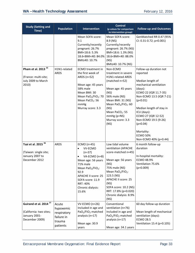
WA – Health Technology Assessment February 12, 2016
Extracorporeal Membrane Oxygenation: Final Evidence Report Page 33
Study (Setting and Time) Population Intervention
Control (p values for comparison
to intervention group) Follow-up and Outcomes
Mean SOFA score: 9.1 Currently/recently pregnant: 26.7% BMI<18.6: 5.3% 18.6<BMI<40: 84.0% BMI≥40: 10.7%
Mean SOFA score: 8.9 (NS) Currently/recently pregnant: 26.7% (NS) BMI<18.6: 1.3% (NS) 18.6<BMI<40: 88.0% (NS) BMI≥40: 10.7% (NS)
GenMatched RR 0.47 (95% CI: 0.31-0.72; p=0.001)
Pham et al. 2013 35 (France: multi-site; July 2009 to March 2010)
H1N1-related ARDS
ECMO treatment in the first week of ARDS (n=52) Mean age: 45 years 58% male Mean BMI: 30 Mean PaO2/FiO2: 70 Mean PaCO2: 56 mmHg Murray score: 3.3
Non-ECMO treatment in severe H1N1-related ARDS (matched n=52) Mean age: 45 years (NS) 56% male (NS) Mean BMI: 31 (NS) Mean PaO2/FiO2: 60 (NS) Mean PaCO2: 55 mmHg (p=NS) Murray score: 3.3 (NS)
Follow-up duration not reported Median length of mechanical ventilation (days): ECMO 22 (IQR 11.7-35) Non-ECMO 13.5 (IQR 7-21) (p<0.01) Median length of stay in ICU (days): ECMO 27 (IQR 12-52) Non-ECMO 19.5 (9-26) (p=0.04) Mortality: ECMO 50% Non-ECMO 40% (p=0.44)
Tsai et al. 2015 36 (Taiwan: single site; January 2007 to December 2012
ARDS
ECMO (n=45) • VV-ECMO
(n=37) • VA-ECMO (n=8) Mean age: 56 years 71% male Mean PaO2/FiO2: 92.9 APACHE II score: 25 SOFA score: 11.9 RRT: 40% Chronic dialysis: 15.6%
Low tidal volume ventilation (APACHE score-matched n=45) Mean age: 56 years (NS) 75% male (NS) Mean PaO2/FiO2: 123.5 (NS) APACHE II score: 25 (NS) SOFA score: 10.2 (NS) RRT: 17.8% (p=0.020) Chronic dialysis: 8.9% (NS)
6 month follow-up duration In-hospital mortality: ECMO 48.9% Ventilation 75.6% (p=0.009)
Guirand et al. 2014 37 (California: two sites; January 2001-December 2009)
Acute hypoxemic respiratory failure in trauma patients
VV-ECMO (n=26) Included in age and PaO2/FiO2-matched analysis (n=17) Mean age: 30.9 years
Conventional ventilation (n=76) Included in age and PaO2/FiO2-matched analysis (n=17) Mean age: 34.1 years
60 day follow-up duration Mean length of mechanical ventilation (days): ECMO 28.5 Ventilation 15.4 (p=0.105)

WA – Health Technology Assessment February 12, 2016
Extracorporeal Membrane Oxygenation: Final Evidence Report Page 34
Study (Setting and Time) Population Intervention
Control (p values for comparison
to intervention group) Follow-up and Outcomes
Defined as PaO2/FiO2≤80 with FiO2>0.9 without evidence of cardiogenic pulmonary edema and Murray score ≥3.0
71% male 88% Blunt trauma Mean PaO2/FiO2: 52.1 Murray score: 3.9 35% RRT
(NS) 88% male (NS) 65% Blunt trauma (NS) Mean PaO2/FiO2: 51.1 (NS) Murray score: 3.8 (NS) 24% RRT (NS)
Mean length of stay in hospital (days): ECMO 45.9 Ventilation 21.1 (0.040) Mean length of stay in ICU (days): ECMO 38.5 Ventilation 18.2 (p=0.064)
NS=non-significant
Summary of Results Across Studies: Mortality The impact of ECMO on in-hospital or post-discharge mortality was mixed in the available evidence. Neither RCT showed an independent mortality benefit for ECMO. Bein et al. described low overall hospital mortality (16.5%), which was not statistically significantly different between groups.2 While Peek et al. described a composite outcome of death or severe disability at 6-months which was improved for ECMO patients versus controls (37% vs. 53%, RR 0.69, CI= 0.05-0.97, p=0.03), the study was not powered to detect differences in survival alone, and indeed did not. In contrast to the RCTs, four of the six observational studies found that use of ECMO resulted in statistically-significant reductions in in-hospital mortality. While populations and extracorporeal technology differed, mortality ranged from 8-49% in the ECMO arms and 35-76% in comparator groups. A single study examined mortality over the longer-term; Kluge et al. found no differences at 28 days or 6 months between patients receiving pECLA and those receiving invasive mechanical ventilation. This study was hampered by relatively low statistical power however, with only 21 patients in each treatment arm.34 Specific study findings are presented in Table 4. Length of Hospitalization The two RCTs showed no significant difference in length of hospital or ICU stay between treatment groups or did not formally present significance testing for the comparison. Bein et al. found no statistically significant differences between groups for either length of stay in ICU or total length of stay in hospital.2 Peek et al. included length of stay in the ICU and length of hospital stay as secondary outcomes, which were longer in the ECMO group (ICU median days: ECMO 24 vs. conventional management 13; hospital median days: ECMO 35 vs. conventional management 17), but did not present statistical testing. Of the four observational studies to include length of stay as outcomes, two described significantly longer hospital or ICU stays among patients treated with ECMO versus non-ECMO therapies. Pham et al. described significantly longer ICU stay among patients treated with ECMO versus non-ECMO (27 days vs. 19.5 days; p=0.04), and Guirand et al. described longer hospital and ICU stays among patients treated with ECMO compared to mechanical ventilation (hospital LOS 45.9 days vs. 21.1 days; p=0.040; ICU LOS 38.5 vs. 18.2; p=0.064). Del Sorbo et al. found no significant difference in hospital or

WA – Health Technology Assessment February 12, 2016
Extracorporeal Membrane Oxygenation: Final Evidence Report Page 35
ICU length of stay between patients treated with or without ECCO2-R in addition to noninvasive ventilation, and Kluge et al. found no significant difference in length of hospital or ICU stay between patients treated with pECLA versus mechanical ventilation. Morbidity and Disability Neither RCT found differences in measures of morbidity or disability between treatment arms. Bein et al. found no statistically significant differences between groups for the Murray Lung Injury Score on day 10.e 2 One of the primary outcomes of interest in the CESAR trial was severe disability at 6 months after randomization. Severe disability was defined as confinement to bed and inability to wash or dress independently. None of these patients had been severely disabled before their presenting illness, and all of them were severely disabled at the time of randomization. The proportion of severe disability among those alive at six months of follow-up and with disability data did not significantly differ between treatment arms (ECMO 0 vs. control 1%). Neither observational study, which compared measures of illness severity found significant differences between treatment arms. Tsai et al. found no differences in APACHE II score, SOFA score, or RIFLE score between treatment arms.f 36 Matched analysis results from Guirand et al. showed no difference in Murray Lung Injury Score between groups.37 Quality of Life and Functional Outcomes Although there was a trend toward higher health-related quality of life and functional outcome measures in one RCT evaluating such outcomes among those treated with ECMO compared to conventional management, these differences were not statistically significant. In the CESAR trial, quality of life and other functional indicators were collected using a number of psychometric instruments at 6-month follow-up.11 Of the patients to participate in follow-up data collection (63% ECMO sample, 51% conventional therapy sample), all assessments favored the ECMO group, but none differed significantly. The proportion of individuals in both arms lacking follow-up data diminishes the statistical power of the study to document differential trends in these longer term outcomes where in fact they might exist. • The EuroQol-5 dimensions (EQ-5D): none in the ECMO group were confined to bed compared to two
in the control group, and there were no differences between groups in the ability to wash or dress independently.
eThe Murray Lung Injury Score (LIS) was proposed in 1988 by Murray et al.90 It has been commonly used as a measure of acute lung injury severity in clinical studies. The four component score was derived empirically by expert consensus to include 1) chest Xray; 2) hypoxemia score; 3) PEEP; and 4) static compliance of respiratory system. The final score is obtained by dividing the aggregate sum by the number of components. The LIS preceded the first American-European Consensus Committee definition of ARDS in 1994. Although it has not been validated as an accurate measure of lung injury severity, LIS has become a standard measure of ARDS severity. It is used both as a description of baseline lung injury characteristics and as a physiologic endpoint.40 f The APACHE II score (Acute Physiology and Chronic Health Evaluation II) is a severity-of-disease classification system used in the ICU. The score considers patient age, alveolar-arterial oxygen difference or PaO2, temperature, mean arterial pressure, pH arterial, heart rate, respiratory rate, sodium, potassium, creatinine, hematocrit, white blood cell count, and Glasgow Coma Scale. A score can range from 0 to 71, with higher scores corresponding to more severe disease and a higher risk of death.38

WA – Health Technology Assessment February 12, 2016
Extracorporeal Membrane Oxygenation: Final Evidence Report Page 36
• The Visual Analogue Scale (VAS, scored 0-100): More of the patients in the ECMO group reported feeling better compared with a year ago than did the control group (10% vs. 2%); this difference was not statistically significant.
• The SF-36 (scored 0-100): Physical functioning, general health, vitality, and mental health scores were not significantly different between ECMO patients than those in the control group.
• St. George’s hospital respiratory questionnaire (SGRQ, scored 0-100): Patients in the ECMO group had lower (i.e., better) total scores than did those in the control group (22.4 vs. 27.6); this difference was not statistically significant.
• The mini mental state examination score (MMSE, 0-100): There were no differences on the MMSE between groups.
• Hospital Anxiety and Depression Scale (HADS, scored 0-21): The depression score was similar between groups. Fewer ECMO patients had clinically significant anxiety than did those in the control group (8% vs. 11%); this difference was not statistically significant
• Strain reported among patient caregivers was higher among the ECMO group than the control group (10% vs. 7%); this difference was not statistically significant.
Use of Mechanical Ventilation The evidence base provides conflicting evidence around the impact of ECMO on the duration of mechanical ventilation between treatment arms. For Bein et al., the primary outcome of interest was the number of days without assisted ventilation in 28-day and 60-day follow-up periods.2 These did not statistically differ across treatment groups (means of 9-10 days in a 28-day period, 29-33 days in a 60-day period). Peek et al found that the ECMO treatment arm received low-volume low-pressure ventilation for more days than patients in the control arm (93% vs. 70% at any time; p<0.0001).11 Of the four observational studies to report length of time on mechanical ventilation, two showed significant differences between treatment arms, but in opposite directions. For Del Sorbo et al., cumulative prevalence of endotracheal intubation during the 28 days after ICU admission was a primary outcome. The decision to intubate was made according to clinical signs by attending physicians uninvolved with the study.33 They reported a Hazard Ratio of 0.27 (95% CI: 0.07-0.98; p=0.047) for endotracheal intubation for ECCO2-R patients compared to those who received only noninvasive ventilation. (Of note, intubation rate itself did not significantly differ between groups.) Pham et al., on the other hand, reported longer time on mechanical ventilation within the ECMO versus non-ECMO group [median days 22 (Interquartile range [IQR] 11.7-35) vs. 13.5 (IQR 7-21); p<0.01]. Kluge et al. and Guirand et al. reported no significant differences in length of time using mechanical ventilation between groups.34,37 ►Surgical Bridge to Transplant In total, our review identified three comparative cohort studies that report perioperative use of ECMO as a bridge to transplantation; no clinical benefit was associated with ECMO other than a decrease in hospital stay. ECMO patients were compared to those who did not require ECMO or those who required conventional cardiopulmonary bypass (CPB). Study populations were lung transplant recipients in two studies and heart lung transplant recipients in one study. Evidence on ECMO’s benefits is inconsistent across these studies; for example, two of the three studies showed higher mortality rates in ECMO-treated patients. The only consistent effect demonstrated for ECMO in this population was shorter

WA – Health Technology Assessment February 12, 2016
Extracorporeal Membrane Oxygenation: Final Evidence Report Page 37
hospital length of stay. Detailed descriptions of major study findings can be found organized by outcome below. Bittner et al. reported on 27 lung transplant recipients (mean age=49, standard deviation [SD]=12) who required VA-ECMO preoperatively (n=9), intraoperatively (n=7), and postoperatively (n=11) with 81 recipients who did not require ECMO (mean age=53, SD=11) in Germany.41 Demographics and transplantation characteristics were balanced at baseline except that a higher proportion of ECMO patients underwent sternotomy than patients without ECMO (22.2% vs 6.2%, p=0.027). Ius compared 46 lung transplantation patients (mean age=42.8, SD=14.4) who required VA-ECMO intraoperatively with 46 (mean age=42.6, SD=16.7) who required conventional cardiopulmonary bypass (CPB) and 211 off-pump patients (age not reported) in terms of their survival during a follow-up of 18 (SD=11) months in Germany.42 Preoperative characteristics of ECMO patients and CPB patients were generally comparable but ECMO patients had a greater prevalence of pulmonary hypertension as the indication for transplantation (37% vs 11%, p=0.003) and preoperative ECMO/iLA support (17% vs 2%, p=0.03), both of which were cited as well-recognized risk factors for mortality in lung transplantation. The authors used propensity score matching and multivariate analyses to create more balanced comparisons between the technologies. Jayarajan et al reviewed 15 heart lung transplant patients (mean age=39.5 years, SD=9.8 years) who required ECMO and 505 who did not require either ECMO or mechanical ventilation (mean age=39.2 years, SD=11.1 years) in the United States and compared their survival at 30 days and 5 years.43 At baseline, the ECMO group had a greater number of total human leukocyte antigen mismatches (4.7) than the control group (4.6) and those requiring MV (4.0; p=0.041). Also, the ECMO group had the highest class I plasma-reactive antigen panel (25.5%) compared with control (9.7%) or the MV group (10.8; p=0.041). In addition, lung allocation scores at the time of match were higher in the ECMO group (45.6) and the MV group (40.2) compared with the control (35.7; p=0.019). But none of these imbalances were found to be significant covariates in Cox proportional regression analysis. Mortality All three studies evaluated short-term or long-term mortality, ranging from 1 month to 5 years. All three are comparative cohort studies based on retrospective database reviews. Overall, patients who received ECMO had higher mortality compared to those who did not require cardiopulmonary support; however, compared to those requiring cardiopulmonary bypass, those treated with ECMO had lower short-term mortality. However, the differences disappeared once the patients survived discharge or the first year post-operation. During a mean of 2.3 years of follow-up in Bittner et al., short-term and long-term survival was significantly reduced in ECMO patients. The 30-day, 90-day, 1-year, and 5-year survival was estimated to be 63%, 44%, 33%, and 21%, respectively, in ECMO patients, compared to 97%, 91%, 83%, and 58% in the patient group without ECMO (p=0.001, log-rank test). However, in patients who survived beyond one year, there was no difference in long-term survival between groups (no statistical test reported). In Ius et al., ECMO patients had lower in-hospital mortality than CPB patients (13% vs 39%, p=0.004),.42 At 3, 9, and 12 months, overall survival was 87%, 81%, and 81%, respectively, in ECMO patients, compared to 70%, 59%, and 56% in CPB patients (p=0.004). However, among those discharged from the hospital, there was no difference in survival between the 2 groups (p=0.42) at 3, 9, and 12 months,

WA – Health Technology Assessment February 12, 2016
Extracorporeal Membrane Oxygenation: Final Evidence Report Page 38
implying that ECMO mainly improved short-term survival. Off-pump patients appeared to have better survival than ECMO patients, but these differences were not statistically significant. Jayarajan et al. found that the ECMO patients had significantly lower survival over the period of follow-up; using multivariate adjustment for demographic and clinical characteristics among both organ donors and recipients, the authors report a hazard ratio of 3.8 (95% C.I.: 1.6-9.1; p=0.003).43 Length of Hospitalization Only Jayarajan reported difference in postoperative length of stay between ECMO patients and controls.43 Length of stay was shorter in ECMO group (mean LOS= 12.4 days, SD= 10.3days) compared with controls (mean LOS= 39.4 days, SD= 46.1 days). The authors suspected that the shorter LOS in ECMO was likely skewed due to the high mortality in these patients. Morbidity and Disability None of the three available studies for this indication examined disability. Neither did the studies report health-related quality of life or functional outcomes. ►Cardiopulmonary Resuscitation (CPR) The evidence base presents an inconsistent picture regarding short- versus long-term outcomes in cardiac arrest patients treated with ECPR compared to conventional CPR, with one study reporting significant findings for ECMO-associated benefit on both mortality and neurologically intact survival, while others report short-term benefit that disappeared in the longer-term. Our review identified five studies evaluating the use of ECMO in patients requiring cardiopulmonary resuscitation. All were good quality comparative cohort studies conducted over a fairly constrained temporal period, and likely represent recent technologic advances in the area of ECPR. Several studies found a significant short-term mortality benefit conferred by ECPR; this disappeared over the longer term (up to three months). In contrast was one study which reported significant mortality benefit associated with ECPR in both the short- and long-term (up to 2 years). It is possible that this study had substantially greater statistical power to document such relative effect within propensity score-matched cohorts. Detailed descriptions of major study findings can be found organized by outcome, beginning on page 40. Limitations to the available evidence in this area include the fact that all studies were carried out in Southeast Asia, limiting the generalizability of the findings to other regions, and as well the bulk of the evidence is from retrospectively analyzed data. Our review identified five good quality comparative cohort studies comparing the use of extracorporeal cardiopulmonary resuscitation (ECPR) to conventional CPR; these studies were described in six publications.44-49 All five studies enrolled patients between 2003 and 2013, and all five studies were conducted in Southeast Asia, representing, therefore, a fairly homogenous temporal and geographic sample. Three studies44,46,48 evaluated the role of ECPR in cardiac arrest occurring in-hospital, while the remaining 2 evaluated its role in out-of-hospital cardiac arrests.45,47 Four of the five comparative cohort studies were retrospective in nature44-46,48,49 , and therefore subject to the implicit bias inherent in this design. Three of the four retrospective studies employed propensity score-matching to minimize the impact of hidden bias.45,46,48 Chou et al. described a retrospective comparative cohort study of 66 adult patients in Taiwan, with sudden in-hospital cardiac arrest due to a diagnosis of acute myocardial infarction, followed by CPR for

WA – Health Technology Assessment February 12, 2016
Extracorporeal Membrane Oxygenation: Final Evidence Report Page 39
more than 10 minutes, treated with ECPR (VA circuit, Centrifugal pump, Biomedicus Pump Console-560) and conventional CPR respectively, following them until discharge and evaluating survival using multivariate analyses accounting for multiple potentially confounding variables including age.44 Kim et al. described a retrospective comparative cohort study of 499 patients in Korea with out-of-hospital cardiac arrest.45 The study incorporated an analysis of propensity score-matched cohorts with 52 patients each treated with ECPR (T-PLS, or Capiox system) and CCPR respectively, and followed patients until 3 months post-cardiac arrest. Lin et al described a retrospective comparative cohort study of 118 patients in Taiwan, all responders to CPR treatment of in-hospital cardiac arrest of cardiac origin.46 Patients were aged 18-75 years with cardiac arrest of cardiac origin, undergoing CCPR for >10 minutes without sustained ROSC, defined as continuous maintenance of spontaneous circulation for >=20 minutes, subsequently treated to response with either CCPR or ECPR (Medtronic) with ROSC or ROSB. This study incorporated an analysis of propensity score-matched cohorts with 27 patients in each group, and evaluated mortality over a one-year period. Sakamoto et al. described a prospective comparative cohort study of 454 adult patients in Japan, with out-of-hospital cardiac arrest of cardiac origin, with no restoration of spontaneous circulation (ROSC) during the 15 minutes after hospital arrival.47 There were no significant differences in the treatment groups with respect to age, gender, time from emergency call to hospital arrival, or comorbidities present, and the authors evaluated both survival and neurologic outcomes at 6 months post-arrest. Shin et al. described a retrospective comparative cohort study of 406 patients in Korea, with in-hospital cardiac arrest.48,49 The study incorporated an analysis of propensity score-matched cohorts with 60 patients each, and evaluated both survival and neurologic outcomes over a 2-year period post-arrest. These studies are described in more detail in Table 5 below.
Table 5: Summary of evidence for ECMO used as ECPR
Study (Setting and Time)
Patient Population ECPR Conventional CPR Follow-up
Duration Chou et al., 201444 (Single center Taiwan: 2006-2010)
in-hospital cardiac arrest
n=43 Treated with ECPR Mean age 60.5
n=23 Mean age 69.6
Until discharge (NR)
Kim et al., 201445 (Single Center Korea: 2006-2013)
out-of-hospital cardiac arrest
n=52 in propensity matched group Mean age: 54 M/F: 40/12 Comorbidity score: 0
n=52 in propensity matched group Mean age: 54 (NS) M/F: 38/14 (NS) Comorbidity score: 0 (NS)
3 months post-cardiac arrest
Lin et al., 201046 (Single Center Taiwan: 2004-2006)
in-hospital cardiac arrest responders
n=27 in propensity-matched group Mean age 59 Male 77.8%
n=27 in propensity matched group Mean age 60 (NS) 85.2% (NS)
1 year
Sakamoto et al. 201447 (Multicenter Japan: 2008-2011)
out-of-hospital cardiac
n=260 Mean Age: 56.3 Male: 90.4%
n=194 Mean Age: 58.1 (NS) Male: 88.7% (NS)
6 months

WA – Health Technology Assessment February 12, 2016
Extracorporeal Membrane Oxygenation: Final Evidence Report Page 40
Study (Setting and Time)
Patient Population ECPR Conventional CPR Follow-up
Duration Shin et al. (Shin 2011, Shin 2013)48,49 (Korea: 2003-2009)
Patients with witnessed in-hospital cardiac arrests at Samsung Medical Center; ages 18-80
n=60 in propensity-matched group Treated with ECPR (Capiox bypass system)
n=60 in propensity-matched group Treated with CCPR
2 years
Mortality All five identified studies examined mortality, although at varying timepoints and with disparate results. There was an inconsistent pattern of outcomes being relatively better in cardiac arrest patients treated with ECPR compared to conventional CPR, with short-term ECPR benefit diminishing over time being reported in several studies, in contrast to one study reporting maintenance of benefit over the longer term. Chou et al. found that survival for more than 3 days was significantly improved in in-hospital cardiac arrest patients treated with ECPR (p=0.009) in a univariate analysis.44 However, when survival to discharge was evaluated in a multivariate analysis, the effect of ECPR diminished to non-significance (OR 1.9, 95% C.I.: 0.60-6.23; p=0.40). Kim et al. described a higher rate of return of spontaneous beating (ROSB) or return of spontaneous circulation (ROSC)(p<0.001) and a higher rate of survival at 24 hours (p<0.01) within the ECPR group compared to the conventional CPR group (p<0.001) in a cohort of out-of-hospital cardiac arrest patients; however, survival at 3 months post-arrest was numerically superior in the ECPR group, but no longer statistically significant (p=0.358)45 The short-term benefit of ECPR is echoed by Sakamoto et al. finding that survival at 24 hours is substantially higher in in-hospital cardiac arrest patients treated with ECPR group (68.1%) rather than CCPR group (19.1%).47 In distinct contrast to the lack of long-term benefit evidence is a report by Shin et al., describing statistically significant short-term (28 day) and long term (2 year) benefit for in-hospital cardiac arrest patients treated with ECPR compared to CCPR on both survival and survival with minimal neurologic impairment. This paper (Shin et al.) has possibly higher statistical power conferred by greater sample size even after propensity score matching than does the other evaluation of in-hospital cardiac arrest 44, suggesting that there is higher relative benefit of ECPR over CCPR in this subgroup of cardiac arrest patients. Chou et al. found that survival for more than 3 days (35% vs. 22% for ECPR and CPR, respectively) was significantly improved in patients treated with ECPR (p=0.009) in a univariate analysis.44 However, when survival to discharge was evaluated in a multivariate survival analysis also incorporating VT/VF rhythms, STEMI, time to coronary intervention, as well as demographic factors, the effect of ECPR diminished to non-significance. Variables remaining significant in the model were STEMI as a cause (OR 7.5, 95% C.I.: 2.1-26.2; p=0.001) and time from collapse to coronary intervention <210 minutes (OR 4.0, 95% C.I.: 1.2-13.8; p=0.03). Kim et al. described a higher rate of return of spontaneous breathing or return of spontaneous circulation (ROSB/ROSC) within the ECPR group (81%) than the conventional CPR group (39%; p<0.001).45 Survival at 24 hours was also higher in ECPR group (57.7% vs 30.8% in for CPR, p<0.01). However, there were no differences in survival at three months post-arrest, suggesting that the short-term ECMO-associated survival benefit did not persist over a longer period. Lin et al found no significant difference in short-term or 1 year survival when looking at responders to CPR, whether conventional or ECPR.46 These conclusions were derived from observation of both the original and propensity score-matched cohorts.
WA – Health Technology Assessment February 12, 2016
Extracorporeal Membrane Oxygenation: Final Evidence Report Page 41
Sakamoto et al. found survival at 24 hours to be substantially higher in the ECPR group than in the CCPR group, though the statistical significance of this was not reported; 177/260 (68.1%) of the ECPR treated group survived, compared to 37/194 (19.1%) of the CCPR-treated group.47 Shin et al. reported benefit of ECPR compared to CCPR on 28-day survival (p=0.011); 28-day survival with minimal neurologic impairment (OR 0.17, 95% C.I.: 0.04-0.68; p=0.012); 6-month survival (p=0.019); 6-month survival with minimal neurologic impairment (per Modified Glasgow Outcome Score [MGOS]>=4) (HR for ECPR adjusted with propensity score: 0.51 (95% C.I.: 0.34-0.77); 1-year survival (p=0.019), 1-year survival with minimal neurologic impairment (per Modified Glasgow Outcome Score [MGOS]>=4) (HR for ECPR : 0.52, 95% C.I.: 0.35-0.78); 2-year survival (p=0.019); 2-year survival with minimal neurologic impairment (per Modified Glasgow Outcome Score [MGOS]>=4): HR for ECPR : 0.53 (95% C.I.: 0.36-0.80); and death at 2 years with documented hypoxic brain damage (HR for ECPR : 0.42, 95% C.I.: 0.13-1.41).48,49 ECPR therefore significantly increased both overall 2-year survival, and 2-year survival with minimal neurologic impairment, compared to CCPR. Similarly substantial and significant impacts on survival at 1 month, 6 months, and 1 year were reported. Length of Hospitalization The limited evidence base in this area suggests that ECPR provides no benefit on length of hospitalization. Only one study identified in this review evaluated days in the hospital associated with various CPR modalities. Kim et al. reported hospital length of stay (days) was not significantly different between the groups.. Morbidity and Disability The evidence base provides conflicting information regarding the impact of ECPR on CPC outcomes, with one study reporting significant short-term benefit conferred by ECPR diminishing in the longer-term, and another study reporting maintenance of the ECPR benefit on this outcome. Lin et al. reported lower CPC scores (indicating better neurologic outcomes) in the ECPR group at discharge (p=0.011) but no difference by three months.46 However, the authors described a significantly beneficial effect of ECPR on CPC outcome at 3 months in subgroups of patients defined by length of CPR, indicating that ECPR in patients with CPR duration between 21-80 minutes provided a significant treatment benefit over CPR (p=0.026). It is unclear whether the range of categories defined by CPR duration were pre-planned subgroups for study; the five different categorization schemes evaluated evoke concern regarding multiple comparisons. There was no significant difference in CPC scores overall at 3 months (p=0.070). There was no significant difference in short-term or one-year survival when looking at responders to CPR, whether conventional or ECPR. Sakamoto et al. found that significantly higher proportions of patients treated with ECPR achieved favorable neurological outcomes that persisted at 6 months of observation, with 11.2% of the ECPR group maintaining a favorable CPC score of 1 or 2 at 6 months compared to 3.1% in the CCPR group (p=0.002).47
Long Term Outcomes of ECMO Long-term prognosis and outcomes in the years following ECMO use and hospital discharge have rarely been evaluated, irrespective of indication for use.50 There is no clear consensus about whether adult patients treated with ECMO have better or worse long-term outcomes, and there are studies indicating divergent trends. There is no consistent time period for assessing follow-up in this critically ill patient population, and few studies examine long-term outcomes. Of the two RCTs and 16 good-quality

WA – Health Technology Assessment February 12, 2016
Extracorporeal Membrane Oxygenation: Final Evidence Report Page 42
observational studies in our evidence base, only two reported outcomes beyond one year, and two provided data beyond two years of follow-up. From the transplant literature included in this review of the evidence, Bittner et al., Jayarajan et al., and Ius et al. examined outcomes greater than one year after ECMO use.41,51,52 Bittner and Jayarajan reported lower one-year and five-year survival compared to patients who did not receive ECMO, and Ius reported greater survival at one-year compared to patients who received CPB. Two ECPR studies examined outcomes at one-year and two-year follow-up points. Lin found comparable survival at one year following ECPR, and Shin et al., on the other hand, found significant improvement at both one and two years of follow-up.46,48,49 Although Peek et al. suggested comparable or better health-related quality of life scores compared with patients treated with conventional ventilation, the follow-up period was limited to 6 months.11 Other studies outside of our evidence provide information around longer term outcomes. Such studies include that of Hodgson and colleagues which found that only 26% of long-term survivors returned to their previous work at eight months follow-up, and health-related quality of life scores were lower than described in other ARDS patient populations.53 Another study reported relatively normal respiratory function but worsening self-reported pulmonary symptoms at follow-up assessments made at least 12 months following ECMO use among adult ARDS survivors.54 Because ECMO use is more well-established in the pediatric setting, there is a larger evidence base from which to examine long-term outcomes. However, this literature is similarly limited by diverse patient populations, variable follow-up duration across studies, and the challenge of attributing outcomes to ECMO as a treatment strategy versus the underlying disease process. In a study of children treated with ECMO as neonates compared to healthy controls, Hamutcu et al. reported greater incidence of lung injury among ECMO survivors (hyperinflated residual lung volume, greater airway obstruction, and lower oxygen saturation).55 Another study of survivors of neonatal ECMO found that exercise tolerance was reduced at 5, 8, and 12 years follow-up compared to healthy controls, irrespective of underlying diagnosis.56 Sensorineural hearing loss has been associated with ECMO use among children.57 One review of studies published between 1985 and 1996 found that 7.5% (range across study centers 3-21%) of ECMO survivors suffered from sensorineural hearing loss over follow-up durations of 1-10 years.58 Although a similar prevalence (12%) of sensorineural hearing loss was observed in a pediatric RCT, the rate did not differ among those who received conventional treatment.5,57 In contrast, a seven-year follow-up of this same RCT evaluated the cognitive ability of surviving patients; 76% of children achieved a cognitive level within the normal range and learning problems were similar between children treated with ECMO and conventional management.59 Authors of the study attributed long-term morbidity to underlying disease processes rather than the ECMO treatment protocols. Other studies have provided mixed results. Two studies reported normal intelligence levels at five years of follow-up,60,61 but three commonly cited studies have reported that 6-17% of neonatal ECMO survivors have demonstrated neurologic deficits that include epilepsy, cognitive delays, and motor difficulties.61-63
Key Question #2: What are the rates of adverse events and other potential harms associated with ECMO compared to conventional treatment strategies?

WA – Health Technology Assessment February 12, 2016
Extracorporeal Membrane Oxygenation: Final Evidence Report Page 43
Our review identified nine comparative studies that reported harms related to extracorporeal life support. Commonly reported complications included bleeding, cannula site complications, and distal limb ischemia. There is substantial variation in the reported rates of such complications. Furthermore, there is little correlation between the rates and duration of follow-up, and most are peri-operative in nature. It is likely that the noted variations are due instead to the heterogeneous study populations and settings described in the reports. Thus, there is insufficient evidence to fully evaluate whether complications differ by indication or type of ECMO. These studies are described in more detail in Table 6 below, with outcomes described in the sections that follow. Table 6: Summary of evidence for complications associated with ECMO
Study & Indication Patients with Complications Bleeding Limb Ischemia
Cannulation Site
Complications
Follow-up Period
Bein et al. 20132 40 patients with ARDS treated with avECCO2-R
3 (7.5%) - 1 (2.5%) 2 (5%) 60 days
Bittner et al. 201241 Perioperative VA-ECMO support for 27 patients undergoing lung transplantation
- 4 (14.8%) 0 - 5 years
Chamogeorgakis et al. 201327 61 patients treated with VA-ECMO for post-infarction- or decompensated cardiomyopathy-related cardiogenic shock
8 (13.1%) 2 (2.5%)β 6 (7.6%)β 8 (13.1%)π 14 months
Del Sorbo et al. 201533 25 patients with acute hypercapnic respiratory failure due to exacerbation of COPD treated with ECCO2-R
13 (52%) 4 (16%) - 1 (4%) 28 days
Guirand et al. 201437 26 trauma patients with life-threatening acute hypoxemic respiratory failure treated with VV-ECMO
23 (88%) 4 (15%) - 0 60 days
Ius et al. 201242 46 patients undergoing lung transplant were supported perioperatively with VA-ECMO
- - 2 (4.3%) 5 (11%) 18 months
Kim et al. 201464 52 patients with out-of-hospital cardiac arrest treated with ECPR
16 (30.8%) 13 (25%) 3 (6.8%) 12 (23.1%)µ 3 months

WA – Health Technology Assessment February 12, 2016
Extracorporeal Membrane Oxygenation: Final Evidence Report Page 44
Study & Indication Patients with Complications Bleeding Limb Ischemia
Cannulation Site
Complications
Follow-up Period
Peek et al. 200911 90 patients with ARDS randomized to receive VV-ECMO (68 treated)
2 (2%) - - 1 (1%)* 6 months
Pham et al. 201335 123 patients with H1N1-associated ARDS treated with VV- or VA-ECMO
65 (53%) - - - NR (In-ICU)
*Percent of 90 randomized to ECMO (68 patients [75%] actually treated with ECMO) βPercent of total patient population of 61 ECMO and 18 VAD πAll complications were limb complicates related to cannulation site µ12 bleeding events were at cannulation site
►ICU Cardiac Support We identified a single good-quality study that reported harms associated with ECMO in patients requiring cardiac support.65 The study retrospectively reviewed the charts of 79 patients (mean age 55.5; 76% male; 77.8/52.5% post-infarction for VAD, ECMO, respectively) who received VA-ECMO or a short-term VAD between 2006 and 2011 for either post-infarction or decompensated cardiomyopathy cardiogenic shock. The incidence of limb complications related to the arterial cannulation site for the overall study population (12) included limb ischemia (6), compartment syndrome (2), and hyperfusion syndrome (2). Limb complications occurred in 13.1% of ECMO patients, which was not statistically different from the VAD group.65 ►ICU Pulmonary Support Several good-quality studies assessed the harms associated with ECMO or avECCO2-R in patients who required pulmonary support. One RCT of avECCO2-R (described previously on page 26) reported low incidence of avECCO2-R-related adverse events.2 In total, three of 40 patients (7.5%) in the treatment arm experienced a complication, which consisted of one transient lower limb ischemia and two false aneurysms due to arterial cannulation.2 A second RCT, the CESAR trial (described on page 28) reported similar incidence of complications in 90 ARDS patients randomized to receive VV-ECMO support: two serious adverse events occurred, one related to mechanical failure of the oxygen supply during transport to the ECMO center, and a second vessel perforation during cannulation.11 Another good quality retrospective comparative cohort study of patients with ARDS evaluated 123 patients who received ECMO support for H1N1-associated ARDS. Sixty-five patients (53%) experienced at least one complication. Among the most common complications were bleeding events, such as epistaxis (15 [12%]] and cannulation-site bleeding (10 [8%]), and complications related to cannulation or the ECMO device, such as cannula-site infection and/or septicemia 14 [11%], deep vein thrombosis (8 [7%]), or hemolysis (8 [7%]).35 The incidence of adverse events reported in this study are similar to those reported by Del Sorbo and colleagues (2015) in a retrospective cohort analysis of 46 patients who required support with avECCO2-R or conventional ventilation for acute hypercapnic respiratory failure due to exacerbation of COPD.33 Del Sorbo and colleagues reported that 13 (52%) patients experienced adverse events related to avECCO2-R, which consisted of bleeding episodes (3: 1 hematuria, 1 retroperitoneal hematoma, 1 bleeding at groin), vein perforation at cannula insertion (1), and system

WA – Health Technology Assessment February 12, 2016
Extracorporeal Membrane Oxygenation: Final Evidence Report Page 45
malfunctioning (9: 6 clots in circuit, 2 pump malfunctions, 1 membrane lung failure). The incidence of adverse events among patients supported with conventional ventilation was not reported in the study publication. A final retrospective study evaluated ECMO in trauma patients with life-threatening acute hypoxemic respiratory failure treated between 2001 and 2009. Guirand and colleagues found that the overall rate of complications did not statistically differ between patients supported with VV-ECMO and conventional ventilation, however ECMO patients were transfused more packed red blood cells units than patients treated with conventional ventilation (8.4 U vs. 0.6; p<0.001) and experienced more hemorrhagic complications (4 [15%] vs. 1 [1%]; p=0.014). Whereas patients supported with ECMO did not experience pulmonary complications (pneumothorax, pulmonary hemorrhage, or pneumonia), 21 (28%) patients supported with conventional ventilation experienced such complications. Statistical differences disappeared in a matched cohort analysis for all complication types.37 ►Surgical Bridge to Transplant We identified two good-quality comparative cohort studies that evaluated perioperative use of ECMO in patients undergoing lung transplantation.41,42 The first study, from Bittner and colleagues, evaluated 108 patients (63% male; mean age 51.4) who underwent 50 bilateral sequential and 58 single lung transplants for various end-stage lung diseases including idiopathic pulmonary fibrosis (n=49) and chronic obstructive pulmonary disease (n=35). Twenty-seven patients were supported with VA-ECMO (9 preoperatively, 7 intraoperatively, and 11 postoperatively); these patients were compared to eighty-one patients who did not receive perioperative ECMO support. Four patients experienced bleeding complications (the severity of which was not described) in the ECMO group (one with pre-transplant support and three with post-operative support); distal limb ischemia did not occur in any of the ECMO-supported patients. Complications experienced by patients who did not receive perioperative ECMO support were not described.41 A second study from Ius and colleagues evaluated 46 patients who underwent lung transplant with cardiopulmonary bypass support and 46 patients who were supported with ECMO (n=92; 52.2% male; mean age 42.7).42 Post-transplant, CPB patients experienced greater morbidity than ECMO patients: (12 [26%] vs. 2 [4%]; p<0.01) required secondary ECMO/iLA implantation for acute rejection or primary graft dysfunction 18 ± 32 days after lung transplantation. There were no statistical differences between groups in vascular complications, the number of patients with grade 3 primary graft dysfunction, atrial fibrillation, rejection, stroke, or superficial secondary wound infection. Of the ECMO patients, five (1%) experienced complications related to cannulation of the femoral vessels (2 arteriovenous fistulas, 1 type B dissection, and 2 lower limb ischemias). ►Cardiopulmonary Resuscitation (CPR) Our review identified two good-quality retrospective studies that assessed harms related to ECPR compared to conventional CPR in patients who experienced cardiac arrest. In the first study, sixteen patients experienced complications during ECPR, which included bleeding at access site (12/55), lower limb ischemia (3/55), and one intracranial hemorrhage. Patients who experienced fewer ECPR-related complications had better neurologic outcomes; the relationship between complications and neurologic outcomes was not evaluated among those treated with conventional CPR in this study.45 Another study of ECPR reported that non-life-threatening bleeding and hematoma of insertion sites were relatively common complications but did not provide the rates with which these events occurred;

WA – Health Technology Assessment February 12, 2016
Extracorporeal Membrane Oxygenation: Final Evidence Report Page 46
rarer complications included vascular injury, catheter infection, limb ischemia, gastrointestinal bleeding, hemolysis, and stroke.49 We also identified a single systematic review (described on page 13) from Cheng and colleagues, which evaluated twenty studies that reported complication rates for ECMO in 1,866 adult patients who experienced cardiogenic shock or cardiac arrest. Pooled estimate rates of complications included: lower extremity ischemia, 16.9% (95% C.I.: 12.5-22.6); lower extremity amputation, 4.7% (95% C.I.: 2.3-9.3); stroke, 5.9% (95% C.I.:4.2-8.3); neurologic complications, 13.3% (95% C.I.: 9.9-17.7); acute kidney injury, 55.6% (95% C.I.: 35.5-74.0); major or significant bleeding, 40.8% (95% C.I.: 26.8-56.6); and significant infection, 30.4% (95% C.I.: 19.5-44.0).66 Case Series We identified ten case series that met predefined quality criteria and reported ECMO-related harms. Several of these studies accessed the ELSO database for mechanical and patient-related complications.67-70 Two studies looked specifically at the prevalence of infection during extracorporeal life support. Vogel and colleagues examined data from the ELSO database, comparing 2,996 adult patients who experienced infectious complications with those who did not have infectious complications; an infectious complication was defined as the presence of a new organism during ECMO or a white blood cell count below 1500. Adult patients with infectious complications experienced significantly more mechanical (59.2% vs. 34.4%), hemorrhagic (48.8% vs. 39.5%), neurologic (12.4% vs. 15.1%), renal (77.2% vs. 54.6%), cardiovascular (87.6% vs. 72.5%), pulmonary (22.5% vs. 10.7%), and metabolic complications (53.5% vs. 29.1%) than those patients who did not have infections.67 A second study of the ELSO database reported that of the patients recorded as having fungal infections, 34/59 acquired the infection while on VA-ECMO and 16/47 acquired an infection while supported with VV-ECMO.68 Two studies of the ELSO database from Paden and colleagues found cannula site bleeding, surgical site bleeding, oxygenator failure, and cannula problems to be among the most common complications from ECMO.69 Although statistical comparisons were not made, patients who were received ECMO for cardiac support appear to have more bleeding complications than patients who received ECMO for respiratory support.70
Key Question #3: What is the differential effectiveness and safety of ECMO according to sociodemographic factors (e.g., age, sex, race or ethnicity), severity of the condition for which ECMO is used (e.g., Murray score or APACHE score), setting in which ECMO is implemented (e.g., specialized ECMO centers), time of ECMO initiation (early vs. late), and duration of time on ECMO?
There is little evidence describing factors impacting the differential effectiveness of ECMO, with one RCT reporting no interaction between the effect of age and the ECMO treatment effect. There is inconsistent evidence suggesting that age is a predictor of short-term (in hospital) survival, and limited data suggest its association with neurologic outcome at 3 months post-cardiac arrest. More consistent findings suggest that gender is not associated with ECMO outcome, in either the short-term (prior to discharge), or medium-term (3 months post-admission). Limited but consistent evidence suggests that renal replacement therapy (dialysis) is associated with negative outcomes related to ECMO. These findings

WA – Health Technology Assessment February 12, 2016
Extracorporeal Membrane Oxygenation: Final Evidence Report Page 47
suggest that it will be difficult to use the described factors to define subgroups of patients with need for cardiopulmonary support for whom ECMO would be preferentially indicated or contraindicated. There are scant and often conflicting data addressing intervention-associated and patient-based factors that influence outcomes following treatment with ECMO. Several factors (e.g., age, gender, need for renal replacement therapy, and other comorbidities) are often adjusted for in analyses of the effect of ECMO treatment; however, there are few data available to describe differential impact of such factors among those treated with ECMO versus those treated with conventional therapy. While there is a dearth of formal subgroup analyses in this area, there are data describing various factors as independent risk factors for ECMO-related outcomes. These data are described below. We gave priority to evidence from RCTs and comparative cohort studies where available but also augment our analyses with data from case series describing ECMO use in US populations. The lack of evidence evaluating the effect of ECMO setting, time of ECMO initiation, and duration of ECMO treatment precluded its synthesis here. Age Our review identified one RCT11 and four comparative cohort studies35,36,41,45 which evaluated the role of age as an independent predictor of ECMO-related outcomes. In the area of ECMO for pulmonary support, one RCT11 and two comparative cohort studies35,36 described the effect of age on ECMO outcomes. Peek et al. is described earlier; in brief, it is a report on the Conventional ventilation or ECMO for Severe Adult Respiratory failure (CESAR) trial, in which adults with severe but potentially reversible respiratory failure were randomized into two treatment arms: ECMO and conventional management.11 Demographic characteristics and physiologic presentation were similar at baseline between the treatment and control groups., and investigators used an intention to treat analysis. This study reports no significant interaction between the treatment group and age category with respect to the outcome of severe disability or death (p=0.20), suggesting no differential effect of age on treatment with ECMO versus treatment with conventional therapy. While age does not appear to differentially impact the effect of ECMO treatment compared to conventional treatment of patients requiring pulmonary support, there are inconsistent suggestions from comparative cohort studies indicate that it is an independent predictor of treatment outcomes. Pham et al. described the use of ECMO in H1N1 patients with ARDS treated in French ICUs from July 2009 to March 2010, comparing outcomes from 52 pairs of patients: those treated with ECMO in the first week of ARDS matched with patients with severe H1N1-related ARDS not treated with ECMO.35 In this study, younger age was not a significant independent predictor of survival to discharge in patients treated with ECMO (p=0.06). In contrast, Tsai et al. compared the outcomes of 90 ARDS patients, half of whom did and half of whom did not receive ECMO matched with APACHE score.36 In this Japanese study, younger age was a significant independent predictor of survival (p=0.008). Kim and colleagues describe results from a retrospective comparative cohort study of 499 patients in Korea, with out-of-hospital cardiac arrest treated with ECPR or CPR.45 The study incorporated an analysis of propensity score-matched cohorts with 52 patients each in the ECPR treated group and CPR treated groups. In this study, Kim et al. reported that younger age was an independent predictor of better neurologic outcome (CPC score 1, 2) at 3 months post-arrest in those treated with ECPR (p=0.014). In contrast, Bittner et al. reported on 27 lung transplant recipients (mean age=49) who required VA-ECMO compared with 81 recipients who did not require ECMO (mean age=53) in Germany,

WA – Health Technology Assessment February 12, 2016
Extracorporeal Membrane Oxygenation: Final Evidence Report Page 48
finding that there was no significant effect of age on survival.41 We used evidence from several case series with drawing data from US patients to augment the findings around the effect of age on ECMO outcomes. Several such case series evaluated age as an independent risk factor for ECMO outcomes. Reflecting some of the findings from the comparative studies of interest, analysis of a case series of 405 adult patients in the US treated for severe ARDS with ECMO over the period of 1989 through 2003 identified age as an independent predictor of survival to discharge (p=0.01).71 Another case series describing the use ECMO in mixed cardiopulmonary support settings also found age to be an independent predictor of outcomes. Guttendorf et al. described a case series of 212 patients receiving ECMO for cardiac (n=126), or respiratory (n=86) failure during the time period 2005 through 2009 in the US.72 Overall survival to hospital discharge was 33%, with a higher rate of survival in those with a respiratory indication (50%) than with a cardiac indication (33%); older age was an independent risk factor for mortality, with survivors having a mean age of 48 and non-survivors a mean age of 53 (p=0.01). Analysis of data derived from the ELSO registry, which collects data on ECMO used to support cardiopulmonary function from 116 US and international centers, documents a 27% rate of survival to discharge over the period of 1992 to 2007 in 297 adult patients receiving ECPR. In this group, age was not independently associated with survival (p value not reported).73 Another analysis of data derived from the ELSO registry documented survival to discharge in 3846 patients treated with ECMO for cardiogenic shock over the period 2003 through 2013.50 Age less than 38 years was an independent predictor of survival (OR 2.6, 95% C.I.: 2.1-3.2; p<0.0001), as was age between 39 and 52 years (OR 1.7, 95% C.I.: 1.4-2.0; p<0.001). Gender No RCTs evaluated the role of gender on ECMO related outcomes; however, our review identified four comparative cohort studies which did so.35,36,41,45 In the area of ECMO for pulmonary support, gender was not a significant predictor of outcome in the comparative cohort studies from Pham, Tsai, or Bittner. The finding that gender is not an independent predictor of ECMO outcome is reflected in Kim et al., which describes results from a retrospective comparative cohort study of 499 patients in Korea, with out-of-hospital cardiac arrest treated with ECPR or CCPR.45 The study incorporated an analysis of propensity score-matched cohorts with 52 patients each in the ECPR treated group and CCPR treated groups. In this study, Kim et al. reported that male gender was not a significant independent predictor of better neurologic outcome (CPC score 1, 2) in those treated with ECPR (NS). In contrast to the findings from the comparative studies above, analysis of a case series of 405 adult patients in the US treated for severe ARDS with ECMO over the period of 1989 through 2003 identified male gender as an independent predictor of survival (p=0.048).71 Renal Replacement Therapy/Dialysis We identified no RCTs describing the effect of renal replacement therapy on outcomes related to cardiopulmonary support provided by ECMO or other means. We did identify a comparative cohort study reporting that neither renal replacement therapy nor chronic dialysis was a significant predictor of survival to discharge in 90 ARDS patients matched on APACHE II score, half of whom did and half of whom did not receive ECMO.36 In contrast, several analyses of data derived from the ELSO registry documented a significant association of renal dysfunction on ECMO outcomes. Thiagarajan et al. reported a 27% rate of survival to discharge

WA – Health Technology Assessment February 12, 2016
Extracorporeal Membrane Oxygenation: Final Evidence Report Page 49
over the period of 1992 to 2007 in 297 adult patients in the ELSO registry receiving ECPR.73 In this group, the need for dialysis was independently associated with mortality (OR 2.41, 95% C.I. 1.34-4.34; p=0.003). Another analysis of data derived from the ELSO registry documented survival to discharge in 3846 patients treated with ECMO for cardiogenic shock over the period 2003 through 2013.91 In this study, chronic renal failure was an independent predictor of reduced survival (OR 0.42, 95% C.I.: 0.26-0.68; p=0.0001).
Key Question #4: What are the costs and potential cost-effectiveness of ECMO relative to conventional treatment strategies?
Prior Published Evidence on Costs and Cost-Effectiveness As clinical evidence has accumulated on ECMO, data on the costs and potential cost-effectiveness of ECMO in certain populations has been more sparse. Below we summarize the findings of a review of published studies available since 2000. The current review identified the following literature describing costs and cost-effectiveness related to ECMO. Findings from two studies suggest that ECMO meets commonly-accepted thresholds for cost-effectiveness, but both used data from non-US settings. Studies of the budgetary impact of ECMO in the US suggest substantial incremental costs, ranging from $100,000 to nearly $600,000 depending on setting, indication, and timing of analysis. Peek et al. (2009, 2010) The CESAR randomized controlled trial of 180 UK adults with severe but potentially reversible respiratory failure included a concurrent economic evaluation of the cost-effectiveness of ECMO provided at a specialist center compared to conventional ventilator support, as described by Peek and colleagues.11 The analysis used both NHS and societal perspectives in the UK to evaluate the cost-effectiveness of ECMO at 6 months post-randomization and modeled to a lifetime horizon. The societal perspective analysis included costs borne by family and friends visiting or caring for patients. Health care resource utilization was collected for each patient both during hospitalization (within the trial) and after 6 months (via questionnaire), with unit costs applied to calculate total costs. Quality of life utility scores were measured using the EQ-5D at 6 months post-randomization, with an assumption that all patients had quality of life scores of 0 at randomization. Mean costs per patient (in 2005 USD) were $65,519 higher for patients allocated to ECMO than for patients allocated to conventional ventilator support (more than double the cost of conventional treatment), with 0.03 additional QALYs gained at 6 months; the resulting cost-effectiveness estimate at 6 months exceeded $2 million. When extrapolated over a lifetime horizon, cost-effectiveness was calculated as $31,112 per QALY gained (95% C.I.: $12,317-$95,507), with costs and QALYs discounted at 3.5%. The authors also noted that the budget impact of ECMO would likely be small, due to the relatively small number of patients with severe respiratory failure. As an economic evaluation conducted alongside a RCT, this study provides the best evidence to date on the cost-effectiveness of ECMO. However, it should be noted that ECMO was provided in only one experienced specialist center with clinical expertise on ECMO in the UK, and no standardized treatment protocol was used for the conventional treatment arm, so the results of this analysis may not be generalizable to other settings. St-Onge et al. (2015)

WA – Health Technology Assessment February 12, 2016
Extracorporeal Membrane Oxygenation: Final Evidence Report Page 50
St-Onge and colleagues estimated the cost-effectiveness of VA-ECMO in adults with cardiac arrest or cardiotoxicant-induced shock, compared with standard care. This analysis used a societal perspective (including medical and nonmedical costs) and lifetime horizon. Intervention effectiveness (survival) and probabilities used in the model were taken from the Masson et al. observational study of 62 patients (Masson et al. Resuscitation 2012).74 The incremental cost per life-year (LY) gained was estimated to be $7,185/LY in 2013 Canadian dollars, using estimates of 100% survival for cardiac arrest patients and 83% for severe shock patients from the Masson study. However, using survival estimates from other cohort studies in a sensitivity analysis (of 27% survival in cardiac arrest and 39% for severe shock), the incremental cost per LY gained increased to $34,311/LY. The authors noted that the survival estimates and some of the costs used in their analysis were based on a nonrandomized study of a small sample of selected European patients, and so should be confirmed in future studies. In addition, quality of life was not measured, so cost-per-QALY gained could not be calculated. Gregory et al. (2013) Gregory and colleagues developed a budget impact model from the payer perspective of percutaneous cardiac assist devices (pVADs), using data from a commercial claims database from 2009-2011.75 Patients experiencing cardiogenic shock who received surgical support using ECMO or extracorporeal LVADs, in comparison to those receiving non-surgical support using pVAD were included. Their model estimated the per-patient and overall cost of increasing use of pVADs vs. other surgical hemodynamic support, including ECMO and extracorporeal LVAD, from hospitalization to one year. The model estimated mean total allowed costs per case of $457,730 for surgical hemodynamic support during the index hospitalization and up to 30 days following; this was $170,000 (or 59%) higher than the mean cost per case for pVAD. When these patients were tracked for one year following hospitalization, the mean cost per surgical hemodynamic support case increased to $533,284 ($192,244, or 56%, higher than mean pVAD costs). In both cases, most of the difference was due to inpatient costs for the index admission, associated with longer mean length of stay for ECMO patients (30.9 days) that for pVAD patients (20.4 days, p=0.053). Aplin et al. (2015) Aplin and colleagues examined the variables affecting hospital costs from 2008 to 2010, using the AHRQ Nationwide Inpatient Sample database. In a ranking of DRGs by average hospital charge, ECMO or tracheostomy with 96+ hours of mechanical ventilation (DRG 3) was one of the top 10 most costly DRGs, with average charge per admission of $411,061.76 Maxwell et al. (2014) Maxwell and colleagues examined resource use trends in the use of ECMO in critically ill adults using the Nationwide Inpatient Sample database for the years 1998 through 2009. They found an average charge per admission of $344,009 (in 2009 US$). Total national hospital charges for these patients increased from $109.0 million in 1998 to $764.7 million in 2009 (p=0.0016), with mean total charges per admission increasing from less than $200,000 per patient to almost $500,000 per patient over this period (test for trend, p=0.0032). Total charges were highest for patients with heart transplant ($722,123 per patient) and lung transplant ($702,973), intermediate for respiratory failure ($421,037) and cardiogenic shock ($352,559) and lowest for patients post-cardiotomy ($273,429 per patient). Sauer et al. (2015) Sauer and colleagues also examined trends in the use of ECMO in adults using the Nationwide Inpatient Sample database, but for the years 2006 through 2011. Using simple linear regression analyses, they found no significant differences in trend in median cost per day or median cost per patient, with a

WA – Health Technology Assessment February 12, 2016
Extracorporeal Membrane Oxygenation: Final Evidence Report Page 51
median cost per patient of approximately $120,000 in 2011. Differences between the Maxwell and Sauer studies included the use of different ICD-9 codes to identify ECMO (Maxwell used code 39.65 and 39.66, while Sauer used only 39.65), the use of reported charges in Maxwell and HCUP cost-to-charge ratios in Sauer, and the use of regression analyses in Sauer. Higgins et al. (2011) Higgins and colleagues investigated critical care and hospital costs for patients with influenza A/H1N1 who were admitted to ICU in Australia and New Zealand in 2009 (n=762), in a multicenter cohort study.77 ECMO costs were included as one component of overall costs of care for these patients. They calculated the costs of ECMO using a “ground-up” costing method including supplies, labor and capital costs, in 2009 Australian dollars (AU$). For the 7% of patients who required ECMO, median ICU and median total hospital costs were found to be AU$160,735 and AU$177,158 respectively, compared to median ICU and hospital costs of AU$30,807 and AU$47,366, respectively, for the patients who did not receive ECMO (p<0.001 for both comparisons). The mean additional cost for providing ECMO was calculated as AU$13,646 per patient. Hsu et al. (2015) This study examined ECMO expenditures in Taiwan from 2000 to 2010, using retrospective claims data.78 Hsu et al. found that median expenditure per patient was $604,317 in 2000, increasing to $673,888 in 2010 (New Taiwan dollars). The authors also reported that median expenditures for newborns was significantly higher than that for adults, and significantly higher for males than for females, although exact amounts were not provided. In addition, patients receiving ECMO for trauma had significantly lower median expenditures than those receiving ECMO for cardiovascular, respiratory, or other indications. Other studies Mishra et al. (2010) examined the cost of ECMO in a single academic hospital in Norway in 2007. Costs were obtained for 14 consecutive ECMO patients (9 adults and 5 patients <18 years old), with mean estimated total hospital costs (in 2007 US dollars) of $213,246 (SD=$12,265) and estimated median costs of $191,436. Tseng and colleagues (2011) conducted a single-center study of costs associated with extra-corporeal life support in 72 consecutive adult patients treated for postcardiotomy cardiogenic shock, non-postcardiotomy cardiogenic shock or arrest, and ARDS in 2008 and 2009. They found mean and median total hospital costs of $39,845 (SD=$18,911) and $39,262, respectively (in 2010 US dollars). As single-center studies conducted in other countries, these results would be difficult to generalize to U.S. settings.

WA – Health Technology Assessment February 12, 2016
Extracorporeal Membrane Oxygenation: Final Evidence Report Page 52
ICER Integrated Evidence Ratings The ICER integrated evidence rating matrix is shown below; a detailed explanation of the methodology underpinning this rating system can be found in Appendix D to the full report. Separate ratings are provided for each of the indications of ECMO under consideration; the ratings and rationale are described on the following pages. Figure 4: ICER Integrated Evidence Ratings
Comparative Value
a
High
b
Reasonable/Comp
c
Low
Promising but Inconclusive: P/I
Comparative Clinical Effectiveness
Superior: A
Incremental: B+/B
Comparable: C+/C
Insufficient: I
Inferior: D

WA – Health Technology Assessment February 12, 2016
Extracorporeal Membrane Oxygenation: Final Evidence Report Page 53
Specific Intervention/Setting
5. ECMO versus VAD for cardiac support: Insufficient (I/Low Value) 6. ECMO versus mechanical ventilation for pulmonary support: Comparable or Better (C+c/Low
Value) 7. ECMO versus cardiopulmonary bypass as a bridge to heart and/or lung transplant: Insufficient
(I/Low Value) 8. ECMO versus conventional cardiopulmonary resuscitation for cardiac arrest: Comparable
(Cc/Low Value)
Rationale for ICER Ratings This review noted no consistent documentation of the benefit of ECMO on survival, days in the hospital, or disability across the comparisons present in a variety of settings. Randomized trials and other nonrandomized studies showed no distinct benefit for ECMO compared to ventricular assist devices, mechanical ventilation, cardiopulmonary bypass, or conventional resuscitation. Additionally, the use of ECMO in critically ill patients is associated with several complications and harms, although there is also no consistent evidence that rates of key harms differ from that of conventional management. In our view, the benefits and harms associated with ECMO yield a net health benefit rating of “Comparable” (C) when used for cardiopulmonary resuscitation, as the benefit-harm tradeoffs appear to be similar and relatively consistent across multiple available studies. However, despite challenges with the evidence base for pulmonary support, a majority of studies provide evidence of reduced mortality with ECMO, at least over the short term. We therefore consider the net health benefit in this instance to be “Comparable or Better” (C+), but the certainty in this rating to be moderate. Finally, in the case of ICU cardiac support and as a bridge to transplant, the presence of only one good-quality study with a relevant comparator in each indication was insufficient (I) to determine net health benefit. Two cost-effectiveness analyses evaluating the use of ECMO for pulmonary support and cardiac arrest/shock respectively estimated, over a lifetime horizon, cost-effectiveness ratios ranging from $7,000 - $35,000 per life year or QALY gained. However, these evaluations were based on data from single studies conducted in non-US settings with institutional cost structures that are vastly different from those in the US. Because ECMO appears to introduce substantial incremental hospital costs in the US in comparison to alternative means of cardiac or respiratory support (up to or exceeding $500,000 in some studies), we consider its use to represent a low value in all indications in the context of its general functional equivalence to alternative management.

WA – Health Technology Assessment February 12, 2016
Extracorporeal Membrane Oxygenation: Final Evidence Report Page 54
10. Recommendations for Future Research There is substantial heterogeneity with respect to underlying indication of populations in the identified studies, precluding quantitative syntheses. In addition, only two studies were randomized trials, and many of the comparative cohort studies were retrospective in nature, with selection of patients and ECMO systems made at the discretion of the centers, introducing additional layers of heterogeneity related to treatment efficacy. Evaluation of the efficacy of ECMO is challenging given the very nature of its application in the context of critical illness.92 In such settings, many diffuse and undefined factors compete in the determination of outcome, rendering rigorous randomization difficult.92 Additionally, there may be methodological flaws found in many such studies which may diminish their statistical power to document treatment efficacy; evaluations of critical care interventions are commonly negative given inadequate statistical power to identify treatment effects often unrealistically hypothesized.93 Carefully controlled and appropriately powered studies are needed to further characterize the comparative effectiveness of ECMO in the variety of settings and indications in which it is currently used. A better understanding of potential confounders of the relationship between treatment and outcome is required in order to more appropriately design clinical trials. One such potential confounder is role of the ECMO center itself, as might have played a role in one of the RCTs reviewed here. In the CESAR trial, the survival benefit noted for the treatment arm cannot be attributed solely to the use of ECMO; instead, it is more carefully attributable to the treatment strategy of referral to a particular single ECMO center for assessment.11 It may well be that the benefit noted in the CESAR trial is attributable not to ECMO, but rather to the standard of care available at the single center studied. Trials in which treatment allocation is not confounded by the potential impact of differential standard of care available to the intervention arm are required to more fully evaluate efficacy. In addition to more careful control of treatment, patient factors, including the impact of various co-morbidities, require further hypothesis-driven evalution to identify those associated with negative ECMO outcomes. Research into the use of prognostic instruments, such as the RESP score, may be of use in such clinical decision-making.92,94 This score is currently useful for the prediction of survival after ECMO initiation in patients with ARDS. Lacking, however, is the further development of such a score to predict the probability of survival in patients prior to initiation.92,95 Still further down the road is the development of scores to predict the probability of neurologically intact survival. The careful control of factors captured by such scores may provide for more powerful effectiveness studies, and elucidate the patient populations in which ECMO might be of most benefit. Whether long-term mortality or other factors are being affected by modifiable or non-modifiable factors requires additional research, in order to best test new interventions, and shape guidance for offering ECMO. It is difficult to tease out long-term outcomes associated with ECMO among survivors in the current evidence base, particularly in the absence of a robust body of evidence from randomized controlled trials. The difficulty of carrying out RCTs in these populations is recognized. However, the continued study of this issue using quasi-experimental study designs such as propensity score-matched cohort studies may yield a more robust evidence base, and one in which certain more homogeneous subsets of data may be analyzed quantitatively.
WA – Health Technology Assessment February 12, 2016
Extracorporeal Membrane Oxygenation: Final Evidence Report Page 55
References
1. Bein T, Weber F, Philipp A, et al. A new pumpless extracorporeal interventional lung assist in critical hypoxemia/hypercapnia. Critical care medicine. 2006;34(5):1372-1377.
2. Bein T, Weber-Carstens S, Goldmann A, et al. Lower tidal volume strategy (approximately 3 ml/kg) combined with extracorporeal CO2 removal versus 'conventional' protective ventilation (6 ml/kg) in severe ARDS: the prospective randomized Xtravent-study. Intensive care medicine. 2013;39(5):847-856.
3. Del Sorbo L, Cypel M, Fan E. Extracorporeal life support for adults with severe acute respiratory failure. The Lancet. Respiratory medicine. 2014;2(2):154-164.
4. Cooper DJ, Hodgson CL. Extracorporeal Membrane Oxygenation Rescue for H1N1 Acute Respiratory Distress Syndrome. American Journal of Respiratory and Critical Care Medicine. 2013;187(3):224-226.
5. UK collaborative randomised trial of neonatal extracorporeal membrane oxygenation. UK Collaborative ECMO Trail Group. Lancet (London, England). 1996;348(9020):75-82.
6. Morris AH, Wallace CJ, Menlove RL, et al. Randomized clinical trial of pressure-controlled inverse ratio ventilation and extracorporeal CO2 removal for adult respiratory distress syndrome. Am J Respir Crit Care Med. 1994;149(2 Pt 1):295-305.
7. Zapol WM, Snider MT, Hill JD, et al. Extracorporeal membrane oxygenation in severe acute respiratory failure. A randomized prospective study. Jama. 1979;242(20):2193-2196.
8. Roncon-Albuquerque Jr. R, Paiva JA. Recent Advances and Novel Applications of Modern ECMO. In: Vincent J-L, ed. Annual Update in Intensive Care and Emergency Medicine 20132013:621-633.
9. Chalwin RP, Moran JL, Graham PL. The role of extracorporeal membrane oxygenation for treatment of the adult respiratory distress syndrome: review and quantitative analysis. Anaesthesia and intensive care. 2008;36(2):152-161.
10. Gilani FS, Farooqui S, Doddamani R, Gruberg L. Percutaneous Mechanical Support in Cardiogenic Shock: A Review. Clinical Medicine Insights. Cardiology. 2015;9(Suppl 2):23-28.
11. Peek GJ, Mugford M, Tiruvoipati R, et al. Efficacy and economic assessment of conventional ventilatory support versus extracorporeal membrane oxygenation for severe adult respiratory failure (CESAR): a multicentre randomised controlled trial. Lancet (London, England). 2009;374(9698):1351-1363.
12. Davies A, Jones D, Bailey M, et al. Extracorporeal Membrane Oxygenation for 2009 Influenza A(H1N1) Acute Respiratory Distress Syndrome. Jama. 2009;302(17):1888-1895.
13. Gattinoni L, Carlesso E, Langer T. Clinical review: Extracorporeal membrane oxygenation. Critical care (London, England). 2011;15(6):243.
14. Lyon RM. Extra-corporeal cardiopulmonary resuscitation--miracle cure or expensive futility? Resuscitation. 2012;83(11):1311-1312.
15. Lynch JE, Zwischenberger JB. Turning the dial to futility. Chest. 2011;139(1):230-231; author reply 231-232.
16. Schmidt M, Bailey M, Sheldrake J, et al. Predicting survival after extracorporeal membrane oxygenation for severe acute respiratory failure. The Respiratory Extracorporeal Membrane Oxygenation Survival Prediction (RESP) score. American journal of respiratory and critical care medicine. 2014;189(11):1374-1382.

WA – Health Technology Assessment February 12, 2016
Extracorporeal Membrane Oxygenation: Final Evidence Report Page 56
17. Abrams DC, Prager K, Blinderman CD, Burkart KM, Brodie D. Ethical dilemmas encountered with the use of extracorporeal membrane oxygenation in adults. Chest. 2014;145(4):876-882.
18. Hubmayr RD, Farmer JC. Should we "rescue" patients with 2009 influenza A(H1N1) and lung injury from conventional mechanical ventilation? Chest. 2010;137(4):745-747.
19. Mols G, Loop T, Geiger K, Farthmann E, Benzing A. Extracorporeal membrane oxygenation: a ten-year experience. American journal of surgery. 2000;180(2):144-154.
20. Thiagarajan R, Barrett C. ECMO - Indications and Outcomes. 2011; http://www.sccm.org/Communications/Critical-Connections/Archives/Pages/ECMO---Indications-and-Outcomes.aspx. Accessed October 1, 2015.
21. UCSF Medical Center. Extracorporeal Membrane Oxygenation (ECMO). 2015; http://www.ucsfhealth.org/treatments/extracorporeal_mechanical_oxygenation/. Accessed October 1, 2015.
22. New York-Presbyterian. Center for Acute Respiratory Failure--What is ECMO. http://nyp.org/services/carf/what-is-ecmo.html. Accessed October 1, 2015.
23. Cove and MacLaren. Diagrammatic representation of peripheral veno-venous (VV-ECMO) and peripheral veno-arterial (VA-ECMO) extracorporeal membrane oxygenation.: Critical Care; 2010.
24. Zimmermann M, Bein T, Arlt M, et al. Pumpless extracorporeal interventional lung assist in patients with acute respiratory distress syndrome: a prospective pilot study. Critical care (London, England). 2009;13(1):R10.
25. Fitzsimmons M. Weaning from cardiopulmonary bypass (CPB). In: Mark J, ed. UpToDate. Waltham, MA2015.
26. Aroesty J, Jeevanandam V, Eisen H. Short-term mechanical circulatory devices. In: Cutlip D, ed. UpToDate. Waltham, MA2015.
27. Chamogeorgakis T, Rafael A, Shafii AE, Nagpal D, Pokersnik JA, Gonzalez-Stawinski GV. Which is better: A miniaturized percutaneous ventricular assist device or extracorporeal membrane oxygenation for patients with cardiogenic shock? ASAIO Journal. 2013;59(6):607-611.
28. Pozner C. Advanced cardiac life support (ACLS) in adults. In: Walls R, Page R, eds. UpToDate. Waltham, MA2015.
29. Pozner C. Basic life support (BLS) in adults. In: Walls R, Page R, eds. UpToDate. Waltham, MA2015.
30. Courey A, Hyzy R. Orverview of mechanica ventilation. In: Parsons P, ed. UpToDate. Waltham, MA2014.
31. Tramm R, Ilic D, Davies AR, Pellegrino VA, Romero L, Hodgson C. Extracorporeal membrane oxygenation for critically ill adults. (1).
32. Noah MA, Peek GJ, Finney SJ, et al. Referral to an extracorporeal membrane oxygenation center and mortality among patients with severe 2009 influenza A(H1N1). Jama. 2011;306(15):1659-1668.
33. Del Sorbo L, Pisani L, Filippini C, et al. Extracorporeal Co2 removal in hypercapnic patients at risk of noninvasive ventilation failure: A matched cohort study with historical control. Critical care medicine. 2015;43(1):120-127.
34. Kluge S, Braune SA, Engel M, et al. Avoiding invasive mechanical ventilation by extracorporeal carbon dioxide removal in patients failing noninvasive ventilation. Intensive care medicine. 2012;38(10):1632-1639.

WA – Health Technology Assessment February 12, 2016
Extracorporeal Membrane Oxygenation: Final Evidence Report Page 57
35. Pham T, Combes A, Roze H, et al. Extracorporeal membrane oxygenation for pandemic influenza a(h1n1)-induced acute respiratory distress syndrome a cohort study and propensity-matched analysis. American Journal of Respiratory and Critical Care Medicine. 2013;187(3):276-282.
36. Tsai HC, Chang CH, Tsai FC, et al. Acute Respiratory Distress Syndrome With and Without Extracorporeal Membrane Oxygenation: A Score Matched Study. Annals of Thoracic Surgery. 2015;100(2):458-464.
37. Guirand DM, Okoye OT, Schmidt BS, et al. Venovenous extracorporeal life support improves survival in adult trauma patients with acute hypoxemic respiratory failure: a multicenter retrospective cohort study. The journal of trauma and acute care surgery. 2014;76(5):1275-1281.
38. Knaus WA, Draper EA, Wagner DP, Zimmerman JE. APACHE II: a severity of disease classification system. Critical care medicine. 1985;13(10):818-829.
39. Murray D, Grant D, Murali N, Butt W. Unmeasured anions in children after cardiac surgery. The Journal of thoracic and cardiovascular surgery. 2007;133(1):235-240.
40. Kangelaris KN, Calfee CS, May AK, Zhuo H, Matthay MA, Ware LB. Is there still a role for the lung injury score in the era of the Berlin definition ARDS? Annals of intensive care. 2014;4(1):4.
41. Bittner HB, Lehmann S, Rastan A, et al. Outcome of extracorporeal membrane oxygenation as a bridge to lung transplantation and graft recovery. The Annals of thoracic surgery. 2012;94(3):942-949; author reply 949-950.
42. Ius F, Kuehn C, Tudorache I, et al. Lung transplantation on cardiopulmonary support: venoarterial extracorporeal membrane oxygenation outperformed cardiopulmonary bypass. The Journal of thoracic and cardiovascular surgery. 2012;144(6):1510-1516.
43. Jayarajan SN, Taghavi S, Komaroff E, et al. Impact of extracorporeal membrane oxygenation or mechanical ventilation as bridge to combined heart-lung transplantation on short-term and long-term survival. Transplantation. 2014;97(1):111-115.
44. Chou TH, Fang CC, Yen ZS, et al. An observational study of extracorporeal CPR for in-hospital cardiac arrest secondary to myocardial infarction. Emergency Medicine Journal. 2014;31(6):441-447.
45. Kim SJ, Jung JS, Park JH, Park JS, Hong YS, Lee SW. An optimal transition time to extracorporeal cardiopulmonary resuscitation for predicting good neurological outcome in patients with out-of-hospital cardiac arrest: A propensity-matched study. Critical Care. 2014;18(5):1-15.
46. Lin JW, Wang MJ, Yu HY, et al. Comparing the survival between extracorporeal rescue and conventional resuscitation in adult in-hospital cardiac arrests: Propensity analysis of three-year data. Resuscitation. 2010;81(7):796-803.
47. Sakamoto T, Morimura N, Nagao K, et al. Extracorporeal cardiopulmonary resuscitation versus conventional cardiopulmonary resuscitation in adults with out-of-hospital cardiac arrest: a prospective observational study. Resuscitation. 2014;85(6):762-768.
48. Shin TG, Choi JH, Jo IJ, et al. Extracorporeal cardiopulmonary resuscitation in patients with inhospital cardiac arrest: A comparison with conventional cardiopulmonary resuscitation. Critical care medicine. 2011;39(1):1-7.
49. Shin TG, Jo IJ, Sim MS, et al. Two-year survival and neurological outcome of in-hospital cardiac arrest patients rescued by extracorporeal cardiopulmonary resuscitation. International Journal of Cardiology. 2013;168(4):3424-3430.
50. Schmidt M, Hodgson C, Combes A. Extracorporeal gas exchange for acute respiratory failure in adult patients: A systematic review. Critical Care. 2015;19(1).

WA – Health Technology Assessment February 12, 2016
Extracorporeal Membrane Oxygenation: Final Evidence Report Page 58
51. Jayarajan S, Taghavi S, Komaroff E, et al. Long-term outcomes in heart transplantation using donors with a history of past and present cocaine use. European Journal of Cardio-thoracic Surgery. 2015;47(4):e146-e150.
52. Ius F, Sommer W, Tudorache I, et al. Veno-veno-arterial extracorporeal membrane oxygenation for respiratory failure with severe haemodynamic impairment: Technique and early outcomes. Interactive Cardiovascular and Thoracic Surgery. 2015;20(6):761-767.
53. Hodgson CL, Hayes K, Everard T, et al. Long-term quality of life in patients with acute respiratory distress syndrome requiring extracorporeal membrane oxygenation for refractory hypoxaemia. Critical care (London, England). 2012;16(5):R202.
54. Linden VB, Lidegran MK, Frisen G, Dahlgren P, Frenckner BP, Larsen F. ECMO in ARDS: a long-term follow-up study regarding pulmonary morphology and function and health-related quality of life. Acta anaesthesiologica Scandinavica. 2009;53(4):489-495.
55. Hamutcu R, Nield TA, Garg M, Keens TG, Platzker AC. Long-term pulmonary sequelae in children who were treated with extracorporeal membrane oxygenation for neonatal respiratory failure. Pediatrics. 2004;114(5):1292-1296.
56. van der Cammen-van Zijp MH, Gischler SJ, Hop WC, de Jongste JC, Tibboel D, Ijsselstijn H. Deterioration of exercise capacity after neonatal extracorporeal membrane oxygenation. The European respiratory journal. 2011;38(5):1098-1104.
57. Bennett CC, Johnson A, Field DJ. A comparison of clinical variables that predict adverse outcome in term infants with severe respiratory failure randomised to a policy of extracorporeal membrane oxygenation or to conventional neonatal intensive care. Journal of perinatal medicine. 2002;30(3):225-230.
58. Cheung PY, Robertson CM. Sensorineural hearing loss in survivors of neonatal extracorporeal membrane oxygenation. Pediatric rehabilitation. 1997;1(2):127-130.
59. McNally H, Bennett CC, Elbourne D, Field DJ. United Kingdom collaborative randomized trial of neonatal extracorporeal membrane oxygenation: follow-up to age 7 years. Pediatrics. 2006;117(5):e845-854.
60. Glass P, Wagner AE, Papero PH, et al. Neurodevelopmental status at age five years of neonates treated with extracorporeal membrane oxygenation. The Journal of pediatrics. 1995;127(3):447-457.
61. Hanekamp MN, Mazer P, van der Cammen-van Zijp MH, et al. Follow-up of newborns treated with extracorporeal membrane oxygenation: a nationwide evaluation at 5 years of age. Critical care (London, England). 2006;10(5):R127.
62. Nijhuis-van der Sanden MW, van der Cammen-van Zijp MH, Janssen AJ, et al. Motor performance in five-year-old extracorporeal membrane oxygenation survivors: a population-based study. Critical care (London, England). 2009;13(2):R47.
63. Jen HC, Shew SB. Hospital readmissions and survival after nonneonatal pediatric ECMO. Pediatrics. 2010;125(6):1217-1223.
64. Kim SJ, Jung JS, Park JH, Park JS, Hong YS, Lee SW. An optimal transition time to extracorporeal cardiopulmonary resuscitation for predicting good neurological outcome in patients with out-of-hospital cardiac arrest: a propensity-matched study. Critical care (London, England). 2014;18(5):535.
65. Chamogeorgakis T, Lima B, Shafii AE, et al. Outcomes of axillary artery side graft cannulation for extracorporeal membrane oxygenation. Journal of Thoracic and Cardiovascular Surgery. 2013;145(4):1088-1092.

WA – Health Technology Assessment February 12, 2016
Extracorporeal Membrane Oxygenation: Final Evidence Report Page 59
66. Cheng R, Hachamovitch R, Kittleson M, et al. Complications of extracorporeal membrane oxygenation for treatment of cardiogenic shock and cardiac arrest: a meta-analysis of 1,866 adult patients. Annals of Thoracic Surgery. 2014;97:610-616.
67. Vogel AM, Lew DF, Kao LS, Lally KP. Defining risk for infectious complications on extracorporeal life support. Journal of pediatric surgery. 2011;46(12):2260-2264.
68. Pluim T, Halasa N, Phillips SE, Fleming G. The morbidity and mortality of patients with fungal infections before and during extracorporeal membrane oxygenation support. Pediatric critical care medicine : a journal of the Society of Critical Care Medicine and the World Federation of Pediatric Intensive and Critical Care Societies. 2012;13(5):e288-293.
69. Paden ML, Conrad SA, Rycus PT, Thiagarajan RR, Registry E. Extracorporeal Life Support Organization Registry Report 2012. ASAIO journal (American Society for Artificial Internal Organs : 1992). 2013;59(3):202-210.
70. Paden ML, Rycus PT, Thiagarajan RR, Registry E. Update and outcomes in extracorporeal life support. Seminars in perinatology. 2014;38(2):65-70.
71. Hemmila MR, Rowe SA, Boules TN, et al. Extracorporeal life support for severe acute respiratory distress syndrome in adults. Annals of surgery. 2004;240(4):595-597.
72. Guttendorf J, Boujoukos AJ, Ren D, Rosenzweig MQ, Hravnak M. Discharge outcome in adults treated with extracorporeal membrane oxygenation. American journal of critical care : an official publication, American Association of Critical-Care Nurses. 2014;23(5):365-377.
73. Thiagarajan RR, Brogan TV, Scheurer MA, Laussen PC, Rycus PT, Bratton SL. Extracorporeal membrane oxygenation to support cardiopulmonary resuscitation in adults. The Annals of thoracic surgery. 2009;87(3):778-785.
74. Masson R, Colas V, Parienti JJ, et al. A comparison of survival with and without extracorporeal life support treatment for severe poisoning due to drug intoxication. Resuscitation. 2012;83(11):1413-1417.
75. Gregory D, Scotti DJ. A Budget impact model to estimate the cost dynamics of treating high-risk heart failure patients with advanced percutaneous cardiac assist devices: The payer perspective. Journal of Managed Care Medicine. 2013;16(1):61-69.
76. Aplin B, Nazzal M, Qu W, Zelenock G, Kazan V, Abbas J. Patient variables impacting hospital costs from 2008 to 2010. American Journal of Surgery. 2015;210(1):134-140.
77. Higgins AM, Pettila V, Harris AH, et al. The critical care costs of the influenza A/H1N1 2009 pandemic in Australia and New Zealand. Anaesthesia and intensive care. 2011;39(3):384-391.
78. Hsu C-P, Lee W-C, Wei H-M, et al. Extracorporeal membrane oxygenation use, expenditure, and outcomes in taiwan from 2000 to 2010. Journal of epidemiology / Japan Epidemiological Association. 2015;25(4):321-331.
79. Cave DM, Gazmuri RJ, Otto CW, et al. Part 7: CPR Techniques and Devices: 2010 American Heart Association Guidelines for Cardiopulmonary Resuscitation and Emergency Cardiovascular Care. Circulation. 2010;122(18 suppl 3):S720-S728.
80. Fitzgerald M, Millar J, Blackwood B, et al. Extracorporeal carbon dioxide removal for patients with acute respiratory failure secondary to the acute respiratory distress syndrome: a systematic review Critical Care. 2014;18:1-11.
81. Mitchell MD, Mikkelsen ME, Umscheid CA, Lee I, Fuchs BD, Halpern SD. A systematic review to inform institutional decisions about the use of extracorporeal membrane oxygenation during the H1N1 influenza pandemic. Critical care medicine. 2010;38(6):1398-1404.

WA – Health Technology Assessment February 12, 2016
Extracorporeal Membrane Oxygenation: Final Evidence Report Page 60
82. Munshi L, Telesnicki T, A. W, Fan E. Extracorporeal Life Support for Acute Respiratory Failure: A Systematic Review and metaanalysis. Annals of the American Thoracic Society. 2014;11(5):802-810.
83. Zampieri FG, Mendes PV, Ranzani OT, et al. Extracorporeal membrane oxygenation for severe respiratory failure in adult patients: a systematic review and meta-analysis of current evidence. Journal of critical care. 2013;28(6):998-1005.
84. Zangrillo A, Biondi-Zoccai G, Landoni G, et al. Extracorporeal membrane oxygenation (ECMO) in patients with H1N1 influenza infection: a systematic review and meta-analysis including 8 studies and 266 patients receiving ECMO. Critical Care. 2013;17:1-8.
85. Higgins JPT, S G. Cochrane Collaboration Handbook for Systematic Reviews of Interventions. The Cochrane Collaboration and John Wiley & Sons Ltd; 2008.
86. Cook DJ, Mulrow CD, Haynes RB. Systematic reviews: synthesis of best evidence for clinical decisions. Annals of internal medicine. 1997;126(5):376-380.
87. Ollendorf DA, Pearson SD. An integrated evidence rating to frame comparative effectiveness assessments for decision makers. Medical care. 2010;48(6 Suppl):S145-152.
88. Agency for Healthcare Research and Quality. U.S. Preventive Services Task Force Procedure Manual: Publication No. 08-05118-EF. 2008.
89. AHRQ. Methods Guide for Effectiveness and Comparative Effectiveness Reviews. 2014. 90. Murray JF, Matthay MA, Luce JM, Flick MR. An expanded definition of the adult respiratory
distress syndrome. The American review of respiratory disease. 1988;138(3):720-723. 91. Schmidt M, Burrell A, Roberts L, et al. Predicting survival after ECMO for refractory cardiogenic
shock: The survival after veno-arterial-ECMO (SAVE)-score. European Heart Journal. 2015;36(33):2246-2256.
92. Goligher EC, Doufle G, Fan E. Update in Mechanical Ventilation, Sedation, and Outcomes 2014. Am J Respir Crit Care Med. 2015;191(12):1367-1373.
93. Harhay MO, Wagner J, Ratcliffe SJ, et al. Outcomes and statistical power in adult critical care randomized trials. Am J Respir Crit Care Med. 2014;189(12):1469-1478.
94. Schmidt M, Bailey M, Sheldrake J, et al. Predicting survival after extracorporeal membrane oxygenation for severe acute respiratory failure: The Respiratory Extracorporeal Membrane Oxygenation Survival Prediction (RESP) score. American journal of respiratory and critical care medicine. 2014;189(11):1374-1382.
95. Fan E, Pham T. Extracorporeal membrane oxygenation for severe acute respiratory failure: yes we can! (But should we?). Am J Respir Crit Care Med. 2014;189(11):1293-1295.
96. Aigner C, Wisser W, Taghavi S, et al. Institutional experience with extracorporeal membrane oxygenation in lung transplantation. European journal of cardio-thoracic surgery : official journal of the European Association for Cardio-thoracic Surgery. 2007;31(3):468-473; discussion 473-464.
97. Barge-Caballero E, Almenar-Bonet L, Villa-Arranz A, et al. Impact of short-term mechanical circulatory support with extracorporeal devices on postoperative outcomes after emergency heart transplantation: data from a multi-institutional Spanish cohort. Int J Cardiol. 2014;176(1):86-93.
98. Biscotti M, Yang J, Sonett J, Bacchetta M. Comparison of extracorporeal membrane oxygenation versus cardiopulmonary bypass for lung transplantation. J Thorac Cardiovasc Surg. 2014;148(5):2410-2415.

WA – Health Technology Assessment February 12, 2016
Extracorporeal Membrane Oxygenation: Final Evidence Report Page 61
99. Hayanga AJ, Aboagye J, Esper S, et al. Extracorporeal membrane oxygenation as a bridge to lung transplantation in the United States: an evolving strategy in the management of rapidly advancing pulmonary disease. J Thorac Cardiovasc Surg. 2015;149(1):291-296.
100. Hayes D, Jr., Whitson BA, Black SM, Preston TJ, Papadimos TJ, Tobias JD. Influence of age on survival in adult patients on extracorporeal membrane oxygenation before lung transplantation. The Journal of heart and lung transplantation : the official publication of the International Society for Heart Transplantation. 2015;34(6):832-838.
101. Lamarche Y, Cheung A, Ignaszewski A, et al. Comparative outcomes in cardiogenic shock patients managed with Impella microaxial pump or extracorporeal life support. Journal of Thoracic and Cardiovascular Surgery. 2011;142(1):60-65.
102. Ried M, Bein T, Philipp A, et al. Extracorporeal lung support in trauma patients with severe chest injury and acute lung failure: a 10-year institutional experience. Critical care (London, England). 2013;17(3):R110.
103. Taghavi S, Beyer C, Vora H, et al. Noncardiac surgery in patients on mechanical circulatory support. Asaio j. 2014;60(6):670-674.
104. Arlt M, Philipp A, Zimmermann M, et al. Emergency use of extracorporeal membrane oxygenation in cardiopulmonary failure. Artificial organs. 2009;33(9):696-703.
105. Beiderlinden M, Eikermann M, Boes T, Breitfeld C, Peters J. Treatment of severe acute respiratory distress syndrome: role of extracorporeal gas exchange. Intensive care medicine. 2006;32(10):1627-1631.
106. Bermudez CA, Rocha RV, Zaldonis D, et al. Extracorporeal membrane oxygenation as a bridge to lung transplant: midterm outcomes. The Annals of thoracic surgery. 2011;92(4):1226-1231; discussion 1231-1222.
107. Bermudez CA, Shiose A, Esper SA, et al. Outcomes of intraoperative venoarterial extracorporeal membrane oxygenation versus cardiopulmonary bypass during lung transplantation. The Annals of thoracic surgery. 2014;98(6):1936-1942; discussion 1942-1933.
108. Chestovich PJ, Kwon MH, Cryer HG, Tillou A, Hiatt JR. Surgical procedures for patients receiving mechanical cardiac support. The American surgeon. 2011;77(10):1314-1317.
109. Dahlberg PS, Prekker ME, Herrington CS, Hertz MI, Park SJ. Medium-term results of extracorporeal membrane oxygenation for severe acute lung injury after lung transplantation. The Journal of heart and lung transplantation : the official publication of the International Society for Heart Transplantation. 2004;23(8):979-984.
110. Ganslmeier P, Philipp A, Rupprecht L, et al. Percutaneous cannulation for extracorporeal life support. The Thoracic and cardiovascular surgeon. 2011;59(2):103-107.
111. George TJ, Beaty CA, Kilic A, Shah PD, Merlo CA, Shah AS. Outcomes and temporal trends among high-risk patients after lung transplantation in the United States. The Journal of heart and lung transplantation : the official publication of the International Society for Heart Transplantation. 2012;31(11):1182-1191.
112. Klotz S, Rukosujew A, Welp H, Schmid C, Tjan TD, Scheld HH. Primary extracorporeal membrane oxygenation versus primary ventricular assist device implantation in low cardiac output syndrome following cardiac operation. Artificial organs. 2007;31(5):390-394.
113. Lebreton G, Pozzi M, Mastroianni C, Léger P, Pavie A, Leprince P. Extracorporeal life support as a bridge to bridge: A strategy to optimize ventricular assist device results. European Journal of Cardio-thoracic Surgery. 2015;48(5):785-791.
WA – Health Technology Assessment February 12, 2016
Extracorporeal Membrane Oxygenation: Final Evidence Report Page 62
114. Lee HJ, Hwang Y, Hwang HY, et al. Use of Extracorporeal Membrane Oxygenation Prior to Lung Transplantation Does Not Jeopardize Short-term Survival. Transplantation Proceedings. 2015;47(9):2737-2742.
115. Nguyen DN, Huyghens L, Wellens F, Schiettecatte J, Smitz J, Vincent JL. Serum S100B protein could help to detect cerebral complications associated with extracorporeal membrane oxygenation (ECMO). Neurocritical care. 2014;20(3):367-374.
116. Ohman JW, Vemuri C, Prasad S, Silvestry SC, Jim J, Geraghty PJ. The effect of extremity vascular complications on the outcomes of cardiac support device recipients. Journal of vascular surgery. 2014;59(6):1622-1627.
117. Taghavi S, Ankersmit J, Zuckermann A, et al. A retrospective analysis of extracorporel membrane oxygenation versus right ventricular assist device in acute great failure after heart transplantation. Transplant Proc. 2003;35(7):2805-2807.
118. Toyoda Y, Bhama JK, Shigemura N, et al. Efficacy of extracorporeal membrane oxygenation as a bridge to lung transplantation. J Thorac Cardiovasc Surg. 2013;145(4):1065-1070; discussion 1070-1061.

WA – Health Technology Assessment February 12, 2016
Extracorporeal Membrane Oxygenation: Final Evidence Report Page 63
Appendix A: Milestones in the Development of ECMO
1918: McLean and Howell isolate heparin to be able to stop in-circuit coagulation
1954: Gibbon invents heart-lung machine to support patients during cardiac surgery
1968: Kolobow and Zapol develop membrane oxygenator, proving lon
1971: 1st successful use of ECMO in adult
1975: 1st neonatal ECMO
Since 2000: • Protective lung ventilation with low tidal volumes changes standard of care for patients with acute respiratory distress syndrome
• Hollow-fiber oxygenators coated with polymethylpentene replace silicone membrane oxygenators, causing less platelet and plasma protein consumption, more effective gas exchange, and lower resistance to blood flow
• New pumps eliminate stagnation, thrombosis, and heat production of earlier pumps
• Tubing may be coated with biocompatible lining to reduce systemic inflammatory response and risk of thrombosis
• ICU nurse can care for circuit and patient without ECMO specialist present
• ECMO used during H1N1 pandemic
• CESAR trial reports improved survival with ECMO in adults with ARDS
1979: NIH study published comparing ECMO to mechanical ventilation in adults with ARDS; trial ended early after 10% survival in both groups
1986: 18 neonatal centers have ECMO teams with 80% survival in neonatal population

WA – Health Technology Assessment February 12, 2016
Extracorporeal Membrane Oxygenation: Final Evidence Report Page 64
Appendix B: Literature Search Strategy Databases: Ovid Search of Medline, Nursing Database, PsycINFO, DARE, Cochrane Database of Systematic Reviews, Cochrane Central Register of Controlled Trials, EMBASE Search Date: January 4, 2016 Ovid Search Terms
Search String # Syntax
1 exp Extracorporeal Membrane Oxygenation/ or ecmo.ti,ab. or ((extracor* or extra-cor*) and membra* and oxygen*).ti,ab. or (((extracor* or extra-cor*) and (carbon dioxide or co2)) adj3 remov*).ti,ab. Or ((pump* or interventional) adj3 lung?assist*).ti,ab. or (pECLA or iLA).ti,ab.
2 limit 1 to (english language and humans and yr="2000 -Current" and adult)
3 (Abstracts or Academic Dissertations or Addresses or Annual Reports or Comment or Duplicate Publication or Editorial or Guideline or Letter or Meeting Abstracts or Case Report or Clinical Conference).pt.
4 2 not 3
5 Remove duplicates from 4

WA – Health Technology Assessment February 12, 2016
Extracorporeal Membrane Oxygenation: Final Evidence Report Page 65
Embase Search Terms
Search String # Syntax
1 ‘extracorporeal oxygenation’/exp
2 ecmo:ab,ti
3 ((extracor* OR ‘extra?corporeal’) NEAR/3 membra* NEAR/3 oxygen*):ab,ti
4 (extracor* OR extra?corporeal) NEAR/3 (‘carbon dioxide’ OR co2) NEAR/3 remov*
5 ‘lung assist device’/exp
6 Pecla:ab,ti OR ila:ab,ti
7 #1 OR #2 OR #3 OR #4 OR #5 OR #6
8 #7 AND [adult]/lim AND [humans]/lim AND [English]/lim AND [2000-2015]/py
9 [conference abstract]/lim OR [conference review]/lim OR [editorial]/lim OR [erratum]/lim OR [letter]/lim OR [note]/lim OR [review]/lim OR [short survey]/lim
10 #8 NOT #9

WA – Health Technology Assessment February 12, 2016
Extracorporeal Membrane Oxygenation: Final Evidence Report Page 66
Appendix C: Summary Evidence Tables Table 7: Summary Evidence Table of Good Quality Studies
Author & Year of Publication
Study Design Interventions # of
Patients Duration of Follow-up Inclusion Criteria Patient
Characteristics Outcomes Harms
Bein 20132 RCT 1) avECCO2-R 2) Conventional ventilation
n=79 1) 40 2) 39
1) 7.4 d (mean) Outcomes assessed at 60 days
Inclusion: ARDS; no LV failure; age>=18; Hx mechanical ventilation <7d; Plateau pressure>25cmH20; hemodynamic stability Exclusion: Decompensated heart failure; acute coronary syndrome; severe COPD; Advanced malignancy w/ life expectancy <6mos; chronic dialysis; Ltx; Hx of HIT; BMI>40; cirrhosis; acute fulminant hepatic failure; severe peripheral arterial occlusive disease; no limb doppler pulse; acute brain injury
Age 1) 49.8 2) 48.7 Sex 1) 95% male 2) 77% male Murray score 1) 2.8 2) 2.7 Pulmonary ARDS 1) 78% 2) 95% PaO2/FiO2 1) 152 2) 168
Proportion of days w/o assisted ventilation 28-day period: 1) 10.0 +/- 8 2) 9.3 +/- 9 p=0.779 60-day period: 1) 33.2 +/- 20 2) 29.2 +/- 21 p=0.469 Length of stay in hospital, ICU(days): 1) 46.7, 31.3 2) 35.1, 22.9 p=0.113, p=0.144 In-hospital mortality: 1) 7 (17.5%) 2) 6 (15.4%) p=NS
Number of units of RBCs transfused: 1) 3.7 +/- 2.4 2) 1.5 +/- 1.3 p<0.05 Incidence of adverse treatment-related events: 1) 3 (7.5%) [1 transient ischemia of lower limb; 2 "false" aneurysm as result of arterial cannulation
Bittner 201241 Retro-spective comparative cohort
1) VA-ECMO preoperatively, intraoperatively, or postoperatively in lung transplantation 2) No ECMO
n=108 1) 27 preop ECMO (9) intraop ECMO (7) postop ECMO (11) 2) 81
2.26 years (mean)
Inclusion: Underwent single LTx or sequential bilateral LTx for various end stage lung diseases between November 2002 and December 2009 Exclusion: The analysis excluded data for 3 patients with combined heart-lung transplantation.
Age, yr, mean (SD) 1) 49 (12) 2) 52 (11) % female 1) 48.1 2) 37.0 Indication: lung transplantation
30-day survival, %, mean (SD) 1) 97 (1.1) 2) 63 (9.3) OR (multivariate model) 22.94 p<0.001 90-d survival, % 1) 91 (3.2) 2) 44 (9.6) 1-yr survival, % 1) 83 (4.3) 2) 33 (9.1) OR (multivariate model) 9.52 p<0.001 5-yr survival, % 1) 58 (8.4) 2) 21 (9.2)
Bleeding complications in 4 patients: Pre-LTx ECMO support (1) post-LTx ECMO support (3)
Chamogeorgakis Retro-spective 1) Miniaturized n=79 4.5 days Cardiogenic shock from Age, yr Successfully weaned n (%) 15.2% limb

WA – Health Technology Assessment February 12, 2016
Extracorporeal Membrane Oxygenation: Final Evidence Report Page 67
Author & Year of Publication
Study Design Interventions # of
Patients Duration of Follow-up Inclusion Criteria Patient
Characteristics Outcomes Harms
201327 comparative cohort
percutaneous ventricular assist device (mp-VAD) 2) ECMO
1) 18 2) 61
postinfarction (n=46) or decompensated cardiomyopathy [ischemic (n=8) or non-ischemic (n=25)] Cardiogenic shock defined clinically by hypotension and end-organ hypoperfusion Hemodynamic criteria include a cardiac index of <1.8 L/min/m2 and a pulmonary capillary wedge pressure of >20 mmHg
1) 58 2) 53 p=0.121 Male, n (%) 1) 13 (72.2) 2) 49 (80.3) p=0.519 Postinfarction, n (%) 1) 14 (77.8) 2) 32 (52.5) p=0.063
1) 6 (33.3) 2) 12 (19.7) p=0.336 OR 0.286 95% CI: 0.091-0.902 p=0.033 In-hospital survival n (%) 1) 9 (50) 2) 30 (49.2) p>0.999 Bridge to long-term support or transplant n (%) 1) 5 (27.8) 2) 19 (31.1) p>0.999 Univariate OR 0.071 (0.023-0.225), p<0.001 Multivariate OR 0.087 (0.027-0.280), p<0.001
complications, n (%): 1) 4 (22.2) 2) 8 (13.1) p=0.454
Chou 201444 Retro-spective comparative cohort
1) Conventional CPR 2) ECPR
n=66 1) 23 2) 43
Follow-up until discharge; mean/ median duration NR
Inclusion: Age >18 years; sudden cardiac arrest due to AMI, followed by CPR for more than 10 min. Exclusion: Terminal malignancy; severe irreversible brain damage; cardiac arrest due to other diagnosis; CPR with ROSC within 10 min and presence of signed ‘do not attempt resuscitation’ documents
Age 1) 69.6 2) 60.5 p=0.005 % male Survival 1) 21.7 2) 32.6 Nonsurvival 1) 52.2 2) 60.5 p=0.055 Previous heart disease (%) Survival 1) 21.7 2) 4.7 Nonsurvival 1) 39.1 2) 30.2
Survival (n, %) 1) 5 (21.7) 2) 15 (34.9) p=0.000 Survival for patients receiving emergent coronary intervention (n, %) STEMI 1) 2 (8.7 2) 14 (32.6) p=0.041 Non-STEMI 1) 3 (13.0) 2) 1 (2.3) p=0.041 ROSC rate Survival 1) 21.7 2) 34.9 Non-survival
NR

WA – Health Technology Assessment February 12, 2016
Extracorporeal Membrane Oxygenation: Final Evidence Report Page 68
Author & Year of Publication
Study Design Interventions # of
Patients Duration of Follow-up Inclusion Criteria Patient
Characteristics Outcomes Harms
p=0.068 1) 30.4 2) 65.1 p=0.000
Del Sorbo 201533 comparative cohort
1) Noninvasive ventilation + extracorporeal CO2 removal 2) Noninvasive ventilation alone
n=46 1) 25 2) 21
nr Inclusion: Age >18 and <90 years; Arterial pH≤7.3 with PaCO2>20% of baseline; respiratory rate≥30 breaths/min with signs of accessory muscle recruitment Exclusion: Mean arterial pressure < 60 mm Hg despite infusion of fluids and vasoactive drugs; contraindications to anticoagulation; stroke or severe head trauma or intracranial arteriovenous malformation, or cerebral aneurysm, or CNS mass lesion within the previous 3 months; epidural catheter in place or expected to be positioned during the study; history of congenital bleeding diatheses; gastrointestinal bleeding within the 6 weeks prior to study entry; esophageal varices, chronic jaundice, cirrhosis, or chronic ascites; trauma; body weight greater than 120 kg; contraindication to continuation of active treatment; and failure to obtain consent.
Patients treated with noninvasive ventilation for acute hypercapnic respiratory failure due to exacerbation of chronic obstructive pulmonary disease (COPD)
Endotracheal intubation during the 28 days after ICU admission (ref: NIV-only) HR=0.27 95% CI: 0.07-0.98 p=0.047 Intubation rate: 1) 12% 2) 33% p=0.1495 In-hospital mortality: 1) 8% 2) 35% p=0.0347 Median length of stay in hospital (days): 1) 24 (IQR 21-28) 2) 22 (IQR 13-36) p=0.8007 Median length of stay in ICU (days): 1) 8 (IQR 7-10) 2) 12 (IQR 6-15) p= 0.1943
Thirteen patients (52%) experienced adverse events related to extracorporeal Co2 removal. Bleeding episodes were observed in three patients, and one patient experienced vein perforation. Malfunctioning of the system caused all other adverse events.
Guirand 201437 Retro-spective comparative cohort
1) VV-ECMO 2) Mechanical ventilation
n=102 1) 26 2) 76 Matched analysis: 1) 17 2) 17
60 days Trauma patients between 16 and 55 years of age with life-threatening acute hypoxemic respiratory failure treated between January 2001 and December 2009
Matched cohorts: Age 1) 30.9 2) 34.1 p=0.413 % male 1) 71 2) 88 p=0.398
Mortality (full cohort) ECMO AOR: 0.193 95% CI: 0.042-0.884 p=0.034 Matched cohorts: Mortality ECMO AOR: 0.038 95% CI: 0.004-0.407 p=0.007
Matched cohorts: Any complication (n, %) 1) 16 (94) 2) 16 (94) p=1 Hemorrhagic complication (n, %) 1) 3 (18)

WA – Health Technology Assessment February 12, 2016
Extracorporeal Membrane Oxygenation: Final Evidence Report Page 69
Author & Year of Publication
Study Design Interventions # of
Patients Duration of Follow-up Inclusion Criteria Patient
Characteristics Outcomes Harms
Injury severity score 1) 30.6 2) 29.4 p=0.796 Murray lung injury score: 3.9
Injury severity score AOR: 1.123 95% CI: 1.029-1.1226 p=0.009 ICU length of stay (days, SD) 1) 38.5 (36.9) 2) 18.2 (22.9) p=0.064 Hospital length of stay (days, SD) 1) 45.9 (22.9) 2) 21.1 (23.6) p=0.040
2) 0 p=0.227 Pulmonary complication (n, %) 1) 0 2) 3 (18) p=0.227 Renal complication 1) 16 (94) 2) 16 (94) p=1
Ius 201242 Retro-spective comparative cohort
1) Cardiopulmonary bypass (CPB) 2) VA-ECMO
n=92 1) 46 2) 46
18 months Patients who underwent lung transplantation at single institution between August 2008 and September 2011 with ECMO or CPB
Age: 42.7 % female 1) 39 2) 56 p=0.09 Preoperative ECMO/iLA 1) 6 2) 12
Rejection (n. %) 1) 18 (39) 2) 15 (33) p=NS ICU stay (days) 1) 28,9 +/-32.1 2) 19.1 +/- 18.4 p=NS In-hospital mortality (n, %) 1) 18 (39) 2) 6 (13) p=0.004 3-month survival (%) 1) 70 2) 87 9-month survival (%) 1) 59 2) 81 12-month survival (%) 1) 56 2) 81 p=0.004
Stroke (n, %) 1) 1 (2) 2) 2 (4) New requirement for dialysis (n, %) 1) 22 (48) 2) 6 (13) p<0.01 Vascular complications (n, %) 1) 1 (2) 2) 5 (11)
Jayarajan 201443 Retro-spective comparative cohort
1) Control 2) ECMO
n=542 1) 505 2) 15
1365.8 days Heart-lung transplant patients treated between 1995 and 2011 with data registerd in United Network for Organ
Age: 39.5 % male 1) 40
Median survival (days) 1) 1547 2) 10 p<0.001
NR

WA – Health Technology Assessment February 12, 2016
Extracorporeal Membrane Oxygenation: Final Evidence Report Page 70
Author & Year of Publication
Study Design Interventions # of
Patients Duration of Follow-up Inclusion Criteria Patient
Characteristics Outcomes Harms
3) Ventilator 3) 22 Sharing database Matched by age, gender, ethnicity, ischemic time, cardiac output, pulmonary vascular resistance, race mis-match, and class II plasma-reactive antigen panel
2) 60 3) 36.4 p=NS Heart/lung retransplantations 1) 1/5 2) 2/0 3) 1/2 p<0.001/p<0.001
Survival at 30 days (%) 1) 83.5 2) 20% One-year survival 1) 71.5 2) 20.0 5-year survival 1) 47.4 2) 20.0 p<0.001 Length of stay (days) 1) 39.4 +/- 46.1 2) 12.4 +/- 10.3 3) 60.7 +/- 40.6 p=0.024
Kim 201464 Retro-spective comparative cohort
1) ECPR 2) CCPR
n=499 1) 55 2) 444 Propensity score 1:1 matched pairs: 52
3 months Inclusion: Patients age>=18 who experienced out-of-hospital cardiac arrest, with no traumatic origin, and data registerd in CPR registry. Exclusion: Patients who were transferred from the ED to other hospitals after ROSC and those who had missed the CPR duration date
Age: 54 Male:Female 1) 40:12 2) 38;14 Pre-existing comorbidities: 1 CPR duration (min) 1) 62.5 2) 60.5 SAPS III: 91
Rate of ROSB/ROSC (≥20 minutes) (n, %) 1) 42 2) 20 P<0.001 Hospital length of stay (days) 1) 30 (14-60) 2) 28 (16-50) p=0.766 Survival at 3-months 1) 15.4% 2) 7.5% p=0.358 CPC score at discharge/3 months (n, %) Score 1 1) 7 (13.5) / 7 (13.5) 2) 1 (1.9) / 1 (1.9) Score 2 1) 1 (1.9) / 1 (1.9) 2) 0 / 0 Score 3 1) 0 / 0 2) 2 (3.8) / 2 (3.8) Score 4
Complications during E-CPR: 16 Bleeding at access site: 12 Lower limb ischemia: 3 Intracranial hemorrhage: 1

WA – Health Technology Assessment February 12, 2016
Extracorporeal Membrane Oxygenation: Final Evidence Report Page 71
Author & Year of Publication
Study Design Interventions # of
Patients Duration of Follow-up Inclusion Criteria Patient
Characteristics Outcomes Harms
1) 1 (1.9) / 0 2) 8 (15.4) / 1 (1.9) Score 5 1) 43 (82.7) / 44 (84.6) 2) 41 (78.8) / 48 (92.3) p=0.011 / p=0.070
Kluge 201234 Retro-spective comparative cohort
1) pECLA 2) Mechanical ventilation (MV)
n=42 1) 21 2) 21
6 months 1) Non-intubated patients with potentially reversible acute hypercapnic respiratory failure for whom endotracheal intubation carried a high risk of secondary complications; treated between 1 January 2007 and 31 December 2010. 2) Patients admitted with acute hypercapnic respiratory failure who failed non-invasive ventilation. Matched 1:1 based on underlying diagnosis;age; SAPS II; pH before pECLA or intubation
Age: 58 % female: 54.5 SAPS II: 40 Duration of non-invasive ventilation prior to pECLA or MV: 7 hours
28-day mortality (n, %) 1) 5 (24) 2) 4 (19) Adjusted p-value: 0.845 6-month mortality (n, %) 1) 7 (33) 2) 7 (33) Adjusted p-value: 0.897 Length of ICU stay (days) (median, range) 1) 15 (4-137) 2) 30 (4-66) Adjusted p-value: 0.263 Length of hospital stay (days) (median, range) 1) 23 (4-137) 2) 42 (4-248) Adjusted p-value: 0.056 Time on pECLA/MV (days) (median, range) 1) 9 (1-116) 2) 21 (1-47)
NR
Lin 201046 Retro-spective comparative cohort
1) ECPR 2) Conventional CPR
n=118 1) 55 2) 63
1 year Adult patients (age 18-75) with in-hospital cardiac arrest receiving in-hospital CPR from 2004-2006. Matched 1:1 extracorporeal-assisted CPR responders and conventional CPR responders with equalized baseline prognostic factors. Controls underwent conventional CPR >10 min for an arrest of
Age 1) 59.0 2) 60.6 p=NS % male 1) 85.5 2) 65.1 p=0.011 Acute coronary syndrome (%) 1) 65.5
Hospital stay (mean days) 1) 19.2 2) 17.5 p=0.752 Survival to discharge (n, %) 1) 16 (29.1) 2) 14 (22.2) p=0.394 OR: 1.436 95% CI: 0.6250-3.298 Cerebral Performance
nr
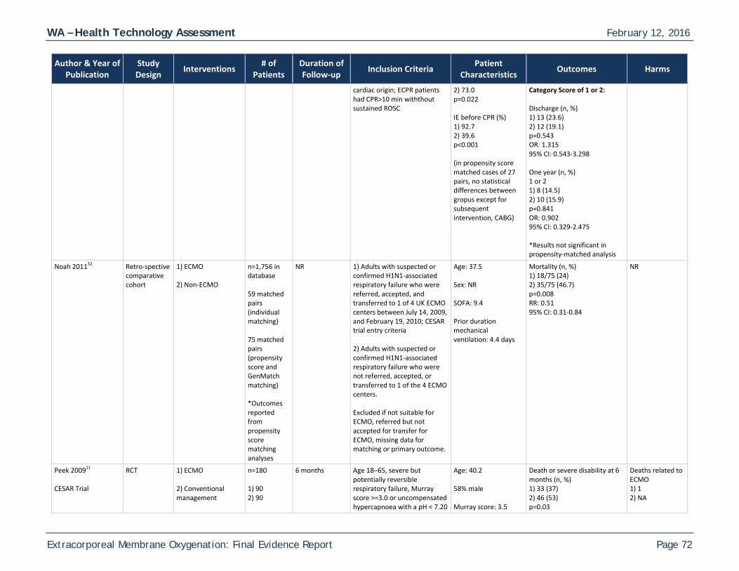
WA – Health Technology Assessment February 12, 2016
Extracorporeal Membrane Oxygenation: Final Evidence Report Page 72
Author & Year of Publication
Study Design Interventions # of
Patients Duration of Follow-up Inclusion Criteria Patient
Characteristics Outcomes Harms
cardiac origin; ECPR patients had CPR>10 min withthout sustained ROSC
2) 73.0 p=0.022 IE before CPR (%) 1) 92.7 2) 39.6 p<0.001 (in propensity score matched cases of 27 pairs, no statistical differences between gropus except for subsequent intervention, CABG)
Category Score of 1 or 2: Discharge (n, %) 1) 13 (23.6) 2) 12 (19.1) p=0.543 OR: 1.315 95% CI: 0.543-3.298 One year (n, %) 1 or 2 1) 8 (14.5) 2) 10 (15.9) p=0.841 OR: 0.902 95% CI: 0.329-2.475 *Results not significant in propensity-matched analysis
Noah 201132 Retro-spective comparative cohort
1) ECMO 2) Non-ECMO
n=1,756 in database 59 matched pairs (individual matching) 75 matched pairs (propensity score and GenMatch matching) *Outcomes reported from propensity score matching analyses
NR 1) Adults with suspected or confirmed H1N1-associated respiratory failure who were referred, accepted, and transferred to 1 of 4 UK ECMO centers between July 14, 2009, and February 19, 2010; CESAR trial entry criteria 2) Adults with suspected or confirmed H1N1-associated respiratory failure who were not referred, accepted, or transferred to 1 of the 4 ECMO centers. Excluded if not suitable for ECMO, referred but not accepted for transfer for ECMO, missing data for matching or primary outcome.
Age: 37.5 Sex: NR SOFA: 9.4 Prior duration mechanical ventilation: 4.4 days
Mortality (n, %) 1) 18/75 (24) 2) 35/75 (46.7) p=0.008 RR: 0.51 95% CI: 0.31-0.84
NR
Peek 200911 CESAR Trial
RCT 1) ECMO 2) Conventional management
n=180 1) 90 2) 90
6 months Age 18–65, severe but potentially reversible respiratory failure, Murray score >=3.0 or uncompensated hypercapnoea with a pH < 7.20
Age: 40.2 58% male Murray score: 3.5
Death or severe disability at 6 months (n, %) 1) 33 (37) 2) 46 (53) p=0.03
Deaths related to ECMO 1) 1 2) NA

WA – Health Technology Assessment February 12, 2016
Extracorporeal Membrane Oxygenation: Final Evidence Report Page 73
Author & Year of Publication
Study Design Interventions # of
Patients Duration of Follow-up Inclusion Criteria Patient
Characteristics Outcomes Harms
despite optimum conventional treatment Excluded if on high pressure or high FiO2 ventilation >7 days, contraindication to limited heparinization or continuation of active treatment
APACHE II: 19.8
RR: 0.69 95% CI: 0.05-0.97 Died 6 mos or before discharge 1) 33 2) 45 p=0.07 RR: 0.73 95% CI: 0.52-1.03 Days between randomization and death 1) 15 2) 5 p=NR Length of Stay ICU, hospital days (median) 1) 24, 35 1) 13, 17 p=NR VAS Overall 1) 67.9 2) 65.9 p=NR Upper limb movement restriction: 1) 3% 2) 6% p=NR
Pham 201335 Retro-spective comparative cohort
1) ECMO 2) Non-ECMO
n=104 (propensity score matched) 1) 52 2) 52
NR Patients infected with influenza A(H1N1)pdm with ARDS, admitted to 114 participating French ICUs between July 2009 to March 2010, and recorded in web-based registry (REVA-SRLFH1N1)
Age: 45 57% male SOFA score: 9.6 SAPS3 score: 59
Mortality (n, %) 1) 26 (50) 2) 21 (40) P=NS Length of ICU stay (days) 1) 27 2) 19.5 p=0.04 Median duration of ECMO (days): 11 Median duration of intubation: 28
Deaths in ICU Multiorgan failure: 22 Refractory hypoxemia: 8 Refractory shock: 6 Intracranial hemorrhage: 5 Unspecified: 3

WA – Health Technology Assessment February 12, 2016
Extracorporeal Membrane Oxygenation: Final Evidence Report Page 74
Author & Year of Publication
Study Design Interventions # of
Patients Duration of Follow-up Inclusion Criteria Patient
Characteristics Outcomes Harms
Patients with one or more ECMO-related complication: 65
Sakamoto 201447 Prospective comparative cohort
1) ECPR: percutaneous cardiopulmonary support 2) non-eCPR: conventional CPR
n=454 1) eCPR n=260 1) non-eCPR n=194
NR Patients with out-of-hospital cardiac arrest (OHCA) of cardiac origin, with core body temp >30 degC; VF/VT on initial ECG; cardiac arrest on hospital arrival within 45 minutes from reception of emergency call or onset of cardiac arrest to hospital arrival; no restoration of spontaneous circulation (ROSC) during the 15 minutes after hospital arrival despite conventional CPR; age 20-75
Age 1) 56.3 2) 58.1 p=NS Male 1) 90.4% 2) 88.7% p=NS Time from 911 call to hospital arrival: 1) 29.8 minutes 2) 30.5 minutes p=NS Acute coronary syndrome: NS 1) 63.5% 2) 59.3% p=NS
Favorable CPC (Glasgow-Pittsburgh Cerebral Performance and Overall Performance Categories) scores 1 or 2 at 1 month: 1) 32/260 2) 3/194 p<0.0001
Favorable CPC scores 1 or 2 at 6 months: 1) 32/260 2) 3/194 p=0.001
Survival at 24 hours: 1) 177/260 (68.1%) 2) 37/194 (19.1%) p=NR
NR
Shin 201148 Retro-spective comparative cohort
1) ECPR: 2) Conventional CPR
n=406 1) n=85 2) n=321
2 years (See Shin 2013)
Patients with witnessed in-hospital cardiac arrests at Samsung Medical Center; ages 18-80
Age 1) 59.9 (SD 15.3) 2) 61.6 (14.2) p=NS Male 1) 53 (62.4%) 2) 201 (62.6%) p=NS Diabetes 1) 17 (20.2%) 2) 98 (30.5%) p=0.055 Chronic renal disease 1) 5 (5.9%) 2) 48 (15.0%) p=0.027 Primary disease,
(Propensity score-matched outcome analysis of 60 patients in each group) CPC score <=2 at discharge 1)14 (23.3) 2) 3 (5.0) p=0.013 28 day survival: 1) 19 (31.7) 2) 6 (10.0) p=0.011 CPC score <=2 at 6 months: 1)14 (23.3) 2) 3 (5.0) p=0.013 6-month survival: 1) 16 (26.7)
Bleeding and hematoma of insertion sites; vascular injury, catheter infection, limb ischemia, gastrointestinal bleeding, hemolysis, and stroke (rates not reported)

WA – Health Technology Assessment February 12, 2016
Extracorporeal Membrane Oxygenation: Final Evidence Report Page 75
Author & Year of Publication
Study Design Interventions # of
Patients Duration of Follow-up Inclusion Criteria Patient
Characteristics Outcomes Harms
cardiac: p=0.004 1) 63 (74.1%) 2) 182 (56.7%) Cause of arrest, cardiac: p=0.010 1) 79 (92.9) 2) 261 (81.3) Pre-arrest SOFA: NS 1) 6.3 (3.5 SD) 2) 5.9 (3.6 SD) Deyo-Charlson score: NS 1) 2.1 (2.3 SD) 2) 2.1 (1.9 SD)
2) 5 (8.3) p=0.019 HR for eCPR: 0.52 95% CI: 0.35-0.79
Shin 201349 (Follow-up to Shin 2011)
See Shin 2011 1) ECPR: 2) Conventional CPR
See Shin 2011
See Shin 2011 See Shin 2011 See Shin 2011 1-year survival: 1) 13 (21.6) 2) 5 (8.3) p=0.019 HR for eCPR: 0.55 95% CI: 0.37-0.83 2-year survival: 1) 12 (20.0) 2) 5 (8.3) p=0.019 HR for eCPR: 0.56 95% CI: 0.37-0.84 2-year survival with minimal neurologic impairment (per Modified Glasgow Outcome Score [MGOS]>=4): 1) 12 (20.0) 2) 3 (5.0) p=0.002 HR for eCPR : 0.53 95% CI: 0.36-0.80 Death at 2 years with documented hypoxic brain damage 1) 5 (8.3) 2) 6 (10.0) p=NS
See Shin 2011

WA – Health Technology Assessment February 12, 2016
Extracorporeal Membrane Oxygenation: Final Evidence Report Page 76
Author & Year of Publication
Study Design Interventions # of
Patients Duration of Follow-up Inclusion Criteria Patient
Characteristics Outcomes Harms
HR for eCPR : 0.42 95% CI: 0.13-1.41
Tsai 201536 Retro-spective case control
1) ECMO 2) Non-ECMO
n=90 1) 45 2) 45
Up to 6 months Inclusion: The medical records of all patients with ARDS admitted to the ICU from January 2007 to December 2012 were reviewed. ECMO and non-ECMO patients were matched. Patients were paired when the difference in their APACHE II scores was within 3 points and their age difference was 3 years. Exclusion: 126 patients who could not be matched
Age mean (SD) 56 (2.4) both groups Sex Male n (%) 1) 32 (71) 2) 34 (75) APACHE II score mean (SD) 25 (1.1) both groups SOFA mean (SD) 1) 11.9 (0.5) 2) 10.2 (0.8) RIFLE 1) 1.2 (0.2) 2) 1.0 (0.2)
Mortality n (%) 1) 22 (48.9) 2) 34 (75.6) p=0.009
NR

WA – Health Technology Assessment February 12, 2016
Extracorporeal Membrane Oxygenation: Final Evidence Report Page 77
Table 8: Summary Evidence Table of Fair Quality Studies
Author & Year of Publication Study Design Interventions # of
Patients Duration of Follow-up Inclusion Criteria Patient
Characteristics Outcomes Harms
Aigner 200796 Retrospective comparative cohort
1) ECMO used peri-operatively 2) No ECMO used peri-operatively (but some CPB)
n=306 1) 147 2) 149
3 years Lung transplantation Age: 1) 42 +/- 16 2) 49 +/- 13 p<0.01 Sex, % male: 1) 55% 2) 58% p=0.55 Mean waiting list days: 1) 87 +/- 86 2) 96 +/- 84 p=0.45 Lobar/split lung transplant: 1) 39 2) 10 p<0.001
ICU days, n (range): 1) ECMO: Intraoperative ECMO 11.5 (1-137) Prolonged ECMO 12 (1-55) 2) Non-ECMO: No support 5.5 (1-55) p=0.06 CPB 23.5 (10-87) p<0.01 Hospital days, n (range): 1) Intraoperative ECMO 25.5 (1-173) Prolonged ECMO 26 (1-100) 2) No support 23 (8-124) p=0.13 CPB 51 (26-87) p=0.02 3-month survival: 1) 85.4% 2) No support 93.5%, CPB 74.0% 1-year survival: 1) 74.2% 2) No support 91.9%, CPB 65.9% 3-year survival: 1) 67.6% 2) No support 86.5%, CPB 57.7% Survival ECMO vs. no support p<0.001 Survival ECMO vs. CPB p=0.41
Bleeding: 1) 31 2) 11 p=0.001 25 other complications (vascular complications, thromboses, circuit problems, cerebral bleeding, lymphatic fistulae.
Barge-Caballero 201497
Prospective comparative cohort
1) VA-ECMO 2) VAD: Pulsatile VAD and Continuous-flow VAD 3) Control Mechanical circulatory support (MCS) = VA-ECMO+VAD
n=101 1) 23 2) VAD (78) Pulsatile VAD (53) Continuous-flow VAD (25) 3) 568
Post-transpant follow-up, median (IQR) 2.9 (0.2-5) years
Underwent emergency heart transplant in 15 Spanish hospitals between 2000 and 2009
Age, mean (SD) 1) 54.1 (10) 2) 46.2 (13) 3) Control 50.9 (12) % female 1) MCS 38% 2) Control 16% Preoperative INTERMACS Status (% of status 1/2/3/4) 1) MCS 39/50/11/1 2) Control 28/39/28/5
Post-transplant mortality (vs. control) 1) VA-ECMO HR 0.51 95% CI 0.21-1.25 p=NR 2) Pulsatile VAD HR 2.21 95% CI: 1.48-3.30 3) Continuous-flow VAD HR 2.24 95% CI: 1.20-4.19 Mean cold ischemic times, min, mean (SD) 1) VA-ECMO 194 (57) 2) VAD 226 (57) p=0.022
Primary graft failure, % 1) MCS 36.6% 2) Control 21.5% p=0.042 Major surgical bleeding, % 1) MCS 30.7% 2) Control 21.5% p=0.042 Need for cardiac reoperation, % 1) MCS 21.8%

WA – Health Technology Assessment February 12, 2016
Extracorporeal Membrane Oxygenation: Final Evidence Report Page 78
Author & Year of Publication Study Design Interventions # of
Patients Duration of Follow-up Inclusion Criteria Patient
Characteristics Outcomes Harms
Mean bypass time, min, mean (SD) 1) VA-ECMO 139 (43) 2) VAD 169 (79) p=0.031
2) Control 13.2% p=0.024 Postoperative infection, % 1) MCS 50.5% 2) 38.6% p=0.024
Biscotti 201498 Prospective comparative cohort
1) Intraoperative ECMO 2) Intraoperative CPB
n=102 1) 47 2) 55
1 year Received lung transplant at study center between January 1, 2008, and July 13, 2013 and required intraoperative cardiopulmonary support
Age, yr, mean (SD) 1) 50.8 (14.9) 2) 46.9 (15.9) Sex, female, n (%) 1) 22 (46.8) 2) 28 (50.9) LAS (lung allocation score), mean (SD) 1) 62.0 (22.8) 2) 61.9 (20.0)
Postoperative ECMO n (%) 1) 5 (10.6%) 2) 3 (5.5%) p= 0.465 Secondary ECMO n (%) 1) 4 (8.5%) 2) 4 (7.3%) p>0.999 ICU stay, d, n (%) 1) 10.4 (8.4) 2) 13.0 (13.1) p= 0.25 30-d survival n (%) 1) 44 (93.6%) 2) 53 (96.4%) p= 0.66 FEV1 (% predicted), mean (SD) 1) 52.5 (15.2) 2) 57.0 (19.3) p= 0.22 Any PGD at 24 h n (%) 1) 23 (48.9) 2) 41 (74.5) p= 0.008 Any PGD at 72 h n (%) 1) 26 (56.5) 2) 42 (76.4) p= 0.034
CVA n (%) 1) 3 (6.4) 2) 2 (3.6) p=0.66 Hemodialysis n (%) 1) 4 (8.5) 2) 8 (14.5) p= 0.346 Tracheostomy n (%) 1) 10 (21.3) 2) 18 (32.7) p= 0.196 Reoperation n (%) 1) 7 (14.9) 2) 21 (38.2) p= 0.009 Vascular complications n (%) 1 3 (6.4) 2) 2 (3.6) p= 0.66 Bleeding n (%) 1) 3 (6.,4) 2) 15 (27.3) p= 0.006 CPR or cardiac arrest n (%) 1) 3 (6.4) 2) 7 (12.7)

WA – Health Technology Assessment February 12, 2016
Extracorporeal Membrane Oxygenation: Final Evidence Report Page 79
Author & Year of Publication Study Design Interventions # of
Patients Duration of Follow-up Inclusion Criteria Patient
Characteristics Outcomes Harms
p= 0.335
Hayanga 201599 Retrospective comparative cohort
1) ECMO 2) Non-ECMO
n=12,458 1) 119 2) 12,339
1 year Consecutive U.S. adult lung transplant recipients who underwent transplantation between January 2000 and December 2011 with data registered in Scientific Registry of Transplant Recipints Standard Transplant Analysis and Research
Age 1) 51 (34-60) 2) 57 (48-63) p<0.001 % male 1) 62.2 2) 56.8
1-year survival (%) 2000-2002 1) 25 2) 81 ECMO mortality HR: 7.15 95% CI: 2.23-22.89) p=0.001 2003-2005 1) 76.5 2) 84.5 ECMO mortality HR: 1.62 95% CI: 0.61-4.35) p=0.34 2006-2008 1) 47.1 2) 84.2 ECMO mortality HR: 6.24 95% CI: 3.77-10.33) p<0.001 2009-2011 1) 74.4 2) 85.7 ECMO mortality HR: 1.96 95% CI: 1.20-3.21) p=0.007
NR
Hayes 2015100 Retrospective comparative cohort
1) ECMO 2) Non-ECMO
n=17,556 1) 198 2) 17,358
NR Inclusion: All first-time adult (>=18 years) lung transplants from January 1 , 2000 to September 6, 2013 registered in the Organ Procurement and Transplant Network Standard Transplant Analysis and Research database Exclusion: Missing or duplicated data entries, non-cadaveric donor, unmatched controls
Age 1) 47.34 2) 53.63 p<0.001 % male 1) 60.61 2) 56.65 p=NS Year of transplant 1) 2010 2) 2007 p<0.001
Multivariate survival on ECMO (n=15,553) HR: 1.845 95% CI: 1.450-2.347 p<0.001 1:5 matching of 1,005 patients ECMO mortality HR: 2.010 95% CI: 1.47-2.748 p<0.001 Propensity score matched pairs of 364 patients ECMO mortality HR: 2.5 95% CI: 1.525-4.099 p<0.001
NR

WA – Health Technology Assessment February 12, 2016
Extracorporeal Membrane Oxygenation: Final Evidence Report Page 80
Author & Year of Publication Study Design Interventions # of
Patients Duration of Follow-up Inclusion Criteria Patient
Characteristics Outcomes Harms
Lamarche 2011101 Retro-spective comparative cohort
1) VA-ECMO 2) VADs
1) 32 2) 29
30 days Patients with postcardiotomy shock deemed to have potential for recovery (e.g., no multiorgan dysfunction)
Male (% 1) 62.5 2) 82.8 p=0.08 Age mean (SD) 1) 50.4 (14.2) 2) 53.7 (13.1) p=0.35 Idiopathic dilated cardiomyopathy %* 1) 3.1 2) 24.1 Postcardiotomy %* 1) 43.8 2) 13.8 *causes of shock p=0.008
30-day mortality n (%) 1) 14 (43.8) 2) 11 (37.9) p=0.16
PRBCs median (IQR) 1) 18.0 (9-34) 2) 4 (2-9) p<0.001
Ried 2013102 Retrospective comparative cohort
1) pECLA 2) Miniaturized VV-ECMO
n=52 1) 26 2) 26
NR (until discharge)
Trauma with acute lung failure (ALF) defined by partial pressure of arterial oxygen (PaO2)/fraction of inspired oxygen (FiO2) ration <80 mmHg, a maximum positive end-expiratory pressure (18cmH2O) and persistent respiratory acidosis (ph<7.25) despite optimized mechanical ventilation and optimization of conservative treatment options.
Male n (%) 1) 25 (96) 2) 24 (92) Age mean (SD) 1) 34.5 (14.3) 2) 29.3 (13.2) BMI mean (SD) 1) 26.7 (4.5) 2) 29.6 (7.3) Prior resuscitation n (%) 1) 4 (15.4) 2) 4 (15.4) Injury Severity Score mean (SD) 1) 57.8 (10.9) 2) 59.4 (11.2) Lung Injury Score mean (SD) 1) 3.06 (0.65) 2) 3.53 (0.36)
ECMO duration (days) 1) 7.6 (SD 4) 2) 6.3 (SD 3.1) p=ns
ICU stay (days) 1) 23 (range 18-25) 2) 17 (range 13-30) p=NR
Hospital stay (days) 1) 25 (21-39) 2) 24 (13-44) p=NR In-hospital mortality n (%) 1) 6 (23.1%) 2) 5 (19.2%) p=NR Death on ECMO n (%) 1) 6 (15.4%) 2) 6 (15.4%) p=NR
Cannula-related complications 1) 19% 2) 12%

WA – Health Technology Assessment February 12, 2016
Extracorporeal Membrane Oxygenation: Final Evidence Report Page 81
Author & Year of Publication Study Design Interventions # of
Patients Duration of Follow-up Inclusion Criteria Patient
Characteristics Outcomes Harms
SOFA score mean (SD) 1) 9.2 (3) 2) 11.8 (2.4)
Taghavi 2014c103 Retrospective comparative cohort
1) Left ventricular assist device (LVAD) 2) ECMO
n=40 1) 29 2) 11
NR All patients who received mechanical circulatory support and noncardiac surgery at Temple University Hospital from January 2002 to December 2012; noncardiac surgical procedures included abdominal exploration/bowel resection, tracheostomy, extremity/vascular surgery, urological procedure, gynecological surgery, oral surgery, and other surgery
Age mean (SD) LVAD 53.6 (14.3) ECMO Sex male n (%) LVAD 20 (71.4) ECMO
Postoperative outcomes mean (SD) total length of stay (d) 1) 37.0 ± 33.6 2) 30.1 ± 42.2 p=0.52 ICU stay (hr) 1) 28.7 ± 33.2 2) 24.9 ± 38.8 p=0.71 Requirement of postoperative mechanical ventilation, n (%) 1) 32 (68.1) 2) 15 (100.0) p=0.01 Requirement of postoperative vasopressor support, n (%) 1) 19 (36.2) 2) 9 (19.1) p=0.24 Require blood transfusion w/in 24 hrs of surgery, n (%) 1) 12 (25.5) 2) 11 (73.3) p=0.002 Perioperative mortality, n (%) LVAD 3 (6.4) ECMO 7 (46.7) p=0.001 Univariate regression for survival ECMO HR: 2.90 95% CI 1.46-5.78 p=0.002 Median survival, d LVAD 142.5 ECMO 6.0 p=0.002
NR

WA – Health Technology Assessment February 12, 2016
Extracorporeal Membrane Oxygenation: Final Evidence Report Page 82
Table 9: Summary Evidence Table of Poor Quality Studies
Author & Year of
Publication
Study Design Interventions # of Patients Duration of
Follow-up Inclusion Criteria Patient Characteristics Outcomes Harms
Arlt 2009104 Prospective comparative cohort
1) pECLA 2) VV-ECMO 3) VA-ECMO
n=53 1) 20 2) 20 3) 13
1) 5.3 days 2) 8.2 days 3) 3.5 days
Severe pulmonary and cardiopulmonary failure
Age: 1) 33 2) 41 3) 51 65-85% male
Weaned from ECMO: 1) 65% 2) 70% 3) 85% p=NR Discharged from hospital: 1) 50% 2) 60% 3) 62% p=NR Mortality: 1) 35% 2) 0% 3) 15% P=NR
3) Compartment syndrome: 6 Heparin-induced thrombocytopenia: 1
Beiderlinden 2006105
Prospective comparative cohort
1) Multimodal treatment with ECMO 2) Multimodal treatment without ECMO (conservative)
n=150 1) 32 2) 118 Patients with acute community-acquired pneumonia (CAP): n=47 1) 17 2) 30
NR Inclusion: ARDS; referred to study’s ICU between January 1998 and September 2003; Lung Injury Score (LIS) > 2.5; previously mechanically ventilated in other ICUs; age <70 years, weight >15 kg. Exclusion: Malignancy; end-stage lung disease; intracranial bleeding
Total Age, yr, mean (SD) 1) 42.2 (13) 2) 41.9 (16) Sex NR Lung injury score, mean (SD) 1) 3.8 (0.3) 2) 3.3 (0.4) SAPS score (Simplified Acute Physiology Score), mean (SD) 1) 52 (14) 2) 43 (12) SOFA score (Sepsis-Related Organ Failure Assessment), mean (SD) 1) 14 (3.3) 2) 10 (3.5)
Hospital Mortality Total 1) 46.9% 2) 28.8% p=0.059 Among patients with CAP 1) 29.4% 2) 23.3% p=0.73
NR

WA – Health Technology Assessment February 12, 2016
Extracorporeal Membrane Oxygenation: Final Evidence Report Page 83
Author & Year of
Publication
Study Design Interventions # of Patients Duration of
Follow-up Inclusion Criteria Patient Characteristics Outcomes Harms
Bermudez 2011106
Retrospective comparative cohort
1) Preoperative ECMO 2) Control (no ECMO)
n=1,288 1) 17 2) 1,271
2.3 years (mean) Patients who underwent lung transplant (primary and re-transplantation), while on ECMO support, from March 1991 to October 2010; patients who had lung transplant without the use of preoperative ECMO during the period analyzed served as a control group.
Age, yr, mean (SD) 1) 40 (14) 2) 51 (13) Sex, male, n (%) 1) 7 (41) 2) 659 (51)
30-day survival 1) 81% 2) 93% p=NS 1-yr survival 1) 74% 2) 78% p=NS 3-yr survival 1) 65% 2) 62% p=NS 2-yr survival 1) 5 out of 9 2) 7 out of 8 p=NS
Adverse envents related to ECMO included significant bleeding from the arterial femoral cannulation site requiring intervention in 1 patient and transient encephalopathy of unclear etiology while on ECMO support with spontaneous resolution in 1 patient.
Bermudez 2014107
Retrospective comparative cohort
1) Intraoperative ECMO 2) Intraoperative CPB
n=271 1) ECMO n=49 2) CPB n=222
Up to 1 yr Inclusion: Underwent primary LT Between July 2007 and April 2013 in study institution. Exclusion: Began LTx on ECMO and switched to CPB; Redo LTxs
Age, yr, mean (SD) 1) 50.3 (15.0) 2) 54.4 (14.1) Sex, male, n (%) 1) 27 (55.1) 2) 130 (58.6) lung allocation score 1) 73.3 (22.0) 2) 52.9 (20.2) p<0.001
Mechanical ventilation, total, hr, mean (SD) 1) 250.3 (393.4) 2) 380.2 (654.8) p=0.06 Reintubation, % 1) 20.4 2) 35.6 p=0.04 Temporary tracheostomy 1) 28.6 2) 44.6 p=0.05 ICU LOS (days), mean (SD) 1) 15.1 (20.5) 2) 21.9 (31.3) p=0.06 Hospital LOS (days), mean (SD) 1) 49 (44.3)
Major intraoperative complications n (%) 1) 1 (2) 2) 1 (0.5) Reoperation for bleeding n (%) 1) 4 (8.2) 2) 39 (17) Renal failure requiring dialysis n(%) 1) 4 (8.2) 2) 49 (22.1) p=0.03 Postoperative ECMO (severe PGD) n (%) 1) 9 (18.3) 2) 34 (15.3) p= 0.83 30-d mortality, n (%) 1) 2 (4.1) 2) 11 (5) p=1.00 6-mo mortality, n(%)

WA – Health Technology Assessment February 12, 2016
Extracorporeal Membrane Oxygenation: Final Evidence Report Page 84
Author & Year of
Publication
Study Design Interventions # of Patients Duration of
Follow-up Inclusion Criteria Patient Characteristics Outcomes Harms
2) 52 (47.2) p=0.55
1) 7 (14.3) 2) 32 (14.4) p=1.00 1-yr mortality, n(%) 1) 9 (19.1) 2) 42 (18.9) p=NR
Chestovich 2011108
Retrospective comparative cohort
1) ECMO 2) ventricular assist devices (VAD)
n=69 1) 14 2) 55
NR All adult patients who received mechanical cardiac support and underwent a noncardiac surgical procedure (NCP) during a 12-year periord from July 1998 through June 2010 at UCLA.
Age, yr, mean (range) 1) 50.6 (22-74) 2) 51.2 (18-74) Sex, male, n (%) 1) 7 (50) 2) 40 (73)
30-d mortality n (%) 1) ECMO 12 (86) 2) VAD 14 (25) p<0.001
NR
Dahlberg 2004109
comparative cohort
1) ECMO 2) overall group, including ECMO and non-ECMO
n=171 1) 15 2) 156
up to 2 years All patients who underwent lung transplantation from January 1997 to December 2002; ECMO support was initiated in 15 patients with persistent pulmonary allograft failure (PGF).
Age, yr, mean (SD) 1) 43 (14) 2) 50 (12) Sex, male, % 1) 33 2) 43
Hospital stay (days), median 1) 48 2) 16 p<0.05 90-day mortality, n (%) 1) 6 (40%) 2) 21 (12%) p=NR 2-yr survival 1) 46% 2) 70% p=NR FEV1 at 2 month, mean (SD) 1) 55 (12) 2) 60 (21) p=NR FEV1 at 1 year, mean (SD) 1) 63 (11) 2) 74 (17) p=NR FEV1 at 2 year, mean (SD)
Bacterial or fungal sepsis, % 1) 33% 2) 3% p< 0.001 Renal failure requiring dialysis % 1) 33% 2) 4.5% p<0.001 multisystem organ failure % 1) 13% 2) 2% p=0.053

WA – Health Technology Assessment February 12, 2016
Extracorporeal Membrane Oxygenation: Final Evidence Report Page 85
Author & Year of
Publication
Study Design Interventions # of Patients Duration of
Follow-up Inclusion Criteria Patient Characteristics Outcomes Harms
1) 63 (12) 2) 68 (28) p=NR
Davies 200912 Retrospective comparative cohort
1) ECMO 2) Mechanical ventilation
n= 194 1) 61 2) 133
NR Inclusion: Adult and pediatric patients treated with ECMO or mechanical ventilation between June 1 and August 31, 2009 in Australia and New Zealand with confirmed or strongly suspected cases of 2009 influenza A(H1N1)–related respiratory disease. Exclusion: Neonates; patients treated with ECMO for primary cardiac failure; patients with an alternative diagnosis and who had no virus isolated;
Age, yr, median (IQR) 1) 36 (27-45) 2) 44 (31-54) Sex, male, n (%) 1) 29 (48) 2) 63 (47) APACHE III comorbidity, n (%) has at least one comorbidity 1) 5 (8) 2) 30 (23)
Mechanical LOS (days), median (IQR) 1) 19 (9-27) 2) 8 (4-14) p=0.001 ICU LOS, (days), median (IQR) 1) 22 (13-32) 2) 12 (7-18) p=0.001 Hospital LOS (days), Median (IQR) 1) 28 (15-43) 2) 20 (13-31) p=0.07 Mortality in ICU, n (%) 1) 14 (23) 2) 12 (9) p=0.01 Mortality in hospital, n (%) 1) 14 (23) 2) 17 (13) p=0.06
NR
Ganslmeier 2011110
Retrospective comparative cohort
1) pECLA in patients with respiratory compromise only 2) VV-ECMO 3) VA-ECMO in acute circulatory failure
n=464 1) 196 2) 110 3) 158
NR Supported with extracorporeal life support at study institution between January 2004 and December 2009 (University Medical Center Regensburg, Regensburg, Germany)
Age, yr, mean (SD) 1) 43 (16.0) 2) 51 (14.0) 3) 55 (16.0) Sex, male, n (%) 1) 157 (80.1) 2) 73 (66.4) 3) 110 (69.6)
Survival n (%) 1) 83 (43) 2) 53 (48) 3) 32 (20) Death after explant n (%) 1) 32 (16) 2) 17 (16) 3) 32 (21) Death on system n (%) 1) 81 (41)
Difficulties during cannula insertion: 25 (5.4%) Bleeding after cannulation: 32 (6.9%) Limb ischemia: 15 (3.2%)

WA – Health Technology Assessment February 12, 2016
Extracorporeal Membrane Oxygenation: Final Evidence Report Page 86
Author & Year of
Publication
Study Design Interventions # of Patients Duration of
Follow-up Inclusion Criteria Patient Characteristics Outcomes Harms
2) 40 (36) 3) 93 (59)
George 2012111 comparative cohort
1) ECMO 2) no mechanical support 3) ventilator support
n=2,522 1) 22 2) 1,874 3) 526
Up to 2 years Inclusion: All adults (≥18 years) who underwent LTx from May 2005 to June 2011 Exclusion: Patients undergoing combined heart-lung or multivisceral organ transplantation
Age, yr, mean (SD) 1) 48 (16) 2) 55 (13) 3) 51 (16) Sex, male, n (%) 1) 74 (60.7) 2) 304 (57.8) 3) 1151 (61.4) LAS score, mean (SD) 1) 73.9 (21.4) 2) 64.9 (22.9) 3) 65.4 (14.5)
30-day mortality (vs. no support) ECMO HR=4.38, 95% CI: 2.44-7.87, p<0.001
Ventilatory support HR=1.90, 95% CI: 1.26-2.86, p=0.002 1-year mortality (vs. no support) ECMO HR=3.03, 95% CI: 2.00-4.59, p<0.001 Ventilatory support, HR=1.99, 95% CI: 1.58-2.51, p<0.001 LOS (days), median (IQR) 1) 32 (16.5-60) 2) 17 (11-30) 3) 30 (19-50) p<0.001
Drug-treated infection n (%) 1) 9 (64.3) 2) 64 (69.6) 3) 144 (51.6) p=0.01 Renal replacement therapy n (%) 1) 42 (35.6) 2) 137 (7.4) 3) 72 (13.7) p<0.001 Stroke n (%) 1) 3 (2.6) 2) 41 (2.2) 3) 19 (3.6) p=0.2 Biopsy-proven rejection n (%) 1) 1 (0.8) 2) 31 (1.7) 3) 7 (1.3) p=0.8
Klotz 2007112 Retrospective comparative cohort
1) VAD 2) ECMO 3) ECMO + VAD
n=183 1) 20 2) 150 3) 13
NR Patients implanted with VAD, ECMO, or both with low cardiac output (cardiac index<2.0 L/min despite adequate filling volumes and use of different inotropic agents) Excluded: pediatric patients<18
Age 1) 41.7 2) 65.9 3) 45.9 p<0.001 for (2) vs. (1) and (3) % male 1) 85 2) 69 3) 77 Reoperation (n, %) 1) 4 (20) 2) 23 (15) 3) 5 (38)
Survived/Died 1) 10/10 2) 38/112 3) 7/6 30-day mortality 1) 50% 2) 75% 3) 46% p<0.001 for (2) vs. (1) and (3)
NR

WA – Health Technology Assessment February 12, 2016
Extracorporeal Membrane Oxygenation: Final Evidence Report Page 87
Author & Year of
Publication
Study Design Interventions # of Patients Duration of
Follow-up Inclusion Criteria Patient Characteristics Outcomes Harms
Lebreton 2015113
Retrospective comparative cohort
1) Bridge to bridge (using ECMO as a bridge to longer term mechanical circulatory support) 2) Long-term mechanical circulatory support as first-line therapeutic strategy
1) 49 2) 48
5 years Decompensated heart failure under inotropic support Signs of cardiogenic shock
Age, yr, mean (SD) 1) 54.0 (12.6) 2) 48.3 (12.0) p=0.025 Male n (%) 1) 40 (83.3) 2) 40 (81.6) Chronic ischemic heart failure n (%) 1) 19 (41.7) 2) 8 (16.3) p=0.014 Acute coronary heart failure n (%) 1) 9 (18.8) 2) 19 (38.8) p=0.014
Overall survival at 36 months 51.5% 1) 51% 2) NR p=0.76
NR
Lee HJ 2015114 Retrospective comparative cohort
1) ECMO pretransplantation 2) mechanical ventilation support pretransplantation
1) 12 2) 15
24 months Patients who underwent lung transplantation at research site
Age, yr, median (IQR) 1) 51.5 (35.6-58.7) 2) 48.8 (32.6-58.6) p=0.981 Male n (%) 1) 8 (66.7) 2) 7 (46.7) BMI 1) 21.2 2) 17.9 p=0.047 Days between registration and transplantation (IQR) 1) 16 (8-48) 2) 38 (26-90) p=0.025 PaO2/FiO2 before invasive respiratory support mmHg (IQR) 1) 60.5 (46.4-65.4) 2) 162 (106.2-247.6)
Ventilator weaning days 1) 17 2) 9 p=0.427 ICU LOS (days) 1) 21 2) 17 p=0.256 Hospital LOS (days) 1) 81 2) 47.5 p=0.317 Post-transplantation survival at 24 months 1) 61.1% 2) 66.0% p=0.540
NR

WA – Health Technology Assessment February 12, 2016
Extracorporeal Membrane Oxygenation: Final Evidence Report Page 88
Author & Year of
Publication
Study Design Interventions # of Patients Duration of
Follow-up Inclusion Criteria Patient Characteristics Outcomes Harms
p<0.001
Mols 200019 Prospective comparative cohort
1) VV ECMO 2) Non-ECMO (conventional ventilation, incl. permissive hypercapneia, prone positioning, NO inhalation, hemodynamic support, infection control)
n=245 1) 62 2) 183
NR ARDS patients: "Immediate entry": PaO2<=40mmHg "Fast entry": PaO2/FiO2<=50mmHg, PEEP>=10cmH2O for 2 hrs "Slow entry": FiO2>0.6 fpr several days w/o improvement
Age, yrs 1) 35 +/- 11 2) 43 +/- 17 p=0.001 Ventilation days 1) 10 +/- 7 2) 2 +/- 3 p<0.0001 PaO2/FiO2 (mmHg): 1) 96 +/- 51 2) 126 +/- 46 p<0.0001 Number organ failures 1) 2.1 +/- 1.0 2) 2.0 +/- 1.1 Lung injury score 1) 3.2 +/- 0.4 2) 2.7 +/- 0.6 P<0.0001
1) Survivor (n=34) vs. non-survivor (n=28): 61% survival Age, kidney failure associated w/ non-survivors
ECMO-related complications: 15 Other complications: 7 Surgical interventions: 6
Nguyen 2014115 Retrospective comparative cohort
1) VV or VA ECMO 2) Non-ECMO (inotropic, vasopressors, and intraaortic balloon pump)
n=32 1) 15 2) 17
NR Mechanical ventilated ICU patients with acute refractory respiratory or cardiorespiratory failure following septic shock, cardiogenic shock, or ARDS
Age: 67 78% male APACHE: 75 SOFA: 9
ICU Stay (days) 1) 18 2) 24 p=0.61 ICU Mortality (n, %) 1) 8 (53) 2) 6 (35) p=0.74
Cerebral complications (n, %) 1) 3 (20) 2) 2 (12) p=0.64 Nosocomial infection (n, %) 1) 13 (87) 2) 9 (53) p=0.061
Ohman 2014116 Retrospective comparative cohort
1) Temporary VAD 2) Permanent VAD 3) ECMO
n=208 1) 38 2) 146 3) 24
NR All patients who received temporary and permanent cardiac support devices from 7/1/2010 to /30/2012 at a single institution; ECMO patients were prospectively enrolled in database after 7/1/2011. For patients who had been placed on ECMO between 7/1/2010 and 7/1/2011, a retrospective chart review was undertaken for all study
Age 1) 51.2 2) 51.5 3) 53.5 Male 1) 30/38 2) 119/146
30-day mortality (n, %) Experienced extremity vascular complication (EVC) 1) 8 (80) 2) 2 (15.4) 3) 4 (50
Extremity vascular complication (n) 1) 10 2) 13 3) 8 Amputation (n) 1) 0 2) 1

WA – Health Technology Assessment February 12, 2016
Extracorporeal Membrane Oxygenation: Final Evidence Report Page 89
Author & Year of
Publication
Study Design Interventions # of Patients Duration of
Follow-up Inclusion Criteria Patient Characteristics Outcomes Harms
variables Cardiogenic shock 1) 31 2) 68 3) 4
Did not experience EVC 1) 10 (35.7) 2) 6 (4.5) 3) 11 (68.8) p=NR
3) 1 30-day, 2-year mortality in patients with embolic complication (n, %) 1) 4/5 (80), 4/5 (80) 2) 2/8 (25), 5/8 (62.5) 3) 3/3 (100), 3/3 (100) p=NR 30-day, 2-year mortality in patients with cannulation complication (n, %) 1) 4/5 (80), 4/5 (80) 2) 0/5 (0), 2/5 (40) 3) 1/5 (20), 2/5 (40)
Taghavi 2003117 Prospective comparative cohort
1) Right ventricular assist device (RVAD) 2) ECMO
n=25 1) 15 2) 10
NR Htx with acute graft failure, where neither long reperfusion time nor maximal drug therapy (inotropics and vasodilators) facilitated weaning from CPB
Age mean (SD) 55 (12.8) Sex (M/F) 1) 15/0 2) 9/1 Pulmonary vascular wedge pressure (PCWP) 1) 22.6 (7.96) 2) 29.8 (10.83) Left ventricular ejection fraction (%) 1) 21.3 (11.13) 2) 15.13 (3.29)
Mortality (n) 1) 6/10 2) 5/15 Duration of device mean (stab) 1) 86.1 (63.62) 2) 123.2 (71.29) Weaned (n) 1) 7/10 2) 2/15 P=NR
NR
Toyoda 2013118 Retrospective comparative cohort
1) Pretransplant ECMO 2) Control (lung transplantation without pretransplant ECMO)
n=715 1) 24 2) 691
2 years Consecutive patients who underwent lung transplant (primary and retransplantation) from May 2005 to September 2011). Data obtained from the University of Pittsburgh Medical Center transplant database and patient charts ECMO used in patients with advanced
Age 1) 46 2) 57 p<0.01 % male 1) 42 2) 55 p=NS
Median hospital stay (days) 1) 46 2) 27 p=0.16 Deaths during ECMO as bridge to transplant: 7
NR

WA – Health Technology Assessment February 12, 2016
Extracorporeal Membrane Oxygenation: Final Evidence Report Page 90
Author & Year of
Publication
Study Design Interventions # of Patients Duration of
Follow-up Inclusion Criteria Patient Characteristics Outcomes Harms
cardiopulmonary failure unresponsive to maximal medical therapy, and/or who presented a rapid deterioration of a chronic lung disease
Single/double transplant (%) 1) 0/100 2) 25/75 p<0.01 Post-transplant ECMO (%) 1) 54 2) 6 p<0.01 LAS 1) 87 2) 44 p<0.01
30 day mortality (n,%) 1) 1 (4) 2) 24 (3) p=NR 90 day mortality (n, %) 1) 3 (13) 2) 43 (6) p=NR Discharged (n, %) 1) 20 (83) 2) 649 (91) Actuarial survival after transplantation at months 1, 3, 6, 12, and 24 did not statistically differ between groups

WA – Health Technology Assessment February 12, 2016
Extracorporeal Membrane Oxygenation: Final Evidence Report Page 91
Appendix D: ICER Integrated Evidence Ratings Formulary decisions require a rigorous evaluation of available evidence, a process that entails judgments regarding the quality of individual clinical studies and, ultimately, an assessment of the entire body of evidence regarding a therapeutic agent. To support this latter step, the Institute for Clinical and Economic Review (ICER) has developed the ICER Evidence Rating Matrix™. This user’s guide to the ICER Matrix was developed with funding provided by the Comparative Effectiveness Research Collaborative Initiative (CER-CI), a joint initiative of the Academy of Managed Care Pharmacy, the International Society of Pharmacoeconomics and Outcomes Research, and the National Pharmaceutical Council (http://www.npcnow.org/issue/cer-collaborative-initiative). The ICER Matrix presents a framework for evaluating the comparative benefits and risks of therapies in a consistent, transparent system leading to an evidence rating that can guide coverage and formulary placement decisions. The purpose of this user’s guide is to help members of Pharmacy and Therapeutics Committees and other decision-makers understand the approach embodied in the matrix, and to help them apply it in a reliable, consistent fashion.
The updated ICER Evidence Rating Matrix is shown below, with a key to the single letter ratings on the following page. Fundamentally, the evidence rating reflects a joint judgment of two critical components:
a. The magnitude of the difference between a therapeutic agent and its comparator in “net health benefit” – the balance between clinical benefits and risks and/or adverse effects (horizontal axis); AND
b. The level of certainty that you have in your best point estimate of net health benefit (vertical axis).
Negative Comparable Small Substantial Net Benefit Net Benefit Net Benefit Net Benefit
High Certainty
Moderate Certainty
Low Certainty
ABCD
I
IP/I
C+
B+

WA – Health Technology Assessment February 12, 2016
Extracorporeal Membrane Oxygenation: Final Evidence Report Page 92
The letter ratings are listed below, according to the level of certainty in the best estimate of net health benefit.
High Certainty A = Superior B = Incremental C = Comparable D = Inferior
Moderate Certainty
B+=Incremental or Better C+=Comparable or Better P/I = Promising but Inconclusive I = Insufficient
Low Certainty
I = Insufficient
Steps in Applying the ICER Evidence Rating Matrix
1. Establish the specific focus of the comparison to be made and the scope of evidence you will be considering. This process is sometimes referred to as determining the “PICO” – the Population, Intervention, Comparator(s), and Outcomes of interest. Depending on the comparison, it is often helpful to also define the specific Time Horizon and Setting that will be considered relevant.
2. Estimate the magnitude of the comparative net health benefit. Working from the scope of evidence established, it is important to quantify findings from the body of evidence on specific clinical benefits, risks, and other potentially important outcomes, such as adherence, so you can compare these side-by-side for the therapeutic agent and comparator. Some organizations compare each outcome, risk, etc. separately without using a quantitative measure to try to sum the overall comparative balance of benefits and risks between the therapeutic agent and the comparator. For these organizations the estimate of comparative net health benefit must be made qualitatively. Other organizations summarize the balance of benefits and risks using formal mathematical approaches such as health utility analysis, which generates a quantitative summary measure known as the quality-adjusted life year (QALY). What is most important, however, is full and transparent documentation of your rationale for assigning the magnitude of comparative net health benefit into one of four possible categories:
• Negative: the drug produces a net health benefit inferior to that of the comparator • Comparable: the drug produces a net health benefit comparable to that of the
comparator • Small: the drug produces a small positive net health benefit relative to the comparator • Substantial: the drug produces a substantial (moderate-large) positive net health
benefit relative to the comparator

WA – Health Technology Assessment February 12, 2016
Extracorporeal Membrane Oxygenation: Final Evidence Report Page 93
3. Assign a level of certainty to the estimate of comparative net health benefit. Given the strength of the evidence on comparative benefits and risks, a “conceptual confidence interval” around the original estimate of comparative net health benefit can be made, leading you to an assignment of the overall level of certainty in that estimate. Rather than assigning certainty by using a fixed equation weighting different attributes of the body of evidence, we recommend formal documentation of the consideration of 5 major domains related to strength of evidence: (1) Level of Bias—how much risk of bias is there in the study designs that comprise the entire evidence base? (2) Applicability—how generalizable are the results to real-world populations and conditions? (3) Consistency—do the studies produce similar treatment effects, or do they conflict in some ways? (4) Directness—are direct or indirect comparisons of therapies available, and/or are direct patient outcomes measured or only surrogate outcomes, and if surrogate outcomes only, how validated are these measures? (5) Precision—does the overall database include enough robust data to provide precise estimates of benefits and harms, or are estimates/confidence intervals quite broad? If you believe that your “conceptual confidence interval” around the point estimate of comparative net health benefit is limited to the boundaries of one of the four categories of comparative net health benefit above, your level of certainty is “high.” “Moderate” certainty reflects conceptual confidence interval s extending across two or three categories, and may include drugs for which your conceptual confidence interval includes a small likelihood of a negative comparative net health benefit. When the evidence cannot provide enough certainty to limit your conceptual confidence interval within two to three categories of comparative net health benefit, then you have “low” certainty.
4. Assign a joint rating in the Evidence Rating Matrix. The final step is the assignment of the joint rating of magnitude of comparative net health benefit and level of certainty. As shown again in the figure on the following page, when your certainty is “high,” the estimate of net benefit is relatively assured, and so there are distinct labels available: a rating of A indicates a high certainty of a substantial comparative net benefit. As the magnitude of comparative net health benefit decreases, the rating moves accordingly, to B (incremental), C (comparable), and finally D, indicating an inferior or negative comparative net health benefit for the therapeutic agent relative to the comparator. When the level of certainty in the point estimate is only “moderate,” the summary ratings differ based on the location of the point estimate and the ends of the boundaries of the conceptual confidence interval for comparative net health benefit. The ratings associated with moderate certainty include B+ (incremental or better), which indicates a point estimate of small or substantial net health benefit and a conceptual confidence interval whose lower end does not extend into the comparable range. The rating C+ (comparable or better) reflects a point estimate of either comparable, small, or substantial net health benefit and a lower bound of the conceptual confidence interval that does not extend into the inferior range. These ratings may be particularly useful for new drugs that have been tested using noninferiority trial designs, or those involving modifications to an existing agent to provide adherence or safety advantages. Another summary rating reflecting moderate certainty is P/I (promising but inconclusive). This rating is used to describe an agent with evidence suggesting that it provides a comparable, small, or substantial net benefit over the comparator. However, in contrast to ratings B+ and C+, P/I is the rating given when the conceptual confidence interval includes a small likelihood that the comparative net health benefit might actually be negative. In our experience the P/I rating is a common rating when assessing the evidence on novel agents that have received regulatory approval
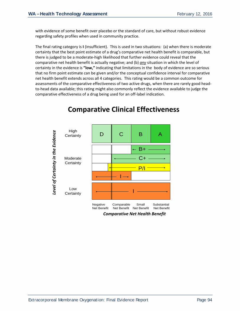
WA – Health Technology Assessment February 12, 2016
Extracorporeal Membrane Oxygenation: Final Evidence Report Page 94
with evidence of some benefit over placebo or the standard of care, but without robust evidence regarding safety profiles when used in community practice. The final rating category is I (insufficient). This is used in two situations: (a) when there is moderate certainty that the best point estimate of a drug’s comparative net health benefit is comparable, but there is judged to be a moderate-high likelihood that further evidence could reveal that the comparative net health benefit is actually negative; and (b) any situation in which the level of certainty in the evidence is ”low,” indicating that limitations in the body of evidence are so serious that no firm point estimate can be given and/or the conceptual confidence interval for comparative net health benefit extends across all 4 categories. This rating would be a common outcome for assessments of the comparative effectiveness of two active drugs, when there are rarely good head-to-head data available; this rating might also commonly reflect the evidence available to judge the comparative effectiveness of a drug being used for an off-label indication.
Comparative Clinical Effectiveness
Negative Comparable Small Substantial Net Benefit Net Benefit Net Benefit Net Benefit
High Certainty
Moderate Certainty
Low Certainty
ABCD
I
IP/I
C+
B+
Comparative Net Health Benefit
Leve
l of C
erta
inty
in th
e Ev
iden
ce