extracellular matrix: composition and...
TRANSCRIPT
1
Extracellular matrix: Composition and Remodelling Specific processes: Tumor metastasis, Wound healing, Liver fibrosis, Angiogenesis
Introduction The extracellular matrix is a network of proteins and carbohydrates that supports and
surrounds the cells in connective tissues. In addition to their structural-mechanical
functions ECM molecules regulate diverse cellular functions, and play a significant role
in several physiological and pathological processes, such as embryonic development,
inflammation and wound healing, hemostasis, fibrosis of various organs, tumor growth
and metastasis, and angiogenesis.
The interaction of ECM molecules with the cells is conducted through cell-matrix
receptors including integrins (see. the seminar material on Cell adhesion).
The extracellular matrix is not static: it is remodelled constantly, which implies its
constant breakdown by proteases, notably matrix metalloproteinases.
During the seminar, we will discuss this topic according to the following scheme:
1. Review the components of the extracellular matrix
2. Introduce the family of matrix metalloproteinases
3. Discuss selected figures from original papers on the role of MMPs in tumor metastasis
and/or present a review on the role of MMPs in tumor metastasis
4. Review the process of wound healing and scarring
1. Components of the extracellular matrix The extracellular matrix presents as two clearly identifiable structures:
1, the basement membrane: a condensed matrix layer formed adjacent to epithelial cells,
and other covering cell sheets (e.g. mesothelium), or muscle cells, adipocytes, etc.
2, the interstitial matrix: a space-filling structure of huge variety that determines the main
characteristics of a given connective tissue
Both of these structures are defined by an insoluble collagen scaffold to which soluble
adhesive glycoproteins and proteoglycans adhere. (Reticular fibrils and elastic fibrils –
other insoluble components of the ECM- are not discussed during this seminar)
Collagens
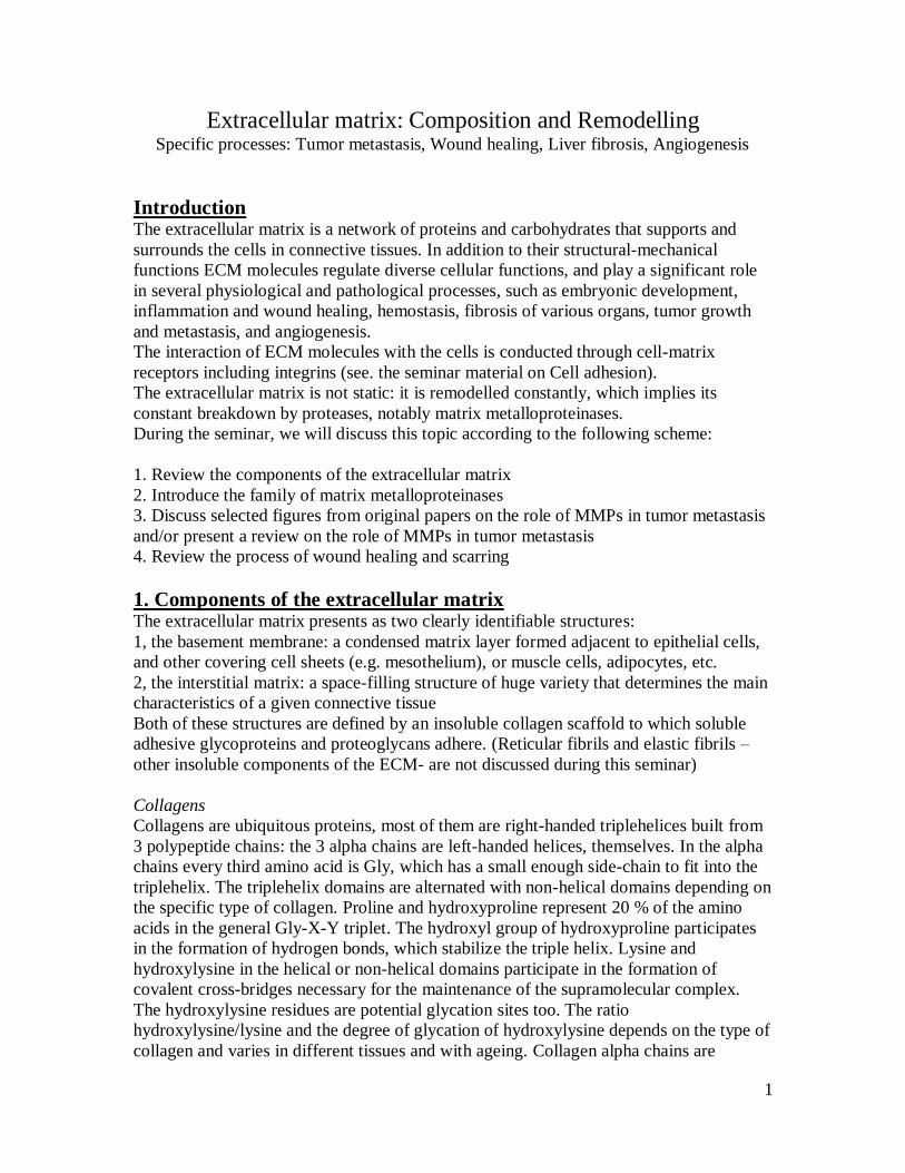
Collagens are ubiquitous proteins, most of them are right-handed triplehelices built from
3 polypeptide chains: the 3 alpha chains are left-handed helices, themselves. In the alpha
chains every third amino acid is Gly, which has a small enough side-chain to fit into the
triplehelix. The triplehelix domains are alternated with non-helical domains depending on
the specific type of collagen. Proline and hydroxyproline represent 20 % of the amino
acids in the general Gly-X-Y triplet. The hydroxyl group of hydroxyproline participates
in the formation of hydrogen bonds, which stabilize the triple helix. Lysine and
hydroxylysine in the helical or non-helical domains participate in the formation of
covalent cross-bridges necessary for the maintenance of the supramolecular complex.
The hydroxylysine residues are potential glycation sites too. The ratio
hydroxylysine/lysine and the degree of glycation of hydroxylysine depends on the type of
collagen and varies in different tissues and with ageing. Collagen alpha chains are
2
synthesized following the secretory pathway with several co-, and posttranslational
covalent modifications and formation of the triplehelical structure in the ER. The
supramolecular collagen structure is formed following the secretion of procollagen into
the ECM and type-specific additional modifications.
More than 20 genetically distinct collagen types have been identified and classified based
on their structures.
Fibrillar collagen types (I, II, III, V and XI) self-assemble into fibrils after their secretion
into the ECM. The intracellular precursor, pro-collagen contains non-helical regions at
the two terminals of the triple helical region (N- and C-telopeptides, N- and C-
propeptides). Following secretion the propeptides are cleaved by specific N- and C-
peptidases and thus the newly exposed telopeptides direct the axial arrangement in the
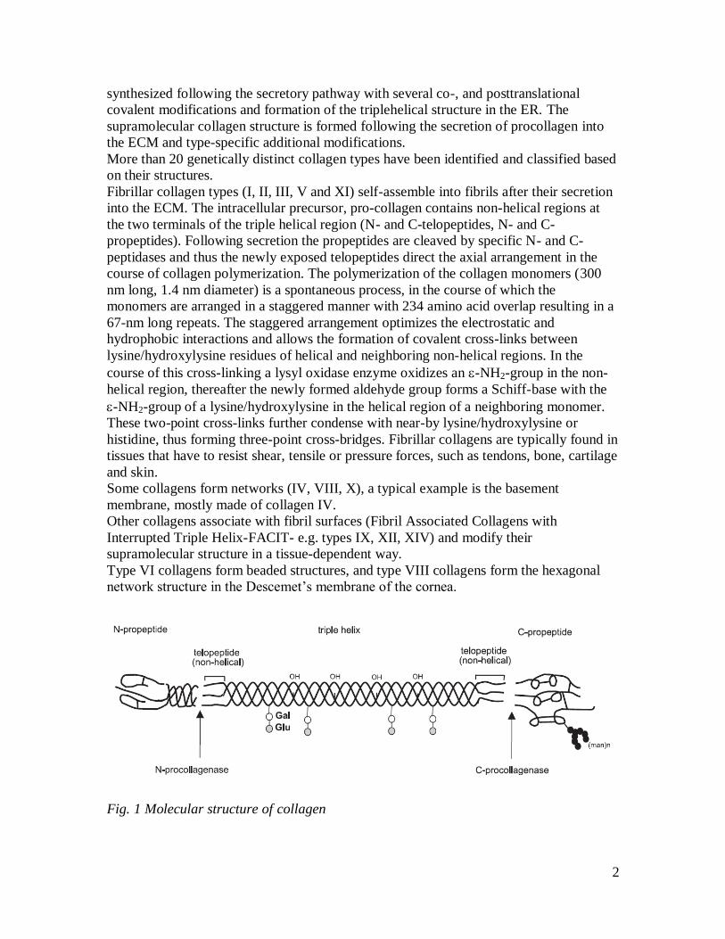
course of collagen polymerization. The polymerization of the collagen monomers (300
nm long, 1.4 nm diameter) is a spontaneous process, in the course of which the
monomers are arranged in a staggered manner with 234 amino acid overlap resulting in a
67-nm long repeats. The staggered arrangement optimizes the electrostatic and
hydrophobic interactions and allows the formation of covalent cross-links between
lysine/hydroxylysine residues of helical and neighboring non-helical regions. In the
course of this cross-linking a lysyl oxidase enzyme oxidizes an -NH2-group in the non-
helical region, thereafter the newly formed aldehyde group forms a Schiff-base with the
-NH2-group of a lysine/hydroxylysine in the helical region of a neighboring monomer.
These two-point cross-links further condense with near-by lysine/hydroxylysine or
histidine, thus forming three-point cross-bridges. Fibrillar collagens are typically found in
tissues that have to resist shear, tensile or pressure forces, such as tendons, bone, cartilage
and skin.
Some collagens form networks (IV, VIII, X), a typical example is the basement
membrane, mostly made of collagen IV.
Other collagens associate with fibril surfaces (Fibril Associated Collagens with
Interrupted Triple Helix-FACIT- e.g. types IX, XII, XIV) and modify their
supramolecular structure in a tissue-dependent way.
Type VI collagens form beaded structures, and type VIII collagens form the hexagonal
network structure in the Descemet’s membrane of the cornea.
Fig. 1 Molecular structure of collagen
3
Fig. 2 Supramolecular assembly of collagen fibrils
Proteoglycans
Proteoglycans consist of glucosaminoglycan(GAG) chains posttranslationally added to a
core protein by glycosyltransferases in the ER, followed by further elongation and
modification of the GAG chain in the Golgi. The GAG chains are composed of repeated
disaccharides. In chondroitin-sulphates the (beta1-3)glucuronic acid-N-acetyl-
glucosamine unit is sulphated on C4 (chondroitin-sulphate A) or C6 (chondroitin-
sulphate C) of the aminosugar moiety. Dermatan-sulphate (chondroitin-sulphate B)
contains at least one iduronic acid, formed from glucuronic acid by postsynthetic
epimerization. In heparan-sulphate, the repeated disaccharide is (beta1-4)glucuronic
acid-N-acetyl-glucosamine, where glucuronic acid can be epimerized to iduronic acid,
and both sugars can be sulphated, as well. Keratan-sulphate is composed of (beta1-
4)galactose-N-acetyl-glucosamine. Hyaluronic acid contains (beta1-3)glucuronic acid-N-
acetyl-glucosamine that are not sulphated, and it is the only GAG that is not attached to a
core protein. Instead, hyaluronic acid can be found in tissues as an extremely long, free
GAG chain, with Mw up to 10 000 kDa. Hyaluronic acids can form aggregates and
networks, very frequently co-aggregate with proteoglycans, as well.
The sulphation of GAG chains frequently varies even within the same GAG molecule,
and the same core-protein can carry different number of GAG chains, or GAG chains of
various lengths, which results in the highly heterogeneous nature of proteoglycans.
Proteoglycans are the space fillers in the ECM: due to the presence of uronic acids and
sulphate groups they are highly negative molecules that form a highly hydrated gel-like
substance. This substance is responsible for the volume of the ECM and is also resistant
to compression.
In addition to their structural functions, proteoglycans have been implicated in several
processes, where they can modulate cell-cell, and cell-matrix interactions, regulate
cellular behaviour including motility, proliferation, differentiation.
Based on their localization they are classified as matrix proteoglycans (e.g. aggrecan in
cartilage, perlecan in the basal membrane, versican in vessel wall, or the
decorin/biglycan/fibromodulin group in various tissues) or membrane-associated
proteoglycans (e.g. syndecans and glypicans, CD44, thrombomodulin)
Adhesive glycoproteins

4
Adhesive glycoproteins connect the cells to the matrix structure, most of them are ligands
for cell adhesion receptors and regulate cellular behaviour. They can also bind to the
structural matrix components, thereby modulating the matrix structure and organizing the
cells and ECM structures.
In addition to fibronectin and laminins described in the seminar material on Cell
adhesion, glycoproteins, such as nidogen, tenascin, thrombospondin, osteopontin belong
to this group.
Fig. 3 Relative size of the core (green) and sugar (magenta) component of extracellular
matrix proteins.
Structure of the basement membrane (also referred to as basal lamina)
The basement membrane is a thin (40-120 nm thick), though flexible sheet of ECM
molecules that not only underlies all epithelia, but also surrounds certain individual non-
epithelial cells, such as muscle cells, adipocytes, or Schwann cells. It separates these cells
from the adjacent connective tissue, and provides a mechanical connection between the
two layers, as well. Epidermolysis bullosa, a blistering, sometimes lethal skin disease is
due to a genetic defect of certain basement membrane components, which leads to an
improper anchorage of skin epidermis to the underlying dermis. In addition to its
structural role, basal lamina components also influence cellular functions and serve as
highways of cell migration. Like other extracellular matrices, the basement membrane
contains fibrous proteins (collagen type IV), adhesive glycoproteins (laminins and
nidogen), as well as proteoglycans (perlecan carrying heparan-sulphate side chains).
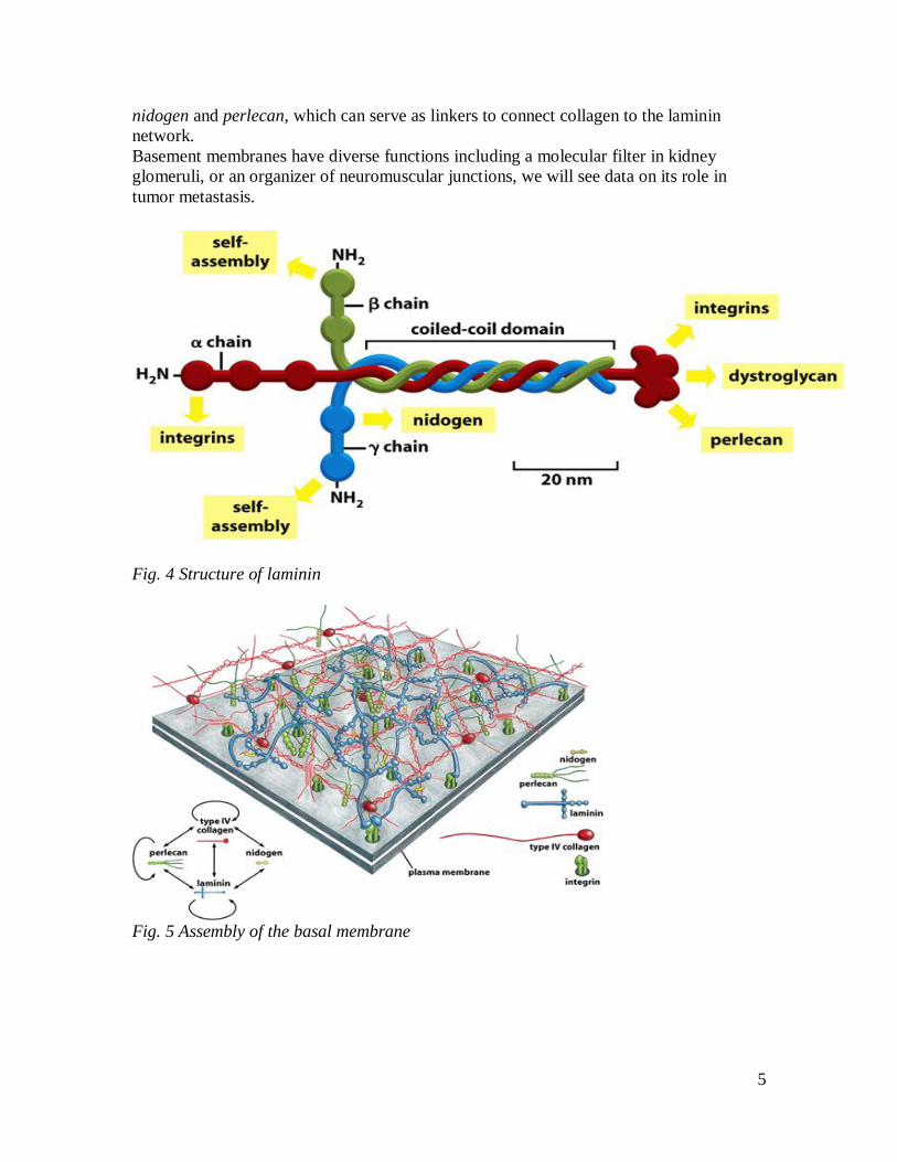
Laminin-1 is a large, flexible protein composed of three polypeptide chains (alpha, beta,
and gamma) held together by disulfide bonds. It looks like a bunch of three flowers
whose stems are twisted together at the foot, but whose heads remain separate. These
heterotrimers self-assemble into a network through interactions between their heads and
interaction between their foot and the cells producing them help to organize the network
into an orderly sheet. Laminin anchorage occurs mostly via integrin receptors, another
important receptor is dystroglycan, a proteoglycan with a transmembrane core protein
and an extracellular GAG chain. This laminin network then presumably coordinates the
assembly of the other basal lamina components, since laminin has multiple binding sites
for nidogen and perlecan. Type IV collagen, like other collagens, is a superhelix of three
polypeptide chains, however, its triplehelical structure is interrupted in more than 20
regions, allowing multiple bending of the molecule. Collagen type IV can bind to both
5
nidogen and perlecan, which can serve as linkers to connect collagen to the laminin
network.
Basement membranes have diverse functions including a molecular filter in kidney
glomeruli, or an organizer of neuromuscular junctions, we will see data on its role in
tumor metastasis.
Fig. 4 Structure of laminin
Fig. 5 Assembly of the basal membrane

6
2. ECM Remodelling: matrix metalloproteinases (MMPs) and tissue
inhibitors of matrix metalloproteinases (TIMPs) Matrix metalloproteinases (MMPs) are a family of zinc-dependent endopeptidases that
consist of more than 21 human MMPs. Based on their domain structure they can be
divided into eight classes, three of which are membrane-bound (see Figure 2 in the
review below). The MMPs can collectively degrade most of the components of the
extracellular matrix (ECM), including collagens (collagenases), denatured or partially
degraded collagens (gelatinases), laminins, as well as cell-adhesion molecules, growth
factors, and growth factor receptors, through which they also influence cellular signalling
and function.
Fig. 6 Tertiary structure of MMPs (the catalytic site Zn is shown in yellow)
The MMPs are synthesized as inactive zymogens, where a Cys residue in the propeptide
domain forms a bridge with the zinc in the catalytic center, and prevents enzymatic
activity. Activation occurs when this Cys-Zn2+ interaction is disrupted and the
propeptide is proteolytically removed. The activation occurs in two steps: in the 1st step,
the Cys-Zn2+ interaction is perturbed as a result of a conformational change (induced
chemically by e.g. APMA-amino phenyl mercuric acid-) or proteolysis by a serine-
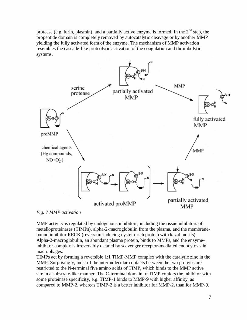
7
protease (e.g. furin, plasmin), and a partially active enzyme is formed. In the 2nd
step, the
propeptide domain is completely removed by autocatalytic cleavage or by another MMP
yielding the fully activated form of the enzyme. The mechanism of MMP activation
resembles the cascade-like proteolytic activation of the coagulation and thrombolytic
systems.
Fig. 7 MMP activation
MMP activity is regulated by endogenous inhibitors, including the tissue inhibitors of
metalloproteinases (TIMPs), alpha-2-macroglobulin from the plasma, and the membrane-
bound inhibitor RECK (reversion-inducing cystein-rich protein with kazal motifs).
Alpha-2-macroglobulin, an abundant plasma protein, binds to MMPs, and the enzyme-
inhibitor complex is irreversibly cleared by scavenger receptor-mediated endocytosis in
macrophages.
TIMPs act by forming a reversible 1:1 TIMP-MMP complex with the catalytic zinc in the
MMP. Surprisingly, most of the intermolecular contacts between the two proteins are
restricted to the N-terminal five amino acids of TIMP, which binds to the MMP active
site in a substrate-like manner. The C-terminal domain of TIMP confers the inhibitor with
some proteinase specificity, e.g. TIMP-1 binds to MMP-9 with higher affinity, as
compared to MMP-2, whereas TIMP-2 is a better inhibitor for MMP-2, than for MMP-9.
8
In addition to acting as a reversible MMP inhibitor, TIMP-2 plays a cofactor role in the
activation of proMMP-2 (for details, discuss Figure 4. in the recommended review below,
and the paper on pericellular proMMP-2 activation by MT1-MMP).
Many of the MMP genes are inducible, and the effectors include not only the soluble
growth factors, cytokines and chemicals, but cell-matrix and cell-cell interactions have
been shown to influence MMP gene expression, as well. Enhanced MMP gene expression
may be down-regulated by suppressive factors (e.g. TGF1, glucocorticoids).
3. Matrix metalloproteinases in tumor metastasis MMPs have been implicated in cancer invasion and metastasis as proteases degrading the
ECM barrier in the direction of invasion. To carry out such a function, MMPs are
expected to act at the leading edge of invading cancer cells. MT1-MMP was identified as
the first membrane-anchored type MMP acting as a key enzyme responsible for the
degradation of the pericellular ECM. Unlike the soluble MMPs, MT1-MMP activation
occurs in the trans-Golgi by furin, during the secretory pathway, so a catalytically active
enzyme reaches the cell surface. In addition to MT1-MMP, MMP-2 and MMP-9, two
types of type IV collagenases have been implicated in cancer invasion. Even though these
MMPs are soluble enzymes produced by fibroblasts located in the stroma surrounding
cancer cell nests, type IV collagenase activity was found to associate with the cancer cell
surface. As it turns out, the expression of MT1-MMP confers the cells the ability to bind
and to activate proMMP-2.
MT1-MMP, in addition to its role in proMMP-2 activation, is a membrane-bound
collagenase, itself. “Clearing a path” in the ECM, however, is not the only mechanism
that promotes tumor cell invasion. MT1-MMP also interacts with cell adhesion receptors,
such as CD44, a hyaluronic acid receptor, as well as with syndecan-1, a transmembrane
proteoglycan, which results in enhanced cell migration. MT1-MMP cleaves laminin-5, a
basal lamina protein, and the cleaved laminin-5 also promotes cell migration through the
basement membrane.
Angiogenesis is the formation of new vessels from existing vessels and is required for an
adequate blood supply in tumor growth. During this process endothelial cells need to
detach from neighbouring cells, invade into the surrounding stroma, proliferate and
generate a tube structure. These steps include the degradation of the basal lamina, and the
collagen-rich stroma. Several other MMPs have been shown to play a role in
angiogenesis, however, in an experimental collagen-rich matrix, only the MT1-MMP
knock out mice failed to show neovessel formation. MT1-MMP also up-regulates the
gene expression of VEGF by tumor cells, a growth factor important for angiogenesis.
Angiogenesis-a brief review
Angiogenesis is the process of vessel growing and branching throughout the body. The
formation of the first rudiments of vessels from the early embryonic endothelial cells in
the course of the embryonic development is called vasculogenesis, but further branching
and maturation of the vessel structure is a similar process throughout our lives.
Eventually, almost every cell is located within 50-100 m of a blood capillary to ensure
adequate blood supply. The renewal and development of the vasculature is required not
only during embryonic development and body growth, but is also necessary in tissue
9
repair, as well as in tumor growth and metastasis. Angiogenesis follows a carefully
orchestrated sequence of events: 1, an endothelial tip cell is selected, that will start to
form a new capillary branch; 2, the endothelial tip cell, with many filopodia, grows into
the surrounding tissue; 3, endothelial stalk cells trail behind the tip cell, and hollow out to
form the lumen of the new vessel; 4, the newly formed vasculature is stabilized by the
recruitment of mural cells (e.g. smooth muscle cells, fibroblasts) and the deposition of an
extracellular matrix, which altogether ensure that parenchymal cells receive adequate
nutrient supply.
The signals controlling this whole event are rather complex, but vascular endothelial
growth factor (VEGF) is a key factor. VEGF is a relative of platelet-derived growth
factor (PDGF), and a survival factor for endothelial cells (its main target cells). VEGF
receptors belong to the group of receptor-tyrosine kinases, and activate the phosphatidyl-
inositol-3-kinase/protein kinase B pathway, as well as the MAP-kinase pathway. The
biological effects of VEGF on endothelial cells include proliferation, production of
proteases (MMPs and serine proteases) that digest the surrounding ECM to help
sprouting. The endothelial tip cells detect the VEGF gradient and move toward its source
that signalled the need for angiogenesis. Increased vascular permeability and vasodilation
(NO-mediated) lead to the formation of a provisional matrix around the new branch, and
integrin receptor-matrix molecule interactions help the endothelial cells to migrate along
these matrix molecules. Angiopoetins in the presence of VEGF facilitate sprouting and
the formation of a new “immature” vascular network. The maturation of newly formed
vessels is required to reach branching and vessel wall structure appropriate for the site
and vessel-type (e.g. capillary, arteriole, artery, vein, or lymphatic vessels). PDGF
secreted from endothelial cells in response to VEGF recruits mural cells and promotes
their proliferation. Angiopoetins secreted by mural cells and endothelial cells facilitate
endothelial cell-mural cell interactions, as well as endothelial cell-matrix interactions.
TGF promotes the production of ECM and proteases by mural cells and endothelial
cells and regulates the final differentiation of vessel wall cell-types. An excessive
formation of blood vessels is undesired, inhibitors of angiogenesis limit the number of
vessels and the deposition of ECM molecules. Such inhibitors include angiostatin, a
cleavage product of plasminogen, and endostatin, a cleavage product of collagen type
XVIII.
There are several situations that may necessitate angiogenesis, a well-studied example is
hypoxia, and the hypoxia-inducible factor-1(HIF-1). HIF-1 is an alpha-beta heterodimer,
the beta subunit is a constitutively expressed nuclear protein, whereas HIF-1alpha is
induced by a shortage of oxygen. HIF-1 alpha/beta was first recognized as a DNA-
binding protein that up-regulates the transcription of erythropoietin, and later turned out
to be an oxygen-sensor in a wide variety of cell types. In well-oxygenated cells the
concentration of HIF-1alpha is low because of its continuous degradation. Non-heme,
iron-, and 2-oxoglutarate-dependent dioxygenases, that require molecular oxygen, can
hydroxylate HIF-1alpha on two Pro, and an Asn residues. One oxygen atom of the O2
molecule creates the hydroxyl group, and the other one oxidizes 2-oxoglutarate to
succinate with the release of CO2. Hydroxylation at the Pro residues mediates interactions
with the von Hippel-Lindau (VHL) E3 ubiquitin ligase complex that targets HIF-1alpha
for proteasomal degradation. The second hydroxylation-dependent control is the beta-
hydroxylation of an Asn residue, which blocks the interaction of the HIF-1alpha C-
10
terminus with the transcriptional coactivator, p300 (this is a histone acetyl-transferase). In
oxygenated cells, this dual mechanism (proteolytic destruction and inhibition of
transcriptional activity) inhibits the HIF-dependent transcription of proangiogenic factors,
e.g. VEGF.
The role of the HIF-pathway in the regulation of angiogenesis is underlined in von Hippel
Lindau syndrome. People affected carry just one functional copy of the VHL gene, and
when the other one gets mutated, the lack of a functional protein leads to high HIF-1alpha
levels, regardless of oxygen availability, which leads to the development of
hemangioblastomas, tumors with dense masses of blood vessels.
VEGF mRNA is up-regulated in solid tumors, (both cancer cells and stromal cells
produce VEGF) and angiogenesis is stimulated in order to supply the growing tumor
mass. The structure of the vasculature, however, is abnormal with uneven wall thickness,
leaky endothelial cell lining, altered expression of endothelial cell markers and adhesion
molecules. These abnormalities are considered to result from an imbalance between pro-,
and anti-angiogenic molecules. Inhibition of angiogenesis may limit tumor growth and
metastasis formation, e.g. anti-VEGF treatments potentiate the anti-tumor effects of
conventional radiation therapy and chemotherapy.
4. Wound healing and tissue repair Damage to tissues can result from various acute and chronic stimuli, including
mechanical injury, infections, metabolic abnormalities, and autoimmune reactions. The
repair process involves two stages: a first, regenerative phase, where injured cells are
replaced by cells of the same type; and a second phase in which connective tissue is
deposited with the possible restoration of the original organ structure. If this process goes
out of control, it results in substantial remodelling of the ECM, and the formation of
permanent scar tissue. In some cases, such as liver fibrosis, parenchymal tissue cannot be
regenerated, and is replaced by connective tissue, instead.
Following tissue injury, damaged epithelial/endothelial cells release inflammatory
mediators that initiate blood clot formation including platelet aggregation and fibrin
deposition. Activated platelets release chemokines and growth factors, such as PDGF
(platelet-derived growth factor) and TGF (tumor growth factor-beta). Leukocytes from
the circulation are recruited to the site and activated by chemokines (e.g. PDGF, TGF).
Neutrophils (arriving the earliest) and macrophages (arriving after 1-2 days) eliminate
tissue debris, dead cells, invading organisms. Activated leukocytes secrete profibrotic
cytokines, such as IL-13 and TGF, which further activate macrophages and fibroblasts.
Activated fibroblasts transform into myofibroblasts expressing -SMC (alpha-smooth
muscle cell actin- the histological marker of myofibroblasts) that combine the contractile
properties of smooth muscle cells with the fibroblasts’ ability to produce ECM
components including fibrillar collagens.
At the molecular level, TGF (transforming growth factor ) is a cytokine vital to tissue
repair, however, its excessive action has also been implicated in fibrotic scarring leading
to organ damage. TGF is a dimer of a 12 kDa polypeptide, and belongs to the
superfamily of cytokines regulating embryonal development, cell differentiation, and
tissue repair. Platelets contain high concentrations of TGF, which is released upon their
degranulation. TGF is chemoattractant for leukocytes, induces angiogenesis, control the
production of several cytokines and inflammatory mediators. Two additional features of

11
TGF action may easily lead to an out-of-control healing process: 1, the autoinduction of
TGF-production in myofibroblasts, which may lead to persistent activation, and 2, the
induction of ECM deposition by TGF via increased synthesis of all major ECM
components and a simultaneous blockage of matrix degradation (increased TIMP,
decreased MMP expression). Supporting the potential role of TGF in fibrosis, anti-
TGF antibodies capable of blocking the binding of TGF to its cell surface receptor
have been successfully applied in some cases.
The term wound healing covers the restoration of the ECM integrity following
mechanical injury. This is not a simple task, because in the basal state the structural
elements of the ECM form a network, which is anchored under strain on the parenchymal
cells. The existence of mechanical strain in the tissues is supported by the fact that the
length of blood vessels and nerves is 25 – 30 % smaller in isolated state compared to their
length in situ in the tissues. This mechanical strain provides an elastic protective shield
around the parenchymal cells and in certain tissues it is a prerequisite for the basic organ
function (e.g. in the lung alveoli). Thus, in the course of wound healing not only the ECM
continuity should be restored, but the strain of the structure should be tuned accordingly.
This process called mechano-regulation requires the coordinated function of many
molecular and cellular components. The changing mechanic strain at the site of injury
initiates the secretion of transforming growth factor 1 (TGF-1) in the neighboring
fibroblasts, whereas the platelets arriving from the damaged blood vessels secrete
platelet-derived growth factor (PDGF). Both cytokines can initiate the differentiation of
myofibroblasts manifested primarily in the massive production of -SM actin. As a result
of this the myofibroblasts can exert long-lasting contraction forces coupled to the events
of the extracellular structural rearrangements. The contraction is based on the interactions
of actin microfilaments and myosin (non-muscle type), which are enabled by the
phosphorylation of the myosin light chain (MLC). This phosphorylation can be catalyzed
by two kinases: Ca2+
-dependent MLC kinase (MLCK) and Rho-kinase (RhoA small G-
protein activated kinase). The elevation of the intracellular Ca2+
-level results in fast (the
affinity of MLCK for MLC is high), but short-lasting contraction (the myosin
phosphatase removes the phosphate groups from the phosphorylated MLC also with high
affinity). In contrast, MLC is not so good substrate of Rho-kinase and thus the
contraction provoked by it is slow. However, the myosin-binding subunit of myosin
phosphatase is also a substrate of Rho-kinase and in phosphorylated state it does not bind
myosin, the Rho-kinase initiated contraction is long-lasting and energetically more
suitable to maintain the isometric strain the newly forming connective tissue. The exact
mechanisms, which activate the Rho-kinase and adjust the strength of contraction, are not
known. The initial active contraction is followed by structural rearrangement of ECM
around the myofibroblasts (Fig. 8). These cells express transmembrane adhesion
complexes, which connect the extracellular collagen fibers with the intracellular
microfilaments. Consequently, the cell contraction results in stretching of the initial loose
collagen meshwork (Fig. 8b, “B” fibroblast). This deformation of the initial collagen
matrix is accompanied by mechanical strain in the opposite direction compared to the
final structure. This is resolved by the matrix metalloproteinases produced by the
myofibroblasts (MMP-1,-2,-3,-9). MMP-1 removes the outer collagen layer, and n
accordance with their substrate specificity (see MMP chapter), the MMP-2 and 9
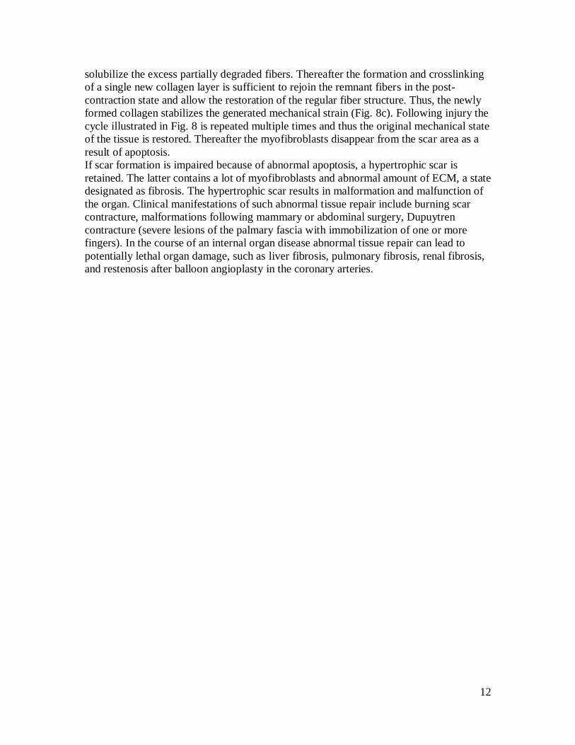
12
solubilize the excess partially degraded fibers. Thereafter the formation and crosslinking
of a single new collagen layer is sufficient to rejoin the remnant fibers in the post-
contraction state and allow the restoration of the regular fiber structure. Thus, the newly
formed collagen stabilizes the generated mechanical strain (Fig. 8c). Following injury the
cycle illustrated in Fig. 8 is repeated multiple times and thus the original mechanical state
of the tissue is restored. Thereafter the myofibroblasts disappear from the scar area as a
result of apoptosis.
If scar formation is impaired because of abnormal apoptosis, a hypertrophic scar is
retained. The latter contains a lot of myofibroblasts and abnormal amount of ECM, a state
designated as fibrosis. The hypertrophic scar results in malformation and malfunction of
the organ. Clinical manifestations of such abnormal tissue repair include burning scar
contracture, malformations following mammary or abdominal surgery, Dupuytren
contracture (severe lesions of the palmary fascia with immobilization of one or more
fingers). In the course of an internal organ disease abnormal tissue repair can lead to
potentially lethal organ damage, such as liver fibrosis, pulmonary fibrosis, renal fibrosis,
and restenosis after balloon angioplasty in the coronary arteries.
13
Fig. 8 Mechano-regulation in the course of wound healing
14
Recommended presentations:
1, Please, summarize the components of the extracellular matrix, based on your
previous studies in anatomy and biochemistry.
2, Summarize the structure and function of the MMP family, the mechanism of proMMP
activation and their TIMP-type inhibitors. Use the scheme above and relevant parts of the
following reviews:
2.1 Brinckerhoff CE, Matrisian LM. Matrix metalloproteinases: a tail of a frog that
became a prince. Nature Rev Mol Cell Biol 2002, 3:207-214 (Figs 2, 4)
2.2 Nagase H, Woessner JF Jr. Matrix metalloproteinases. J Biol Chem 1999, 274:21491-
21494.
3, Discuss the roles of MMPs in tumor metastasis formation. You may choose to
present the relevant parts of the review article (3.1) or present and discuss a few selected
figures from the original papers illustrating the experimental nature of research in this
field (3.2-3.4). If the latter use Figure 1 from the review paper for a scaffold.
3.1 A selected review on pericellular proteolysis and its role in tumor metastasis and
angiogenesis:
Itoh Y, Seiki M. MT1-MMP: A potent modifier of pericellular microenvironment. J Cell
Physiol 2006, 206:1-8 Note Figures 1, and 2.
3.2 An original paper on proMMP-2 activation:
Will H, Atkinson SJ, Butler GS, Smith B, Murphy G. The soluble catalytic domain of
membrane type 1 matrix metalloproteinase cleaves the propeptide of progelatinase A and
initiates autoproteolytic activation. J Biol Chem 1996, 271: 17119-17123.
Figures 2 and 3 present kinetic and gel electrophoretic data on the activation of
proMMP2 by MT1-MMP, and its inhibition by TIMP-2
3.3 MMPs promote cell migration-original paper
Gianelli G, Falk-Marzillier J, Schiraldi O, Stetler-Stevenson WG, Quaranta V. Induction
of cell migration by matrix metalloproteinase-2 cleavage of laminin-5. Science 1997,
277:225-228
Summary:
Cell migration across ECM boundaries is required in several important processes,
including tissue remodelling and tumor invasion. Laminin-5 is a component of basement
membranes. Cells adhere to and migrate on ECM molecules by means of integrin
receptors, and laminin-5 interaction with integrins has been shown to be essential for the
adhesion of epithelial cells to basement membranes. In an experimental model, the so-
called transwell migration assay, the migration of epithelial cells through filters was
promoted specifically by laminin-5 predigested by MMP2. Pretreatment of laminin-5 by

15
MMP9 or plasmin (two other potentially relevant proteases) was not promigratory, nor
were other basement membrane components, such as type IV collagen, laminin-1 or
fibronectin. Using monoclonal antibodies that specifically block various epitopes on
laminin-5, as well as on integrin receptors, the adhesion of the breast epithelial cells to
MMP2-cleaved or uncleaved laminin-5 occurs via integrin a3b1, a laminin receptor.
Cleavage of laminin-5 by MMP2 results in the appearance of a new epitope on the alpha3
subunit, which is not involved in cell adhesion, but directly stimulates cell motility.
Analysis of various mouse tissue samples with immunoblotting demonstrated the
presence of this new epitope in tissues undergoing remodelling (mammary tissue from a
pregnant rat) and mouse carcinoma, whereas it was undetectable in quiescent tissues,
(such as tongue or mammary tissue from a sexually immature female rat).
3.4 ECM degradation by MMPs-original paper
Several MMPs have been shown to be able to degrade several components of the
extracellular matrix, here we recommend just one original paper with a few simple
Figures as an illustration.
Ohuci E, Imai K, Fujii Y, Sato H, Seiki M, Okada Y. Membrane type 1 matrix
metalloproteinase digests interstitial collagens and other extracellular matrix
macromolecules. J Biol Chem 1997, 272: 2446-2451.
Figures 4, and 5 present data on the MT1-MMP cleavage patterns of collagen types I, II,
and III, type I gelatine, fibronectin, vitronectin and laminin-1 followed by SDS-gel
electrophoresis. Figure 6 shows that MT1-MMP and MMP2 synergistically cleave
fibrillar collagens. Kinetic parameters are summarized in Table I.
4, Summary of the basic wound healing process and the role of TGF in it. Use the
following figures from reviews.
4.1 Wynn TA. Common and unique mechanisms regulate fibrosis in various
fibroproliferative diseases. J Clin Invest 2007, 117:524-529. Figure 1.
4.2 Border WA, Ruoshlati E. Transforming growth factor- in disease: the dark side of
tissue repair. J Clin Invest 1992, 90:1-7. Figure 2.
Liver fibrosis as a clinical example for abnormal tissue repair:
4.3 Iredale JP. Cirrhosis: new research provides a basis for rational and targeted
treatments. Br Med J 2003, 327:143-147.