extra pulmonary tuberculosis · tb intensive san antonio, texas december 1-3, 2010 extrapulmonary...
TRANSCRIPT
TB IntensiveSan Antonio, TexasDecember 1-3, 2010
Extrapulmonary TBRobert Longfield, MD, FACP; TCID
December 3, 2010
Extra Pulmonary Tuberculosis
TB Intensive CourseDecember 2010
Robert N. Longfield, MD, FACP
Spectrum of EPTB:
• CNS– Meningitis– Tuberculoma
• Skeletal– Spinal– Joint– Osteomyelitis– Myositis
• Lymphadenitis
• Pleural– Effusion– Empyema
• Genitourinary• Gastrointestinal• Pericardial• Cutaneous• Other
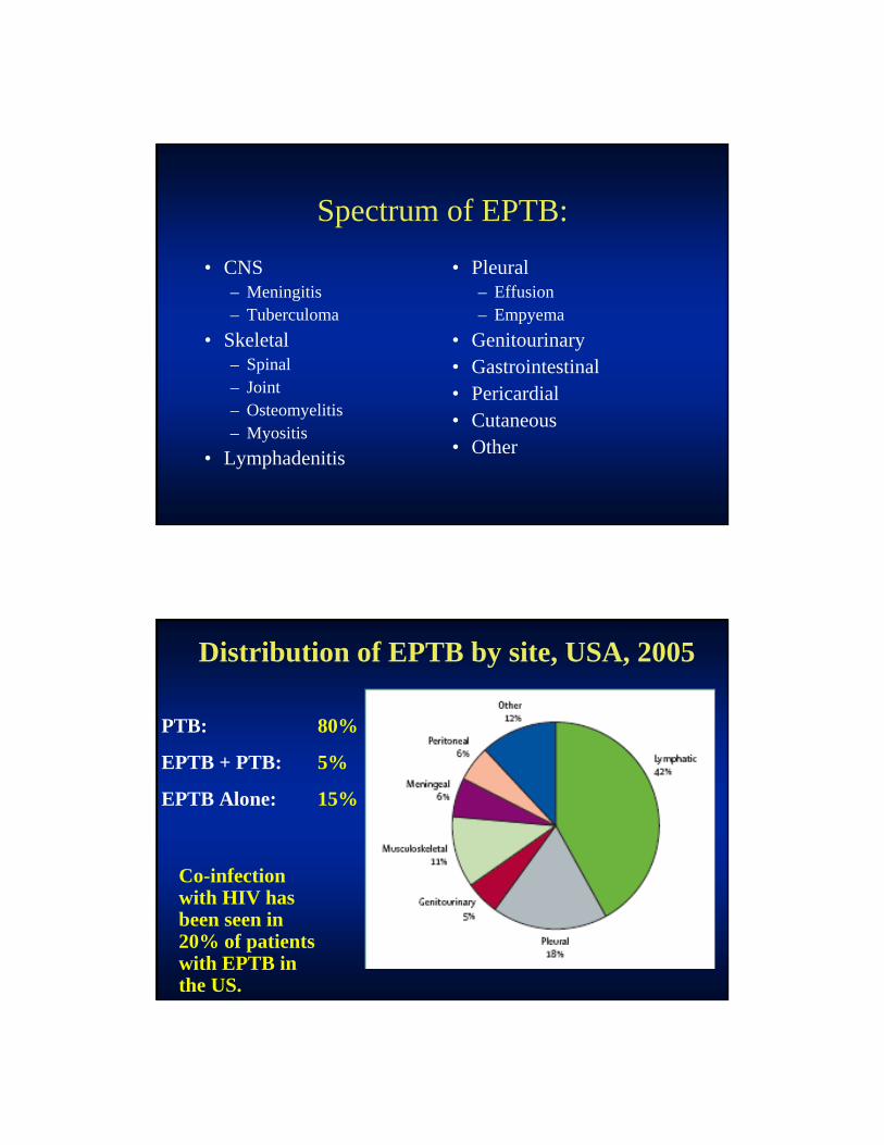
Distribution of EPTB by site, USA, 2005
PTB: 80%
EPTB + PTB: 5%
EPTB Alone: 15%
Co-infection with HIV has been seen in 20% of patients with EPTB in the US.

CNS Tuberculosis:
• CNS TB includes: – Meningitis,
– Tuberculoma
– Spinal Arachnoiditis
• CNS infection reported frequently from high incidence countries.
INTRODUCTION
• Approximately 300 to 400 cases of TB meningitis occur each year in the US. – 1% of all TB disease.
• Despite effective treatment regimens, case fatality remains high - 15% to 40%.
• Early recognition of CNS-TB is critical!
INTRODUCTION
• Empiric Rx should be started with:– a meningitis syndrome and
– low CSF glucose, elevated protein, and lymphocytic pleocytosis and
– evidence of TB elsewhere in the body or
– if prompt w/u fails to establish another diagnosis.
Pathogenesis:
• Post-primary infection:
– Infants and young children
– Advanced HIV infection
– TNF-I incident infection
– Transplant patients.
• Reactivation bacillemia:
– Immune deficiency of aging,
– TNF-I Rx,
– Alcoholism,
– Malnutrition,
– Diabetes,
– Malignancy
– Transplant patients.
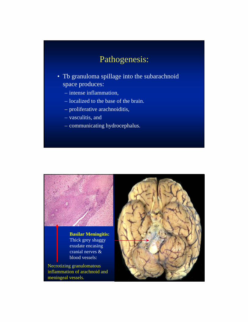
Pathogenesis:
• Tb granuloma spillage into the subarachnoid space produces:– intense inflammation,
– localized to the base of the brain.
– proliferative arachnoiditis,
– vasculitis, and
– communicating hydrocephalus.
Basilar Meningitis:Thick grey shaggy exudate encasing cranial nerves & blood vessels:
Necrotizing granulomatous inflammation of arachnoid and meningeal vessels.

CLINICAL FEATURES:
1. Prodromal phase, 2 to 3 weeks, with a) insidious malaise, lassitude,
b) headache,
c) low-grade fever, and
d) personality changes.
2. Meningitic phase
3. Paralytic phase
CLINICAL FEATURES:
2. Meningitic phase witha) meningismus,
b) protracted headache,
c) vomiting,
d) confusion, and
e) varying cranial nerve and long-tract signs.
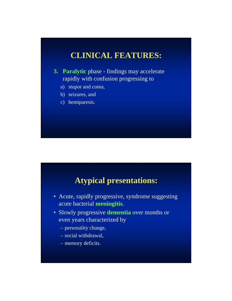
CLINICAL FEATURES:
3. Paralytic phase - findings may accelerate rapidly with confusion progressing to
a) stupor and coma,
b) seizures, and
c) hemiparesis.
Atypical presentations:
• Acute, rapidly progressive, syndrome suggesting acute bacterial meningitis.
• Slowly progressive dementia over months or even years characterized by – personality change,
– social withdrawal,
– memory deficits.
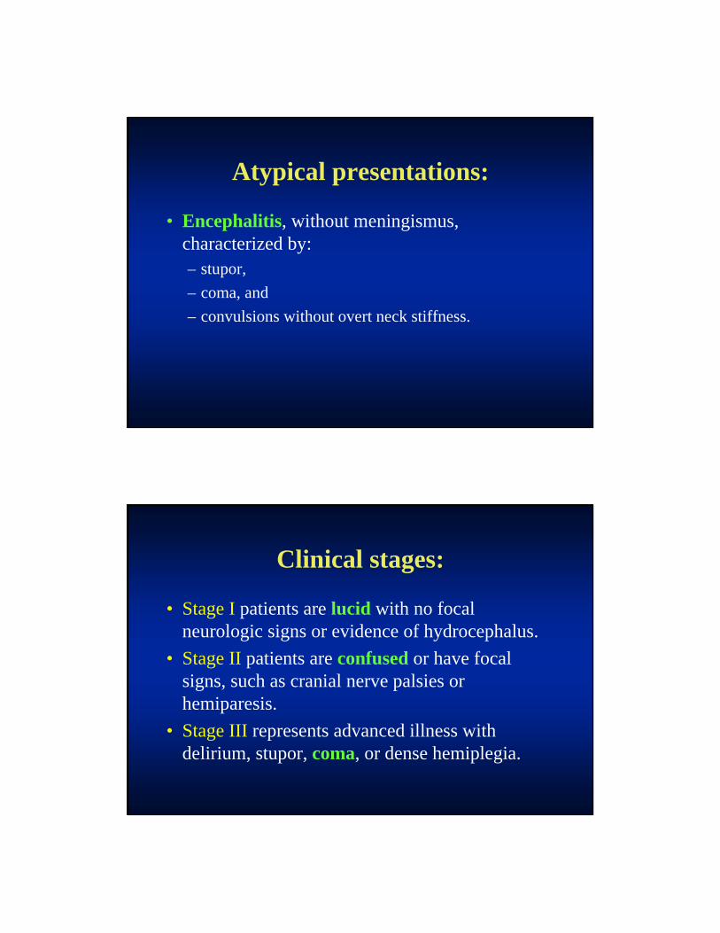
Atypical presentations:
• Encephalitis, without meningismus, characterized by: – stupor,
– coma, and
– convulsions without overt neck stiffness.
Clinical stages:
• Stage I patients are lucid with no focal neurologic signs or evidence of hydrocephalus.
• Stage II patients are confused or have focal signs, such as cranial nerve palsies or hemiparesis.
• Stage III represents advanced illness with delirium, stupor, coma, or dense hemiplegia.

HIV infection:
• In one study, cerebral tuberculomas were more common in the HIV-infected group:
(60% versus 14%).
• Otherwise, HIV co-infection did not alter the – clinical manifestations,
– CSF findings, or
– response to therapy.
DIAGNOSIS:
• Serial examination of the CSF by AFB stain and culture is the best diagnostic approach.
• Smears and cultures may yield positive results days to weeks after therapy has been initiated.

DIAGNOSIS CSF Examination :
• Diagnosis (Dx) can be difficult.
• Proper CSF samples are critical for early Dx.
• Maintain a high degree of suspicion.
• Initiate empiric therapy promptly.
DIAGNOSIS CSF Examination :
• Typical CSF formula shows
– elevated protein (100 to 500 mg/dL)
– low glucose and
– a lymphocytic pleocytosis.
• Hydrocephalus with subarachnoid block may have protein levels of 2 to 6 g/dL, xanthochromia and a poor prognosis.

DIAGNOSIS CSF Examination :
• Early in the course of illness, the CSF cellular reaction may be atypical with
– only a few cells or
– a predominance of PMN’s.
• Usually changes rapidly to lymphocytic predominance.
DIAGNOSIS AFB smears:
• Repeated, careful CSF examination and culture for M. tuberculosis is critically important!
• In one series, only 37% of cases were diagnosed on the basis of an initial positive AFB smear.
• The yield increased to 87% when, despite therapy, up to 4 serial specimens were examined.

DIAGNOSIS AFB smears:
• At minimum, 3 daily LP’s should be performed.
• Empiric Tb Rx should not be delayed.
DIAGNOSIS AFB smears:
• The sensitivity of the AFB smear may be enhanced by:– Using the last fluid removed by LP.
– Removing a large volume (10 to 15 mL) of CSF.
– Examining a smear of the clot or centrifuged sediment (cyto-centrifuge).
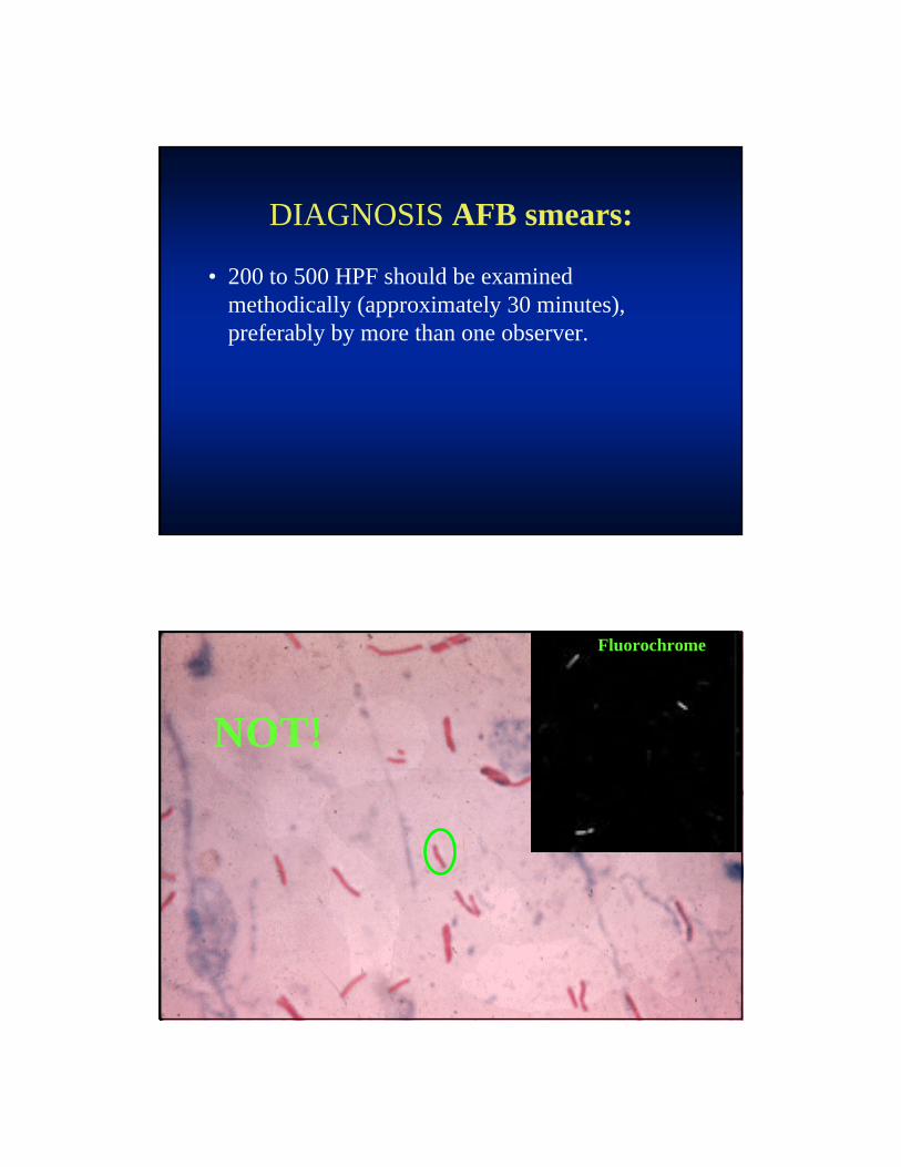
DIAGNOSIS AFB smears:
• 200 to 500 HPF should be examined methodically (approximately 30 minutes), preferably by more than one observer.
NOT!
Fluorochrome
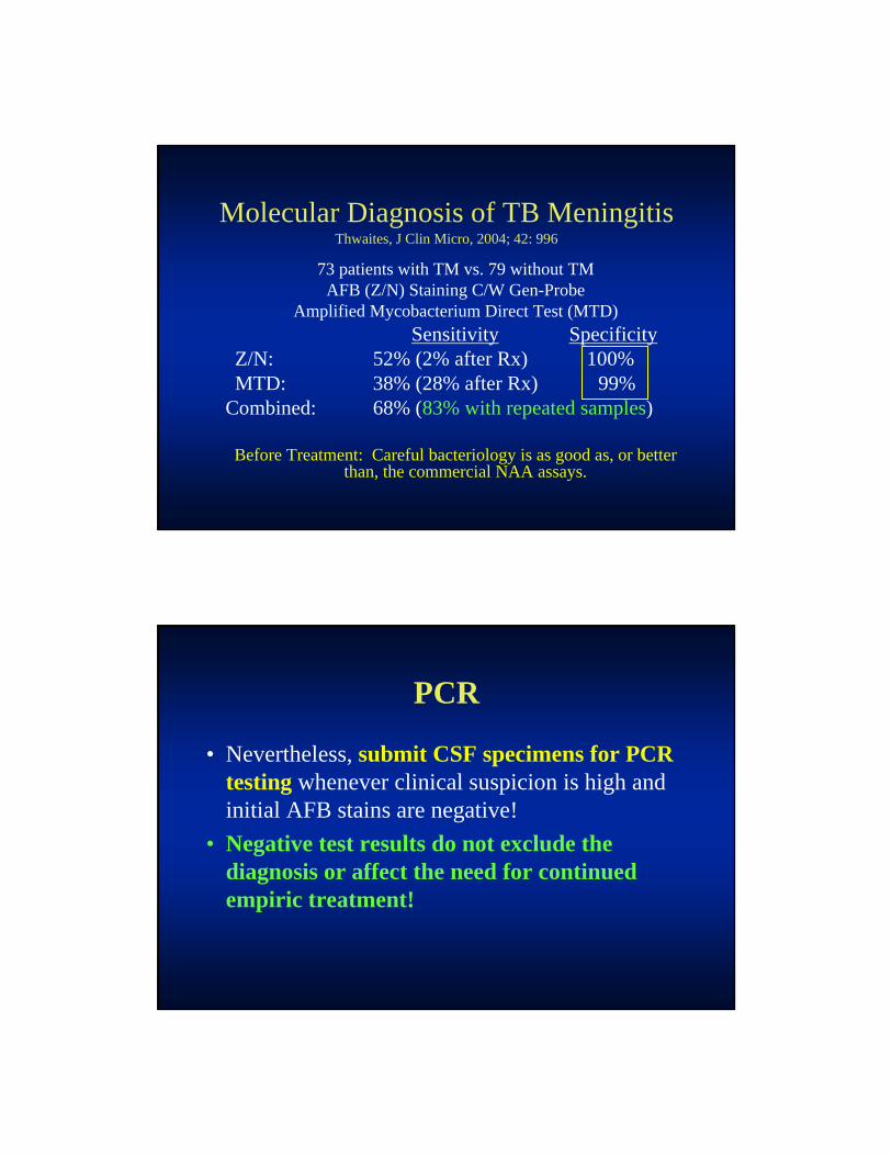
Molecular Diagnosis of TB MeningitisThwaites, J Clin Micro, 2004; 42: 996
73 patients with TM vs. 79 without TMAFB (Z/N) Staining C/W Gen-Probe
Amplified Mycobacterium Direct Test (MTD)Sensitivity Specificity
Z/N: 52% (2% after Rx) 100%MTD: 38% (28% after Rx) 99%
Combined: 68% (83% with repeated samples)
Before Treatment: Careful bacteriology is as good as, or betterthan, the commercial NAA assays.
PCR
• Nevertheless, submit CSF specimens for PCR testing whenever clinical suspicion is high and initial AFB stains are negative!
• Negative test results do not exclude the diagnosis or affect the need for continued empiric treatment!
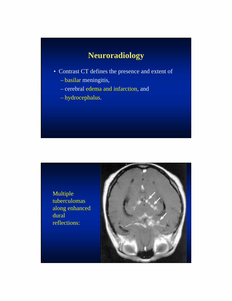
Neuroradiology
• Contrast CT defines the presence and extent of
– basilar meningitis,
– cerebral edema and infarction, and
– hydrocephalus.
Multiple tuberculomasalong enhanced duralreflections:

Basilar Enhancement & Hydrocephalus:
Tuberculoma & Hydrocephalus:
Neuroradiology
• Compatible clinical features, CT evidence of basilar meningeal enhancement AND any degree of hydrocephalus is strongly suggestive of TB meningitis.
• Hydrocephalus combined with marked basilar enhancement is indicative of advanced disease and carries a poor prognosis.
Neuroradiology
• Marked basilar enhancement correlates well with vasculitis and the risk for basal ganglia infarction.
• MRI is superior to CT in defining lesions of the basal ganglia, midbrain, and brain stem and for evaluating all forms of suspected spinal TB.

Other Conditions Mimicking TB Meningitis Radiographically:
• Cryptococcal Meningitis
• Coccidioides Meningitis
• Viral Encephalitis
• Sarcoidosis
• Meningeal Metastases
• Lymphoma
THERAPY Recommended regimens:
• Drug resistance should be considered in individuals – from countries where resistant TB is prevalent,
– in Pts with a history of prior TB treatment, and
– in Pts with exposure to drug-resistant source case.
Duration of therapy
• There are no randomized trials to establish the optimal duration of therapy.
• It is recommended that therapy be administered for 12 months in the cases of drug-sensitive CNS Tb.
Duration of therapy
• If PZA cannot be given, treatment should be extended to 18 months.
• There are no guidelines for the length of therapy in patients with MDR or XDR-TB.
• Therapy is often extended for 18 to 24 months, based on severity of illness, clinical response, and the pt’s immune status.
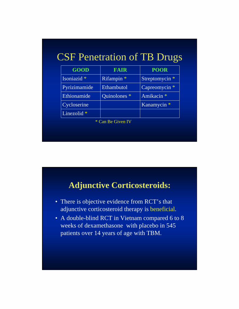
CSF Penetration of TB DrugsGOOD FAIR POOR
Isoniazid * Rifampin * Streptomycin *
Pyrizimamide Ethambutol Capreomycin *
Ethionamide Quinolones * Amikacin *
Cycloserine Kanamycin *
Linezolid *
* Can Be Given IV
Adjunctive Corticosteroids:
• There is objective evidence from RCT’s that adjunctive corticosteroid therapy is beneficial.
• A double-blind RCT in Vietnam compared 6 to 8 weeks of dexamethasone with placebo in 545 patients over 14 years of age with TBM.

Adjunctive Corticosteroids:
Mortality
(%)Stage I: Stage II: Stage III: Total:
Steroid
Group:17 31 55 32
Non-Steroid:
20 40 60NS
41
Adjunctive Corticosteroids:
• There was no reduction in residual neurologic deficits and disability among surviving patients at nine months.
• I.E., Morbidity not improved by steroids.
Adjunctive Corticosteroids:
• The survival benefit associated with a reduction in severe adverse events (9.5 versus 16.6%), particularly drug related hepatitis that required changes to TB drug regimens.
• No mortality benefit from dexamethasone was seen in 98 HIV-infected patients included in the study.
Adjunctive Corticosteroids:
• Adjunctive corticosteroid therapy is recommended for
all children and adults treated for TB meningitis.
Adjunctive Corticosteroids:
• Specific clinical indications include: – Acute "encephalitis," high CSF pressure or brain
edema.
– Clinical worsening after treatment onset.
– Spinal block or impending block (CSF protein >500 mg/dL and rising).
– CT evidence of marked basilar enhancement.
– Symptomatic tuberculomas.
Adjunctive Corticosteroids:
• Concurrent treatment with Rifampin may reduce effective levels of corticosteroids by induction of the hepatic cytochrome P-450 system.

Recommended Corticosteroid regimens:
• Children — Prednisone 2 to 4 mg/kg per day tapered over 4wks.
• Adults either:– Prednisone 60 mg per day tapered gradually over six
weeks, or– Dexamethasone IV for the first 3 weeks (initially
0.4 mg/kg per day, tapering to 0.1 mg/kg per day) followed by oral 4 mg per day, tapered over 3 - 4 weeks.
Surgery
• Hydrocephalus may require urgent shunting.
• Serial LP and steroid therapy may suffice for Stage I pts awaiting response to antibiotics.
• Shunting should not be delayed in patients with stupor, coma or progressive neurologic signs.

Skeletal Tuberculosis:
• Vertebral Osteomyelitis – Pott’s Disease
• Arthritis
• Myositis
• Osteomyelitis, non-axial skeleton
History:
• 39 y.o. Hispanic male presented with F, wt. loss & upper back pain made worse by cough.
• No prior Hx TB or TB treatment.
• Abnl CXR c/w far advanced TB.
• Sputum AFB positive, PCR – MTb.
History 2:
• CT spine and MRI spine revealed discitis, vertebral osteo. and epidural abscess at T2 – 4.
• Sterile pyuria, anemia and hyponatremia.
• Patient started on RIPE.
Exam:
• T 102.0oF, HR 88, BP 84/42, 5’3”, 111 lbs.
• Lungs – upper lobe crackles.
• Neurologic:– Strength 4/5 BLE, weak ankle dorsiflexors.
– Numbness below T8.
– DTR’s 3/4 BLE.
– Asymmetrical plantar flexor reflexes.

BU
L, F
ar A
dvan
ced,
Cav
itar
y T
B.
T1-
T2-
T3-
T4-


Hospital Course:
• Daily fevers.
• Decreased LE strength led to transfer for neurosurgical care.
• Neuro exam revealed reduced strength Rt > Lt, weak right ankle dorsiflexor, numbness below nipples, but intact toe position sense.
Laminectomy T2 – T5:
• Procedure: Posterior laminectomy.
• No discrete epidural mass encountered.
• Thecal sac decompressed.
• Corticosteroids were given during the early post operative phase, later tapered & stopped.

Further Management:
• Pt returned to TCID on P.O.D. 10, afebrile with improved cough.
• He was able to do transfers with improved L.E. strength.
• Pt completed 12 months Tb Rx (INH & RIF).
• Pain free and able to walk at discharge.
Potts Disease:

Potts Paravertebral Abscess:Potts T10 & T11:
24 yo Nigerian Female with 1 yr history of
back pain:
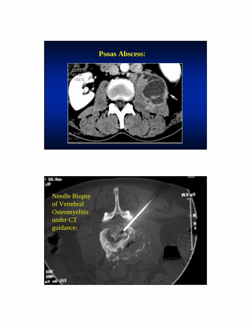
Psoas Abscess:
Needle Biopsy of Vertebral Osteomyelitis under CT guidance:

TB Osteomyelitis Distal Femur:

TB Arthritis of the wrist:
TREATMENT
• Duration of therapy —
• Traditionally, 12 to 18 months of therapy has been advocated.
• At issue: drug penetration into necrotic bone.
• Several studies recommended shorter treatment.
TREATMENT
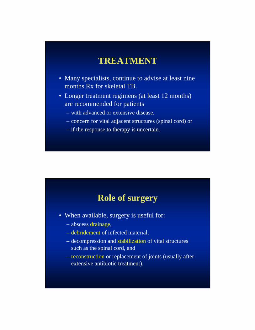
• Many specialists, continue to advise at least nine months Rx for skeletal TB.
• Longer treatment regimens (at least 12 months) are recommended for patients – with advanced or extensive disease,
– concern for vital adjacent structures (spinal cord) or
– if the response to therapy is uncertain.
Role of surgery
• When available, surgery is useful for:– abscess drainage,
– debridement of infected material,
– decompression and stabilization of vital structures such as the spinal cord, and
– reconstruction or replacement of joints (usually after extensive antibiotic treatment).
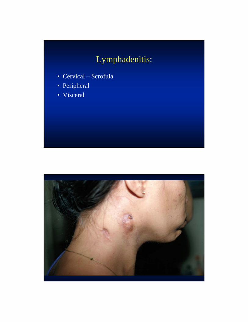
Lymphadenitis:
• Cervical – Scrofula
• Peripheral
• Visceral
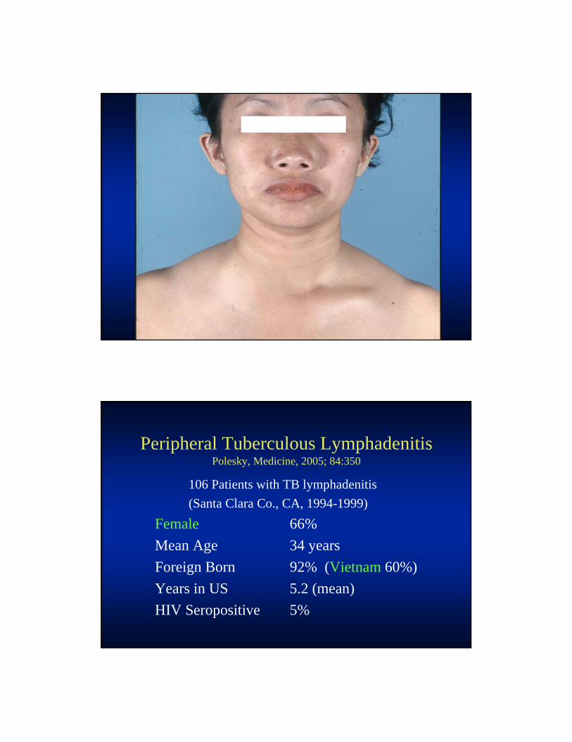
Peripheral Tuberculous LymphadenitisPolesky, Medicine, 2005; 84:350
106 Patients with TB lymphadenitis
(Santa Clara Co., CA, 1994-1999)
Female 66%
Mean Age 34 years
Foreign Born 92% (Vietnam 60%)
Years in US 5.2 (mean)
HIV Seropositive 5%

Necrotic Lymph Node by CT:
43 y.o. HIV+ (CD4=46)Pulmonary TB Started on RIPE Started on HAART
– AZT/3TC/efavirenz
17 Days later….
Immune Reconstitution TB LAD:
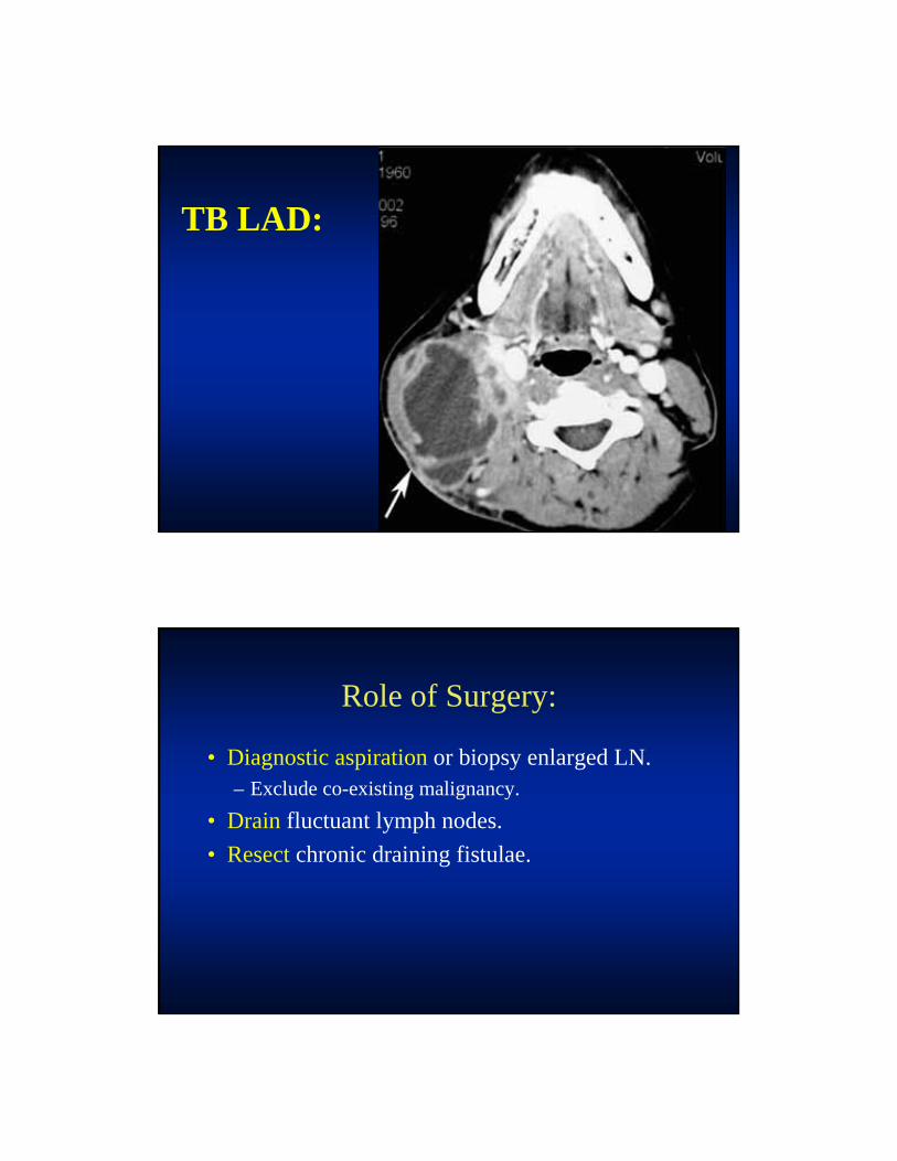
TB LAD:
Role of Surgery:
• Diagnostic aspiration or biopsy enlarged LN.– Exclude co-existing malignancy.
• Drain fluctuant lymph nodes.
• Resect chronic draining fistulae.
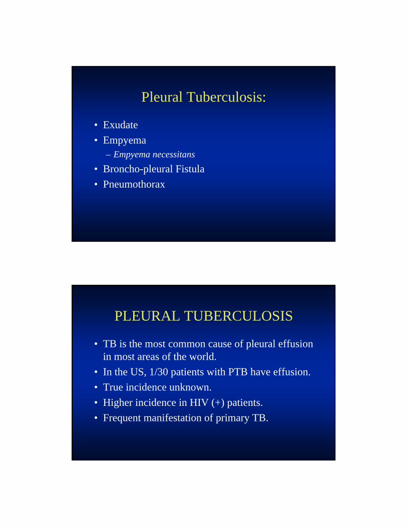
Pleural Tuberculosis:
• Exudate
• Empyema– Empyema necessitans
• Broncho-pleural Fistula
• Pneumothorax
PLEURAL TUBERCULOSIS
• TB is the most common cause of pleural effusion in most areas of the world.
• In the US, 1/30 patients with PTB have effusion.
• True incidence unknown.
• Higher incidence in HIV (+) patients.
• Frequent manifestation of primary TB.


Therapy of Pleural TB:
• Standard RIPE therapy advised for PTB(S).
• AVOID CHEST TUBE – for simple exudates.
• NO DECORTICATION FOR 3-4 MONTHS– Pleural thickening often decreases with time.
– Entrapped lung occasionally requires surgery.
Tb Empyema:
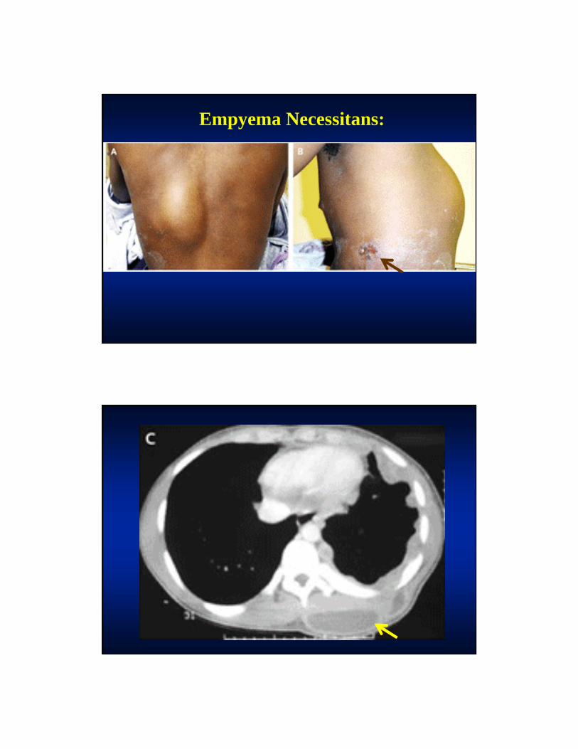
Empyema Necessitans:
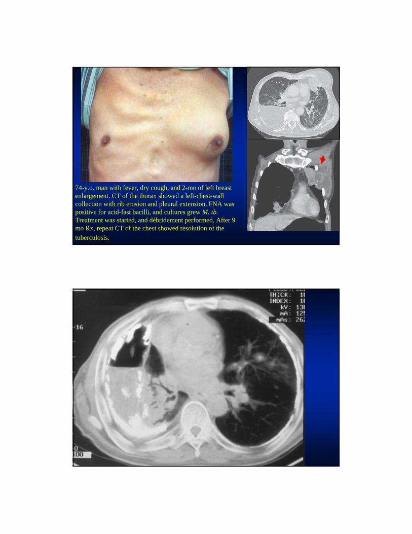
74-y.o. man with fever, dry cough, and 2-mo of left breast enlargement. CT of the thorax showed a left-chest-wall collection with rib erosion and pleural extension. FNA was positive for acid-fast bacilli, and cultures grew M. tb. Treatment was started, and débridement performed. After 9 mo Rx, repeat CT of the chest showed resolution of the
tuberculosis.

Therapy of Pleural TB:
• BP FISTULA (air-fluid level in pleural space):– Does not heal spontaneously.– 90-120 days therapy before decortication.
Role of Surgery:
• Tube thoracostomy management of pneumothorax or empyema.
• Decortication of thickened pleura.
• Release of entrapped lung.
• Closure of BP fistulae with persistent air leak.
• Resection of necrotic pulmonary tissue.
• Muscle flap placement to fill voids.
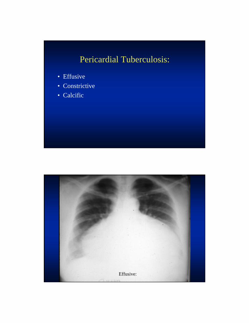
Pericardial Tuberculosis:
• Effusive
• Constrictive
• Calcific
Effusive:

Pericardial Calcification:
Chronic Pericarditis:
Fibrinous Pericarditis:
Constrictive Pericarditis:

TB Pericarditis:
• Large pericardial effusion and inversion of the right atrium, caused by elevated pericardial pressure, in late diastole and early systole.
• A parasternal short-axis view shows that the right ventricular outflow tract is compressed in diastole because of the elevated pericardial pressure.
Nardell E. N Engl J Med 2004; 351:1804-1805
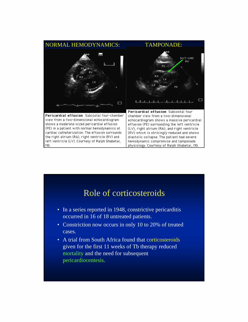
TAMPONADE:NORMAL HEMODYNAMICS:
Role of corticosteroids
• In a series reported in 1948, constrictive pericarditis occurred in 16 of 18 untreated patients.
• Constriction now occurs in only 10 to 20% of treated cases.
• A trial from South Africa found that corticosteroidsgiven for the first 11 weeks of Tb therapy reduced mortality and the need for subsequent pericardiocentesis.

Role of corticosteroids
• The following benefits were noted in the prednisolone group:– More rapid reduction in pulse rate and JVP
– Improved functional status
– Lower mortality during follow-up (4% versus 11%)
– Lower need for pericardiectomy (21% versus 30%)
Role of corticosteroids
• Mortality with effusion drainage, TB therapy and prednisolone was 3%.
• Mortality with drainage and TB therapy was 14%.
Role of corticosteroids
• The ATS, CDC, and IDSA joint guidelines recommend corticosteroids as an adjunct during the first 11 weeks of therapy for TB pericarditis.
Role of corticosteroids
• The recommended regimens are as follows:– For adults, prednisone 60 mg/day for 4 weeks,
• 30 mg/day for 4 weeks,
• 15 mg/day for 2 weeks, then
• 5 mg/day for week 11.
– For children, prednisone 1 mg/kg daily as the initial dose for 4 weeks, with a decreasing dose over time as described for adults.
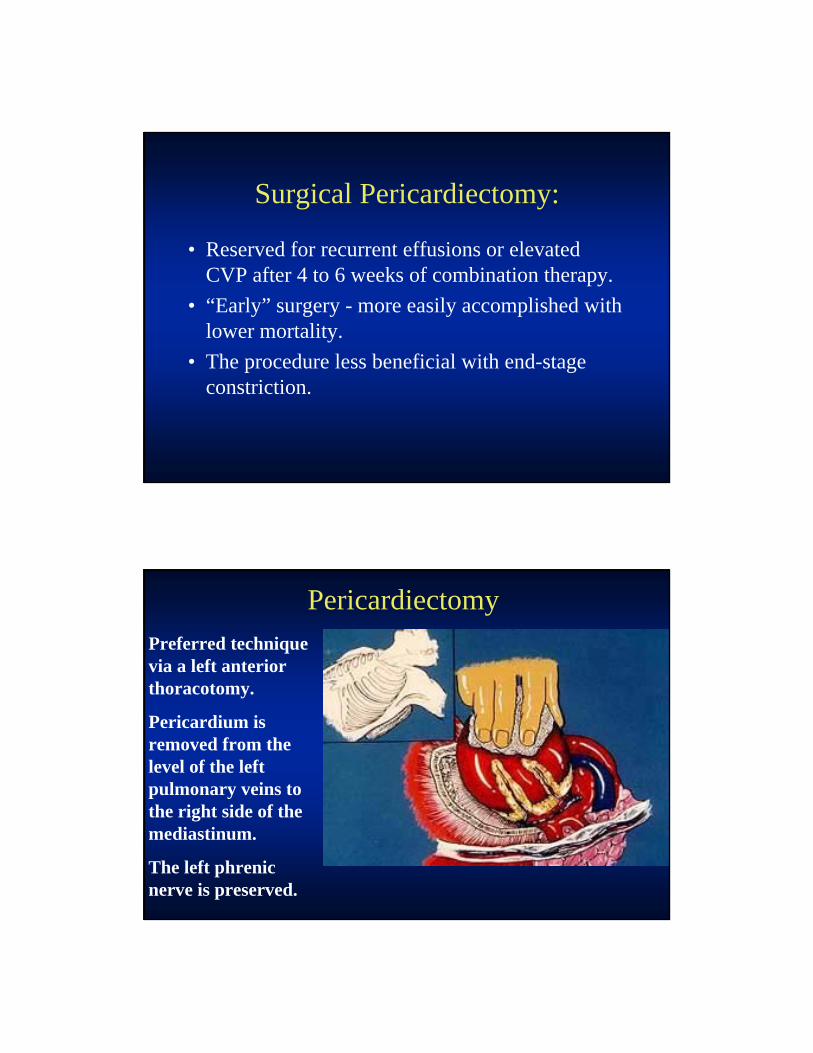
Surgical Pericardiectomy:
• Reserved for recurrent effusions or elevated CVP after 4 to 6 weeks of combination therapy.
• “Early” surgery - more easily accomplished with lower mortality.
• The procedure less beneficial with end-stage constriction.
Preferred technique via a left anterior thoracotomy.
Pericardium is removed from the level of the left pulmonary veins to the right side of the mediastinum.
The left phrenic nerve is preserved.
Pericardiectomy
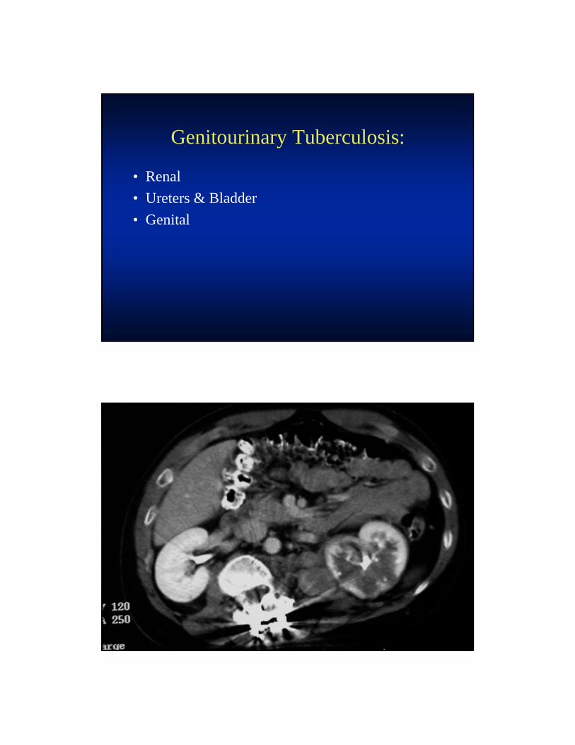
Genitourinary Tuberculosis:
• Renal
• Ureters & Bladder
• Genital
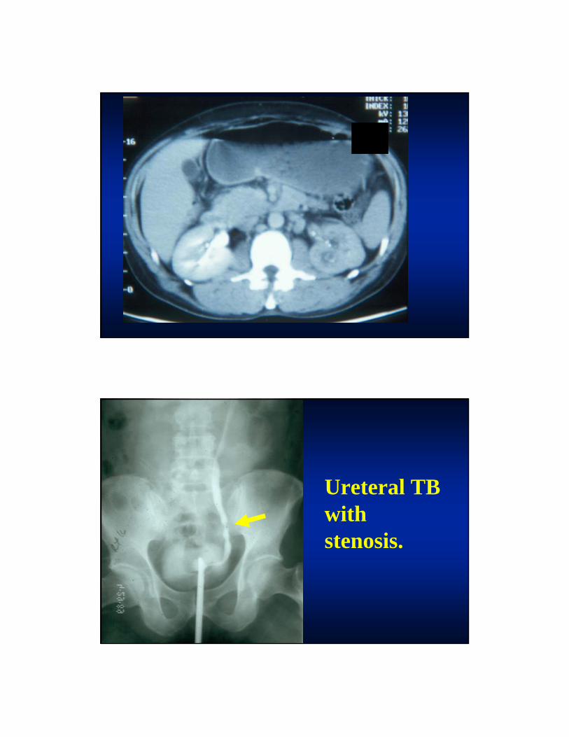
Ureteral TB with stenosis.
Blunting of the calyces (caliectasis) and two long ureteral strictures (arrows) are seen. Although the caliceal changes can be seen in other disorders (such as reflux nephropathy), the concurrent ureteral abnormalities are virtually diagnostic of tuberculosis.
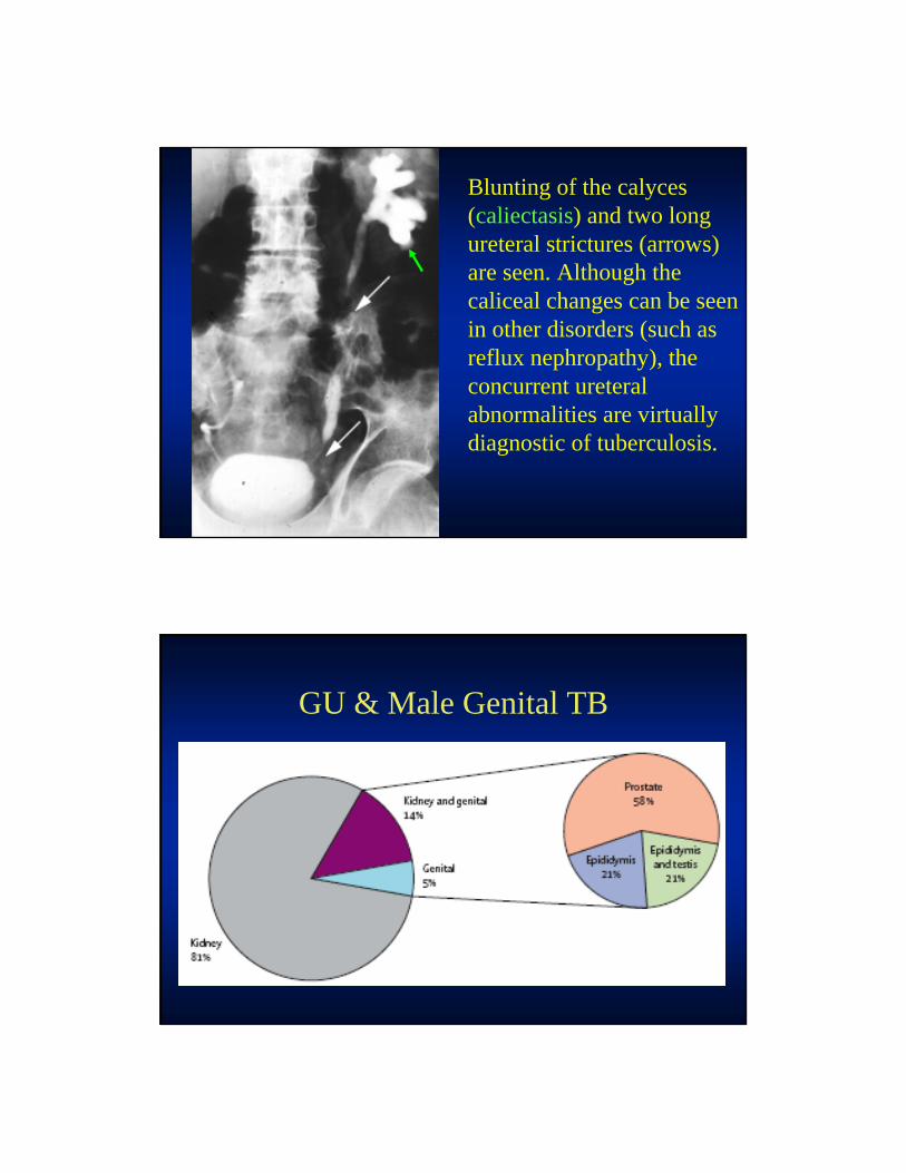
GU & Male Genital TB
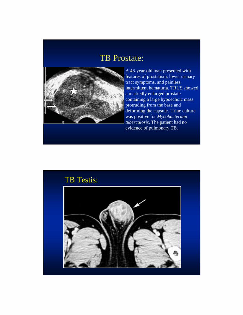
TB Prostate:A 46-year-old man presented with features of prostatism, lower urinary tract symptoms, and painless intermittent hematuria. TRUS showed a markedly enlarged prostate containing a large hypoechoic mass protruding from the base and deforming the capsule. Urine culture was positive for Mycobacterium tuberculosis. The patient had no evidence of pulmonary TB.
TB Testis:
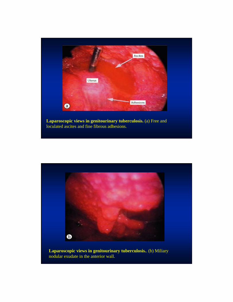
Laparoscopic views in genitourinary tuberculosis. (a) Free and loculated ascites and fine fibrous adhesions.
Laparoscopic views in genitourinary tuberculosis.. (b) Miliary nodular exudate in the anterior wall.
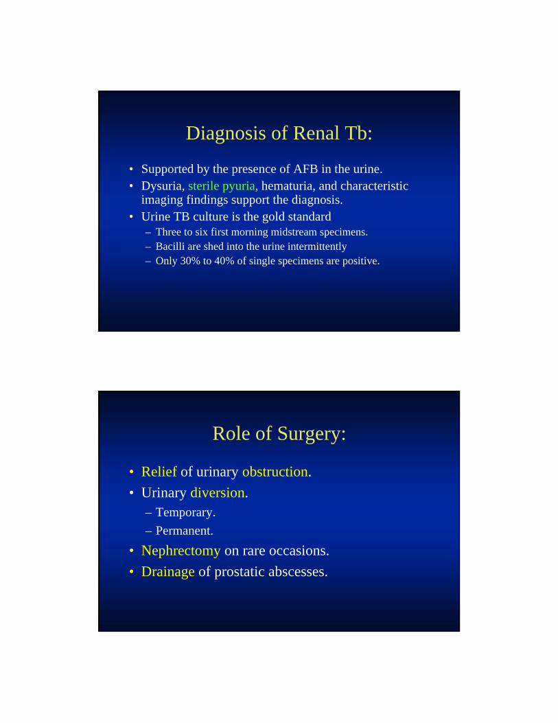
Diagnosis of Renal Tb:
• Supported by the presence of AFB in the urine. • Dysuria, sterile pyuria, hematuria, and characteristic
imaging findings support the diagnosis. • Urine TB culture is the gold standard
– Three to six first morning midstream specimens.– Bacilli are shed into the urine intermittently– Only 30% to 40% of single specimens are positive.
Role of Surgery:
• Relief of urinary obstruction.
• Urinary diversion.– Temporary.
– Permanent.
• Nephrectomy on rare occasions.
• Drainage of prostatic abscesses.
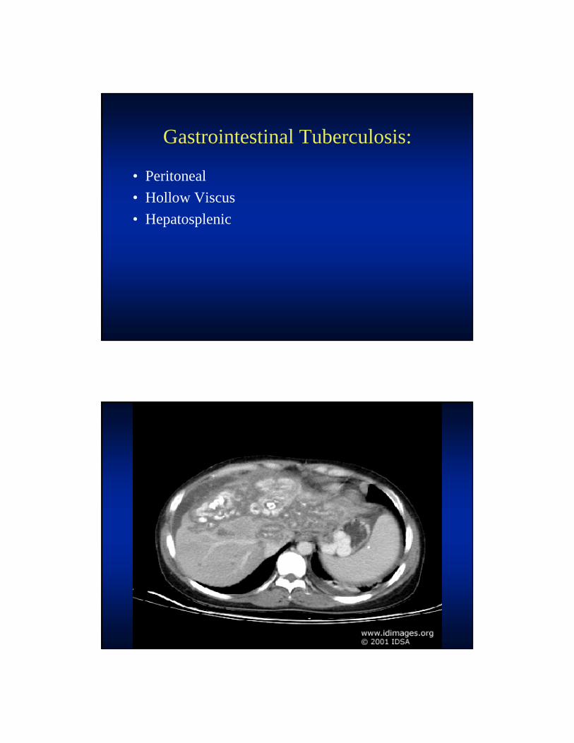
Gastrointestinal Tuberculosis:
• Peritoneal
• Hollow Viscus
• Hepatosplenic
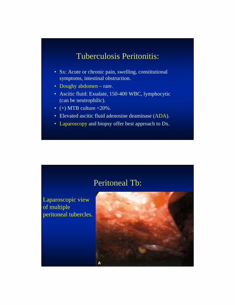
Tuberculosis Peritonitis:
• Sx: Acute or chronic pain, swelling, constitutional symptoms, intestinal obstruction.
• Doughy abdomen – rare.
• Ascitic fluid: Exudate, 150-400 WBC, lymphocytic (can be neutrophilic).
• (+) MTB culture <20%.
• Elevated ascitic fluid adenosine deaminase (ADA).
• Laparoscopy and biopsy offer best approach to Dx.
Peritoneal Tb:
Laparoscopic view of multiple peritoneal tubercles.

Peritoneal Tb.
Laparoscopic view of peritoneal ‘‘spider web’’adhesions. Note the ascitic fluid at the rightbottom corner.
Hollow Viscus Tuberculosis:
• Chronic pain (R L Q), weight loss, anorexia fever
• Colitis (ulcerative, hypertrophic) with predilection for ileocecum.
• Strictures.
• Perforation.
• Endoscopic findings are not definitive, – full thickness biopsy with AFB smear and culture.
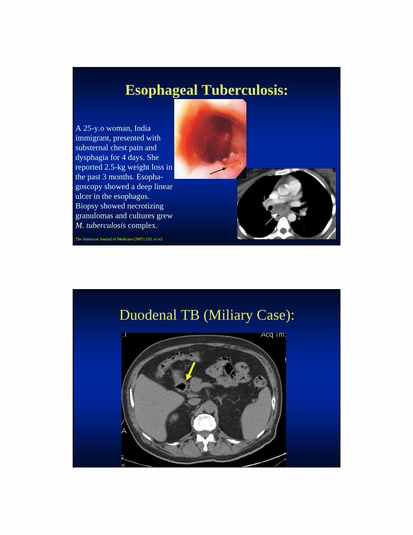
Esophageal Tuberculosis:
A 25-y.o woman, India immigrant, presented with substernal chest pain and dysphagia for 4 days. She reported 2.5-kg weight loss in the past 3 months. Esopha-goscopy showed a deep linear ulcer in the esophagus. Biopsy showed necrotizing granulomas and cultures grew M. tuberculosis complex.
The American Journal of Medicine (2007) 120, e1-e2
Duodenal TB (Miliary Case):

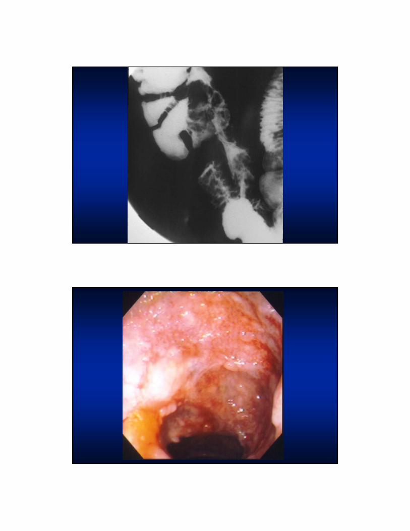
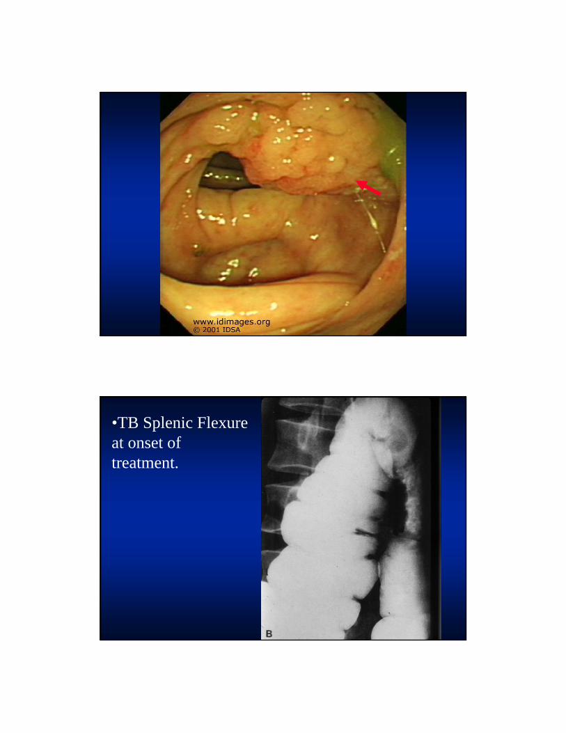
•TB Splenic Flexure at onset of treatment.

•TB Splenic Flexure after 10 weeks of Rx.
84 y.o. WM with change in bowel habits and an normal CXR.
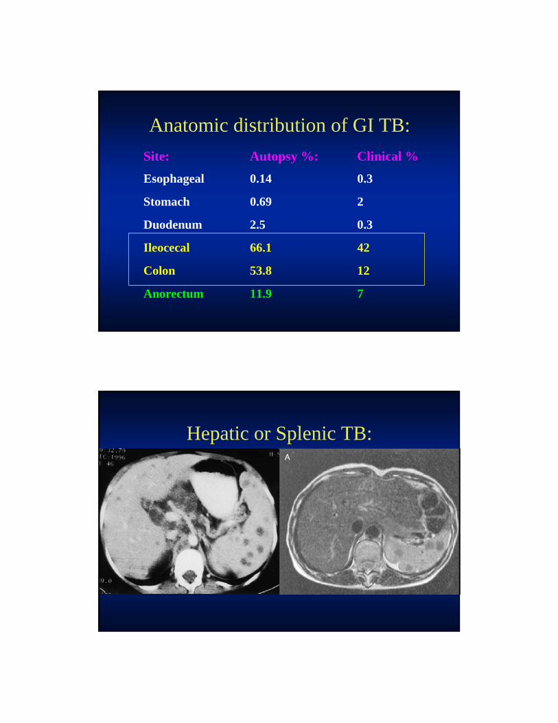
Anatomic distribution of GI TB:
Site: Autopsy %: Clinical %
Esophageal 0.14 0.3
Stomach 0.69 2
Duodenum 2.5 0.3
Ileocecal 66.1 42
Colon 53.8 12
Anorectum 11.9 7
Hepatic or Splenic TB:

Surgical Considerations:
• Laparascopic biopsy to Dx peritonitis.
• Management of strictures, fistulae and perforation which do not respond to medical therapy.
ENT Tuberculosis:
• Laryngitis
• Otitis
• Mastoiditis
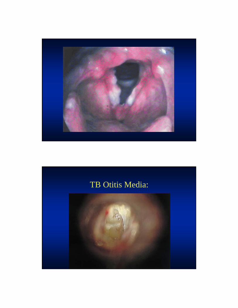
TB Otitis Media:
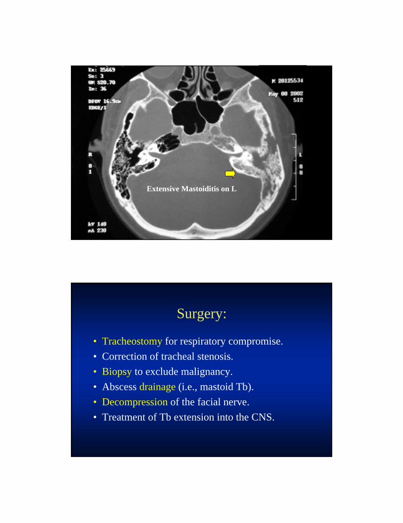
Extensive Mastoiditis on L
Surgery:
• Tracheostomy for respiratory compromise.
• Correction of tracheal stenosis.
• Biopsy to exclude malignancy.
• Abscess drainage (i.e., mastoid Tb).
• Decompression of the facial nerve.
• Treatment of Tb extension into the CNS.

Other EPTB:
• Adrenal
• Thyroid
• Myocardium
• Retina – choroidal tubercles
• Skin – Lupus vulgaris
Adrenal Calcifications from Tb:
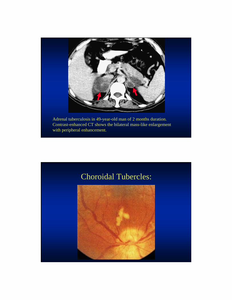
Adrenal tuberculosis in 49-year-old man of 2 months duration. Contrast-enhanced CT shows the bilateral mass-like enlargement with peripheral enhancement.
Choroidal Tubercles:

Lupus vulgaris Reddish-brown plaque. Note nodular infiltration, scaling of the helix, and atrophic scarring in the center of the plaque.
16 y.o. male with PTB & Skin TB x 5yrs:

Miliary TB:
• Wide dissemination throughout the body.
• Tiny size of the lesions.
• Distinctive pattern seen on chest X-ray. – With or without other types of infiltrates.
• Appearance similar to millet seeds, thus the term "miliary" tb.
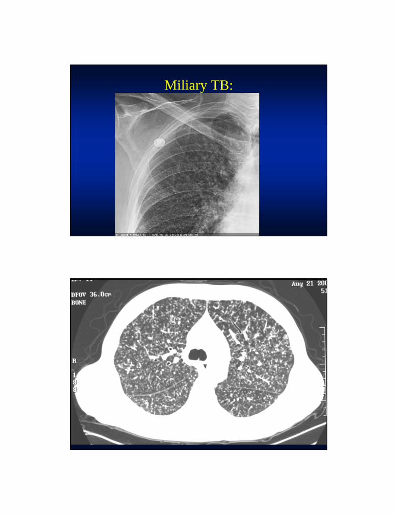
Miliary TB:
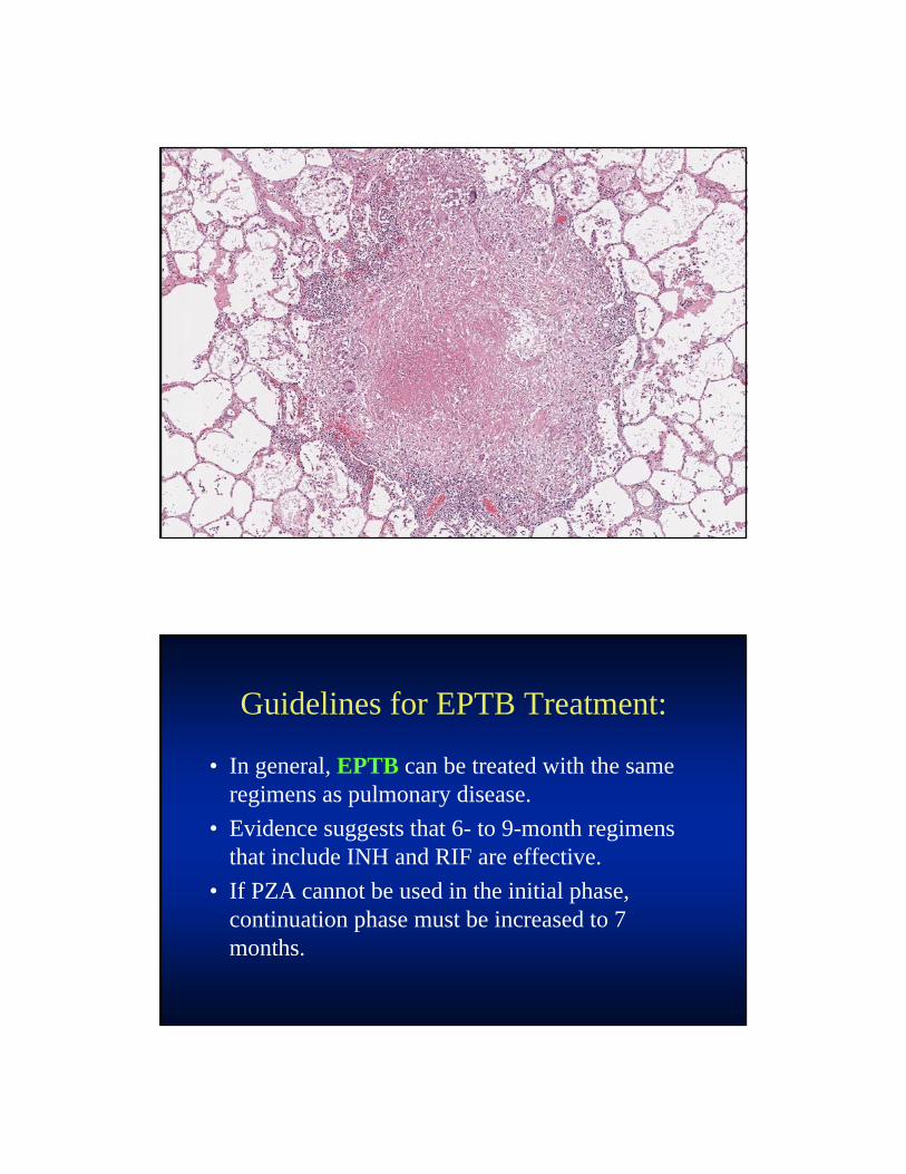
Guidelines for EPTB Treatment:
• In general, EPTB can be treated with the same regimens as pulmonary disease.
• Evidence suggests that 6- to 9-month regimens that include INH and RIF are effective.
• If PZA cannot be used in the initial phase, continuation phase must be increased to 7 months.
Guidelines for EPTB Treatment:
• For disseminated TB and TB meningitis, 9--12 months of treatment is recommended.
• Prolongation of therapy also should be considered for TB in any site that is slow to respond.
• The adjunctive corticosteroids are recommended for TB pericarditis and TB meningitis.