extended report nerve growth factor inhibition with
TRANSCRIPT

EXTENDED REPORT
Nerve growth factor inhibition with tanezumabinfluences weight-bearing and subsequent cartilagedamage in the rat medial meniscal tear modelTimothy P LaBranche,1,2 Alison M Bendele,3 Brian C Omura,3 Kathryn E Gropp,4
Susan I Hurst,4 Cedo M Bagi,4 Thomas R Cummings,4 Lonnie E Grantham II,5
David L Shelton,6 Mark A Zorbas7
Handling editor Tore K Kvien
▸ Additional material ispublished online only. To view,please visit the journal online(http://dx.doi.org/10.1136/annrheumdis-2015-208913).
1Pfizer Inc, Cambridge,Massachusetts, USA2Blueprint Medicines,Cambridge, Massachusetts,USA3Bolder BioPATH, Inc.,Boulder, Colorado, USA4Pfizer Inc, Groton,Connecticut, USA5Arbor Analytics, LLC,Ann Arbor, Michigan, USA6Pfizer Inc, SouthSan Francisco, California, USA7Pfizer Inc, San Diego,California, USA
Correspondence toDr Timothy P LaBranche,Blueprint Medicines, 38 SidneyStreet, Suite 200, Cambridge,MA 02139, USA; [email protected]
Mr Omura died on 10 July2015.
Received 17 November 2015Revised 31 March 2016Accepted 29 April 2016Published Online First5 July 2016
To cite: LaBranche TP,Bendele AM, Omura BC,et al. Ann Rheum Dis2017;76:295–302.
ABSTRACTObjective To investigate whether the effects of nervegrowth factor (NGF) inhibition with tanezumab on ratswith medial meniscal tear (MMT) effectively modelrapidly progressive osteoarthritis (RPOA) observed inclinical trials.Methods Male Lewis rats underwent MMT surgery andwere treated weekly with tanezumab (0.1, 1 or 10 mg/kg), isotype control or vehicle for 7, 14 or 28 days. Gaitdeficiency was measured to assess weight-bearing onthe operated limb. Joint damage was assessed viahistopathology. A second arm, delayed onset oftreatment (starting 3–8 weeks after MMT surgery) wasused to control for analgesia early in the diseaseprocess. A third arm, mid-tibial amputation, evaluatedthe dependency of the model on weight-bearing.Results Gait deficiency in untreated rats was present3–7 days after MMT surgery, with a return to normalweight-bearing by days 14–28. Prophylactic treatmentwith tanezumab prevented gait deficiency and resultedin more severe cartilage damage. When onset oftreatment with tanezumab was delayed to 3–8 weeksafter MMT surgery, there was no increase in cartilagedamage. Mid-tibial amputation completely preventedcartilage damage in untreated MMT rats.Conclusions These data suggest that analgesia due toNGF inhibition during the acute injury phase isresponsible for increased voluntary weight-bearing andsubsequent cartilage damage in the rat MMT model.This model failed to replicate the hypotrophic boneresponse observed in tanezumab-treated patients withRPOA.
INTRODUCTIONKnee osteoarthritis (OA) is a condition charac-terised by pain, inflammation and functional dis-ability.1 OA pain is complex and involves bothinflammatory and neuropathic componentsmediated through persistent tissue injury andrelease of inflammatory mediators.2 Pain treatmentfor OA is problematic because many standard ther-apies provide minimal pain relief and do notaddress underlying mechanisms driving diseasepathophysiology.3
The neurotrophin, nerve growth factor (NGF), isconsidered a key modulator of pain perception inseveral chronic pain conditions, including OA.4–7
Tanezumab, a humanised monoclonal antibody,
binds NGF and prevents interaction with its recep-tors (high-affinity transmembrane tyrosine kinasereceptor (TrkA) and the low-affinity NGF receptor[p75]).8 Tanezumab provided significant improve-ment in pain, physical function and patients’ globalassessments in a number of chronic pain condi-tions.9–16
Investigator reports of adverse events initiallydescribed as osteonecrosis leading to total jointreplacement during the conduct of phase III clinicalOA studies led the US Food and DrugAdministration to put trials of all NGF inhibitorson partial clinical hold.17–19 Blinded adjudicationof the results showed that there was no increase inosteonecrosis, nor frequency of total joint replace-ment with tanezumab monotherapy. Tanezumabtreatment was however associated with increasedincidence of rapidly progressive osteoarthritis(RPOA). A retrospective analysis of the data sug-gested ways to mitigate this risk, and based onthese data the clinical hold was lifted to allowfurther trials to test these risk mitigationapproaches.17–20
The increased frequency of RPOA was unex-pected, as no issues with bone or joints were seenin non-clinical studies of anti-NGF therapy usinglarge multiples of the clinical dose.21 Also, no evi-dence of abnormal bone or joint phenotypes existsin humans with TrkA or p75 null mutations otherthan that observed in congenital pain insensitivitymutations.5 Last, in an experimental fracturemodel, anti-NGF therapy was shown to amelioratefracture pain without impacting bone healing.22
Meniscal injury and acute meniscectomy areknown to increase the risk of knee OA.23 Up to80% of patients with knee OA, as well as a highpercentage of age-matched controls, have evidenceof meniscal injury at the time of diagnosis yetabnormal load-bearing and joint instability stem-ming from meniscal injury resulting in substantialOA lesions may take years.23 Attempts to modelthis in animals have produced varied results.24 25
The medial meniscal tear (MMT)-induced jointdamage model in rats has many features attractivefor an animal model. These animals show jointinstability and tibial cartilage damage in as little as7–14 days after surgery.25 MMT-induced jointdamage lesions are highly reproducible and includearticular cartilage proteoglycan loss, chondrocyte
LaBranche TP, et al. Ann Rheum Dis 2017;76:295–302. doi:10.1136/annrheumdis-2015-208913 295
Basic and translational research on F
ebruary 21, 2022 by guest. Protected by copyright.
http://ard.bmj.com
/A
nn Rheum
Dis: first published as 10.1136/annrheum
dis-2015-208913 on 5 July 2016. Dow
nloaded from

degeneration and loss of matrix. Although erosion of cartilage isa feature of this model, rarely does it progress to ulcerationwithin 14 or 28 days. Subchondral bone sclerosis and osteo-phyte formation, which are compensatory responses to alteredmechanical loading and joint instability are present in thismodel.24 26–28
The objective of this study was to characterise the impact ofNGF inhibition with tanezumab on voluntary weight-bearingand subsequent articular cartilage damage in MMT rats to see ifthis model would be useful for investigating potential mechan-isms underlying the clinical findings of increased RPOA inpatients treated with tanezumab.
METHODSAnimals and tanezumab administrationFor animal care and use, see online supplementary text S1.
All animals underwent MMT or sham surgery on study day0. In MMT surgery rats, the medial meniscus in the right hindlimb was cut through the full thickness to simulate a completetear.24 25 In sham surgery animals, the knee was opened and themedial meniscus was touched only (not cut). Sham surgery ratsreceived vehicle, and animals that underwent MMT surgerywere treated with either isotype control (a non-specific antibodyof the same type (immunoglobulin G) and class (2a) as tanezu-mab), vehicle (10 mM trehalose buffer) or tanezumab (0.1, 1 or10 mg/kg) subcutaneously once a week for 7, 14 or 28 days.
In the first arm of the study, treatment began on the day ofsurgery (day 0, figure 1A).29 To examine the impact of analgesiaon weight-bearing and lesion severity, a second arm evaluateddelaying the onset of tanezumab treatment (0.1 mg/kg weekly)until gait deficiency was no longer evident (day 23); half theanimals started receiving treatment on day 23, while the otherhalf were delayed another month (day 57) before treatment wasinitiated (figure 1B). In both arms of this study, anti-drug anti-bodies in plasma were measured, as well as concentration ofparent compound in serum to evaluate the potential for clear-ance of tanezumab via immunologic response.
To understand the role of weight-bearing in the progressionof knee joint damage in the rat MMT model, a third arm of thestudy evaluated the impact of mid-tibial amputation on MMTlesion progression, eliminating weight-bearing as a variable. Theknees were collected 14 days after MMT for micro-CT (mCT)imaging, followed by histopathology (figure 1C). In the first twostudy arms, the impact of NGF inhibition on subchondral bone,osteophyte size and growth plate thickness was evaluated inMMTrats to assess secondary effects on the overall joint.
Gait deficiencyDifference in dynamic weight-bearing between the ipsilateral(MMT operated; right side) limb and contralateral (control; leftside) limb is an established approach to measuring gait in ratand mouse models of OA.30 31 Voluntary weight-bearing bynon-amputated MMTrats was assessed on days −3, 3, 7, 14 and28 in the first arm of the study and additional days 37, 42, 56,63 and 71 in the delayed-treatment study to confirm that therewas no pain relapse in any animals (see online supplementarytext S2).
Histopathology and quantitative image analysisMicroscopy-based semiquantitative scoring of cartilage andbone changes is an established endpoint for describing responseto compounds in MMT rats. Cohorts of animals were sacrificed7, 14 or 28 days after surgery or 14 days after treatment onset(day 23 or 57). Knees were collected at necropsy and evaluated
via light microscopy by a veterinary pathologist as described(see online supplementary text S3).25
Radiography and mCT imagingKnees were X-rayed with a MX20 specimen scanner (FaxitronBioptics, Tucson, Arizona, USA) using recommended settings;exposure 12–18 s at 31−35 kV. Radiographs were used to assessgross anatomy of the region of interest (ROI) evaluated by mCTand to inspect bone samples for presence of abnormalities.
mCTwas conducted on the right tibial epiphysis and metaphy-sis using a MicroCT 100 system (Scanco Medical, Bassersdorf,Switzerland) with the following parameters: 800 slices, 10 mmresolution, total scanned area of 8.0 mm2 and source energy of70 kVp, 115 mA at 8 W to capture the entire proximal tibia
Figure 1 Study designs for (A) the initial arm (28-day study), (B) thesecond arm (delayed-treatment onset study) and (C) the weight-bearing(amputation) study. GA, gait analysis; HA, histopathology analysis;MMT, medial meniscal tear; SC, subcutaneous.
296 LaBranche TP, et al. Ann Rheum Dis 2017;76:295–302. doi:10.1136/annrheumdis-2015-208913
Basic and translational research on F
ebruary 21, 2022 by guest. Protected by copyright.
http://ard.bmj.com
/A
nn Rheum
Dis: first published as 10.1136/annrheum
dis-2015-208913 on 5 July 2016. Dow
nloaded from

section. Cortical and cancellous bone of the entire epiphysiswere scanned and analysed in an ROI on 100 consecutiveslices with 1.0 mm thickness that best represented the centralsegment of the epiphysis. Parameters included tissue volume(TV), bone volume (BV), BV/TV ratio and bone mineraldensity (BMD). Cortical and cancellous bone of the proximalmetaphysis were scanned and analysed in an ROI on 100 con-secutive slices 1 mm below the growth plate with a thicknessof 1.0 mm that best represented the central metaphysissegment. Parameters included TV, BV, BV/TV and BMD.Cancellous bone compartment of the metaphysis was analysed1 mm below the growth plate and extending 3 mm distally toinclude only the secondary spongiosa. Cancellous bone wasevaluated in an ROI drawn on 100 consecutive slices with athickness of 1.0 mm that best represented the central segmentof the tibia.32 Cancellous bone parameters included TV, BV,BV/TV, trabecular number, trabecular thickness, trabecular sep-aration and BMD.
Pharmacokinetics and anti-drug antibodyTo evaluate the potential for clearance of compound viaimmunologic response, we measured anti-drug antibodies inplasma as well as concentration of tanezumab in serum pharma-cokinetic (PK) samples. Blood samples were collected on days 3,7, 14 and 21. An additional PK sample was taken 4 days priorto termination. In the delayed-treatment study, blood samplesfor PK and anti-drug antibodies were collected on days 16, 30,57 and 64. PK plasma concentrations of tanezumab were mea-sured using a validated ELISA at ICON Development Solutions(Whitesboro, New York, USA). The lower limit of quantitationin Lewis rat plasma was 50 ng/mL. Concentrations below thelimit of quantitation were treated as 0 ng/mL in the PK calcula-tions. Anti-drug antibodies concentrations in serum were mea-sured using a validated ELISA at ICON Development Solutions.Final assessment of induction of an anti-drug antibodiesresponse was based on comparison of predose and postdoseresults for each animal.
Figure 2 Weight-bearing corresponds with severity of cartilage damage in MMT rats. (A) Gait deficiency in tanezumab-treated MMT rats comparedwith control treatment and sham surgery rats. Data expressed as per cent decrease from mean of right and left (SE). Starting on day 7, 10 animalsper treatment group were removed for microscopic evaluation. (B) Substantial tibial cartilage degeneration width, as determined by histopathologyon days 7, 14 and 28 (n=10 rats/group). (C) Representative photomicrographs (50× magnification) from the isotype and sham surgery controls andtanezumab treatment groups on day 28. Days reflect days after MMT surgery. *p<0.05 versus isotype control; **p<0.01 versus isotype control;***p<0.001 versus isotype control. CD, cartilage degeneration; MMT, medial meniscal tear.
LaBranche TP, et al. Ann Rheum Dis 2017;76:295–302. doi:10.1136/annrheumdis-2015-208913 297
Basic and translational research on F
ebruary 21, 2022 by guest. Protected by copyright.
http://ard.bmj.com
/A
nn Rheum
Dis: first published as 10.1136/annrheum
dis-2015-208913 on 5 July 2016. Dow
nloaded from

Statistical analysesBody weight change, weight-bearing (gait) deficiency at eachtime point and histopathology outcomes were analysed. Vehicleand sham surgery groups were compared with the isotype groupusing a two-sided t test (with a Welch correction for unequalvariances if necessary) or Mann-Whitney U test. Eachtanezumab-treated group was compared with the isotype groupusing a two-sided Dunnett or Dunn multiple comparisons test.Significance for all tests was set at p≤0.05. Gait deficiency wasassessed by Dunnett and t-tests.
RESULTSTanezumab treatment influences weight-bearing andsubsequent cartilage damage in the rat MMT modelAlthough MMTrats with intact hind limbs appeared to ambulatenormally after surgery, they experienced a measurable weight-bearing (gait) deficiency on the operated limb. Gait deficiency inMMTrats administered vehicle or isotype control peaked 3 daysafter surgery (11.8% and 12.2% decrease, respectively), contin-ued through day 7 and resolved by days 14–28 (figure 2A). Incontrast, tanezumab-treated MMT rats exhibited no gait defi-ciency at any point, indicating that tanezumab-treated MMTratswere bearing full weight on the operated limbs throughout thestudy (figure 2). Tanezumab-treated rats exhibited more severetotal tibial cartilage substantial degeneration width than eithervehicle or isotype controls on days 7, 14 and 28 (figure 2B). Nosignificant differences in body weight gain were observed overthe course of the study. Focal areas of alopecia along the mouth/muzzle began developing on day 14 and were seen in mostanimals in the 1 mg/kg tanezumab group by day 28. Gait analysiswas also performed as part of the delayed-treatment study (seeonline supplementary table S1).
There was evidence of a robust, compensatory (hypertrophic)response to the partially destabilised joint. Namely, a time-dependent increase in subchondral bone sclerosis was observedin untreated MMTrats (table 1), which was exacerbated by tane-zumab (statistically significant greater subchondral sclerosis com-pared with isotype controls on days 7, 14 and 28). Largerosteophyte size was also observed in tanezumab-treated MMTrats compared with isotype controls on days 7 (tanezumab10 mg/kg, p<0.05), 14 (tanezumab 1 mg/kg, p<0.01) and 28(tanezumab 0.1, 1 and 10 mg/kg, p<0.001 for all; table 1), indi-cating a time–dose relationship with increasing effect. No osteo-phytes were observed in sham surgery animals. Last, a significantfocal increase in growth plate thickness was also observed intanezumab-treated MMT rats compared with isotype control-treated rats (1 and 10 mg/kg on days 14 and 28, p≤0.05, table 1).
Delaying onset of tanezumab treatment protects againstcartilage damage in MMT ratsGait deficiency began shortly after MMT surgery and appearedto resolve by days 14–28. To confirm whether changes inweight-bearing during this early postoperative period was theprincipal driver of increased joint damage in the tanezumab-treated MMT rats, we delayed the onset of treatment from day0 to day 23 (cohort 1) or day 57 (cohort 2). The second cohortin particular initiated treatment well past the time that gaitabnormalities normalised. Tanezumab-treated MMTrats had sig-nificantly greater cartilage degeneration width than MMT con-trols when treatment began on day 23 (p=0.035, figure 3).However, delaying treatment until weight-bearing fully returnedto normal (ie, day 57 but not day 23) resulted in complete pro-tection from worsening of joint damage when compared withcontrol treatments (figure 3).
Table 1 Histopathology summary, n=10/group; mean (SE)
Parameter/day Isotype control Vehicle control Sham surgeryTanezumab0.1 mg/kg/week
Tanezumab1 mg/kg/week
Tanezumab10 mg/kg/week
Growth plate difference (medial-lateral), mm
7 41.33 (5.43) 33.33 (2.22) 5.33 (2.95)*** 50.67 (10.29) 60.00 (8.94) 70.67 (11.60)
14 68.00 (15.83) 50.67 (7.38) 46.67 (5.71) 74.67 (8.93) 164.00 (26.15)** 141.33 (22.59)*
28 33.33 (4.10) 32.00 (4.07) 16.00 (5.55)* 65.33 (10.60) 185.33 (50.73)*** 161.33 (76.24)**
Medial collateral ligament, mm
7 865.33 (26.70) 893.33 (13.91) 870.67 (35.84) 1068.00 (33.46)** 1041.33 (48.42)* 1128.00 (60.65)***
14 637.33 (26.28) 633.33 (31.13) 641.33 (28.63) 664.00 (24.49) 689.33 (31.74) 688.00 (29.02)
28 476.00 (16.03) 466.67 (15.78) 472.00 (20.09) 530.67 (17.87) 570.67 (22.82)** 545.33 (15.71)*
Bone damage score†
7 2.00 (0.15) 2.00 (0.00) 0.00 (0.00)*** 2.40 (0.16) 2.20 (0.13) 2.20 (0.13)
14 2.80 (0.25) 2.60 (0.16) 0.00 (0.00)*** 3.10 (0.31) 2.60 (0.16) 2.70 (0.30)
28 3.00 (0.26) 3.40 (0.16) 0.00 (0.00)*** 4.60 (0.16)** 4.40 (0.34)** 4.20 (0.29)*
Bone sclerosis score‡
7 1.30 (0.15) 1.20 (0.13) 0.20 (0.13)*** 2.30 (0.15)** 2.10 (0.10)* 2.70 (0.26)***
14 2.20 (0.20) 2.50 (0.17) 0.10 (0.10)*** 3.50 (0.17)** 3.70 (0.15)*** 3.80 (0.13)***
28 2.80 (0.25) 3.70 (0.26)* 0.20 (0.13)*** 4.60 (0.16)*** 4.80 (0.13)*** 4.70 (0.15)***
Tibial osteophyte size, mm
7 119.33 (26.79) 121.33 (26.88) 0 (0.00)*** 167.33 (21.79) 150.33 (29.65) 223.67 (14.59)*
14 309.67 (10.90) 299.67 (16.03) 0 (0.00)*** 382.67 (27.93) 476.67 (43.62)** 417.67 (41.30)
28 581.67 (34.20) 601.67 (27.38) 0 (0.00)*** 1106.67 (49.20)*** 1068.33 (75.30)*** 1093.33 (50.63)***
*p<0.05 versus isotype control; **p<0.01 versus isotype control; ***p<0.001 versus isotype control.†Damage to calcified cartilage and subchondral bone was scored on a 6-point numerical scale (0=no changes to 5=increased basophilia; marked to severe fragmentation of calcifiedcartilage, mesenchymal change in marrow involves up to three-fourths of the total area and articular cartilage has collapsed into the epiphysis to a depth of >250 mm from tidemark).‡Medial tibial subchondral/epiphysial bone sclerosis was scored on a 6-point numerical scale (0=no changes to 5=76%–100% increase in subchondral or epiphysial trabecular bonethickness in medial versus lateral; very little marrow space remains in medial tibia).
298 LaBranche TP, et al. Ann Rheum Dis 2017;76:295–302. doi:10.1136/annrheumdis-2015-208913
Basic and translational research on F
ebruary 21, 2022 by guest. Protected by copyright.
http://ard.bmj.com
/A
nn Rheum
Dis: first published as 10.1136/annrheum
dis-2015-208913 on 5 July 2016. Dow
nloaded from

Limb amputation protects against joint damage inuntreated MMT ratsAs reported,26 as well as in this study (figure 4) signs of tibialarticular cartilage damage appear in normal weight-bearingMMT rats in as early as 7 days after surgery. In this study, thosechanges were prevented by tibial amputation (figure 4). MMTrats with tibial amputations are not placing weight on theiroperated limbs, as evidenced by less cortical and cancellousbone in amputated rats than unamputated rats, regardless ofwhether they had MMT surgery performed (table 2 andfigure 4C).
PK and anti-drug antibody levelsFor PK and anti-drug antibodies results, see online supplemen-tary text S4.
DISCUSSIONThe serious joint adverse events observed in placebo-controlledclinical trials of anti-NGF antibodies have been adjudicated aseither normal or RPOA, the latter characterised byatrophic bone with subchondral insufficiency and/or occasionalfoci of secondary osteonecrosis.33 This clinical finding wasunexpected, since extensive non-clinical safety studies failed tofind adverse events of bone or joint. The current study wasundertaken to see if a commonly used and relevant arthritismodel, the rat MMT, would be useful for further understandingthe finding.
Treating MMT rats with tanezumab did not recapitulate theatrophic bone destruction associated with RPOA in clinicaltrials. Although anti-NGF treatment early in the MMT diseaseprocess influenced weight-bearing and drove additional cartilagedamage, secondary changes in subchondral bone were insteadhypertrophic (subchondral bone thickening, with osteophytes
present). There were however notable findings related to weight-bearing and its impact on joint damage, highlighting that thismodel is ideal for evaluating the relationship between jointinstability, changes in weight/load-bearing and health of articularcartilage damage.24 27 Cartilage damage and subsequent effectson bony tissues (ie, subchondral bone sclerosis and osteophyteformation) appear to be exquisitely sensitive to weight-bearingearly in the rat MMT disease process (ie, 0–14 days after menis-cal tear). In contrast, treatment in the chronic phase after gaitdeficiency had already normalised, similar to the clinical trialexperience, had no adverse effects on the joint.
Mid-tibial amputation protected against MMT-induced jointdamage, further suggesting that dynamic weight-bearing is thekey driver for joint damage in this model. For instance, therewas more severe decreased bone volume in the proximal tibialmetaphysis of amputated rats versus normal rats. The idea thatweight-bearing drives joint damage in the MMT rat is also sup-ported by observations that MMTrats given intra-articular injec-tions of irritating substances leading to joint swelling and painactually result in decreased cartilage damage and bone sclerosisscores, largely because the rats are unwilling to bear weight onthe joint (A. Bendele, unpublished observations).30
In this study, MMT rats exhibited gait deficiency on the oper-ated limb shortly after surgery, which returned to baseline bydays 14–28. Transection of the meniscus leads to this gait abnor-mality, not general surgery trauma, as animals undergoing shamsurgery exhibited no change in weight-bearing at any point.Treatment with tanezumab at ≥0.1 mg/kg resulted in MMT ratsbehaving similarly to sham surgery rats, since they did notexhibit altered weight-bearing. These results, combined withother work investigating the role of NGF in a meniscal transec-tion rat model of OA, suggest that analgesia can significantlyinfluence voluntary weight-bearing.
Figure 3 Delaying onset of treatmentfrom day 0 until after gait deficiencyreturns to normal (day 57 but not day23) resulted in no difference incartilage damage between treated anduntreated MMT rats sacrificed 14 daysafter treatment. Data points includemean substantial tibial cartilagedegeneration width measurements(tanezumab 0.1 mg/kg/week for14 days; n=10 rats/group). Days reflectdays after MMT surgery. CD, cartilagedegeneration; MMT, medial meniscaltear.
LaBranche TP, et al. Ann Rheum Dis 2017;76:295–302. doi:10.1136/annrheumdis-2015-208913 299
Basic and translational research on F
ebruary 21, 2022 by guest. Protected by copyright.
http://ard.bmj.com
/A
nn Rheum
Dis: first published as 10.1136/annrheum
dis-2015-208913 on 5 July 2016. Dow
nloaded from
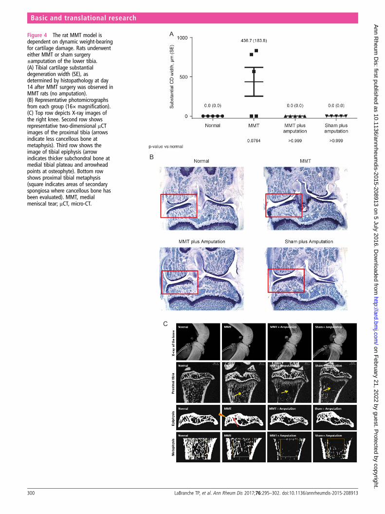
Figure 4 The rat MMT model isdependent on dynamic weight-bearingfor cartilage damage. Rats underwenteither MMT or sham surgery±amputation of the lower tibia.(A) Tibial cartilage substantialdegeneration width (SE), asdetermined by histopathology at day14 after MMT surgery was observed inMMT rats (no amputation).(B) Representative photomicrographsfrom each group (16× magnification).(C) Top row depicts X-ray images ofthe right knee. Second row showsrepresentative two-dimensional μCTimages of the proximal tibia (arrowsindicate less cancellous bone atmetaphysis). Third row shows theimage of tibial epiphysis (arrowindicates thicker subchondral bone atmedial tibial plateau and arrowheadpoints at osteophyte). Bottom rowshows proximal tibial metaphysis(square indicates areas of secondaryspongiosa where cancellous bone hasbeen evaluated). MMT, medialmeniscal tear; μCT, micro-CT.
300 LaBranche TP, et al. Ann Rheum Dis 2017;76:295–302. doi:10.1136/annrheumdis-2015-208913
Basic and translational research on F
ebruary 21, 2022 by guest. Protected by copyright.
http://ard.bmj.com
/A
nn Rheum
Dis: first published as 10.1136/annrheum
dis-2015-208913 on 5 July 2016. Dow
nloaded from

Results suggest that at least two separable phases of painbehaviours occur in the rat MMT model: (1) an early phase, inwhich gait abnormality is present and dynamic weight-bearingsignificantly alters the future trajectory of joint damage, and (2)a later phase, in which there is no detectable gait abnormalityand tissue destruction is no longer sensitive to NGF inhibition.From this, we propose that increased joint damage seen intanezumab-treated MMT rats was due to an increase in volun-tary weight-bearing early in the disease process and not due todirect toxic effect of tanezumab on cartilage or bone. Tosupport this, an additional arm of the study was conducteddelaying onset of tanezumab treatment from day 0 to day 23 or57 after MMT surgery when gait deficiency was no longerpresent. Overall, the results presented here support the conclu-sion that analgesia may exacerbate joint damage when it ispresent during the early phase. This is likely due to mechanicaloverloading shortly after meniscal injury. The relevance of thismodel to RPOA reported in human clinical trials should be con-sidered. Specifically, results from this study may help guidefuture clinical trial design by suggesting exclusion criteria (ie,patients deemed sensitive to mechanical overloading/radio-graphic evidence of bone or joint insufficiency).
Acknowledgements The authors acknowledge the excellent technical assistanceof Phil Bendele at Bolder BioPATH for the tibial amputation model, as well as ICONlaboratories for the determination of PK and anti-drug antibodies parameters. Theauthors also acknowledge Drs Larry Whiteley, Nasir Khan and Lloyd Dethloff (Pfizer)for their guidance and critical review of this work. Medical writing support wasprovided by Joseph Oleynek at Engage Scientific Solutions and was funded by EliLilly & Co. and Pfizer.
Contributors TPL, KEG, TRC, DLS and MAZ designed the study. BCO, AMB, SIH andCMB generated data for the study. LEG performed the statistical analyses. All authorswere involved in data interpretation and writing and reviewing the manuscript.
Funding This study was funded by Pfizer, Inc. and Eli Lilly & Co.
Competing interests KEG, SIH, CMB, TRC and MAZ are employees of Pfizer andown stock and/or stock options in Pfizer. TPL and DLS were employees of Pfizer atthe time of these studies and own stock and/or stock options in Pfizer. LEG is acontracted resource of and owns stock in Pfizer.
Ethics approval Oversight by local institutional animal care and use committees.
Provenance and peer review Not commissioned; externally peer reviewed.
Open Access This is an Open Access article distributed in accordance with theCreative Commons Attribution Non Commercial (CC BY-NC 4.0) license, whichpermits others to distribute, remix, adapt, build upon this work non-commercially,and license their derivative works on different terms, provided the original work isproperly cited and the use is non-commercial. See: http://creativecommons.org/licenses/by-nc/4.0/
REFERENCES1 Dillon CF, Rasch EK, Gu Q, et al. Prevalence of knee osteoarthritis in the United
States: arthritis data from the Third National Health and Nutrition ExaminationSurvey 1991–94. J Rheumatol 2006;33:2271–9.
2 Kidd BL, Langford RM, Wodehouse T. Arthritis and pain. Current approaches in thetreatment of arthritic pain. Arthritis Res Ther 2007;9:214.
3 Hochman JR, French MR, Bermingham SL, et al. The nerve of osteoarthritis pain.Arthritis Care Res (Hoboken) 2010;62:1019–23.
4 Hefti FF, Rosenthal A, Walicke PA, et al. Novel class of pain drugs based onantagonism of NGF. Trends Pharmacol Sci 2006;27:85–91.
5 Mantyh PW, Koltzenburg M, Mendell LM, et al. Antagonism of nerve growthfactor-TrkA signaling and the relief of pain. Anesthesiology 2011;115:189–204.
6 Walsh DA, McWilliams DF, Turley MJ, et al. Angiogenesis and nerve growth factorat the osteochondral junction in rheumatoid arthritis and osteoarthritis.Rheumatology (Oxford) 2010;49:1852–61.
7 Watson JJ, Allen SJ, Dawbarn D. Targeting nerve growth factor in pain: what is thetherapeutic potential? BioDrugs 2008;22:349–59.
8 Abdiche YN, Malashock DS, Pons J. Probing the binding mechanism and affinity oftanezumab, a recombinant humanized anti-NGF monoclonal antibody, using arepertoire of biosensors. Protein Sci 2008;17:1326–35.
9 Brown MT, Murphy FT, Radin DM, et al. Tanezumab reduces osteoarthritic kneepain: results of a randomized, double-blind, placebo-controlled phase III trial. J Pain2012;13:790–8.
10 Brown MT, Murphy FT, Radin DM, et al. Tanezumab reduces osteoarthritic hip pain:results of a randomized, double-blind, placebo-controlled phase III trial. ArthritisRheum 2013;65:1795–803.
11 Ekman E, Gimbel J, Bello A, et al. Efficacy and Safety of Intravenous Tanezumab inOsteoarthritis Hip and Knee Pain: comparison to Placebo and Naproxen in TwoPhase III Studies (NCT00830063 & NCT00863304). J Pain 2011;12:55. http://www.ampainsoc.org/abstract/view/4761/
12 Evans RJ, Moldwin RM, Cossons N, et al. Proof of concept trial of tanezumab for thetreatment of symptoms associated with interstitial cystitis. J Urol 2011;185:1716–21.
13 Katz N, Borenstein DG, Birbara C, et al. Efficacy and safety of tanezumab in thetreatment of chronic low back pain. Pain 2011;152:2248–58.
14 Kivitz AJ, Gimbel JS, Bramson C, et al. Efficacy and safety of tanezumab versusnaproxen in the treatment of chronic low back pain. Pain 2013;154:1009–21.
Table 2 Bone parameters evaluated ex vivo by m-CT at proximal tibial epiphysis and metaphysis
Parameter Normal rats MMT surgery MMT plus amputation Sham plus amputation
Proximal tibial epiphysis (cortical and cancellous bone)
TV, mm3 1.65±0.11 1.87±0.14 1.83±0.07 1.67±0.11
BV, mm3 0.76±0.10 0.74±0.09 0.67±0.04†,‡ 0.64±0.05†,‡
BV/TV, ratio 0.46±0.03 0.43±0.02 0.37±0.03††,‡ 0.38±0.02††,‡
BMD, g/cm2 985.14±16.44 990.85±14.89 983.37±14.97 1005.05±11.05
Proximal tibial metaphysis (cortical and cancellous bone)
TV, mm3 2.81±0.05 2.88±0.04 2.87±0.05 2.80±0.14
BV, mm3 0.73±0.06 0.57±0.06††† 0.24±0.07†††,‡‡‡ 0.26±0.05†††,‡‡‡
BV/TV, ratio 0.26±0.02 0.20±0.02††† 0.08±0.02†††,‡‡‡ 0.09±0.02†††,‡‡‡
BMD, g/cm2 929.74±32.06 922.53±57.59 848.15±40.35 943.72±68.42
Proximal tibial metaphysis (cancellous bone only)
TV, mm3 0.93±0.01 0.95±0.01 0.94±0.02 0.96±0.00
BV, mm3 0.15±0.02 0.07±0.01††† 0.01±0.01†††,‡‡‡ 0.03±0.02†††,‡‡‡
BV/TV, ratio 0.16±0.02 0.08±0.01††† 0.01±0.01†††,‡‡‡ 0.03±0.02†††,‡‡‡
TbN, 1/mm 2.82±0.35 1.65±0.26††† 0.32±0.19†††,‡‡‡ 0.63±0.32†††,‡‡‡
TbT, ìm 0.056±0.002 0.048±0.005† 0.039±0.007†††,‡‡‡ 0.041±0.010†,‡
TbS, ìm 0.30±0.04 0.57±0.09††† 4.94±4.64†††,‡‡‡ 2.01±1.23†††,‡‡‡
BMD, g/cm2 918.43±19.86 923.92±37.10 864.96±41.79 921.60±45.66
†p<0.05 versus normal; ‡p<0.05 versus MMT; ††p<0.01 versus normal; ‡‡p<0.01 versus MMT; †††p<0.001 versus normal and ‡‡‡p<0.001 versus MMT.BMD, bone mineral density; BV, bone volume; MMT, medial meniscal tear; TbN, trabecular number; TbS, trabecular separation; TbT, trabecular thickness; TV, tissue volume; μCT, micro-CT.
LaBranche TP, et al. Ann Rheum Dis 2017;76:295–302. doi:10.1136/annrheumdis-2015-208913 301
Basic and translational research on F
ebruary 21, 2022 by guest. Protected by copyright.
http://ard.bmj.com
/A
nn Rheum
Dis: first published as 10.1136/annrheum
dis-2015-208913 on 5 July 2016. Dow
nloaded from
15 Lane NE, Schnitzer TJ, Birbara CA, et al. Tanezumab for the treatment of pain fromosteoarthritis of the knee. N Engl J Med 2010;363:1521–31.
16 Spierings EL, Fidelholtz J, Wolfram G, et al. A phase III placebo- andoxycodone-controlled study of tanezumab in adults with osteoarthritis pain of thehip or knee. Pain 2013;154:1603–12.
17 Food and Drug Administration Center for Drug Evaluation and Research.Background Materials. Meeting of the Arthritis Advisory Committee (AAC). http://www.fda.gov/downloads/AdvisoryCommittees/CommitteesMeetingMaterials/Drugs/ArthritisDrugsAdvisoryCommittee/UCM295202.pdf (accessed 10 Apr 2012).
18 Food and Drug Administration Center for Drug Evaluation and Research.Background Materials Addendum. http://www.fda.gov/downloads/AdvisoryCommittees/CommitteesMeetingMaterials/Drugs/ArthritisDrugsAdvisoryCommittee/UCM295203.pdf (accessed 10 Apr 2012).
19 Pfizer Inc. Arthritis Advisory Committee Briefing Document. http://www.fda.gov/downloads/AdvisoryCommittees/CommitteesMeetingMaterials/Drugs/ArthritisDrugsAdvisoryCommittee/UCM295205.pdf (accessed 10 Apr 2012).
20 Schnitzer TJ, Ekman EF, Spierings EL, et al. Efficacy and safety of tanezumabmonotherapy or combined with non-steroidal anti-inflammatory drugs in thetreatment of knee or hip osteoarthritis pain. Ann Rheum Dis 2015;74:1202–11.
21 Zorbas M, Hurst S, Shelton D, et al. A multiple-dose toxicity study of tanezumab incynomolgus monkeys. Regul Toxicol Pharmacol 2011;59:334–42.
22 Koewler NJ, Freeman KT, Buus RJ, et al. Effects of a monoclonal antibody raisedagainst nerve growth factor on skeletal pain and bone healing after fracture of theC57BL/6J mouse femur. J Bone Miner Res 2007;22:1732–42.
23 Bhattacharyya T, Gale D, Dewire P, et al. The clinical importance of meniscal tearsdemonstrated by magnetic resonance imaging in osteoarthritis of the knee. J BoneJoint Surg Am 2003;85-A:4–9.
24 Bendele AM. Animal models of osteoarthritis. J Musculoskelet Neuronal Interact2001;1:363–76.
25 Gerwin N, Bendele AM, Glasson S, et al. The OARSI histopathology initiative—recommendations for histological assessments of osteoarthritis in the rat.Osteoarthritis Cartilage 2010;18(Suppl 3):S24–34.
26 Bendele AM. Animal models of osteoarthritis in an era of molecular biology.J Musculoskelet Neuronal Interact 2002;2:501–3.
27 Bove SE, Laemont KD, Brooker RM, et al. Surgically induced osteoarthritis in the ratresults in the development of both osteoarthritis-like joint pain and secondaryhyperalgesia. Osteoarthr Cartil 2006;14:1041–8.
28 Janusz MJ, Bendele AM, Brown KK, et al. Induction of osteoarthritis inthe rat by surgical tear of the meniscus: inhibition of joint damageby a matrix metalloproteinase inhibitor. Osteoarthr Cartil 2002;10:785–91.
29 Shelton DL, Zeller J, Ho WH, et al. Nerve growth factor mediates hyperalgesia andcachexia in auto-immune arthritis. Pain 2005;116:8–16.
30 Ashraf S, Mapp PI, Burston J, et al. Augmented pain behavioural responses tointra-articular injection of nerve growth factor in two animal models ofosteoarthritis. Ann Rheum Dis 2014;73:1710–18.
31 Poulet B, de Souza R, Knights CB, et al. Modifications of gait as predictors ofnatural osteoarthritis progression in STR/Ort mice. Ann Rheum Dis2014;66:1832–42.
32 Hanson NA, Bagi CM. Alternative approach to assessment of bone quality usingmicro-computed tomography. Bone 2004;35:326–33.
33 Hochberg MC. Serious joint-related adverse events in randomized controlled trials ofanti-nerve growth factor monoclonal antibodies. Osteoarthritis Cartilage 2015;23(Suppl 1):S18–21.
302 LaBranche TP, et al. Ann Rheum Dis 2017;76:295–302. doi:10.1136/annrheumdis-2015-208913
Basic and translational research on F
ebruary 21, 2022 by guest. Protected by copyright.
http://ard.bmj.com
/A
nn Rheum
Dis: first published as 10.1136/annrheum
dis-2015-208913 on 5 July 2016. Dow
nloaded from