expression, purification and isolation of the mine protein by arsalan wasim and nicholas wong
TRANSCRIPT

Expression, Purification and Isolation of the MinE protein
By Arsalan Wasim and Nicholas Wong
Incubation of bacteria containing the MinE gene
• Using the PET system which is a strain of E coli bacteria that has the ability to clone proteins at a very quick rate
• The specific gene sequence that is desired is inserted into this E coli plasmid which is under strong T7 RNA polymerase control
• T7 RNA polymerase control can allow the cell to spend almost all of the cell resources on making the desired protein until the cell itself is over 50% filled with the desired product
Incubation of bacteria containing the MinE gene
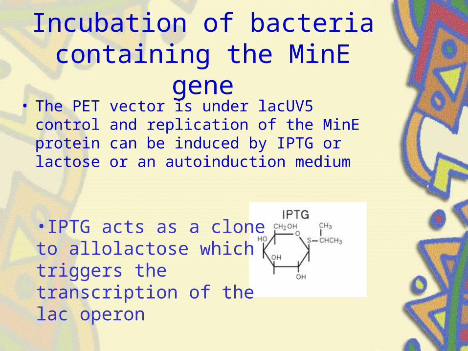
• The PET vector is under lacUV5 control and replication of the MinE protein can be induced by IPTG or lactose or an autoinduction medium
•IPTG acts as a clone to allolactose which triggers the transcription of the lac operon
Incubation of bacteria containing the MinE gene
• Cells are plated on agar mediums and allowed to grow for 48 hours
• Bacterial colonies are then cultivated using a sterile toothpick and incubated in 3mL of Luria-Bertani broth at 37oC for 12 hours in a shaking incubator – shaking allows good exchange of oxygen and
carbon dioxide while making sure all bacteria in the broth is suspended instead of sedentary
Luria-Bertani Broth
• Liquid medium high in nutrients used for the incubation of bacteria
• Generally composed of– 10g tryptone (short peptide used as a protein
source)– 5g yeast extract– 10g NaCl (variable depending on the
bacteria)– 1L deionized water
• Autoclave at 121oC and keep sterile
Isolation of the MinE Protein
• Resuspend the bacteria in LB broth by vortexing the tube
• Transfer 1mL of the broth mixture to a 1.5mL microcentrifuge tube
• Centrifuge at 13000rpm for 30 seconds to pellet bacteria
Isolation of the MinE Protein
• Pipette off supernatant leaving the pellet
• Repeat until all 3mL of broth has been pelleted
• Resuspend pellet in 150ul lysis buffer

Lysis Buffer
• The lysis buffer is composed of– 10mM tris-HCl– 5mM EDTA– 200mM NaCl– 0.2% Sodium Dodecyl Sulfate (SDS)
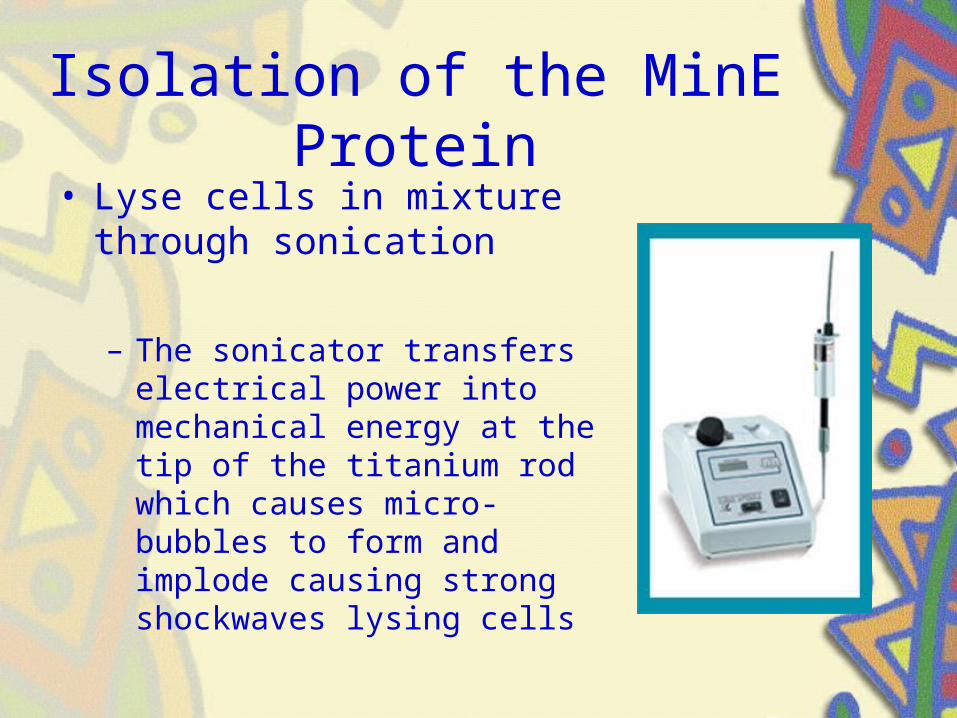
Isolation of the MinE Protein• Lyse cells in mixture through
sonication
– The sonicator transfers electrical power into mechanical energy at the tip of the titanium rod which causes micro-bubbles to form and implode causing strong shockwaves lysing cells
Isolation of the MinE Protein
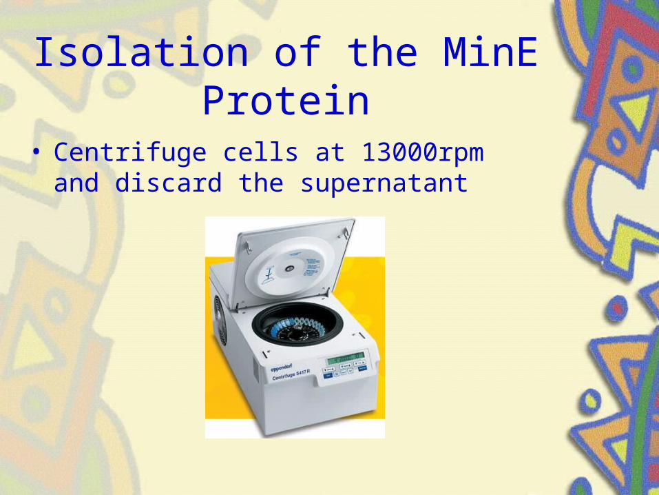
• Centrifuge cells at 13000rpm and discard the supernatant
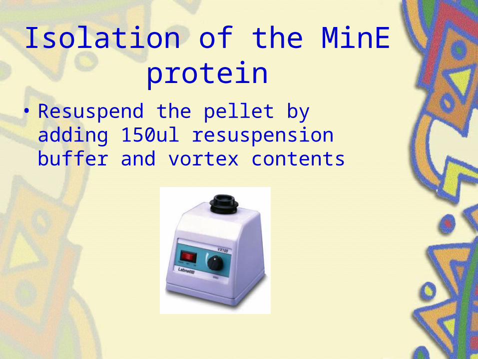
Isolation of the MinE protein
• Resuspend the pellet by adding 150ul resuspension buffer and vortex contents
Resuspension buffer
• The resuspension buffer is composed of– 25mM Tris-HCl– 10mM EDTA– 50mM glucose
Isolation of the MinE Protein
• Pour the mixture into the His-Bind Nickel column– The His-Bind Nickel column has a solid
stationary phase which binds to Histidine through chelation with the nickel ions on the column
– Other contaminants that do not bind through chelation would elute out of the column
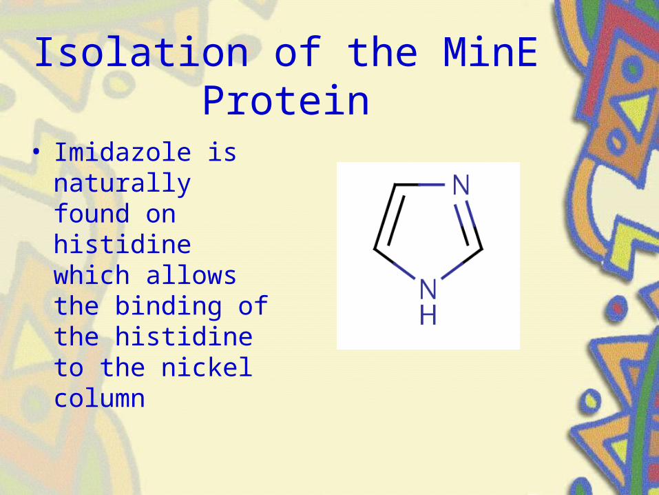
Isolation of the MinE Protein
• Imidazole is naturally found on histidine which allows the binding of the histidine to the nickel column
Purification of the MinE protein
• Elute non specifically bound proteins and contaminants using 300ul wash buffer
• Repeat 3 times
Wash Buffer
• The wash buffer is composed of– 50mM sodium phosphate– 300mM NaCl– 5mM imidazole
Purification of the MinE protein
• Elute the now purified MinE protein using 300ul of elution buffer
• Repeat 3 times

Elution Buffer
• Elution buffer is composed of– 50mM sodium phosphate– 300mM NaCl– 150mM imidazole

Purification of the MinE protein
• The high concentration of imidazole will displace the his-bound proteins and elute the protein
Western Blot
• Bradford Assay used to determine the amount of protein present
- Prepare standards using stock solution of bovine serum albumin (BSA)
- Serial Dilutions
- Add Bradford Reagent (900µl)
- Read Absorbance at 595nm
Western Blot
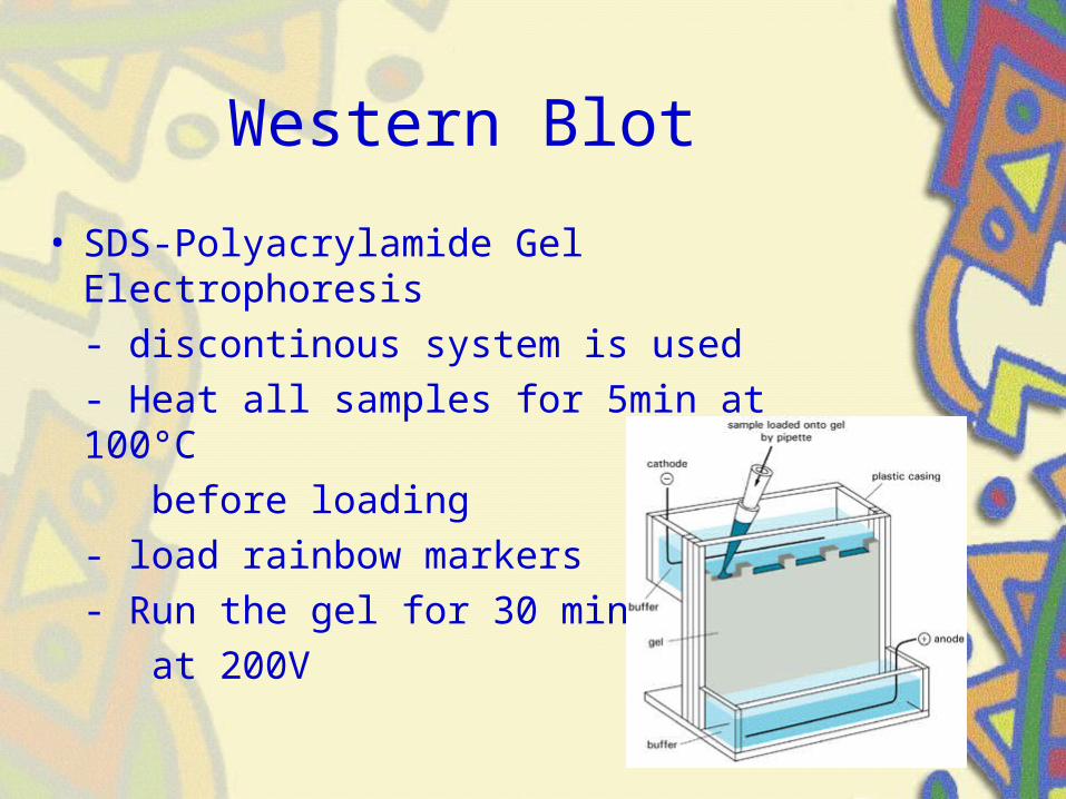
• SDS-Polyacrylamide Gel Electrophoresis
- discontinous system is used
- Heat all samples for 5min at 100°C
before loading
- load rainbow markers
- Run the gel for 30 min
at 200V
Western Blot
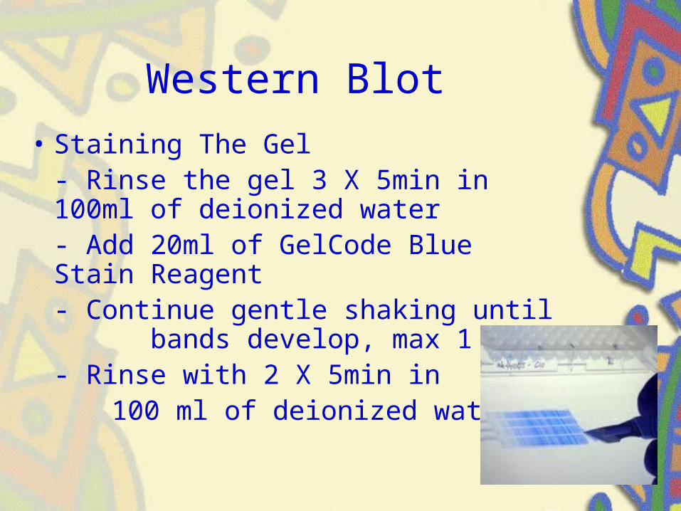
• Staining The Gel- Rinse the gel 3 X 5min in 100ml of deionized water- Add 20ml of GelCode Blue Stain Reagent- Continue gentle shaking until bands develop, max 1 hr- Rinse with 2 X 5min in
100 ml of deionized water
Western Blot
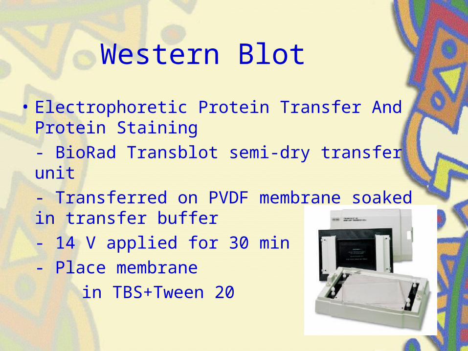
• Electrophoretic Protein Transfer And Protein Staining
- BioRad Transblot semi-dry transfer unit
- Transferred on PVDF membrane soaked in transfer buffer
- 14 V applied for 30 min
- Place membrane
in TBS+Tween 20
Western Blot
• TBS+Tween 20 is composed of:– Trizma base – 61g– NaCl – 90g– Distilled water – 1000ml– Tween 20 – 5ml
Western Blot
• Blocking Non-Specific Binding Sites & Incubation with Primary Antibody– Place membrane in dilute solution of BSA– Protein attaches to the membrane in
places where the target protein has not been attached
– Add antibody, attaches on sites specific to the target protein
Western Blot
• Blocking Non-Specific Binding Sites & Incubation with Primary Antibody– Wash membrane in 100ml blocking buffer
(nonfat dry milk ) for 1 hr– Wash with TBS+Tween 20, 1X15min,
2X5min– Place membrane in antibody
for 1hr– Wash with TBS+Tween 20,
1X15min, 2X5min
Western Blot
• Detection Of The Antigen-Antibody Complex– Place the membrane in 2° antibody,
horseradish peroxidase for 30 min– Wash 1X15 min and 4X5min with
TBS+Tween 20– Wash with water 2x10min– Incubate in 5 ml Western Blue Stabilized
Substrate for Alkaline Phosphatase– Wash with water 2X5min to stop reaction
Summary
1. Incubate the MinE protein using the PET vector in LB broth induced with IPTG
2. Resuspend and pellet bacteria discard supernatant
3. Resuspend bacteria and lyse using lysis buffer4. Sonicate bacterial cells to lyse contents5. Centrifuge contents and discard supernatant6. Resuspend pellet in resuspension buffer7. Purify using a his-affinity nickel column8. Wash with wash buffer9. Elute with elution buffer
Summary
1. Bradford Assay, prepare standards from stock solution of BSA
2. Perform Serial dilutions and read abs at 595nm3. SDS-Polyacrylamide Gel Electrophoresis4. Staining The Gel with 20ml of GelCode Blue
Stain Reagent5. Electrophoretic Protein Transfer And Protein
Staining using the BioRad Transblot semi-dry transfer unit
6. Blocking Non-Specific Binding Sites & Incubation with Primary Antibody
7. Detection Of The Antigen-Antibody Complex