exploring amino-acid radicals and quinone redox chemistry ...199750/fulltext01.pdf · amino-acid...
TRANSCRIPT

Exploring amino-acid radicals and quinone redox chemistry in model proteins
Kristina Westerlund

©Kristina Westerlund, Stockholm 2008 ISBN 978-91-7155-632-5 pp. 1-60 Printed in Sweden by Universitetsservice AB, Stockholm 2008 Distributor: Department of Biochemistry and Biophysics, Stockholm University

Tillägnad min familj


List of publications
This thesis is based on the following publications, which will be referred to by their Roman numerals (I-V) in the text:
I Westerlund, K., Berry, B.W., Privett, H.K. and Tommos, C. (2005) Exploring amino acid radical chemistry: Protein engineer-ing and de novo design* Biochim. Biophys. Acta 1707, 103-116.
II Westerlund, K., Moran, S.D., Privett, H.K., Hay, S., Jarvet, J., Gibney, B.R. and Tommos, C. (2008) Making a single-chain four-helix bundle for redox chemistry studies* Protein Eng., Des. Sel. doi:10.1093/protein/gzn043
III Hay, S.,‡ Westerlund, K.‡ and Tommos, C. (2005) Moving a hydroxyl group from the surface to the interior of a protein: Ef-fects on the phenol potential and pKa* Biochemistry 44, 11891-11902.
IV Westerlund, K.,‡ Berry, B.W.,‡ Hay, S. and Tommos, C. (2008) Probing proton-coupled electron transfer in model tyrosyl radical proteins Manuscript in preparation
V Hay, S., Westerlund, K. and Tommos, C. (2007) Redox charac-teristics of a de novo quinone protein* J. Phys. Chem. B 111, 3488-3495.
‡These authors have contributed equally.
*Published papers are reproduced with permission from the publisher.


Contents
1. Introduction to thesis...........................................................................................13 2. Amino-acid radical systems................................................................................14
2.1. Amino-acid radical enzymes......................................................................14 2.2. The ribonucleotide reductases – a well studied class of amino-acid
radical enzymes ....................................................................................................15 2.3. Glycyl radical enzymes – Important enzymes in anaerobic
metabolism ............................................................................................................17 2.4. Amino-acid radicals and oxidative stress ...............................................17 2.5. Studying amino-acid radicals in natural systems .................................18 2.6. Redox properties of amino-acid radicals in natural systems .............18 2.7. Significance of thesis project, part I........................................................20
3. Quinones .................................................................................................................21 3.1. Quinones in biology .....................................................................................21 3.2. Protein-derived cofactors present in quinoproteins .............................21 3.3. Quinones involved in biological energy transduction...........................22 3.4. Binding sites for non-covalently bound quinones.................................23 3.5. Redox chemistry of para-quinones in solution ......................................23 3.6. The photosynthetic reaction center of R. sphaeroides – a well
studied quinone-containing system .................................................................24 3.7. Electrochemical characterization of quinones in proteins...................27 3.8. Significance of thesis project, part II ......................................................27
4. De novo protein design .......................................................................................28 4.1. What is de novo protein design? ..............................................................28 4.2. Protein sequence space and the inverse folding problem ..................28 4.3. Design of coiled coils and helical bundles – an example of iterative
rational design ......................................................................................................29 4.4. Template-assembled synthetic proteins .................................................30 4.5. Designed combinatorial libraries of de novo proteins .........................30 4.6. Computational protein design ...................................................................31 4.7. Significance of thesis project, part III ....................................................32
5. Results and discussion.........................................................................................33 Making a single-chain four-helix bundle for redox chemistry studies
(Papers I-II) ..........................................................................................................33 Moving a phenol hydroxyl group from the surface to the interior of a
protein: Effects on the phenol potential and pKA (Paper III) .....................35

Probing proton-coupled electron transfer in model tyrosyl radical proteins
(Paper IV) ..............................................................................................................39 Redox characteristics of a de novo quinone protein (Paper V)..................43 Summary and Future Perspectives ..................................................................47
6. Acknowledgements...............................................................................................49 7. References..............................................................................................................50
Abbreviations
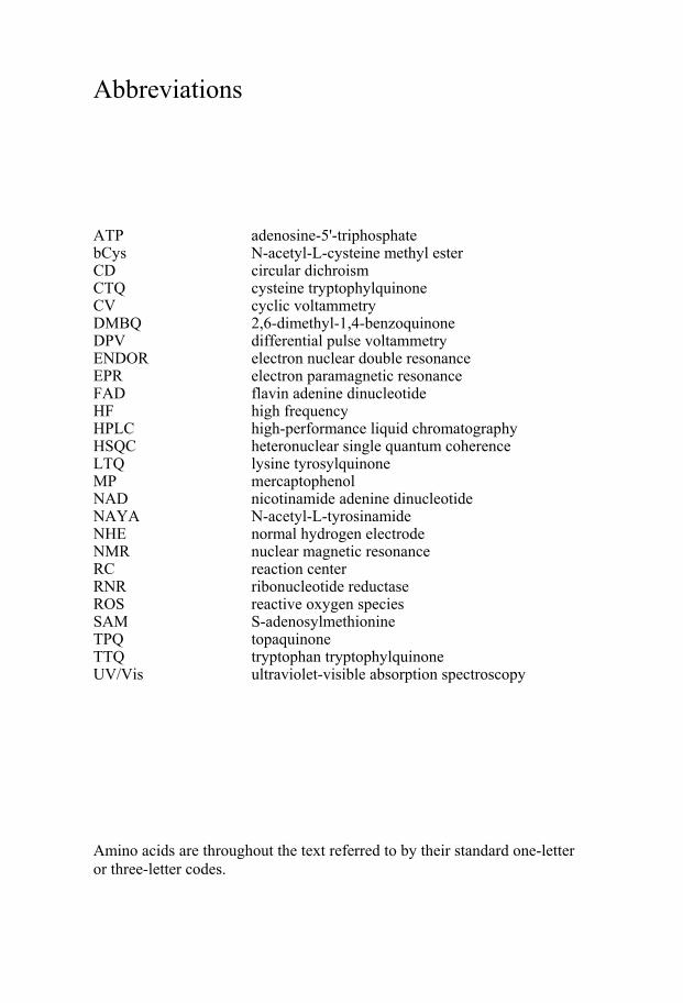
ATP adenosine-5'-triphosphate bCys N-acetyl-L-cysteine methyl ester CD circular dichroism CTQ cysteine tryptophylquinone CV cyclic voltammetry DMBQ 2,6-dimethyl-1,4-benzoquinone DPV differential pulse voltammetry ENDOR electron nuclear double resonance EPR electron paramagnetic resonance FAD flavin adenine dinucleotide HF high frequency HPLC high-performance liquid chromatography HSQC heteronuclear single quantum coherence LTQ lysine tyrosylquinone MP mercaptophenol NAD nicotinamide adenine dinucleotide NAYA N-acetyl-L-tyrosinamide NHE normal hydrogen electrode NMR nuclear magnetic resonance RC reaction center RNR ribonucleotide reductase ROS reactive oxygen species SAM S-adenosylmethionine TPQ topaquinone TTQ tryptophan tryptophylquinone UV/Vis ultraviolet-visible absorption spectroscopy Amino acids are throughout the text referred to by their standard one-letter or three-letter codes.

Nomenclature
Tyr-122 tyrosine 122 Tyr-122• tyrosine 122 as a neutral radical Ile-M256 isoleucine 256 in subunit M
Abstract
Amino-acid radical enzymes have been studied extensively for 30 years but the experimental barriers to determine the thermodynamic properties of their key radical cofactors are so challenging that only a handful of reports exist in the literature. This is a major drawback when trying to understand the long-range radical transfer and/or catalytic mechanisms of this important family of enzymes. Here this issue is addressed by developing a library of well-structured model proteins specifically designed to study tyrosine and trypto-phan radicals. The library is based on a 67-residue three-helix bundle (α3W) and a 117-residue four-helix bundle (α4W). α3W and α4W are single-chain and uniquely structured proteins. They are redox inert except for a single radical site (position 32 in α3W and 106 in α4W). Papers I and II describe the design process and the protein characteristics of α4W as well as a volt-ammetry study of its unique tryptophan. Paper III and V describe two pro-jects based on α3C, which is a Trp-32 to Cys-32 variant of α3W. In Paper III we use α3C to investigate what effect the degree of solvent exposure of the phenolic OH group has on the redox characteristics of tyrosine analogs. We show that the potential of the PhO•/PhOH redox pair is dominated by inter-actions with the OH group and that the environment around the hydrophobic part of the phenol has no significant impact. In addition, we observe that interactions between the phenolic OH group and the protein matrix can raise the phenol potential by 0.11-0.12 V relative to solution values. The α3C sys-tem is extended in Paper V to study quinone redox chemistry. Papers III and V contain protocols to generate the cofactor-containing α3C systems and descriptions of their protein properties. Paper IV describes efforts to redesign α3Y (a Trp-32 to Tyr-32 variant of α3W) to contain an interacting Tyr-32/histidine pair. The aim is to engineer and study the effects of a redox-induced proton acceptor in the Tyr-32 site.

1. Introduction to thesis Amino-acid radicals are involved in functional as well as harmful redox chemistry in living organisms and, yet, very little is known about their ther-modynamic properties. The main experimental barrier arises from the typi-cally highly oxidizing potentials of these species, which severely hamper electrochemical investigations. The central strategy of this thesis project is to use designed proteins that contain the redox-active species of interest as well as features that facilitate spectroscopic, structural and, most importantly, electrochemical characterization of the model proteins. The studies included in this thesis are based on the structurally characterized de novo α3W three-helix bundle protein (Papers III-V) and the recently constructed α4W four-helix bundle protein (Paper I-II). Paper I-II represents mainly protein-design oriented efforts while Papers III-IV describe functionally oriented studies on tyrosyl radicals. Paper V illustrates a successful extension of the three-helix bundle system to study protein quinone redox chemistry. To provide a back-ground to Papers I-V, we briefly summarize the main characteristics of amino-acid radical systems and quinone-containing enzymes, and then de-scribe the basics of de novo protein design.
13

2. Amino-acid radical systems
2.1. Amino-acid radical enzymes Amino-acid radical enzymes catalyze a number of fundamental biochemical reactions including DNA and hormone synthesis, carbohydrate metabolism, cell detoxification reactions and energy transduction (Table 1) (1-5). Amino acids acting as redox cofactors involved in these processes are shown below.
Figure 1. Amino-acid radical enzymes contain redox-active (A) tyrosine, (B) trypto-phan, (C) cysteine, and (D) glycine residues. Post-translationally modified tyrosyl radical cofactors including covalently cross-linked (E) Tyr-Cys and (F) Tyr-His species are also used. The amino acids serve as single electron redox cofactors involved in electron-transfer or proton coupled electron-transfer reactions.
Table 1 lists enzymes shown to contain amino-acids radicals. The ribonu-cleotide reductases (RNRs), photosystem II, galactose oxidase, pyruvate formate lyase, prostaglandin H synthase, and cytochrome c peroxidase are long-standing members of this class of enzymes and the essential function of the radicals in these systems is firmly established. For other enzymes, such as linoleate diol synthase, bovine catalase and the katG proteins, the role of the observed radicals need further investigations. Some exciting recent de-velopments in the radical enzyme field include: i) The discovery of the Radical-SAM protein superfamily (6), which most likely will lead to a rapid increase of known glycyl radical proteins (5). ii) Reports of the possible involvement of tyrosyl radicals in the reaction mechanisms of human mono-amide oxidase A (7) and cytochrome P450cam (8), both of which are of considerable medical and pharmaceutical importance. iii) That tryptophan- and tyrosine-based radical transfer chains may be involved in the action of bluelight-activated photoreceptors. These systems mediate developmental, growth and/or circadian responses in a variety of species (9-11).
14
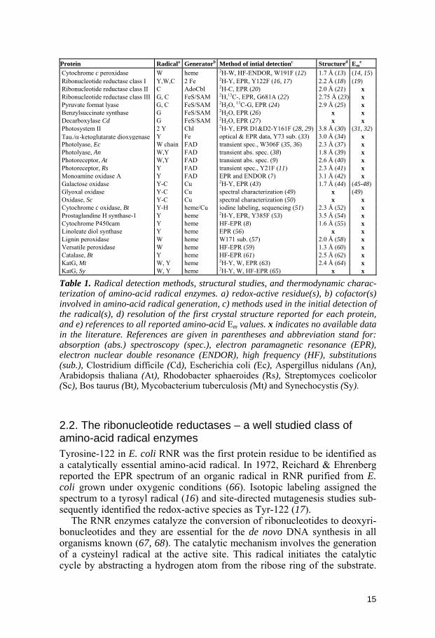
Protein Radicala Generatorb Method of intial detectionc Structured Em
e
Cytochrome c peroxidase W heme 2H-W, HF-ENDOR, W191F (12) 1.7 Å (13) (14, 15) Ribonucleotide reductase class I Y,W,C 2 Fe 2H-Y, EPR, Y122F (16, 17) 2.2 Å (18) (19) Ribonucleotide reductase class II C AdoCbl 2H-C, EPR (20) 2.0 Å (21) x Ribonucleotide reductase class III G, C FeS/SAM 2H,13C-, EPR, G681A (22) 2.75 Å (23) x Pyruvate format lyase G, C FeS/SAM 2H2O, 13C-G, EPR (24) 2.9 Å (25) x Benzylsuccinate synthase G FeS/SAM 2H2O, EPR (26) x x Decarboxylase Cd G FeS/SAM 2H2O, EPR (27) x x Photosystem II 2 Y Chl 2H-Y, EPR D1&D2-Y161F (28, 29) 3.8 Å (30) (31, 32) Tau./α-ketoglutarate dioxygenase Y Fe optical & EPR data, Y73 sub. (33) 3.0 Å (34) x Photolyase, Ec W chain FAD transient spec., W306F (35, 36) 2.3 Å (37) x Photolyase, An W,Y FAD transient abs. spec. (38) 1.8 Å (39) x Photoreceptor, At W,Y FAD transient abs. spec. (9) 2.6 Å (40) x Photoreceptor, Rs Y FAD transient spec., Y21F (11) 2.3 Å (41) x Monoamine oxidase A Y FAD EPR and ENDOR (7) 3.1 Å (42) x Galactose oxidase Y-C Cu 2H-Y, EPR (43) 1.7 Å (44) (45-48) Glyoxal oxidase Y-C Cu spectral characterization (49) x (49) Oxidase, Sc Y-C Cu spectral characterization (50) x x Cytochrome c oxidase, Bt Y-H heme/Cu iodine labeling, sequencing (51) 2.3 Å (52) x Prostaglandine H synthase-1 Y heme 2H-Y, EPR, Y385F (53) 3.5 Å (54) x Cytochrome P450cam Y heme HF-EPR (8) 1.6 Å (55) x Linoleate diol synthase Y heme EPR (56) x x Lignin peroxidase W heme W171 sub. (57) 2.0 Å (58) x Versatile peroxidase W heme HF-EPR (59) 1.3 Å (60) x Catalase, Bt Y heme HF-EPR (61) 2.5 Å (62) x KatG, Mt W, Y heme 2H-Y, W, EPR (63) 2.4 Å (64) x KatG, Sy W, Y heme 2H-Y, W, HF-EPR (65) x x
Table 1. Radical detection methods, structural studies, and thermodynamic charac-terization of amino-acid radical enzymes. a) redox-active residue(s), b) cofactor(s) involved in amino-acid radical generation, c) methods used in the initial detection of the radical(s), d) resolution of the first crystal structure reported for each protein, and e) references to all reported amino-acid Em values. x indicates no available data in the literature. References are given in parentheses and abbreviation stand for: absorption (abs.) spectroscopy (spec.), electron paramagnetic resonance (EPR), electron nuclear double resonance (ENDOR), high frequency (HF), substitutions (sub.), Clostridium difficile (Cd), Escherichia coli (Ec), Aspergillus nidulans (An), Arabidopsis thaliana (At), Rhodobacter sphaeroides (Rs), Streptomyces coelicolor (Sc), Bos taurus (Bt), Mycobacterium tuberculosis (Mt) and Synechocystis (Sy).
2.2. The ribonucleotide reductases – a well studied class of amino-acid radical enzymes Tyrosine-122 in E. coli RNR was the first protein residue to be identified as a catalytically essential amino-acid radical. In 1972, Reichard & Ehrenberg reported the EPR spectrum of an organic radical in RNR purified from E. coli grown under oxygenic conditions (66). Isotopic labeling assigned the spectrum to a tyrosyl radical (16) and site-directed mutagenesis studies sub-sequently identified the redox-active species as Tyr-122 (17).
The RNR enzymes catalyze the conversion of ribonucleotides to deoxyri-bonucleotides and they are essential for the de novo DNA synthesis in all organisms known (67, 68). The catalytic mechanism involves the generation of a cysteinyl radical at the active site. This radical initiates the catalytic cycle by abstracting a hydrogen atom from the ribose ring of the substrate.
15
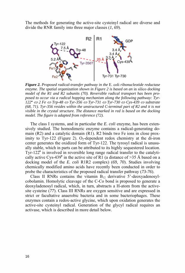
The methods for generating the active-site cysteinyl radical are diverse and divide the RNR family into three major classes (1, 69).
Figure 2. Proposed radical-transfer pathway in the E. coli ribonucleotide reductase enzyme. The spatial organization shown in Figure 2 is based on an in silico docking model of the R1 and R2 subunits (70). Reversible radical transport has been pro-posed to occur via a radical hopping mechanism along the following pathway: Tyr-122• ⇔ 2 Fe ⇔ Trp-48 ⇔ Tyr-356 ⇔ Tyr-731 ⇔ Tyr-730 ⇔ Cys-439 ⇔ substrate (68, 71). Tyr-356 resides within the unstructured C-terminal part of R2 and it is not visible in the crystal structure. The distance marked in red is based on the docking model. The figure is adapted from reference (72).
The class I systems, and in particular the E. coli enzyme, has been exten-sively studied. The homodimeric enzyme contains a radical-generating do-main (R2) and a catalytic domain (R1). R2 binds two Fe ions in close prox-imity to Tyr-122 (Figure 2). O2-dependent redox chemistry at the di-iron center generates the oxidized form of Tyr-122. The tyrosyl radical is unusu-ally stable, which in parts can be attributed to its highly sequestered location. Tyr-122• is involved in reversible long range radical transfer to the catalyti-cally active Cys-439• in the active site of R1 (a distance of >35 Å based on a docking model of the E. coli R1R2 complex) (69, 70). Studies involving chemically modified amino acids have recently been conducted in order to probe the characteristics of the proposed radical transfer pathway (73-76).
Class II RNRs contains the vitamin B12 derivative 5’-deoxyadenosyl-cobolamin. Homolytic cleavage of the C-Co bond is proposed to generate a deoxyladenosyl radical, which, in turn, abstracts a H-atom from the active-site cysteine (77). Class III RNRs are oxygen sensitive and are expressed in strict or facultative anaerobic bacteria and in some bacteriophages. These enzymes contain a redox-active glycine, which upon oxidation generates the active-site cysteinyl radical. Generation of the glycyl radical requires an activase, which is described in more detail below.
16

2.3. Glycyl radical enzymes – Important enzymes in anaerobic metabolism Glycyl radical enzymes operate under anaerobic conditions. The peptide bond adjacent to the glycyl radical is cleaved upon oxygen exposure and the system is irreversible inactivated. The members of this enzyme family are involved in a broad range of metabolic pathways including anaerobic break-down of pyruvate (24), toluene metabolism (26), fermentative production of para-cresol (27) and glycerol fermentation (78). Class III RNRs belong to this protein family. All glycyl radical enzymes are expressed in an inactive precursor form and require activation by a specific activase. The activases are members of the Radical-SAM superfamily and contain an iron-sulfur cluster and S-adenosylmethionine (SAM). Reductive cleavage of SAM by the iron-sulfur cluster produces a transient 5’-deoxyadenosyl radical, which abstracts a H-atom from the α-carbon of the strictly conserved glycyl resi-due. From sequence homology studies, putative new glycyl radical enzymes with yet unknown functions have been identified in E. coli and other faculta-tive and strict anaerobic bacteria and archaea. Glycyl radical enzymes may play an important role in the anaerobic metabolism in various species and the number of identified enzymes is likely to increase in the near future (79).
2.4. Amino-acid radicals and oxidative stress Oxidative stress results from exposure to high levels of reactive oxygen spe-cies (ROS) that have not been detoxified by the system. ROS are generally small, highly reactive molecules formed by an incomplete reduction of oxy-gen. ROS can be formed by ionizing radiation of biological molecules, by exogenous agents such as asbestos or ozone, or as biproducts in the respira-tory electron transport (80). ROS can oxidize virtually all cellular compo-nents including DNA, lipids and proteins. Increased levels of ROS are asso-ciated with aging and many diseases such as cardiovascular diseases, diabe-tes mellitus (81) and rhematoid arthritis (82). In proteins, ROS can oxidize sites at the backbone and/or the side chains and even cause cleavage of the peptide bond. Backbone oxidation in proteins occurs mainly by H-atom ab-straction at the α-carbon and the resulting radical is stabilized by electron delocalization onto the peptide backbone. Glycine can adopt particularly favorable backbone configurations upon oxidation. All amino acid side-chains are susceptible to ROS oxidation but aromatic residues and sulfur-containing amino acids are the major sites of oxidations in proteins. Thus, the four residues used for functional redox chemistry in radical enzymes (Figure 1) represent particularly vulnerable positions under oxidative stress conditions. Most oxidative damage is non-repairable and may lead to delete-rious consequences on protein structure and function.
17

2.5. Studying amino-acid radicals in natural systems Dysfunctional radical chemistry typically occurs infrequently during the normal course of cellular processes. It is consequently difficult to obtain a high steady-state concentration of the reactive radical, which hampers ex-perimental investigations. Studies on the experimentally more controllable amino-acid radical enzymes have been performed at a more molecular level. Crystal structures are available for the majority of these proteins, including several membrane-bound multisubunit enzymes. We note that only the reso-lution of the first crystal structure reported for each system is shown in Table 1 and that more refined structural data are available for many of the listed proteins. In addition, structures from different organisms and/or structures representing different redox states exist for several of the proteins listed in Table 1. An impressive array of spectroscopic techniques, including multi-frequency EPR-based methods, Fourier transform infrared and transient op-tical spectroscopy, has been used to identify the radical species and to derive detailed structural and kinetic information.
In sharp contrast to the extensive structural and spectroscopic work, virtu-ally nothing is known about the reduction potentials of amino-acid radicals and their dependence on solution pH and the protein environment. The sheer size and complexity of amino-acid radical enzymes combined with the highly oxidizing nature of their radical cofactors severely hamper electro-chemical measurements. Voltammetry and spectroelectrochemical meas-urements must typically be performed in the +1.0 V potential range, which for most enzymes will lead to oxidation of other cofactors, chromophores, and/or non-active amino acids with potential destruction of the protein as a consequence. Voltammetry experiments performed in this potential range in aqueous buffer will furthermore have a substantial background signal arising from water oxidation occurring at the surface of the working electrode. This complicates the analysis of the voltammogram, particularly if it reflects mul-tiple redox events. Studies based on optical detection are further limited by the low extinction coefficients of amino-acid radicals. The weak spectral features of amino-acid radicals can easily be obscured by the spectral bands of other chromophores and/or added redox mediators.
2.6. Redox properties of amino-acid radicals in natural systems The experimental limitations described above are so significant that no more than 10 published reports are available in the literature on the potentials of amino-acid redox cofactors (Table 1). In fact, the Cu-ligated, cysteine cross-linked Tyr-272 species in the active site of galactose oxidase is the only amino-acid radical system that has been studied systematically as a function of the protein microenvironment and solution pH (Figure 3; see legend for more details on this enzyme).
18
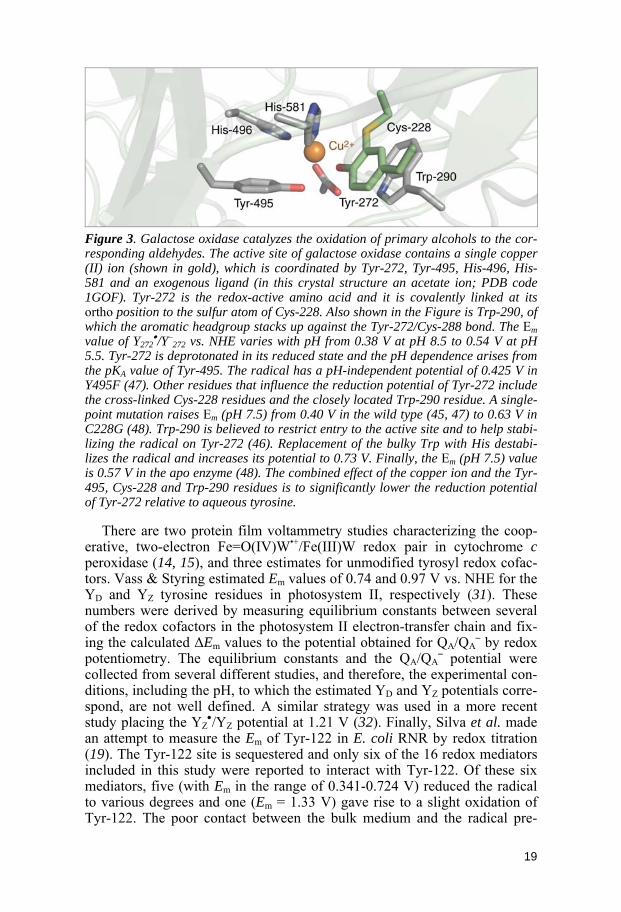
Figure 3. Galactose oxidase catalyzes the oxidation of primary alcohols to the cor-responding aldehydes. The active site of galactose oxidase contains a single copper (II) ion (shown in gold), which is coordinated by Tyr-272, Tyr-495, His-496, His-581 and an exogenous ligand (in this crystal structure an acetate ion; PDB code 1GOF). Tyr-272 is the redox-active amino acid and it is covalently linked at its ortho position to the sulfur atom of Cys-228. Also shown in the Figure is Trp-290, of which the aromatic headgroup stacks up against the Tyr-272/Cys-288 bond. The Em value of Y272
•/Y–272 vs. NHE varies with pH from 0.38 V at pH 8.5 to 0.54 V at pH
5.5. Tyr-272 is deprotonated in its reduced state and the pH dependence arises from the pKA value of Tyr-495. The radical has a pH-independent potential of 0.425 V in Y495F (47). Other residues that influence the reduction potential of Tyr-272 include the cross-linked Cys-228 residues and the closely located Trp-290 residue. A single-point mutation raises Em (pH 7.5) from 0.40 V in the wild type (45, 47) to 0.63 V in C228G (48). Trp-290 is believed to restrict entry to the active site and to help stabi-lizing the radical on Tyr-272 (46). Replacement of the bulky Trp with His destabi-lizes the radical and increases its potential to 0.73 V. Finally, the Em (pH 7.5) value is 0.57 V in the apo enzyme (48). The combined effect of the copper ion and the Tyr-495, Cys-228 and Trp-290 residues is to significantly lower the reduction potential of Tyr-272 relative to aqueous tyrosine.
There are two protein film voltammetry studies characterizing the coop-erative, two-electron Fe=O(IV)W•+/Fe(III)W redox pair in cytochrome c peroxidase (14, 15), and three estimates for unmodified tyrosyl redox cofac-tors. Vass & Styring estimated Em values of 0.74 and 0.97 V vs. NHE for the YD and YZ tyrosine residues in photosystem II, respectively (31). These numbers were derived by measuring equilibrium constants between several of the redox cofactors in the photosystem II electron-transfer chain and fix-ing the calculated ΔEm values to the potential obtained for QA/QA
– by redox potentiometry. The equilibrium constants and the QA/QA
– potential were collected from several different studies, and therefore, the experimental con-ditions, including the pH, to which the estimated YD and YZ potentials corre-spond, are not well defined. A similar strategy was used in a more recent study placing the YZ
•/YZ potential at 1.21 V (32). Finally, Silva et al. made an attempt to measure the Em of Tyr-122 in E. coli RNR by redox titration (19). The Tyr-122 site is sequestered and only six of the 16 redox mediators included in this study were reported to interact with Tyr-122. Of these six mediators, five (with Em in the range of 0.341-0.724 V) reduced the radical to various degrees and one (Em = 1.33 V) gave rise to a slight oxidation of Tyr-122. The poor contact between the bulk medium and the radical pre-
19

vented a true equilibrium potential to be determined and an apparent value of 1.0 V (pH 7.6) was given for the Y122•/Y122 pair.
2.7. Significance of thesis project, part I Amino-acid radical enzymes have been studied for over 30 years and to date there is only one system, galactose oxidase (Figure 3), for which protein structural features have been systematically correlated with changes in the reduction potential of the active amino-acid radical. It appears safe to con-clude that a complete chemical understanding of these biochemically impor-tant redox species cannot be obtained by concentrating experimentally solely on the naturally systems. The main motivation for the work described in this thesis was to develop an alternative approach to study the thermodynamic properties of amino-acid radicals. The goal was to gain insights into essential and unresolved issues such as if, and by how much, protein amino-acid radi-cal potentials are modulated by solvent accessibility and the hydrogen-bonding environment. Papers III and IV describe such efforts and are dis-cussed in more detail in Section 5.
20
3. Quinones
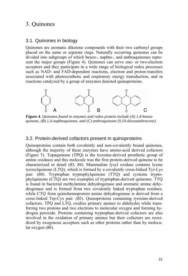
3.1. Quinones in biology Quinones are aromatic diketone compounds with their two carbonyl groups placed on the same or separate rings. Naturally occurring quinones can be divided into subgroups of which benzo-, naphto-, and anthraquinones repre-sent the major groups (Figure 4). Quinones can serve one- or two-electron acceptors and they participate in a wide range of biological redox processes such as NAD- and FAD-dependent reactions, electron and proton-transfers associated with photosynthetic and respiratory energy transduction, and in reactions catalyzed by a group of enzymes denoted quinoproteins.
Figure 4. Quinones found in enzymes and redox protein include (A) 1,4-benzo-quinone, (B) 1,4-naphtoquinone, and (C) anthraquinone (9,10-dioxoanthracene).
3.2. Protein-derived cofactors present in quinoproteins Quinoproteins contain both covalently and non-covalently bound quinones, although the majority of these enzymes have amino-acid derived cofactors (Figure 5). Topaquinone (TPQ) is the tyrosine-derived prosthetic group of amine oxidases and this molecule was the first protein-derived quinone to be characterized in detail (83, 84). Mammalian lysyl oxidase contains lysine tyrosylquinone (LTQ), which is formed by a covalently cross-linked Tyr-Lys pair. (84). Tryptophan tryptophylquinone (TTQ) and cysteine trypto-phylquinone (CTQ) are two examples of tryptophan-derived quinones. TTQ is found in bacterial methylamine dehydrogenase and aromatic amine dehy-drogenase and is formed from two covalently linked tryptophan residues, while CTQ from quinohemoprotein amine dehydrogenase is derived from a cross-linked Trp-Cys pair. (85). Quinoproteins containing tyrosine-derived cofactors, TPQ and LTQ, oxidize primary amines to aldehydes while trans-ferring two protons and two electrons to molecular oxygen and forming hy-drogen peroxide. Proteins containing tryptophan-derived cofactors are also involved in the oxidation of primary amines but their cofactors are reoxi-dized by exogenous acceptors such as other proteins rather than by molecu-lar oxygen (86).
21

Figure 5. The protein-derived redox cofactors found in quinoproteins include (A) topaquinone (TPQ), (B) lysine tyrosylquinone (LTQ), (C) tryptophan trypto-phylquinone (TTQ), and (D) cysteine tryptophylquinone (CTQ).
3.3. Quinones involved in biological energy transduction Due to the ability of quinones to carry both electrons and protons (see Sec-tion 3.5 below) they play an important biochemical role in coupling electron transfer to proton movement. Thus quinones are essential for the formation of electrochemical proton gradients in photosynthetic and respiratory sys-tems. Ubiquinone (Figure 6A) has been given its name because of its ubiqui-tous presence in nature and this molecule is found, for example, in the mito-chondrial respiratory chain. Both ubi- (Figure 6A) and menaquinones (Fig-ure 6B) are present in reaction centers (RCs) isolated from photosynthetic bacteria while plastoquinone (Figure 6C) is involved in electron and proton transfers in plant chloroplasts. An essential feature of the quinones involved in energy transduction is their long isoprenoid chain. The hydrophobic tail makes the quinones more soluble in the lipid bilayer of biological mem-branes and allow the oxidized and reduced cofactors to diffuse between membrane-bound protein complexes.
Figure 6. Quinones involved in biological energy transduction include (A) ubiquinone, (B) menaquinone, and (C) plastoquinone. The length of the isoprenoid chain depends on the species.
22

3.4. Binding sites for non-covalently bound quinones Quinones can be covalently bound, as is the case with the quinoproteins described above, or, more commonly, associated to proteins by non-covalent forces including hydrogen bonding to the carbonyl/hydroxyl groups, and van der Waals interactions with the aromatic ring and the aliphatic isoprenoid chain. Interactions at the binding sites can have a profound effect on the redox characteristics of the quinones and different types of binding sites are associated with different functional properties of quinones (87). A well-studied example is the RC from the purple bacterium R. sphaeroides in which the two electron acceptor-side ubiquinones have very different redox characteristics although they are chemically identical. In order to discuss this system in more detail, the redox properties of para-quinones in solution will first be briefly described.
3.5. Redox chemistry of para-quinones in solution Quinones have nine possible redox states (Figure 7) due to their ability to bind two electrons and two protons (88, 89). In aprotic media a quinone is reduced in two steps, the first forming the anionic semiquinone species, Q–, and the second forming the dianionic quinol Q2–. Due to electrostatic effects, the formation of Q2– requires more energy than the formation of Q– and, consequently, the redox behavior of quinones in aprotic media is relatively simple with two reversible and typically well-resolved n = 1 reactions (90).
Figure 7. Different para-quinone states resulting from electron and proton binding. Horizontal arrows indicate uptake of an electron while vertical arrows indicate protonation. States marked in bold are those available at pH > 0 (91).
23

In a protic solution the redox behavior of quinones becomes complicated by the protonic reactions associated with the hydroxyl groups. Most semi-quinones have pKA values around 3-5, which is lower then the pKA values for the corresponding quinol (91). The quinol states are greatly stabilized relative to the semiquinone states and, as a result, the reduction of quinone in a protic media give rise to an n = 2 behavior. This overall n = 2 conversion of quinone to quinol can be treated as two n = 1 reactions, rate-limited by the first energetically unfavorable one-electron transfer (90).
3.6. The photosynthetic reaction center of R. sphaeroides – a well studied quinone-containing system Reaction centers are integral membrane protein complexes found in photo-synthetic organisms such as the purple bacterium R. sphaeroides. In this system, the RC is part of a light-driven “redox loop” that gives rise to a transmembrane proton gradient subsequently used for ATP synthesis. Cen-tral to this mechanism are the QA and QB ubiquinones, which are both buried within the RC. The secondary quinone QB is reduced in two one-electron transfer steps via the primary quinone QA and is released as a protonated quinol, QBH2, to the membrane bilayer (Figure 8; see legend for more de-tails). For each QBH2 produced, the RC goes through two light-induced turn-overs and two protons are taken up from the cytosolic side of the membrane. After dissociation from the RC, QBH2 is reoxidized by cytochrome bc1, where the release of two protons to the periplasm completes a “proton trans-location redox loop” (92). Although QA and QB are chemically identical molecules they serve two very different roles in the RC quinone redox cycle. Clearly the protein environment plays a major role in tuning the redox prop-erties of the two quinones.
In the QA site (Figure 9) only the fully oxidized QA and anionic semi-quinone QA
– are found. QA is bound 20 times tighter than QB to the RC (93) and does not dissociate from the protein during the reduction cycle. The QA site is highly hydrophobic and no proton path is associated with the reduc-tion of the quinone. Protonation is needed to stabilize the fully reduced qui-nol state and inaccessibility to protons may be the reason why QA only func-tions as a one-electron acceptor under normal conditions (94). The side chain of Ile-M256 is in van der Waal contact with several atoms of the QA head group and changing this residue to a polar Ser or Thr lowers the midpoint potential for the QA/QA
− pair by as much as 60 and 115 mV (pH 8), respec-tively (95). As a contrast, a replacement of Ile-M256 with a Val has a negli-gible effect on the midpoint potential of the ubiquinone (95, 96). A lowered QA midpoint potential is consistent with a destabilization of the anionic semiquinone state relative to the oxidized quinone. The destabilization may arise from a change in the polarity of the binding site, which will affect the negatively charged QA
− more than the neutral quinone, and/or a change in the hydrogen-bonding pattern to the quinone head group (95, 96). Similar effects on the midpoint potential were observed when replacing the native ubiquinone with anthraquinone (Figure 4C) indicating that this effect does
24

not have a large steric component (96). Substitution of other residues in the vicinity of QA (e.g. Met-M218 and Trp-M252) have a similar effect by in-creasing the equilibrium constant for electron transfer to QB consistent with a negative shift in the midpoint potential of QA (95).
Figure 8. The ubiquinone reduction cycle in the photosynthetic reaction center of R. sphaeroides. Different stages in the reduction cycle are numbered by Roman numer-als. Figure adapted from references (92, 94). Stage I represents the light-induced electron transfer from the primary electron donor P, a bacteriochlorophyll dimer, to the primary electron acceptor quinone QA. The electron transfer occurs through a series of intermediates before reducing QA to an anionic semiquinone forming the P+QA
−QB state. P+ is re-reduced to P by cytochrome c2 after each photooxidation event. Stage II involves the first electron transfer step from QA
− to QB forming the PQAQB
− state and a sub-stoichiometric (H+/e−<1) proton uptake predominantly to Glu-L212 near QB. After a second light-induced electron transfer from P (stage III), QA is reduced to QA
− forming the PQA−QB
− state. Stage IV involves a fast, super-stoichiometric proton uptake and direct protonation of QB
− via Asp-L213 and Ser-L223 forming the PQA
−QBH state. This is followed by stage V in which a second, slow, electron transfer from QA
− occurs forming the PQAQBH− state. In stage VI the doubly reduced QBH− is protonated via Glu-L212 forming the PQAQBH2 state. The fully reduced and protonated quinol QBH2 can now leave the binding pocket and be replaced by a fully oxidized ubiquinone molecule from the membrane pool in stage VII (92, 94).
25
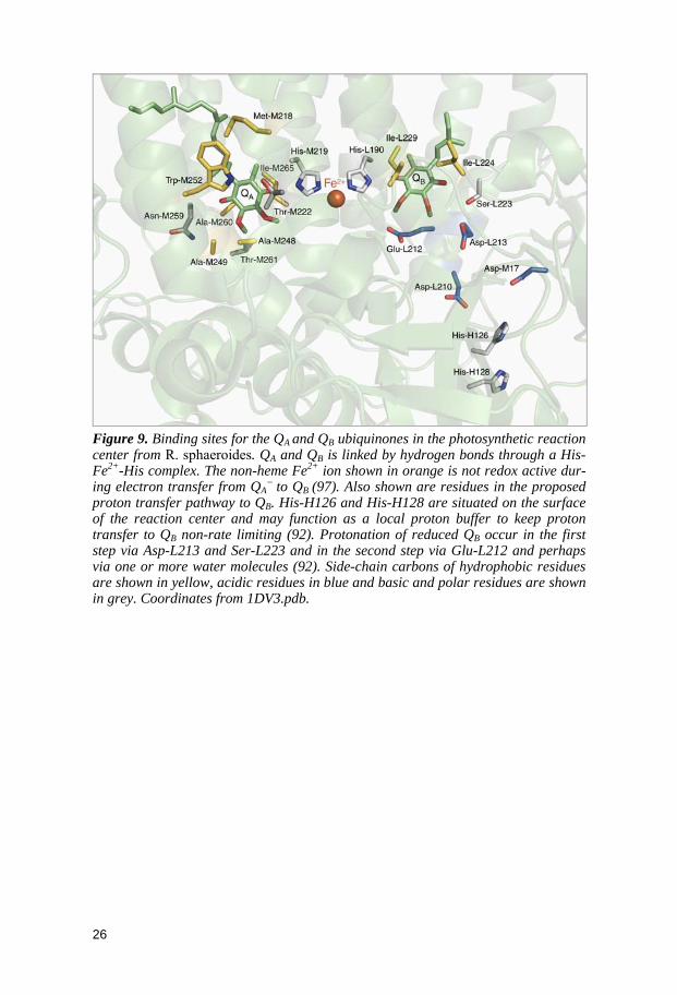
Figure 9. Binding sites for the QA
and QB ubiquinones in the photosynthetic reaction center from R. sphaeroides. QA and QB is linked by hydrogen bonds through a His-Fe2+-His complex. The non-heme Fe2+ ion shown in orange is not redox active dur-ing electron transfer from QA
− to QB (97). Also shown are residues in the proposed proton transfer pathway to QB. His-H126 and His-H128 are situated on the surface of the reaction center and may function as a local proton buffer to keep proton transfer to QB non-rate limiting (92). Protonation of reduced QB occur in the first step via Asp-L213 and Ser-L223 and in the second step via Glu-L212 and perhaps via one or more water molecules (92). Side-chain carbons of hydrophobic residues are shown in yellow, acidic residues in blue and basic and polar residues are shown in grey. Coordinates from 1DV3.pdb.
26
3.7. Electrochemical characterization of quinones in proteins Since quinones are involved in many biological redox processes, the sol-vated quinone/quinol redox couple is a very well-studied system electro-chemically (91). The para-quinone/para-quinol pair (Figure 6) is possibly the most studied of all organic redox couples by electrochemical methods. In contrast to the numerous articles on the redox behavior of quinone cofactors free in solution (98-101) or covalently attached to the working electrode (102, 103), reports on the redox behavior of quinones in proteins are scarce. Quinone-containing enzymes are often large, multi-subunit and multi-cofactor complexes and, in addition, several of them are membrane bound. These factors have hampered electrochemical characterization and only a few systems have been studied by other electrochemical methods than sim-ple redox potentiometry. A weak electrochemical response from the CTQ cofactor (Figure 5D) in P. denitrificans amine dehydrogenase was detected when using standard cyclic voltammetry (CV). Reversible CV and square wave voltammograms exhibiting resolved waveforms of two bacteriochloro-phyll species and QA cofactors in the R. sphaeroides RC were recorded by Kong et al. (104). Two types of protein film samples (i.e. protein samples deposited directly on the surface of the working electrode) were used to achieve rapid electron transfer between the electrode and the protein cofac-tors. The RC complex was either incorporated into multiple lipid bilayers or positioned between layers of polyion molecules (104). Gray and coworkers synthesized a diethylaniline-terminated oligo-(phenyl-ethynyl)-thiol “elec-tron wire” with the aim to electronically connect to the sequestered TPQ cofactor (Figure 5A) in the active site of A. globiformis amine oxidase. The electron wire molecule was covalently attached to the surface of the working electrode and then bound to the protein via its 20 Å hydrophobic substrate channel. Rapid electron tunneling between the buried cofactor and the work-ing electrode was successfully obtained by this method (105). Haehnel and coworkers covalently attached ubi- and menaquinones to a template-assembled four-helix bundle. Characterization of the de novo quinone pro-teins included determination of reduction potentials by cyclic voltammetry (106).
3.8. Significance of thesis project, part II Quinones are important cofactors in enzymes and redox proteins and volt-ammetry-based methods represent a key tool to study the properties of these cofactors as a function of the protein environment. As described above, the size and complexity of most quinone proteins preclude however voltam-metry characterization. The second motivation for this thesis project was to construct a model quinone protein which redox properties could be studied by voltammetry. These efforts are described in Paper V and discussed in Section 5.
27
4. De novo protein design
4.1. What is de novo protein design? The construction of proteins from scratch, de novo protein design (107), represents a model system approach to explore protein folding, pro-tein/cofactor interactions, and protein-based catalysis. Various methods in-cluding iterative rational design, template-assembled strategies, combinato-rial libraries, and computational methods have been used to create α-helical, β-sheet and mixed α/β-protein folds, cofactor- and metal-containing designs, and systems exhibiting some degree of catalytic activity.
4.2. Protein sequence space and the inverse folding problem De novo protein design is ultimately about finding amino-acid sequences that fold into structures with desired properties, a problem sometimes re-ferred to as “the inverse folding problem” (108). The sequence space, the number of different possible sequence combinations, is enormous even for a very small protein. For example, a protein of 50 residues, in which all 20 naturally occurring amino acids are permitted at every position, have 2050≈1065 different amino-acid sequence combinations. These sequences would weigh in at 1042 kilograms which is about 1017 times the mass of the Earth (109). Given the vastness of protein sequence space it is easy to see that the optimal sequence for a given application will never be found using a totally random search. A number of complementary methods exists to ex-plore the sequence space more efficiently and to enhance the probability of a successful de novo design.
Rational protein design is based on selecting specific folding patterns and/or known stabilizing features, e.g. propensities of the amino acids to stabilize or destabilize specific secondary structures, construct typically a small number of variants containing these features, and then examine their physical properties to connect the design with the product. The strategy of template-assembled methods is to reduce the complexity of the system by connecting peptide chains to a template, e.g. a metal or a small circular pep-tide. In combinatorial protein design, randomness and elements of rational design are combined to make libraries of de novo proteins. The libraries are then screened to select molecules with the desired properties. Finally, com-putational protein design algorithms search through sequence space to find low energy sequences that satisfy the target fold. Advances using these dif-ferent methods are briefly summarized below.
28

4.3. Design of coiled coils and helical bundles – an example of iterative rational design The coiled coil is a widespread structural motif found in a variety of proteins including transcription factors, chaperones, and structural proteins such as keratin (110). It is a strikingly simple motif consisting of α-helices wrapped around each other in a highly organized fashion. The oligomeric state of coiled coils can vary from 2 to 5 and the helices can pack in either a parallel or an anti-parallel manner. The contributing helices may come from a single peptide chain or from separate chains in an oligomeric protein. Most coiled coils consist of interacting amphipathic helices of which the amino acids are organized in a characteristic heptad (7-residue) repeat pattern (Figure 10). The heptad repeat pattern was first discovered in tropomycin (111, 112). Tropomycin is a parallel two-stranded coiled coil involved in the contraction and relaxation of skeletal muscles.
Figure 10. Interactions between residues in a dimeric coiled coil. The positions of the amino acids in a heptad segment are denoted abcdefg (111). Residues a and d are normally non-polar and their side chains pack against each other forming the main part of the hydrophobic core in the coiled-coil structure. Residues e and g are often charged and may be involved in inter-helical salt bridges. Residues in the remaining positions are usually polar and form the surface of the coiled coil.
The first de novo designed coiled coils were constructed in 1981 by Hodges and coworkers (113). Since then, many groups have used the heptad-repeat pattern to construct coiled coils and helical bundles (114). The con-struction of the α3D three-helical bundle by DeGrado and coworkers (115) provides an excellent example of iterative rational design and highlights several important features for the successful design of a helical bundle in-cluding: I) the importance of loop regions to promote a monomeric structure, II) the introduction of helix capping interactions to stabilize helix formation, III) the addition of features of “negative design” to destabilize unwanted topologies, and IV) the importance of a well-defined hydrophobic core (115). An early variant of α3D suffered from an ill-defined hydrophobic core made up of all Leu residues (115). Leucine is a big and bulky residue and has many possible rotamers close in energy and an overabundance of Leu may result in a dynamic and/or “over-packed” core (116, 117). The introduc-tion of more sterically constrained aromatic and β-branched amino acids at
29

core positions tend to make de novo proteins less dynamic and more “native-like” (118, 119). The NMR solution structure of the repacked α3D protein was found to be in good agreement with the original design (120) although, interestingly, substitutional (121) and NMR side-chain dynamics (122) stud-ies on α3D showed a more dynamic behavior at core positions relative to natural proteins studied at this level of detail.
4.4. Template-assembled synthetic proteins The unfavorable loss of main-chain and side-chain entropy occurring upon protein folding increases with the number of possible unfolded conforma-tions. Consequently, the loss in peptide entropy upon folding will be less if the number of unfolded conformations for a given peptide chain is reduced. In template-assembled protein design, peptides are attached to a rigid mo-lecular scaffold with the aim to reduce the size of the ensemble of unfolded conformations. The peptides are pre-ordered on the template prior to folding and the entropic cost of folding the peptide chain is thereby lessened. The templates varies and include peptides, carbohydrates, porphyrin derivatives, steroids, transition metals, and cavitands (rigid organic molecules with an internal cavity) (123, 124). The vast majority of template-assembled de novo proteins are α-helical with 3-6 interacting helices. The helices can have iden-tical or different sequences and they can be oriented in a parallel or anti-parallel fashion. Rational, combinatorial and computational methods have been used to generate this type of synthetic proteins (123, 124). Haehnel and coworkers have used the template-assembled synthetic protein methodology to design, synthesize and characterize a number of de novo proteins includ-ing heme proteins (125), heme proteins with catalytic activity (126), chloro-phyll binding proteins (127), quinoproteins (106), and proteins with copper-binding sites (128).
4.5. Designed combinatorial libraries of de novo proteins Hecht and coworkers have constructed combinatorial libraries of de novo four-helix bundles using a simple binary pattern. The binary pattern ap-proach is based on the assumption that the ability of a peptide chain to form amphipatic secondary structure elements is sufficient to promote the chain to fold into a globular structure. The binary pattern specifies the order of hy-drophilic and hydrophobic residues in the peptide sequence but the identity of the amino acid at each site is allowed to vary combinatorially (129). Li-braries of synthetic genes were constructed based on the binary pattern and the organization of the genetic code that allowed for the combinatorial diver-sity. Protein expression tests conducted on the first-generation library showed that about 60 % of randomly chosen clones generated soluble pro-teins. Some of the expressed proteins showed native-like characteristics, but most of them displayed properties consistent with fluctuating structures (130, 131). A second-generation library was created by redesigning one of the non-native like proteins characterized from the original library. The heli-
30

ces were elongated and several elements of rational design were allowed to influence the redesign (132). The NMR solution structure of a well-folded de novo four-helix bundle protein from this second-generation library has been presented (133).
4.6. Computational protein design With more powerful computers and efficient algorithms, computational de-sign strategies have gained in popularity. In silico de novo protein-design methods have the potential to explore the protein sequence space more ex-tensively then experimentally based methods. However, a full de novo de-sign of even a small 100-residue protein remains a considerable challenge (134). Even if only the most preferred amino-acid rotamers are taken into account, the complexity of the computational problem quickly grows into overwhelming proportions. For example, a 100-residue protein for which the ∼100 most preferred side-chain conformations of the 20 naturally occurring amino acids are considered at each position, have in total 100100=10200 possi-ble conformations (134). In addition, natural proteins typically have a struc-tural flexibility that allows the back-bone to relax upon mutations. Freezing the back-bone position during the calculations reduces the computational complexity greatly, but this approach has its limitations in de novo design since the starting structure by definition is not well defined (134).
The ORBIT program by Mayo and coworkers is constructed to optimize the sequence that will stabilize a fixed template structure (135). The ORBIT methodology has been used for the full sequence design of a 28-residue zinc finger motif (135), redesign of calmodulin to improve the binding of one of its target peptides, redesign of 24 core positions in T4 lysozyme, and in an attempt to design a α/β-barrel with an “idealized” artificial back-bone fold (134).
The RosettaDesign software, developed by Baker, Kuhlman and cowork-ers (136), uses an iterative design approach and alternates between atomic-resolution structure prediction and Monte Carlo-based sequence optimiza-tion. Back-bone flexibility is incorporated by considering a set of initially fixed back-bone templates (134). The Rosetta design methodology has been used for protein structure prediction (137), to redesign and stabilize natural proteins (138, 139), for the design of a peptide sequence that can switch between a trimeric coiled coil and a zinc finger-like fold (140), and for the de novo design of Top7 a protein with a novel fold (136). Recently, Rosetta was used to design novel catalysts for a retro-aldol reaction in which a car-bon bond is broken in an unnatural substrate. 44 % of the 72 experimentally characterized de novo proteins displayed retro-aldolase activity with rate enhancement up to four orders of magnitude as compared to the uncatalyzed reaction (141). Two of the active enzymes, which were designed to display different protein folds and different active-site architectures, were crystal-lized. In both cases the crystal structures were in good agreement with the design models (141).
31

4.7. Significance of thesis project, part III Our group has previously utilized de novo design to construct a stable and well-structured single-chain three-helical bundle. This system has been used to study amino-acid redox chemistry (Papers III and IV) and quinone redox chemistry (Paper V) occurring within a protein environment. This thesis describes the characterization of a single-chain four-helix bundle protein that was designed using the methods outlined in Section 4.3. The goal was to construct a larger protein scaffold that will be used in future studies similar to those performed with the three-helix bundle system. These efforts are described in Papers I and II and discussed in Section 5.
32
5. Results and discussion
Making a single-chain four-helix bundle for redox chemistry studies (Papers I-II) The three-helix bundle system used in Papers III-V exhibits some key fea-tures. The protein scaffold is well structured and, in addition, uncomplicated with respect to its optical properties. Earlier work and data presented here demonstrate that NMR as well as UV/Vis, fluorescence and CD spectros-copy can be used to derive detailed structural information of the protein as a whole (142, 143) and of the radical/quinone site specifically (e.g. Paper IV). The 67-residue, single-chain protein scaffold is “redox inert” except for a dedicated site, position 32, in the core of the protein (Figure 6, Paper I). We define “redox inert” as tolerating voltammetry measurements in the oxida-tive range of up to +1.3 V vs. NHE without triggering uncontrolled redox reactions. The inert nature of the scaffold is essential as it allows us to probe the redox properties of highly oxidizing molecules without triggering un-wanted side reactions. Thus far three main variants have been developed in which position 32 is either occupied by a tryptophan (α3W), a tyrosine (α3Y) or a cysteine (α3C). In the latter, Cys-32 is used as a tag to ligate phenol (Paper III) or quinone (Paper V) molecules to the protein.
A valid criticism of the three-helix bundle scaffold is that it is small in size relative to naturally occurring radical and quinone-containing enzymes. In addition, its structural architecture is simple which can provide limitations (see Discussion Paper IV). The goal with this project was to create a protein of roughly twice the molecular mass of α3W (7.5 kDa) but otherwise exhibit-ing the same overall properties as the smaller system. That is, the larger pro-tein should be well structured. It should have a redox inert sequence except for a single site containing a redox-active molecule, which, in turn, should give rise to a Faradaic response in voltammetry investigations. The initial design of this larger protein, a single chain four-helix bundle with a molecule mass of about 13 kDa, was first introduced in Paper I. The refined design and the characteristics of the final protein are described in Paper II.
In contrast to the de novo α3W protein, a natural protein was used as a starting point for the four-helix bundle design. The sequence of the E. coli DNA-binding Rop protein was chosen as the template. Thus this project does not represent de novo design, but rather redesign of a natural protein. The redesign of Rop followed classical iterative design principles and in-volved multiple changes in the primary sequence with large-scale changes in inter-helical interactions and bundle topology as a result.
In solution, Rop is a homo dimeric four-helix bundle with its two helix-turn-helix domains arranged in an anti-parallel manner. The sequences of the helical regions in Rop follow the heptad repeat pattern described in Section 4.3. Wild-type Rop has eight hydrophobic packing layers with small residues at heptad a positions and large residues at d positions with the exception of
33

the equivalent layers 2 and 2’ (see Figure 1, Paper II) in which the small versus large residue packing pattern is reversed.
The anti-parallel topology of Rop places the C- and N-termini of each monomer on opposite sides of the protein. In our redesign, 20 out of the 32 heptad a and d core residues were changed following a small versus large packing pattern to induce and stabilize a ∼180° flip of one of the subunits (illustrated schematically in Supplementary Figure S1, Paper II). The C- and N-termini of the two subunits were linked by the introduction of a five-residue glycine loop and multiple inter-helical sites were changed to avoid potentially unfavorable electrostatic interactions. The single-chain sequence was made to contain no tyrosine, tryptophan or cysteine residues except for a unique tryptophan at heptad a position 106 (Figure 1, Paper II).
Characterization of the first protein product revealed a predominately helical structure (71.0 ± 0.6% helical, pH 5.5-10.0) with a poor global stabil-ity (~ –2.7 ± 0.5 kcal mol-1). The protein appears monomeric at low protein concentrations (≤ 100 μM) but broadened NMR spectral lines suggest that aggregation occurs at higher concentrations (Figure 4A, Paper II). The sub-sequent redesign process was guided by two main goals: To improve the protein stability (which hopefully would resolve the aggregation problem) and to make the protein more suitable for NMR structural studies by increas-ing the overall chemical-shift dispersion. Two Thr (T19 and T80) were re-placed by apolar amino acids and two Phe were placed in predicted core positions (T80F and L109F). The introduction of aromatic side chains was predicted to provide ring current shifts and influence the chemical shifts of nearby atoms. The changes were made in a stepwise fashion and the physical properties of each variant were investigated (Table I, Paper II).
The final protein is a three-site variant (T19I, T80F, L109F) of the initial design. This protein forms a stable helical structure in water. Its free energy of unfolding is –4.7 kcal/mol (25° C, pH 7.2) and the protein was shown to be thermostable with an estimated midpoint temperature of unfolding above 355 K. The helical content is high and displays little variance over the 5.5 – 10.0 pH range (69.8 ± 0.8 %). Analytical ultracentrifugation and NMR data showed that the protein is monomeric up to at least 0.5 mM. 1D proton and 2D 15N-HSQC data display narrow spectral lines consistent with a uniquely structured protein. The spectral resolution of the 2D 15N-HSQC spectrum (Figure 4C, Paper II) is excellent considering the all-helical nature of the protein and suggests that resonance assignment of the protein backbone should be straight forward. The blue-shifted fluorescence spectrum of Trp-106 suggests that the aromatic residue is buried, as indented. Finally, a dif-ferential pulse voltammetry investigation showed a Faradaic response of a single proton-coupled oxidation reaction consistent with the unique Trp-106.
In conclusion, the efforts described in Papers I and II resulted in the ex-tention of our model protein library with a stable and well-structured 13 kDa radical protein. This protein is denoted α4W to reflect that it is a single chain four-helix bundle with a unique tryptophan. It is our hope that the α4W pro-tein can be explored in a manner similar to the α3W system and that new information can be gained by comparing the two.
34

Moving a phenol hydroxyl group from the surface to the interior of a protein: Effects on the phenol potential and pKA (Paper III) Despite the number and biochemical importance of enzymes utilizing tyro-sine redox chemistry, experimental studies characterizing the thermody-namic properties of tyrosyl radicals are scarce, essentially nonexistent, in the literature (see Table 1 and discussion in Section 2.6.). Since very little is know, virtually any aspect of “tyrosine redox properties vs. the protein envi-ronment” is important to strive to understand. The work described in Paper III aims to address a very basic issue: How does the degree of solvent expo-sure of the phenol hydroxyl group influences the redox characteristics of a tyrosine? Experimentally we wanted to gradually alter the solvent exposure of the phenol hydroxyl group but without introducing large-scale changes in the overall environment of the aromatic head group. A “phenol rotation strategy” was developed that consisted of ligating phenols containing a SH tag in the para, meta and ortho position (site 4, 3 and 2 relative to the phenol hydroxyl site 1) to the unique cysteine in α3C.
Figure 11. Chemical structures of (A) 4-mercaptophenol, (B) 3-mercaptophenol, and (C) 2-mercaptophenol attached via an S–S bond to a cysteine. (D) Tyrosine is also included to provide a structural comparison. R’ and R’’ represents the peptide back-bone in the α3C systems. For the solution reference compounds, R’ and R’’ represents –COCH3 and –OCH3, respectively.
Trp-32 in α3W has an accessible surface area of < 3% (143) and the smaller side chain of the corresponding Cys-32 residue in α3C is therefore predicted to be buried within the hydrophobic core. The tyrosine analogues 4-mercaptophenol (4MP), 3-mercaptophenol (3MP) and 2-mercaptophenol (2MP) were covalently attached to α3C via a disulfide bond. Chemical repre-sentations of the three Cys-reacted phenols are shown in Figure 11. The sim-ple heptad design of the three-helix bundle, which was confirmed by the structural characterization of α3W (Figure 6, Paper I), predicts that the pro-tein scaffold will force the geometry of the bound mercaptophenols rather that the other way around. That is, we anticipated that binding a mecapto-phenols to Cys-32 would not give rise to large-scale shifts in the position of the cysteine and that the phenol OH group of 4MP-α3C would be more ex-posed to the bulk solvent relative to the phenol OH group in the 3MP-α3C and 2MP-α3C proteins. The aim of the “phenol rotation strategy” is shown in the second Figure in Paper III. We note that the purpose of Figure 2 is only to illustrate the design and not to make detailed structural predictions.
35

Paper III describes the synthesis and purification of the three MP-α3C proteins as well as characterization of their properties relative to α3C and α3W. It was shown that homogenous MP-α3C protein samples could be ob-tained. UV/Vis spectra and extinction coefficients of the protein-bound phe-nols were measured and described. The MP-α3C proteins were shown to be stable and highly helical. These results are summarized in Table 1 in Paper III. One-dimensional NMR spectra of the MP-α3C proteins display chemical shift dispersion in the amide region that is similar to the α3W 1D NMR spec-trum. This indicates that no catastrophic changes, i.e. conversion from a structured scaffold to a molten globule, occur upon binding of the phenols. This conclusion has more recently been confirmed by multi-dimensional NMR data, which show characteristics fully consistent with well-structured proteins (Tommos et al. unpublished).
Two data sets suggest that the phenol OH group of 4MP-α3C is signifi-cantly more solvent exposed relative to the OH group of the phenols in the 3MP-α3C and 2MP-α3C proteins. Firstly, the global stability of 3MP-α3C and 2MP-α3C (–3.1 kcal mol–1) is lower than the stability of 4MP-α3C (–3.7 kcal mol–1) and α3C (–4.1 kcal mol–1). The core of α3C is made of Leu, Ile and Val residues and solvating a hydrophilic OH group within this highly hydrophobic environment is expected to have a destabilizing effect on the protein. Secondly, the CD spectrum of 4MP-α3C is virtually identical at pH 7.6 and at pH 10 (Figure 8, Paper III). The phenol pKA of 4MP-α3C is about 9.5, which means that at pH 7.6 the phenol is predominately protonated and at pH 10 it is predominately deprotonated. To form a charge inside the pro-tein is expected to have a significant impact on the secondary structures of the protein and the fact that there is no change at all strongly suggests that the OH group resides at or near the surface of the protein.
The last part of Paper III describes an electrochemical characterization of the MP-α3C proteins. In order to isolate the effects of the protein matrix on the redox properties of the bound phenols, all electrochemical data derived from the MP-α3C proteins were compared to data derived from aqueous reference compounds. Thus, we are not interested in the absolute phenol potentials but rather on the shifts introduced as the phenols are bound to the protein relative to being freely solvated in aqueous buffer. The reference compounds were made by reacting the three mercaptophenols with the blocked, neutral Cys derivate N-acetyl-L-cysteine methyl ester (bCys). The preparation and purification of these compounds are described in Paper III.
Differential pulse voltammetry (DPV) measurements have been used pre-viously to estimate the reduction potentials and pKA values of tyrosine and tryptophan in solution and in α3W and α3Y (142). The average width of the DPV peaks representing the MP-α3C proteins and the MP-bCys compounds were broader than expected for an n = 1 reversible reaction (144), suggesting irreversible redox chemistry. Cyclic voltammetry traces of 2MP-α3C and 2MP-bCys confirmed this conclusion (not shown). Irreversible electrochem-istry is typical for organic radicals, since these species usually have life times that are short relative to the time scale of the experiment. For a fully reversible redox reaction with no kinetic complications, the average DPV
36

peak potential approximates the formal reduction potential (E0’) (145). Epeak values derived by voltammetry from aqueous tyrosine and tryptophan are very close to E0’ values derived from pulse radiolysis equilibrium studies. We estimate that Epeak = E0’ ± 0.02 V for the freely solvated species (142). For the protein samples it is more difficult to estimate a direct relationship between Epeak and the true reduction potential, although we note that scan rates were deliberately kept low in order to avoid any kinetic effects on Epeak. At this point, we assume that the model proteins behave similarly to the sol-vated compounds and that their Epeak values can be compared directly.
Figure 12. Redox states of phenol (PhOH) in aqueous solution as a function of pH. (A) At a solution pH below the pKA for the oxidized species (pKOX) the potential becomes pH-independent and PhOH•+/PhOH is the predominant redox couple. (B) Between pKOX and pKRED the redox reaction is pH dependent with a slope of –ln(10)RT/nF (59 mV per pH unit for a one electron, one proton redox event at 25° C). The major redox couple in this region is PhO•/PhOH. (C) At a pH higher than the pKA of the reduced species (pKRED) the redox reaction becomes pH independent again with the PhO•/PhO– as the predominant redox couple.
The redox reactions of a phenol are dependent on the solution pH, as il-lustrated in Figure 12. DPV measurements were performed in the pH 4 to 10 range to investigate what effect the protein matrix has on the potential and pKA values of the bound mercaptophenols. Epeak vs. pH plots for 2MP, 3MP, and 4MP ligated to α3C and to bCys are shown in Figure 7, Paper III, and the results from these data sets are summarized in Table 2 below. The pKA values of the phenolate/phenol couples were determined electrochemically (pKRED) and optically via the absorption of the phenolate (pKA). The values derived by these two methods were found to be in good agreement.
Species pKA pKRED -ln(10)RT/nF Epeak,7 ΔEpeak,7 4MP-bCys 9.3 9.7 0.059 0.80 4MP-α3C 9.5 9.4 0.064 0.79 -0.01 3MP-bCys 9.6 9.8 0.059 0.96 3MP-α3C >10 >10 0.058 1.07 0.11 2MP-bCys 9.0 8.8 0.060 0.79 2MP-α3C >10 >10 0.057 0.91 0.12
Table 2. Electrochemical properties of 4MP, 3MP, and 2MP bound to a3C and bCys. All potentials are in the unit of V and are given vs. NHE.
37
Our study of the MP-α3C proteins and their corresponding solution refer-ence compounds provided two main results. As described above, the charac-terization of 4MP-α3C suggests that the protein folds around the hydropho-bic part of the ligated phenol and that the phenol OH group resides at or near the protein surface. Clearly the protein-ligated phenol resides in a very dif-ferent environment relative to the solvated species (schematically illustrated in Figure 13) and yet their redox properties are remarkably similar (Figure 7, Paper III; Table 2). The Epeak (pH 7.0) values of 4MP-α3C and 4MP-bCys are within the error margin of the experiment. The average pKA values de-rived from the electrochemical and phenolate titrations are 9.45 and 9.50 ± 0.2 for 4MP-α3C and 4MP-bCys, respectively. In all its simplicity, this is a surprising and important finding that represents the first experimental dem-onstration that the interactions with the OH group dominate the redox char-acteristics of the PhO•/PhOH redox couple and that interactions with the remaining part of the aromatic molecule have no significant impact.
Figure 13. Schematic representation of the environment for 4MP when (A) solvated in water and (B) attached to the α3C protein. The aqueous compound experiences a homogenous environment with a high dielectric constant. The protein-bound spe-cies, in contrast, resides in a highly heterogeneous environment as it extends from the hydrophobic core (marked in dark blue), through the charged surface of the protein (red) and into the bulk phase (light blue). That the redox properties of 4MP in Figures A and B are essentially identical, indicate that interactions to the phenol OH group are the key parameters tuning the phenol reduction potential.
In contrast to 4MP, the electrochemical characterization of 3MP and 2MP show distinct differences when the phenols are ligated to α3C relative to bCys. The average pKA values for 3MP-bCys and 2MP-bCys are 9.7 ± 0.1 and 8.9 ± 0.1, respectively. These values increase to >10 when the phenols are ligated to α3C. The increase in the pKA values observed for 3MP-α3C and 2MP-α3C relative to the solution compound suggests that the phenol OH group is more shielded in the protein. Epeak (pH 7.0) is elevated by 0.11 and 0.12 V for 3MP-α3C and 2MP-α3C, respectively, relative to the solution systems. The Epeak vs. pH plots for 3MP-α3C and 2MP-α3C display slopes close to 59 mV per pH unit over the whole pH 4 to 10 pH range. This is also true for 3MP-and 2MP-bCys at a pH < pKA and show that PhO•/PhOH is the relevant redox couple in this region. Factors that may influence the potential of the PhO•/PhOH couple are hydrogen-bonding interactions of the phenol and/or the phenoxyl radical, the fate of the phenolic proton and its associated charge upon forming the neutral radical, and potential redox induced struc-tural changes. The ~59 mV/pH slopes show that the MP-α3C proteins remain
38

overall charge neutral upon oxidation suggesting that charge is not involved in raising the potentials of 3MP-α3C and 2MP-α3C. As discussed at some length on page 11899, Paper III, it is doubtful that structural changes are involved as well. Most likely differences in hydrogen bonding between the aqueous and protein-bound 3MP and 2MP molecules is the main parameter influencing the potential of the PhO•/PhOH redox couple.
To summarize, Paper III describes two interesting observations. Firstly, the potential of the PhO•/PhOH redox pair is dominated by interactions with the phenol OH group while interactions with the hydrophobic part of the aromatic ring appears to have little or no impact. Secondly, we propose that hydrogen-bonding interactions between the 3MP and 2MP species and the protein matrix either stabilize the reduced phenol state and/or destabilize the radical state, relative to the solvated compounds. Although not part of this thesis project, NMR structural characterization of the three MP-α3C proteins is in progress to investigate this further (Tommos et al. unpublished).
Probing proton-coupled electron transfer in model tyrosyl radical proteins (Paper IV) Oxidation of a protonated tyrosine will generate the neutral radical state in an aqueous milieu. This is the result of the very low pKOX value of tyrosine (<0; (146)). Here we aim to understand how the chemical nature of the ac-ceptor(s) of the phenolic proton influences the thermodynamic properties of tyrosine. The project is based experimentally on the α3Y model protein, which sequence contains a single tyrosine at position 32 and originally no histidine residues. The goal is to engineer a histidine side chain in close vi-cinity of the Tyr-32 phenolic oxygen so that the imidazole ring acts as a pro-ton acceptor or part of a proton-acceptor network involved in the proton-transfer reactions coupled to the electrochemical oxidation of Tyr-32. His-tidine was chosen as the primary target for the α3Y re-engineering project since work on natural systems has shown that tyrosine/histidine interactions can have significant influences on tyrosine oxidation/reduction rates and radical yield ((147); see also page 108 in Paper I).
Overall the project involves an initial modeling step to identify a set of single-site α3Y/histidine variants in which the imidazole side chain is pre-dicted to reside close to the phenolic oxygen of Tyr-32, the generation and purification of these proteins followed by spectroscopic and structural screening studies to probe for Tyr-32/histidine interactions. The proteins that pass these screening steps are studied electrochemically to further character-ize the nature of the Tyr-32/histidine interactions and to probe for potential effects on the tyrosine redox properties. The major part of this work, includ-ing a detailed spectroscopic and structural characterization of α3Y and his-tidine variants has been completed. These results are shown in the manu-script labeled Paper IV in this thesis. The electrochemical characterization has more recently been conducted and detailed analyses of the obtained data sets have yet to be finalized. A summary of the results obtained thus far and a preliminary statement of the electrochemical studies is provided below.
39

To identify potential histidine sites in α3Y, a model of this protein was made from the α3W NMR structure (Figure 1 Paper IV). Tyr-32 rotamers were visually inspected to identify residues within 5 Å of the phenolic oxy-gen. Histidine was modeled into each identified site and the Tyr-32 rotamers were paired with all available histidine rotamers to get a rough idea of rela-tive ring geometries and distances. This study suggested the following nine sites for histidine incorporation: 9, 12, 13, 29, 33, 36, 58, 61 and 62.
The modeling was coarse and not intended to make detailed predictions but rather our re-engineering approach relied on a subsequent spectroscopic screening step to identify α3Y/His variants of interest. The α3Y-V9H, α3Y-L12H, α3Y-E13H, α3Y-K29H, α3Y-E33H, α3Y-K36H, α3Y-L58H, α3Y-E61H and α3Y-I62H proteins were made and purified. We take advantage of the sensitivity in the electronic absorption and emission properties of tyro-sine as a function of its environment in the characterization of these proteins. Absorption and fluorescence spectra were obtained from the nine α3Y/His variants and compared to corresponding spectra obtained from α3Y. It is well known that the absorption energy of phenolic compounds shifts in re-sponse to the dielectric medium and hydrogen-bonding interactions (142, 148). This effect is evident when comparing the blueshifted absorption spec-trum of aqueous N-acetyl-L-tyrosinamide (NAYA; λmax 275.3 nm) relative to the α3Y spectrum (λmax 277.8 nm) (Figure 2A Paper IV). Shifts in absorption maximum, interestingly both to the blue and to the red relative to the α3Y absorption maximum, were detected for a main fraction of the α3Y/His pro-teins (e.g. Figure 2B Paper IV) indicating changes in the microenvironment of Tyr-32 in these systems. A more sensitive structural probe is provided by the fluorescence spectrum of tyrosine. For example, aqueous NAYA has an emission maximum at 302 nm, which redshifts to 319 and 335 nm upon hydrogen bonding to acetate and imidazole, respectively (149). The excita-tion spectra of the hydrogen-bonded complexes are ~2 nm redshifted relative to the excitation spectrum of aqueous NAYA (149). In contrast, upon form-ing a hydrogen-bond to an amide C=O group the NAYA emission remains unchanged while the excitation spectrum shifts up to 10 nm (148). These results are particularly relevant since the available hydrogen-bonding part-ners to reduced Tyr-32 are water, the NH and C=O groups of the protein backbone, glutamate, and the introduced histidine. The fluorescence of α3Y-E13H, α3Y-E33H, α3Y-E58H, and α3Y-E61H look essentially identical to the fluorescence of α3Y (e.g. Figure 2D). In contrast, the excitation and emission spectra of α3Y-V9H, α3Y-L12H, α3Y-K29H, α3Y-K36H and α3Y-I62H are shifted relative to the fluorescence of α3Y (e.g. Figures 2C & E), indicating changes in the environment of Tyr-32 in these proteins.
Five of the nine proteins passed the initial spectroscopic screening. The next step was to determine whether the observed spectral shifts are due to a global perturbation of the protein scaffold rather than to a local change in the vicinity of Tyr-32. The results from the structural screening are displayed in Figures 3 and 4, and summarized in Table I in Paper IV. Briefly, it was de-termined that the α-helical contents and the global stability of α3Y-L12H and α3Y-I62H (Figure 3B) were significantly lower relative to α3Y, α3Y-
40

V9H, α3Y-K29H and α3Y-K36H (Figure 3A). The pKapp values of Tyr-32 and the introduced histidine were determined by pH titrations monitored optically (tyrosinate absorbance at 293 nm) or by NMR (chemical shift of the imidazole C2 proton). Significantly broadened NMR spectral lines were observed for α3Y-L12H and α3Y-I62H (not shown) consistent with poorly structured proteins. The NMR pH titration of α3Y-V9H showed that the pKapp of His-9 is significantly lower (< 5) relative to aqueous histidine (6.0) and that the protonation of this residue is tightly coupled to the unfolding of the protein (not shown). A CD study monitoring the degree of α-helical con-tent as a function of pH confirmed this conclusion (Figure 4B). The impor-tance of this observation is that the initial selection of α3Y-V9H was based on optical data obtained at pH 5. However, the spectral shifts observed are most likely due to global unfolding rather than to a specific change around Tyr-32. On the basis of these results, α3Y-V9H, α3Y-L12H and α3Y-I62H were excluded from further characterizations.
A more refined fluorescence study could be conducted on α3Y-K29H and α3Y-K36H following the characterization of their protein properties. Fluo-rescence spectra of Tyr-32 were obtained ± 2 pH units around the pKapp of the introduced histidine in these two proteins (Figures 5B & C Paper IV) and corresponding control spectra were obtained from α3Y (Figure 5A). The intensity of the Tyr-32 emission spectrum is different in the two histidine-containing proteins relative to α3Y and, in addition, pH-induced spectral changes are significantly more pronounced for the histidine variants. The low and high pH spectra obtained from α3Y-K29H are also significantly different from those obtained from α3Y-K36H both with respect to intensity (Figure 5B vs. 5C) and emission maximum (Figure 5D). These results show that the local environment of Tyr-32 is different in the three proteins.
A few clarifications should be made to strengthen this statement. Firstly, the fluorescence spectra were obtained ≥ 1.7 pH units below the pKapp of Tyr-32 (Figure 4 & Table I) to avoid spectral contributions from tyrosinate fluorescence. For α3Y-K29H, spectra were obtained both a pH 8.7 (2 pH units below pKapp of Tyr-32) and at pH 9.0 (2 pH units above pKapp of His-29) to check that there were essentially identical. Secondly, changes in the α-helical content were measured by CD spectroscopy in the pH region ± 2 pH units around the pKapp value of the histidine in α3Y-K29H and α3Y-K36H. α3Y-K29H and α3Y-K36H are 66.8 ± 2.5 and 64.7 ± 0.8 percent helical between pH 5.1 and 9.1, respectively. From these results we conclude that pH-induced spectral changes arising from global unfolding are minimal in this pH range.
Fluorescence pH titrations were conducted to probe for more direct evi-dence of Tyr-32/His interactions in α3Y-K29H and α3Y-K36H. Figure 6A shows the emission center of mass as a function of pH for Tyr-32 in α3Y. A small, 1.0 nm shift was observed between pH 5.1 and 9.1 and fitting the α3Y fluorescence titration plot to a single pK yields a value 8.1 ± 0.1. A signifi-cantly more substantial shift of 4.3 nm, and titrating with a pKapp of 7.1 ± 0.1, was observed for α3Y-K36H. This value is exactly consistent with the 7.1 pKapp of His-36 suggesting an interaction between Tyr-32 and His-36.
41

Finally, the fluorescence pH plot of α3Y-K29H titrates with a pKapp of 7.4 ± 0.1. This suggests that another residue or residues in the addition to His-29 influence the fluorescence properties of Tyr-32 in α3Y-K29H.
To briefly mention the recently obtained electrochemical data, there is no major change in the potential of Tyr-32 in the three proteins but there are differences with respect to the protonic reactions coupled to the oxidation event (the change in Epeak vs. pH is 47 mV/pH unit for α3Y & 53 mV/pH unit for α3Y-K36H). The Epeak vs. pH plot of α3Y-K29H is not linear and most likely reflects redox-induced shifts in the pKA values of nearby residues. Interestingly, the pH dependence of the DPV peak height (reflecting the number of molecules oxidized) is significantly different in the three proteins.
In conclusion, the data obtained thus far show that α3Y-K29H and α3Y-K36H are stable and well-folded proteins. The protonation state of their sin-gle histidine residue influences the fluorescence of Tyr-32 suggesting an interaction between the His and Tyr side chains in these proteins. NMR NOE experiments in the aromatic region combined with protein modeling studies will be conducted to try to get a sense of the relative geometry of the two aromatic side chains in α3Y-K29H and α3Y-K36H. This information will be coupled to detailed analyses of the electrochemical data (see reference (150) for an example).
As a final remark, ideally we wish to make a stable Tyr/His complex that involves a direct phenolic oxygen – imidazole nitrogen hydrogen bond. The architecture of our three-helix bundle model protein might be too limited to be able to place the two residues in a geometry that allows a direct Tyr/His hydrogen bond to be formed. The four-helix bundle system described above, or potentially a natural protein such as e.g. ubiqutin, will be explored in fu-ture efforts along these lines. The practical lessons learned from this study will definitely help in the construction of such systems.
42

Redox characteristics of a de novo quinone protein (Paper V) There are only a handful of voltammetry studies on quinone-containing pro-teins present in the literature, which induced us to try to extend the α3C sys-tem and study quinone redox chemistry in addition to radical reactions. These efforts are described in Paper V and summarized below.
Figure 14. Chemical structures of (A) free 2,6-dimethyl-1,4-benzoquinone, and (B) 2,6-dimethyl-1,4-benzoquinone ligated to a Cys residue. R’ and R’’ represents the peptide back-bone in the α3C protein. For the solution reference compounds, R’ and R’’ represents –COCH3 and –OCH3 groups, respectively.
With respect to sample preparation, the ligation chemistry used in this project was different relative to that used earlier. In Paper III, the mercapto-phenols were ligated to α3C via a disulfide linkage (Figure 12) while in Pa-per V 2,6-dimethyl-1,4-benzoquinone (DMBQ) was bound to α3C via a 1,4-Michael-type thiol addition reaction (Figure 14). The former reaction was very efficient and, when using a 1:5 protein/mercaptophenol ratio at alkaline pH, the binding reaction went to completion and all of the apo-α3C was con-sumed. The latter reaction was less efficient and gave rise to side products. An additional redox dialysis step was introduced in the purification to maximize the amount of oxidized DMBQ-α3C in the reaction mixture. This simplified the subsequent HPLC purification step significantly. The final yield of DMBQ-α3C was about 25% relative to the starting amount of α3C.
The protein characterization of DMBQ-α3C provided results similar to those obtained for the MP-α3C proteins. It was shown that homogenous pro-tein samples could be generated. UV/Vis spectra of the oxidized quinone and reduced hydroquinone form of DMBQ-α3C were obtained and compared to the spectra representing aqueous DMBQ and DMBQ-bCys. As shown in Figure 1, Paper V, the absorption characteristics of DMBQ-α3C are distinct relative to those of freely solvated DMBQ and DMBQ-bCys. Clearly, the protein-bound DMBQ experiences a significantly different milieu relative to the solvated species. A 273 nm extinction coefficient of 10600 ± 900 M–1 cm–1 was estimated for the oxidized form of DMBQ-α3C. As shown in Fig-ure 1C, Paper V, DMBQ-α3C displays a classic CD spectrum of a helical structure with double minima at 222 and 208 nm. The DMBQ-reacted α3C protein was found to be 65% helical at pH 5.5 by comparing its 222 nm CD intensity with that of the structurally-characterized α3W protein (143). This value is close to the 72% α-helical content of α3C and suggests that the bind-ing of DMBQ does not significantly alter the α-helical content of the α3C scaffold. The global stability of α3C and DMBQ-α3C are -4.0 ± 0.1 and -2.7
43

± 0.1 kcal mol-1, respectively. The decrease in stability upon DMBQ binding most likely reflects the presence of a quinone oxygen atom in the hydropho-bic core of α3C. The conclusion from the characterization of DMBQ-α3C is that the protein remains folded and stable in aqueous solution and that no large-scale structural reorganizations occur upon DMBQ binding.
There are similarities as well as several distinct differences between the electrochemical characterization of DMBQ-α3C relative to the MP-α3C pro-teins: i) In both studies the properties of the protein-bound redox cofactor are compared to those of the corresponding aqueous species. The non-zwitterionic N-acetyl-L-cysteine methyl ester compound was used to mimic Cys-32 in the phenol and quinone aqueous systems. As noted earlier, we are interested in relative “protein vs. water” potentials and not absolute poten-tials. ii) The potential range probed in the mercaptophenol study was about 0.4 to 1.3 V vs. NHE. The higher number represents a practical upper limit for voltammetry studies on aqueous sample since the baseline arising from bulk water oxidation becomes increasing severe at higher potentials. The range used in the quinone study, 0.2 to about 0.9 V, was considerably more modest and solvent oxidation did not cause any significant problems. ii) The potential ranges that we had to use for the two different cofactors determined the type of working electrode. For the mercaptophenol study, we used a glassy carbon electrode (water oxidation starts at ~0.7 V) while a chemically modified gold electrode (water oxidation starts at ~0.5 V) was utilized for the quinone study. Naturally, a bare gold electrode cannot be used since the protein would be denatured on the electrode surface. All electrochemical measurements described in Paper V were preformed using a gold electrode modified with a self-assembled layer of 3-mercaptopropionic acid. iii) The radical cofactors studied in Paper III exhibit one-electron, irreversible elec-trochemistry. For this study, DPV was our main method of choice since this technique is more sensitive than cyclic voltammetry (CV) and, in addition, provides more distinct waveforms (i.e. easier to determine Epeak values). In contrast, two-electron, quasi-reversible electrochemistry was observed for the DMBQ-based systems. For the quinone study we used CV since this method has the potential to provide more information of the Faradaic system studied (e.g. kinetic information, see below). iv) The small-molecule aque-ous systems and the model radical proteins display diffusion control electro-chemistry (Tommos, 1999; unpublished data). However, for DMBQ-α3C we could obtain diffusion controlled as well as protein film data. The main re-sults from the electrochemical characterization of DMBQ, DMBQ-bCys and DMBQ-α3C are described below.
DMBQ and DMBQ-bCys. The CV characteristics of aqueous DMBQ and DMBQ-bCys were investigated as a function of pH. The obtained CV traces display broad and widely separated anodic and cathodic wave forms (Figure 2A, Paper V). The half-height width of the anodic and cathodic peaks, the difference between the anodic and cathodic peak potentials (ΔEpeak), and the ratio between the anodic and cathodic peak currents (ipa/ipc) were all pH-dependent and their values consistent with the typical quasi-reversible, n = 2 electrochemical behavior of solvated quinones (90).
44

Figure 15. Redox states of DMBQ-bCys (Q) in aqueous buffer. (A) At a solution pH below pK(1) the potential has a pH dependence of 59 mV per pH unit for a two elec-tron, two proton redox event at 25° C and Q/QH2 is the predominant redox couple. (B) Between pK(1) and pK(2) the redox reaction is pH dependent with 29 mV per pH unit slope for a two electron, one proton redox event at 25° C. The major redox couple in this region is Q/QH–. (C) At a pH above the pK(2) of the redox reaction becomes pH independent with the Q/Q2- as the predominant redox couple.
DMBQ is a well-studied system, which allowed us to check the values we derived from our CV measurements against the literature. For DMBQ, a half-wave potential at pH 7.0, E1/2 (pH 7.0), of 179 mM was measured. This value is within the 160-180 mV range reported for aqueous DMBQ com-pounds. The 10.4 ± 0.3 (pK(1)) and 12. 6 ± 0.6 (pK(2)) values derived from the DMBQ E1/2 vs. pH plot (Figure 2C, Paper V) are fully consistent with the pKA values of 10.35 and 12.4 derived for DMBQ by optical methods (151). pK(1) and pK(2) were assigned to the hydroquinone QH- + H+ ↔ QH2 and Q2- + H+ ↔ QH- equilibria, respectively (see Figure 7 for quinone redox states).
Only a small change in E1/2 (pH 7.0) was observed for DMBQ-bCys rela-tive to DMBQ (190 mV vs. 179 mV; Table 2 Paper V) but a substantial ef-fect of the ring sulfur substitution was noted on the acidity of the quinone. Linear fits of the DMBQ-bCys E1/2 vs. pH data show a pH dependence of about 59 mV per pH unit below pH 7.0 and 29 mV per pH unit above pH 7.0. This indicates that the predominant redox couple is different in these two pH regions. A 59 mV per pH unit dependence at 25 °C is indicative of a two electron/two proton redox reaction (Q + 2e– + 2H+ ↔ QH2; reaction A in Figure 15) while a 29 mV per pH unit dependence is indicative of a two electron/one proton redox reaction (Q + 2e– + H+ ↔ QH–; reaction B in Fig-ure 15). The pKA for the QH– + H+ ↔ QH2 equilibrium of DMBQ-bCys was determined to 6.84 ± 0.09, which is significantly lower than the 10.4 ± 0.4 values derived for DMBQ. This shift may arise from the stronger electro-negativity of sulfur relative to hydrogen, which predicts a stabilization of QH– relative to QH2.
DMBQ-α3C. The CV traces of DMBQ-α3C are significantly different relative to those of the two DMBQ solution systems (Figure 2B, Paper V). ΔEp is reduced by one order of magnitude in the diffusion controlled DMBQ-α3C data relative to the two DMBQ solution systems and is close to the expected value of 30 mV for a two-electron redox process. Non-unity
45

ipa/ipc values and broadened peak widths were observed for DMBQ-α3C traces at all pH values measured. These quasi-reversible characteristics are common when other reactions, such as protonic reactions, are coupled to the electron transfer. It is also possible, and even likely, that protein movements are coupled to the quinone n = 2 redox change studied here.
The pH dependence of E1/2 was investigated between pH 4.8 and 8.6 for DMBQ-α3C. In this case, a clear increase in E1/2 (pH 7.0) was detected for DMBQ-α3C relative to DMBQ-bCys (297 mV vs. 190 mV; Table 2 Paper V) while there was no significant change in the pKA (QH– + H+ ↔ QH2) value (6.84 ± 0.09 vs. 6.98 ± 0.08). The interaction between the surface of the elec-trode and DMBQ-α3C was found to be pH dependent and both diffusion-controlled and thin layer data could be obtained (for details, see page 3493, Paper V). The E1/2 was investigated as a function of scan rate for the DMBQ-α3C monolayer at pH 7 and found to remain constant at 286 ± 4 mV in the range of scan rates analyzed. The measured E1/2 value appears thus to be a true equilibrium value not governed by slow electron tunneling between the surface of the electrode and the protein bound quinone. We conclude that the protein matrix does influence the potential while the sulfur substitution of the ring is the main parameter controlling the acidity of the quinone.
To summarize, in Paper V we show that the α3C system could be ex-tended also to study protein quinone redox chemistry. The cyclic voltammo-grams of DMBQ-α3C display overall characteristics similar to the few natu-ral systems studied thus far. It appears likely that DMBQ-α3C will serve as a useful model protein and provide insights into the parameters that govern protein redox chemistry. This is particularly true once detailed structural characterization can be connected to the redox properties.
46

Summary and Future Perspectives Although amino-acid radicals represent key components in processes rang-ing from energy transduction in plants to carcinogenesis in humans, virtually nothing is known about the thermodynamic properties of these species. The overall aim of this thesis was to develop a library of well-structured model proteins in which to study how specific protein features influence the elec-trochemical and thermodynamic properties of tyrosine and tryptophan radi-cals (Figure 16). The original members of this protein library are the α3Y and α3W de novo designed three-helix bundles (142). The primary sequences of α3Y and α3W are identical except for position 32, which is occupied by a tyrosine in α3Y and by a tryptophan in α3W. Both α3Y and α3W are uniquely structured proteins (142) and a high-resolution NMR structural model has been obtained of the latter (143). These proteins are “redox inert” (i.e. solvent oxidation ultimately limits the electrochemical measurements and not spurious oxidation of the protein scaffold) and robust against changes in the solution pH (i.e. α3W is 76 ± 1% α-helical between pH 4-10 (142, 143)). These are key features in order to probe the properties of the radicals generated in these systems in a meaningful way.
There are many published studies in which designed proteins are used to study redox reactions, but an overwhelming majority of the generated model proteins are not well structured. Detailed structural information can thus not be obtained and correlated with the redox characteristics observed for these systems. A major experimental effort of this thesis has been to strive for structural uniqueness so that both structural and electrochemical data can be obtained on the model radical systems. The α4W project hopefully conveys this stance clearly. Here our aim was to make a single chain α-helical protein about twice the size of the three-helix bundle system and containing an elec-trochemically active tryptophan. The initial protein design is described in the experimental part of Paper I and the refined design in Paper II. The data described in Paper II strongly suggest that the α4W system can be used in the future to correlate radical characteristics with structural features, i.e., we demonstrate that electrochemical data can be obtained and that a structural NMR study is feasible. We anticipate that the larger scaffold of α4W will broaden our possibilities to vary the properties of the radical site.
Paper III describes an extension of the three-helix bundle system with a third main variant denoted α3C. This protein was ligated with three different mercaptophenols (Figure 16) with the aim to study how the redox properties of a phenol are influenced by the solvent exposure of its hydroxyl group. We could show that the potential of the PhO•/PhOH redox pair is dominated by interactions to the phenolic OH group and that these interactions can elevate the protein potentials by 0.11-12 V relative to the phenol solution potentials. To investigate this further, magnetic resonance including both NMR and EPR techniques will be applied to structurally characterize the MP-α3C pro-teins and their radical sites. To gain detailed structural information of the MP-α3C proteins is likely to benefit a related project. In Paper V, Cys-32 is ligated to a quinone and a detailed cyclic voltammetry study was conducted on the resulting DMBQ-α3C protein. This started as a bit of the side project
47

but the considering the well-behaved and interesting electrochemical proper-ties of DMBQ-α3C, this system is likely to be expanded in the future.
Perhaps the overall most challenging project in this thesis is to make his-tidine variants of α3Y in which the imidazole side chain is involve in pro-tonic reactions induced by the oxidation of Tyr-32 (Paper IV). As described earlier, spectroscopic and structural screening narrowed initially nine α3Y/His variants to two that displayed properties suggestion a connection between the aromatic side chains of Tyr-32 and the introduced histidine. Electrochemical data have been obtained on these two proteins and α3Y although more analysis is required in order to draw firm conclusions from these data sets. To make a Tyr/His pair, and most ideally a directly hydro-gen-bonded Tyr/His pair, represents an important goal and further attempts using either the α4W system or a naturally occurring well-characterized pro-tein will be made along these lines.
Figure 16. Model proteins made to study various aspects of tryptophan and tyrosine radicals and quinone redox chemistry.
48

6. Acknowledgements Jag vill tacka Först och främst vill jag tacka min handledare Cecilia Tommos – Tack för att du gav mig möjligheten att göra det här och tack för all hjälp och stöd på vägen. Jag är och kommer alltid att vara imponerad av vilken fantastisk fors-kare du är.
Astrid Gräslund och Stefan Nordlund som får institutionen att fungera på ett föredömligt sätt.
Bruce W. Berry – for being a great colleague and friend. Room A350a has been very empty this last year. I’ll see you at your dissertation!
Sam Hay – for fruitful collaborations and for being so patient when teach-ing me the mysteries of electrochemistry.
Margaret Elvekrog – thanks for all good times in and out of the lab and for being such a great hostess in New York. I’ll be back to take advantage of your hospitality again soon.
Sean D. Moran, Heidi K. Privett, Jüri Jarvet and Brian R. Gibney – for great collaborations.
Josh Wand and all the people at the Wand lab, UPenn. Eeva-Liisa Karjalainen – tack för att du är en fantastisk vän som alltid
ställer upp när jag behöver dig. Elsa Bárány-Wallje – tack för trevligt sällskap på och utanför jobbet. Jag
tittar gärna förbi Göteborg. Jesper Lind – för att du (nästan) alltid har haft tid för en kopp kaffe. Tack
också för hjälp med Pymol och korrekturläsning. Sofia Unnerståle – för våra diskussioner om livets väsentligheter. Ulrika Flock, Kristina Faxén och Lina Salomonsson – tack för en rolig
vecka (med mycket shoppande) i Kalifornien. Haidi och Torbjörn Astlind – som får allt att fungera på biofysik.
Alla vänner utanför jobbet! Ni är fantastiska. Och så förstås tack till min familj,
Kenneth och Mikael – ni är de bästa bröder en (något bortskämd) lillasys-ter kan tänka sig! Vad gjorde jag utan er?
Mamma och Pappa – tack för allt stöd under alla år. Jag älskar er. Slutligen tack till alla på biofysik som har gjort avdelningen till en trevlig plats att jobba på.
49

7. References 1. Stubbe, J., and van der Donk, W. A. (1998) Protein radicals in enzyme ca-
talysis, Chem. Rev., 98, 705-762. 2. Pesavento, R. P., and van der Donk, W. A. (2001) Tyrosyl radical cofactors,
in Adv. Protein Chem., 58, 317-385. 3. Himo, F., and Siegbahn, P. E. M. (2003) Quantum chemical studies of radical
containing enzymes, Chem. Rev., 103, 2421-2456. 4. Hoganson, C. W., and Tommos, C. (2004) The function and characteristics of
tyrosyl radical cofactors, Biochim. Biophys. Acta, 1655, 116-122. 5. Wang, S. C., and Frey, P. A. (2007) S-adenosylmethionine as an oxidant: The
radical SAM superfamily, Trends Biochem. Sci., 32, 101-110. 6. Sofia, H. J., Chen, G., Hetzler, B. G., Reyes-Spindola, J. F., and Miller, N. E.
(2001) Radical SAM, a novel protein superfamily linking unresolved steps in familiar biosynthetic pathways with radical mechanisms: Functional charac-terization using new analysis and information visualization methods, Nucleic Acids Res., 29, 1097-1106.
7. Rigby, S. E. J., Hynson, R. M. G., Ramsay, R. R., Munro, A. W., and Scrut-ton, N. S. (2005) A stable tyrosyl radical in monoamine oxidase A, J. Biol. Chem., 280, 4627-4631.
8. Schunemann, V., Lendzian, F., Jung, C., Contzen, J., Barra, A. L., Sligar, S. G., and Trautwein, A. X. (2004) Tyrosine radical formation in the reaction of wild type and mutant cytochrome P450cam with peroxy acids - A multifre-quency EPR study of intermediates on the millisecond time scale, J. Biol. Chem., 279, 10919-10930.
9. Giovani, B., Byrdin, M., Ahmad, M., and Brettel, K. (2003) Light induced electron transfer in a cryptochrome blue light photoreceptor, Nat. Struct. Biol., 10, 489-490.
10. Zeugner, A., Byrdin, M., Bouly, J. P., Bakrim, N., Giovani, B., Brettel, K., and Ahmad, M. (2005) Light-induced electron transfer in Arabidopsis cryptochrome-1 correlates with in vivo function, J. Biol. Chem., 280, 19437-19440.
11. Gauden, M., Grinstead, J. S., Laan, W., van Stokkum, H. M., Avila-Perez, M., Toh, K. C., Boelens, R., Kaptein, R., van Grondelle, R., Hellingwerf, K. J., and Kennis, J. T. M. (2007) On the role of aromatic side chains in the photoactivation of BLUF domains, Biochemistry, 46, 7405-7415.
12. Huyett, J. E., Doan, P. E., Gurbiel, R., Houseman, A. L. P., Sivaraja, M., Goodin, D. B., and Hoffman, B. M. (1995) Compound ES of cytochrome c peroxidase contains a Trp pi-cation radical - Characterization by CW and pulsed Q-band ENDOR spectroscopy, J. Am. Chem. Soc., 117, 9033-9041.
13. Finzel, B. C., Poulos, T. L., and Kraut, J. (1984) Crystal structure of yeast cytochrome c peroxidase refined at 1.7 Å resolution, J. Biol. Chem., 259, 3027-3036.
14. Mondal, M. S., Fuller, H. A., and Armstrong, F. A. (1996) Direct measure-ment of the reduction potential of catalytically active cytochrome c peroxi-
50

dase compound I: Voltammetric detection of a reversible, cooperative two-electron transfer reaction, J. Am. Chem. Soc., 118, 263-264.
15. Mondal, M. S., Goodin, D. B., and Armstrong, F. A. (1998) Simultaneous voltammetric comparisons of reduction potentials, reactivities, and stabilities of the high potential catalytic states of wild-type and distal pocket mutant (W51F) yeast cytochrome c peroxidase, J. Am. Chem. Soc., 120, 6270-6276.
16. Sjöberg, B. M., Reichard, P., Gräslund, A., and Ehrenberg, A. (1977) Nature of free radical in ribonucleotide reductase from Escherichia coli, J. Biol. Chem., 252, 536-541.
17. Larsson, A., and Sjöberg, B. M. (1986) Identification of the stable free radical tyrosine residue in ribonucleotide reductase, EMBO J., 5, 2037-2040.
18. Nordlund, P., Sjöberg, B. M., and Eklund, H. (1990) Three dimensional structure of the free radical protein of ribonucleotide reductase, Nature, 345, 593-598.
19. Silva, K. E., Elgren, T. E., Que, L., and Stankovich, M. T. (1995) Electron transfer properties of the R2 protein of ribonucleotide reductase from Es-cherichia coli, Biochemistry, 34, 14093-14103.
20. Licht, S., Gerfen, G. J., and Stubbe, J. A. (1996) Thiyl radicals in ribonucleo-tide reductases, Science, 271, 477-481.
21. Sintchak, M. D., Arjara, G., Kellogg, B. A., Stubbe, J., and Drennan, C. L. (2002) The crystal structure of class II ribonucleotide reductase reveals how an allosterically regulated monomer mimics a dimer, Nat. Struct. Biol., 9, 293-300.
22. Sun, X. Y., Ollagnier, S., Schmidt, P. P., Atta, M., Mulliez, E., Lepape, L., Eliasson, R., Gräslund, A., Fontecave, M., Reichard, P., and Sjöberg, B. M. (1996) The free radical of the anaerobic ribonucleotide reductase from Es-cherichia coli is at glycine 681, J. Biol. Chem., 271, 6827-6831.
23. Logan, D. T., Andersson, J., Sjöberg, B. M., and Nordlund, P. (1999) A gly-cyl radical site in the crystal structure of a class III ribonucleotide reductase, Science, 283, 1499-1504.
24. Wagner, A. F. V., Frey, M., Neugebauer, F. A., Schafer, W., and Knappe, J. (1992) The free radical in pyruvate formate lyase is located on glycine-734, Proc. Natl. Acad. Sci. U. S. A., 89, 996-1000.
25. Becker, A., Fritz-Wolf, K., Kabsch, W., Knappe, J., Schultz, S., and Wagner, A. F. V. (1999) Structure and mechanism of the glycyl radical enzyme pyru-vate formate lyase, Nat. Struct. Biol., 6, 969-975.
26. Krieger, C. J., Roseboom, W., Albracht, S. P. J., and Spormann, A. M. (2001) A stable organic free radical in anaerobic benzylsuccinate synthase of Azoar-cus sp. strain T, J. Biol. Chem., 276, 12924-12927.
27. Yu, L., Blaser, M., Andrei, P. I., Pierik, A. J., and Selmer, T. (2006) 4-hydroxyphenylacetate decarboxylases: Properties of a novel subclass of gly-cyl radical enzyme systems, Biochemistry, 45, 9584-9592.
28. Barry, B. A., and Babcock, G. T. (1987) Tyrosine radicals are involved in the photosynthetic oxygen evolving system, Proc. Natl. Acad. Sci. U. S. A., 84, 7099-7103.
51

29. Debus, R. J., Barry, B. A., Babcock, G. T., and McIntosh, L. (1988) Site directed mutagenesis identifies the specific tyrosine residues shown to be re-dox components of photosystem II, Biophys. J., 53, A270-A270.
30. Zouni, A., Witt, H. T., Kern, J., Fromme, P., Krauss, N., Saenger, W., and Orth, P. (2001) Crystal structure of photosystem II from Synechococcus elon-gatus at 3.8 Å resolution, Nature, 409, 739-743.
31. Vass, I., and Styring, S. (1991) pH dependent charge equilibria between tyro-sine-D and the S-states in photosystem II. Estimation of relative midpoint re-dox potentials, Biochemistry, 30, 830-839.
32. Grabolle, M., and Dau, H. (2005) Energetics of primary and secondary elec-tron transfer in photosystem II membrane particles of spinach revisited on ba-sis of recombination fluorescence measurements, Biochim. Biophys. Acta, 1708, 209-218.
33. Ryle, M. J., Liu, A., Muthukumaran, R. B., Ho, R. Y. N., Koehntop, K. D., McCracken, J., Que, L., and Hausinger, R. P. (2003) O2- and α-ketoglutarate-dependent tyrosyl radical formation in TauD, an α-keto acid-dependent non-heme iron dioxygenase, Biochemistry, 42, 1854-1862.
34. Elkins, J. M., Ryle, M. J., Clifton, I. J., Hotopp, J. C. D., Lloyd, J. S., Burzlaff, N. I., Baldwin, J. E., Hausinger, R. P., and Roach, P. L. (2002) X-ray crystal structure of Escherichia coli taurine/α-ketoglutarate dioxygenase complexed to ferrous iron and substrates, Biochemistry, 41, 5185-5192.
35. Li, Y. F., Heelis, P. F., and Sancar, A. (1991) Active site of DNA photolyase - Tryptophan-306 is the intrinsic hydrogen atom donor essential for flavin radical photoreduction and DNA repair in vitro, Biochemistry, 30, 6322-6329.
36. Aubert, C., Vos, M. H., Mathis, P., Eker, A. P. M., and Brettel, K. (2000) Intraprotein radical transfer during photoactivation of DNA photolyase, Na-ture, 405, 586-590.
37. Park, H. W., Kim, S. T., Sancar, A., and Deisenhofer, J. (1995) Crystal struc-ture of DNA photolyase from Escherichia coli, Science, 268, 1866-1872.
38. Aubert, C., Mathis, P., Eker, A. P. M., and Brettel, K. (1999) Intraprotein electron transfer between tyrosine and tryptophan in DNA photolyase from Anacystis nidulans, Proc. Natl. Acad. Sci. U. S. A., 96, 5423-5427.
39. Tamada, T., Kitadokoro, K., Higuchi, Y., Inaka, K., Yasui, A., deRuiter, P. E., Eker, A. P. M., and Miki, K. (1997) Crystal structure of DNA photolyase from Anacystis nidulans, Nat. Struct. Biol., 4, 887-891.
40. Brautigam, C. A., Smith, B. S., Ma, Z. Q., Palnitkar, M., Tomchick, D. R., Machius, M., and Deisenhofer, J. (2004) Structure of the photolyase like do-main of cryptochrome 1 from Arabidopsis thaliana, Proc. Natl. Acad. Sci. U. S. A., 101, 12142-12147.
41. Anderson, S., Dragnea, V., Masuda, S., Ybe, J., Moffat, K., and Bauer, C. (2005) Structure of a novel photoreceptor, the BLUF domain of AppA from Rhodobacter sphaeroides, Biochemistry, 44, 7998-8005.
42. De Colibus, L., Li, M., Binda, C., Lustig, A., Edmondson, D. E., and Mattevi, A. (2005) Three dimensional structure of human monoamine oxidase A (MAO A): Relation to the structures of rat MAO A and human MAO B, Proc. Natl. Acad. Sci. U. S. A., 102, 12684-12689.
52
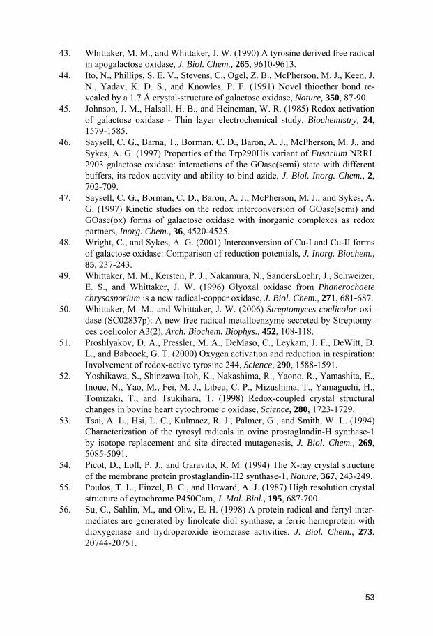
43. Whittaker, M. M., and Whittaker, J. W. (1990) A tyrosine derived free radical in apogalactose oxidase, J. Biol. Chem., 265, 9610-9613.
44. Ito, N., Phillips, S. E. V., Stevens, C., Ogel, Z. B., McPherson, M. J., Keen, J. N., Yadav, K. D. S., and Knowles, P. F. (1991) Novel thioether bond re-vealed by a 1.7 Å crystal-structure of galactose oxidase, Nature, 350, 87-90.
45. Johnson, J. M., Halsall, H. B., and Heineman, W. R. (1985) Redox activation of galactose oxidase - Thin layer electrochemical study, Biochemistry, 24, 1579-1585.
46. Saysell, C. G., Barna, T., Borman, C. D., Baron, A. J., McPherson, M. J., and Sykes, A. G. (1997) Properties of the Trp290His variant of Fusarium NRRL 2903 galactose oxidase: interactions of the GOase(semi) state with different buffers, its redox activity and ability to bind azide, J. Biol. Inorg. Chem., 2, 702-709.
47. Saysell, C. G., Borman, C. D., Baron, A. J., McPherson, M. J., and Sykes, A. G. (1997) Kinetic studies on the redox interconversion of GOase(semi) and GOase(ox) forms of galactose oxidase with inorganic complexes as redox partners, Inorg. Chem., 36, 4520-4525.
48. Wright, C., and Sykes, A. G. (2001) Interconversion of Cu-I and Cu-II forms of galactose oxidase: Comparison of reduction potentials, J. Inorg. Biochem., 85, 237-243.
49. Whittaker, M. M., Kersten, P. J., Nakamura, N., SandersLoehr, J., Schweizer, E. S., and Whittaker, J. W. (1996) Glyoxal oxidase from Phanerochaete chrysosporium is a new radical-copper oxidase, J. Biol. Chem., 271, 681-687.
50. Whittaker, M. M., and Whittaker, J. W. (2006) Streptomyces coelicolor oxi-dase (SC02837p): A new free radical metalloenzyme secreted by Streptomy-ces coelicolor A3(2), Arch. Biochem. Biophys., 452, 108-118.
51. Proshlyakov, D. A., Pressler, M. A., DeMaso, C., Leykam, J. F., DeWitt, D. L., and Babcock, G. T. (2000) Oxygen activation and reduction in respiration: Involvement of redox-active tyrosine 244, Science, 290, 1588-1591.
52. Yoshikawa, S., Shinzawa-Itoh, K., Nakashima, R., Yaono, R., Yamashita, E., Inoue, N., Yao, M., Fei, M. J., Libeu, C. P., Mizushima, T., Yamaguchi, H., Tomizaki, T., and Tsukihara, T. (1998) Redox-coupled crystal structural changes in bovine heart cytochrome c oxidase, Science, 280, 1723-1729.
53. Tsai, A. L., Hsi, L. C., Kulmacz, R. J., Palmer, G., and Smith, W. L. (1994) Characterization of the tyrosyl radicals in ovine prostaglandin-H synthase-1 by isotope replacement and site directed mutagenesis, J. Biol. Chem., 269, 5085-5091.
54. Picot, D., Loll, P. J., and Garavito, R. M. (1994) The X-ray crystal structure of the membrane protein prostaglandin-H2 synthase-1, Nature, 367, 243-249.
55. Poulos, T. L., Finzel, B. C., and Howard, A. J. (1987) High resolution crystal structure of cytochrome P450Cam, J. Mol. Biol., 195, 687-700.
56. Su, C., Sahlin, M., and Oliw, E. H. (1998) A protein radical and ferryl inter-mediates are generated by linoleate diol synthase, a ferric hemeprotein with dioxygenase and hydroperoxide isomerase activities, J. Biol. Chem., 273, 20744-20751.
53
57. Doyle, W. A., Blodig, W., Veitch, N. C., Piontek, K., and Smith, A. T. (1998) Two substrate interaction sites in lignin peroxidase revealed by site-directed mutagenesis, Biochemistry, 37, 15097-15105.
58. Poulos, T. L., Edwards, S. L., Wariishi, H., and Gold, M. H. (1993) Crystal-lographic refinement of lignin peroxidase at 2 Å, J. Biol. Chem., 268, 4429-4440.
59. Pogni, R., Baratto, M. C., Teutloff, C., Giansanti, S., Ruiz-Duenas, F. J., Choinowski, T., Piontek, K., Martinez, A. T., Lendzian, F., and Basosi, R. (2006) A tryptophan neutral radical in the oxidized state of versatile peroxi-dase from Pleurotus eryngii - A combined multifrequency EPR and density functional theory study, J. Biol. Chem., 281, 9517-9526.
60. Perez-Boada, M., Ruiz-Duenas, F. J., Pogni, R., Basosi, R., Choinowski, T., Martinez, M. J., Piontek, K., and Martinez, A. T. (2005) Versatile peroxidase oxidation of high redox potential aromatic compounds: Site-directed mutagenesis, spectroscopic and crystallographic investigation of three long-range electron transfer pathways, J. Mol. Biol., 354, 385-402.
61. Ivancich, A., Jouve, H. M., and Gaillard, J. (1996) EPR evidence for a tyrosyl radical intermediate in bovine liver catalase, J. Am. Chem. Soc., 118, 12852-12853.
62. Fita, I., Silva, A. M., Murthy, M. R. N., and Rossmann, M. G. (1986) The refined structure of beef liver catalase at 2.5 Å resolution, Acta Crystallogr., Sect. B: Struct. Sci., 42, 497-515.
63. Chouchane, S., Girotto, S., Yu, S. W., and Magliozzo, R. S. (2002) Identifi-cation and characterization of tyrosyl radical formation in Mycobacterium tu-berculosis catalase-peroxidase (KatG), J. Biol. Chem., 277, 42633-42638.
64. Bertrand, T., Eady, N. A. J., Jones, J. N., Nagy, J. M., Jamart-Gregoire, B., Raven, E. L., and Brown, K. A. (2004) Crystal structure of Mycobacterium tuberculosis catalase peroxidase, J. Biol. Chem., 279, 38991-38999.
65. Ivancich, A., Jakopitsch, C., Auer, M., Un, S., and Obinger, C. (2003) Protein based radicals in the catalase peroxidase of Synechocystis PCC6803: A multi-frequency EPR investigation of wild type and variants on the environment of the heme active site, J. Am. Chem. Soc., 125, 14093-14102.
66. Ehrenberg, A., and Reichard, P. (1972) Electron spin resonance of iron con-taining protein B2 from ribonucleotide reductase, J. Biol. Chem., 247, 3485-3488.
67. Poole, A. M., Logan, D. T., and Sjöberg, B. M. (2002) The evolution of the ribonucleotide reductases: Much ado about oxygen, J. Mol. Evol., 55, 180-196.
68. Stubbe, J., Nocera, D. G., Yee, C. S., and Chang, M. C. Y. (2003) Radical initiation in the class I ribonucleotide reductase: Long-range proton-coupled electron transfer?, Chem. Rev., 103, 2167-2201.
69. Eklund, H., Uhlin, U., Färnegårdh, M., Logan, D. T., and Nordlund, P. (2001) Structure and function of the radical enzyme ribonucleotide reductase, Prog. Biophys. Mol. Biol., 77, 177-268.
70. Uhlin, U., and Eklund, H. (1994) Structure of ribonucleotide reductase pro-tein R1, Nature, 370, 533-539.
54
71. Nordlund, P., and Reichard, P. (2006) Ribonucleotide reductases, Annu. Rev. Biochem., 75, 681-706.
72. Yee, C. S., Chang, M. C. Y., Ge, J., Nocera, D. G., and Stubbe, J. (2003) 2,3-difluorotyrosine at position 356 of ribonucleotide reductase R2: A probe of long-range proton-coupled electron transfer, J. Am. Chem. Soc., 125, 10506-10507.
73. Yee, C. S., Seyedsayamdost, M. R., Chang, M. C. Y., Nocera, D. G., and Stubbe, J. (2003) Generation of the R2 subunit of ribonucleotide reductase by intein chemistry: Insertion of 3-nitrotyrosine at residue 356 as a probe of the radical initiation process, Biochemistry, 42, 14541-14552.
74. Seyedsayamdost, M. R., and Stubbe, J. (2006) Site-specific replacement of Y-356 with 3,4-dihydroxyphenylalanine in the β2 subunit of E. coli ribonu-cleotide reductase, J. Am. Chem. Soc., 128, 2522-2523.
75. Seyedsayamdost, M. R., Yee, C. S., Reece, S. Y., Nocera, D. G., and Stubbe, J. (2006) pH rate profiles of FnY356-R2s (n=2, 3, 4) in Escherichia coli ri-bonucleotide reductase: Evidence that Y-356 is a redox-active amino acid along the radical propagation pathway, J. Am. Chem. Soc., 128, 1562-1568.
76. Seyedsayamdost, M. R., Xie, J., Chan, C. T. Y., Schultz, P. G., and Stubbe, J. (2007) Site-specific insertion of 3-aminotyrosine into subunit α2 of E. coli ri-bonucleotide reductase: Direct evidence for involvement of Y-730 and Y-731 in radical propagation, J. Am. Chem. Soc., 129, 15060-15071.
77. Kolberg, M., Strand, K. R., Graff, P., and Andersson, K. K. (2004) Structure, function, and mechanism of ribonucleotide reductases, Biochim. Biophys. Acta, 1699, 1-34.
78. Raynaud, C., Sarcabal, P., Meynial-Salles, I., Croux, C., and Soucaille, P. (2003) Molecular characterization of the 1,3-propanediol (1,3-PD) operon of Clostridium butyricum, Proc. Natl. Acad. Sci. U. S. A., 100, 5010-5015.
79. Selmer, T., Pierik, A. J., and Heider, J. (2005) New glycyl radical enzymes catalysing key metabolic steps in anaerobic bacteria, Biol. Chem., 386, 981-988.
80. Davies, M. J. (2005) The oxidative environment and protein damage, Bio-chim. Biophys. Acta, 1703, 93-109.
81. Kojda, G., and Harrison, D. (1999) Interactions between NO and reactive oxygen species: pathophysiological importance in atherosclerosis, hyperten-sion, diabetes and heart failure, Cardiovasc. Res., 43, 562-571.
82. Gelderman, K. A., Hultqvist, M., Olsson, L. M., Bauer, K., Pizzolla, A., Olofsson, P., and Holmdahl, R. (2007) Rheumatoid arthritis: The role of reac-tive oxygen species in disease development and therapeutic strategies, Anti-oxid. Redox Signal., 9, 1541-1567.
83. Mure, M., and Klinman, J. P. (1995) Model studies of Topa quinone - Syn-thesis and characterization of Topa quinone derivates, in Methods Enzymol., 258, 39-52.
84. Mure, M. (2004) Tyrosine derived quinone cofactors, Acc. Chem. Res., 37, 131-139.
85. Davidson, V. L. (2007) Protein derived cofactors. Expanding the scope of post translational modifications, Biochemistry, 46, 5283-5292.
55

86. Klinman, J. P. (2001) Commentary - How many ways to craft a cofactor?, Proc. Natl. Acad. Sci. U. S. A., 98, 14766-14768.
87. Fisher, N., and Rich, P. R. (2000) A motif for quinone binding sites in respi-ratory and photosynthetic systems, J. Mol. Biol., 296, 1153-1162.
88. Rich, P. R., and Bendall, D. S. (1979) Mechanism for the reduction of cyto-chromes by quinols in solution and its relevance to biological electron trans-fer reactions, FEBS Lett., 105, 189-194.
89. Zhu, Z. Y., and Gunner, M. R. (2005) Energetics of quinone-dependent elec-tron and proton transfers in Rhodobacter sphaeroides photosynthetic reaction centers, Biochemistry, 44, 82-96.
90. Rich, P. R. (2004) The quinone chemistry of bc complexes, Biochim. Bio-phys. Acta, 1658, 165-171.
91. Chambers, J. Q. (1988) Electrochemistry of quinones, in The chemistry of quinonoid compounds, vol. II, (Patai, S., and Rappoport, Z., Eds.).
92. Wraight, C. A. (2006) Chance and design - Proton transfer in water, channels and bioenergetic proteins, Biochim. Biophys. Acta, 1757, 886-912.
93. McComb, J. C., Stein, R. R., and Wraight, C. A. (1990) Investigations on the influence of headgroup substitution and isoprene side chain lenght in the function of primary and secondary quinones of bacterial reaction centers, Biochim. Biophys. Acta, 1015, 156-171.
94. Okamura, M. Y., Paddock, M. L., Graige, M. S., and Feher, G. (2000) Proton and electron transfer in bacterial reaction centers, Biochim. Biophys. Acta, 1458, 148-163.
95. Rinyu, L., Martin, E. W., Takahashi, E., Maroti, P., and Wraight, C. A. (2004) Modulation of the free energy of the primary quinone acceptor QA in reaction centers from Rhodobacter sphaeroides: Contributions from the pro-tein and protein-lipid(cardiolipin) interactions, Biochim. Biophys. Acta, 1655, 93-101.
96. Takahashi, E., Wells, T. A., and Wraight, C. A. (2001) Protein control of the redox potential of the primary quinone acceptor in reaction centers from Rhodobacter sphaeroides, Biochemistry, 40, 1020-1028.
97. Hermes, S., Bremm, O., Garczarek, F., Derrien, V., Liebisch, P., Loja, P., Sebban, P., Gerwert, K., and Haumann, M. (2006) A time resolved iron spe-cific X-ray absorption experiment yields no evidence for an Fe2+ -> Fe3+ tran-sition during QA
- −>QB electron transfer in the photosynthetic reaction center, Biochemistry, 45, 353-359.
98. Mure, M., and Klinman, J. P. (1993) Synthesis and spectroscopic characteri-zation of model compounds for the active site cofactor in copper amine oxi-dases, J. Am. Chem. Soc., 115, 7117-7127.
99. Itoh, S., Ogino, M., Haranou, S., Terasaka, T., Ando, T., Komatsu, M., Oh-shiro, Y., Fukuzumi, S., Kano, K., Takagi, K., and Ikeda, T. (1995) A model compound of novel cofactor tryptophan tryptophylquinone of bacterial me-thyiamine dehydrogenases - Synthesis and physicochemical properties, J. Am. Chem. Soc., 117, 1485-1493.
100. Mure, M., Wang, S. X., and Klinman, J. P. (2003) Synthesis and characteriza-tion of model compounds of the lysine tyrosyl quinone cofactor of lysyl oxi-dase, J. Am. Chem. Soc., 125, 6113-6125.
56

101. Murakami, Y., Tachi, Y., and Itoh, S. (2004) A model compound of the novel organic cofactor CTQ (cysteine tryptophylquinone) of quinohemoprotein amine dehydrogenase, Eur. J. Org. Chem., 14, 3074-3079.
102. Katz, E., Schlereth, D. D., and Schmidt, H. L. (1994) Electrochemical study of pyrroloquinoline quinone covalently immobilized as a monolayer onto a cystamine-modified gold electrode, J. Electroanal. Chem., 367, 59-70.
103. Sato, Y., Fujita, M., Mizutani, F., and Uosaki, K. (1996) Electrochemical properties of the 2-mercaptohydroquinone monolayer on a gold electrode. Ef-fect of solution pH, adsorption time and concentration of the modifying solu-tion, J. Electroanal. Chem., 409, 145-154.
104. Kong, J. L., Lu, Z. Q., Lvov, Y. M., Desamero, R. Z. B., Frank, H. A., and Rusling, J. F. (1998) Direct electrochemistry of cofactor redox sites in a bac-terial photosynthetic reaction center protein, J. Am. Chem. Soc., 120, 7371-7372.
105. Hess, C. R., Juda, G. A., Dooley, D. M., Amii, R. N., Hill, M. G., Winkler, J. R., and Gray, H. B. (2003) Gold electrodes wired for coupling with the deeply buried active site of Arthrobacter globiformis amine oxidase, J. Am. Chem. Soc., 125, 7156-7157.
106. Li, W. W., Hellwig, P., Ritter, M., and Haehnel, W. (2006) De novo design, synthesis, and characterization of quinoproteins, Chem. Eur. J., 12, 7236-7245.
107. Richardson, J. S., Richardson, D. C., and Erickson, B. W. (1984) De novo design and synthesis of a protein, Biophys. J., 45, A25-A25.
108. Pabo, C. (1983) Molecular technology - Designing proteins and peptides, Nature, 301, 200-200.
109. (1995) Mass, dimensions, and other parameters of the earth, in CRC Hand-book of chemistry and physics, (Lide, D. R., Ed.).
110. Burkhard, P., Stetefeld, J., and Strelkov, S. V. (2001) Coiled coils: a highly versatile protein folding motif, Trends Cell Biol., 11, 82-88.
111. Sodek, J., Hodges, R. S., Smillie, L. B., and Jurasek, L. (1972) Amino-acid sequence of rabbit skeletal tropomyosin and its coiled coil structure, Proc. Natl. Acad. Sci. U. S. A., 69, 3800-3804.
112. McLachlan, A. D., and Stewart, M. (1975) Tropomyosin coiled coil interac-tions: Evidence for an unstaggered structure, J. Mol. Biol., 98, 293-304.
113. Hodges, R. S., Saund, A. K., Chong, P. C. S., St.-Pierre, S. A., and Reid, R. E. (1981) Synthetic model for two-stranded α-helical coiled-coils - Design, synthesis, and characterization of an 86-residue analog of Tropomyosin, J. Biol. Chem., 256, 1214-1224.
114. Baltzer, L., Nilsson, H., and Nilsson, J. (2001) De novo design of proteins - What are the rules?, Chem. Rev., 101, 3153-3163.
115. Bryson, J. W., Desjarlais, J. R., Handel, T. M., and DeGrado, W. F. (1998) From coiled coils to small globular proteins: Design of a native-like three-helix bundle, Protein Sci., 7, 1404-1414.
116. Munson, M., Balasubramanian, S., Fleming, K. G., Nagi, A. D., Obrien, R., Sturtevant, J. M., and Regan, L. (1996) What makes a protein a protein? Hy-drophobic core designs that specify stability and structural properties, Protein Sci., 5, 1584-1593.
57

117. Willis, M. A., Bishop, B., Regan, L., and Brunger, A. T. (2000) Dramatic structural and thermodynamic consequences of repacking a protein's hydro-phobic core, Structure, 8, 1319-1328.
118. Dolphin, G. T., Brive, L., Johansson, G., and Baltzer, L. (1996) Use of aro-matic amino acid residues to restrict the dynamics in the hydrophobic core of a designed helix-loop-helix dimer, J. Am. Chem. Soc., 118, 11297-11298.
119. Hill, R. B., and DeGrado, W. F. (1998) Solutions structure of αD-2, a native-like de novo designed protein, J Am Chem Soc, 120, 1138-1145.
120. Walsh, S. T. R., Cheng, H., Bryson, J. W., Roder, H., and DeGrado, W. F. (1999) Solution structure and dynamics of a de novo designed three-helix bundle protein, Proc. Natl. Acad. Sci. U. S. A., 96, 5486-5491.
121. Walsh, S. T. R., Sukharev, V. I., Betz, S. F., Vekshin, N. L., and DeGrado, W. F. (2001) Hydrophobic core malleability of a de novo designed three-helix bundle protein, J. Mol. Biol., 305, 361-373.
122. Walsh, S. T. R., Lee, A. L., DeGrado, W. F., and Wand, A. J. (2001) Dynam-ics of a de novo designed three-helix bundle protein studied by 15N, 13C, and 2H NMR relaxation methods, Biochemistry, 40, 9560-9569.
123. Jensen, K. J., and Brask, J. (2002) Carbohydrates as templates for control of distance-geometry in de novo-designed proteins, Cell. Mol. Life Sci., 59, 859-869.
124. Singh, Y., Dolphin, G. T., Razkin, J., and Dumy, P. (2006) Synthetic peptide templates for molecular recognition: Recent advances and applications, Chembiochem, 7, 1298-1314.
125. Rau, H. K., and Haehnel, W. (1998) Design, synthesis, and properties of a novel cytochrome b model, J. Am. Chem. Soc., 120, 468-476.
126. Monien, B. H., Drepper, F., Sommerhalter, M., Lubitz, W., and Haehnel, W. (2007) Detection of heme oxygenase activity in a library of four-helix bundle proteins: Towards the de novo synthesis of functional heme proteins, J. Mol. Biol., 371, 739-753.
127. Rau, H. K., Snigula, H., Struck, A., Robert, B., Scheer, H., and Haehnel, W. (2001) Design, synthesis and properties of synthetic chlorophyll proteins, Eur. J. Biochem., 268, 3284-3295.
128. Schnepf, R., Haehnel, W., Wieghardt, K., and Hildebrandt, P. (2004) Spec-troscopic identification of different types of copper centers generated in syn-thetic four-helix bundle proteins, J. Am. Chem. Soc., 126, 14389-14399.
129. Kamtekar, S., Schiffer, J. M., Xiong, H. Y., Babik, J. M., and Hecht, M. H. (1993) Protein design by binary patterning of polar and nonpolar amino acids, Science, 262, 1680-1685.
130. Roy, S., Ratnaswamy, G., Boice, J. A., Fairman, R., McLendon, G., and Hecht, M. H. (1997) A protein designed by binary patterning of polar and nonpolar amino acids displays native-like properties, J. Am. Chem. Soc., 119, 5302-5306.
131. Roy, S., and Hecht, M. H. (2000) Cooperative thermal denaturation of pro-teins designed by binary patterning of polar and nonpolar amino acids, Bio-chemistry, 39, 4603-4607.
58

132. Wei, Y. N., Liu, T., Sazinsky, S. L., Moffet, D. A., Pelczer, I., and Hecht, M. H. (2003) Stably folded de novo proteins from a designed combinatorial li-brary, Protein Sci., 12, 92-102.
133. Wei, Y. N., Kim, S., Fela, D., Baum, J., and Hecht, M. H. (2003) Solution structure of a de novo protein from a designed combinatorial library, Proc. Natl. Acad. Sci. U. S. A., 100, 13270-13273.
134. Fung, H. K., Welsh, W. J., and Floudas, C. A. (2008) Computational de novo peptide and protein design: Rigid templates versus flexible templates, Ind. Eng. Chem. Res., 47, 993-1001.
135. Dahiyat, B. I., and Mayo, S. L. (1997) De novo protein design: Fully auto-mated sequence selection, Science, 278, 82-87.
136. Kuhlman, B., Dantas, G., Ireton, G. C., Varani, G., Stoddard, B. L., and Baker, D. (2003) Design of a novel globular protein fold with atomic level accuracy, Science, 302, 1364-1368.
137. Bowers, P. M., Strauss, C. E. M., and Baker, D. (2000) De novo protein structure determination using sparse NMR data, J. Biomol. NMR, 18, 311-318.
138. Dantas, G., Kuhlman, B., Callender, D., Wong, M., and Baker, D. (2003) A large scale test of computational protein design: Folding and stability of nine completely redesigned globular proteins, J. Mol. Biol., 332, 449-460.
139. Dantas, G., Corrent, C., Reichow, S. L., Havranek, J. J., Eletr, Z. M., Isern, N. G., Kuhlman, B., Varani, G., Merritt, E. A., and Baker, D. (2007) High resolution structural and thermodynamic analysis of extreme stabilization of human procarboxypeptidase by computational protein design, J. Mol. Biol., 366, 1209-1221.
140. Ambroggio, X. I., and Kuhlman, B. (2006) Computational design of a single amino acid sequence that can switch between two distinct protein folds, J. Am. Chem. Soc., 128, 1154-1161.
141. Jiang, L., Althoff, E. A., Clemente, F. R., Doyle, L., Rothlisberger, D., Zang-hellini, A., Gallaher, J. L., Betker, J. L., Tanaka, F., Barbas, C. F., Hilvert, D., Houk, K. N., Stoddard, B. L., and Baker, D. (2008) De novo computational design of retro-aldol enzymes, Science, 319, 1387-1391.
142. Tommos, C., Skalicky, J. J., Pilloud, D. L., Wand, A. J., and Dutton, P. L. (1999) De novo proteins as models of radical enzymes, Biochemistry, 38, 9495-9507.
143. Dai, Q. H., Tommos, C., Fuentes, E. J., Blomberg, M. R. A., Dutton, P. L., and Wand, A. J. (2002) Structure of a de novo designed protein model of radical enzymes, J. Am. Chem. Soc., 124, 10952-10953.
144. Bard, A. J., and Faulkner, L. R. (2001) Electrochemical methods: Fundamen-tals and applications, John Wiley & Sons, Inc., New York.
145. Parry, E. P., and Osteryoung, R. A. (1965) Evaluation of analytical pulse polarography, Anal. Chem., 37, 1634-1637.
146. Dixon, W. T., and Murphy, D. (1976) Determination of acidity constants of some phenol radical cations by means of electron-spin resonance, J. Chem. Soc., Faraday Trans.2, 72, 1221-1230.
147. Tommos, C. (2002) Electron, proton and hydrogen-atom transfers in photo-synthetic water oxidation, Philos. Trans. R. Soc. London, B, 357, 1383-1394.
59
60
148. Lee, J., and Ross, R. T. (1998) Absorption and fluorescence of tyrosine hy-drogen-bonded to amide-like ligands, J. Phys. Chem. B, 102, 4612-4618.
149. Lee, J. K., Ross, R. T., Thampi, S., and Leurgans, S. (1992) Resolution of the properties of hydrogen-bonded tyrosine using a trilinear model of fluores-cence, J. Phys. Chem., 96, 9158-9162.
150. Sibert, R., Josowicz, M., Porcelli, F., Veglia, G., Range, K., and Barry, B. A. (2007) Proton-coupled electron transfer in a biomimetic peptide as a model of enzyme regulatory mechanisms, J. Am. Chem. Soc., 129, 4393-4400.
151. Bishop, C. A., and Tong, L. K. J. (1965) Equilibria of substituted semi-quinones at high pH, J. Am. Chem. Soc., 87, 501-505.