exploitation of an iron transporter for bacterial protein ... · exploitation of an iron...
TRANSCRIPT
Exploitation of an iron transporter for bacterial proteinantibiotic importPaul Whitea, Amar Joshia,1, Patrice Rassama, Nicholas G. Housdena, Renata Kaminskaa, Jonathan D. Goulta,Christina Redfielda, Laura C. McCaugheya,b, Daniel Walkerc, Shabaz Mohammeda, and Colin Kleanthousa,2
aDepartment of Biochemistry, University of Oxford, Oxford OX1 3QU, United Kingdom; biThree Institute, University of Technology Sydney, Sydney, NSW2007, Australia; and cInstitute of Infection, Immunity & Inflammation, College of Medical, Veterinary & Life Sciences, University of Glasgow, Glasgow G128QQ, United Kingdom
Edited by Hiroshi Nikaido, University of California, Berkeley, CA, and approved September 28, 2017 (received for review August 3, 2017)
Unlike their descendants, mitochondria and plastids, bacteria donot have dedicated protein import systems. However, paradoxi-cally, import of protein bacteriocins, the mechanisms of which arepoorly understood, underpins competition among pathogenic andcommensal bacteria alike. Here, using X-ray crystallography, iso-thermal titration calorimetry, confocal fluorescence microscopy,and in vivo photoactivatable cross-linking of stalled translocationintermediates, we demonstrate how the iron transporter FpvAI inthe opportunistic pathogen Pseudomonas aeruginosa is hijackedto translocate the bacteriocin pyocin S2 (pyoS2) across the outermembrane (OM). FpvAI is a TonB-dependent transporter (TBDT) thatactively imports the small siderophore ferripyoverdine (Fe-Pvd) bycoupling to the proton motive force (PMF) via the inner membrane(IM) protein TonB1. The crystal structure of the N-terminal domainof pyoS2 (pyoS2NTD) bound to FpvAI (Kd = 240 pM) reveals that thepyocin mimics Fe-Pvd, inducing the same conformational changes inthe receptor. Mimicry leads to fluorescently labeled pyoS2NTD beingimported into FpvAI-expressing P. aeruginosa cells by a processanalogous to that used by bona fide TBDT ligands. PyoS2NTD inducesunfolding by TonB1 of a force-labile portion of the plug domain thatnormally occludes the central channel of FpvAI. The pyocin is thendragged through this narrow channel following delivery of its ownTonB1-binding epitope to the periplasm. Hence, energized nutrienttransporters in bacteria also serve as rudimentary protein importsystems, which, in the case of FpvAI, results in a protein antibiotic60-fold bigger than the transporter’s natural substrate being trans-located across the OM.
Pseudomonas aeruginosa | pyocin | outer membrane receptor | transporter
Bacteriocins are peptide or protein antibiotics produced bybacteria to kill their neighbors, usually in response to envi-
ronmental stress, that play a fundamental role in shaping bacterialcommunities (1–3) and are implicated in the invasion mechanismsof pathogens (4, 5). Bacteriocins are currently the focus of in-tense efforts to develop them as much needed antibiotics againstmultidrug-resistant bacteria (6, 7) and as antiinfectives for use inagriculture (8). The present work centers on the mode of action ofprotein bacteriocins, which are species-specific protein antibioticsthat are widespread agents of competition in gram-negative bac-teria (9). Protein bacteriocins are known to parasitize a variety ofcell envelope proteins (10), but how they exploit these systems topromote their import has remained unresolved since their dis-covery (11). We reveal the mechanism by which the nucleasebacteriocin pyocin S2 (pyoS2) crosses the outer membrane (OM)of Pseudomonas aeruginosa, a path that is likely to be used byother TonB-dependent protein bacteriocins.Protein bacteriocins are 40- to 80-kDa toxins that carry a
single cytotoxic domain at their C terminus into the cell. Celldeath ensues through depolarization of the cell by an ionophoreor enzymatic cleavage of peptidoglycan precursors in the peri-plasm or nucleic acids (DNA, tRNA, or rRNA) in the cytoplasm(12). The best studied of the protein bacteriocins are the colicinsthat target and kill Escherichia coli. Colicins exploit a variety of
OM proteins as their primary receptor, including the vitamin B12transporter BtuB and the siderophore transporter FepA (13, 14).Colicin entry into cells requires contact with proton motive force(PMF)-linked systems in the inner membrane (IM) that span theperiplasm: Tol-Pal for group A colicins and Ton for group Bcolicins (15). Tol-Pal is a multiprotein complex involving threeIM proteins (TolA, TolQ, and TolR), a periplasmic protein(TolB), and an OM lipoprotein (Pal). The Ton system comprisesthree IM proteins (TonB, ExbB, and ExbD). Both systems, whichare virulence factors in pathogenic bacteria, energize processesat the OM: Tol-Pal stabilizes the membrane, while Ton catalyzesimport of scarce nutrients such as iron and vitamins across themembrane. Group A colicins generally require additional OMproteins, usually porins, to contact the Tol-Pal system (16). Fewtranslocator proteins have been identified for group B colicins(10). How the Ton or Tol-Pal system catalyzes import of aprotein bacteriocin across the OM is unknown. Once in theperiplasm, nuclease colicins are translocated across the IM bythe AAA+ ATPase FtsH, which also proteolytically releases thenuclease domain to the cytoplasm (17, 18). In the present work,we reveal how pyoS2, a Ton-dependent protein bacteriocin,translocates across the OM through its receptor and show thatthis mechanism has strong parallels with that used by the en-dogenous ligand for the receptor.
Significance
The outer membrane (OM) excludes antibiotics such as vanco-mycin that kill gram-positive bacteria, and so is a major con-tributor to multidrug resistance in gram-negative bacteria. Yet,the OM is readily bypassed by protein bacteriocins, which aretoxins released by bacteria to kill their neighbors during com-petition for resources. Discovered over 60 y ago, it has been amystery how these proteins cross the OM to deliver their toxicpayload. We have discovered how the bacteriocin pyocin S2(pyoS2), which degrades DNA, enters Pseudomonas aeruginosacells. PyoS2 tricks the iron transporter FpvAI into transporting itacross the OM by a process that is remarkably similar to thatused by its endogenous ligand, the siderophore ferripyoverdine.
Author contributions: P.W., A.J., N.G.H., and C.K. designed research; P.W., P.R., N.G.H.,J.D.G., C.R., and S.M. performed research; P.W., N.G.H., R.K., L.C.M., and D.W. contributednew reagents/analytic tools; P.W., A.J., P.R., N.G.H., J.D.G., C.R., and S.M. analyzed data;and P.W., C.R., and C.K. wrote the paper.
The authors declare no conflict of interest.
This article is a PNAS Direct Submission.
This open access article is distributed under Creative Commons Attribution-NonCommercial-NoDerivatives License 4.0 (CC BY-NC-ND).
Data deposition: The atomic coordinates and structure factors have been deposited in theProtein Data Bank, www.wwpdb.org (PDB ID code 5ODW).1Present address: Lonza Biologics Plc, Slough SL1 4DX, United Kingdom.2To whom correspondence should be addressed. Email: [email protected].
This article contains supporting information online at www.pnas.org/lookup/suppl/doi:10.1073/pnas.1713741114/-/DCSupplemental.
www.pnas.org/cgi/doi/10.1073/pnas.1713741114 PNAS | November 7, 2017 | vol. 114 | no. 45 | 12051–12056
MICRO
BIOLO
GY
ResultsThe N-Terminal Domain of PyoS2 Mimics Ferripyoverdine Binding toFpvAI. PyoS2 is a 74-kDa endonuclease bacteriocin that is ef-fective in the treatment of P. aeruginosa-induced pneumonia inmice and in eradicating P. aeruginosa biofilms (6, 19). The pri-mary receptor for pyoS2 is FpvAI (20–22), a TonB-dependenttransporter (TBDT) (23) that actively imports ferripyoverdine(Fe-Pvd) (24). FpvAI is a classical 22-stranded β-barrel with acentral channel that is completely occluded by a globular “plug”domain, which must be reconfigured for substrate transport bycoupling to the PMF via TonB1 (25).Our starting point for elucidating the pyoS2 mechanism of
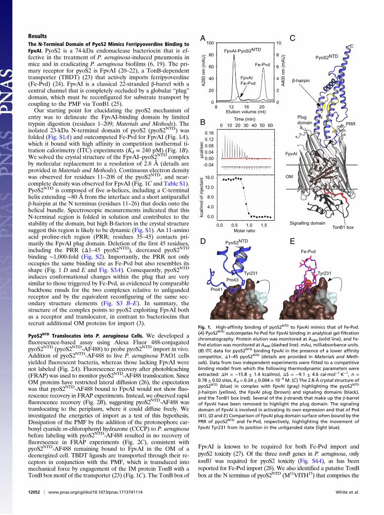
entry was to delineate the FpvAI-binding domain by limitedtrypsin digestion (residues 1–209; Materials and Methods). Theisolated 23-kDa N-terminal domain of pyoS2 (pyoS2NTD) wasfolded (Fig. S1A) and outcompeted Fe-Pvd for FpvAI (Fig. 1A),which it bound with high affinity in competition isothermal ti-tration calorimetry (ITC) experiments (Kd = 240 pM) (Fig. 1B).We solved the crystal structure of the FpvAI–pyoS2NTD complexby molecular replacement to a resolution of 2.8 Å (details areprovided in Materials and Methods). Continuous electron densitywas observed for residues 11–208 of the pyoS2NTD, and near-complete density was observed for FpvAI (Fig. 1C and Table S1).PyoS2NTD is composed of five α-helices, including a C-terminalhelix extending ∼80 Å from the interface and a short antiparallelβ-hairpin at the N terminus (residues 11–26) that docks onto thehelical bundle. Spectroscopic measurements indicated that thisN-terminal region is folded in solution and contributes to thestability of the domain, but high B-factors in the crystal structuresuggest this region is likely to be dynamic (Fig. S1). An 11-aminoacid proline-rich region (PRR; residues 35–45) contacts pri-marily the FpvAI plug domain. Deletion of the first 45 residues,including the PRR (Δ1–45 pyoS2NTD), decreased pyoS2NTD
binding ∼1,000-fold (Fig. S2). Importantly, the PRR not onlyoccupies the same binding site as Fe-Pvd but also resembles itsshape (Fig. 1 D and E and Fig. S3A). Consequently, pyoS2NTD
induces conformational changes within the plug that are verysimilar to those triggered by Fe-Pvd, as evidenced by comparablebackbone rmsds for the two complexes relative to unligandedreceptor and by the equivalent reconfiguring of the same sec-ondary structure elements (Fig. S3 B–E). In summary, thestructure of the complex points to pyoS2 exploiting FpvAI bothas a receptor and translocator, in contrast to bacteriocins thatrecruit additional OM proteins for import (3).
PyoS2NTD Translocates into P. aeruginosa Cells. We developed afluorescence-based assay using Alexa Fluor 488-conjugatedpyoS2NTD (pyoS2NTD-AF488) to probe pyoS2NTD import in vivo.Addition of pyoS2NTD-AF488 to live P. aeruginosa PAO1 cellsyielded fluorescent bacteria, whereas those lacking FpvAI werenot labeled (Fig. 2A). Fluorescence recovery after photobleaching(FRAP) was used to monitor pyoS2NTD-AF488 translocation. SinceOM proteins have restricted lateral diffusion (26), the expectationwas that pyoS2NTD-AF488 bound to FpvAI would not show fluo-rescence recovery in FRAP experiments. Instead, we observed rapidfluorescence recovery (Fig. 2B), suggesting pyoS2NTD-AF488 wastranslocating to the periplasm, where it could diffuse freely. Weinvestigated the energetics of import as a test of this hypothesis.Dissipation of the PMF by the addition of the protonophore car-bonyl cyanide m-chlorophenyl hydrazone (CCCP) to P. aeruginosabefore labeling with pyoS2NTD-AF488 resulted in no recovery offluorescence in FRAP experiments (Fig. 2C), consistent withpyoS2NTD-AF488 remaining bound to FpvAI in the OM of adeenergized cell. TBDT ligands are transported through their re-ceptors in conjunction with the PMF, which is transduced intomechanical force by engagement of the IM protein TonB with aTonB box motif of the transporter (23) (Fig. 1C). The TonB box of
FpvAI is known to be required for both Fe-Pvd import andpyoS2 toxicity (27). Of the three tonB genes in P. aeruginosa, onlytonB1 was required for pyoS2 toxicity (Fig. S4A), as has beenreported for Fe-Pvd import (28). We also identified a putative TonBbox at the N terminus of pyoS2NTD (M11VITH15) that comprises the
A
B
D E
C
Fig. 1. High-affinity binding of pyoS2NTD to FpvAI mimics that of Fe-Pvd.(A) PyoS2NTD outcompetes Fe-Pvd for FpvAI binding in analytical gel-filtrationchromatography. Protein elution was monitored at A280 (solid line), and Fe-Pvd elution was monitored at A400 (dashed line). mAu, milliabsorbance units.(B) ITC data for pyoS2NTD binding FpvAI in the presence of a lower affinitycompetitor, Δ1–45 pyoS2NTD (details are provided in Materials and Meth-ods). Data from two independent experiments were fitted to a competitivebinding model from which the following thermodynamic parameters wereextracted: ΔH = −15.8 ± 1.4 kcal/mol, ΔS = −9.1 ± 4.6 cal·mol−1·K−1, n =0.78 ± 0.02 sites, Kd = 0.24 ± 0.004 × 10⁻⁹ M. (C) The 2.8-Å crystal structure ofpyoS2NTD (blue) in complex with FpvAI (gray) highlighting the pyoS2NTD
β-hairpin (yellow), the FpvAI plug (brown) and signaling domains (black),and the TonB1 box (red). Several of the β-strands that make up the β-barrelof FpvAI have been removed to highlight the plug domain. The signalingdomain of FpvAI is involved in activating its own expression and that of Pvd(41). (D and E) Comparison of FpvAI plug domain surface when bound by thePRR of pyoS2NTD and Fe-Pvd, respectively, highlighting the movement ofFpvAI Tyr231 from its position in the unliganded state (light blue).
12052 | www.pnas.org/cgi/doi/10.1073/pnas.1713741114 White et al.

first β-strand of the β-hairpin. TonB-dependent bacteriocins suchas colicin B, which targets E. coli, generally have their own TonBbox (29). Direct binding of pyoS2NTD to TonB1 was shown by ITC(Kd ∼ 1 μM), as well as by cross-linking, and was abolished when theβ-hairpin was deleted (Δ1–30 pyoS2NTD) (Fig. S4 B–D). Deletion ofthe TonB1 box (Δ1–21 pyoS2NTD-AF488) also abolished fluores-cence recovery in FRAP experiments, as well as pyoS2 cytotoxicity(Fig. S5 A and B). Finally, Δ1–21 pyoS2NTD-AF488 fluorescence,when bound to cells, was completely removed by trypsin treat-ment, in contrast to pyoS2NTD-AF488 (Fig. S5 C–E). We concludethat as well as being the receptor-binding domain for the bacteriocin,pyoS2NTD carries all of the information necessary for translocationacross the OM and that the process requires at least FpvAI, thePMF, and TonB1 in the IM. Thereafter, import to the cytoplasm ismediated by the bacteriocin nuclease domain FtsH and a conservedDPY motif present in all nuclease bacteriocins (9, 17, 30).
PyoS2NTD Translocation Through FpvAI Mirrors That of a TBDT Ligand.We developed a cross-linking strategy to map the pyoS2 importroute that capitalized on its PMF dependence and involved trappingpyoS2NTD during translocation. The PMF delivers ∼20 pN me-chanical force (31), and so we fused pyoS2NTD to GFP (pyoS2NTD-GFP), which can resist up to 100 pN (32). In contrast to pyoS2NTD-AF488, FRAP experiments with pyoS2NTD-GFP bound to P.aeruginosa cells did not show fluorescence recovery, consistent witha block in translocation (Fig. 3A). We substituted 13 pyoS2NTD resi-dues, distributed throughout the pyoS2NTD-GFP construct, for theUV-inducible cross-linker para-Benzoylphenylalanine (pBpa)(Materials and Methods and Fig. 3B). The interfacial siteTyr46pBpa was included as a positive control. All pBpa variantswere purified and first complexed with FpvAI in vitro. Of the13 variants, cross-linking was only observed for Tyr46pBpa, itscross-link to FpvAI Met431 at the protein–protein interface
confirmed by liquid chromatography-tandem mass spectrome-try (LC-MS/MS) (Fig. S6). After ensuring pBpa labeling did notinterfere with pyoS2 cytotoxicity (Fig. S4E), we developed amethod for cross-linking and purifying pyoS2NTD-GFP trans-location intermediates from live P. aeruginosa cells and iden-tified cross-linking sites by LC-MS/MS (Materials and Methods).In vivo cross-links were observed for all 13 pBpa variants,suggesting that translocation intermediates were indeed trap-ped by this strategy (Fig. 3C). We were able to map cross-linksfor seven pBpa sites (Gln17, Ile23, Ala29, Lys70, Ala87,Gln135, and Gln184), all of which were to transmembrane re-gions of FpvAI that are inaccessible to pyoS2NTD in its groundstate complex with FpvAI (Fig. 1C). PyoS2NTD must translocatesignificant distances to satisfy the cross-linking data, as in thecase of pyoS2NTD Gln184, which moves 76 Å to meet FpvAIVal197 (Fig. 3 B–D). We identified six principal cross-link sitesin FpvAI, although LC-MS/MS data suggested residues to eitherside of these sites are also hit, but at lower frequency (Figs. S7 andS8). Five of six residues are within the plug domain that occludesthe channel, and one was to the barrel wall (Met766) (Fig. 3B).Four pBpa sites (Gln17, Ile23, Ala87, and Gln135) cross-link tothe same FpvAI plug domain residue, Met177, suggesting muchof the pyoS2NTD polypeptide chain translocates past this residue.We have interpreted our cross-linking data in the context of
recent atomic force microscopy (AFM) studies on other TBDTs,the vitamin B12 transporter BtuB, and the ferric hydroxamatereceptor FhuA from E. coli (Fig. 4). Hickman et al. (33) haveshown that the plug domains of BtuB and FhuA are composed offorce-resistant and force-labile subdomains. Displacement of theforce-labile subdomain by engagement of TonB with the ligand-bound transporter opens an ∼13-Å-wide channel. Structuralalignment of the plug domains of BtuB (34) and FpvAI identifiesan equivalent force-labile subdomain (Fig. S9). PyoS2NTD
translocation therefore involves at least three steps (Fig. 4). Step1 is binding of pyoS2NTD to FpvAI, where Fe-Pvd mimicry in-duces recruitment of TonB1 in the periplasm. Step 2 is PMF-driven unfolding of the FpvAI force-labile subdomain, release ofthe N-terminal β-hairpin, and delivery of the pyocin’s TonB1 boxthrough the channel, where it engages either the same or anothercopy of TonB1. The pBpa substitutions at pyoS2NTD residuesGln17, Ile23, and Ala29 all cross-link to sites within the channel,either to the nonlabile subdomain (Asp239) or to the barrel wall(Met766). Displacement of the β-hairpin from the helical bundleof pyoS2NTD destabilizes the domain (Fig. S1C), expediting im-port. Step 3 is TonB1-dependent unfolding of pyoS2NTD. Themajority of observed cross-links are for this final state in which fivepyoS2NTD residues spanning >100 amino acids become cross-linked to three hyperreactive FpvAI labile subdomain residues(Pro166, Met177, and Leu182). PyoS2NTD residues Gln17 andIle23 are among the sites that cross-link to FpvAI Met177, which,along with their cross-links to Met776, explains why these residuesgive two cross-linked adducts on SDS/PAGE (Fig. 3C). Finally, asingle cross-link was observed between pyoS2NTD Gln184pBpaand FpvAI Val197, likely denoting the entrapment by GFP of thetranslocating pyocin within the FpvAI channel.
DiscussionOur data show that PMF-coupled TonB1 imports pyoS2NTD bydragging it through a narrow channel in FpvAI created byTonB1-induced displacement of the labile plug subdomain, apathway ordinarily used by its ligand Fe-Pvd. TBDT plug dis-placement is thought to be facilitated by waters at the plug–barrel interface, which could lubricate pyoS2 translocation (35).Two mechanisms seem reasonable as to how the remaining∼500 amino acids of pyoS2 translocate through FpvAI; either thePMF continues to be involved or refolding of pyoS2NTD in theperiplasm provides the driving force. Force-dependent unfolding of
PyoS2NTD-AF488
B
CPyoS2NTD-AF488 + CCCP
Brightfield Pre-bleach Bleach(t=0 mins)
Post-bleach(t=2 mins)
PAO1fpvAI+
PW5036fpvAI−
A Brightfield Fluorescence
Fig. 2. PyoS2NTD translocates across the P. aeruginosa OM. (A) Fluorescencelabeling of live FpvAI-expressing P. aeruginosa PAO1 with pyoS2NTD-AF488.The PAO1 fpvA− transposon mutant PW5036 (fpvA-H02::ISlacZ/hah) exhibitsno labeling. (Scale bars, 5 μm.) FRAP experiments on P. aeruginosaPAO1 labeled with pyoS2NTD-AF488 (B) and pyoS2NTD-AF488 (C) are shown,where cells were first treated with 100 μM CCCP. The bleached region ishighlighted (dashed circle). The absence of FRAP in these experiments sug-gests pyoS2NTD remains bound to FpvAI in the OM, whereas FRAP suggestspyoS2NTD-AF488 has translocated to the periplasm, where it can diffuselaterally. (Scale bars, 1 μm.)
White et al. PNAS | November 7, 2017 | vol. 114 | no. 45 | 12053
MICRO
BIOLO
GY

the bacteriocin during import disrupts the high-affinity complexesof the pyocin with FpvAI (Kd ’ pM) and the C-terminal nucleasewith its immunity protein (Kd ’ fM) (36). Immunity proteinsprotect nuclease bacteriocin-producing cells but are displaced atthe cell surface during import (37). AFM studies have shown thatforce-dependent remodeling of a colicin nuclease accelerates theimmunity protein dissociation rate (31). PyoS2 unfolding duringimport could provide this remodeling force. Ton-dependent bac-teriocins are widely distributed in bacteria, delivering differentcytotoxic activities (nucleases, peptidoglycan hydrolases, and ion-ophores) (12). The subversion of FpvAI by pyoS2 demonstrates amechanism that is likely to be the basis of import for many Ton-dependent bacteriocins and reveals how TBDTs moonlight asprotein transporters.The protein import mechanism we have uncovered in bacteria
has similarities to the classical protein import pathway of mito-chondria catalyzed by the translocase at outer membrane com-plex (38). Both systems rely on an N-terminal sequence motif,the presequence in mitochondria and the TonB box in bacteria,for translocation across the OM. These sequences direct proteinimport through β-barrel transporters, driven by protein com-plexes in the IM coupled to the PMF. Unlike mitochondria,however, where the presequence is recognized by a receptorwithin the translocase, in bacteria, the imported proteins
themselves are recognized by specific receptors that also actas translocases.
Materials and MethodsProtein Expression and Purification. His-tagged pyoS2NTD and derivatives wereexpressed in BL21 (DE3) cells and were purified by nickel-affinity chroma-tography and size-exclusion chromatography (SEC). The TonB1 periplasmicdomain (residues 109–342) was purified using the same method with theaddition of tobacco etch virus-protease removal of the His-tag. FpvAI wasexpressed in E. coli TNE012 cells (ompA−, ompB−, and tsx−) (39) transformedwith pNGH183 carrying the fpvAI gene from P. aeruginosa PAO1 with anE. coli ompF signal sequence. After isolation of the OM fraction, FpvAI waspurified by anion exchange chromatography and SEC. Details are describedin Supporting Information.
Limited Trypsin Proteolysis and Peptide Mass Fingerprinting. A total of 2 mg offull-length pyoS2 (purified using the same method as described for thepyoS2NTD) in a 1:1 molar complex with FpvAI was digested with 2 μg ofSequencing Grade Modified Trypsin (Promega) at room temperature over-night in 25 mM Tris·HCl (pH 8.0), 150 mM NaCl, and 1% (wt/vol) n-octyl-β-D-glucopyranoside (β-OG). Digestion was stopped with 1 mM PMSF, andfragments were copurified with FpvAI on a Superdex 200 10/300 GL column(GE Healthcare). Fragments were resolved by SDS/PAGE, and bands wereexcised for in-gel trypsin digestion and peptide mass fingerprinting as de-scribed in Supporting Information.
m/z500 1000 1500
100
50
Rel
ativ
ein
tens
ity
E = 1.07E-13 MH+ = 3739.7604 Charge = +5
y4 β +1486.30
y6 β +1730.43
y6 α +3267.06
y12 α +2761.34
b7 α +3711.86
b4 β +31440.70
y7 β +1859.47
y14 α +31045.49
y15 α +31078.33
y3 β +1374.25
b6 α +1607.33
b2 α +1239.12
y9 α +2586.78
D
11666.2
45.0
35.0
kDa Mar
ker
M11
XQ
17X
I23X
A29
XY
46X
K70
X
A96
X
A87
XK
92X
A11
9XQ
135X
Q18
4XV
204X
Crosslinks
FpvAIPyoS2NTD-GFP
A B
C
P166 M177
L182
M766
D239
V197
Q184
V204
A96
K92A87
A119
Q135
Y46
A29
Q17
M11
GFP
I23
K7076 Å
Wild
-type
210
191181
FpvAI α
PyoS2 β
H T P G I T V S A Y D T D R N N Y Y A R
Q T S X E L E N K A R
191
Brightfield Pre-bleach Bleach(t=0 mins)
Post-bleach(t=2 mins)
Fig. 3. Mapping the pyoS2NTD translocation pathway by photo cross-linking. (A) FRAP experiment on a P. aeruginosa PAO1 cell labeled with pyoS2NTD-GFPdemonstrates stalled import. The bleached region is highlighted (dashed circle). (Scale bar, 1 μm.) (B) Structure of the pyoS2NTD–FpvAI complex showing thesites of pBpa incorporation in pyoS2NTD-GFP (yellow diamonds) and the major cross-link sites in FpvAI (yellow circles), most of which are located in the plugdomain of the TBDT. The red dotted line indicates the cross-link detected between pBpa184 in pyoS2NTD and Val197 in FpvAI, which requires this region of thepyocin to translocate at least 76 Å. (C) Ten percent SDS/PAGE gel showing Coomassie-stained, in vivo cross-linked adducts of translocated pyoS2NTD-GFPpBpavariants following purification by nickel affinity chromatography from OM extracts of P. aeruginosa PAO1 cells. Only cross-links to FpvAI were observed by LC-MS/MS, confirming that GFP traps the translocating pyoS2NTD within FpvAI (details are provided in Materials and Methods). (D) Fragmentation spectrum ofpBpa184-Val197 cross-linked peptides (X = pBpa) with b- and y-ions indicated, along with the E-value, precursor ion mass (MH+), and precursor ion charge.
12054 | www.pnas.org/cgi/doi/10.1073/pnas.1713741114 White et al.

Analytical SEC. PyoS2NTD at 5 μMwas added to a 5 μM stoichiometric complexof FpvAI and Fe-Pvd (Sigma) in 25 mM Tris·HCl (pH 8.0), 150 mM NaCl, and1% (wt/vol) β-OG, and was incubated at room temperature for 6 h. Themixture was separated on a Superdex 200 10/300 GL column equilibrated inthe same reaction buffer monitoring for protein elution at A280 and for Fe-Pvd elution at A400.
Crystallization and Structure Determination. The FpvAI–pyoS2NTD complex at8 mg/mL, purified as described in Supporting Information, was crystallizedby the sitting-drop vapor diffusion method in 96-well MRC two-drop plates(SWISSCI) at 18 °C. Drops consisted of 100 nL of protein and 100 nL ofcrystallization solution dispensed using a Mosquito robot (TTP Labtech).Crystals of the complex were grown in 0.35M ammonium sulfate, 13.5% (wt/vol)PEG3350, and 0.05 M sodium acetate (pH 4.0), and were harvested after 30 d.Crystals were cryoprotected in the crystallization solution supplemented with1% (vol/vol) n-octyl-polyoxyethylene and 25% (vol/vol) ethylene glycol beforeflash-cooling into liquid nitrogen. X-ray data were collected at the EuropeanSynchrotron Radiation Facility on beamline ID23-2 from a single cryocooledcrystal (100 K) using a 225-mm MarMOSAIC CCD detector. Data collection,processing, refinement, and model building are described in Supporting In-formation. The atomic coordinates and structure factors for the pyoS2NTD
–FpvAI complex [Protein Data Bank (PDB) ID code 5ODW] have been depositedin the PDB.
Fluorescence Microscopy.Alexa Fluor 488 was conjugated onto the C terminusof pyoS2NTD as described in Supporting Information. P. aeruginosa PAO1 cellswere grown in M9-glucose media (6.78 g/L Na2HPO4, 3 g/L KH2PO4, 0.5 g/LNaCl, 10 mM D-glucose, 1 mg/mL NH4Cl, 2 mM MgSO4) at 37 °C, and werelabeled with 1 μM fluorophore-conjugated pyoS2 construct for 15 min (details
are provided in Supporting Information). FRAP experiments were performedusing a PerkinElmer spinning disk confocal microscope with a 100× oil-immersion objective (1.4 N.A.). Images were acquired using the 488-nm laser at10% power, and bleaching was performed at 50% laser power at maximumspeed. Recovery images were acquired over a time course up to 2 min.Bright-field images were recorded for each FRAP experiment.
ITC. For FpvAI-pyoS2NTD–binding experiments, proteins were prepared in50 mM potassium phosphate (pH 7.0) and 1% (wt/vol) β-OG. Experiments wereperformed using aMicroCal iTC200 thermostat at 25 °C. The cell contained FpvAI at3.5 μM, and the syringe contained pyoS2NTD at 70 μM. Competition ITC experi-ments were performed with 10 μM FpvAI and 15 μM Δ1–45 pyoS2NTD in the celland with 70 μMpyoS2NTD in the syringe. Because of the smallΔH, TonB1-pyoS2NTD–binding experiments were performed using a MicroCal PEAQ-ITC isothermal ti-tration calorimeter. Proteins were prepared in 50 mM potassium phosphate(pH 7.0) and 150 mM NaCl. The cell contained TonB1 at 15 μM, and the syringecontained either pyoS2NTD or Δ1–30 pyoS2NTD at 150 μM. The first injectionof 0.5 μL was followed by 19 injections of 2 μL, with each injection spaced by180 s. Binding isotherms were fitted using the manufacturer’s software.
Growth Inhibition Assays. PyoS2 cytotoxic activity was assayed by plate-basedgrowth inhibition assays. Typically, a 10-mL culture of P. aeruginosa wasgrown at 37 °C to an OD600 of 0.6. Lawns were prepared by addition of 200 μLof culture to 5 mL of molten soft LB-agar [0.75% (wt/vol) agar in LB] at 42 °Cand were poured over LB-agar plates. Once set and dry, 2 μL of serially di-luted wild-type or variant pyoS2 was applied to the lawn. Lawns wereallowed to grow overnight at 37 °C, and cytotoxicity was determined byobservation of clearance zones.
29239
23
76617
184
197
17
23
135
87
70
182
177
166
46 431
OM
FpvAI
PyoS2NTD
TonB1 box
TonB1
GFP GFP
GFP
PMF-dependent
force
PMF-dependent
force
TonB1 box
Step 1 Step 2 Step 3
Fig. 4. Model for pyoS2NTD translocation through FpvAI. The three-step model of pyoS2NTD translocation through FpvAI supported by pBpa cross-linkingdata (Fig. 3 and Figs. S6–S9) is shown. In step 1, binding of pyoS2NTD to FpvAI mimics Fe-Pvd, activating the receptor for substrate transport and recruiting theC-terminal domain of TonB1 in the periplasm. In step 2, a PMF-dependent mechanical force, applied via the ExbB–ExbD–TonB1 complex in the IM (not shown),drives unfolding of the labile half of the plug domain. The N terminus of pyoS2NTD enters the ∼13-Å-wide cavity that is created, allowing it to present its ownTonB1 box in the periplasm. In step 3, the pyoS2NTD TonB1 box is bound by another copy of TonB1; at this time, the mechanical force is used to drivetranslocation of pyoS2NTD through the FpvAI lumen. Further translocation is blocked by the force-resistant GFP. The multiple cross-links observed between thelabile portion of the FpvAI plug domain and translocated pyoS2NTD residues are presumed to involve unfolded polypeptide chains in the periplasm.
White et al. PNAS | November 7, 2017 | vol. 114 | no. 45 | 12055
MICRO
BIOLO
GY

LC-MS/MS Identification of in Vivo Photoactivated Cross-Links. A 500-mL cul-ture of P. aeruginosa PAO1 was grown for 24 h at 30 °C in iron-free succinatemedium (6.1 g/L K2HPO4, 3 g/L KH2PO4, 4.1 g/L succinic acid) supplementedwith 7.5 mM (NH4)2SO4, 1 mM MgSO4, and 1× trace metal solution [preparedas PTM4(Fe−) as described elsewhere (40)]. Cells were washed and resuspendedin 35 mL of fresh medium, incubated for 60 min in the presence of 100 nMpyoS2NTD-GFPpBpa variants, and UV-irradiated at 365 nm for 30 min in a CL-1000 UV cross-linker. Cells were then resuspended in 10 mM Tris·HCl (pH 8.0),0.25% (wt/vol) lithium diiodosalicyclic acid, and 2% (vol/vol) Triton X-100, andwere lysed through sonication. The cleared lysate was ultracentrifuged at200,000 × g for 45 min to isolate the OM fraction, which was solubilized in10 mM Tris·HCl (pH 8.0), 5 mM EDTA, and 2% (wt/vol) β-OG. Cross-linkedcomplexes were purified using EDTA-resistant cOmplete His-Tag Purification
Resin (Roche) and were resolved by SDS/PAGE. Cross-linking sites weremappedby LC-MS/MS and pLink software as described in Supporting Information.
ACKNOWLEDGMENTS. We thank Iain Lamont for kindly providing the tonBmutant and parent strains PAO6609, K1040, K1407, and MS231. We thankSvenja Hester for preparing cross-linking gel slices for LC-MS/MS. We thankEd Lowe and European Synchrotron Radiation Facility ID23-2 beamline stafffor assistance with X-ray data collection, and we thank David Staunton forassistance with molecular biophysics measurements. This work was fundedby the Wellcome Trust (Grant 201505/Z/16/Z) and Biotechnology and Biolog-ical Sciences Research Council (Grant BB/G020671/2). P.W. was funded by theWellcome Trust Doctoral Training Centre for Cellular and Structural Biology.We acknowledge Grant NIH P30 DK089507 for use of the two-allele librarystrain PW5036.
1. Kirkup BC, Riley MA (2004) Antibiotic-mediated antagonism leads to a bacterial gameof rock-paper-scissors in vivo. Nature 428:412–414.
2. Wiener M, Freymann D, Ghosh P, Stroud RM (1997) Crystal structure of colicin Ia.Nature 385:461–464.
3. Housden NG, et al. (2013) Intrinsically disordered protein threads through the bac-terial outer-membrane porin OmpF. Science 340:1570–1574.
4. Nedialkova LP, et al. (2014) Inflammation fuels colicin Ib-dependent competition ofSalmonella serovar Typhimurium and E. coli in enterobacterial blooms. PLoS Pathog10:e1003844.
5. Holt KE, et al. (2013) Tracking the establishment of local endemic populations of anemergent enteric pathogen. Proc Natl Acad Sci USA 110:17522–17527.
6. McCaughey LC, Ritchie ND, Douce GR, Evans TJ, Walker D (2016) Efficacy of species-specific protein antibiotics in a murine model of acute Pseudomonas aeruginosa lunginfection. Sci Rep 6:30201.
7. Cotter PD, Ross RP, Hill C (2013) Bacteriocins - A viable alternative to antibiotics? NatRev Microbiol 11:95–105.
8. Schulz S, et al. (2015) Broad and efficient control of major foodborne pathogenicstrains of Escherichia coli by mixtures of plant-produced colicins. Proc Natl Acad SciUSA 112:E5454–E5460.
9. Sharp C, Bray J, Housden NG, Maiden MCJ, Kleanthous C (2017) Diversity and distri-bution of nuclease bacteriocins in bacterial genomes revealed using Hidden MarkovModels. PLOS Comput Biol 13:e1005652.
10. Kleanthous C (2010) Swimming against the tide: Progress and challenges in our un-derstanding of colicin translocation. Nat Rev Microbiol 8:843–848.
11. Fredericq P (1957) Colicins. Annu Rev Microbiol 11:7–22.12. Braun V, Patzer SI, Hantke K (2002) Ton-dependent colicins and microcins: Modular
design and evolution. Biochimie 84:365–380.13. Kurisu G, et al. (2003) The structure of BtuB with bound colicin E3 R-domain implies a
translocon. Nat Struct Biol 10:948–954.14. Devanathan S, Postle K (2007) Studies on colicin B translocation: FepA is gated by
TonB. Mol Microbiol 65:441–453.15. Cascales E, et al. (2007) Colicin biology. Microbiol Mol Biol Rev 71:158–229.16. Housden NG, et al. (2010) Directed epitope delivery across the Escherichia coli outer
membrane through the porin OmpF. Proc Natl Acad Sci USA 107:21412–21417.17. Walker D, Mosbahi K, Vankemmelbeke M, James R, Kleanthous C (2007) The role of
electrostatics in colicin nuclease domain translocation into bacterial cells. J Biol Chem282:31389–31397.
18. Chauleau M, Mora L, Serba J, de Zamaroczy M (2011) FtsH-dependent processing ofRNase colicins D and E3 means that only the cytotoxic domains are imported into thecytoplasm. J Biol Chem 286:29397–29407.
19. Smith K, et al. (2012) Activity of pyocin S2 against Pseudomonas aeruginosa biofilms.Antimicrob Agents Chemother 56:1599–1601.
20. Ohkawa I, Shiga S, Kageyama M (1980) Effect of iron concentration in the growthmedium on the sensitivity of Pseudomonas aeruginosa to pyocin S2. J Biochem 87:323–331.
21. Denayer S, Matthijs S, Cornelis P (2007) Pyocin S2 (Sa) kills Pseudomonas aeruginosastrains via the FpvA type I ferripyoverdine receptor. J Bacteriol 189:7663–7668.
22. Elfarash A, Wei Q, Cornelis P (2012) The soluble pyocins S2 and S4 from Pseudomonasaeruginosa bind to the same FpvAI receptor. MicrobiologyOpen 1:268–275.
23. Noinaj N, Guillier M, Barnard TJ, Buchanan SK (2010) TonB-dependent transporters:Regulation, structure, and function. Annu Rev Microbiol 64:43–60.
24. Greenwald J, et al. (2007) Real time fluorescent resonance energy transfer visuali-zation of ferric pyoverdine uptake in Pseudomonas aeruginosa. A role for ferrousiron. J Biol Chem 282:2987–2995.
25. Adams H, Zeder-Lutz G, Schalk I, Pattus F, Celia H (2006) Interaction of TonB with theouter membrane receptor FpvA of Pseudomonas aeruginosa. J Bacteriol 188:5752–5761.
26. Rassam P, et al. (2015) Supramolecular assemblies underpin turnover of outer mem-brane proteins in bacteria. Nature 523:333–336.
27. McCaughey LC, et al. (2016) Discovery, characterization and in vivo activity of pyocinSD2, a protein antibiotic from Pseudomonas aeruginosa. Biochem J 473:2345–2358.
28. Poole K, Zhao Q, Neshat S, Heinrichs DE, Dean CR (1996) The Pseudomonas aerugi-nosa tonB gene encodes a novel TonB protein. Microbiology 142:1449–1458.
29. Hilsenbeck JL, et al. (2004) Crystal structure of the cytotoxic bacterial protein colicin Bat 2.5 A resolution. Mol Microbiol 51:711–720.
30. Mosbahi K, et al. (2002) The cytotoxic domain of colicin E9 is a channel-forming en-donuclease. Nat Struct Biol 9:476–484.
31. Farrance OE, et al. (2013) A force-activated trip switch triggers rapid dissociation of acolicin from its immunity protein. PLoS Biol 11:e1001489.
32. Dietz H, Rief M (2004) Exploring the energy landscape of GFP by single-moleculemechanical experiments. Proc Natl Acad Sci USA 101:16192–16197.
33. Hickman SJ, Cooper REM, Bellucci L, Paci E, Brockwell DJ (2017) Gating of TonB-dependent transporters by substrate-specific forced remodelling. Nat Commun 8:14804.
34. Chimento DP, Mohanty AK, Kadner RJ, Wiener MC (2003) Substrate-induced trans-membrane signaling in the cobalamin transporter BtuB. Nat Struct Biol 10:394–401.
35. Chimento DP, Kadner RJ, Wiener MC (2005) Comparative structural analysis of TonB-dependent outer membrane transporters: Implications for the transport cycle.Proteins 59:240–251.
36. Wallis R, Moore GR, James R, Kleanthous C (1995) Protein-protein interactions incolicin E9 DNase-immunity protein complexes. 1. Diffusion-controlled association andfemtomolar binding for the cognate complex. Biochemistry 34:13743–13750.
37. Vankemmelbeke M, et al. (2009) Energy-dependent immunity protein release duringtol-dependent nuclease colicin translocation. J Biol Chem 284:18932–18941.
38. Wiedemann N, Pfanner N (2017) Mitochondrial machineries for protein import andassembly. Annu Rev Biochem 86:685–714.
39. Taylor R, Burgner JW, Clifton J, CramerWA (1998) Purification and characterization ofmonomeric Escherichia coli vitamin B12 receptor with high affinity for colicin E3.J Biol Chem 273:31113–31118.
40. Greenwald J, Zeder-Lutz G, Hagege A, Celia H, Pattus F (2008) The metal dependenceof pyoverdine interactions with its outer membrane receptor FpvA. J Bacteriol 190:6548–6558.
41. Shen J, Meldrum A, Poole K (2002) FpvA receptor involvement in pyoverdine bio-synthesis in Pseudomonas aeruginosa. J Bacteriol 184:3268–3275.
42. Kabsch W (2010) Xds. Acta Crystallogr D Biol Crystallogr 66:125–132.43. Evans PR, Murshudov GN (2013) How good are my data and what is the resolution?
Acta Crystallogr D Biol Crystallogr 69:1204–1214.44. Winn MD, et al. (2011) Overview of the CCP4 suite and current developments. Acta
Crystallogr D Biol Crystallogr 67:235–242.45. McCoy AJ, et al. (2007) Phaser crystallographic software. J Appl Cryst 40:658–674.46. Greenwald J, et al. (2009) FpvA bound to non-cognate pyoverdines: Molecular basis
of siderophore recognition by an iron transporter. Mol Microbiol 72:1246–1259.47. Adams PD, et al. (2010) PHENIX: A comprehensive Python-based system for macro-
molecular structure solution. Acta Crystallogr D Biol Crystallogr 66:213–221.48. Bricogne GBE, et al. (2010) BUSTER (Global Phasing Ltd, Cambridge, United Kingdom),
Version 2.10.2.49. Emsley P, Cowtan K (2004) Coot: Model-building tools for molecular graphics. Acta
Crystallogr D Biol Crystallogr 60:2126–2132.50. Chen VB, et al. (2010) MolProbity: All-atom structure validation for macromolecular
crystallography. Acta Crystallogr D Biol Crystallogr 66:12–21.51. Krissinel E, Henrick K (2007) Inference of macromolecular assemblies from crystalline
state. J Mol Biol 372:774–797.52. Delano WL (2002) The PyMOL Molecular Graphics System (DeLano Scientific, Palo
Alto, CA).53. Chin JW, Martin AB, King DS, Wang L, Schultz PG (2002) Addition of a photo-
crosslinking amino acid to the genetic code of Escherichiacoli. Proc Natl Acad Sci USA99:11020–11024.
54. Marino F, et al. (2014) Characterization and usage of the EASY-spray technology aspart of an online 2D SCX-RP ultra-high pressure system. Analyst (Lond) 139:6520–6528.
55. Yang B, et al. (2012) Identification of cross-linked peptides from complex samples. NatMethods 9:904–906.
56. Schanda P, Kupce E, Brutscher B (2005) SOFAST-HMQC experiments for recordingtwo-dimensional heteronuclear correlation spectra of proteins within a few seconds.J Biomol NMR 33:199–211.
57. Braunschweiler L, Ernst RR (1983) Coherence transfer by isotropic mixing–Applicationto proton correlation spectroscopy. J Magn Reson 53:521–528.
58. Delaglio F, et al. (1995) NMRPipe: A multidimensional spectral processing systembased on UNIX pipes. J Biomol NMR 6:277–293.
59. Vranken WF, et al. (2005) The CCPN data model for NMR spectroscopy: Developmentof a software pipeline. Proteins 59:687–696.
12056 | www.pnas.org/cgi/doi/10.1073/pnas.1713741114 White et al.