experimental productio onf midgut …jeb.biologists.org/content/jexbio/35/2/251.full.pdfin...
TRANSCRIPT
VOLUME 35, No. 2 JUNE 1958
EXPERIMENTAL PRODUCTION OF MIDGUT TUMOURSIN PERIPLANETA AMERICANA L.
BY JANET E. HARKER
Department of Zoology, University of Cambridge
(Received 3 October 1957)
(With Plate 8)
INTRODUCTION
In earlier papers (Harker, 1955; 1956) it has been shown that the sub-oesophagealganglion of Periplaneta americana L. undergoes a 24 hr. rhythm of secretory activityto which is related the 24 hr. rhythm of locomotor activity. Furthermore, the sub-oesophageal ganglion retains its secretory rhythm even when it is implanted into theabdomen of another animal, and the animal into which it is implanted will take upa locomotor activity rhythm in phase with the secretory rhythm of the implant,provided that the animal is not already showing a strong rhythm.
Now that this autonomous ' clock' has been found it may be possible to upset thecycle of 24 hr. to which the activity rhythm seems bound, and this might be doneby continuous stimulation to activity through the ganglion secretion. Since thefactors which control the secretory rhythm are change from light to darkness andthe length of time spent in each of these environments within 24 hr. cycles, it is notpossible to bring about continuous stimulation through the animal's own sub-oesophageal ganglion; but implanted ganglia can be used to provide a secretionduring the inactive phase of the animals.
In the following pages the effect of this secondary secretion on the tissues of thealimentary canal is described. The effect on the locomotor activity rhythm will bediscussed in another paper.
METHODS
Adult P. americana of both sexes were used in the experiments. Animals were keptin alternating light and darkness, of periods of 12 hr. each, for at least 3 weeks beforethe experiments were begun, and the locomotor activity rhythms were recorded toensure that the timing of the phases of the rhythm was known. Cockroaches here-after described as having a normal 24 hr. activity rhythm were kept in darkness from10 p.m. to 10 a.m., those described as having a reversed rhythm were kept indarkness from 10 a.m. to 10 p.m.
Prior to the implantation of sub-oesophageal ganglia a small polythene ring wassealed with wax around a circular incision on the dorsal surface of the abdomen ofeach cockroach; the haemolymph flowed into the ring. The sub-oesophageal gangliawere then inserted, as rapidly as possible after dissection, and the ring was sealedwith a fragment of cover-slip. It was found that the cuticle grew up the sides of the
17 Exp. BioL 35, 2

252 JANET E. HARKER
ring rather than across the wound, so that renewal of the implant did not necessitatedamaging the cuticle repeatedly. Great care was taken in making the articularincision to ensure that it did not penetrate the muscle layer beneath thecuticle.
EFFECT ON THE GUT OF IMPLANTED SUB-OESOPHAGEAL GANGLIA
Sub-oesophageal ganglia removed from cockroaches known to have a reversedactivity rhythm were implanted, one each, into thirty-five cockroaches showingnormal activity rhythms. The implants were replaced by fresh ganglia every 24 hr.at 10.0 a.m., that is at the time of change of light conditions. The implanted gangliawere sectioned after removal and it was found that they had not been encapsulatedby blood cells and that the neurosecretory cells appeared to be in a normal condition.
In this series of experiments the cockroaches receiving implants were supposedlyreceiving secretions from the sub-oesophageal ganglia in two phases, in one phasefrom their own ganglion, and in the other from the implant.
The cockroaches were divided into groups of five animals, and implantation wascontinued for from 1 to 4 days in different groups. The treatment and the time ofkilling are given in Table 1. The alimentary canal of each animal was examined, andit was found that a small swelling appeared in the midgut of many. The guts of allanimals were sectioned and the swellings were found to be small tumours; detaileddescriptions of these are given in the next section.
Table 1. Sub-oesophageal ganglia taken from cockroaches with reversed 24 hr. loco-motor activity rhythms and implanted into cockroaches with normal rhythms.
Groups offive Tiim«l«
A
B
C
D
E
F
G
i
Implanted
Implanted
Implanted
Implanted
Implanted
Implanted
Implanted
1
Implanted
Implanted
Implanted
Implanted
Implanted
Implanted
—
3
Implanted
Implanted
Implanted
Implanted
Implanted
—
4
Implanted
Implanted
Implanted
Implanted
Implanted
—
6
—
—
—
—
—
—
8
KiUed.no
—
—
—
—
1 0
—
Killed, tmall
in twoim'mitf
—
—
—
—
—
Killed, amalltumourin two
—
—
14
—
—
—
Killed,tumour in
—
—
16
—
—
—
—
Killed,tumour inall aniroaU
Killed,tumourin three
Killed, notumour*
It can be seen from Table 1 that when implantation was performed from two tofour times the recipient cockroaches, in all but one case, formed tumours by thesixteenth day from the beginning of the experiment. The tumours all appeared inthe midgut, irrespective of the region of the abdomen in which the implant wasmade. To confirm the lack of dependence of the position of the tumour on the
Experimental production of midgut tumours 253
position of the implant, implantation into the thorax was performed daily for 4 dayson three cockroaches; at the end of 18 days a tumour was found in the midgut ofeach animal.
As a control similar implantations were made into two groups of five animals each,but, instead of sub-oesophageal ganglia, brains were implanted in one group andcorpora allata in the other. Implantation was repeated for 8 days in each case. Therewas no sign of tumour formation, after 28 days, in any of the animals.
Sub-oesophageal ganglia taken from cockroaches showing normal activityrhythms were implanted into ten cockroaches which also had normal activityrhythms. In this series of experiments the recipient cockroaches were supposedlyreceiving secretions from both sub-oesophageal ganglia at the same time, that is,during only part of the 24 hr. Implantation was continued daily for 4 days in fiveanimals, and for 8 days in five animals. None of the animals showed any sign oftumour formation, even after 28 days from the beginning of the experiment.
Extracts of sub-oesophageal ganglia were prepared by grinding a single ganglionin 0-05 ml. of Ringer solution, and a fresh extract was injected into the abdomen ofeach of five cockroaches during the inactive phase. This was repeated daily for12 days. No tumours were formed in any of these animals. Similar extracts wereprepared using five ganglia in 0-05 ml. of Ringer solution, and these were injecteddaily for 5 days into five other animals. After 21 days three of the cockroaches werefound to have a small tumour in the midgut. An extract of this strength (five gangliain 0-05 ml. Ringer) injected into five cockroaches every day during their activephase, and repeated for 5 days, did not produce tumours in any of the animals.
Previous experiments (Harker, 1956 and unpublished) have shown that, althoughphases of the locomotor activity rhythm of Periplaneta can be determined by thephases of the light:dark rhythm to which they are subjected, not all the processesin the animal which undergo a 24 hr. rhythm are controlled in this way. Forinstance, there is a variation over 24 hr. in the type of reaction of headless cockroachesto injections of sub-oesophageal ganglia extracts, and the type of reaction appearsto be related to the time of day and not to the light: dark treatment, nor to theresultant locomotor activity rhythm of the animal before decapitation. It seemslikely then that, although the phases of the locomotor activity rhythm are reversedin cockroaches which have been subjected to reversed light and darkness, the phasesof some metabolic rhythms persist in the normal form. If this is so, cockroachesmight show some variation at different times of day in their sensitivity to implantedsub-oesophageal ganglia; the following experiment was performed to test thishypothesis.
Five cockroaches were kept in reversed light and darkness for 3 weeks; theirlocomotor activity rhythms were found to be reversed. Into these cockroaches wereimplanted sub-oesophageal ganglia taken from animals with normal activityrhythms; implantation was repeated daily for 4 days. No tumours had formed inthese animals by the twentieth day. The experiments were repeated, this time thetreatment was continued for 8 days. Two of the animals had formed tumours inthe midgut by the twentieth day. In this experiment the secretory activity of the
17-2
254 JANET E. HARKER
sub-oesophageal ganglia is supposedly similar to that in the previous experiments, yetthe incidence of tumours is lower and treatment had to be continued for a longerperiod before tumours were formed. It seems likely that either the gut shows arhythm of sensitivity to sub-oesophageal secretion, a rhythm which is not reversedby reversed light:dark conditions, or that some other organ affected by the sub-oesophageal ganglia secretion and involved in tumour production has a rhythmindependent of light: dark conditions. In either case it appears that Periplaneta isnot as sensitive to the sub-oesophageal ganglion hormone at the time when thehormone would normally be present as at the time when normally none would bepresent.
HISTOLOGY AND THE DEVELOPMENT OF THE TUMOURS
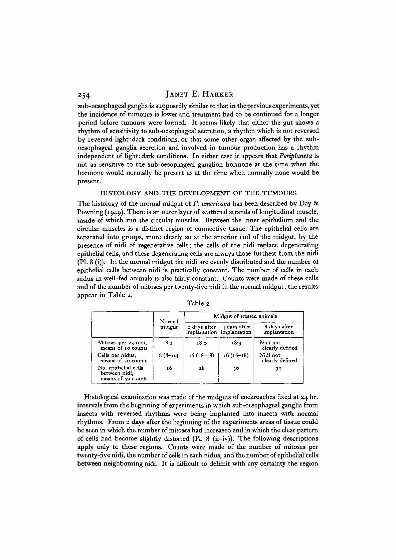
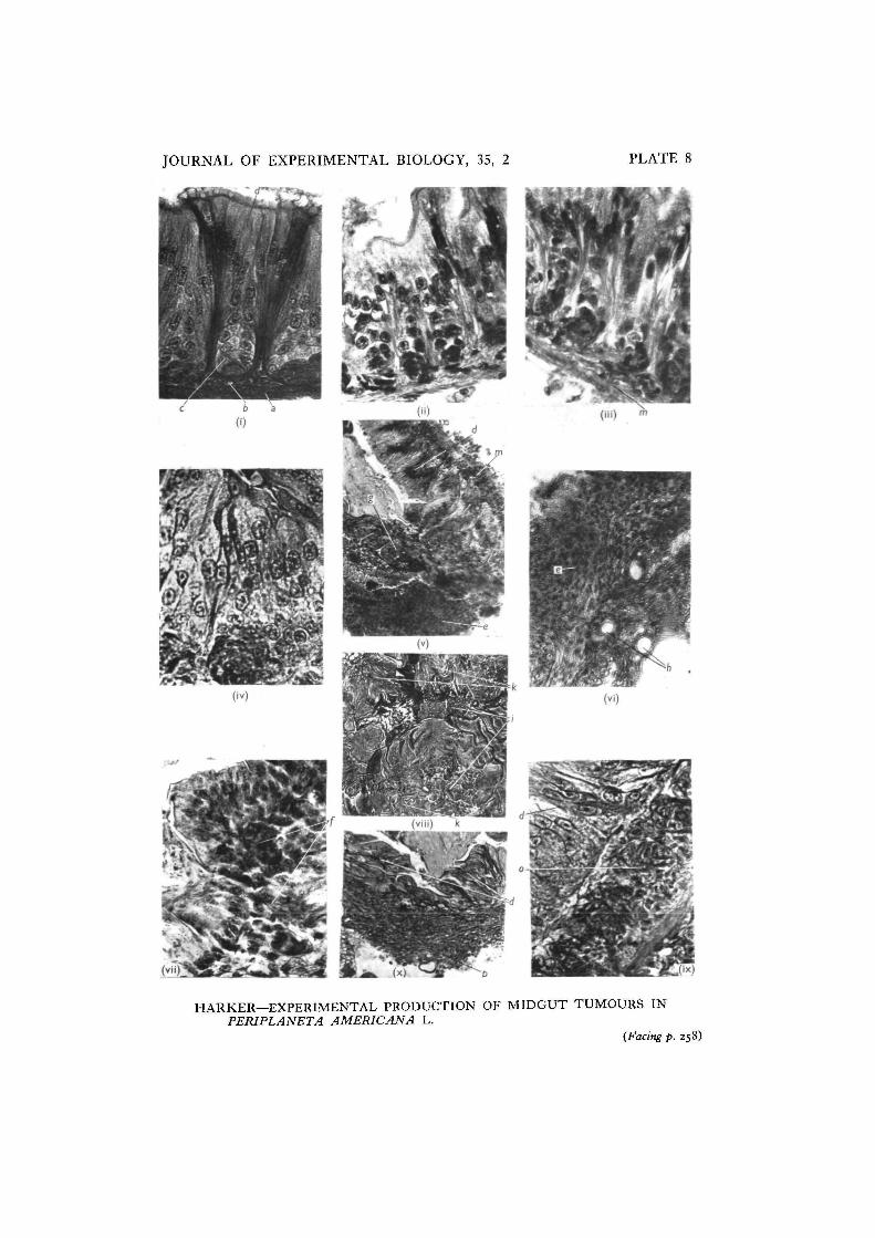
The histology of the normal midgut of P. americana has been described by Day &Powning (1949). There is an outer layer of scattered strands of longitudinal muscle,inside of which run the circular muscles. Between the inner epithelium and thecircular muscles is a distinct region of connective tissue. The epithelial cells areseparated into groups, more clearly so at the anterior end of the midgut, by thepresence of nidi of regenerative cells; the cells of the nidi replace degeneratingepithelial cells, and these degenerating cells are always those furthest from the nidi(PI. 8 (i)). In the normal midgut the nidi are evenly distributed and the number ofepithelial cells between nidi is practically constant. The number of cells in eachnidus in well-fed animals is also fairly constant. Counts were made of these cellsand of the number of mitoses per twenty-five nidi in the normal midgut; the resultsappear in Table 2.
Table 2
Mitoses per 25 nidi,means of 10 counts
Cells per nidus,means of 50 counts
No. epithelial cellsbetween nidi,means of 50 counts
midgut
8-i
8 (8-10)
16
Midgut of treated animals
2 days afterimplantation
18-0
16(16-18)
38
4 days afterimplantation
18-3
16(16-18)
3°
8 days afterimplantation
Nidi notclearly defined
Nidi notclearly defined
30
Histological examination was made of the midguts of cockroaches fixed at 24 hr.intervals from the beginning of experiments in which sub-oesophageal ganglia frominsects with reversed rhythms were being implanted into insects with normalrhythms. From 2 days after the beginning of the experiments areas of tissue couldbe seen in which the number of mitoses had increased and in which the clear patternof cells had become slightly distorted (PI. 8 (ii-iv)). The following descriptionsapply only to these regions. Counts were made of the number of mitoses pertwenty-five nidi, the number of cells in each nidus, and the number of epithelial cellsbetween neighbouring nidi. It is difficult to delimit with any certainty the region
Experimental production of midgut tumours 255
affected, and the numbers given in Table 2 cannot be taken as accurate; where theyerr it will be because normal cells have been included and so the numbers will be low.
From the results appearing in Table 2 it can be seen that the number of mitosesincreases rapidly after treatment, more than doubling after 2 days; the number ofcells in each nidus also increases, but it appears that some cells are moving up intothe epithelium. Seven days after the beginning of treatment, in the majority ofcases, there is a double line of cells in the epithelium (PI. 8 (ii), (iv)); in determiningthis, it is necessary to ensure that sections are cut at right angles to the main axisof the gut, and that the region of suspected tumour formation is not at either endof the midgut, where a double line of epithelial cells may normally be present.
At the same time as the mitotic activity increases, that is from the second day,cells from the nidi are seen to have moved into the connective tissue layer adjacentto the muscle layer; it appears to be these cells which form the tumour. After14-18 days the epithelial cells begin to break down (PI. 8 (iv), (v)); the tumourthen consists of small cells with very little cytoplasm and which stain densely withhaematoxylin; these cells form a whorled pattern and a number of tracheae runbetween them.
Small metastases have been found in eight cockroaches after 24-30 days; theyappear in other regions of the midgut, the foregut and hindgut and in the salivaryglands (PI. 8 (vii), (viii)). The metastases may arise from cells moving in theconnective tissue layer of the gut as well as in the haemocoele, for cells appear tomigrate in both.
Transplants of small portions of the tumours, about 0-5-1-0 mm.2, were placedin the haemocoele of twenty cockroaches. The implants from two animals wereexamined after 2 days and were seen to be surrounded by a sheath of blood cells.Two cockroaches were killed every 2 days for 14 days, and it appeared that thetransplanted portions had increased in size; however, after 4 days it is difficult todifferentiate between the surrounding blood cells and the transplanted cells. Of theremaining cockroaches, two were examined at 20, 24 and 28 days respectively; threeof these six animals had developed tumours in the midgut, and one in the hindgut.
Small portions of normal gut tissue approximately equal in size to the transplantsof tumour tissue were implanted into ten control animals. None of these animalsshowed any sign of tumour formation after 28 days.
The sequence of tumour formation was examined in three groups of four animalsinto which tumour tissue had been transplanted 8, 14 and 28 days previously. Thesequence appeared to differ in some details from that of primary tumour formation.Eight days after the transplantation had been made cells were seen to be invadingthe connective tissue layer below the nidi of the midgut; the width of the connectivetissue layer increased about fourfold in the next 5 days (PI. 8 (ix)), and it was notuntil this stage was reached that the epithelial cells (in all but one animal) showedany abnormal appearance. In the one exception the epithelial cells had begun todegenerate by the eighth day. The nidi had lost their normal form by the eighth dayin two animals, and in all four of the animals killed on the fourteenth day. The nidiof the surrounding tissue showed no increase in mitosis until after the epithelium
256 JANET E. HARKER
had begun to degenerate; in this type of tumour formation it appears that the nidido not supply cells to the tumour, but only carry out their normal function ofreplacing epithelial cells. After the epithelium had begun to break down, that is,between the eighth and fourteenth day, blood cells began to accumulate along themuscle layer and these added superficially to the size of the swelling. The swellingat this stage is invaded by tracheae.
In insects swellings due to wound healing have something of the appearance ofmalignant growth (Day, 1952; Wigglesworth, 1937), but that neither the primarynor transplanted tumours are swellings of this type appears evident (i) from thecontrol experiments, (ii) from the fact that in the formation of primary tumours thesite of implantation is not related to the region in which the tumour is formed, and(iii) from the evidence of increase in mitotic activity of the cells of the nidi.
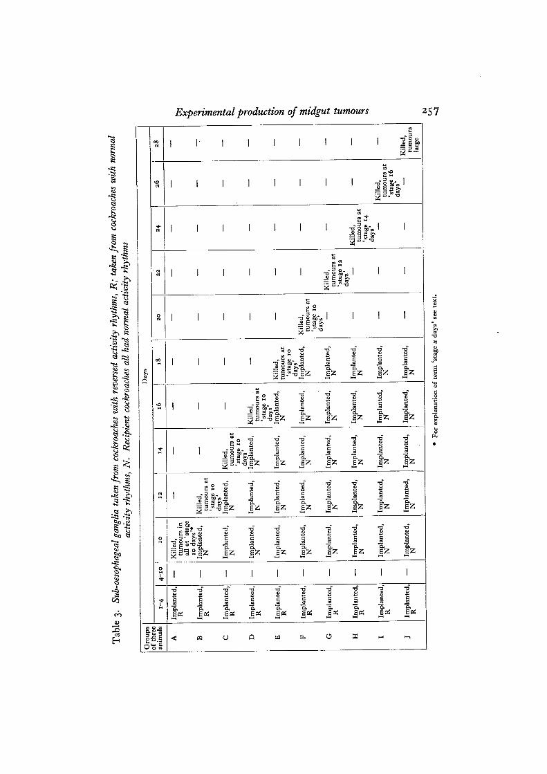
TEMPORARY CONTROL OF THE TUMOURS
Ten groups of three cockroaches were implanted daily for 4 days. Sub-oesophagealganglia taken from cockroaches with reversed activity rhythms were implanted dailyfor 4 days into ten groups of three cockroaches each. Implantation was discontinuedfrom day 4 to day 10. On day 10 one group was killed; tumours were found to bedeveloping in all three animals, and had reached the stage in which the number ofepithelial cells had increased, but there was no breakdown as yet of the epithelialtissue (see Table 3). Daily implantations were made into the remaining groups fromday 10 until day 20, using sub-oesophageal ganglia from animals whose rhythmswere normal and in phase with those of the recipient cockroaches. Every secondday one group of animals was killed and the guts sectioned. The sections showedthat tumours had not developed beyond the stage already reached when the secondseries of implantations began on day 10. The number of mitoses in the nidi hadapparently returned to normal, but the affected region was recognizable by theincreased number of cells. On day 20 implantation was discontinued and theexamination of one group every 2 days continued. By day 24 the tumour size hadincreased considerably, the epithelial layer had been broken down, and the generalappearance was that of a mass of small cells in whorled formation. This stage isreferred to as 'stage 14 days' in Table 3; the term is only one of convenience andit is not claimed that certain stages are closely correlated with the number of daysfrom the beginning of the treatment.
In this experiment the presence of excess sub-oesophageal ganglion hormone atthe time when some secretion would normally be present appears to control thegrowth of the tumours. That the cells of the tumour have undergone an irreversiblechange, however, is shown by the increased growth as soon as the hormone levelreturns to normal.
The number of animals used in this experiment, as in those previously described,has had to be limited because of the time which every operation takes, and becauseof the large numbers of cockroaches necessary to supply the sub-oesophageal gangliafor implantation.
258 JANET E. HARKER
DISCUSSION
The three main conclusions which might be drawn from these results are that thepresence of excess amounts of sub-oesophageal ganglion secretion brings aboutformation of transplantable tumours in the gut of Periplaneta, that the midgut is theregion most frequently affected, and lastly, the most striking conclusion, that thetimtTig of the presence of the secretion is of first importance in tumour formation.
The importance of timing, both in the production of tumourous cells and in thegrowth of the tumours, apparently results from the interaction of 24 hr. rhythms ofseveral processes. Although the introduction at any time of day of large excessesof hormone is followed by the formation of tumourous cells, at a low level ofconcentration not only is the 24 hr. rhythm of secretion concerned, but also thesensitivity of the gut, which itself shows a 24 hr. rhythm. The hormone continuesto act when the cells have become tumourous and again it is not just the presenceof the hormone which is involved, but the timing of its presence, for this samehormone which was responsible for the formation of the tumour can control itsgrowth, if present at the normal time. Perhaps it is also significant that the cellswhich give rise to the tumour are cells which probably show a 24 hr. rhythm ofmitosis under normal conditions; this has not been shown, but it appears likely,since the regeneration of the epithelium is connected with feeding, and feedingfollows the 24 hr. rhythm of locomotory activity.
Nothing is known of the chemical nature of the sub-oesophageal ganglion secre-tion, nor of the way in which it acts on the animal in regulating the locomotoractivity rhythm. Whether it acts through one or several systems, and whether it isthe secretion itself or a secondary system which is responsible for the formation oftumours, is unknown, but, however tumour formation is brought about, the essentialfeature appears to be the continuous presence of the hormone. The normal animalseems to be well safeguarded from continuous secretion; even if the sub-oesophagealganglion could maintain a continuous supply, the secretory phase appears to bebound to a 24 hr. rhythm which has, as yet, proved unalterable by experimentalmeans.
SUMMARY
1. Sub-oesophageal ganglia taken from Periplaneta americana L. which had beenkept in reversed conditions of light and darkness have been implanted into theabdomens of cockroaches living in normal light: dark conditions. When implantationwas continued for 4 days transplantable tumours appeared by the eighteenth day inthe midgut of the majority of cockroaches.
2. No tumours appeared in the guts of cockroaches when the implanted sub-oesophageal ganglia had been taken from animals kept in normal conditions of lightand darkness, nor when brains or corpora allata were implanted.
3. Injection of an extract of five sub-oesophageal ganglia in 005 ml. Ringersolution produced tumours only when injection took place during the inactivephase of the 24 hr. locomotory rhythm.
JOURNAL OF EXPERIMENTAL BIOLOGY, 35, 2 PLATE 8
'*)
HARKER—EXPERIMENTAL PRODUCTION OF MIDGUT TUMOURS INPERIPLANETA AMERICANA L.
(Facing p. 258)
