experimental methods for examining synaptic...
TRANSCRIPT
Topic Introduction
Experimental Methods for Examining Synaptic Plasticityin Drosophila
Douglas P. Olsen and Haig Keshishian
The Drosophila neuromuscular junction (NMJ) ranks as one of the preeminent model systems forstudying synaptic development, function, and plasticity. In this article, we review the experimentalgenetic methods that include the use of mutated or reengineered ion channels tomanipulate the synap-tic connections made by motor neurons onto larval body-wall muscles. We also provide a consider-ation of environmental and rearing conditions that phenocopy some of the genetic manipulations.
BACKGROUND
The Drosophila larval NMJ has been extensively studied as a model system for examining all aspects ofsynaptic development, function, and plasticity. Several excellent and up-to-date reviews of this systemhave been published over the last few years (Budnik and Ruiz-Canada 2006; Collins and Diantonio2007). In this article, we provide an overview of the genetic methods most often used for manipulatingthe NMJ for the purpose of studying synaptic development and plasticity. There is a multitude ofmutations that yield NMJ phenotypes. Here we focus on the better understood genetic approaches:those that directly affect neurophysiological functions to alter levels of membrane excitability; con-structs that alter either presynaptic transmitter release or postsynaptic receptor function; and manip-ulations of second messengers and signal transduction cascades, cell adhesion molecules (CAMs),retrograde growth factors, and transcription factors.
In addition, we consider environmental and rearing conditions that phenocopy the geneticapproaches that affect synaptic growth and function. The article is organized around seven tablesthat summarize the major experimental tools, the genes that are involved, comments about themethod, source material, and relevant references.
METHODS FOR MANIPULATING SYNAPTIC CONNECTIONS
Membrane Excitability
We begin with a review of the genetic tools that are commonly used to manipulate membrane excit-ability, summarized in Table 1. Two approaches have been adopted. The earliest methods made use ofmutations that affect either the expression or function of ion channels. The more recent secondapproach is to use the directed expression of modified ion channels in neurons or muscles. Thesechannels have often been reengineered to introduce useful properties, such as altered activation volt-ages or green fluorescent protein (GFP) tagging.
Adapted from Drosophila Neurobiology (ed. Zhang et al.). CSHL Press, Cold Spring Harbor, NY, USA, 2010.
© 2012 Cold Spring Harbor Laboratory PressCite this article as Cold Spring Harbor Protoc; 2012; doi:10.1101/pdb.top067785
162
Cold Spring Harbor Laboratory Press on June 29, 2018 - Published by http://cshprotocols.cshlp.org/Downloaded from

Ion Channel Mutations
In general, these approaches involve mutations that have one of three primary effects: They alter themain conductance subunit of the channel itself, such as paralytic (para) or Shaker (Sh); they affectother genes required for channel function, such as temperature-induced paralytic E (tipE); or theyaffect the expression of the ion channel subunits, such asmaleless (mlenap-ts1). An important propertyof many of the classical ion channel mutations is the availability of temperature-sensitive (ts) con-ditional alleles. In most cases, the ts mutations have a modest phenotype at the permissive tempera-ture; however, at the restrictive temperature a full-fledged phenotype is obtained. The conditionalquality of the ts mutations is of value for studies of neural plasticity and function. For example, paraly-tic hypoactivity mutations that would otherwise be lethal at the embryonic stage are used to acutelysuppress activity at later times of development for phenocritical analyses.
Elevated membrane excitability is obtained using loss of function of ion channels that carryoutward currents that repolarize the membrane voltage, such as the K+ channel mutations Sh andether-a-gogo (eag), which are often used in combination (eag Sh). Hyperexcitability can also beobtained by increasing the dosage of Na+ channels, for example, by a duplication of the para gene.The most powerful bouts of acute hyperexcitability involve mutant combinations of ts “seizure”mutations, such as the comatose Kum double mutations comts; CaP60Akumts. At the restrictive temp-erature these animals experience brief bouts of very high-frequency action potential firing, followedby paralysis (Hoeffer et al. 2003). In general, presynaptic membrane hyperexcitability leads to the
TABLE 1. Methods to alter membrane excitability
Effect Tool (gene symbol) CommentCommonly used alleles(allele type: stock #)a Reference(s)b
Increasedexcitability
Shaker (Sh) KV1.2 K+ channel Sh14 (null: 3563); Sh7 (anti);Sh21 (hypo); Sh16; Sh5 (111)
Budnik et al. (1990)
ether-a-gogo (eag) K+ channel subunit eagsc29 (null: 1442); eag1 (hypo:3561); eag4PM
Budnik et al. (1990)
Hyperkinetic (Hk) Channel subunit; oxidoreductase Hk1 (3562); Hk2 (55) Budnik et al. (1990)comatose; Ca-P60A(comt; Ca-P60A)
Temperature-dependent seizure Comt6; CaP60AKum170 Hoeffer et al. (2003)
Dp para + Duplication of chromosomalregion containing para locus
Dp(1;4)r + l (5273) Budnik et al. (1990)
UAS-SDN Shaker dominant negative Mosca et al. (2005)UAS-eag-DN eag dominant negative (8178) Broughton et al. (2004)UAS-TrpA1 Warm-activated trp channel (26263, 26264) Hamada et al. (2008)UAS-TRPM8 Cold-activated trp channel Peabody et al. (2009)UAS-NaChBac Bacterial Na channel Nitabach et al. (2006)UAS-P2X2 Light activation of channel agonist Lima and Miesenbock (2005)UAS-Channel-rhodopsin-2(UAS-ChR2)
Light-gated cation channel Schroll et al. (2006), Atamanet al. (2008)
Decreasedexcitability
maleless (mle) Regulates Na+ channelexpression
mlenap-ts1(GOF) Budnik et al. (1990), Jareckiand Keshishian (1995)
paralytic (para) Na+ channel paralk2 (null); parats1 (hypo: 1572) Budnik et al. (1990), Jareckiand Keshishian (1995)
Temperature-inducedparalytic E (tipE)
Channel subunit tipE1 Jarecki and Keshishian (1995)
UAS-Kir2.1 Inward rectifier K+ channel Baines et al. (2001), Paradiset al. (2001)
UAS-EKO Voltage-gated reengineeredSh channel
White et al. (2001)
UAS-dORK Constitutively open K+ channel Nitabach et al. (2002)
aThe allele nomenclature and annotation of allele type listed in this and subsequent tables is derived from FlyBase. Please refer to FlyBase (http://flybase.org/) for synon-ymous alleles. The stock number listed is from the Bloomington Drosophila Stock Center (http://flystocks.bio.indiana.edu/). Abbreviations of allele type: null, amorph;hypo, hypomorph; anti, antimorph; GOF, gain of function.bIn general, the references cited in this and subsequent tables refer to examples of the use of the genetic tools at the neuromuscular junction (NMJ). In cases in which a toolhas not been used at the NMJ, the reference cited refers to the original characterization/development of the genetic tool.
Cite this article as Cold Spring Harbor Protoc; 2012; doi:10.1101/pdb.top067785 163
Methods for Examining Synaptic Plasticity
Cold Spring Harbor Laboratory Press on June 29, 2018 - Published by http://cshprotocols.cshlp.org/Downloaded from
expansion of the synapse, including an increase in the number of synaptic boutons and nerve terminalbranches, as well as increased neurotransmitter release (Budnik et al. 1990; Schuster et al. 1996b).
Reduced membrane excitability is often achieved by loss of function of channels carrying inwardcurrents, most commonly the Na+ channel para, or through loss of function of genes required forNa+ channel function or expression, such as tipE or mlenap-ts1. In contrast to the growth effectsobserved with hyperactivity, reduced neural activity impairs the proper refinement of synaptic con-nections at the NMJ (Jarecki and Keshishian 1995; White et al. 2001). Although hypoactivemutants have relatively normal NMJ growth, their individual synaptic boutons are significantlysmaller (Lnenicka et al. 2003).
Expression of Ion Channels
The directed expression of modified ion channels to manipulate neural excitability represents themost promising direction for controlling neural activity, in both a constitutive and a conditionalmanner. The tools are expressed as upstream activating sequence (UAS) effectors, controlled byeither conventional Gal4 drivers or by inducible methods, such as the steroid-activated GeneSwitchGal4 drivers (Osterwalder et al. 2001; Roman et al. 2001). Three channels have been widely used tosuppress membrane excitability: UAS-EKO, UAS-Kir2.1, and UAS-dORK. Each is a K+ channelthat shunts the membrane voltage toward the equilibrium potential of potassium (EK), a voltagethat is usually more negative than the resting voltage. Whereas the dORK channel introduces a con-stitutively open K+ shunt, both the Kir2.1 and EKO channels have voltage dependence (Baines et al.2001; Paradis et al. 2001; White et al. 2001; Nitabach et al. 2002). Kir2.1 is a modified GFP-taggedinward rectifier channel, and it is most effective near the resting membrane voltage (Baines et al.2001; Paradis et al. 2001). In contrast, EKO is an extensively modified variant of the fast activatingShaker K+ channel, in which the fast inactivation function has been disabled. Although EKO has aconstitutive K+ conductance at the resting voltage, it rapidly activates with depolarization, intercept-ing the membrane voltage to drive it back toward EK when the cell initiates an action potential. Theconstruct also has a GFP tag, to help monitor its localization. As EKO is based on a native K+ channel(Shaker), it also localizes to membrane sites in which the Dlg adapter protein is found, such as thesubsynaptic reticulum of the NMJ (White et al. 2001).
Several effective tools have also been introduced for acutely elevating membrane excitability. Forexample, both the eag and Sh proteins can be suppressed by dominant negative constructs (Broughtonet al. 2004; Mosca et al. 2005). SDN (Shaker Dominant Negative) is a truncated Shaker subunit thatabolishes Sh function when expressed in neurons or muscles. It functions as a dominant negative con-struct, presumably by disrupting the proper assembly of the Sh channel during its biogenesis (Moscaet al. 2005). However, SDNwill have an effect only in cells that express the Shaker channel. Moreover,the degree of increased excitation is limited to that achieved by a Shmutant. As an alternative, a moregeneral tool for elevating membrane excitability is NaChBac, a bacterially derived Na+ channel thatdepolarizes any cell in which it is expressed (Nitabach et al. 2006).
Several recently introduced approaches allow the investigator to control neural excitability in aconditional fashion using either light or temperature. These involve the expression of either the light-activated protein Channelrhodopsin-2 (ChR2) or the temperature-activated channels TrpA1 andTRPM8 (Schroll et al. 2006; Ataman et al. 2008; Hamada et al. 2008; Peabody et al. 2009). Thesetools allow the investigator to elevate membrane excitability transiently in targeted neurons or inmuscles, on illumination with the appropriate wavelength or by a temperature shift. A caveat ofChR2 is that Drosophila requires that retinal be provided to assemble a functional light-gatedchannel (Schroll et al. 2006; Ataman et al. 2008). An alternative and effective method for light acti-vation involves P2X2, a purine receptor that can be activated by using photolytically cleavedligands. However, this approach requires that the caged ligand be provided to the animal beforelight activation (Lima and Miesenbock 2005).
Finally, the researcher has the option of using the directed expression of double-stranded UASRNA interference (RNAi) constructs to knock down specific ion channels (Dietzl et al. 2007).
164 Cite this article as Cold Spring Harbor Protoc; 2012; doi:10.1101/pdb.top067785
D.P. Olsen and H. Keshishian
Cold Spring Harbor Laboratory Press on June 29, 2018 - Published by http://cshprotocols.cshlp.org/Downloaded from
When combined with inducible Gal4 expression systems, this knockdown can be performed in a con-ditional manner in the cells of interest, bypassing early lethality that might result from constitutivechannel knockdown. The increasing availability of these lines for a wide range ofDrosophila ion chan-nels provides a powerful alternative to the several methods described above. The use of transgenicRNAi lines is also a powerful tool for loss of function studies for any of the signaling cascades describedlater in the article.
Overall, a wide range of options is available to the researcher interested in manipulating neuralexcitability, but care must be exercised in selecting the appropriate tool for the task at hand. If thegoal is to examine the effects of long-term changes in membrane excitability, any of the classic ionchannel mutations are a good choice. However, these will usually affect neurons and/or musclesthroughout the organism, complicating cell-specific analyses. Also, to have a direct effect, the genebeing mutated must be expressed in the neuron or muscle of interest. For example, the para Na+
channel is not expressed in muscles, whereas the Shaker K+ channel is not expressed in all neuronsnor at all developmental stages. A second consideration is to control for genetic backgroundeffects, because many synaptic phenotypes have limited penetrance or result in modest changes inNMJ size or function. One solution to this problem is to use genetic suppression as a control.Thus the triple mutant eag Sh mlenap-ts1 line is often used as a control for eag Sh, in which thereduced Na+ channel expression due to mlenap-ts1 suppresses the membrane hyperexcitability pheno-type caused by eag Sh (Budnik et al. 1990).
The conditionally activated channels such as UAS-ChR2, UAS-TrpA1, or UAS-TRPM8 have tre-mendous power, and they are expected to become the methods of choice when rapid and acute exci-tation of specific neurons is desired. It is likely that corresponding methods will be introduced foracute suppression of activity, also based on optical methods, such as those that involve the use ofhalorhodopsins. An exciting prospect for the investigator will be animals in which distinct wave-lengths of light could be used to acutely elevate and suppress membrane excitability in specificneurons.
A protocol is available forMonitoring Membrane Excitability inDrosophila Expressing ModifiedShaker Constructs (Olsen and Keshishian 2012).
Synaptic Transmission
The next class of genetic tools target chemical synapses, with the goal of either elevating or decreasingsynaptic transmission (Table 2). As we saw with methods used to manipulate membrane excitability(Table 1), these tools make use of both classic mutations as well as a variety of transgenic constructs.
Functional plasticity at the NMJ typically involves changes in either quantal content (the amountof neurotransmitter quanta released per action potential) or quantal size (the postsynaptic responsedue to the release of a single transmitter quantum). There are numerous manipulations that affect
TABLE 2. Methods to alter synaptic transmission
EffectTool
(gene symbol) Comment
Commonly usedalleles (allele type:
stock #) Reference
Decreased presynapticrelease
UAS-shits1 Temperature-sensitiveDynamin mutation
Kitamoto (2001)
UAS-TNT Tetanus toxin light chain Sweeney et al. (1995)Decreased postsynapticsensitivity
DGluRIIA Mutations in postsynapticglutamate receptor subunit
DGluRIIAAD9;DGluRIIASP16
Petersen et al. (1997)
UAS-DGluRIIAM614R
Channel dead glutamatereceptor subunit
Diantonio et al. (1999)
UAS-DGluRIIB Wild-type glutamate receptorsubunit
Diantonio et al. (1999)
Increased postsynapticsensitivity
UAS-DGluRIIAsubunit
Wild-type glutamate receptor Petersen et al. (1997)
Cite this article as Cold Spring Harbor Protoc; 2012; doi:10.1101/pdb.top067785 165
Methods for Examining Synaptic Plasticity
Cold Spring Harbor Laboratory Press on June 29, 2018 - Published by http://cshprotocols.cshlp.org/Downloaded from
either parameter, as will be discussed in later sections. However, here we focus on genetic tools thataffect either the exocytosis/endocytosis machinery of the synaptic terminal or neurotransmitter recep-tors in muscle in a direct, specific, and well-controlled fashion.
Tools Targeting Presynaptic Function
The first widely used genetic tool for controlling synaptic transmission was the ts allele of the dynamingene Shibire (Shits). At the restrictive temperature, Shits animals rapidly collapse and remain paralyzeduntil cooled to the permissive temperature (Grigliatti et al. 1973). For acute blockade of chemicalsynaptic transmission throughout the nervous system, this is a powerful tool, but it has been sup-planted by the introduction of a UAS Shits effector, permitting dominant, cell-specific, and con-ditional silencing of synaptic release on shifting to the restrictive temperature (Waddell et al. 2000;Kitamoto 2001). For example, the rapid activation and conditional nature of the construct hasallowed researchers to examine with impressive detail when cell-specific synaptic activity is requiredduring the establishment of long-term memories (Waddell et al. 2000). However, because dynaminfunction is required for membrane turnover and neuronal growth, suppressing function for pro-longed periods during development is problematic, especially when the structural plasticity ofsynapses is being studied.
A direct approach for silencing synaptic transmission is to disrupt the SNARE (soluble NSF attach-ment protein receptor) complex involved in vesicle exocytosis. The method of choice uses tetanustoxin light chain (TetTxLC), expressed in a cell-specific manner under the control of theUAS-TNT effector. TetTxLC is a protease that degrades the vSNARE synaptobrevin, resulting in acomplete blockade of evoked synaptic transmission (Sweeney et al. 1995). The silencing is essentiallyirreversible; therefore, to restrict expression to specific postembryonic stages of development requiresthe use of inducible Gal4 expression systems, such as GeneSwitch or the Gal80-based TARGETsystem. Even so, the researcher should be aware that all existing inducible systems have a low levelof leakiness. Given the very potent nature of TetTxLC, there may be an effect on evoked transmissioneven in the absence of induction. Expression of inactive forms of the tetanus toxin light chain can beused as a control.
Tools Targeting Postsynaptic Function
The suppression and elevation of the postsynaptic response at the NMJ is most directly achieved byaltering glutamate receptor function. Drosophila body-wall muscles express two tetrameric DGluRIIreceptor complexes, which differ by whether they include a DGluRIIA or DGluRIIB subunit. Quantalsize increases in proportion to the ratio of DGluRIIA to DGluRIIB. Thus, an effective method to sup-press synaptic drive at the NMJ is to use either the DGluRIIA mutation or alternatively to driveexpression of the dominant negative transgene UAS DGluRIIA-DN. Quantal size can also bedecreased by driving expression of DGluRIIB, once again reducing the ratio of DGluRIIA/DGluRIIB.It was through the use of these lines that the first evidence for a retrograde homeostatic regulation ofpresynaptic neurotransmitter release at the NMJ was discovered (Petersen et al. 1997; Diantonio et al.1999). Quantal size is effectively increased by driving expression of DGluRIIA.
Signal Transduction and Second Messengers
Cyclic AMP
The cyclic AMP (cAMP) cascade is a potent regulator of both functional and structural synaptic plas-ticity at the NMJ. The genetic tools most widely used to manipulate the levels of intracellular cAMP atthe synapse are the adenylyl cyclase rutabaga (rut) and the cAMP phosphodiesterase dunce (dnc).Levels of cAMP can be increased by using dnc mutations or expression of UAS-rut, whereas lowlevels of cAMP can be achieved by using rut mutations or UAS-dnc (Table 3). Loss-of-functionmutations of either rut or dnc disrupt short-term plasticity, as measured by paired pulse facilitation(PPF) or posttetanic potentiation (PTP) (Zhong and Wu 1991). However, the increased levels of
166 Cite this article as Cold Spring Harbor Protoc; 2012; doi:10.1101/pdb.top067785
D.P. Olsen and H. Keshishian
Cold Spring Harbor Laboratory Press on June 29, 2018 - Published by http://cshprotocols.cshlp.org/Downloaded from
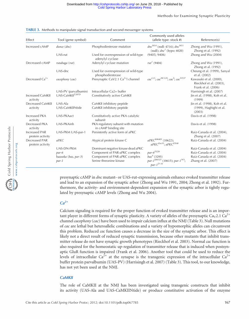
presynaptic cAMP in dncmutant- or UAS-rut-expressing animals enhance evoked transmitter releaseand lead to an expansion of the synaptic arbor (Zhong and Wu 1991, 2004; Zhong et al. 1992). Fur-thermore, the activity- and environment-dependent expansion of the synaptic arbor is tightly regu-lated by presynaptic cAMP levels (Zhong and Wu 2004).
Ca2+
Calcium signaling is required for the proper function of evoked transmitter release and is an impor-tant player in different forms of synaptic plasticity. A variety of alleles of the presynaptic CaV2.1 Ca
2+
channel cacophony (cac) have been used to impair calcium influx at the NMJ (Table 3). Null mutationsof cac are lethal but heteroallelic combinations and a variety of hypomorphic alleles can circumventthis problem. Reduced cac function causes a decrease in the size of the synaptic arbor. This effect islikely not a direct result of reduced synaptic transmission, because other mutants that inhibit trans-mitter release do not have synaptic growth phenotypes (Rieckhof et al. 2003). Normal cac function isalso required for the homeostatic up-regulation of transmitter release that is induced when postsyn-aptic GluR function is impaired (Frank et al. 2006). Another tool that could be used to reduce thelevels of intracellular Ca2+ at the synapse is the transgenic expression of the intracellular Ca2+
buffer protein parvalbumin (UAS-PV) (Harrisingh et al. 2007) (Table 3). This tool, to our knowledge,has not yet been used at the NMJ.
CaMKII
The role of CaMKII at the NMJ has been investigated using transgenic constructs that inhibitits activity (UAS-Ala and UAS-CaMKIINtide) or produce constitutive activation of the enzyme
TABLE 3. Methods to manipulate signal transduction and second messenger systems
Effect Tool (gene symbol) CommentCommonly used alleles(allele type: stock #) Reference(s)
Increased cAMP dunce (dnc) Phosphodiesterase mutation dncM14 (null: 4714); dncM11
(null); dnc1 (hypo: 6020)Zhong and Wu (1991),Zhong et al. (1992)
UAS-rut Used for overexpression of wild-typeadenylyl cyclase
(9405; 9406) Zhong and Wu (2004)
Decreased cAMP rutabaga (rut) Adenylyl cyclase mutation rut1 (9404) Zhong and Wu (1991),Zhong et al. (1992)
UAS-dnc Used for overexpression of wild-typephosphodiesterase
Cheung et al. (1999), Sanyalet al. (2002)
Decreased Ca2+ cacophony (cac) Presynaptic CaV2.1 Ca2+l channel cacL13; cacHC129; cacS; cacNT27 Kawasaki et al. (2000),Rieckhof et al. (2003),Frank et al. (2006)
UAS-PV (parvalbumin) Intracellular Ca2+ buffer Harrisingh et al. (2007)Increased CaMKIIactivity
UAS-CaMKIIT287D Constitutively active CaMKII Jin et al. (1998), Koh et al.(1999)
Decreased CaMKIIactivity
UAS-AlaUAS-CaMKIINtide
CaMKII inhibitory peptideCaMKII inhibitory peptide
Jin et al. (1998), Koh et al.(1999), Haghighi et al.(2003)
Increased PKAactivity
UAS-PKAact Constitutively active PKA catalyticsubunit
Davis et al. (1998)
Decreased PKAactivity
UAS-PKAinh PKA regulatory subunit with mutationin cAMP binding site
Davis et al. (1998)
Increased PARprotein activity
UAS-PKM UAS-par-1 Persistently active form of aPKC Ruiz-Canada et al. (2004),Zhang et al. (2007)
Decreased PARprotein activity
aPKC Atypical protein kinase C aPKCk06403 (10622);aPKCExc55; aPKCEX48
Ruiz-Canada et al. (2004)
UAS-DN-PKM Dominant negative kinase dead aPKC Ruiz-Canada et al. (2004)par-6 Component of PAR-aPKC complex par-6D226 Ruiz-Canada et al. (2004)bazooka (baz, par-3) Component of PAR-aPKC complex baz4 (3295) Ruiz-Canada et al. (2004)par-1 Serine threonine kinase par-1k06323 (10615); par-1W3;
par-1D-16Zhang et al. (2007)
Cite this article as Cold Spring Harbor Protoc; 2012; doi:10.1101/pdb.top067785 167
Methods for Examining Synaptic Plasticity
Cold Spring Harbor Laboratory Press on June 29, 2018 - Published by http://cshprotocols.cshlp.org/Downloaded from
(UAS-CaMKIIT287D) (Table 3). A role for CaMKII in regulating the synaptic localization of the PDZprotein Discs large (Dlg) and bouton morphology was shown by altering CaMKII activity both pre-and postsynaptically (Koh et al. 1999). Postsynaptic CaMKII has been shown to negatively regulate theretrograde signaling that controls the homeostasis of transmitter release (Haghighi et al. 2003).
Protein Kinase A
The role of protein kinase A (PKA) in postsynaptic muscles has been investigated using transgenictools that increase (UAS-PKAact) or inhibit (UAS-PKAinh) PKA activity (Table 3). These studieshave shown that postsynaptic PKA regulates quantal size in a GluRIIA-dependent fashion. IncreasedPKA activity decreases quantal size, whereas inhibition of PKA leads to an increase in quantal size.These effects are eliminated in the absence of the GluRIIA subunit (Davis et al. 1998).
Proline- and Acidic Amino Acid-Rich Proteins
Experiments have shown roles for the baz/par-6/aPKC complex as well as the par-1 serine-threoninekinase at the NMJ. The tools available to study the proline- and acidic amino acid-rich (PAR) proteinsinclude loss of function mutations (par-6, baz, aPKC, par-1) and transgenic lines that express domi-nant negative (UAS-DN-PKM), persistently active (UAS-PKM), or wild-type forms of these proteins(UAS-par-1) (Table 3). Because null mutations in the baz/par-6/aPKC complex are embryonic lethal,hypomorphic alleles of aPKC and heterozygous mutation of baz and par-6 have been used (Ruiz-Canada et al. 2004). Both loss- and gain-of-function manipulations of the baz/par-6/aPKC compleximpair synaptic growth and disrupt postsynaptic GluR distribution (Ruiz-Canada et al. 2004). Theregulation of the cytoskeleton by the baz/par-6/aPKC complex is important for the normal growthof the synapse but the specific role of baz/par-6/aPKC during activity-dependent plasticity is notwell understood. Loss of function and overexpression studies have shown that par-1 negatively regu-lates synapse formation and synaptic strength via its ability to phosphorylate Dlg (Zhang et al. 2007).
CAMs and Growth Factors
CAMs
The growth and plasticity of the larval NMJ is directly influenced by both CAMs expressed at thesynaptic contacts and by at least two transsynaptic growth factor signaling systems.
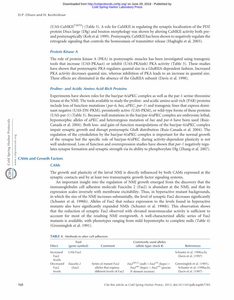
An important insight into the regulation of NMJ growth emerged from the discovery that theimmunoglobulin cell adhesion molecule Fasciclin 2 (Fas2) is abundant at the NMJ, and that itsexpression scales inversely with membrane excitability. Thus, in hyperactive mutant backgrounds,in which the size of the NMJ increases substantially, the level of synaptic Fas2 decreases significantly(Schuster et al. 1996b). Alleles of Fas2 that reduce expression to the levels found in hyperactivemutants also have significantly expanded NMJs (Schuster et al. 1996b). This observation showsthat the reduction of synaptic Fas2 observed with elevated neuromuscular activity is sufficient toaccount for most of the resulting NMJ overgrowth. A well-characterized allelic series of Fas2mutants is available, with phenotypes ranging from mild hypomorphs to complete nulls (Table 4)(Grenningloh et al. 1991).
TABLE 4. Methods to alter cell adhesion
EffectTool
(gene symbol) CommentCommonly used alleles(allele type: stock #) References
IncreasedFas2levels
UAS-Fas2 Schuster et al. 1996(a,b),Davis et al. (1997)
DecreasedFas2levels
Fasciclin 2(Fas2)
Series of mutant Fas2alleles that expressdifferent levels of Fas2
Fas2EB112 (null) > Fas2e76 (hypo) >Fas2e86 (hypo) > Fas2e93 (preciseP-element excision)
Grenningloh et al. (1991),Schuster et al. (1996a,b),Davis et al. (1997)
168 Cite this article as Cold Spring Harbor Protoc; 2012; doi:10.1101/pdb.top067785
D.P. Olsen and H. Keshishian
Cold Spring Harbor Laboratory Press on June 29, 2018 - Published by http://cshprotocols.cshlp.org/Downloaded from

Fas2 levels can also be elevated by expression of the UAS-Fas2 effector in either motor neurons ormuscles. Although overexpression of Fas2 in muscle generally suppresses NMJ growth, it should benoted that a balanced overexpression of Fas2 on both sides of the synapse results, surprisingly, inthe expansion of the NMJ (Ashley et al. 2005).
Growth Factors
The transsynaptic signaling systems that affect the size of the Drosophila NMJ include the TGF-βgrowth factor Glass bottom boat (Gbb), a member of the BMP family, and the Wnt growth factorWingless (Wg). Both growth factors have been shown to be essential for normal NMJ development(Aberle et al. 2002; Marques et al. 2002; Packard et al. 2002; McCabe et al. 2003). In addition tomutant alleles of the Gbb ligand and its type II receptor Wishful Thinking (Wit), the TGF-β signalingcascade can be manipulated using a variety of transgenic lines (Table 5). It should be noted that witand gbbmutations are generally not homozygosed, but are used in heteroallelic combinations, such asgbb1/gbb2 and witA12/witB11.
For gain of function using directed cell-specific expression, the most common approach has beento drive the constitutively active BMP receptor in motor neurons (the BMP type I co-receptorUAS-Act-Tkv). It is also possible to overexpress the ligand in muscle, using the UAS-Gbb effector.For loss of function studies in specific cells, there are dominant-negative transgenes that suppressdownstream signaling components, such as the transcription factor Mad (UAS-Mad1), as well asexpressible forms of inhibitory proteins that suppress TGF-β signaling (UAS-Dad).
Wg signaling can be suppressed using both classic mutations targeting Wg or its receptor frizzled,as well as a DN form of the frizzled receptor. To bypass early lethality caused by Wg loss of function,hypomorphic or ts alleles are used. Retrograde signaling from synapse to cell body can be disruptedusing dominant negative transgenes that target the p150 dynactin component of the dynein-dynactincomplex (UAS-GluedDN or UAS-ΔGl) (McCabe et al. 2003; Mathew et al. 2005). This approach hasbeen extensively used to show the need for retrograde growth factor transport to the cell body, but
TABLE 5. Methods to alter growth factor signaling
Effect Tool (gene symbol) CommentCommonly used alleles(allele type: stock #) Reference(s)
Increased BMPsignaling
Daughters against dpp(Dad)
Loss of function ofinhibitory Smad
Dad271-68; Dadj1E4 Sweeney and Davis (2002),McCabe et al. (2004)
UAS-Gbb Used for overexpressionof BMP ligand
McCabe et al. (2004)
UAS-Act-Tkv Constitutively activetype I BMP receptor
McCabe et al. (2004), Collins et al.(2006), Wang et al. (2007),O’Connor-Giles et al. (2008)
Decreased BMPsignaling
glass bottom boat (gbb) Secreted BMP ligand gbb1 (null); gbb2 (null); gbb3 (hypo);gbb4 (hypo)
McCabe et al. (2003)
wishful thinking (wit) Type II BMP receptor witA12 (5173); witB11 (5174); witHA1
(hypo); witHA2; witHA3; witHA4;witHA5; witS126215
Aberle et al. (2002), Marques et al.(2002)
UAS-Dad Used for overexpressionof an inhibitory Smad
McCabe et al. (2004), Goold andDavis (2007)
UAS-Mad1 Mutant Smad that lacksDNA-binding activity
Takaesu et al. (2005)
Decreased Wgsignaling
wingless (wg) Secreted growth factor wgI-12 (null: 7000); wg1 (hypo: 2978) Packard et al. (2002), Ataman et al.(2008)
frizzled 2 (fz2) wg receptor fz2C1 Mathew et al. (2005)UAS-DFz2DN Dominant negative
frizzledPackard et al. (2002), Mathewet al. (2005)
Disrupted retro-gradetransport
UAS-GluedDN/UAS-ΔGl
DN disruption of dynein/dynactin complex
McCabe et al. (2003), Mathewet al. (2005)
Cite this article as Cold Spring Harbor Protoc; 2012; doi:10.1101/pdb.top067785 169
Methods for Examining Synaptic Plasticity
Cold Spring Harbor Laboratory Press on June 29, 2018 - Published by http://cshprotocols.cshlp.org/Downloaded from
must be applied with caution, as it undoubtedly disrupts multiple retrograde signals, as well as poten-tially affecting orthograde transport as well.
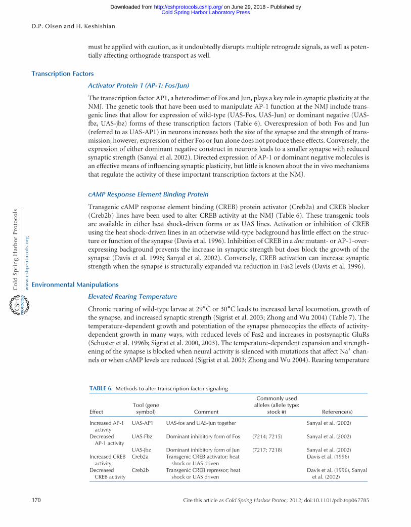
Transcription Factors
Activator Protein 1 (AP-1: Fos/Jun)
The transcription factor AP1, a heterodimer of Fos and Jun, plays a key role in synaptic plasticity at theNMJ. The genetic tools that have been used to manipulate AP-1 function at the NMJ include trans-genic lines that allow for expression of wild-type (UAS-Fos, UAS-Jun) or dominant negative (UAS-fbz, UAS-jbz) forms of these transcription factors (Table 6). Overexpression of both Fos and Jun(referred to as UAS-AP1) in neurons increases both the size of the synapse and the strength of trans-mission; however, expression of either Fos or Jun alone does not produce these effects. Conversely, theexpression of either dominant negative construct in neurons leads to a smaller synapse with reducedsynaptic strength (Sanyal et al. 2002). Directed expression of AP-1 or dominant negative molecules isan effective means of influencing synaptic plasticity, but little is known about the in vivo mechanismsthat regulate the activity of these important transcription factors at the NMJ.
cAMP Response Element Binding Protein
Transgenic cAMP response element binding (CREB) protein activator (Creb2a) and CREB blocker(Creb2b) lines have been used to alter CREB activity at the NMJ (Table 6). These transgenic toolsare available in either heat shock-driven forms or as UAS lines. Activation or inhibition of CREBusing the heat shock-driven lines in an otherwise wild-type background has little effect on the struc-ture or function of the synapse (Davis et al. 1996). Inhibition of CREB in a dncmutant- or AP-1-over-expressing background prevents the increase in synaptic strength but does block the growth of thesynapse (Davis et al. 1996; Sanyal et al. 2002). Conversely, CREB activation can increase synapticstrength when the synapse is structurally expanded via reduction in Fas2 levels (Davis et al. 1996).
Environmental Manipulations
Elevated Rearing Temperature
Chronic rearing of wild-type larvae at 29˚C or 30˚C leads to increased larval locomotion, growth ofthe synapse, and increased synaptic strength (Sigrist et al. 2003; Zhong and Wu 2004) (Table 7). Thetemperature-dependent growth and potentiation of the synapse phenocopies the effects of activity-dependent growth in many ways, with reduced levels of Fas2 and increases in postsynaptic GluRs(Schuster et al. 1996b; Sigrist et al. 2000, 2003). The temperature-dependent expansion and strength-ening of the synapse is blocked when neural activity is silenced with mutations that affect Na+ chan-nels or when cAMP levels are reduced (Sigrist et al. 2003; Zhong and Wu 2004). Rearing temperature
TABLE 6. Methods to alter transcription factor signaling
EffectTool (genesymbol) Comment
Commonly usedalleles (allele type:
stock #) Reference(s)
Increased AP-1activity
UAS-AP1 UAS-fos and UAS-jun together Sanyal et al. (2002)
DecreasedAP-1 activity
UAS-Fbz Dominant inhibitory form of Fos (7214; 7215) Sanyal et al. (2002)
UAS-Jbz Dominant inhibitory form of Jun (7217; 7218) Sanyal et al. (2002)Increased CREBactivity
Creb2a Transgenic CREB activator; heatshock or UAS driven
Davis et al. (1996)
DecreasedCREB activity
Creb2b Transgenic CREB repressor; heatshock or UAS driven
Davis et al. (1996), Sanyalet al. (2002)
170 Cite this article as Cold Spring Harbor Protoc; 2012; doi:10.1101/pdb.top067785
D.P. Olsen and H. Keshishian
Cold Spring Harbor Laboratory Press on June 29, 2018 - Published by http://cshprotocols.cshlp.org/Downloaded from
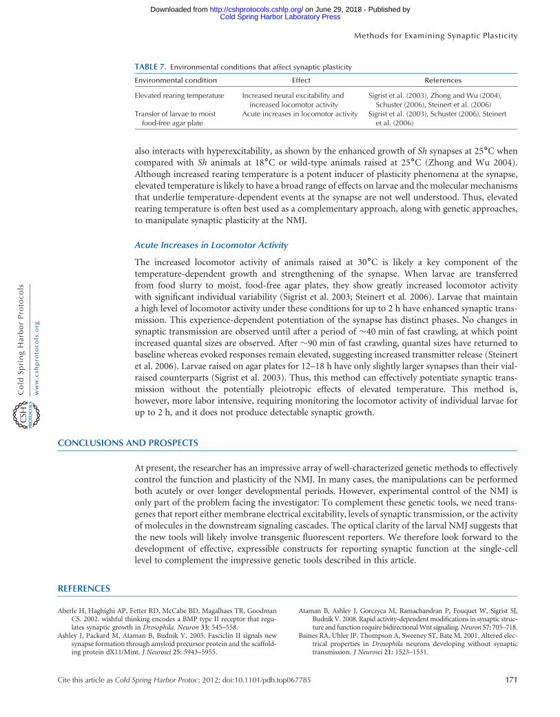
also interacts with hyperexcitability, as shown by the enhanced growth of Sh synapses at 25˚C whencompared with Sh animals at 18˚C or wild-type animals raised at 25˚C (Zhong and Wu 2004).Although increased rearing temperature is a potent inducer of plasticity phenomena at the synapse,elevated temperature is likely to have a broad range of effects on larvae and the molecular mechanismsthat underlie temperature-dependent events at the synapse are not well understood. Thus, elevatedrearing temperature is often best used as a complementary approach, along with genetic approaches,to manipulate synaptic plasticity at the NMJ.
Acute Increases in Locomotor Activity
The increased locomotor activity of animals raised at 30˚C is likely a key component of thetemperature-dependent growth and strengthening of the synapse. When larvae are transferredfrom food slurry to moist, food-free agar plates, they show greatly increased locomotor activitywith significant individual variability (Sigrist et al. 2003; Steinert et al. 2006). Larvae that maintaina high level of locomotor activity under these conditions for up to 2 h have enhanced synaptic trans-mission. This experience-dependent potentiation of the synapse has distinct phases. No changes insynaptic transmission are observed until after a period of �40 min of fast crawling, at which pointincreased quantal sizes are observed. After �90 min of fast crawling, quantal sizes have returned tobaseline whereas evoked responses remain elevated, suggesting increased transmitter release (Steinertet al. 2006). Larvae raised on agar plates for 12–18 h have only slightly larger synapses than their vial-raised counterparts (Sigrist et al. 2003). Thus, this method can effectively potentiate synaptic trans-mission without the potentially pleiotropic effects of elevated temperature. This method is,however, more labor intensive, requiring monitoring the locomotor activity of individual larvae forup to 2 h, and it does not produce detectable synaptic growth.
CONCLUSIONS AND PROSPECTS
At present, the researcher has an impressive array of well-characterized genetic methods to effectivelycontrol the function and plasticity of the NMJ. In many cases, the manipulations can be performedboth acutely or over longer developmental periods. However, experimental control of the NMJ isonly part of the problem facing the investigator: To complement these genetic tools, we need trans-genes that report either membrane electrical excitability, levels of synaptic transmission, or the activityof molecules in the downstream signaling cascades. The optical clarity of the larval NMJ suggests thatthe new tools will likely involve transgenic fluorescent reporters. We therefore look forward to thedevelopment of effective, expressible constructs for reporting synaptic function at the single-celllevel to complement the impressive genetic tools described in this article.
REFERENCES
Aberle H, Haghighi AP, Fetter RD, McCabe BD, Magalhaes TR, GoodmanCS. 2002. wishful thinking encodes a BMP type II receptor that regu-lates synaptic growth in Drosophila. Neuron 33: 545–558.
Ashley J, Packard M, Ataman B, Budnik V. 2005. Fasciclin II signals newsynapse formation through amyloid precursor protein and the scaffold-ing protein dX11/Mint. J Neurosci 25: 5943–5955.
Ataman B, Ashley J, Gorczyca M, Ramachandran P, Fouquet W, Sigrist SJ,Budnik V. 2008. Rapid activity-dependent modifications in synaptic struc-ture and function require bidirectionalWnt signaling.Neuron 57: 705–718.
Baines RA, Uhler JP, Thompson A, Sweeney ST, Bate M. 2001. Altered elec-trical properties in Drosophila neurons developing without synaptictransmission. J Neurosci 21: 1523–1531.
TABLE 7. Environmental conditions that affect synaptic plasticity
Environmental condition Effect References
Elevated rearing temperature Increased neural excitability andincreased locomotor activity
Sigrist et al. (2003), Zhong and Wu (2004),Schuster (2006), Steinert et al. (2006)
Transfer of larvae to moistfood-free agar plate
Acute increases in locomotor activity Sigrist et al. (2003), Schuster (2006), Steinertet al. (2006)
Cite this article as Cold Spring Harbor Protoc; 2012; doi:10.1101/pdb.top067785 171
Methods for Examining Synaptic Plasticity
Cold Spring Harbor Laboratory Press on June 29, 2018 - Published by http://cshprotocols.cshlp.org/Downloaded from

Broughton SJ, Kitamoto T, Greenspan RJ. 2004. Excitatory and inhibitoryswitches for courtship in the brain of Drosophila melanogaster. CurrBiol 14: 538–547.
Budnik V, Ruiz-Cañada C. 2006. The fly neuromuscular junction: Structureand function. In International review of neurobiology, 2nd ed. (ed.Bradley R, Harris RA, Jenner P), Vol. 75, pp. 1–406. Academic Press,New York.
Budnik V, Zhong Y, Wu CF. 1990. Morphological plasticity of motor axonsin Drosophila mutants with altered excitability. J Neurosci 10:3754–3768.
Cheung US, Shayan AJ, Boulianne GL, Atwood HL. 1999. Drosophila larvalneuromuscular junction’s responses to reduction of cAMP in thenervous system. J Neurobiol 40: 1–13.
Collins CA, Diantonio A. 2007. Synaptic development: Insights from Droso-phila. Curr Opin Neurobiol 17: 35–42.
Collins CA, Wairkar YP, Johnson SL, DiAntonio A. 2006. Highwire re-strains synaptic growth by attenuating a MAP kinase signal. Neuron51: 57–69.
Davis GW, Schuster CM, Goodman CS. 1996. Genetic dissection of struc-tural and functional components of synaptic plasticity. III. CREB isnecessary for presynaptic functional plasticity. Neuron 17: 669–679.
Davis GW, Schuster CM, Goodman CS. 1997. Genetic analysis of the mech-anisms controlling target selection: Target-derived Fasciclin II regulatesthe pattern of synapse formation. Neuron 19: 561–573.
Davis GW, Diantonio A, Petersen SA, GoodmanCS. 1998. Postsynaptic PKAcontrols quantal size and reveals a retrograde signal that regulates pre-synaptic transmitter release in Drosophila. Neuron 20: 305–315.
Diantonio A, Petersen SA, Heckmann M, Goodman CS. 1999. Glutamatereceptor expression regulates quantal size and quantal content at theDrosophila neuromuscular junction. J Neurosci 19: 3023–3032.
Dietzl G, Chen D, Schnorrer F, Su KC, Barinova Y, Fellner M, Gasser B,Kinsey K, Oppel S, Scheiblauer S, et al. 2007. A genome-wide transgenicRNAi library for conditional gene inactivation in Drosophila. Nature448: 151–156.
Frank CA, Kennedy MJ, Goold CP, Marek KW, Davis GW. 2006. Mechan-isms underlying the rapid induction and sustained expression of synap-tic homeostasis. Neuron 52: 663–677.
Goold CP, Davis GW. 2007. The BMP ligand Gbb gates the expression ofsynaptic homeostasis independent of synaptic growth control. Neuron56: 109–123.
Grenningloh G, Rehm EJ, Goodman CS. 1991. Genetic analysis of growthcone guidance in Drosophila: Fasciclin II functions as a neuronal recog-nition molecule. Cell 67: 45–57.
Grigliatti TA, Hall L, Rosenbluth R, Suzuki DT. 1973. Temperature-sensitivemutations in Drosophila melanogaster. XIV. A selection of immobileadults. Mol Gen Genet 120: 107–114.
HaghighiAP,McCabeBD, Fetter RD, Palmer JE,HomS,GoodmanCS. 2003.Retrograde control of synaptic transmission by postsynaptic CaMKII atthe Drosophila neuromuscular junction. Neuron 39: 255–267.
Hamada FN, Rosenzweig M, Kang K, Pulver SR, Ghezzi A, Jegla TJ, GarrityPA. 2008. An internal thermal sensor controlling temperature prefer-ence in Drosophila. Nature 454: 217–220.
HarrisinghMC,Wu Y, Lnenicka GA, NitabachMN. 2007. Intracellular Ca2+
regulates free-running circadian clock oscillation in vivo. J Neurosci 27:12489–12499.
Hoeffer CA, Sanyal S, Ramaswami M. 2003. Acute induction of conservedsynaptic signaling pathways in Drosophila melanogaster. J Neurosci 23:6362–6372.
Jarecki J, Keshishian H. 1995. Role of neural activity during synaptogenesisin Drosophila. J Neurosci 15: 8177–8190.
Jin P, Griffith LC, Murphey RK. 1998. Presynaptic calcium/calmodulin-dependent protein kinase II regulates habituation of a simple reflex inadult Drosophila. J Neurosci 18: 8955–8964.
Kawasaki F, Felling R, Ordway RW. 2000. A temperature-sensitive paralyticmutant defines a primary synaptic calcium channel in Drosophila.J Neurosci 20: 4885–4889.
Kitamoto T. 2001. Conditional modification of behavior in Drosophila bytargeted expression of a temperature-sensitive shibire allele in definedneurons. J Neurobiol 47: 81–92.
Koh YH, Popova E, Thomas U, Griffith LC, Budnik V. 1999. Regulation ofDLG localization at synapses by CaMKII-dependent phosphorylation.Cell 98: 353–363.
Lima SQ, Miesenbock G. 2005. Remote control of behavior through geneti-cally targeted photostimulation of neurons. Cell 121: 141–152.
Lnenicka GA, Spencer GM, Keshishian H. 2003. Effect of reduced impulseactivity on the development of identified motor terminals inDrosophilalarvae. J Neurobiol 54: 337–345.
Marques G, Bao H, Haerry TE, Shimell MJ, Duchek P, Zhang B, O’ConnorMB. 2002. The Drosophila BMP type II receptor wishful thinking regu-lates neuromuscular synapse morphology and function. Neuron 33:529–543.
Mathew D, Ataman B, Chen J, Zhang Y, Cumberledge S, Budnik V. 2005.Wingless signaling at synapses is through cleavage and nuclear importof receptor DFrizzled2. Science 310: 1344–1347.
McCabe BD, Marques G, Haghighi AP, Fetter RD, Crotty ML, Haerry TE,Goodman CS, O’Connor MB. 2003. The BMP homolog Gbb providesa retrograde signal that regulates synaptic growth at theDrosophila neu-romuscular junction. Neuron 39: 241–254.
McCabe BD, Hom S, Aberle H, Fetter RD, Marques G, Haerry TE, Wan H,O’Connor MB, Goodman CS, Haghighi AP. 2004. Highwire regulatespresynaptic BMP signaling essential for synaptic growth. Neuron 41:891–905.
Mosca TJ, Carrillo RA, White BH, Keshishian H. 2005. Dissection of synap-tic excitability phenotypes by using a dominant-negative Shaker K+
channel subunit. Proc Natl Acad Sci 102: 3477–3482.Nitabach MN, Blau J, Holmes TC. 2002. Electrical silencing of Drosophila
pacemaker neurons stops the free-running circadian clock. Cell 109:485–495.
Nitabach MN, Wu Y, Sheeba V, Lemon WC, Strumbos J, Zelensky PK,White BH, Holmes TC. 2006. Electrical hyperexcitation of lateralventral pacemaker neurons desynchronizes downstream circadianoscillators in the fly circadian circuit and induces multiple behavioralperiods. J Neurosci 26: 479–489.
O’Connor-Giles KM, Ho LL, Ganetzky B. 2008. Nervous wreck interactswith thickveins and the endocytic machinery to attenuate retrogradeBMP signaling during synaptic growth. Neuron 58: 507–518.
Olsen DP, Keshishian H. 2012. Monitoring membrane excitability in Droso-phila expressing modified Shaker constructs. Cold Spring Harb Protocdoi: 10.1101/pdb.prot067801.
Osterwalder T, Yoon KS, White BH, Keshishian H. 2001. A conditionaltissue-specific transgene expression system using inducible GAL4.Proc Natl Acad Sci 98: 12596–12601.
Packard M, Koo ES, Gorczyca M, Sharpe J, Cumberledge S, Budnik V. 2002.The Drosophila Wnt, wingless, provides an essential signal for pre- andpostsynaptic differentiation. Cell 111: 319–330.
Paradis S, Sweeney ST, Davis GW. 2001. Homeostatic control of presynapticrelease is triggered by postsynaptic membrane depolarization. Neuron30: 737–749.
Peabody NC, Pohl JB, Diao F, Vreede AP, Sandstrom DJ, Wang H, ZelenskyPK, White BH. 2009. Characterization of the decision network for wingexpansion in Drosophila using targeted expression of the TRPM8channel. J Neurosci 29: 3343–3353.
Petersen SA, Fetter RD, Noordermeer JN, Goodman CS, Diantonio A. 1997.Genetic analysis of glutamate receptors in Drosophila reveals a retro-grade signal regulating presynaptic transmitter release. Neuron 19:1237–1248.
Rieckhof GE, Yoshihara M, Guan Z, Littleton JT. 2003. Presynaptic N-typecalciumchannels regulate synaptic growth. J Biol Chem 278: 41099–41108.
Roman G, Endo K, Zong L, Davis RL. 2001. P{Switch}, a system for spatialand temporal control of gene expression in Drosophila melanogaster.Proc Natl Acad Sci 98: 12602–12607.
Ruiz-Canada C, Ashley J, Moeckel-Cole S, Drier E, Yin J, Budnik V. 2004.New synaptic bouton formation is disrupted bymisregulation of micro-tubule stability in aPKC mutants. Neuron 42: 567–580.
Sanyal S, Sandstrom DJ, Hoeffer CA, Ramaswami M. 2002. AP-1 functionsupstream of CREB to control synaptic plasticity in Drosophila. Nature416: 870–874.
Schroll C, Riemensperger T, Bucher D, Ehmer J, Voller T, Erbguth K, GerberB, Hendel T, Nagel G, Buchner E, et al. 2006. Light-induced activationof distinct modulatory neurons triggers appetitive or aversive learningin Drosophila larvae. Curr Biol 16: 1741–1747.
Schuster C. 2006. Glutamatergic synapses of Drosophila neuromuscularjunctions: A high-resolution model for the analysis of experience-dependent potentiation. Cell Tissue Res 326: 287–299.
172 Cite this article as Cold Spring Harbor Protoc; 2012; doi:10.1101/pdb.top067785
D.P. Olsen and H. Keshishian
Cold Spring Harbor Laboratory Press on June 29, 2018 - Published by http://cshprotocols.cshlp.org/Downloaded from
Schuster CM, Davis GW, Fetter RD, Goodman CS. 1996a. Genetic dissectionof structural and functional components of synaptic plasticity. I.Fasciclin II controls synaptic stabilization and growth. Neuron 17:641–654.
Schuster CM, Davis GW, Fetter RD, Goodman CS. 1996b. Genetic dissec-tion of structural and functional components of synaptic plasticity. II.Fasciclin II controls presynaptic structural plasticity. Neuron 17:655–667.
Sigrist SJ, Thiel PR, Reiff DF, Lachance PE, Lasko P, Schuster CM. 2000.Postsynaptic translation affects the efficacy and morphology of neuro-muscular junctions. Nature 405: 1062–1065.
Sigrist SJ, Reiff DF, Thiel PR, Steinert JR, Schuster CM. 2003. Experience-dependent strengthening of Drosophila neuromuscular junctions. JNeurosci 23: 6546–6556.
Steinert JR, Kuromi H, Hellwig A, Knirr M, Wyatt AW, Kidokoro Y, Schus-ter CM. 2006. Experience-dependent formation and recruitment oflarge vesicles from reserve pool. Neuron 50: 723–733.
Sweeney ST. Davis GW. 2002. Unrestricted synaptic growth in spinster-a lateendosomal protein implicated in TGF-<gb>-mediated synaptic growthregulation. Neuron 36: 403–416.
Sweeney ST, Broadie K, Keane J, Niemann H, O’Kane CJ. 1995. Targetedexpression of tetanus toxin light chain inDrosophila specifically elimin-ates synaptic transmission and causes behavioral defects. Neuron 14:341–351.
Takaesu NT, Herbig E, Zhitomersky D, O’Connor MB, Newfeld SJ. 2005.DNA-binding domain mutations in SMAD genes yield dominant-negative proteins or a neomorphic protein that can activate WGtarget genes in Drosophila. Development 132: 4883–4894.
Waddell S, Armstrong JD, Kitamoto T, Kaiser K, Quinn WG. 2000. Theamnesiac gene product is expressed in two neurons in the Drosophilabrain that are critical for memory. Cell 103: 805–813.
Wang X, Shaw WR, Tsang HT, Reid E, O’Kane CJ. 2007. Drosophilaspichthyin inhibits BMP signaling and regulates synaptic growth andaxonal microtubules. Nat Neurosci 10: 177–185.
White BH,Osterwalder TP, Yoon KS, JoinerWJ,WhimMD, Kaczmarek LK,Keshishian H. 2001. Targeted attenuation of electrical activity inDroso-phila using a genetically modified K+ channel. Neuron 31: 699–711.
Zhang Y, Guo H, Kwan H, Wang JW, Kosek J, Lu B. 2007. PAR-1 kinasephosphorylates Dlg and regulates its postsynaptic targeting at the Dro-sophila neuromuscular junction. Neuron 53: 201–215.
Zhong Y, Wu CF. 1991. Altered synaptic plasticity in Drosophila memorymutants with a defective cyclic AMP cascade. Science 251: 198–201.
Zhong Y, Wu CF. 2004. Neuronal activity and adenylyl cyclase in environ-ment-dependent plasticity of axonal outgrowth in Drosophila. J Neuro-sci 24: 1439–1445.
Zhong Y, Budnik V, Wu CF. 1992. Synaptic plasticity inDrosophilamemoryand hyperexcitable mutants: Role of cAMP cascade. J Neurosci 12:644–651.
Cite this article as Cold Spring Harbor Protoc; 2012; doi:10.1101/pdb.top067785 173
Methods for Examining Synaptic Plasticity
Cold Spring Harbor Laboratory Press on June 29, 2018 - Published by http://cshprotocols.cshlp.org/Downloaded from

doi: 10.1101/pdb.top067785Cold Spring Harb Protoc; Douglas P. Olsen and Haig Keshishian
DrosophilaExperimental Methods for Examining Synaptic Plasticity in
ServiceEmail Alerting click here.Receive free email alerts when new articles cite this article -
CategoriesSubject Cold Spring Harbor Protocols.Browse articles on similar topics from
(114 articles)RNA Interference (RNAi)/siRNA (43 articles)Mutagenesis
(85 articles)Electrophysiology (236 articles)Drosophila
(659 articles)Developmental Biology (119 articles)Analysis of Gene Expression, general
http://cshprotocols.cshlp.org/subscriptions go to: Cold Spring Harbor Protocols To subscribe to
© 2012 Cold Spring Harbor Laboratory Press
Cold Spring Harbor Laboratory Press on June 29, 2018 - Published by http://cshprotocols.cshlp.org/Downloaded from