experiment one basic methods of microbiological laboratory basic methods of microbiological...
TRANSCRIPT
Experiment one
Basic methods of microbiological laboratoryBasic methods of microbiological laboratory Bacterial cultureBacterial culture Staining of bacteriaStaining of bacteria Use of oil-immersion microscopeUse of oil-immersion microscope Observation of several special bacteria Observation of several special bacteria
Isolation and culture of bacteriaIsolation and culture of bacteria(streak plat(streak plate culture)e culture) Pyogenic cocciPyogenic cocci Pathogenic enterobacteriaPathogenic enterobacteria
Bacterial Culture and Growth State Observation
PURPOSE :PURPOSE : To obtain information about bacterial biological charaTo obtain information about bacterial biological chara
cteristics cteristics METHODS :METHODS : streak plate method isolation and culturestreak plate method isolation and culture
agar slope method agar slope method
liquid medium culture pure culture liquid medium culture pure culture
stab culture stab culture
anaerobic culture anaerobic culture
Streak Plate Method
PURPOSE:PURPOSE:
Isolation and culture of bacteria growing togetIsolation and culture of bacteria growing together in a specimenher in a specimen
MATERIALS: MATERIALS:
l. Mixed broth culture of Escherichia coli l. Mixed broth culture of Escherichia coli
and staphylococcus aureus. and staphylococcus aureus.
2. Nutrient agar plate. 2. Nutrient agar plate.
Streak Plate Method
PROCEDURE:PROCEDURE: Flame your inoculating loop until the wire Flame your inoculating loop until the wire
glows red. glows red. Allow the loop to cool and get a loopful of the susAllow the loop to cool and get a loopful of the sus
pension of sample.pension of sample.Pick up your plate and streak the surface, Flame thPick up your plate and streak the surface, Flame th
e loop before streaking next section. When streakie loop before streaking next section. When streaking, be care ng, be care not to cut into the agar and not to be far not to cut into the agar and not to be far away from flame. away from flame.
Streak Plate Method PROCEDURE: PROCEDURE:
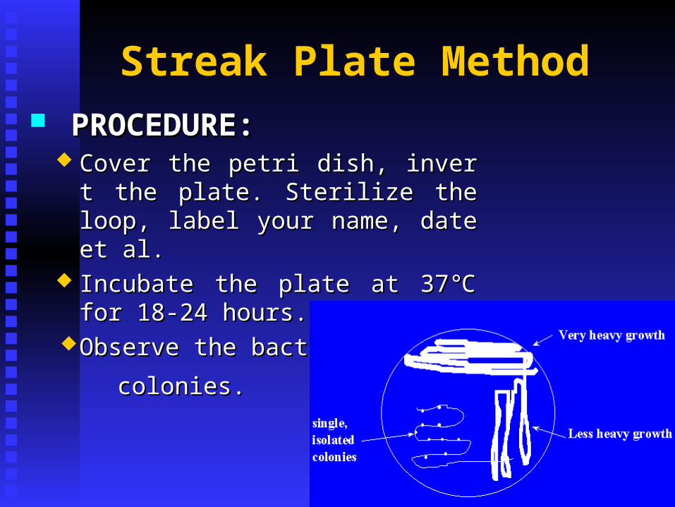
Cover the petri dish, invert the plate. Cover the petri dish, invert the plate. Sterilize the loop, label your name, dSterilize the loop, label your name, date et al. ate et al.
Incubate the plate at 37 for 18-24 ℃Incubate the plate at 37 for 18-24 ℃hours. hours.
Observe the bacterialObserve the bacterial
colonies.colonies.
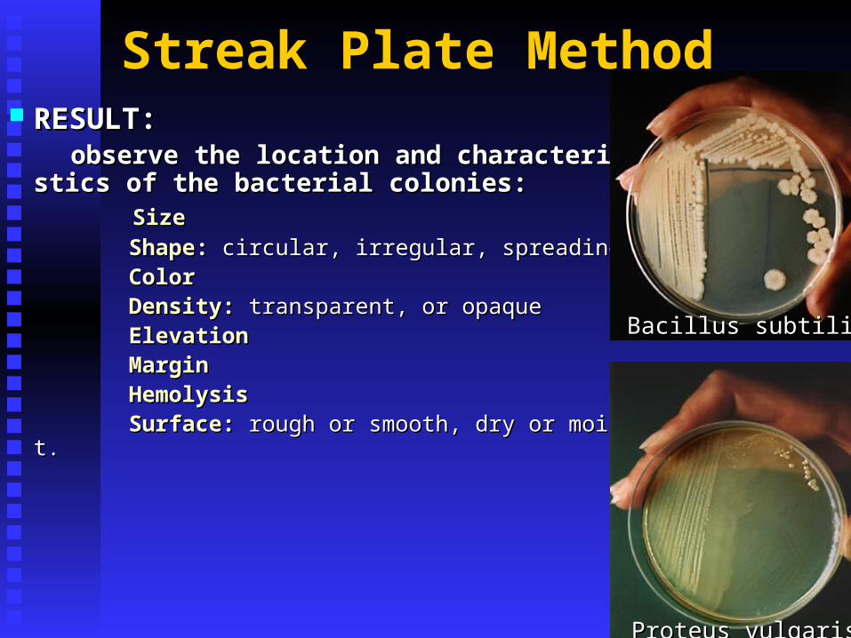
Streak Plate Method RESULT:RESULT: observe the location and characteristics of the baobserve the location and characteristics of the ba
cterial colonies: cterial colonies: SizeSize
ShapeShape:: circular, irregular, spreading circular, irregular, spreading ColorColor Density:Density: transparent, or opaque transparent, or opaque ElevationElevation MarginMargin HemolysisHemolysis Surface:Surface: rough or smooth, dry or moist. rough or smooth, dry or moist.
Bacillus subtilisBacillus subtilis
Proteus vulgarisProteus vulgaris

Streptococcus pyogenesStreptococcus pyogenesStaphylococcus aureusStaphylococcus aureus
mucoidmucoid
Agar Slope MethodAgar Slope Method MATERIALS :MATERIALS :
1. Agar slope 1. Agar slope 2. Colonies on agar plate 2. Colonies on agar plate PROCEDURE :PROCEDURE : 1. With the flame-sterilized wire inoculating loop, 1. With the flame-sterilized wire inoculating loop, transfer a small amount of bacteria from the transfer a small amount of bacteria from the colony on agar plate. Then streak on the agar slope. colony on agar plate. Then streak on the agar slope. 2. Sterile the mouth of tubes, replug the test tubes and 2. Sterile the mouth of tubes, replug the test tubes and flame the loop. flame the loop. 3. Label and incubate at 37 for 18-24 hours ℃3. Label and incubate at 37 for 18-24 hours ℃ 4. Observe your result. 4. Observe your result.

Agar Slope MethodAgar Slope Method
RESULTS :RESULTS :
There are many similar wet colonies on There are many similar wet colonies on the surface. If there are some other forms, the surface. If there are some other forms, it indicates culture sample is not pure. it indicates culture sample is not pure.
Liquid Medium CultureLiquid Medium Culture MATERIALSMATERIALS : : 1. Peptone water 1. Peptone water 2. Colonies on agar plate2. Colonies on agar plate
PROCEDUREPROCEDURE :: 1. Flame -sterilize the wire inoculating loop.1. Flame -sterilize the wire inoculating loop. 2. Insert the wire loop containing a small amount 2. Insert the wire loop containing a small amount of bacteria into the liquid culture robe.of bacteria into the liquid culture robe. 3. Scratch the wall of tube over the broth in order 3. Scratch the wall of tube over the broth in order to let bacteria drop into the liquid.to let bacteria drop into the liquid.
Liquid Medium CultureLiquid Medium Culture
PROCEDUREPROCEDURE :: 4. Flame the mouth of the tube and reinsert the 4. Flame the mouth of the tube and reinsert the cotton plug. Flame-sterilize the wire loop.cotton plug. Flame-sterilize the wire loop. 5. Label the tube, incubate at 37 for 24 hours℃5. Label the tube, incubate at 37 for 24 hours℃ 6. Observe the result.6. Observe the result.
RESULTS:RESULTS: turbid, flocculent, pellicle turbid, flocculent, pellicle
Stab CultureStab Culture
METHODS:METHODS: 1. Flame-sterilize inoculating needle.1. Flame-sterilize inoculating needle. 2. Insert the needle with a small bacteria to the 2. Insert the needle with a small bacteria to the center of the culture, be care not to touch the center of the culture, be care not to touch the bottom of the tube, then draw it out in the same way.bottom of the tube, then draw it out in the same way. 3. Flame the mouth of the tube and reinsert the cotton 3. Flame the mouth of the tube and reinsert the cotton plug. Flame-sterilize the needle.plug. Flame-sterilize the needle. 4. Label the tube, incubate for 24 hours at 37 . ℃4. Label the tube, incubate for 24 hours at 37 . ℃ 5. Observe the result5. Observe the result. .

Stab CultureStab Culture
RESULTS: RESULTS:
Motile bacteria will migrate from the line Motile bacteria will migrate from the line of inoculation to form a diffuse turbidity iof inoculation to form a diffuse turbidity in the surrounding medium; nonmobile ban the surrounding medium; nonmobile bacteria will grow only along the line of inocteria will grow only along the line of inoculation.culation.
Staining of Bacteria PURPOSE :PURPOSE : To make bacteria more easily observable To make bacteria more easily observable To acquaint you with Gram stainTo acquaint you with Gram stain MATERIALS:MATERIALS:
Simple stainSimple stainGram stainGram stain Acid-fast stainAcid-fast stain Special stainSpecial stain
Spore stain Spore stain Capsule stain Capsule stain Flagella stain Flagella stain Metachromatic granules stain. Metachromatic granules stain.
Gram stain
purpose: purpose: differentiating bacteriadifferentiating bacteria
MATERIALS :MATERIALS :Slant cultures of and Escherichia coli and S.aureuSlant cultures of and Escherichia coli and S.aureu
s (18 to 24 hours old)s (18 to 24 hours old) Crystal violet, iodine solution, 95% alcohol, safraCrystal violet, iodine solution, 95% alcohol, safra
nin nin Microscope slides Microscope slides
Gram stain
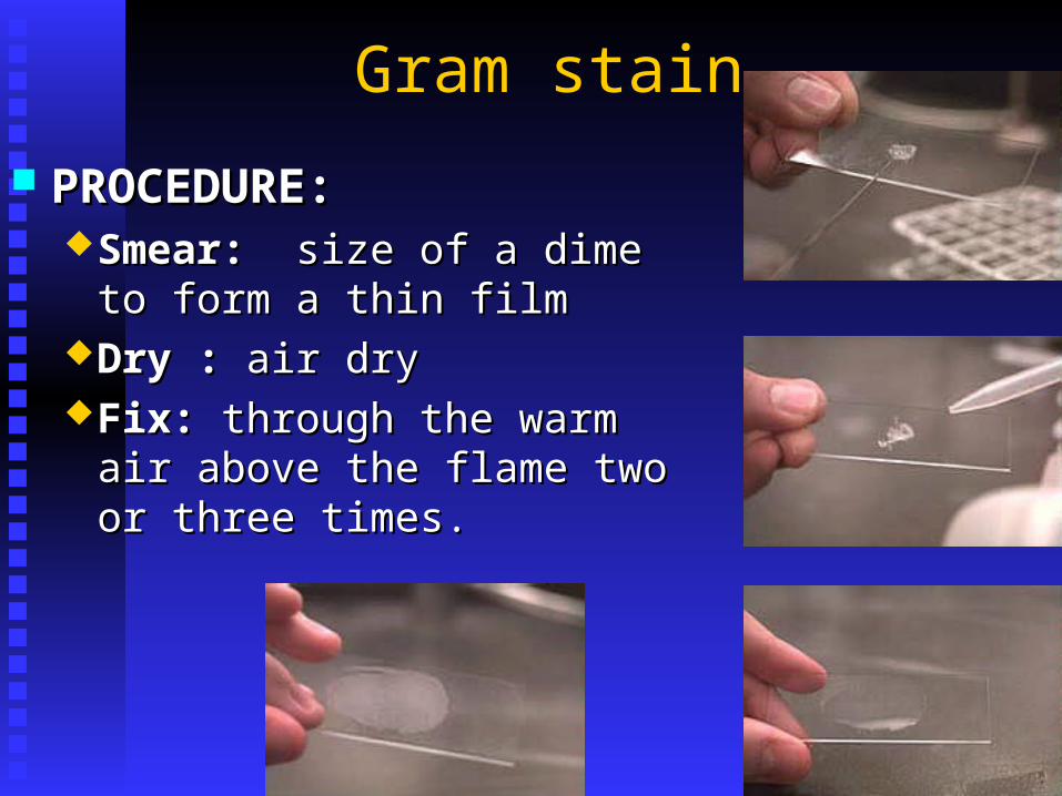
PROCEDURE:PROCEDURE:Smear: Smear: size of a dime to form a size of a dime to form a
thin filmthin filmDry : Dry : air dryair dryFix: Fix: through the warm air above through the warm air above
the flame two or three times.the flame two or three times.
Gram stain PROCEDURE:PROCEDURE:
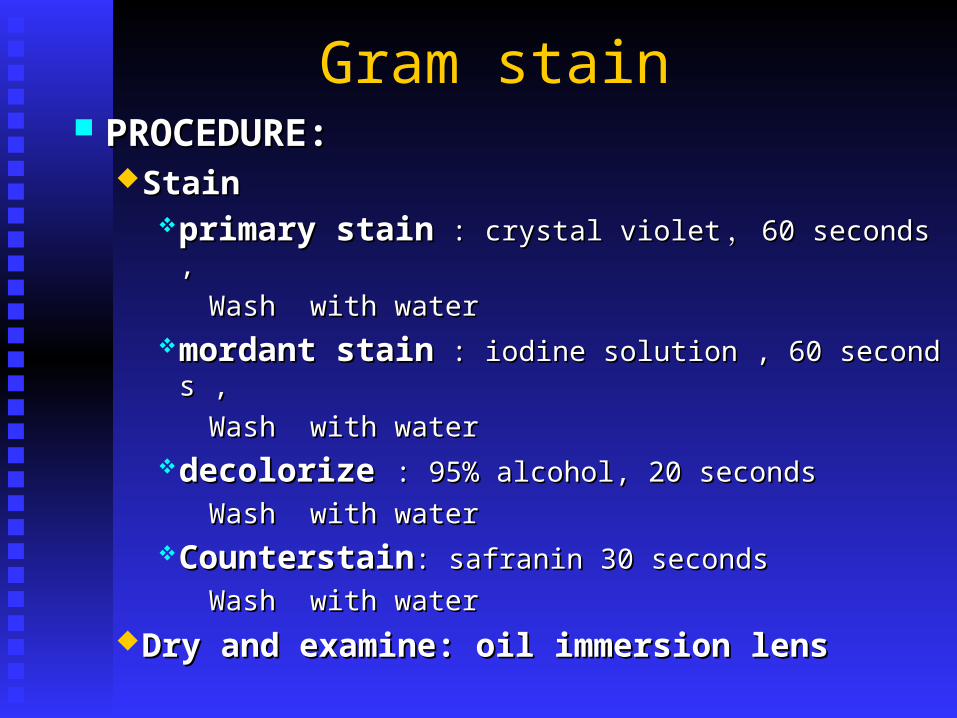
StainStainprimary stainprimary stain : crystal violet : crystal violet ,, 60 seconds , 60 seconds ,
Wash with waterWash with watermordant stainmordant stain : iodine solution , 60 seconds , : iodine solution , 60 seconds ,
Wash with waterWash with waterdecolorize decolorize : 95% alcohol, 20 seconds: 95% alcohol, 20 seconds
Wash with waterWash with waterCounterstainCounterstain: safranin 30 seconds : safranin 30 seconds
Wash with waterWash with water
Dry and examine: oil immersion lens Dry and examine: oil immersion lens
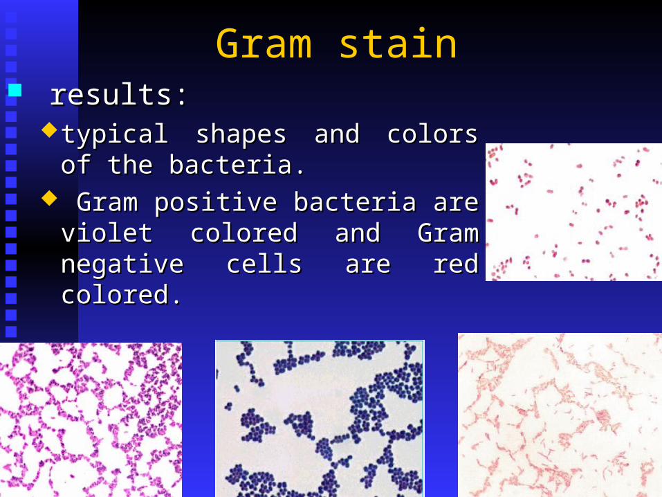
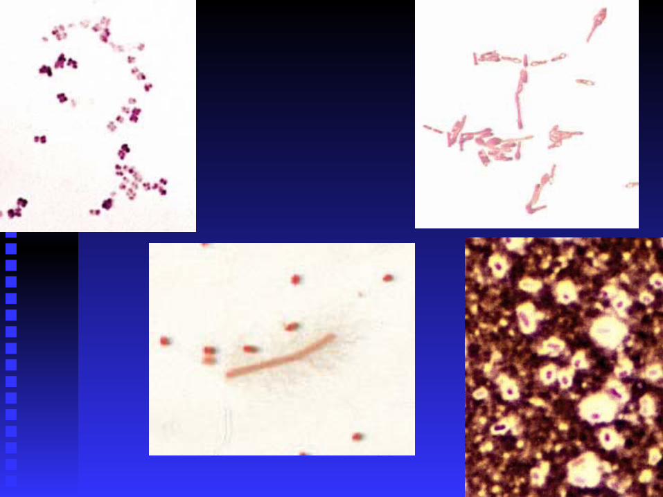
Gram stain results:results:
typical shapes and colors of the typical shapes and colors of the bacteria.bacteria.
Gram positive bacteria are violet Gram positive bacteria are violet colored and Gram negative cells are colored and Gram negative cells are red colored.red colored.

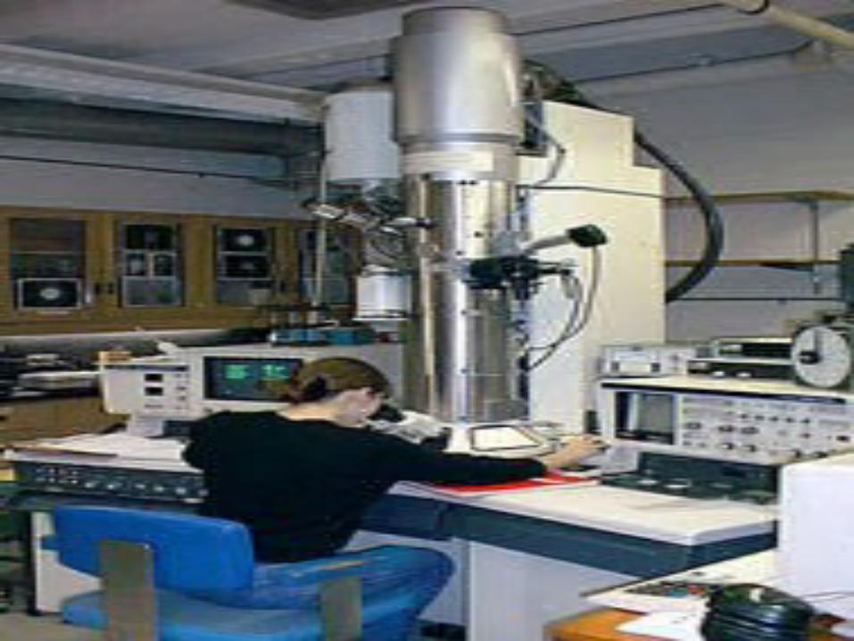
Use of The Oil-immersion Use of The Oil-immersion
MicroscopeMicroscope
study morphologystudy morphology structurestructure staining characteristicsstaining characteristics motility of different microorganisms. motility of different microorganisms.
Use of The Oil-immersion Use of The Oil-immersion
MicroscopeMicroscope
MATERIALS
Microscope, immersion oil, lens Microscope, immersion oil, lens
paper, glass slides, and cover slips. paper, glass slides, and cover slips.
Prepared stained slides of several Prepared stained slides of several
types of bacteria.types of bacteria.

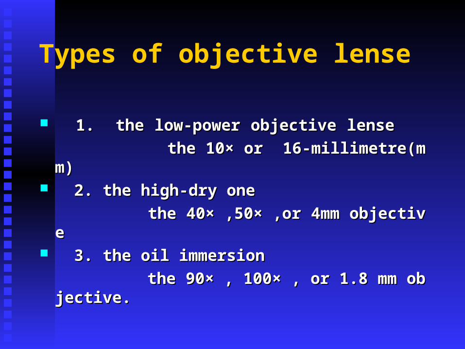
Types of objective lense
1. the low-power objective lense 1. the low-power objective lense
the 10× or 16-millimetre(mm)the 10× or 16-millimetre(mm) 2. the high-dry one 2. the high-dry one
the 40× ,50× ,or 4mm objectivethe 40× ,50× ,or 4mm objective 3. the oil immersion 3. the oil immersion
the 90× , 100× , or 1.8 mm objective.the 90× , 100× , or 1.8 mm objective.
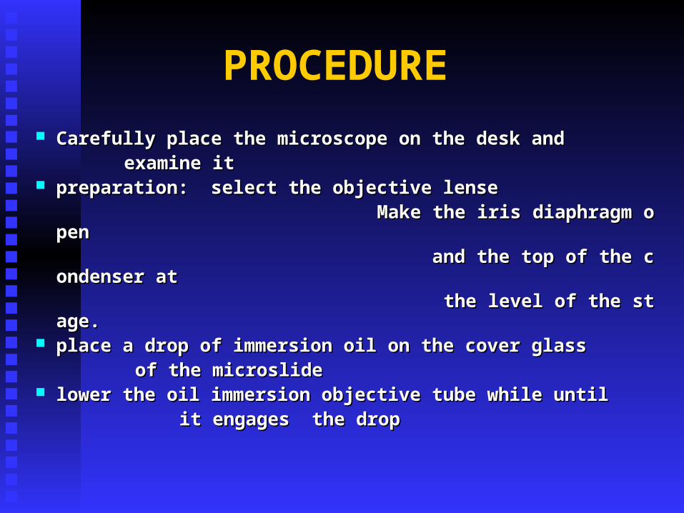
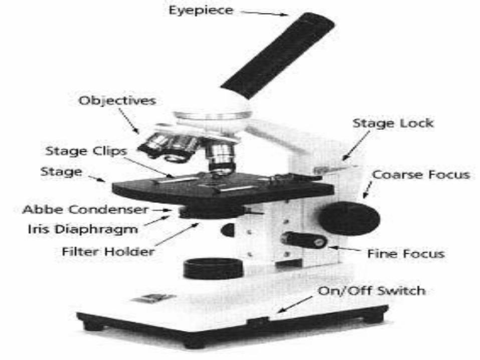
PROCEDURE
Carefully place the microscope on the desk and Carefully place the microscope on the desk and examine itexamine it preparation: select the objective lensepreparation: select the objective lense Make the iris diaphragm open Make the iris diaphragm open and the top of the condenser at and the top of the condenser at the level of the stage.the level of the stage. place a drop of immersion oil on the cover glass place a drop of immersion oil on the cover glass of the microslide of the microslide lower the oil immersion objective tube while until lower the oil immersion objective tube while until it engages the drop it engages the drop
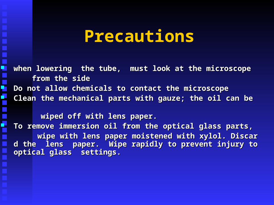
Precautions
when lowering the tube, must look at the microscope when lowering the tube, must look at the microscope from the side from the side Do not allow chemicals to contact the microscope Do not allow chemicals to contact the microscope Clean the mechanical parts with gauze; the oil can be Clean the mechanical parts with gauze; the oil can be wiped off with lens paper.wiped off with lens paper. To remove immersion oil from the optical glass parts, To remove immersion oil from the optical glass parts, wipe with lens paper moistened with xylol. Discard the lwipe with lens paper moistened with xylol. Discard the l
ens paper. Wipe rapidly to prevent injury to optical glasens paper. Wipe rapidly to prevent injury to optical glass settings.s settings.
focus upward slowly until the image appear.focus upward slowly until the image appear. Complete focus with the fine adjustment Complete focus with the fine adjustment Clean the microscope when this experiment is Clean the microscope when this experiment is endedended
Leave the microscope with the lowest power Leave the microscope with the lowest power
objective in the working positionobjective in the working position Remove immersion oil from prepared practice Remove immersion oil from prepared practice
slides. Return these to instructor.slides. Return these to instructor. Place a plastic cover over the microscope or Place a plastic cover over the microscope or
place it in its case. Record the status of the place it in its case. Record the status of the microscope.microscope.

Laboratory Diagnosis of PathogeniLaboratory Diagnosis of Pathogeni
c c
Enterobacterial InfectionEnterobacterial Infection
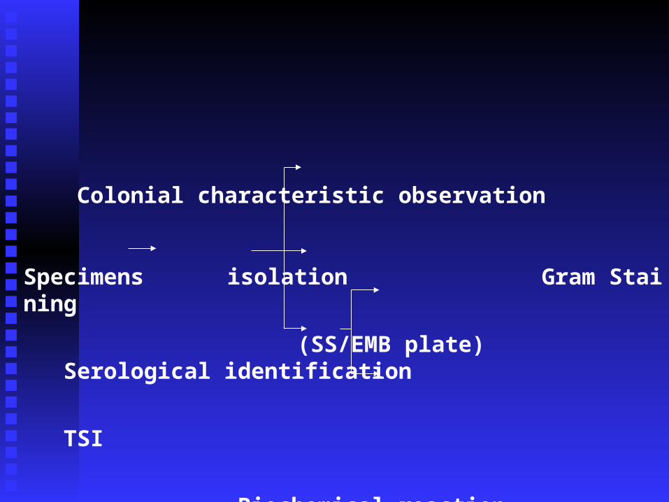
Colonial characteristic observation
Specimens isolation Gram Staining
(SS/EMB plate) Serological identification
TSI
Biochemical reaction