exosomes: extracellular organelles important in
TRANSCRIPT
J O U R N A L O F P R O T E O M I C S 7 3 ( 2 0 1 0 ) 1 9 0 7 – 1 9 2 0
ava i l ab l e a t www.sc i enced i r ec t . com
www.e l sev i e r . com/ loca te / j p ro t
Review
Exosomes: Extracellular organelles important inintercellular communication
Suresh Mathivanan, Hong Ji, Richard J. Simpson⁎
Ludwig Institute for Cancer Research, Parkville, Victoria, Australia
A R T I C L E I N F O
Abbreviations: MVBs, multivesicular bodiePM, plasma membrane; LBPA, lyosbisphosph⁎ Corresponding author. Ludwig Institute fo
Fax: +61 3 9341 3192.E-mail address: richard.simpson@ludwig.
1874-3919/$ – see front matter © 2010 Elsevidoi:10.1016/j.jprot.2010.06.006
A B S T R A C T
Keywords:
In addition to intracellular organelles, eukaryotic cells also contain extracellular organellesthat are released, or shed, into the microenvironment. These membranous extracellularorganelles include exosomes, shedding microvesicles (SMVs) and apoptotic blebs (ABs),many of which exhibit pleiotropic biological functions. Because extracellular organelleterminology is often confounding, with many preparations reported in the literature beingmixtures of extracellular vesicles, there is a growing need to clarify nomenclature and toimprove purification strategies in order to discriminate the biochemical and functionalactivities of these moieties. Exosomes are formed by the inward budding of multivesicularbodies (MVBs) and are released from the cell into the microenvironment following thefusion of MVBs with the plasma membrane (PM). In this review we focus on variousstrategies for purifying exosomes and discuss their biophysical and biochemical properties.An update on proteomic analysis of exosomes from various cell types and body fluids isprovided and host-cell specific proteomic signatures are also discussed. Because theectodomain of ~42% of exosomal integral membrane proteins are also found in thesecretome, these vesicles provide a potential source of serum-based membrane proteinbiomarkers that are reflective of the host cell. ExoCarta, an exosomal protein and RNAdatabase (http://exocarta.ludwig.edu.au), is described.© 2010 Elsevier B.V. All rights reserved.
ExosomesMicrovesiclesExtracellular vesiclesShedding microvesiclesApoptotic blebsBiomarkers
Contents
1. Introduction . . . . . . . . . . . . . . . . . . . . . . . . . . . . . . . . . . . . . . . . . . . . . . . . . . . . . . . . . 19082. Exosomes . . . . . . . . . . . . . . . . . . . . . . . . . . . . . . . . . . . . . . . . . . . . . . . . . . . . . . . . . . . 1908
2.1. Shedding microvesicles (SMVs) . . . . . . . . . . . . . . . . . . . . . . . . . . . . . . . . . . . . . . . . . . . 19082.2. Apoptotic blebs (ABs) . . . . . . . . . . . . . . . . . . . . . . . . . . . . . . . . . . . . . . . . . . . . . . . . 1908
3. Microvesicles: the case for more stringent nomenclature. . . . . . . . . . . . . . . . . . . . . . . . . . . . . . . . . 19084. Current status of exosome protein composition . . . . . . . . . . . . . . . . . . . . . . . . . . . . . . . . . . . . . . . 19095. Exosomesmediate cell-to-cell communication . . . . . . . . . . . . . . . . . . . . . . . . . . . . . . . . . . . . . . . . 1913
s; ILVs, intraluminal vesicles; ESCRT, Endosomal Sorting Complexes Required for Transport;atidic acid; SMVs, shedding microvesicles; ABs, apoptotic blebs.r Cancer Research, PO Box 2008, Royal Melbourne Hospital, Parkville, Vic 3050, Australia.
edu.au (R.J. Simpson).
er B.V. All rights reserved.
1908 J O U R N A L O F P R O T E O M I C S 7 3 ( 2 0 1 0 ) 1 9 0 7 – 1 9 2 0
6. Exosome purification protocols. . . . . . . . . . . . . . . . . . . . . . . . . . . . . . . . . . . . . . . . . . . . . . . 19137. Clinical studies involving exosomes . . . . . . . . . . . . . . . . . . . . . . . . . . . . . . . . . . . . . . . . . . . . 19158. Exosomes as a rich source for discovering potential blood-based biomarkers . . . . . . . . . . . . . . . . . . . . . 19159. Exocarta: a manually curated database of exosomal proteins and RNA. . . . . . . . . . . . . . . . . . . . . . . . . 1916
10. Summary. . . . . . . . . . . . . . . . . . . . . . . . . . . . . . . . . . . . . . . . . . . . . . . . . . . . . . . . . . . 1916Keynotes . . . . . . . . . . . . . . . . . . . . . . . . . . . . . . . . . . . . . . . . . . . . . . . . . . . . . . . . . . . . . . 1917Acknowledgements . . . . . . . . . . . . . . . . . . . . . . . . . . . . . . . . . . . . . . . . . . . . . . . . . . . . . . . . 1918References . . . . . . . . . . . . . . . . . . . . . . . . . . . . . . . . . . . . . . . . . . . . . . . . . . . . . . . . . . . . . 1918
1. Introduction
Molecules that perform specific cellular functions are segre-gated and compartmentalized into dynamic and distinctlystructured organelles, which are composed of both residentand transient molecules that carry out specific functions. Themolecular components of organelles are exchanged constant-ly with the rest of the cell and fluctuate with physiologicalperturbations [1]. While the majority of organelles residewithin the cell, some, such as exosomes, shedding micro-vesicles (SMVs) and apoptotic blebs (ABs) are released into theextracellular space.
2. Exosomes
Exosomes are 40–100 nm diameter membranous vesicles ofendocytic origin that are released by a variety of cell typesinto the extracellular space [2]. Exosomes were first reportedin 1983 by Johnstone and colleagues while culturing reticu-locytes [3]. Inward budding of endosomal membranes resultsin the progressive accumulation of intraluminal vesicles(ILVs) within large multivesicular bodies (MVBs). Transmem-brane proteins are incorporated into the invaginating mem-brane while the cytosolic components are engulfed withinthe ILVs [4]. Based on their biochemical properties, intracel-lular MVBs can either traffic to lysosomes where they aresubjected to proteosomal degradation (i.e., ‘degradativeMVBs’) or, alternatively, to the plasma membrane (PM)where upon fusion with the PM they release their contents(ILVs) into the extracellular space (the so-called ‘exocyticMVBs’ — Fig. 1A); ILVs released into the extracellular spaceare referred to as ‘exosomes’ [5]. While this ‘degradative’/‘exocytic’ MVB phenomenon has been reported to occur inoligodendrocytes and involves changes in the ceramidechemistry of MVB membranes [6], it is not clear whetherthis phenomenon is generally applicable and occurs in all celltypes. Furthermore, it is not clear whether there are twoclasses of MVBs (i.e., exocytic and degradative) or whetherMVBs contain exocytic and degradative ILVs. To date,exosomes are the only type of membranous vesicles origi-nating from intracellular compartments such as the MVBs.Endosomal Sorting Complexes Required for Transport(ESCRTs), multiprotein complexes, are involved in themechanism governing the biogenesis/degradation of MVBs[7,8] in a ubiquitinylation-dependent [9] manner. With theaid of three-dimensional structural studies, key links be-tween the components of the ESCRT multiprotein complex,
phospholipids and ubiquitin are beginning to shed newinsights into MVB biogenesis and trafficking [10].
2.1. Shedding microvesicles (SMVs)
SMVs are large membranous vesicles (>100 nm diameter) thatare shed from the PM of a wide variety of cell types [11–13].Following blebbing (outward protrusion) of the PM, fission ofthe PM stalk detaches the cytoplasmic protrusions, resultingin the formation of SMVs [14]. Regulation of this processinvolves several enzymes such as calpain, flippase, floppase,scramblase and gelsolin [15]. Platelet-derived SMVs arereported to contain components of membrane lipid rafts(e.g., flotilin-1 (FLOT1)), lineage markers (e.g., platelet/endo-thelial cell adhesion molecule (PECAM-1) [15]), while onco-genic growth factor receptors (e.g., EGFRvIII) and tissue factor(CD142) are present in SMVs from gliomas [12] and plasma[16,17], respectively.
2.2. Apoptotic blebs (ABs)
Apoptotic or dying cells release membrane vesicles into theextracellular environment via blebbing of the PM. Thesemembrane vesicles, which are condensed remnants of theshrinking apoptotic cell [18], are referred to as apoptotic blebs(ABs). While SMVs are also released during the early stages ofapoptosis, ABs are released during the late stages of cell death[19]. SMVsandABsare 100–1000 nmand50–500 nmindiameter,respectively, and are heterogeneous in shape; by contrast,exosomes are much smaller in size (40–100 nm diameter) andhomogeneouswith respect to shape. Exosomes are cup-shapedand float at a density of 1.10–1.21 g/mL in sucrose gradient,which uniquely differentiates them from non-exosomal vesi-cles that are of irregular shape and float at higher densities(>1.23 g/mL) [20].
3. Microvesicles: the case for morestringent nomenclature
The extracellular microenvironment including body fluidssuch as ascites and blood contains a mixed population ofexosomes, SMVs and ABs [14]. These microvesicles have beenstudied over the years using a variety of isolation strategiesand have been categorized by their distinct structural andbiochemical properties [2]. However, studies aimed at eluci-dating the mechanism of their biogenesis are under repre-sented; moreover, the heterogeneous materials from which

Fig. 1 – Schematic representation of the release of extracellular membranous microvesicles into the extracellular space. A,Release of exosomes and SMVs is shown. In early endosomes, proteins are either recycled to the PM or sequested in ILVs of thelarger MVBs. ILVs of MVBs are generated by budding from the limiting membrane into the lumen of endosomes [96]. Owing tothe biophysical properties, MVBs either can be degradative (evolving into lysosomes) regulated by ESCRT or ubiquitination[9,97,98] or can be exocytic (i.e., fuse with PM with sub sequel release of their contents— exosomes). SMVs are released by theprocess of blebbing or shedding from the PM. B, Apoptotic or dying cells with cell shrinkage, a hallmark of apoptosis, leadsin generation of ABs. These vesicles are remnants of the degrading apoptotic cell with nuclear and cytoplasmic content.
1909J O U R N A L O F P R O T E O M I C S 7 3 ( 2 0 1 0 ) 1 9 0 7 – 1 9 2 0
sampleswere derived have led to confusing terminologies. Forexample, different nomenclatures have been used to namesecreted vesicles, resulting in diverse terminologies such asexosomes, microparticles, nanoparticles, microvesicles, shed-ding microvesicles, ectosomes, exosome-like vesicles, apo-ptotic blebs, promininosomes, prostasomes, dexosomes,texosomes, dex, tex, epididimosomes, argosomes, archeo-somes and oncosomes [21]. Confusion in terminology has ledto typical exosome preparations sometimes being referred asmicrovesicles and vice versa. However, these terminologiesneed to be refined and general consensus in the nomenclatureagreed upon. As a first step towards standardizing thenomenclature, it is worthwhile to take into account thethree known mechanisms by which membrane vesicles arereleased into the extracellular microenvironment: exocyticfusion of MVBs resulting in exosomes, budding of vesiclesdirectly from the PM resulting in SMVs (Fig. 1A) and cell deathleading to ABs and SMVs (Fig. 1B). Perhaps, the term‘microvesicles’ should be used to indicate a mixed populationof vesicles that contains exosomes, SMVs and ABs. Whenisolating microvesicles it is of paramount importance toclearly distinguish exosomes from SMVs and ABs to avoidcross contamination, which will undoubtedly confound inter-
pretation of biochemical data. Table 1 summarizes variousattributes of microvesicles. The biochemical and functionalaspects of ABs and SMVs, which are documented in previousreviews [14,21,22], will not be discussed in detail in this review.
4. Currentstatusofexosomeproteincomposition
Exosomes contain a distinct set of proteins such as the Alix,TSG101, HSP70 and the tetraspanins CD63, CD81 and CD9. Theprotein content of exosomes has been extensively analyzedfrom various cell types and body fluids by MS, Westernblotting, fluorescence-activated cell sorting and immuno-electron microscopy. A detailed analysis of 19 proteomicstudies (each qualified study identified at least 30 proteins)revealed a more generic outlook of exosomal proteins (Fig. 2).Proteins identified in at least 26% (5/19) of the studies aredepicted in Fig. 2. The 19 exosomal studies used for thisanalysiswere derived fromdendritic cells [20],melanoma cells[23], urine [24,25], microglia [26], mast cells [27], colorectalcancer cells [28,29], mesothelioma cells [30], brain tumor [31],oligodendrocytes [32], tracheobronchial cells [33], hepatocytes
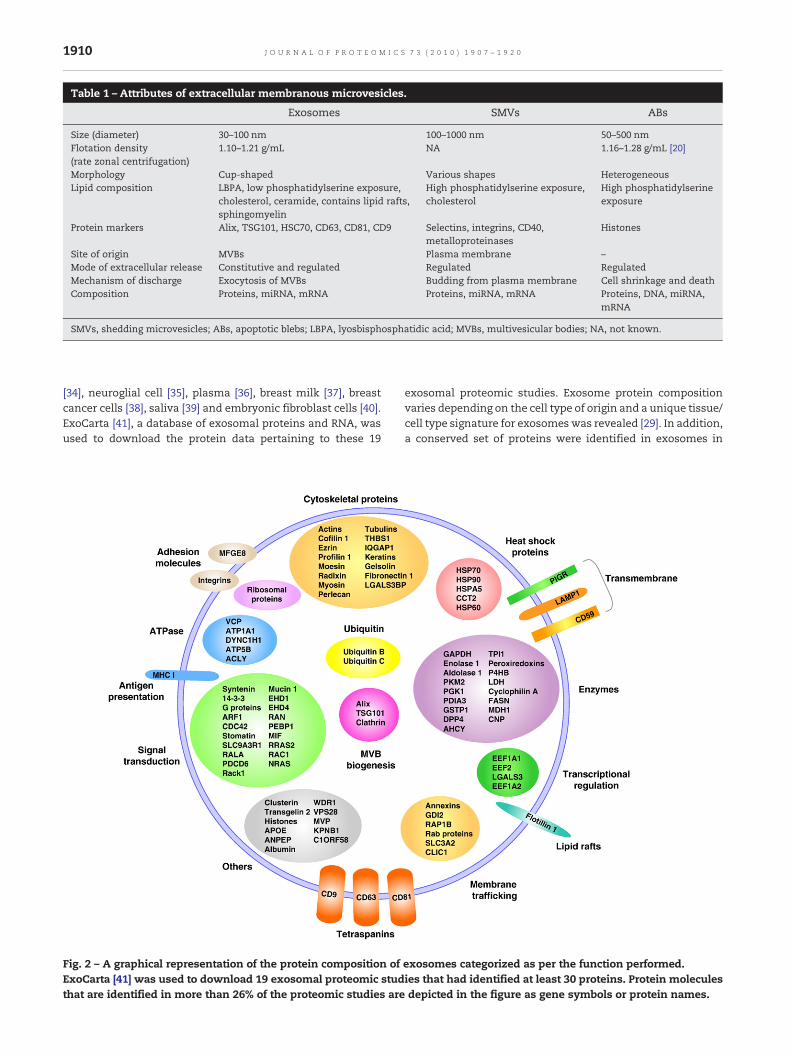
Table 1 – Attributes of extracellular membranous microvesicles.
Exosomes SMVs ABs
Size (diameter) 30–100 nm 100–1000 nm 50–500 nmFlotation density(rate zonal centrifugation)
1.10–1.21 g/mL NA 1.16–1.28 g/mL [20]
Morphology Cup-shaped Various shapes HeterogeneousLipid composition LBPA, low phosphatidylserine exposure,
cholesterol, ceramide, contains lipid rafts,sphingomyelin
High phosphatidylserine exposure,cholesterol
High phosphatidylserineexposure
Protein markers Alix, TSG101, HSC70, CD63, CD81, CD9 Selectins, integrins, CD40,metalloproteinases
Histones
Site of origin MVBs Plasma membrane –Mode of extracellular release Constitutive and regulated Regulated RegulatedMechanism of discharge Exocytosis of MVBs Budding from plasma membrane Cell shrinkage and deathComposition Proteins, miRNA, mRNA Proteins, miRNA, mRNA Proteins, DNA, miRNA,
mRNA
SMVs, shedding microvesicles; ABs, apoptotic blebs; LBPA, lyosbisphosphatidic acid; MVBs, multivesicular bodies; NA, not known.
1910 J O U R N A L O F P R O T E O M I C S 7 3 ( 2 0 1 0 ) 1 9 0 7 – 1 9 2 0
[34], neuroglial cell [35], plasma [36], breast milk [37], breastcancer cells [38], saliva [39] and embryonic fibroblast cells [40].ExoCarta [41], a database of exosomal proteins and RNA, wasused to download the protein data pertaining to these 19
Fig. 2 – A graphical representation of the protein composition ofExoCarta [41] was used to download 19 exosomal proteomic studthat are identified in more than 26% of the proteomic studies are
exosomal proteomic studies. Exosome protein compositionvaries depending on the cell type of origin and a unique tissue/cell type signature for exosomes was revealed [29]. In addition,a conserved set of proteins were identified in exosomes in
exosomes categorized as per the function performed.ies that had identified at least 30 proteins. Protein moleculesdepicted in the figure as gene symbols or protein names.
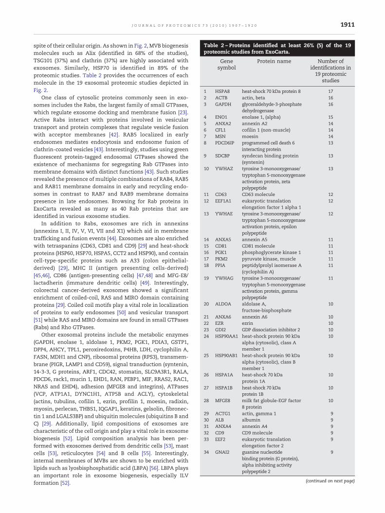
Table 2 – Proteins identified at least 26% (5) of the 19proteomic studies from ExoCarta.
Genesymbol
Protein name Number ofidentifications in19 proteomic
studies
1 HSPA8 heat-shock 70 kDa protein 8 172 ACTB actin, beta 163 GAPDH glyceraldehyde-3-phosphate
dehydrogenase16
4 ENO1 enolase 1, (alpha) 155 ANXA2 annexin A2 146 CFL1 cofilin 1 (non-muscle) 147 MSN moesin 148 PDCD6IP programmed cell death 6
interacting protein13
9 SDCBP syndecan binding protein(syntenin)
13
10 YWHAZ tyrosine 3-monooxygenase/tryptophan 5-monooxygenaseactivation protein, zetapolypeptide
13
11 CD63 CD63 molecule 1212 EEF1A1 eukaryotic translation
elongation factor 1 alpha 112
13 YWHAE tyrosine 3-monooxygenase/tryptophan 5-monooxygenaseactivation protein, epsilonpolypeptide
12
14 ANXA5 annexin A5 1115 CD81 CD81 molecule 1116 PGK1 phosphoglycerate kinase 1 1117 PKM2 pyruvate kinase, muscle 1118 PPIA peptidylprolyl isomerase A
(cyclophilin A)11
19 YWHAG tyrosine 3-monooxygenase/tryptophan 5-monooxygenaseactivation protein, gammapolypeptide
11
20 ALDOA aldolase A,fructose-bisphosphate
10
21 ANXA6 annexin A6 1022 EZR ezrin 1023 GDI2 GDP dissociation inhibitor 2 1024 HSP90AA1 heat-shock protein 90 kDa
alpha (cytosolic), class Amember 1
10
25 HSP90AB1 heat-shock protein 90 kDaalpha (cytosolic), class Bmember 1
10
26 HSPA1A heat-shock 70 kDaprotein 1A
10
27 HSPA1B heat-shock 70 kDaprotein 1B
10
28 MFGE8 milk fat globule-EGF factor8 protein
10
29 ACTG1 actin, gamma 1 930 ALB albumin 931 ANXA4 annexin A4 932 CD9 CD9 molecule 933 EEF2 eukaryotic translation
elongation factor 29
34 GNAI2 guanine nucleotidebinding protein (G protein),alpha inhibiting activitypolypeptide 2
9
(continued on next page)
1911J O U R N A L O F P R O T E O M I C S 7 3 ( 2 0 1 0 ) 1 9 0 7 – 1 9 2 0
spite of their cellular origin. As shown in Fig. 2, MVB biogenesismolecules such as Alix (identified in 68% of the studies),TSG101 (37%) and clathrin (37%) are highly associated withexosomes. Similarly, HSP70 is identified in 89% of theproteomic studies. Table 2 provides the occurrences of eachmolecule in the 19 exosomal proteomic studies depicted inFig. 2.
One class of cytosolic proteins commonly seen in exo-somes includes the Rabs, the largest family of small GTPases,which regulate exosome docking and membrane fusion [23].Active Rabs interact with proteins involved in vesiculartransport and protein complexes that regulate vesicle fusionwith acceptor membranes [42]. RAB5 localized in earlyendosomes mediates endocytosis and endosome fusion ofclathrin-coated vesicles [43]. Interestingly, studies using greenfluorescent protein-tagged endosomal GTPases showed theexistence of mechanisms for segregating Rab GTPases intomembrane domains with distinct functions [43]. Such studiesrevealed the presence of multiple combinations of RAB4, RAB5and RAB11 membrane domains in early and recycling endo-somes in contrast to RAB7 and RAB9 membrane domainspresence in late endosomes. Browsing for Rab proteins inExoCarta revealed as many as 40 Rab proteins that areidentified in various exosome studies.
In addition to Rabs, exosomes are rich in annexins(annexins I, II, IV, V, VI, VII and X1) which aid in membranetrafficking and fusion events [44]. Exosomes are also enrichedwith tetraspanins (CD63, CD81 and CD9) [29] and heat-shockproteins (HSP60, HSP70, HSPA5, CCT2 and HSP90), and containcell-type-specific proteins such as A33 (colon epithelial-derived) [29], MHC II (antigen presenting cells-derived)[45,46], CD86 (antigen-presenting cells) [47,48] and MFG-E8/lactadherin (immature dendritic cells) [49]. Interestingly,colorectal cancer-derived exosomes showed a significantenrichment of coiled-coil, RAS and MIRO domain containingproteins [29]. Coiled coil motifs play a vital role in localizationof proteins to early endosomes [50] and vesicular transport[51] while RAS and MIRO domains are found in small GTPases(Rabs) and Rho GTPases.
Other exosomal proteins include the metabolic enzymes(GAPDH, enolase 1, aldolase 1, PKM2, PGK1, PDIA3, GSTP1,DPP4, AHCY, TPL1, peroxiredoxins, P4HB, LDH, cyclophilin A,FASN, MDH1 and CNP), ribosomal proteins (RPS3), transmem-brane (PIGR, LAMP1 and CD59), signal transduction (syntenin,14-3-3, G proteins, ARF1, CDC42, stomatin, SLC9A3R1, RALA,PDCD6, rack1, mucin 1, EHD1, RAN, PEBP1, MIF, RRAS2, RAC1,NRAS and EHD4), adhesion (MFGE8 and integrins), ATPases(VCP, ATP1A1, DYNC1H1, ATP5B and ACLY), cytoskeletal(actins, tubulins, cofilin 1, ezrin, profilin 1, moesin, radixin,myosin, perlecan, THBS1, IQGAP1, keratins, gelsolin, fibronec-tin 1 and LGALS3BP) and ubiquitinmolecules (ubiquitins B andC) [29]. Additionally, lipid compositions of exosomes arecharacteristic of the cell origin and play a vital role in exosomebiogenesis [52]. Lipid composition analysis has been per-formed with exosomes derived from dendritic cells [53], mastcells [53], reticulocytes [54] and B cells [55]. Interestingly,internal membranes of MVBs are shown to be enriched withlipids such as lyosbisphosphatidic acid (LBPA) [56]. LBPA playsan important role in exosome biogenesis, especially ILVformation [52].

Table 2 (continued)
Genesymbol
Protein name Number ofidentifications in19 proteomic
studies
35 GNB2 guanine nucleotide bindingprotein (G protein), betapolypeptide 2
9
36 GSTP1 glutathione S-transferasepi 1
9
37 MYH9 myosin, heavy chain 9,non-muscle
9
38 PFN1 profilin 1 939 RDX radixin 940 YWHAB tyrosine 3-monooxygenase/
tryptophan 5-monooxygenaseactivation protein, betapolypeptide
9
41 ANXA1 annexin A1 842 ARF1 ADP-ribosylation factor 1 843 GNB1 guanine nucleotide binding
protein (G protein),beta polypeptide 1
8
44 HSPA5 heat-shock 70 kDa protein5 (glucose-regulated protein,78 kDa)
8
45 IQGAP1 IQ motif containing GTPaseactivating protein 1
8
46 PRDX1 peroxiredoxin 1 847 RAB5C RAB5C, member RAS
oncogene family8
48 RAP1B RAP1B, member of RASoncogene family
8
49 THBS1 thrombospondin 1 850 TPI1 triosephosphate
isomerase 18
51 TUBA1A tubulin, alpha 1a 852 VCP valosin-containing protein 853 YWHAH tyrosine 3-monooxygenase/
tryptophan 5-monooxygenaseactivation protein, etapolypeptide
8
54 CCT2 chaperonin containing TCP1,subunit 2 (beta)
7
55 CLIC1 chloride intracellularchannel 1
7
56 CLTC clathrin, heavy chain (Hc) 757 CLU clusterin 758 EHD1 EH-domain containing 1 759 ITGB1 integrin, beta 1 (fibronectin
receptor, beta polypeptide,antigen CD29 includes MDF2,MSK12)
7
60 LDHB lactate dehydrogenase B 761 MUC1 mucin1, cell surfaceassociated 762 P4HB procollagen-proline,
2-oxoglutarate 4-dioxygenase(proline 4-hydroxylase),beta polypeptide
7
63 PIGR polymeric immunoglobulinreceptor
7
64 RAB11B RAB11B, member RASoncogene family
7
65 SLC3A2 solute carrier family 3(activators of dibasic andneutral amino acidtransport), member 2
7
Table 2 (continued)
Genesymbol
Protein name Number ofidentifications in19 proteomic
studies
66 TSG101 tumor susceptibility gene 101 767 TUBA1C tubulin, alpha 1c 768 TUBB5 tubulin, beta 5 769 YWHAQ tyrosine 3-monooxygenase/
tryptophan 5-monooxygenaseactivation protein, thetapolypeptide
7
70 ACTN4 actinin, alpha 4 671 ANPEP alanyl (membrane)
aminopeptidase6
72 ANXA11 annexin A11 673 APOE apolipoprotein E 674 ATP1A1 ATPase, Na+/K+
transporting, alpha 1polypeptide
6
75 CDC42 cell division cycle 42(GTP bindingprotein, 25 kDa)
6
76 EHD4 EH-domain containing 4 677 FASN fatty acid synthase 678 FN1 fibronectin 1 679 GNAI3 guanine nucleotide
binding protein (G protein),alpha inhibiting activitypolypeptide 3
6
80 GNAQ guanine nucleotide bindingprotein (G protein),q polypeptide
6
81 GNAS GNAS complex locus 682 GSN gelsolin (amyloidosis,
Finnish type)6
83 HIST4H4 histone cluster 4, H4 684 KRT10 keratin 10 685 LDHA lactate dehydrogenase A 686 MIF macrophage migration
inhibitory factor(glycosylation-inhibitingfactor)
6
87 PEBP1 phosphatidylethanolaminebinding protein 1
6
88 PRDX2 peroxiredoxin 2 689 RAB11A RAB11A, member RAS
oncogene family6
90 RAN RAN, member RAS oncogenefamily
6
91 TAGLN2 transgelin 2 692 UBB ubiquitin B 693 ACLY ATP citrate lyase 594 AHCY S-adenosylhomocysteine
hydrolase5
95 ANXA7 annexin A7 596 ARF3 ADP-ribosylation factor 3 597 ATP5B ATP synthase,
H+transporting,mitochondrial F1 complex,beta polypeptide
5
98 C1ORF58 chromosome 1 open readingframe 58
5
99 CD59 CD59 molecule, complementregulatory protein
5
100 CNP 2′,3′-cyclic nucleotide3′ phosphodiesterase
5
1912 J O U R N A L O F P R O T E O M I C S 7 3 ( 2 0 1 0 ) 1 9 0 7 – 1 9 2 0

Table 2 (continued)
Genesymbol
Protein name Number ofidentifications in19 proteomic
studies
101 DPP4 dipeptidyl-peptidase 4 5102 DYNC1H1 dynein, cytoplasmic 1, heavy
chain 15
103 EEF1A2 eukaryotic translationelongation factor 1 alpha 2
5
104 FLOT1 flotillin 1 5105 GNA11 guanine nucleotide binding
protein (G protein), alpha 11(Gq class)
5
106 GNB2L1 guanine nucleotide bindingprotein (G protein), betapolypeptide 2-like 1
5
107 HIST1H4A histone cluster 1, H4a 5108 HIST1H4B histone cluster 1, H4b 5109 HLA-A major histocompatibility
complex, class I, A5
110 HSPD1 heat-shock 60 kDa protein 1(chaperonin)
5
111 HSPG2 heparan sulfateproteoglycan 2
5
112 ITGA6 integrin, alpha 6 5113 ITGAV integrin, alpha V
(vitronectin receptor,alpha polypeptide,antigen CD51)
5
114 KPNB1 karyopherin (importin)beta 1
5
115 KRT15 keratin 15 5116 KRT5 keratin 5 5117 LAMP1 lysosomal-associated
membrane protein 15
118 LGALS3 lectin, galactoside-binding,soluble, 3
5
119 LGALS3BP lectin, galactoside-binding,soluble, 3 binding protein
5
120 MDH1 malate dehydrogenase 1,NAD (soluble)
5
121 MVP major vault protein 5122 NRAS neuroblastoma RAS viral
(v-ras) oncogene homolog5
123 PDCD6 programmed cell death 6 5124 PDIA3 protein disulfide
isomerase family A,member 3
5
125 RAB10 RAB10, member RASoncogene family
5
126 RAB13 RAB13, member RASoncogene family
5
127 RAB14 RAB14, member RASoncogene family
5
128 RAB35 RAB35, member RASoncogene family
5
129 RAB5A RAB5A, member RASoncogene family
5
130 RAB5B RAB5B, member RASoncogene family
5
131 RAB7 RAB7, member RASoncogene family
5
132 RAC1 ras-related C3 botulinumtoxin substrate 1(rho family, small GTPbinding protein Rac1)
5
(continued on next page)
Table 2 (continued)
Genesymbol
Protein name Number ofidentifications in19 proteomic
studies
133 RALA v-ral simian leukemiaviral oncogene homologA (ras related)
5
134 RPS3 ribosomal protein S3 5135 RRAS2 related RAS viral (r-ras)
oncogene homolog 25
136 SFN stratifin 5137 SLC9A3R1 solute carrier family 9
(sodium/hydrogenexchanger),member 3 regulator 1
5
138 STOM stomatin 5139 TUBA1B tubulin, alpha 1b 5140 TUBB4 tubulin, beta 4 5141 UBC ubiquitin C 5142 VPS28 vacuolar protein sorting
28 homolog (S. cerevisiae)5
143 WDR1 WD repeat domain 1 5
1913J O U R N A L O F P R O T E O M I C S 7 3 ( 2 0 1 0 ) 1 9 0 7 – 1 9 2 0
5. Exosomesmediate cell-to-cell communication
Cellular interactions are pivotal for the progression, angiogen-esis and invasiveness of tumors [21]. Such interactions arepresumed to be regulated bymembrane surfacemolecules (e.g.,EGFR) and soluble secreted proteins (e.g., IL-12) that activate thetarget cells by interacting with the target cell surface receptors.Recently, another mode of intercellular communication thathad gained immense scientific interest is mediated by exo-somes. Possiblemechanismsbywhich exosomes communicatewith the target cell are shown in Fig. 3. As shown in Fig. 3A,exosomalmembraneproteins can interactwith the target cell ina juxtacrine fashion, thereby activating the target cell. Likewise,exosomal membrane proteins can be cleaved by proteases andthe resulting fragmentcanactas ligands for cell surface receptorin the target cell (Fig. 3B). Interestingly, some of the exosomalmembrane proteins are not identified in the cell surface of theoriginating cell (e.g., LAMP-2) [20]. Perhaps, this observationholds immense promise in terms of juxtacrine and ectodomaincleavage mediated cell-to-cell signaling events. In addition tojuxtacrine and ectodomain cleavage-based signaling, exosomescan fuse with the target cell resulting in the non-selectivetransfer of exosomal proteins andRNA (Fig. 3C) to the target cell.Additionally, such fusionmight change some of themembranefeatures of the target cell (e.g., arachidonic acid transfer fromplatelets-derived SMVs to leukocytes and endothelial cells [57])including varied lipid concentrations and the transfer ofexosomal membrane proteins on the target cell surface (e.g.,CD41 antigen from platelets-derived SMVs to tumor andendothelia cell surface [58,59]).
6. Exosome purification protocols
Detailed biochemical and functional analyses of exosomesare confounded by the technical difficulty in isolating and

Table 3 – Recommended procedures for isolation/purification of exosomes.
Method Comments
Differentialcentrifugation
Differential centrifugation coupled withmembrane filtration (0.1–0.2 μm) can eliminatelarge contaminating extracellular vesicles(SMVs). Small contaminating vesicles (ABs) canstill be present in the preparation.
Rate zonalcentrifugation
Pure exosomal preparation can be obtained(usually 1.10–1.21 g/mL). Exosomal markerproteins can be used to identify the flotationdensity of the exosomal population.
Immunoaffinitycapture
Based on the specificity and availability of theantibodies, pure exosomal preparation can beobtained. The yield will be low as exosomesubpopulations with the localized antigen ofchoice will be isolated.
Fig. 3 – Possible mechanisms of intercellular communication by exosomes. A, Exosomal membrane proteins can interactwith receptors in a target cell and activate intracellular signaling (juxtacrine fashion). B, Exosomal membrane proteins canbe cleaved by proteases in the extracellular space. Cleaved fragments can then act as a soluble ligand which binds to thetarget cell surface receptor. This mechanism in turn activates the signaling cascade within the target cell. C, Exosomes canfuse with the target cell membrane and release their contents inside the recipient target cell in a non-selective manner. Thesurface membrane of the target cells in turn can be modified by the addition of new membrane receptors (from exosomemembranes) and different lipid compositions. Exosomal molecules (protein, mRNA and miRNA) can activate a multitude ofsignaling events in the recipient target cell.
1914 J O U R N A L O F P R O T E O M I C S 7 3 ( 2 0 1 0 ) 1 9 0 7 – 1 9 2 0
purifying them to homogeneity. Without stringent purifi-cation, exosomes are typically contaminated by othermembranous vesicles such as SMVs and ABs. In contrastto intracellular organelles, which are purified from complextissue homogenates, extracellular organelle ‘exosomes’ arerelatively easy to purify. For example, by combiningdifferential centrifugation, membrane filtration, concentra-tion, rate zonal centrifugation and immunocapture, exo-somes can be isolated from a multitude of cell line and bodyfluids [2]. Characterization of isolated exosomes is typicallyperformed using electron microscopy, FACS, LC-MS/MS andWestern blotting [2,4]. A major issue with these studies thatlimits comparative analyses is that diverse purificationstrategies are employed. For example, many exosomepreparations are heavily contaminated with SMVs andABs, and vice versa. Even though the biophysical propertiesof exosomes and other vesicles are distinct (Table 1),relatively a few studies have exploited biophysical proper-ties such as flotation density to isolate and characterizeexosome preparations (sucrose or OptiPrep density gradi-ent). The majority of studies employ a simpler approach ofdifferential centrifugation wherein the preparations can besignificantly contaminated by SMVs, ABs and cellular
debris. The far simpler approach of using membrane filters(0.1–0.2 μm) in combination with differential centrifugationcould eliminate the large vesicles and result in a more

1915J O U R N A L O F P R O T E O M I C S 7 3 ( 2 0 1 0 ) 1 9 0 7 – 1 9 2 0
reliable exosome preparation [40]. Table 3 lists the recom-mended exosome isolation and purification procedures.
Immunoaffinity capture using magnetic beads has beenemployed recently to isolate highly-purified exosomes [60,61].For example, immunoaffinity capture based on the HER2antibody was used to isolate tumor exosomes from breastadenocarcinoma cell line culture supernatant and ascitesfrom ovarian cancer patients [61]. Similarly, A33 [62–64]-expressing tumor exosomes were collected from the culturesupernatant of the colon carcinoma cell lines, LIM1215 [29]and LIM1863 (Simpson, unpublished data) using A33-antibodycoated Dynabeads. Immunoaffinity capture-based exosomeisolation can be performed in cell culture media containingfetal calf serum and high-Mr protein oligomers (e.g., hapto-globin), which may otherwise contaminate conventionalpreparations. By these means, exosomes can be purified freeof contaminating large-Mr proteins and oligomers (e.g.,proteosomal complexes) that co-sediment with exosomes athigh centrifugal force [29]. Interestingly, the immunoaffinitycapture approach resulted in depletion of histones (markers ofABs) as compared to crude exosomes (prepared by differentialcentrifugation and membrane filtration) which, presumably,resulted from contaminating ABs that are in the same sizerange as exosomes (50–100 nm diameter).
7. Clinical studies involving exosomes
It is well known that a variety of solid human tumors arespontaneously infiltrated by T cells and that memory effectorT cells associate with favorable clinical outcomes whileoverwhelming regulatory T cells compromise long-termsurvival [65]. This has led to the identification of manytumor-associated antigens capable of eliciting cytotoxic Tcell responses and vaccine immunotherapy as an approach tocancer treatment. Recently, there has been much interest inthe application of exosomes as a potential viable vaccine forclinical immunotherapy; dendritic cell-derived exosomes areenriched in the components necessary to function as anantigen presenting entities [66]. Exosome-based clinical stud-ies have been carried out on lung cancer and melanomapatients [67–70]. These clinical studies employed a robustmacroscale-method, developed by Lamparski and colleaguesfor isolating clinical-grade dendritic cell-derived exosomes[68]. The method relies on a combination of ultrafiltration(500 K membrane) and ultracentrifugation into a 30% sucrose/deuterium oxide (98%) cushion (density, 1.21 g/mL) to harvestexosomes from dendritic cell culture supernatant in high yield(40–50%). Two Phase 1 clinical trials for melanoma and lungcancer, using peptide antigens [67,68], were completed usingthis purification strategy. Phase I clinical studies in melanomapatients showed that the injection of dendritic cells-derivedexosomes is safe and associated with some tumor regressionand long-term stabilization [69]. Importantly, the studyrevealed that the injected dendritic cells-derived exosomessignificantly increased circulating natural killer cells andnatural killer group 2 member D-dependent functions in themajority of melanoma patients [71]. Similarly, Phase I clinicaltrial that evaluated the tolerance of dendritic cell-derived
exosomes in patients with stage III/IV lung cancer showedthat the exosome injection is safe and allowed long-termstabilization in 4 of the 12 patients [70] (for a furthercommentary on clinical trials and application of exosomes,discussed at the ‘Workshop on the Biological Significance ofExosomes in Montreal, Canada, May 20–21, 2005’, refer to thereview by Johnstone [72]).
A potential confounding factor in exosome isolation,particularly for clinical applications, is possible retroviralcontamination. For example, exosomes and HIV-1 particleshave similar biophysical properties such as size (30–100 nmand ~100 nm, respectively) and buoyant density (1.13–1.21 g/L[20,55] and 1.13–1.21 g/L [73], respectively), as well as theirability to activate immune cells. While earlier studies reportedthat exosomes carried virion cargo [35,74,75], recent exosomepurification strategies deploying immunoaffinity capture [76]or a combination of immunoaffinity capture and densitygradient centrifugation using iodixanol (OptiPrep™) [77]demonstrate that exosomes from haematopoietic cells canbe purified free of virions like HIV-1.
Exosomes have been reported in diverse physiologicalfluids such as blood and ascites fluid. One of the first studiesto provide evidence for exosomes in blood of healthy donorswas performed by Caby and colleagues [78] using differentialcentrifugation or immunocapture using anti-CD63 mAb latexbeads. These exosomes were characterized by electronmicroscopy (50–90 nm diameter), flotation density (1.15–1.27 g/mL) and proteome analysis (e.g., tetraspanins CD63,CD9, CD81, and LAMP-2 and class I and II MHC molecules).More recently, circulating EpCAM-positive exosomes wereisolated from sera of patients with early stage ovarian cancerby a modified activated cell sorting (MACS) procedure usingantibody beads [79]. Subsequent miRNA profiling of theseexosomes indicate the potential use of circulating tumorexosomes as surrogate diagnostic markers for biopsy profilingand possible utility to screen asymptomatic populations [79].In 2008, Levine and coworkers [80] reported the presence of48 K TNFR1-containing exosome-like vesicles (density of 1.09to 1.11 g/mL on sucrose gradient) in human plasma and alsosera where they co-segregate with LDL particles. In morerecent studies using a multidimensional purification schemeincorporating gel-exclusion chromatography, rate zonal cen-trifugation through continuous sucrose gradients and high-speed centrifugation, the same group reports a distinctpopulation of PPARγ-containing exosomes in circulatinghuman plasma [36]. Using anti-HLA class II mAb coatedDynabeads™, Klibi and colleagues report the immunomag-netic capture of galectin-9-containing exosomes in the plasmaof nasopharyngeal (NPC)-patients or mice xenografted withNPC cell lines [81].
8. Exosomes as a rich source for discoveringpotential blood-based biomarkers
The release of exosomes into the extracellular space affordsan opportunity to examine exosomes in body fluids such asblood, urine and malignant ascites. Accessing these bioactivevesicles in a non-invasive manner may lead to potential

Fig. 4 – Circulating exosomes can be a rich source foridentifying potential biomarkers. Patient plasma containsexosomes that are released by disease cells (e.g., colorectalcancer cells), normal counterpart (e.g., normal colon cells) andother normal cells (e.g., liver). Exosomal tissue signatures canbe used to isolate disease cell-derived exosomes for proteomicand transcriptomic profiling.
1916 J O U R N A L O F P R O T E O M I C S 7 3 ( 2 0 1 0 ) 1 9 0 7 – 1 9 2 0
diagnostic biomarkers of disease conditions. Exosomes wereobserved in vivo in blood from healthy donors [78] with similarbiophysical properties as that of previously described exo-somes released from various cell types in vitro. A magneticbead immune capture strategy was employed to isolatecirculating epithelial cell adhesion molecule-positive exo-somes from the plasma of ovarian cancer [79] and lung cancer[82] patients. Interestingly, these studies revealed that plas-ma-exosome levels were increased in patients with advanceddisease (e.g., mean 2.85 mg/mL exosomes for lung canceradenocarcinoma patients compared with 0.77 mg/mL exo-somes in the blood of normal volunteers [82]). These studiessuggest that circulating exosomes in the body may play a rolein pathogenesis and cell–cell or organ–organ communicationsby transporting molecules that need to reach distant celltargets [78]. The increased levels of tumor-derived exosomesin plasma andmalignant effusions of patients with cancer [83]suggest that exosomes can be a rich source for the discovery ofblood-based diagnostic biomarkers of disease (Fig. 4). Exo-somes have also been isolated from physiological fluids suchas normal urine [24,25,84,85], malignant and pleural effusions[83,86], bronchial lavage fluid [87], ocular fluids [88], humansemen [89,90], amniotic fluid [91], human saliva [39,92], breastmilk [37], pregnancy-associated sera [93] and synovial fluid[94]. Urine contains exosomes that are derived from varioustypes of kidney cells that come into contact with the urinaryspace, including glomerular podocytes and renal tubule cells.In a recent study, prostate cancer biomarkers, PCA-3 andTMPRSS2: ERG, were detected in exosomes isolated from theurine of prostate cancer patients [95].
A possible obstacle in such body fluid based exosomeanalysis is the presence of contaminating exosomes secretedby normal cells (e.g., normal colon cells as compared to coloncancer cells) and other cell types (e.g., non colon cell types). Tobetter understand the molecular properties of disease cell-derived exosomes, such exosomes need to be isolated free ofnormal cell-derived exosomes from the complex mixture inblood. Purification strategies that discriminate between com-plex exosome mixtures in blood could be aided by theknowledge of exosome tissue-specific signatures, especiallydisease cell signature, which can be obtained from theExoCarta [41] compendium of exosomal proteins and RNA.
9. Exocarta: a manually curated database ofexosomal proteins and RNA
ExoCarta is a database of previous exosomal proteomic andtranscriptomic studies. It is a manually curated repositorycontaining proteins and RNA that were cataloged from 75published exosome studies. A total of 2624 proteins, 901mRNA and 274 miRNA from 4 different organisms are presentin this compendium. Additionally, the compendium listsproteins that are more often identified in exosomal studiesbased on the number of occurrences of these molecules in the75 published proteomic studies. Some of these molecules canbe used as reliable exosomal markers, which is currently oneof the important steps in characterizing the presence ofexosomes in the preparation. Interestingly, tissue-specific
proteins that are identified in exosomes are also listed inExoCarta. For example, the A33-antigen is specific to colorectalcancer-derived exosomes [29] and future studies based onimmunoaffinity capture of colon-specific exosomes can beperformed with this knowledge. ExoCarta is freely availablefor the scientific community and the entire data encompass-ing 75 studies can be freely downloaded (http://exocarta.ludwig.edu.au).
10. Summary
Exosomes are secreted by various cell types and playimportant roles in cellular communication. Heterogeneousmicrovesicle studies over the years had led to confoundingterminologies due to the cross-contamination of SMVs and,possibly, ABs. However, the known underlying mechanismsby which microvesicles are released into the extracellularspace are limited to three: exocytic exosomes from theintracellular MVBs, SMVs from the PM and ABs from cells
1917J O U R N A L O F P R O T E O M I C S 7 3 ( 2 0 1 0 ) 1 9 0 7 – 1 9 2 0
undergoing apoptosis. Consensus in the nomenclature ofnaming these microvesicles is needed at this juncture and theonus is on the investigators to name the vesicle populationcorrectly in order to avoid any further confusion. In contrast toSMVs and ABs, exosomes are relatively smaller in size andfloat at a different density (1.10 g/mL–1.21 g/mL). In addition toa common set of proteins, in spite of their cellular origin,exosomes also have tissue- or cell-type-specific protein/RNAmolecules. Such attributes coupled with the extracellularlocation of exosomes, make them ideal starting materials forthe identification of candidate biomarkers relevant to thepathophysiology of a specific disease. For approximately 42%of integral PM proteins found in exosomes, their corre-sponding soluble ectodomain can be found in the secretome(Mathivanan and Simpson, unpublished observations). Asimple non-invasive approach can be employed to isolateexosomes for the purpose of identifying several candidateprotein diagnostic biomarkers.
ExoCarta: a database for exosomal proteins and RNAs
• ExoCarta is a web-based freely accessible compendiumthe published protein, mRNA and miRNA content ofpurified exosomes from a multitude of cell line andbody fluids.
• While the onus for the quality of data contained in thedatabase resides with the researchers who publishedthe data, the database catalogs data from manualcuration of scientific literature.
• Every molecule entry in ExoCarta is accompanied byinformation pertaining to the gene information, data-base cross references, gene ontology annotations, pro-tein–protein interactions and experiment description.
Box 2
Keynotes
• Exosomes are defined as 40–100-nm diameter membranevesicles of endocytic origin that are released from most celltypes upon fusion of multivesicular bodies (MVBs) with theplasma membrane (PM). Within the MVBs, exosomes arereferred to as intraluminal vesicles (ILVs); it is only uponrelease of MVB contents into the microenvironment thatILVs are referred to as exosomes. The nomenclature ofexosomes can be confusing since typical exosomes are oftenreferred to in the literature as ‘microvesicles’.
• Exosomes have a cup-shaped appearance by electronmicroscopy, sediment at 100,000 g, and have a buoyantdensity in sucrose of 1.10–1.21 g/mL.While exosomes have alipid bilayer with the same topography as plasma mem-branes, they are also reported to expose phosphatidylserineon their surface.
• It has been recently reported in oligodendrocytes thatformation of MVBs destined for passage to the plasmamembrane and release of their exosomes cargo into themicroenvironment or those tagged for degradation by theproteosome machinery in lysosomes involve differentceramide-based molecular machineries. It is not clearwhether this observation extrapolates to other cell typesand whether there are two types of MVBs (exocytic anddegradative MVBs) or different types of ILVs within MVBs.
• The protein composition of typical exosomes is oftenconfounded by the exactness of the strategy employed intheir purification. Because many high-Mr oligomeric pro-teins and viruses co-sediment with exosomes at 100,000 g,more robust purification methods such as immune captureand/or rate zonal centrifugation are essential for obtaininghigh-purity exosomes.
• Proteomic cataloging of highly-purified exosomes fromurinary, mast cell and colorectal cancer cell lines hasidentified 31 membrane and cytosolic proteins common tothese cell types. Additionally, exosomes exhibit proteinsignatures that reflect the originating cell type. A searchablecompendium of exosomal proteins and RNA is now
accessible at ExoCarta (http://exocarta.ludwig.edu.au). Typ-ical protein markers for exosomes include Alix, TSG101,CD63, CD9, CD81 and HSP70.
• Exosomes contain inactive forms of both mRNA andmicroRNAs that can be transferred to a neighboring cell,conferring new functional properties to the recipient cellafter the acquisition of the exosomal genetic material.MicroRNAprofiling studies of disease cell-derived exosomesand exosomes circulating in blood offer the potential ofexosomal microRNA profiles for use as diagnostic biomar-kers of disease through non-invasive blood tests.
• Exosomes have pleiotropic effects that influence the phys-iology of neighboring cells. Of these, the best studied (invitro) are the roles of exosomes in various stages of theimmune response (interactions with immune cells). Theserange from exosomes being a vehicle for antigen presenta-tion to antigen-independent roles that can inhibit (immu-nosuppressive properties) or promote immune responses(immune-activating properties). Additionally, exosomesplay role in intercellular communication, being conveyorsof proteins and lipids that affect downstream signalingevents in recipient cells. They can also deliver geneticmaterial that affects the physiology of recipient cells.
Box 1
Extracellular membranous vesicles: Membranous vesi-cles (exosomes, shedding microvesicles (SMVs) andapoptotic blebs (ABs)) that are secreted/shed by cells intothe extracellular space.Exosomes: Upon fusion of the MVBs with the plasmamembrane (PM), the intraluminal vesicles (ILVs) arereleased into the microenvironment and are referred toas exosomes.Shedding microvesicles (SMVs): Vesicles that are sheddirectly from the PM into the extracellular space.Apoptotic blebs (ABs): Vesicles that are released bydying/apoptotic cells.Microvesicles: Mixed population of exosomes, SMVs andABs.

• ExoCarta provides biomedical researchers with infor-mation pertaining to isolation/purification methodsemployed in the published exosomal studies (thisinformation allows researchers to evaluate the purityof any exosome preparation).
• Data present in ExoCarta can be freely downloadedfrom the ExoCarta website.
What ExoCarta is not?
• ExoCarta does not address data quality — the onus ofdata quality resides with the researcher(s) responsiblefor acquiring and publishing the data.
• ExoCarta has not cataloged proteins/RNA from non-exosome extracellular membranous vesicle studies.
ExoCarta URL: http://exocarta.ludwig.edu.au
1918 J O U R N A L O F P R O T E O M I C S 7 3 ( 2 0 1 0 ) 1 9 0 7 – 1 9 2 0
Acknowledgements
This work was supported by the National Health and MedicalResearch Council (Program grant 487922 to RJS).
R E F E R E N C E S
[1] Andersen JS,MannM.Organellar proteomics: turning inventoriesinto insights. EMBO Rep 2006;7:874–9.
[2] Simpson RJ, Jensen SS, Lim JW. Proteomic profiling ofexosomes: current perspectives. Proteomics 2008;8:4083–99.
[3] Pan BT, Johnstone RM. Fate of the transferrin receptorduring maturation of sheep reticulocytes in vitro: selectiveexternalization of the receptor. Cell 1983;33:967–78.
[4] van Niel G, Porto-Carreiro I, Simoes S, Raposo G. Exosomes: acommon pathway for a specialized function. J Biochem(Tokyo) 2006;140:13–21.
[5] Simpson RJ, Lim JW, Moritz RL, Mathivanan S. Exosomes:proteomic insights and diagnostic potential. Expert RevProteomics 2009;6:267–83.
[6] Trajkovic K, Hsu C, Chiantia S, Rajendran L, Wenzel D,Wieland F, Schwille P, Brugger B, SimonsM. Ceramide triggersbudding of exosome vesicles into multivesicular endosomes.Science 2008;319:1244–7.
[7] Babst M. A protein's final ESCRT. Traffic 2005;6:2–9.[8] Babst M. A close-up of the ESCRTs. Dev Cell 2006;10:547–8.[9] Hurley JH. ESCRT complexes and the biogenesis of
multivesicular bodies. Curr Opin Cell Biol 2008;20:4–11.[10] Williams RL, Urbe S. The emerging shape of the ESCRT
machinery. Nat Rev Mol Cell Biol 2007;8:355–68.[11] Hess C, Sadallah S, Hefti A, Landmann R, Schifferli J-A.
Ectosomes released by human neutrophils are specializedfunctional units. J Immunol 1999;163:4564–73.
[12] Al-Nedawi K, Meehan B, Micallef J, Lhotak V, May L, Guha A,Rak J. Intercellular transfer of the oncogenic receptor EGFRvIIIby microvesicles derived from tumour cells. Nat Cell Biol2008;10:619–24.
[13] Heijnen HF, Schiel AE, Fijnheer R, Geuze HJ, Sixma JJ.Activated platelets release two types of membrane vesicles:microvesicles by surface shedding and exosomes derivedfrom exocytosis of multivesicular bodies and alpha-granules.Blood 1999;94:3791–9.
[14] Cocucci E, Racchetti G, Meldolesi J. Shedding microvesicles:artefacts no more. Trends Cell Biol 2009;19:43–51.
[15] Piccin A, Murphy WG, Smith OP. Circulating microparticles:pathophysiology and clinical implications. Blood Rev 2007;21:157–71.
[16] Del Conde I, Bharwani LD, Dietzen DJ, Pendurthi U,Thiagarajan P, LÃ'Pez JA. Microvesicle-associated tissuefactor and Trousseau's syndrome. J Thromb Haemost2007;5:70–4.
[17] del Conde I, Shrimpton CN, Thiagarajan P, Lopez JA.Tissue-factor-bearing microvesicles arise from lipid raftsand fuse with activated platelets to initiate coagulation.Blood 2005;106:1604–11.
[18] Hristov M, Erl W, Linder S, Weber PC. Apoptotic bodies fromendothelial cells enhance the number and initiate thedifferentiation of human endothelial progenitor cells in vitro.Blood 2004;104:2761–6.
[19] Beyer C, and Pisetsky DS. The role of microparticles in thepathogenesisof rheumaticdiseases.NatRevRheumatol 6:21–29.
[20] Thery C, Boussac M, Veron P, Ricciardi-Castagnoli P, RaposoG, Garin J, Amigorena S. Proteomic analysis of dendriticcell-derived exosomes: a secreted subcellular compartmentdistinct from apoptotic vesicles. J Immunol 2001;166:7309–18.
[21] Al-Nedawi K, Meehan B, Rak J. Microvesicles: messengersand mediators of tumor progression. Cell Cycle 2009;8:2014–8.
[22] Ratajczak J, Wysoczynski M, Hayek F, Janowska-Wieczorek A,Ratajczak MZ. Membrane-derivedmicrovesicles: important andunderappreciated mediators of cell-to-cell communication.Leukemia 2006;20:1487–95.
[23] Mears R, Craven RA, Hanrahan S, Totty N, Upton C, Young SL,Patel P, Selby PJ, Banks RE. Proteomic analysis of melanoma-derived exosomes by two-dimensional polyacrylamide gelelectrophoresis and mass spectrometry. Proteomics 2004;4:4019–31.
[24] Pisitkun T, Shen RF, Knepper MA. Identification andproteomic profiling of exosomes in human urine. Proc NatlAcad Sci USA 2004;101:13368–73.
[25] Gonzales PA, Pisitkun T, Hoffert JD, Tchapyjnikov D, Star RA,Kleta R, Wang NS, Knepper MA. Large-scale proteomics andphosphoproteomics of urinary exosomes. J Am Soc Nephrol2008.
[26] Potolicchio I, Carven GJ, Xu X, Stipp C, Riese RJ, Stern LJ,Santambrogio L. Proteomic analysis of microglia-derivedexosomes: metabolic role of the aminopeptidase CD13 inneuropeptide catabolism. J Immunol 2005;175:2237–43.
[27] Valadi H, Ekstrom K, Bossios A, Sjostrand M, Lee JJ, Lotvall JO.Exosome-mediated transfer of mRNAs and microRNAs is anovel mechanism of genetic exchange between cells. Nat CellBiol 2007;9:654–9.
[28] Choi DS, Lee JM, Park GW, Lim HW, Bang JY, Kim YK, KwonKH, Kwon HJ, Kim KP, Gho YS. Proteomic analysis ofmicrovesicles derived from human colorectal cancer cells. JProteome Res 2007;6:4646–55.
[29] Mathivanan S, Lim JW, Tauro BJ, Ji H, Moritz RL, Simpson RJ.Proteomic analysis of A33-immunoaffinity-purifiedexosomes released from the human colon tumor cell lineLIM1215 reveals a tissue-specific protein signature. Mol CellProteomics 2009.
[30] Hegmans JP, Bard MP, Hemmes A, Luider TM, Kleijmeer MJ,Prins JB, Zitvogel L, Burgers SA, Hoogsteden HC, LambrechtBN. Proteomic analysis of exosomes secreted by humanmesothelioma cells. Am J Pathol 2004;164:1807–15.
[31] Graner MW, Alzate O, Dechkovskaia AM, Keene JD, SampsonJH, Mitchell DA, Bigner DD. Proteomic and immunologicanalyses of brain tumor exosomes. FASEB J 2008.
[32] Eva-Maria Krämer-Albers NB, Tenzer Stefan, WintersteinChristine, Möbius Wiebke, Berger Hendrik, Nave Klaus-Armin,
1919J O U R N A L O F P R O T E O M I C S 7 3 ( 2 0 1 0 ) 1 9 0 7 – 1 9 2 0
Schild Hansjörg, Trotter Jacqueline. Oligodendrocytes secreteexosomes containing major myelin and stress-protectiveproteins: trophic support for axons? Proteomics: Clin Appl2007;1:1446–61.
[33] KesimerM, Scull M, Brighton B, Demaria G, Burns K, O'NealW,Pickles RJ, Sheehan JK. Characterization of exosome-likevesicles released from human tracheobronchial ciliatedepithelium: a possible role in innate defense. FASEB J 2009;23:1858–68.
[34] Conde-Vancells J, Rodriguez-Suarez E, Embade N, Gil D,Matthiesen R, Valle M, Elortza F, Lu SC, Mato JM, Falcon-PerezJM. Characterization and comprehensive proteome profilingof exosomes secreted by hepatocytes. J Proteome Res 2008;7:5157–66.
[35] Fevrier B, Vilette D, Archer F, Loew D, FaigleW, Vidal M, LaudeH, Raposo G. Cells release prions in association withexosomes. Proc Natl Acad Sci USA 2004;101:9683–8.
[36] Looze C, Yui D, Leung L, InghamM, Kaler M, Yao X, Wu WW,Shen RF, Daniels MP, Levine SJ. Proteomic profiling of humanplasma exosomes identifies PPARgamma as anexosome-associated protein. Biochem Biophys Res Commun2009;378:433–8.
[37] Admyre C, Johansson SM, Qazi KR, Filen JJ, Lahesmaa R,Norman M, Neve EP, Scheynius A, Gabrielsson S. Exosomeswith immune modulatory features are present in humanbreast milk. J Immunol 2007;179:1969–78.
[38] Staubach S, Razawi H, Hanisch FG. Proteomics ofMUC1-containing lipid rafts from plasma membranes andexosomes of human breast carcinoma cells MCF-7. Proteomics2009;9:2820–35.
[39] Gonzalez-Begne M, Lu B, Han X, Hagen FK, Hand AR, Melvin JE,Yates JR. Proteomic analysis of human parotid gland exosomesbyMultidimensional Protein IdentificationTechnology (MudPIT).J Proteome Res 2009.
[40] Ji H, Erfani N, Tauro BJ, Kapp EA, Zhu HJ, Moritz RL, Lim JW,Simpson RJ. Difference gel electrophoresis analysis ofRas-transformed fibroblast cell-derived exosomes.Electrophoresis 2008;29:2660–71.
[41] Mathivanan S, Simpson RJ. ExoCarta: a compendium ofexosomal proteins and RNA. Proteomics 2009;9:4997–5000.
[42] Corbeel L, Freson K. Rab proteins and Rab-associated proteins:major actors in themechanismof protein-trafficking disorders.Eur J Pediatr 2008;167:723–9.
[43] Stenmark H. Rab GTPases as coordinators of vesicle traffic.Nat Rev Mol Cell Biol 2009;10:513–25.
[44] Futter CE, White IJ. Annexins and endocytosis. Traffic 2007;8:951–8.
[45] Denzer K, van EijkM, KleijmeerMJ, Jakobson E, de Groot C, GeuzeHJ. Follicular dendritic cells carry MHC class II-expressingmicrovesicles at their surface. J Immunol 2000;165:1259–65.
[46] Raposo G, Tenza D, Mecheri S, Peronet R, Bonnerot C,Desaymard C. Accumulation of major histocompatibilitycomplex class II molecules inmast cell secretory granules andtheir release upon degranulation. Mol Biol Cell 1997;8:2631–45.
[47] RaposoG,NijmanHW,StoorvogelW,LiejendekkerR,HardingCV,Melief CJ, Geuze HJ. B lymphocytes secrete antigen-presentingvesicles. J Exp Med 1996;183:1161–72.
[48] Segura E, Nicco C, Lombard B, Veron P, Raposo G, Batteux F,Amigorena S, Thery C. ICAM-1 on exosomes from maturedendritic cells is critical for efficient naive T-cell priming.Blood 2005;106:216–23.
[49] Veron P, Segura E, Sugano G, Amigorena S, Thery C.Accumulation of MFG-E8/lactadherin on exosomes fromimmature dendritic cells. Blood Cells Mol Dis 2005;35:81–8.
[50] Raiborg C, Bremnes B, Mehlum A, Gillooly DJ, D'Arrigo A, StangE, Stenmark H. FYVE and coiled-coil domains determine thespecific localisation of Hrs to early endosomes. J Cell Sci2001;114:2255–63.
[51] Nair J, Muller H, Peterson M, Novick P. Sec2 protein contains acoiled-coil domain essential for vesicular transport and adispensable carboxy terminal domain. J Cell Biol 1990;110:1897–909.
[52] Chu Z, Witte DP, Qi X. Saposin C-LBPA interaction inlate-endosomes/lysosomes. Exp Cell Res 2005;303:300–7.
[53] Laulagnier K, Motta C, Hamdi S, Roy S, Fauvelle F, Pageaux JF,Kobayashi T, Salles JP, Perret B, Bonnerot C, Record M. Mastcell- and dendritic cell-derived exosomes display a specificlipid composition and an unusual membrane organization.Biochem J 2004;380:161–71.
[54] Vidal M, Sainte-Marie J, Philippot JR, Bienvenue A. Asymmetricdistribution of phospholipids in the membrane of vesiclesreleased during in vitromaturation of guinea pig reticulocytes:evidenceprecludinga role for “aminophospholipid translocase”. JCell Physiol 1989;140:455–62.
[55] Wubbolts R, Leckie RS, Veenhuizen PT, Schwarzmann G, MobiusW,Hoernschemeyer J, Slot JW,GeuzeHJ, StoorvogelW.Proteomicand biochemical analyses of human B cell-derived exosomes.Potential implications for their function andmultivesicular bodyformation. J Biol Chem 2003;278:10963–72.
[56] Kobayashi T, Gu F, Gruenberg J. Lipids, lipid domains andlipid–protein interactions in endocytic membrane traffic.Semin Cell Dev Biol 1998;9:517–26.
[57] Barry OP, FitzGerald GA. Mechanisms of cellular activation byplatelet microparticles. Thromb Haemost 1999;82:794–800.
[58] Janowska-Wieczorek A, Wysoczynski M, Kijowski J,Marquez-Curtis L, Machalinski B, Ratajczak J, Ratajczak MZ.Microvesicles derived from activated platelets inducemetastasisandangiogenesis in lungcancer. Int JCancer2005;113:752–60.
[59] Barry OP, Pratico D, Savani RC, FitzGerald GA. Modulationof monocyte–endothelial cell interactions by plateletmicroparticles. J Clin Invest 1998;102:136–44.
[60] Clayton A, Court J, Navabi H, Adams M, Mason MD, Hobot JA,Newman GR, Jasani B. Analysis of antigen presenting cellderived exosomes, based on immuno-magnetic isolation andflow cytometry. J Immunol Meth 2001;247:163–74.
[61] Koga K, Matsumoto K, Akiyoshi T, Kubo M, Yamanaka N,Tasaki A, Nakashima H, Nakamura M, Kuroki S, Tanaka M,Katano M. Purification, characterization and biologicalsignificance of tumor-derived exosomes. Anticancer Res2005;25:3703–7.
[62] RitterG,CohenLS,NiceEC,CatimelB, BurgessAW,MoritzRL, JiH,Heath JK,White SJ,Welt S, Old LJ, SimpsonRJ. Characterization ofposttranslational modifications of human A33 antigen, a novelpalmitoylated surface glycoprotein of human gastrointestinalepithelium. Biochem Biophys Res Commun 1997;236:682–6.
[63] Ji H, Moritz RL, Reid GE, Ritter G, Catimel B, Nice E, Heath JK,White SJ,Welt S, Old LJ, Burgess AW, Simpson RJ. Electrophoreticanalysis of the novel antigen for the gastrointestinal-specificmonoclonal antibody, A33. Electrophoresis 1997;18:614–21.
[64] Heath JK, White SJ, Johnstone CN, Catimel B, Simpson RJ,Moritz RL, Tu GF, Ji H, Whitehead RH, Groenen LC, Scott AM,Ritter G, Cohen L, Welt S, Old LJ, Nice EC, Burgess AW. Thehuman A33 antigen is a transmembrane glycoprotein and anovel member of the immunoglobulin superfamily. Proc NatlAcad Sci USA 1997;94:469–74.
[65] Curiel TJ, Coukos G, Zou L, Alvarez X, Cheng P, Mottram P,Evdemon-Hogan M, Conejo-Garcia JR, Zhang L, Burow M, ZhuY, Wei S, Kryczek I, Daniel B, Gordon A, Myers L, Lackner A,Disis ML, Knutson KL, Chen L, Zou W. Specific recruitment ofregulatory T cells in ovarian carcinoma fosters immuneprivilege and predicts reduced survival. Nat Med 2004;10:942–9.
[66] Zitvogel L, Regnault A, Lozier A,Wolfers J, Flament C, Tenza D,Ricciardi-Castagnoli P, Raposo G, Amigorena S. Eradication ofestablished murine tumors using a novel cell-free vaccine:dendritic cell-derived exosomes. Nat Med 1998;4:594–600.

1920 J O U R N A L O F P R O T E O M I C S 7 3 ( 2 0 1 0 ) 1 9 0 7 – 1 9 2 0
[67] Morse MA, Clay TM, Lyerly HK. Current status of adoptiveimmunotherapy of malignancies. Expert Opin Biol Ther2002;2:237–47.
[68] Lamparski HG, Metha-Damani A, Yao JY, Patel S, Hsu DH,Ruegg C, Le Pecq JB. Production and characterization ofclinical grade exosomes derived from dendritic cells. JImmunol Meth 2002;270:211–26.
[69] Escudier B, Dorval T, Chaput N, Andre F, Caby MP, Novault S,Flament C, Leboulaire C, Borg C, Amigorena S, Boccaccio C,Bonnerot C, Dhellin O, Movassagh M, Piperno S, Robert C, SerraV, Valente N, Le Pecq JB, Spatz A, Lantz O, Tursz T, Angevin E,Zitvogel L. Vaccination of metastatic melanoma patients withautologous dendritic cell (DC) derived-exosomes: results of thefirst phase I clinical trial. J Transl Med 2005;3:10.
[70] Morse MA, Garst J, Osada T, Khan S, Hobeika A, Clay TM,Valente N, Shreeniwas R, Sutton MA, Delcayre A, Hsu DH,Le Pecq JB, Lyerly HK. A phase I study of dexosomeimmunotherapy in patients with advanced non-small celllung cancer. J Transl Med 2005;3:9.
[71] Viaud S, Terme M, Flament C, Taieb J, Andre F, Novault S,Escudier B, Robert C, Caillat-Zucman S, Tursz T, Zitvogel L,Chaput N. Dendritic cell-derived exosomes promote naturalkiller cell activation and proliferation: a role for NKG2Dligands and IL-15Ralpha. PLoS ONE 2009;4:e4942.
[72] Johnstone RM. Exosomes biological significance: a concisereview. Blood Cells Mol Dis 2006;36:315–21.
[73] Wang JJ, Horton R, Varthakavi V, Spearman P, Ratner L.Formation and release of virus-like particles by HIV-1 matrixprotein. AIDS 1999;13:281–3.
[74] Fevrier B, Vilette D, Laude H, Raposo G. Exosomes: a bubbleride for prions? Traffic 2005;6:10–7.
[75] Robertson C, Booth SA, Beniac DR, Coulthart MB, Booth TF,McNicol A. Cellular prion protein is released on exosomesfrom activated platelets. Blood 2006;107:3907–11.
[76] Coren LV, Shatzer T, Ott DE. CD45 immunoaffinity depletionof vesicles from Jurkat T cells demonstrates that exosomescontain CD45: no evidence for a distinct exosome/HIV-1budding pathway. Retrovirology 2008;5:64.
[77] Cantin R, Diou J, Belanger D, Tremblay AM, Gilbert C.Discrimination between exosomes and HIV-1: purification ofboth vesicles from cell-free supernatants. J Immunol Meth2008;338:21–30.
[78] Caby MP, Lankar D, Vincendeau-Scherrer C, Raposo G,Bonnerot C. Exosomal-like vesicles are present in humanblood plasma. Int Immunol 2005;17:879–87.
[79] Taylor DD, Gercel-Taylor C. MicroRNA signatures oftumor-derived exosomes as diagnostic biomarkers ofovarian cancer. Gynecol Oncol 2008;110:13–21.
[80] Zhang J, Hawari FI, Shamburek RD, Adamik B, Kaler M,Islam A, Liao D-W, Rouhani FN, Ingham M, Levine SJ.Circulating TNFR1 exosome-like vesicles partition with theLDL fraction of human plasma. Biochem Biophys ResCommun 2008;366:579–84.
[81] Klibi J,NikiT,RiedelA, Pioche-DurieuC, SouquereS, RubinsteinE,Le Moulec S, Guigay J, Hirashima M, Guemira F, Adhikary D,Mautner J, Busson P. Blood diffusion and Th1-suppressive effectsof galectin-9-containing exosomes released by Epstein–Barrvirus-infected nasopharyngeal carcinoma cells. Blood 2008;113:1957–66.
[82] Rabinowits G, Gercel-Taylor C, Day JM, Taylor DD, Kloecker GH.Exosomal microRNA: a diagnostic marker for lung cancer. ClinLung Cancer 2009;10:42–6.
[83] Andre F, Schartz NE, Movassagh M, Flament C, Pautier P,Morice P, Pomel C, Lhomme C, Escudier B, Le Chevalier T,Tursz T, Amigorena S, Raposo G, Angevin E, Zitvogel L.Malignant effusions and immunogenic tumour-derivedexosomes. Lancet 2002;360:295–305.
[84] Hoorn EJ, Pisitkun T, Zietse R, Gross P, Frokiaer J, Wang NS,Gonzales PA, Star RA, Knepper MA. Prospects for urinaryproteomics: exosomes as a source of urinary biomarkers.Nephrology (Carlton) 2005;10:283–90.
[85] Gonzales P, Pisitkun T, Knepper MA. Urinary exosomes: isthere a future? Nephrol Dial Transplant 2008;23:1799–801.
[86] Bard MP, Hegmans JP, Hemmes A, Luider TM, Willemsen R,Severijnen LA, van Meerbeeck JP, Burgers SA, Hoogsteden HC,Lambrecht BN. Proteomic analysis of exosomes isolatedfrom human malignant pleural effusions. Am J Respir CellMol Biol 2004;31:114–21.
[87] Admyre C, Grunewald J, Thyberg J, Gripenback S, Tornling G,Eklund A, Scheynius A, Gabrielsson S. Exosomes with majorhistocompatibility complex class II and co-stimulatorymolecules are present in human BAL fluid. Eur Respir J2003;22:578–83.
[88] Perkumas KM, Hoffman EA, McKay BS, Allingham RR,Stamer WD. Myocilin-associated exosomes in human ocularsamples. Exp Eye Res 2007;84:209–12.
[89] Sullivan R, Saez F, Girouard J, Frenette G. Role of exosomesin sperm maturation during the transit along the malereproductive tract. Blood Cells Mol Dis 2005;35:1–10.
[90] Poliakov A, Spilman M, Dokland T, Amling CL, Mobley JA.Structural heterogeneity and protein composition ofexosome-like vesicles (prostasomes) in human semen.Prostate 2009;69:159–67.
[91] Asea A, Jean-Pierre C, Kaur P, Rao P, Linhares IM, Skupski D,Witkin SS. Heat shock protein-containing exosomes inmid-trimester amniotic fluids. J Reprod Immunol 2008;79:12–7.
[92] Ogawa Y, Kanai-Azuma M, Akimoto Y, Kawakami H,Yanoshita R. Exosome-like vesicles with dipeptidyl peptidaseIV in human saliva. Biol Pharm Bull 2008;31:1059–62.
[93] Taylor DD, Akyol S, Gercel-Taylor C. Pregnancy-associatedexosomes and their modulation of T cell signaling. J Immunol2006;176:1534–42.
[94] Skriner K, Adolph K, Jungblut PR, Burmester GR. Associationof citrullinated proteins with synovial exosomes. ArthritisRheum 2006;54:3809–14.
[95] Nilsson J, Skog J, Nordstrand A, Baranov V, Mincheva-NilssonL, Breakefield XO, Widmark A. Prostate cancer-derived urineexosomes: a novel approach to biomarkers for prostatecancer. Br J Cancer 2009;100:1603–7.
[96] van Deurs B, Holm PK, Kayser L, Sandvig K, Hansen SH.Multivesicular bodies in HEp-2 cells are maturing endosomes.Eur J Cell Biol 1993;61:208–24.
[97] Kim PK, Hailey DW, Mullen RT, Lippincott-Schwartz J.Ubiquitin signals autophagic degradation of cytosolicproteins and peroxisomes. Proc Natl Acad Sci USA 2008;105:20567–74.
[98] Piper RC, Luzio JP. Ubiquitin-dependent sorting of integralmembrane proteins for degradation in lysosomes. Curr OpinCell Biol 2007;19:459–65.

本文献由“学霸图书馆-文献云下载”收集自网络,仅供学习交流使用。
学霸图书馆(www.xuebalib.com)是一个“整合众多图书馆数据库资源,
提供一站式文献检索和下载服务”的24 小时在线不限IP
图书馆。
图书馆致力于便利、促进学习与科研,提供最强文献下载服务。
图书馆导航:
图书馆首页 文献云下载 图书馆入口 外文数据库大全 疑难文献辅助工具