exosomal microrna-503-3p derived from macrophages
TRANSCRIPT
Huang et al. Cell Death Discovery (2021) 7:119
https://doi.org/10.1038/s41420-021-00492-2 Cell Death Discovery
ART ICLE Open Ac ce s s
Exosomal microRNA-503-3p derived frommacrophages represses glycolysis and promotesmitochondrial oxidative phosphorylation in breastcancer cells by elevating DACT2Shulin Huang1, Peizhi Fan2, Chaojie Zhang2, Jing Xie2, Xiaowen Gu2, Shanshan Lei2, Zihua Chen1 andZhongcheng Huang3
AbstractMicroRNAs (miRNAs) are emerging drivers in tumor progression, while the role of miR-503-3p in breast cancer (BC)remains largely unknown. We aimed to explore the impact of macrophage-derived exosomal miR-503-3p in thedevelopment of BC by regulating disheveled-associated binding antagonist of beta-catenin 2 (DACT2). miR-503-3pand DACT2 expression in BC tissues and cells was assessed, and the expression of Wnt/β-catenin signaling pathway-related proteins in BC cells was also evaluated. Macrophages were induced and exosomes were extracted. Thescreened BC cell lines were, respectively, treated with exosomes, miR-503-3p inhibitor/mimic or upregulated/inhibitedDACT2, and then the phenotypes, glucose intake, oxygen consumption rate, and adenosine-triphosphate (ATP) levelof BC cells were determined. Cell growth in vivo was also observed. MiR-503-3p was elevated, DACT2 was reduced,and Wnt/β-catenin signaling pathway was activated in BC cells. Macrophage-derived exosomes, upregulated miR-503-3p or inhibited DACT2 promoted malignant behaviors of BC cells, glucose intake, and activity of the Wnt/β-cateninsignaling pathway, while repressed oxygen consumption rate and ATP level in BC cells. Reversely, reduced miR-503-3por upregulated DACT2 exerted opposite effects. This study revealed that reduction of macrophage-derived exosomalmiR-503-3p repressed glycolysis and promoted mitochondrial oxidative phosphorylation in BC by elevating DACT2and inactivating Wnt/β-catenin signaling pathway. Our research may provide novel targets for BC treatment.
IntroductionCurrently, breast cancer (BC) is the most common
malignancy in women and is a main cause of death. Therewere 1.6 million cases diagnosed with BC in the worldevery year1. Women living in low- and middle-income
countries account for 53% of newly diagnosed BC casesand 62% of cancer-related death worldwide2. About5–10% of all BC cases are resulted from genetic disorders,while 90–95% of cases are associated with environmentalfactors and life style3. Fortunately, in recent decades, themortality of BC has been declined due to promotedawareness, advanced detection, and better treatments4.Patients with early and advanced-stage BC are treated withsurgery combined with radiotherapy and chemotherapy.However, the prognosis of most patients to some che-motherapy drugs is poor because of multidrug resistance5.Hence, novel biomarkers of BC remain to be explored.Exosomes derived from differentially activated macro-
phages have been reported to affect dormancy or resurgence
© The Author(s) 2021OpenAccessThis article is licensedunder aCreativeCommonsAttribution 4.0 International License,whichpermits use, sharing, adaptation, distribution and reproductionin any medium or format, as long as you give appropriate credit to the original author(s) and the source, provide a link to the Creative Commons license, and indicate if
changesweremade. The images or other third partymaterial in this article are included in the article’s Creative Commons license, unless indicated otherwise in a credit line to thematerial. Ifmaterial is not included in the article’s Creative Commons license and your intended use is not permitted by statutory regulation or exceeds the permitted use, you will need to obtainpermission directly from the copyright holder. To view a copy of this license, visit http://creativecommons.org/licenses/by/4.0/.
Correspondence: Zihua Chen ([email protected]) orZhongcheng Huang ([email protected])1The Hunan Provincial Key Lab of Precision Diagnosis and Treatment forGastrointestinal Tumor, Xiangya Hospital, Central South University, Changsha410008 Hunan, China2Department of Breast and Thyroid Surgery, Hunan Provincial People’s Hospital(The First Affiliated Hospital of Hunan Normal University), Changsha 410005Hunan, ChinaFull list of author information is available at the end of the articleEdited by Maria Victoria Niklison Chirou
Official journal of the Cell Death Differentiation Association
1234
5678
90():,;
1234
5678
90():,;
1234567890():,;
1234
5678
90():,;

of BC cells6. Exosomes can transport microRNAs (miRNAs)to promote intercellular communication and regulateimmune response7. Exosomal miRNAs are more stable andexosomes transferring miRNAs contribute to the develop-ment of multiple tumor types8. For example, exosome-mediated miR-222 has been recorded to promote migrationand invasion of BC cells9. MiR-503-3p is one of the miRNAsthat has been verified to affect BC progression10. Never-theless, the role of macrophage-derived exosomal miR-503-3p has not been investigated yet. Moreover, we found fromthe bioinformatic prediction that there were binding sitesbetween miR-503-3p and disheveled-associated antagonistof β-catenin (DACT) 2, a member of the DACT family.DACT2 locus frequently harbors loss of heterozygosity inhuman cancers11 and DACT2 shows an association with BCdevelopment12,13. It has been implied that DACT2 couldretard the processes of BC via inactivating Wnt/beta-cateninsignaling13. Based on those reports, this research was per-formed to explore the impact of macrophage-derived exo-somal miR-503-3p in biological processes of BC cells viamodulating DACT2 and Wnt/β-catenin signaling pathway,and we inferred that the inhibition of macrophage-derivedexosomal miR-503-3p may contribute to malignant beha-viors of BC cells through targeting DACT2 and activatingWnt/β-catenin signaling pathway.
Materials and methodsStudy subjectsOne hundred and forty-one primary BC samples were
collected from patients (26–80 years old, with a mean ageof 51 years) in Hunan Provincial People’s Hospitalbetween January 2012 and December 2015 and classifiedbased on the 7th edition of Cancer Staging Manual pro-posed by the American Joint Committee on Cancer in2010. According to the tumor, node and metastasis(TNM) stage, 26 cases were in stage І, 77 cases in stage ІI,32 cases in stage ІII, and 6 cases in stage IV. Relative non-cancerous mammary epithelial tissues were harvested asthe controls and stored in liquid nitrogen.
Macrophage culture and inductionTHP-1 has a monocyte phenotype which provides a
tractable standardized substitute for human monocyte-derived macrophages. Human THP-1 cells were culturedwith Roswell Park Memorial Institute (RPMI) 1640medium supplemented with 10% fetal bovine serum(FBS). Then, cell suspension (10 μL) was centrifuged, andTHP-1 cell pellets were resuspended in 10% FBS-RPMI1640 medium, seeded into culture dishes at 1 × 10⁶ cells/well, and incubated with 200 nmol/mL propylene glycolmethyl ether acetate (PMA) for 72 h. The adherent andpolygonal cells were differentiated macrophages and wereused for subsequent experiments14.
Identification of macrophagesMacrophages were trypsinized, passaged, and incubated
for 5 min. The cells were extracted, resuspended, andcentrifuged. The sediment was added with fresh mediumand cells were extracted and resuspended, and the cellsuspension was used for counting. A total of 1 × 10⁷ cellswere mixed, centrifuged, and then resuspended byphosphate-buffered saline (PBS), and this step was repe-ated three times. Cells were fixed with 4% formaldehydeand centrifuged, and the formaldehyde was removed.Centrifuged again, the cells were blocked with blockingbuffer containing 5% bovine serum albumin (BSA) for 1 hand centrifuged, then appended with prediluted primaryantibodies (anti-CD68, anti-CD204, and anti-CD206) at4 °C for 1 h. Subsequently, the cells were centrifuged andincubated with secondary antibody for 45min, thenexamined by a flow cytometer.
Macrophage treatment and groupingP4-P6 macrophages at 80% confluence were transfected
with miR-503-3p inhibitor, miR-503-3p mimic, or thenegative control (NC) (GenePharma Co., Ltd., Shanghai,China) based on lipofectamineTM 2000 kit instruction(Invitrogen Inc., Carlsbad, CA, USA).
Extraction of exosomesPMA-stimulated macrophages were continuously incu-
bated in 10% FBS-RPMI 1640 medium for 24 h. Exosomeswere extracted using ultracentrifugation and macrophagesin the culture flasks were cultured with serum-free med-ium for 24 h. Then, the supernatant was transferred intofresh tubes and the exosomes were purified. Supernatantwas first centrifuged at 10,000 × g and 4 °C for 15min, andfiltered using a 0.22-μm filter (GSV Filtering Technology,USA). The Centricon Plus-70 centrifugal filtration unit(Millipore Inc., MA, USA) was used for exosome isolation,and the exosomes were stored at −80 °C15.
Identification of exosomesTransmission electron microscopy (TEM) observation:
a TEM was used to observe the morphology and size ofpurified exosomes. The isolated exosomes were sus-pended in PBS (pH= 7.4), fixed with 2.5% glutaraldehyde,and transferred onto the formware/carbon-coated net-work (Iran Medical University, Tehran, Iran) for 20-minculture. Treated in 50 µL uranium oxalate (pH= 7,Merck, Darmstadt, Germany) for 5 min, the network wascoated with methylcellulose/uranyl acetate (Merck) andput on ice for 10 min. Then, the redundant fluid wasabsorbed using the Whatman No. 1 filter paper and thenetwork was air-dried for 5–10min. The morphology andsize of exosomes were observed under the Carl Zeiss Leo906 TEM (Carl Zeiss, Oberkochen, Germany) at 80 kV,
Huang et al. Cell Death Discovery (2021) 7:119 Page 2 of 13
Official journal of the Cell Death Differentiation Association
and the exosome size was confirmed according to thescale bar15.Western blot analysis: CD63, tumor susceptibility gene
101 (TSG101), and CD81 (all 1:1000) were determined.Nanoparticle tracking analysis (NTA): exosome samples
were diluted with PBS at 1:7500 and 40 μL sample wasappended into sample wells and conducted with com-pression (~700 Pa) to make the samples pass through thenanopores at uniform speed. The software Izon ControlSuite 3.3.2.2000 was utilized to calibrate the data ofsamples and standards.
BC cell screening and cultureHuman normal mammary cell line HCF-10A and BC
cell lines MDA-MB-231, CAL-51, HCC1937, SK-BR-3,and T47D (Cell Resource Center of Peking Union MedicalCollege Hospital, Beijing, China) were incubated withDulbecco’s modified Eagle medium (DMEM) containing10% FBS, 0.1 U/L penicillin, and 100mg/L streptomycin(Gibco, Carlsbad, California, USA), and medium waschanged every 2 d. The cells were passaged and P2-P3cells were collected. Reverse transcription-quantitativepolymerase chain reaction (RT-qPCR) was used todetermine miR-503-3p and DACT2 expression in the celllines, and the protein expression of DACT2 was gaugedusing western blot analysis. Cell lines that had the largestand smallest difference in miR-503-3p and DACT2expression from MCF-10A cells were screened for sub-sequent cellular experiments.
Uptake of macrophage-derived exosomesCAL-51 cells and MDA-MB-231 cells were seeded on
the sterile slide at a density of about 30%. Then, FITC-miR-503-3p was transferred to M2 macrophages byelectroporation, exosomes were extracted and stainedwith 10 μg/mL DIL staining agent for 30min. BC cellswere incubated for 48 h, and the co-localization of FITCand DIL was observed under a fluorescence microscope.
Co-culture of exosomes and CAL-51 cells or MDA-MB-231cellsExosomes (20 μg) were resuspended and co-cultured
with cells for 48 h for subsequent experiments.CAL-51 cells were divided into eight groups: PBS group
(cells were treated with PBS), exo group (cells were co-cultured with exosomes from untreated macrophages),miR-503-3p inhibitor-exo group (cells were co-culturedwith exosomes from miR-503-3p inhibitor-transfectedmacrophages), exo+ overexpressed (oe)-negative control(NC) group (macrophages were transfected with DACT2overexpressed vector NC), exo+ oe-DACT2 group(macrophages were transfected with DACT2 over-expression vector), inhibitor NC group (cells were trans-fected with miR-503-3p inhibitor NC), miR-503-3p
inhibitor group (cells were transfected with miR-503-3pinhibitor), and miR-503-3p inhibitor+ short hairpin RNA(sh)-DACT2 group (cells were transfected with miR-503-3p inhibitor and DACT2 silencing vector).MDA-MB-231 cells were divided into PBS group (cells
without any treatment), exo group (cells were co-culturedwith exosomes from untreated macrophages), miR-503-3p mimic-exo group (cells were co-cultured with exo-somes from miR-503-3p mimic-transfected macro-phages), sh-NC-exo group (macrophages were transfectedwith DACT2 low expression vector NC), sh-DACT2-exogroup (macrophages were transfected with DACT2 lowexpression vector), mimic NC group (macrophages weretransfected with miR-503-3p mimic NC), miR-503-3pmimic group (macrophages were transfected with miR-503-3p mimic), and miR-503-3p mimic+Oe-DACT2group (cells were transfected with miR-503-3p mimic andDACT2 overexpression vector).
Detection of intracellular mitochondrial oxygenconsumption rateCells were seeded onto 24-well plates of a Seahorse
bioenergy detector (Seahorse Bioscience Inc, MA, USA)and incubated for 24 h. Subsequently, the cells were,respectively, appended with oligomycin (1.5 μmol/L),mitochondrial uncoupling agent carbonyl cyanide 4-(tri-fluoromethoxy) phenylhydrazone (1.0 μmol/L), antimycin(0.5 μmol/L), and rotenone (1.5 μmol/L). Then, themitochondrial oxygen consumption rate was measured.
Measurement of intracellular adenosine-triphosphate(ATP) levelThe level of ATP in cells was determined by the
ATPliteTM luminescent analysis system (Perkin ElmerInc., MA, USA). ATP level in the PBS group was definedas 100% and the relative ATP level in the experimentalgroup was calculated. The relative ATP level (%)=(fluorescence intensity of the experimental group/fluor-escence intensity of the PBS group) × 100%.
Determination of glucose absorption levelThe glucose absorption level was detected using glucose
detection kits (Beijing Strong Biotechnologies, Inc., Beij-ing, China) (glucose oxidase assay). The cells were addedwith 198 μL detection reagent and 2 μL cell culturesupernatant for 10-min incubation. The absorbance at490 nm was analyzed by a microplate reader. The reagentblank and glucose content of empty wells of unseededcells were set as the basic value, and the relative glucoseabsorption dose of cells in each well was calculated.
Cell counting kit-8 (CCK-8) assayAccording to the instruction of CCK-8 kit (Dojindo
Laboratories, Kumamoto, Japan), CAL-51 cells or MDA-
Huang et al. Cell Death Discovery (2021) 7:119 Page 3 of 13
Official journal of the Cell Death Differentiation Association

MB-231 cells (5 × 10⁴ cells/mL) were seeded at 6-wellplates at 100 μL/well and incubated for 24, 48, and 72 h.Each well was supplemented with 10 μL CCK-8 solutionand incubated for 3 h. The absorbance at 450 nm wasassessed on a microplate reader.
Propidium iodide (PI) single stainingCAL-51 cells or MDA-MB-231 cells were seeded into
12-well plates and adhered, then were starved by serum-free medium. Cells were trypsinized, fixed with 70%ethanol overnight, and incubated with RNaseA (preparedby PBS containing 0.2% Triton-X100, final concentrationat 20 mg/L) for 30 min. Afterward, the cells were incu-bated with PI (50 mg/L) for 5 min and detected by a flowcytometer, then 20,000 cells were collected and analyzedby the ModFit software.
Transwell assayCAL-51 cells or MDA-MB-231 cells were made into cell
suspension (3 × 10⁵ cells/mL) and added into the Trans-well apical chambers (Corning, Tewksbury, MA, USA)coated with serum-free medium-diluted Matrigel(Matrigel was not added in the migration assay). Cellswere incubated with relative reagent for 36 h and cells onfilter membranes were removed. Next, the cells were fixedwith pre-cooled (4 °C) 4% paraformaldehyde (Santa CruzBiotechnology, CA, USA) and stained with crystal violetdye solution. Five fields of each membrane were selectedand the number of transmembrane cells was countedunder a light microscope.
RT-qPCRTrizol kit (TaKaRa, Liaoning, China) was used to extract
the total RNAs in tissue samples or cells. The reversetranscription was performed using Mir-X miRNA kit andPrimeScript RT Master Mix (TaKaRa), and the PCR wasconducted using SYBR Premix Ex Taq (Roche, CA, USA)
to detect the relative expression of miR-503-3p andDACT2. The primers (Sangon, Shanghai, China) wereshown in Table 1 and data were analyzed by 2−ΔΔCt
method. U6 and β-actin were used as the internal refer-ence of miR-503-3p and DACT2, respectively16.
Western blot analysisTotal protein in tissues and cells was extracted and
protein concentration was determined via bicinchoninicacid kits (BOSTER Biological Technology Co., Ltd.,Hubei, China). The extracted proteins were conductedwith 10% sodium dodecyl sulfate-polyacrylamide gelelectrophoresis (BOSTER Biological Technology) andtransferred onto polyvinylidene fluoride membranes,which were blocked with 5% bovine serum albumin for1 h. Then, the membranes were incubated with primaryantibodies DACT2 (1:1000), active-β-catenin (1:5000),Glut1 (1:1000), β-actin (1:1000, all from Abcam Inc., MA,USA), p-β-catenin (1:1000), and lactate dehydrogenase A(LDH-A, 1:1000, both from Cell Signaling Technology,MA, USA) at 4 °C overnight. Next, the membranes wereincubated with relative secondary antibody (ShanghaiMiaotong Biotech Co., Ltd., Shanghai, China) for 1 h anddeveloped using enhanced chemiluminescent reagent andthe Bio-Rad Gel Doc EZ imager (Bio-Rad Laboratories,Hercules, CA, USA). Image J software (National Institutesof Health, Bethesda, Maryland, USA) was employed toanalyze the gray values of the protein bands.
Dual-luciferase reporter gene assayTarget relation between miR-503-3p and DACT2, and
the binding sites of miR-503-3p and DACT2 were pre-dicted at https://cm.jefferson.edu/rna22/Precomputed/.DACT2 3′-untranslated region (3′UTR) promotersequence containing binding site of miR-503-3p wassynthesized, and the DACT2 3′UTR wild type (WT)plasmid (DACT2-WT) and DACT2 3′UTR mutant type(MUT) plasmid (DACT2-MUT) were established. CAL-51 cells were co-transfected with DACT2-WT orDACT2-MUT, and mimic NC or miR-503-3p mimic. Therelative luciferase activity was determined.
Subcutaneous tumorigenesis in nude miceBalb/C nude mice aged 4 w (Hunan SJA Laboratory
Animal Co., Ltd., Hunan, China) were fed under thespecific pathogen-free condition with 26–28 °C, 40–60%humidity, 12 h day/night cycle, and sterile food and water.The nude mice were subcutaneously injected with
0.1 mL CAL-51 cells or MDA-MB-231 cells (2 × 10⁶), andthe grouping was in line with the cell grouping (n= 6).The spirit, diet, defecation, and activity of the mice wereobserved. From the 4th day onward, the length-diameter(a) and width-diameter (b) were measured every 6 d.Tumor volume= 0.5 × a × b2. The tumor growth was
Table 1 Primer sequence.
Gene Sequence
miR-503-3p F: 5′-GGGGUAUUGUUUCCGCUGCCAGG-3′
R: 5′-GGGCAGGGTCCGAGGT-3′
U6 F: 5′-CGCTTCGGCAGCACATATAC-3′
R: 5′-TTCACGAATTTGCGTGTCAT-3′
DACT2 F: 5′-GGCTGAGACAACAGGACATCG-3′
R: 5′-GACCGTCGCTCATCTCGTAAAA-3′
β-actin F: 5′-TGGACATCCGCAAAGACCTGT-3′
R: 5′-CACACGGAGTACTTGCGCTCA-3′
F forward, R reverse, miR-503-3p microRNA-503-3p, DACT2 disheveled-associatedbinding antagonist of beta-catenin 2.
Huang et al. Cell Death Discovery (2021) 7:119 Page 4 of 13
Official journal of the Cell Death Differentiation Association

observed and mice were euthanized by carbon dioxide 4 wlater. The xenografts were collected and weighed.
Statistical analysisAll data analyses were conducted using SPSS 21.0 soft-
ware (IBM Corp., Armonk, NY, USA). miRNA andDACT2 expression in clinical samples were detectedusing paired double-sample Student’s t test. Data ofgroups were compared using Student’s t test or one-wayanalysis of variance (ANOVA) and were expressed asmean ± standard deviation. The correlation between miR-503-3p/DACT2 expression and clinical parameters ofpatients was analyzed using chi-square test. P value < 0.05was indicative of statistically significant difference.
ResultsMiR-503-3p is upregulated while DACT2 is downregulatedin BC tissues and cell linesMiR-503-3p and DACT2 expression in BC and normal
tissues were determined using RT-qPCR and it came outthat (Fig. 1A, B) miR-503-3p expression was increasedwhile DACT2 mRNA expression was decreased in BCtissues versus the normal mammary epithelial tissues(both P < 0.05).miR-503-3p and DACT2 expression were also tested in
cells and the results showed that (Fig. 1C) the BC cell lines(MDA-MB-231, CAL-51, HCC1937, SK-BR-3, and T47D)had higher miR-503-3p expression and lower DACT2expression versus MCF-10A cells (both P < 0.05).
Relationship between miR-503-3p expression andclinicopathological characteristics of BC patientsBC patients were classified into the high and low
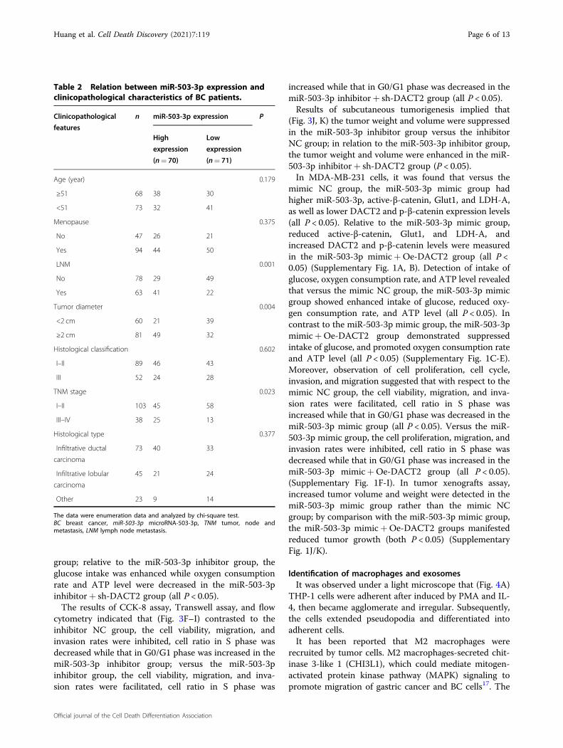
expression groups based on the median of miR-503-3prelative expression, and then the relation between miR-503-3p expression and clinicopathological characteristicsof BC patients was analyzed. We found that (Table 2)
patients with larger tumor, lymph node metastasis (LNM),and advanced TNM stage had an increased ratio of highexpression of miR-503-3p, indicating that miR-503-3pexpression was related to tumor diameter, LNM, andTNM (all P < 0.05), while not to age, menopause andhistologic type (all P > 0.05).
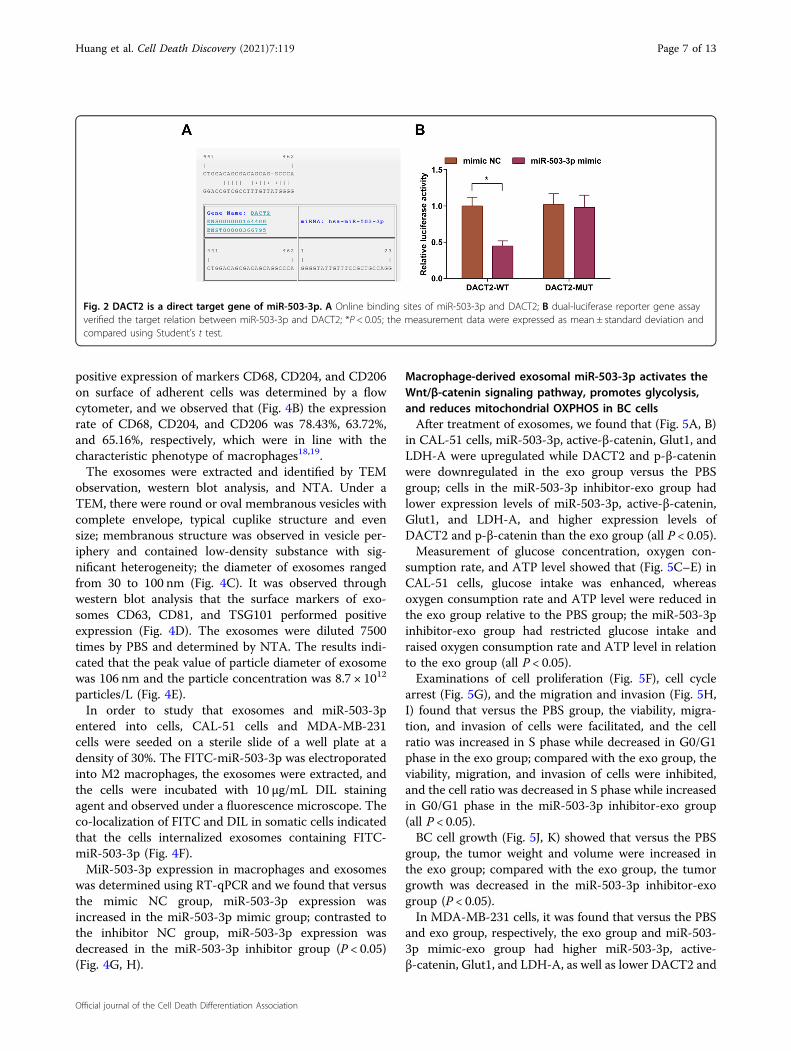
DACT2 is a direct target of miR-503-3pA target relation between miR-503-3p and DACT2 was
predicted by a bioinformatic software (Fig. 2A). Outcomesof dual-luciferase reporter gene assay reflected that (Fig.2B) cells that had been co-transfected with DACT2-WTand miR-503-3p mimic had a decreased luciferase activity(P < 0.05); the co-transfection of DACT2-MUT and miR-503-3p mimic did not influence the luciferase activity (P >0.05), indicating that DACT2 was a direct target of miR-503-3p.
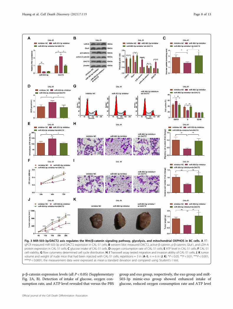
MiR-503-3p/DACT2 axis regulates the Wnt/β-cateninsignaling pathway, glycolysis, and mitochondrial oxidativephosphorylation (OXPHOS) in BC cellsExpression of miR-503-3p, DACT2, active-β-catenin,
p-β-catenin, Glut1, and LDH-A in CAL-51 cells wasevaluated. We observed that (Fig. 3A, B) relative to theinhibitor NC group, the miR-503-3p inhibitor groupshowed lower expression levels of miR-503-3p, active-β-catenin, Glut1, and LDH-A, and higher expressionlevels of DACT2 and p-β-catenin (all P < 0.05); relative tothe miR-503-3p inhibitor group, DACT2 and p-β-cateninwere downregulated, but active-β-catenin, Glut1, andLDH-A were upregulated in the miR-503-3p inhibitor+sh-DACT2 group (all P < 0.05).We detected the intake of glucose (Fig. 3C), oxygen
consumption rate (Fig. 3D), and ATP level (Fig. 3E), andnoticed that versus the inhibitor NC group, the glucoseintake was repressed while oxygen consumption rate andATP level were increased in the miR-503-3p inhibitor
Fig. 1 MiR-503-3p is upregulated while DACT2 is downregulated in BC tissues and cell lines. A, B RT-qPCR measured miR-503-3p and DACT2mRNA expression in clinical samples, n= 141; C RT-qPCR measured miR-503-3p and DACT2 expression in BC cell lines and MCF-10A cells, repetitions= 3; *P < 0.05; **P < 0.01; ***P < 0.001; miR-503-3p and DACT2 expression in clinical samples were detected using paired double-sample Student’st test and data of groups were compared using Student’s t test.
Huang et al. Cell Death Discovery (2021) 7:119 Page 5 of 13
Official journal of the Cell Death Differentiation Association
group; relative to the miR-503-3p inhibitor group, theglucose intake was enhanced while oxygen consumptionrate and ATP level were decreased in the miR-503-3pinhibitor+ sh-DACT2 group (all P < 0.05).The results of CCK-8 assay, Transwell assay, and flow
cytometry indicated that (Fig. 3F–I) contrasted to theinhibitor NC group, the cell viability, migration, andinvasion rates were inhibited, cell ratio in S phase wasdecreased while that in G0/G1 phase was increased in themiR-503-3p inhibitor group; versus the miR-503-3pinhibitor group, the cell viability, migration, and inva-sion rates were facilitated, cell ratio in S phase was
increased while that in G0/G1 phase was decreased in themiR-503-3p inhibitor+ sh-DACT2 group (all P < 0.05).Results of subcutaneous tumorigenesis implied that
(Fig. 3J, K) the tumor weight and volume were suppressedin the miR-503-3p inhibitor group versus the inhibitorNC group; in relation to the miR-503-3p inhibitor group,the tumor weight and volume were enhanced in the miR-503-3p inhibitor+ sh-DACT2 group (P < 0.05).In MDA-MB-231 cells, it was found that versus the
mimic NC group, the miR-503-3p mimic group hadhigher miR-503-3p, active-β-catenin, Glut1, and LDH-A,as well as lower DACT2 and p-β-catenin expression levels(all P < 0.05). Relative to the miR-503-3p mimic group,reduced active-β-catenin, Glut1, and LDH-A, andincreased DACT2 and p-β-catenin levels were measuredin the miR-503-3p mimic+Oe-DACT2 group (all P <0.05) (Supplementary Fig. 1A, B). Detection of intake ofglucose, oxygen consumption rate, and ATP level revealedthat versus the mimic NC group, the miR-503-3p mimicgroup showed enhanced intake of glucose, reduced oxy-gen consumption rate, and ATP level (all P < 0.05). Incontrast to the miR-503-3p mimic group, the miR-503-3pmimic+Oe-DACT2 group demonstrated suppressedintake of glucose, and promoted oxygen consumption rateand ATP level (all P < 0.05) (Supplementary Fig. 1C-E).Moreover, observation of cell proliferation, cell cycle,invasion, and migration suggested that with respect to themimic NC group, the cell viability, migration, and inva-sion rates were facilitated, cell ratio in S phase wasincreased while that in G0/G1 phase was decreased in themiR-503-3p mimic group (all P < 0.05). Versus the miR-503-3p mimic group, the cell proliferation, migration, andinvasion rates were inhibited, cell ratio in S phase wasdecreased while that in G0/G1 phase was increased in themiR-503-3p mimic+Oe-DACT2 group (all P < 0.05).(Supplementary Fig. 1F-I). In tumor xenografts assay,increased tumor volume and weight were detected in themiR-503-3p mimic group rather than the mimic NCgroup; by comparison with the miR-503-3p mimic group,the miR-503-3p mimic+Oe-DACT2 groups manifestedreduced tumor growth (both P < 0.05) (SupplementaryFig. 1J/K).
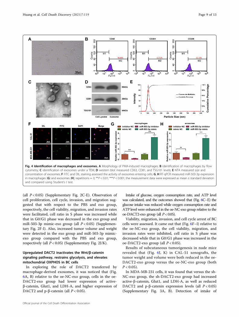
Identification of macrophages and exosomesIt was observed under a light microscope that (Fig. 4A)
THP-1 cells were adherent after induced by PMA and IL-4, then became agglomerate and irregular. Subsequently,the cells extended pseudopodia and differentiated intoadherent cells.It has been reported that M2 macrophages were
recruited by tumor cells. M2 macrophages-secreted chit-inase 3-like 1 (CHI3L1), which could mediate mitogen-activated protein kinase pathway (MAPK) signaling topromote migration of gastric cancer and BC cells17. The
Table 2 Relation between miR-503-3p expression andclinicopathological characteristics of BC patients.
Clinicopathological
features
n miR-503-3p expression P
High
expression
(n= 70)
Low
expression
(n= 71)
Age (year) 0.179
≥51 68 38 30
<51 73 32 41
Menopause 0.375
No 47 26 21
Yes 94 44 50
LNM 0.001
No 78 29 49
Yes 63 41 22
Tumor diameter 0.004
<2 cm 60 21 39
≥2 cm 81 49 32
Histological classification 0.602
I–II 89 46 43
III 52 24 28
TNM stage 0.023
I–II 103 45 58
III–IV 38 25 13
Histological type 0.377
Infiltrative ductal
carcinoma
73 40 33
Infiltrative lobular
carcinoma
45 21 24
Other 23 9 14
The data were enumeration data and analyzed by chi-square test.BC breast cancer, miR-503-3p microRNA-503-3p, TNM tumor, node andmetastasis, LNM lymph node metastasis.
Huang et al. Cell Death Discovery (2021) 7:119 Page 6 of 13
Official journal of the Cell Death Differentiation Association
positive expression of markers CD68, CD204, and CD206on surface of adherent cells was determined by a flowcytometer, and we observed that (Fig. 4B) the expressionrate of CD68, CD204, and CD206 was 78.43%, 63.72%,and 65.16%, respectively, which were in line with thecharacteristic phenotype of macrophages18,19.The exosomes were extracted and identified by TEM
observation, western blot analysis, and NTA. Under aTEM, there were round or oval membranous vesicles withcomplete envelope, typical cuplike structure and evensize; membranous structure was observed in vesicle per-iphery and contained low-density substance with sig-nificant heterogeneity; the diameter of exosomes rangedfrom 30 to 100 nm (Fig. 4C). It was observed throughwestern blot analysis that the surface markers of exo-somes CD63, CD81, and TSG101 performed positiveexpression (Fig. 4D). The exosomes were diluted 7500times by PBS and determined by NTA. The results indi-cated that the peak value of particle diameter of exosomewas 106 nm and the particle concentration was 8.7 × 10¹²
particles/L (Fig. 4E).In order to study that exosomes and miR-503-3p
entered into cells, CAL-51 cells and MDA-MB-231cells were seeded on a sterile slide of a well plate at adensity of 30%. The FITC-miR-503-3p was electroporatedinto M2 macrophages, the exosomes were extracted, andthe cells were incubated with 10 μg/mL DIL stainingagent and observed under a fluorescence microscope. Theco-localization of FITC and DIL in somatic cells indicatedthat the cells internalized exosomes containing FITC-miR-503-3p (Fig. 4F).MiR-503-3p expression in macrophages and exosomes
was determined using RT-qPCR and we found that versusthe mimic NC group, miR-503-3p expression wasincreased in the miR-503-3p mimic group; contrasted tothe inhibitor NC group, miR-503-3p expression wasdecreased in the miR-503-3p inhibitor group (P < 0.05)(Fig. 4G, H).
Macrophage-derived exosomal miR-503-3p activates theWnt/β-catenin signaling pathway, promotes glycolysis,and reduces mitochondrial OXPHOS in BC cellsAfter treatment of exosomes, we found that (Fig. 5A, B)
in CAL-51 cells, miR-503-3p, active-β-catenin, Glut1, andLDH-A were upregulated while DACT2 and p-β-cateninwere downregulated in the exo group versus the PBSgroup; cells in the miR-503-3p inhibitor-exo group hadlower expression levels of miR-503-3p, active-β-catenin,Glut1, and LDH-A, and higher expression levels ofDACT2 and p-β-catenin than the exo group (all P < 0.05).Measurement of glucose concentration, oxygen con-
sumption rate, and ATP level showed that (Fig. 5C–E) inCAL-51 cells, glucose intake was enhanced, whereasoxygen consumption rate and ATP level were reduced inthe exo group relative to the PBS group; the miR-503-3pinhibitor-exo group had restricted glucose intake andraised oxygen consumption rate and ATP level in relationto the exo group (all P < 0.05).Examinations of cell proliferation (Fig. 5F), cell cycle
arrest (Fig. 5G), and the migration and invasion (Fig. 5H,I) found that versus the PBS group, the viability, migra-tion, and invasion of cells were facilitated, and the cellratio was increased in S phase while decreased in G0/G1phase in the exo group; compared with the exo group, theviability, migration, and invasion of cells were inhibited,and the cell ratio was decreased in S phase while increasedin G0/G1 phase in the miR-503-3p inhibitor-exo group(all P < 0.05).BC cell growth (Fig. 5J, K) showed that versus the PBS
group, the tumor weight and volume were increased inthe exo group; compared with the exo group, the tumorgrowth was decreased in the miR-503-3p inhibitor-exogroup (P < 0.05).In MDA-MB-231 cells, it was found that versus the PBS
and exo group, respectively, the exo group and miR-503-3p mimic-exo group had higher miR-503-3p, active-β-catenin, Glut1, and LDH-A, as well as lower DACT2 and
Fig. 2 DACT2 is a direct target gene of miR-503-3p. A Online binding sites of miR-503-3p and DACT2; B dual-luciferase reporter gene assayverified the target relation between miR-503-3p and DACT2; *P < 0.05; the measurement data were expressed as mean ± standard deviation andcompared using Student’s t test.
Huang et al. Cell Death Discovery (2021) 7:119 Page 7 of 13
Official journal of the Cell Death Differentiation Association
p-β-catenin expression levels (all P < 0.05) (SupplementaryFig. 2A, B). Detection of intake of glucose, oxygen con-sumption rate, and ATP level revealed that versus the PBS
group and exo group, respectively, the exo group and miR-503-3p mimic-exo group showed enhanced intake ofglucose, reduced oxygen consumption rate and ATP level
Fig. 3 MiR-503-3p/DACT2 axis regulates the Wnt/β-catenin signaling pathway, glycolysis, and mitochondrial OXPHOS in BC cells. A RT-qPCR measured miR-503-3p and DACT2 expression in CAL-51 cells; B western blot measured DACT2, active-β-catenin, p-β-catenin, Glut1, and LDH-Aprotein expression in CAL-51 cells; C glucose intake of CAL-51 cells, D oxygen consumption rate of CAL-51 cells; E ATP level in CAL-51 cells; F CAL-51cell viability; G flow cytometry determined cell cycle distribution. H, I Transwell assay tested migration and invasion ability of CAL-51 cells; J, K tumorvolume and weight of nude mice that had been injected with CAL-51 cells; repetitions= 3 in (A–I), n= 6 in (J, K); *P < 0.05; **P < 0.01; ***P < 0.001;****P < 0.0001; the measurement data were expressed as mean ± standard deviation and compared using Student’s t test.
Huang et al. Cell Death Discovery (2021) 7:119 Page 8 of 13
Official journal of the Cell Death Differentiation Association
(all P < 0.05) (Supplementary Fig. 2C-E). Observation ofcell proliferation, cell cycle, invasion, and migration sug-gested that with respect to the PBS and exo group,respectively, the cell viability, migration, and invasion rateswere facilitated, cell ratio in S phase was increased whilethat in G0/G1 phase was decreased in the exo group andmiR-503-3p mimic-exo group (all P < 0.05) (Supplemen-tary Fig. 2F-I). Also, increased tumor volume and weightwere detected in the exo group and miR-503-3p mimic-exo group compared with the PBS and exo group,respectively (all P < 0.05) (Supplementary Fig. 2J/K).
Upregulated DACT2 inactivates the Wnt/β-cateninsignaling pathway, restrains glycolysis, and elevatesmitochondrial OXPHOS in BC cellsIn exploring the role of DACT2 transferred by
macrophage-derived exosomes, it was noticed that (Fig.6A, B) relative to the oe-NC-exo group, cells in the oe-DACT2-exo group had lower expression of active-β-catenin, Glut1, and LDH-A, and higher expression ofDACT2 and p-β-catenin (all P < 0.05).
Intake of glucose, oxygen consumption rate, and ATP levelwas calculated, and the outcomes showed that (Fig. 6C–E) theglucose intake was reduced while oxygen consumption rate andATP level were enhanced in the oe-NC-exo group relative to theoe-DACT2-exo group (all P<0.05).Viability, migration, invasion, and cell cycle arrest of BC
cells were assessed. It came out that (Fig. 6F–I) relative tothe oe-NC-exo group, the cell viability, migration, andinvasion rates were inhibited, cell ratio in S phase wasdecreased while that in G0/G1 phase was increased in theoe-DACT2-exo group (all P < 0.05).Results of subcutaneous tumorigenesis in nude mice
revealed that (Fig. 6J, K) in CAL-51 xenografts, thetumor weight and volume were both reduced in the oe-DACT2-exo group versus the oe-NC-exo group (bothP < 0.05).In MDA-MB-231 cells, it was found that versus the sh-
NC-exo group, the sh-DACT2-exo group had increasedactive-β-catenin, Glut1, and LDH-A, as well as reducedDACT2 and p-β-catenin expression levels (all P < 0.05)(Supplementary Fig. 3A, B). Detection of intake of
Fig. 4 Identification of macrophages and exosomes. A Morphology of PMA-induced macrophages; B identification of macrophages by flowcytometry; C identification of exosomes under a TEM; D western blot measured CD63, CD81, and TSG101 levels; E NTA measured size andconcentration of exosomes; F FITC and DIL staining assessed the activity of exosomes entering cells; G, H RT-qPCR measured miR-503-3p expressionin macrophages (G) and exosomes (H); repetitions= 3; **P < 0.01; ***P < 0.001; the measurement data were expressed as mean ± standard deviationand compared using Student’s t test.
Huang et al. Cell Death Discovery (2021) 7:119 Page 9 of 13
Official journal of the Cell Death Differentiation Association

Fig. 5 Macrophage-derived exosomal miR-503-3p activates the Wnt/β-catenin signaling pathway, promotes glycolysis, and reducesmitochondrial OXPHOS in BC cells. A RT-qPCR measured miR-503-3p and DACT2 expression in CAL-51 cells; B western blot analyzed DACT2,active-β-catenin, p-β-catenin, Glut1, and LDH-A protein expression in CAL-51 cells; C glucose intake of CAL-51 cells; D oxygen consumption rate ofCAL-51 cells; E ATP level in CAL-51 cells; F CAL-51 cell viability; G flow cytometry determined cell cycle distribution; H, I Transwell assay testedmigration and invasion ability of CAL-51 cells; J, K tumor volume and weight of nude mice that had been injected with CAL-51 cells; repetitions= 3in (A–I), n= 6 in (J, K); *P < 0.05; **P < 0.01; ***P < 0.001; data of groups were compared using one-way ANOVA, and were expressed as mean ±standard deviation.
Huang et al. Cell Death Discovery (2021) 7:119 Page 10 of 13
Official journal of the Cell Death Differentiation Association

glucose, oxygen consumption rate, and ATP level revealedthat versus the sh-NC-exo group, the sh-DACT2-exogroup showed enhanced intake of glucose, reduced oxy-gen consumption rate, and ATP level (all P < 0.05) (Sup-plementary Fig. 3C-E). Observation of cell proliferation,cell cycle, invasion, and migration suggested that withrespect to the sh-NC-exo group, the cell viability, migra-tion, and invasion rates were facilitated, cell ratio in Sphase was increased while that in G0/G1 phase wasdecreased in the sh-DACT2-exo group (all P < 0.05)(Supplementary Fig. 3F–I). In xenografted tumors, pro-moted tumor growth was observed in the sh-DACT2-exogroup compared with the sh-NC-exo (both P < 0.05)(Supplementary Fig. 3J/K).
DiscussionBC is the most common cause of cancer death in
women and is the 2nd most common cancer death indeveloped counties after lung cancer20. Exosomes, thevital mediators of intercellular communication, have beenidentified to transfer miRNAs to recipient cells21. Weperformed this research to investigate the impact ofmacrophage-derived exosomal miR-503-3p in biologicalprocesses of BC cells, and it was found that the down-regulation of exosomal miR-503-3p restrained glycolysisand promoted mitochondrial OXPHOS in BC cells viaupregulating DACT2 and inhibiting the Wnt/β-cateninsignaling pathway.
We first assessed expression of miR-503-3p, DACT2,and Wnt/β-catenin signaling pathway-related factors. Theoutcomes mirrored that miR-503-3p was highly expres-sed, DACT2 was poorly expressed and the Wnt/β-cateninsignaling pathway was activated in BC tissues and cells.The abnormal expression has been clarified in other lit-eratures. For instance, Zhao et al. have suggested thatmiR-503-3p was upregulated in BC tissues and plasma inrelation to adjacent normal breast tissues and plasmafrom healthy controls10. A former document has illu-strated that DACT2 was decreased in BC tissues12, and ithas been revealed that the abnormal activation of Wnt/β-catenin signaling pathway was able to result in manytypes of cancer, including BC22. Moreover, we discoveredthat the macrophage-derived exosomes elevated theexpression of miR-503-3p in BC cells. Similarly, Nguyenet al. have demonstrated that miR-503 was enriched inatherogenic EVs relative to the controls23. Another find-ing in our study indicated that reduced miR-503-3p andelevated DACT2 were able to inactivate the Wnt/β-catenin signaling pathway. In line with this result, it hasbeen illuminated that DACT2 inhibited the Wnt/β-catenin signaling pathway in human BC cells13. We alsoverified the target relation between miR-503-3p andDACT2 in BC. However, this relationship has not beenuncovered by now.BC cells were treated with macrophage-derived exo-
somes, upregulated/downregulated exosomal miR-503-
Fig. 6 Upregulated DACT2 inactivates the Wnt/β-catenin signaling pathway, restrains glycolysis, and elevates mitochondrial OXPHOS inCAL-51 cells. A RT-qPCR measured miR-503-3p and DACT2 expression in CAL-51 cells; B western blot analyzed DACT2, active-β-catenin, p-β-catenin,Glut1, and LDH-A protein expression in CAL-51 cells; C glucose intake of CAL-51 cells; D oxygen consumption rate of CAL-51 cells; E ATP level in CAL-51 cells; F CAL-51 cell viability; G flow cytometry determined cell cycle distribution; H, I Transwell assay tested migration and invasion ability of CAL-51 cells; J, K tumor volume and weight of nude mice that had been injected with CAL-51 cells; repetitions= 3 in (A–I), n= 6 in (J, K); *P < 0.05; **P <0.01; ***P < 0.001; the measurement data conforming to the normal distribution were expressed as mean ± standard deviation and data of groupswere compared using Student’s t test.
Huang et al. Cell Death Discovery (2021) 7:119 Page 11 of 13
Official journal of the Cell Death Differentiation Association

3p, or silenced/overexpressed DACT2 to investigate theeffects of altered miR-503-3p and DACT2 on BC devel-opment. Results of our in vitro experiments revealed thatthe macrophage-derived exosomes, elevation of exosomalmiR-503-3p, and silenced DACT2 promoted glycolysisand repressed mitochondrial OXPHOS in BC cells, andalso contributed to the malignant behaviors of BC cells.Consistently, Yuan et al. have unraveled that exosomesreleased from Tat-treated macrophages were able totransfer miR-27a to stimulate glycolysis in lung epithelialcells24. A previous research has discovered that cancer-associated fibroblasts-derived exosomes inhibited mito-chondrial OXPHOS, thereby promoting glycolysis incancer cells25. In addition, it has been unearthed thattumor-associated macrophages released exosomes topromote invasion of BC cells26. For the role of miR-503-3p, Zhao et al. have confirmed that miR-503-3p func-tioned as an oncogene of BC cell proliferation, migration,and invasion10, and it has been recently identified that theamplification of miR-503 promoted growth and inhibitedapoptosis of glioblastoma cells27. In addition, Li et al. havefound that DACT2 suppressed BC cell growth andarrested BC cells at G1/S phase13, and it has been unveiledthat the overexpression of DACT2 suppressed prolifera-tion and enhanced apoptosis of glioma cells28. Further-more, results of in vivo experiment revealed thatmacrophage-derived exosomes, elevation of exosomalmiR-503-3p, and silenced DACT2 facilitated growth ofxenografts. In accordance with this finding, oral squa-mous cell carcinoma-derived exosomes have beendemonstrated to transfer miR-29a-3p to promote tumorgrowth in nude mice with xenograft29, and it has beenilluminated that DACT2 decelerated BC cell tumorgrowth in xenograft mice13.In summary, we found that reduction of macrophage-
derived exosomal miR-503-3p repressed glycolysis andpromoted mitochondrial OXPHOS in BC cells, and con-strained malignant phenotypes of BC cells by over-expressing DACT2 and inactivating the Wnt/β-cateninsignaling pathway. This study may further the under-standing of molecular mechanisms in BC.
AcknowledgementsWe would like to acknowledge the reviewers for their helpful comments onthis paper.
Author details1The Hunan Provincial Key Lab of Precision Diagnosis and Treatment forGastrointestinal Tumor, Xiangya Hospital, Central South University, Changsha410008 Hunan, China. 2Department of Breast and Thyroid Surgery, HunanProvincial People’s Hospital (The First Affiliated Hospital of Hunan NormalUniversity), Changsha 410005 Hunan, China. 3Department of General Surgery,Hunan Provincial People’s Hospital (The First Affiliated Hospital of HunanNormal University), Changsha 410005 Hunan, China
Author contributionsZ.C. and Z.H. contributed to study design; S.H. and P.F. contributed tomanuscript editing; C.Z. and J.X. contributed to experimental studies; X.G. andS.L. contributed to data analysis.
Ethics statementWritten informed consents were acquired from all patients before this study.The protocol of this study was confirmed by the Ethic Committee of HunanProvincial People’s Hospital. Animal experiments were carried out strictly inaccordance with the Guide to the Management and Use of LaboratoryAnimals issued by the National Institutes of Health and approved by theInstitutional Animal Care and Use Committee of Hunan Provincial People’sHospital.
Conflict of interestThe authors declare no competing interests.
Publisher’s noteSpringer Nature remains neutral with regard to jurisdictional claims inpublished maps and institutional affiliations.
Supplementary information The online version contains supplementarymaterial available at https://doi.org/10.1038/s41420-021-00492-2.
Received: 7 January 2021 Revised: 30 March 2021 Accepted: 23 April 2021
References1. Thorat, M. A. & Cuzick, J. Preventing invasive breast cancer using endocrine
therapy. Breast 34, S47–S54 (2017).2. Grover, S. et al. Breast cancer and HIV in Sub-Saharan Africa: a complex
relationship. J. Glob. Oncol. 4, 1–11 (2018).3. Kolak, A. et al. Primary and secondary prevention of breast cancer. Ann. Agric.
Environ. Med. 24, 549–553 (2017).4. Geffken, K. & Spiegel, S. Sphingosine kinase 1 in breast cancer. Adv. Biol. Regul.
67, 59–65 (2018).5. Chen, S. et al. WW domain-binding protein 2: an adaptor protein clo-
sely linked to the development of breast cancer. Mol. Cancer 16, 128(2017).
6. Walker, N. D. et al. Exosomes from differentially activated macrophagesinfluence dormancy or resurgence of breast cancer cells within bone marrowstroma. Cell Death Dis. 10, 59 (2019).
7. McDonald, M. K. et al. Functional significance of macrophage-derived exo-somes in inflammation and pain. Pain 155, 1527–1539 (2014).
8. Zheng, P. et al. Exosomal transfer of tumor-associated macrophage-derivedmiR-21 confers cisplatin resistance in gastric cancer cells. J. Exp. Clin. Cancer Res.36, 53 (2017).
9. Ding, J. et al. Exosome-mediated miR-222 transferring: an insight into NF-kappaB-mediated breast cancer metastasis. Exp. Cell Res. 369, 129–138(2018).
10. Zhao, Z. et al. miR-503-3p promotes epithelial-mesenchymal transition inbreast cancer by directly targeting SMAD2 and E-cadherin. J. Genet. Genom.44, 75–84 (2017).
11. Zhang, N. et al. DACT2 modulated by TFAP2A-mediated allelic transcriptionpromotes EGFR-TKIs efficiency in advanced lung adenocarcinoma. Biochem.Pharmacol. 172, 113772 (2020).
12. Guo, L. et al. Methylation of DACT2 contributes to the progression of breastcancer through activating WNT signaling pathway. Oncol. Lett. 15, 3287–3294(2018).
13. Li, J. et al. Methylation of DACT2 promotes breast cancer development byactivating Wnt signaling. Sci. Rep. 7, 3325 (2017).
14. Yu, X. et al. Exosomes from macrophages exposed to apoptotic breast cancercells promote breast cancer proliferation and metastasis. J. Cancer 10,2892–2906 (2019).
15. Behzadi, E., Hosseini, H. M., Halabian, R. & Fooladi, A. A. I. Macrophage cell-derived exosomes/staphylococcal enterotoxin B against fibrosarcoma tumor.Microb. Pathog. 111, 132–138 (2017).
Huang et al. Cell Death Discovery (2021) 7:119 Page 12 of 13
Official journal of the Cell Death Differentiation Association
16. Chi, Y. et al. miR-516a-3p inhibits breast cancer cell growth and EMT by blockingthe Pygo2/Wnt signalling pathway. J. Cell. Mol. Med. 23, 6295–6307 (2019).
17. Chen, Y., Zhang, S., Wang, Q. & Zhang, X. Tumor-recruited M2 macrophagespromote gastric and breast cancer metastasis via M2 macrophage-secretedCHI3L1 protein. J. Hematol. Oncol. 10, 36 (2017).
18. Essafi-Benkhadir, K. et al. Quince (Cydonia oblonga Miller) peel polyphenolsmodulate LPS-induced inflammation in human THP-1-derived macrophagesthrough NF-kappaB, p38MAPK and Akt inhibition. Biochem. Biophys. Res.Commun. 418, 180–185 (2012).
19. Qin, Z. The use of THP-1 cells as a model for mimicking the function andregulation of monocytes and macrophages in the vasculature. Atherosclerosis221, 2–11 (2012).
20. Warrier, S., Tapia, G., Goltsman, D. & Beith, J. An update in breast cancerscreening and management. Women’s Health 12, 229–239 (2016).
21. Hannafon, B. N. et al. Exosome-mediated microRNA signaling from breastcancer cells is altered by the anti-angiogenesis agent docosahexaenoic acid(DHA). Mol. Cancer 14, 133 (2015).
22. Zhang, Y. Ganoderma lucidum (Reishi) suppresses proliferation and migrationof breast cancer cells via inhibiting Wnt/beta-catenin signaling. Biochem.Biophys. Res. Commun. 488, 679–684 (2017).
23. Nguyen, M. A. et al. Extracellular vesicles secreted by atherogenic macro-phages transfer MicroRNA to inhibit cell migration. Arterioscler. Thromb. Vasc.Biol. 38, 49–63 (2018).
24. Yuan, Z. et al. Macrophages exposed to HIV viral protein disrupt lung epithelialcell integrity and mitochondrial bioenergetics via exosomal microRNA shut-tling. Cell Death Dis. 10, 580 (2019).
25. Zhao, H. et al. Tumor microenvironment derived exosomes pleiotropicallymodulate cancer cell metabolism. eLife 5, e10250 (2016).
26. Yang, M. et al. Microvesicles secreted by macrophages shuttle invasion-potentiating microRNAs into breast cancer cells. Mol. Cancer 10, 117(2011).
27. Guo, P. et al. TGF-a1-induced miR-503 controls cell growth and apoptosis bytargeting PDCD4 in glioblastoma cells. Sci. Rep. 7, 11569 (2017).
28. Tan, Y. et al. Upregulation of DACT2 suppresses proliferation and enhancesapoptosis of glioma cell via inactivation of YAP signaling pathway. Cell DeathDis. 8, e2981 (2017).
29. Cai, J., Qiao, B., Gao, N., Lin, N. & He, W. Oral squamous cell carcinoma-derivedexosomes promote M2 subtype macrophage polarization mediated byexosome-enclosed miR-29a-3p. Am. J. Physiol. Cell Physiol. 316, C731–C740(2019).
Huang et al. Cell Death Discovery (2021) 7:119 Page 13 of 13
Official journal of the Cell Death Differentiation Association