exogenous nitric oxide induced early mineralization in rat
TRANSCRIPT

FULL LENGTH Iranian Biomedical Journal 23 (2): 142-152 March 2019
142 Iran. Biomed. J. 23 (2): 142-152
Exogenous Nitric Oxide Induced Early Mineralization
in Rat Bone Marrow Mesenchymal Stem Cells
via Activation of Alkaline Phosphatase
Mohammad Hussein Abnosi* and Sadieeh Pari
Department of Biology, Faculty of Sciences, Markazi, Iran
Received 30 April 2018; revised 15 July 2018; accepted 28 July 2018
ABSTRACT Background: Since the low concentration and short-time treatment with sodium nitroprusside (SNP), a nitric oxide (NO)–donor, cause no harm to rat bone marrow mesenchymal stem cells (MSCs), we studied the impact of SNP on MSCs differentiation. Methods: MSCs were treated with 100 and 1000 µM of SNP for 1 hour in every 48 hours and after 5, 10, 15, and 21 days in osteogenic media. The viability and the level of mineralization were determined using MTT assay and alizarin red staining, respectively. Morphology of the cells was studied using fluorescent dye. Concentration of calcium and the activity of alanine transaminase (ALT), aspartate transaminase (AST), lactate dehydrogenase (LDH), and alkaline phosphatase (ALP) were evaluated by commercial kits. Results: SNP with the concentration of 1000 µM significantly reduced viability from day 5 to day 20, but 100 µM did not affect the viability until the day 15. The low concentration of SNP increased matrix deposition from day 10 and reached almost its maximum (4.40 ± 2.4) at the day 15. Also, increasing the activity of ALP (419 ± 2.2), due to low concentration of SNP, started at day 10 and continued till the day 20, while LDH (2026 ± 11) and AST (25.6 ± 0.4) elevations were observed from day 5 onwards. In case of ALT, we observed a significant decrease (36%) from day 5 till day 20. Conclusion: Based on our findings, low concentrations of SNP might be useful in the promotion of bone repair. DOI: 10.29252/ibj.23.2.142
Keywords: Alkaline phosphatase, Mesenchymal stem cells, Nitroprusside, Osteoblasts
Corresponding Author: Mohammad Hussein Abnosi
Department of Biology, Faculty of Sciences, Arak University, Markazi, Iran; Mobile: (+98-918) 9630568; E-mail: [email protected]
INTRODUCTION
itric oxide (NO), in the cells, is produced by
NO synthase (NOS) and has cell signaling
activity[1]
. In 1772, Joseph Priestley discovered
NO and called it ‘nitrous air’, but till 1987 this
molecule was considered as an air pollutant. Later, it
was shown to be formed in the animal body by NOS
and was determined to act as a vasodilator, which is
used to treat various cardiovascular diseases[2]
. NOS
has three isoenzymes called as neuronal form (type 1;
nNOS), endothelial form (type 3; eNOS) and inducible
form (type 2; iNOS)[3]
, which produce NO in different
cells.
NO has the short half-life of 5-10 seconds; therefore,
its action is confined to the adjacent area. When
entering the cell, NO targets the guanylate cyclase[4]
and increases the formation of cAMP that finally
governs its action. In bone-related cells, such as
mesenchymal stem cells (MSCs), osteoblasts, and
osteocytes, two isoforms of NO synthase (eNOS and
iNOS) are expressed, whereas in osteoclasts, only
nNOS is produced. In osteoblast, the NO acts as a
double agent that regulates cell survival as well as cell
death. At low concentration, NO is associated with cell
proliferation, while high concentrations of NO cause
apoptosis, which can be related to different
pathological conditions[5,6]
.
As quoted before[5,6]
, NO, depending on its
concentration, modulates the activity of both
N
[ D
OI:
10.
2925
2/ib
j.23.
2.14
2 ]
[
Dow
nloa
ded
from
ibj.p
aste
ur.a
c.ir
on
2021
-12-
18 ]
1 / 11

Abnosi & Pari SNP Induces rapid Osteogenic Differentiation
Iran. Biomed. J. 23 (2): 142-152 143
osteoblasts and osteoclasts in vitro. Besides, it has an
anabolic effect on bone tissue and stimulates
mineralization. Production of the large amounts of NO
by the cells inhibits osteoblast proliferation and causes
bone resorption mediated by osteoclasts[7]
. Irrespective
of production in the cells, NO may be generated via
compounds such as nitroglycerine and sodium
nitroprusside (SNP). SNP is an arterial and a venous
vasodilator used in clinical practice to lower blood
pressure. It is a water-soluble sodium salt having Fe2+
,
NO, and five cyanide anions in its structure[8]
. In
biological systems, SNP releases NO via non-
enzymatic and enzymatic processes[9]
. In the body,
SNP functions as a pro-drug, reacting with sulfhydryl
groups on erythrocytes, albumin, and other proteins to
release NO, which immediately decreases the blood
pressure in clinical situations[8]
. Repair of fractured
bone and wound healing after bone surgery requires
differentiation of bone marrow MSCs to osteoblasts[10]
.
MSCs possess two fundamental characteristics: the
ability to proliferate and the capacity to
differentiate[11,12]
.
Considerable in vitro and animal studies suggest that
MSCs have the potential of rapid bone regeneration
that can differentiate to osteoblasts in bone tissues.
Some other investigations have studied the effect of
SNP on bone marrow MSCs. Chu et al.[13]
have found
that the treatment of adult mouse bone marrow
multipotent progenitor cells with 500 to 2000 µM of
SNP for 48 hours can reduce cell proliferation
significantly. Felka et al.[10]
have shown that the
concentration of 10 and 25 μM of SNP activates
respiratory activity to some extent. However,
application of SNP at 100-500 μM reduces the
respiratory activity of MSC to approximately 80%,
whereas 1 mM or higher decreases the respiratory
activity of MSC to 30% or 20%. Recently, we have
demonstrated that the treatment of the MSCs with 100
µM of SNP for 1 hour causes no change in the
morphology and viability of these cells after 24 hours
of incubation. However, incubation of MSCs for 7 and
14 days reduced the numbers of colony. In addition,
biochemical analysis of the cells displayed an increase
in anaerobic metabolism coupled with the reduction of
alkaline phosphatase (ALP) activity, while calcium
concentration remained unchanged[14]
.
To the best of our knowledge, no work has been
published to discuss the possible effect of SNP, as an
NO-releasing agent, on the differentiation property of
MSCs to osteoblasts. It is well documented that the
osteoblasts generate NO enzymatically, showing the
key role in osteoblasts activity. Therefore, in this
research work, we tried to figure out the effect of
exogenous NO on MSCs osteogenic differentiation.
MATERIALS AND METHODS
Isolation and expansion of bone marrow
mesenchymal stem cells
In this experimental study, Wistar rats (6-8 weeks
old) were purchased from Pasteur Institute of Iran
(Tehran) and kept in the animal house of Arak
University (Markazi Province) under standard
condition of light and food. The animals were
sacrificed by excessive chloroform inhalation, and then
their tibia as well as femur were removed and cleaned
from adherent soft tissue. Next, the two ends of the
bones were cut off, and bone marrow was flash out
using 2-ml DMEM (Gibco, Germany) supplemented
with 15% FBS and 1 % penicillin-streptomycin (10
ml/L; both from Gibco, Germany). Bone marrow
content was centrifuged at 2500 rpm for 5 minute and
re-suspended in 5-ml DMEM, then plated in culture
flasks and incubated at 37 °C in an atmosphere of 5%
CO2. After 24 hours, the old medium was replaced by
fresh medium. The medium exchange was repeated
two times a week till the bottom of the flask was
covered with the cells (i.e. till confluency). The cells
were trepsinized (trypsin-EDTA, Gibco, Germany) and
passed to another culture flask as the first passage, and
then the cultures were expanded through two additional
subcultures at which the cells were used for further
investigation[15]
. All the procedures were approved by
the Experimental Animal Ethics Committee of the
Arak University (No. P/97/2S3016).
Osteogenic induction
Mineralization was induced in a confluent monolayer
of cells by the addition of DMEM containing 15%
(v/v) FBS, 1 % (v/v) streptomycin-penicillin (10 ml/L),
and osteogenic supplements (1 mM sodium
glycerophosphate, 50 μg/ml L-ascorbate, and 10-8
M
dexamethasone). All the chemicals were purchased
from Sigma-Aldrich Company (USA) unless
mentioned elsewhere. The culture flasks were then
incubated at 37 °C with 5% CO2, and their medium
was changed every three days for 21 days[15]
.
Exposure to sodium nitroprusside
SNP (Merck, Germany) was used to make 100 and
1000 µM concentration of SNP in the culture media,
and the pH of the solution was adjusted to 7.3. The
treatment with SNP was carried out for one hour in
every 48 hours, and the cells were cultured in media
without SNP for 5, 10, 15 and 21 days in the presence
of osteogenic supplements. The same procedure was
carried out with the control group where no SNP
treatment was done.
[ D
OI:
10.
2925
2/ib
j.23.
2.14
2 ]
[
Dow
nloa
ded
from
ibj.p
aste
ur.a
c.ir
on
2021
-12-
18 ]
2 / 11
SNP Induces rapid Osteogenic Differentiation Abnosi & Pari
144 Iran. Biomed. J. 23 (2): 142-152
Cell viability assays The viability test on control and treated cells was
carried out using MTT (4,5dimethylthiazol-2-yl)-2,5-
diphenyltetrazolium bromide) assay. In live cells, after
4 h of incubation, mitochondrial succinate dehydro-
genase converts yellow color tetrazolium into violet
crystal of formazan. At room temperature, 100 μL of
DMSO was added to each well of the plate, and
formazan crystals were extracted following 30 minute
of incubation. The solution was transferred into
another well, and the absorbance was measured at 505
nm using an automated microplate reader (SCO
Diagnostic, Germany)[15]
.
Analysis of morphological changes Following SNP treatment, the cells were cultured in
an osteogenic medium for 5, 10, 15, and 21 days. To
study the nuclear morphology, the cells were stained
for 5 minutes in a solution of 1 µg/ml of Hoechst
33342 in the dark at room temperature and then
examined under an inverted fluorescence microscope
(Olympus, IX70, Japan). The diameter of the cells was
measured in μm with the help of Motic Image software
(Micro optical group company version 1.2). Hoechst is
a fluorescent dye that penetrates the cells through the
intact plasma membrane and stains the DNA where the
changes in nuclear morphology such as chromatin
condensation and fragmentation can be investigated[16]
.
In addition, using acridine orange, as a fluorescent dye,
the morphology of the cell cytoplasm was investigated.
After staining, the cells were washed twice with PBS,
examined and photographed using an inverted
fluorescence microscope (Olympus, IX70) equipped
with a camera.
Detection and quantification of mineralization Treated and control cells were cultured in 6-well
plates for 5, 10, 15, and 21 days, then were washed
with PBS and fixed in 10% (v/v) formaldehyde at room
temperature for 15 minute. During the staining
procedure, the cells were washed twice with excess of
dH2O, and subsequently, 1 mL of 40 mM alizarin red
solution (pH 4.1) was added per well. The plates were
incubated at room temperature for 20 minute with
gentle shaking, and excess of dye was discarded. The
plates were washed four times with dH2O, and
stained cells were investigated under a light-inverted
microscope and photographed. To quantify the level of
absorbed alizarin red, 800 μL of 10% acetic acid (v/v)
was added to each well, and the plate was incubated at
room temperature for 30 minute with gentle shaking.
Afterwards, the loosely attached cells were scraped
from the plate with a cell scraper and transferred to a
1.5-mL micro-centrifuge tube. After vortexing for 30
second, the slurry was overlaid with 500 μL mineral
oil, heated at 85 °C for 10 minute and kept on ice for 5
minute. The slurry was centrifuged at 12870 g for 15
minutes, 500 μL of the supernatant was transferred to a
new microcentrifuge tube, and 200 μL of 10%
ammonium hydroxide (v/v) was added to neutralize the
acid. An aliquot of the supernatant (100 μL) was read
in triplicate at 405 nm in a microplate reader (SCO
Diagnostic, Germany) and quantified against standard
graph. To prepare alizarin red standards graph,
working alizarin red solution (40 mM) was diluted 20
times with a mixture of 5:2 of 10% acetic acid and
10% ammonium to give a concentration of 2000 μM.
Different standard solutions of 2000 to 31.3 μM were
prepared, and the absorbancess were recorded at 450
nm using a microplate reader. The concentration of the
unknown samples was calculated using the linear
formula Y = 0.099X + 0.101 with R2
= 0.997, where Y
is the absorbance, and X is the concentration (mM) of
alizarin red[16]
.
Preparation of cell extract Control and osteogenic-treated cells were washed
with Tris-HCl buffer. The loosely attached cells were
scraped off the plate with the cell scraper and grinded
in liquid nitrogen, and the cell content was then
extracted with Tris-HCl buffer, followed by
centrifugation at 12870 g for 10 minute. The total
protein content of each sample was determined by
Lowry method[17]
using bovine serum albumin as
standard. Standard graph was plotted, and the
concentration of the unknown protein samples was
calculated using the linear formula Y=0.0021X +
0.0271 with R2 = 0.994, where Y is the absorbance,
and X is the concentration (μg) of the protein in each
sample.
Determination of alkaline phosphatase activity ALP activity of protein lysate was determined by a
commercially available kit (Parsazmon, Iran). In brief,
to determine the activity of enzyme, 20 µl of the
sample was mixed with 1000 µl of the 1st reagent
(diethanolamine [1.0 mol/l; pH 9.8] and magnesium
chloride [0.5 mmol/l]). Subsequently, the tubes were
shaken for 10 second and incubated for 1 minute and
then the 2nd
reagent (p-nitophenylphosphage [10
mmol/l]) was added. Finally, the absorbance was
measured after 1 minute at 405 nm using a
spectrophotometer (T80 + PG instrument Ltd.,
England).
Determination of transaminases and lactate
dehydrogenaseactivity Alanine transaminase (regent 1: 100 mmol/l of Tris
[pH 7.5], 500 mmol/l of L-alanine, ≥1200 U/l of
lactate dehydrogenase and reagent 2: 2-oxogutarate
[ D
OI:
10.
2925
2/ib
j.23.
2.14
2 ]
[
Dow
nloa
ded
from
ibj.p
aste
ur.a
c.ir
on
2021
-12-
18 ]
3 / 11

Abnosi & Pari SNP Induces rapid Osteogenic Differentiation
Iran. Biomed. J. 23 (2): 142-152 145
Fig. 1. The cell viability of BMSCs after 5, 10, 15, and 21
days of treatment with 0, 100, and 1000 µM of SNP in
osteogenic media, based on MTT assay. Values are means ± SD.
The asterisks (*) and (**) represent the level of significant in
each day compared to the control (p < 0.05 and p < 0.001,
respectively; ANOVA, Tukey's test).
[15 mmol/l] and NADH [0.18 mmol/l]), aspartate
transaminase (reagent 1: Tris [80 mmol/l; pH 7.8], L-
aspartate [240 mmol/l], malat dehydrogenase [≥600
U/l] and lactate dehydrogenase (≥600 U/l) and reagent
2: 2-oxoglutarate [12 mmol/l] and NADH [0.18
mmol/l]), and lactate dehydrogenase (regent 1:
phosphate buffer [50 mmol/l ; pH 7.5], pyruvate [0.6
mmol/l] and reagent 2: Good’s buffer (pH 9.6), NADH
0.18 mmol/l) activities were determined (in protein
lysate of control and treated samples based on an equal
amount of protein in each one) according to a
commercial kit instruction (Parsazmon, Iran). In brief,
for determination of transaminase and lactate
dehydrogenase activity, 100 and 10 µl of the sample,
respectively were mixed with 1000 µl of the 1st reagent
for 1 minutes. Then 250 µl of 2nd
reagent was added to
the tubes, and absorbance was measured for 1 minute
at 340 nm using a spectrophotometer (T80, PG
Instrument Ltd., England).
Calcium concentration Both control and treated cells were washed twice
with PBS and then were incubated for 24 hours with 50
μl of 0.5 N HCl to dissolve the calcium content. The
amount of calcium was determined using the
commercial kit (Parsazmon, Iran). In detail, to
determine the concentration of calcium, 10 µl of the
sample was mixed with 1000 µl of the reagent
(phosphate buffer [pH 7.5; 50 mmol/l], 8-
hydroxyquinoline-5-sulfonic acid [5 mol/l], and
arsenazoIII [120 µmol/l]). The tubes were then shaken,
and absorbance of the developed color was measured at
575 nm using a spectrophotometer (T80, PG
Instrument Ltd., England). Using different
concentrations of calcium chloride, a standard curve
was prepared, and the concentration of unknown
samples was calculated using the linear formula as
follows: Y = 0.0763X-0.0039 with R2
= 0.998, where
Y is the absorbance, and X is the concentration (mg/dl)
of calcium.
Statistical analysis Statistical evaluation of the data was performed
using one-way analysis of variance (ANOVA) and
Tukey's test, with the help of SPSS (version 16).
Results were shown as mean ± SD, and p < 0.05 was
accepted statistically as the minimum level of
statistical significance.
RESULTS
Effect of SNP on cell viability MTT assay showed that the 100 µM of SNP did not
change the viability at 5 and 10 days of treatment, but
at 15 and 21 days, it significantly decreased (p < 0.05)
the bone marrow MSC viability under osteogenic
differentiation. On the other hand, the 1000 µM of SNP
caused a highly significant (p < 0.001) reduction in
viability on days 5, 10, 15, and 21, as compared with
the control (Fig. 1).
Morphological changes in differentiated MSCs Morphological study of the nuclei in differentiated
MSCs after 5, 10, 15 and 21 days of treatment with
1000 µM of SNP revealed chromatin condensation and
breakage (Fig. 1), as well as significant reduction (p <
Table 1. Mean nuclear diameter of mesenchymal stem cells after 5, 10, 15, and 21 days of treatment with different doses of SNP
Doses (µM)
Days Nucleus diameter (µm)
5 10 15 21
0 11.70a ± 0.42 11.03a ± 0.17 9.73a ± 0.23 6.58a ± 0.27
100 11.78a ± 0.25 10.64a ± 0.18 9.62a ± 0.23 7.02a ± 0.33
1000 6.53b ± 0.13 6.03b ± 0.16 6.02b ± 0.18 3.7b ± 0.19
Values are means ± SD. Means with the same letter do not differ significantly from each other in a
column (ANOVA, Tukey's test, p > 0.05).
0
0.1
0.2
0.3
0.4
0.5
0.6
0.7
0.8
5 10 15 21
Ab
sorb
tion
(5
05
nm
)
Incubtion period (day)
0
100
1000
c **
** ** **
* *
[ D
OI:
10.
2925
2/ib
j.23.
2.14
2 ]
[
Dow
nloa
ded
from
ibj.p
aste
ur.a
c.ir
on
2021
-12-
18 ]
4 / 11

SNP Induces rapid Osteogenic Differentiation Abnosi & Pari
146 Iran. Biomed. J. 23 (2): 142-152
Table 2. Mean cytoplasm area of mesenchymal stem cells after 5, 10, 15, and 21 days of treatment with
different doses of SNP
Doses (µM)
Days Cytoplasm area (um2)
5 10 15 21
0 2768.92a ± 99.3 1903.17a ± 84.4 1951.27a ± 81.9 1015.37a ± 40.6
100 2735.63a ± 11.38 1896.08a ± 8.59 1900.78a ± 58.5 998.57a ± 68.4
1000 1573.73b ± 11.58 1581.27b ± 13.9 1617.58b ± 84.3 928.85b ± 54.0
Values are means ± SD. Means with the same letter do not differ significantly from each other in a column
(ANOVA, Tukey's test, p > 0.05).
0.05) in nuclei diameter (Table 1). It could also be
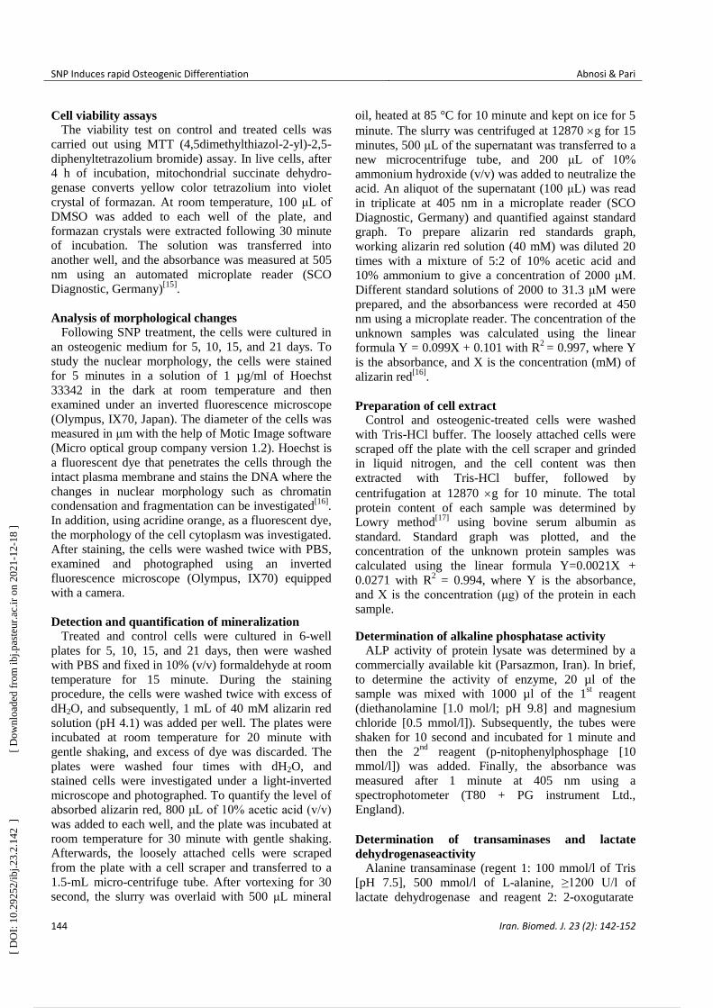
noticed that SNP at 1000 µM concentration caused
remarkable changes in the morphology of cytoplasm
such as shrinkage and complete disappearance of
cytoplasm in some cells (Table 2 and Fig. 2). The
concentration of 100 µM of SNP after 5, 10, 15, and 21
days induced no significant changes in the morphology
of cytoplasm and nuclei (Figs. 2 and 3) as well as
nuclei diameter and cytoplasm area (Tables 1 and 2)
compared to the control. On the other hand, with
passing time from 5 to 21 days of osteogenic
incubation, the control showed morphological changes
from mesenchymal lineage to osteoblast-differentiated
cells. As the size of the nuclei and cytoplasm reduced,
the cells became round, and the nuclei was centric.
Mineralization analysis based on alizarin red and
calcium content
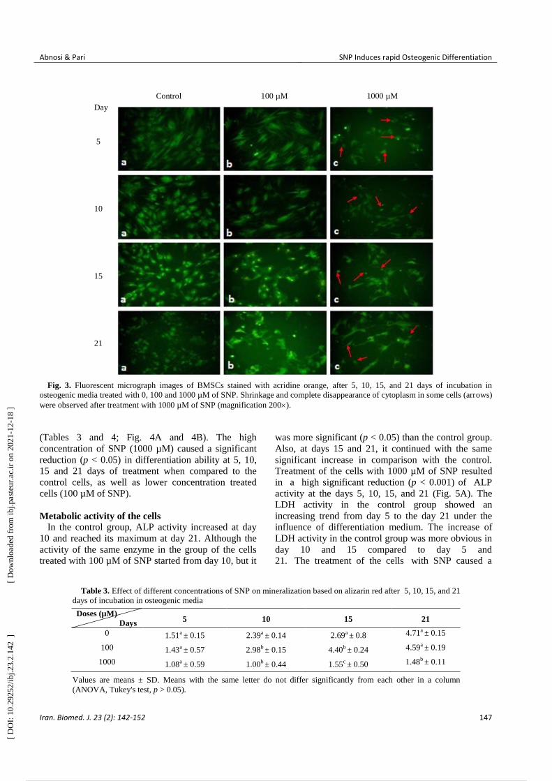
Data analysis showed that the mineralization of
MSCs in the control group started at day 10, but the
presence of 100 µM concentration of SNP significantly
increased (p < 0.05) the mineralization based on
alizarin red and calcium deposition (Tables 3 and 4).
Microscopic picture and camera photograph of alizarin
red confirmed the qualitative analysis when compared
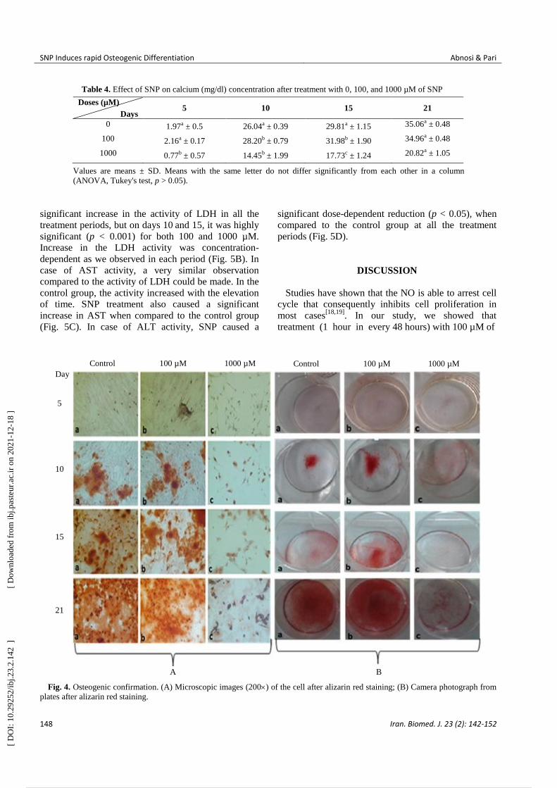
to the control (Fig. 4). In the control group,
mineralization in the absence of SNP mainly started at
day 10 and reached its maximum at day 21; however,
the presence of 100 µM SNP made a highly significant
difference (p < 0.001) on day 15, as compared to the
control, but at day 21, it did not show any difference
Fig. 2. Fluorescent micrograph images of BMSCs stained with Hoechst, after 5, 10, 15, and 21days of incubation in osteogenic
media treated with 0, 100 and 1000 µM of SNP. Nuclear condensation and deformation (arrows) of cells were observed after treatment
with 1000 µM of SNP (magnification 200).
Control 100 µM 1000 µM Day
5
10
15
21
[ D
OI:
10.
2925
2/ib
j.23.
2.14
2 ]
[
Dow
nloa
ded
from
ibj.p
aste
ur.a
c.ir
on
2021
-12-
18 ]
5 / 11
Abnosi & Pari SNP Induces rapid Osteogenic Differentiation
Iran. Biomed. J. 23 (2): 142-152 147
Fig. 3. Fluorescent micrograph images of BMSCs stained with acridine orange, after 5, 10, 15, and 21 days of incubation in
osteogenic media treated with 0, 100 and 1000 µM of SNP. Shrinkage and complete disappearance of cytoplasm in some cells (arrows)
were observed after treatment with 1000 µM of SNP (magnification 200).
(Tables 3 and 4; Fig. 4A and 4B). The high
concentration of SNP (1000 µM) caused a significant
reduction (p < 0.05) in differentiation ability at 5, 10,
15 and 21 days of treatment when compared to the
control cells, as well as lower concentration treated
cells (100 µM of SNP).
Metabolic activity of the cells In the control group, ALP activity increased at day
10 and reached its maximum at day 21. Although the
activity of the same enzyme in the group of the cells
treated with 100 µM of SNP started from day 10, but it
was more significant (p < 0.05) than the control group.
Also, at days 15 and 21, it continued with the same
significant increase in comparison with the control.
Treatment of the cells with 1000 µM of SNP resulted
in a high significant reduction (p < 0.001) of ALP
activity at the days 5, 10, 15, and 21 (Fig. 5A). The
LDH activity in the control group showed an
increasing trend from day 5 to the day 21 under the
influence of differentiation medium. The increase of
LDH activity in the control group was more obvious in
day 10 and 15 compared to day 5 and
21. The treatment of the cells with SNP caused a
Table 3. Effect of different concentrations of SNP on mineralization based on alizarin red after 5, 10, 15, and 21
days of incubation in osteogenic media
Doses (µM)
Days 5 10 15 21
0 1.51a ± 0.15 2.39a ± 0.14 2.69a ± 0.8 4.71a ± 0.15
100 1.43a ± 0.57 2.98b ± 0.15 4.40b ± 0.24 4.59a ± 0.19
1000 1.08a ± 0.59 1.00b ± 0.44 1.55c ± 0.50 1.48b ± 0.11
Values are means ± SD. Means with the same letter do not differ significantly from each other in a column
(ANOVA, Tukey's test, p > 0.05).
Control 100 µM 1000 µM
Day
5
10
15
21
[ D
OI:
10.
2925
2/ib
j.23.
2.14
2 ]
[
Dow
nloa
ded
from
ibj.p
aste
ur.a
c.ir
on
2021
-12-
18 ]
6 / 11
SNP Induces rapid Osteogenic Differentiation Abnosi & Pari
148 Iran. Biomed. J. 23 (2): 142-152
Table 4. Effect of SNP on calcium (mg/dl) concentration after treatment with 0, 100, and 1000 µM of SNP
Doses (µM)
Days 5 10 15 21
0 1.97a ± 0.5 26.04a ± 0.39 29.81a ± 1.15 35.06a ± 0.48
100 2.16a ± 0.17 28.20b ± 0.79 31.98b ± 1.90 34.96a ± 0.48
1000 0.77b ± 0.57 14.45b ± 1.99 17.73c ± 1.24 20.82a ± 1.05
Values are means ± SD. Means with the same letter do not differ significantly from each other in a column
(ANOVA, Tukey's test, p > 0.05).
significant increase in the activity of LDH in all the
treatment periods, but on days 10 and 15, it was highly
significant (p < 0.001) for both 100 and 1000 µM.
Increase in the LDH activity was concentration-
dependent as we observed in each period (Fig. 5B). In
case of AST activity, a very similar observation
compared to the activity of LDH could be made. In the
control group, the activity increased with the elevation
of time. SNP treatment also caused a significant
increase in AST when compared to the control group
(Fig. 5C). In case of ALT activity, SNP caused a
significant dose-dependent reduction (p < 0.05), when
compared to the control group at all the treatment
periods (Fig. 5D).
DISCUSSION
Studies have shown that the NO is able to arrest cell
cycle that consequently inhibits cell proliferation in
most cases[18,19]
. In our study, we showed that
treatment (1 hour in every 48 hours) with 100 µM of
Fig. 4. Osteogenic confirmation. (A) Microscopic images (200) of the cell after alizarin red staining; (B) Camera photograph from
plates after alizarin red staining.
Control 100 µM 1000 µM Control 100 µM 1000 µM
Day
5
10
15
21
A B
[ D
OI:
10.
2925
2/ib
j.23.
2.14
2 ]
[
Dow
nloa
ded
from
ibj.p
aste
ur.a
c.ir
on
2021
-12-
18 ]
7 / 11

Abnosi & Pari SNP Induces rapid Osteogenic Differentiation
Iran. Biomed. J. 23 (2): 142-152 149
Fig. 5. Mean activity of ALP (A), LDH (B), AST (C), and ALT (D) in the cells at osteogenic media after 5, 10, 15 and 21days of
incubation and treatment with 0, 100, and 1000 µM of SNP. Values are means ± SD. The asterisks (*) and (**) represent the level of
significant in each day compared to the control at p < 0.05 and p < 0.001, respectively (ANOVA, Tukey's test).
SNP did not have any toxicity effect within 10 days,
but it was toxic and reduced cell viability in longer
time. Floryszak-Wieczorek et al.[20]
have estimated that
the half-life of SNP is 12 h in a culture system, but in
addition to NO production, SNP also releases cyanide
and iron[21]
. Since SNP with the concentration of 100
μM generates only 1.2 nM of NO[22]
and vanishes very
quickly, the presence of cyanide in the culture medium
could be the reason for viability reduction observed via
cell toxicity from day 5 with respect to 1000 µM
treatment. Evidence has shown that the cyanide
produced by SNP is concentration-dependent[23]
; thus,
our observation with higher concentration might be due
to more production of cyanide that inhibits the cell
respiration via mitochondrial dysfunction[24]
.
In this study, the differentiation medium caused
morphological changes in the control cells, which were
marked by reduction in the cytoplasm area and nuclei
diameter. The changes in cytoplasm started at day 10,
but nuclear diameter reduction started at day 15 and
continued till day 21. SNP at 100 µM concentration did
not bring about differences in the morphology and its
developmental phenomena when compared with the
control group. However, higher concentration (1000
µM) caused the shrinkage of cytoplasm and nuclear
condensation, which all together are considered to be a
sign of apoptosis[25]
. It is well documented that the
high concentration, but not low concentration, of NO
induces apoptosis[26-28]
, which at physiological level is
considered as a signaling molecule[29]
. Researchers
have also mentioned many drastic effects such as
viability reduction, morphological changes, and
formation of cell debris when periodontal ligament
fibroblasts is treated with different concentrations (1 to
4 mM) of SNP for 16 hours[30]
, which is in agreement
with our findings at high concentration. Irrespective of
cell toxicity and mortality caused by the high
concentration of SNP, which mainly might be due to
cyanide toxicity, Huitema et al.[31]
have found that the
treatment of ATDCS cell line with low concentration
(100 μM) of SNP for 24 hours causes the inhibition of
cell mineralization. In addition to cyanide, a toxic
0
100
200
300
400
500
600
700
800
5 10 15 21
Act
ivit
y o
f A
LP
Incubation period (day)
0 100 1000 (A)
0
500
1000
1500
2000
2500
3000
5 10 15 21
Act
ivit
y o
f L
DH
Incubation period (day)
0 100 1000
(B)
*
**
**
**
*
*
**
**
**
**
*
**
0
20
40
60
80
100
120
140
160
180
200
5 10 15 21
Act
ivit
y o
f A
ST
Incubation period (day)
0 100 1000 (C)
* *
*
*
* * * *
0
2
4
6
8
10
12
14
16
18
20
5 10 15 21
Act
ivit
y o
f A
LT
Incubation period (day)
0 100 1000 (D)
*
*
*
*
* *
*
*
*
*
*
[ D
OI:
10.
2925
2/ib
j.23.
2.14
2 ]
[
Dow
nloa
ded
from
ibj.p
aste
ur.a
c.ir
on
2021
-12-
18 ]
8 / 11
SNP Induces rapid Osteogenic Differentiation Abnosi & Pari
150 Iran. Biomed. J. 23 (2): 142-152
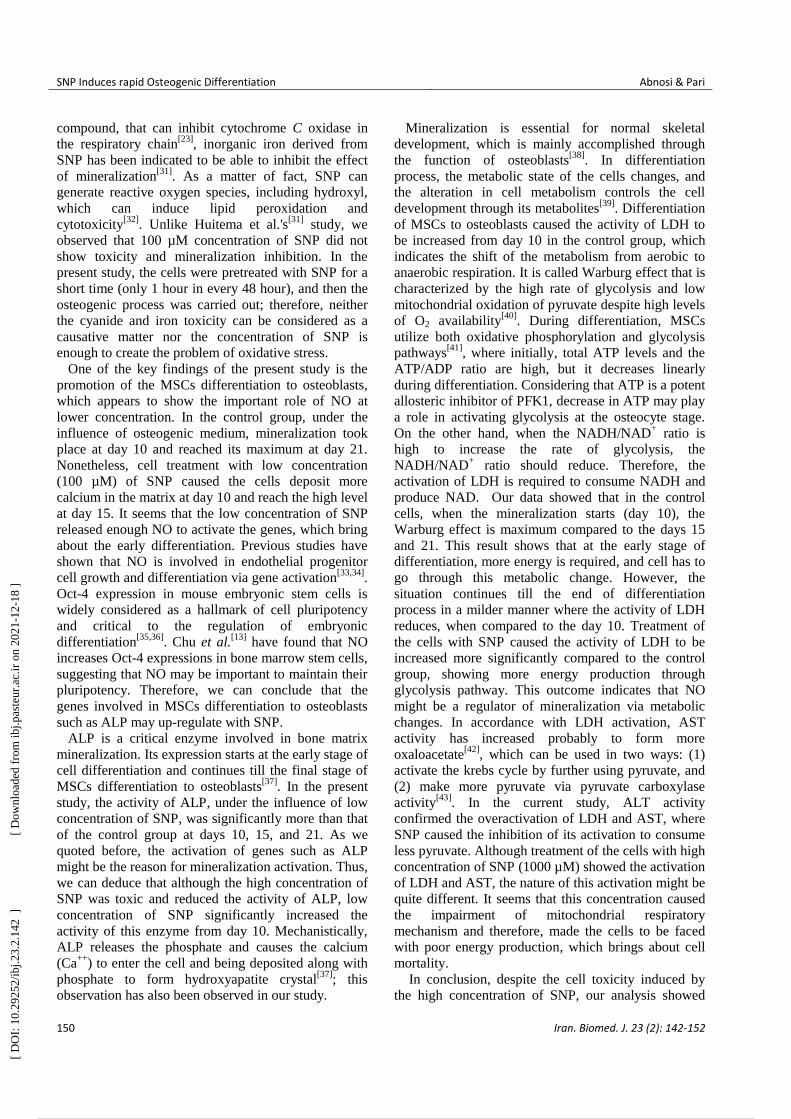
compound, that can inhibit cytochrome C oxidase in
the respiratory chain[23]
, inorganic iron derived from
SNP has been indicated to be able to inhibit the effect
of mineralization[31]
. As a matter of fact, SNP can
generate reactive oxygen species, including hydroxyl,
which can induce lipid peroxidation and
cytotoxicity[32]
. Unlike Huitema et al.'s[31]
study, we
observed that 100 µM concentration of SNP did not
show toxicity and mineralization inhibition. In the
present study, the cells were pretreated with SNP for a
short time (only 1 hour in every 48 hour), and then the
osteogenic process was carried out; therefore, neither
the cyanide and iron toxicity can be considered as a
causative matter nor the concentration of SNP is
enough to create the problem of oxidative stress.
One of the key findings of the present study is the
promotion of the MSCs differentiation to osteoblasts,
which appears to show the important role of NO at
lower concentration. In the control group, under the
influence of osteogenic medium, mineralization took
place at day 10 and reached its maximum at day 21.
Nonetheless, cell treatment with low concentration
(100 µM) of SNP caused the cells deposit more
calcium in the matrix at day 10 and reach the high level
at day 15. It seems that the low concentration of SNP
released enough NO to activate the genes, which bring
about the early differentiation. Previous studies have
shown that NO is involved in endothelial progenitor
cell growth and differentiation via gene activation[33,34]
.
Oct-4 expression in mouse embryonic stem cells is
widely considered as a hallmark of cell pluripotency
and critical to the regulation of embryonic
differentiation[35,36]
. Chu et al.[13]
have found that NO
increases Oct-4 expressions in bone marrow stem cells,
suggesting that NO may be important to maintain their
pluripotency. Therefore, we can conclude that the
genes involved in MSCs differentiation to osteoblasts
such as ALP may up-regulate with SNP.
ALP is a critical enzyme involved in bone matrix
mineralization. Its expression starts at the early stage of
cell differentiation and continues till the final stage of
MSCs differentiation to osteoblasts[37]
. In the present
study, the activity of ALP, under the influence of low
concentration of SNP, was significantly more than that
of the control group at days 10, 15, and 21. As we
quoted before, the activation of genes such as ALP
might be the reason for mineralization activation. Thus,
we can deduce that although the high concentration of
SNP was toxic and reduced the activity of ALP, low
concentration of SNP significantly increased the
activity of this enzyme from day 10. Mechanistically,
ALP releases the phosphate and causes the calcium
(Ca++
) to enter the cell and being deposited along with
phosphate to form hydroxyapatite crystal[37]
; this
observation has also been observed in our study.
Mineralization is essential for normal skeletal
development, which is mainly accomplished through
the function of osteoblasts[38]
. In differentiation
process, the metabolic state of the cells changes, and
the alteration in cell metabolism controls the cell
development through its metabolites[39]
. Differentiation
of MSCs to osteoblasts caused the activity of LDH to
be increased from day 10 in the control group, which
indicates the shift of the metabolism from aerobic to
anaerobic respiration. It is called Warburg effect that is
characterized by the high rate of glycolysis and low
mitochondrial oxidation of pyruvate despite high levels
of O2 availability[40]
. During differentiation, MSCs
utilize both oxidative phosphorylation and glycolysis
pathways[41]
, where initially, total ATP levels and the
ATP/ADP ratio are high, but it decreases linearly
during differentiation. Considering that ATP is a potent
allosteric inhibitor of PFK1, decrease in ATP may play
a role in activating glycolysis at the osteocyte stage.
On the other hand, when the NADH/NAD+ ratio is
high to increase the rate of glycolysis, the
NADH/NAD+ ratio should reduce. Therefore, the
activation of LDH is required to consume NADH and
produce NAD. Our data showed that in the control
cells, when the mineralization starts (day 10), the
Warburg effect is maximum compared to the days 15
and 21. This result shows that at the early stage of
differentiation, more energy is required, and cell has to
go through this metabolic change. However, the
situation continues till the end of differentiation
process in a milder manner where the activity of LDH
reduces, when compared to the day 10. Treatment of
the cells with SNP caused the activity of LDH to be
increased more significantly compared to the control
group, showing more energy production through
glycolysis pathway. This outcome indicates that NO
might be a regulator of mineralization via metabolic
changes. In accordance with LDH activation, AST
activity has increased probably to form more
oxaloacetate[42]
, which can be used in two ways: (1)
activate the krebs cycle by further using pyruvate, and
(2) make more pyruvate via pyruvate carboxylase
activity[43]
. In the current study, ALT activity
confirmed the overactivation of LDH and AST, where
SNP caused the inhibition of its activation to consume
less pyruvate. Although treatment of the cells with high
concentration of SNP (1000 µM) showed the activation
of LDH and AST, the nature of this activation might be
quite different. It seems that this concentration caused
the impairment of mitochondrial respiratory
mechanism and therefore, made the cells to be faced
with poor energy production, which brings about cell
mortality.
In conclusion, despite the cell toxicity induced by
the high concentration of SNP, our analysis showed
[ D
OI:
10.
2925
2/ib
j.23.
2.14
2 ]
[
Dow
nloa
ded
from
ibj.p
aste
ur.a
c.ir
on
2021
-12-
18 ]
9 / 11
Abnosi & Pari SNP Induces rapid Osteogenic Differentiation
Iran. Biomed. J. 23 (2): 142-152 151
that the SNP at low concentration not only does not
show any adverse effect but also reduces the
differentiation time of MSCs to osteoblasts and
increases matrix mineralization via the activation of
ALP activity and metabolic changes.
ACKNOWLEDGMENTS
The authors appreciate the financial support
approved by Arak University (Markazi, Iran), which
enabled us to carry out this research work.
CONFLICT OF INTEREST. None declared.
REFERENCES
1. Förstermann U, Sessa WC. Nitric oxide synthases:
regulation and function. European heart journal 2012;
33(7): 829-837.
2. Omer N, Rohilla A, Rohilla S, Kushnoor A. Nitric
oxide: role in human biology. International journal of
pharmaceutical sciences and drug research 2012; 4(2):
105-109.
3. Wimalawansa SJ. Nitric oxide: novel therapy for
osteoporosis. Expert opinion on pharmacotherapy 2008;
9(17): 3025-3044.
4. Morgan L. Nitric oxide: a challenge to chiropractic. The
Journal of the Canadian chiropractic association 2000;
44(1): 40-48.
5. Ho WP, Chen TL, Chiu WT, Tai YT, Chen RM. Nitric
oxide induces osteoblast apoptosis through a
mitochondria‐dependent pathway. Annals of the New
York academy of sciences 2005; 1042(1): 460-470.
6. Saura M, Tarin C, Zaragoza C. Recent insights into the
implication of nitric oxide in osteoblast differentiation
and proliferation during bone development. The
scientific world journal 2010; 10: 624-632.
7. Klein-Nulend J, Van Oers RF, Bakker AD, Bacabac
RG. Nitric oxide signaling in mechanical adaptation of
bone. Osteoporosis international 2014; 25(5): 1427-37.
8. Hottinger DG, Beebe DS, Kozhimannil T, Prielipp RC,
Belani KG. Sodium nitroprusside in 2014: A clinical
concepts review. Journal of anaesthesiology clinical
pharmacology 2014; 30(4): 462-471.
9. Feelisch M. The use of nitric oxide donors in
pharmacological studies. Naunyn Schmiedeberg's
archives of pharmacology 1998; 358(1): 113-122.
10. Felka T, Ulrich C, Rolauffs B, Mittag F, Kluba T,
DeZwart P, Gunnar Ochs, Bonin M, Nieselt K, Hart
ML, Aicher WK. Nitric oxide activates signaling by c-
Raf, MEK, p-JNK, p38 MAPK and p53 in human
mesenchymal stromal cells and inhibits their osteogenic
differentiation by blocking expression of Runx2.
Journal of stem cell research and therapy 2014; 4: 195.
11. Baghaban Eslaminejad M, Nazarian H, Falahi F,
Taghiyar L, Daneshzadeh MT. Ex vivo expansion and
differentiation of mesenchymal stem cells from goat
bone marrow. Iranian journal of basic medical sciences
2009; 12(2): 70-79.
12. Baghaban Eslaminejad M, Talkhabi M, Zeynali B.
Effect of lithium chloride on proliferation and bone
differentiation of rat marrow-derived mesenchymal stem
cells in culture. Iranian journal of basic medical
sciences 2008; 11(3): 143-151.
13. Chu L, Jiang Y, Hao H, Xia Y, Xu J, Liu Z, Verfaillie
CM, Zweier JL, Liu Z. Nitric oxide enhances Oct-4
expression in bone marrow stem cells and promotes
endothelial differentiation. European journal of
pharmacology 2008; 591(1): 59-65.
14. Pari S, Abnosi MH, Pakyari R. Sodium nitroprusside
changed the metabolism of mesenchymal stem cells to
an anaerobic state while viability and proliferation
remained intact. Cell journal 2016; 19(1): 146-158.
15. Abnosi MH, Jafari Yazdi Z. Sodium arsenite caused
mineralization impairment in rat bone marrow
mesenchymal stem cells differentiating to osteoblasts.
Iranian journal of toxicology 2012; 6(16): 577-587.
16. Abnosi MH, Shojafar E. Biochemical and
morphological changes in bone marrow mesenchymal
stem cells induced by treatment of rats with p-
Nonylphenol. Iranian journal of basic medical sciences
2015; 18(4): 317-324.
17. Lowry OH, Rosebrough, NJ, Farr AL, Randall RJ.
Protein measurement with the Folin phenol
reagent. Journal of biological chemistry 1951; 193 (1):
265-275.
18. Kanno S, Kim PK, Sallam K, Lei J, Billiar TR, Shears
LL 2nd. Nitric oxide facilitates cardiomyogenesis in
mouse embryonic stem cells. Proceedings of the
national academy of sciences of the United States of
America 2004; 101(33): 12277-12281.
19. Villalobo A. Nitric oxide and cell proliferation. The
FEBS journal 2006; 273(11): 2329-2344.
20. Floryszak-Wieczorek J, Milczarek G, Arasimowicz M,
Ciszewski A. Do nitric oxide donors mimic endogenous
NO-related response in plants? Planta 2006; 224(6):
1363-1372.
21. Rao DR, Cederbaum AI. Generation of reactive oxygen
species by the redox cycling of nitroprusside.
Biochimica et biophysica acta (BBA)-general subjects.
1996; 1289(2): 195-202.
22. Marks GS, McLaughlin BE, Jimmo SL, Poklewska-
Koziell M, Brien JF, Nakatsu K. Time-dependent
increase in nitric oxide formation concurrent with
vasodilation induced by sodium nitroprusside, 3-
morpholinosydnonimine, and S-nitroso-N-acetyl-
penicillamine but not by glyceryl trinitrate. Drug
metabolism and disposition 1995; 23(11): 1248-52.
23. Lockwood A, Patka J, Rabinovich M, Wyatt K,
Abraham P. Sodium nitroprusside-associated cyanide
toxicity in adult patients-fact or fiction. Journal of
clinical trials 2010; 2: 133-148.
24. Chen RM, Chen TL, Chiu WT, Chang CC. Molecular
mechanism of nitric oxide‐induced osteoblast apoptosis.
Journal of orthopaedic research 2005; 23(2): 462-468.
25. Elmore S. Apoptosis: a review of programmed cell
[ D
OI:
10.
2925
2/ib
j.23.
2.14
2 ]
[
Dow
nloa
ded
from
ibj.p
aste
ur.a
c.ir
on
2021
-12-
18 ]
10 / 11

SNP Induces rapid Osteogenic Differentiation Abnosi & Pari
152 Iran. Biomed. J. 23 (2): 142-152
death. Toxicologic pathology 2007; 35(4): 495-516.
26. Heneka MT, Löschmann PA, Gleichmann M, Weller M,
Schulz JB, Wüllner U, Klockgether T. Induction of
nitric oxide synthase and nitric oxide‐mediated
apoptosis in neuronal PC12 cells after stimulation with
tumor necrosis factor‐α/lipopolysaccharide. Journal of
neurochemistry 1998; 71(1): 88-94.
27. Messmer UK, Brüne B. Nitric oxide-induced apoptosis:
p53-dependent and p53-independent signalling
pathways. Biochemical journal 1996; 319(1): 299-305.
28. Oyadomari S, Takeda K, Takiguchi M, Gotoh T,
Matsumoto M, Wada I, Akira S, Araki E, Mori M.
Nitric oxide-induced apoptosis in pancreatic β cells is
mediated by the endoplasmic reticulum stress pathway.
Proceedings of the national academy of sciences 2001;
98(19): 10845-10850.
29. Snyder CM, Shroff EH, Liu J, Chandel NS. Nitric oxide
induces cell death by regulating anti-apoptotic BCL-2
family members. PLoS one 2009; 4(9): e7059.
30. Seo T, Cha S, Woo KM, Park YS, Cho YM, Lee JS, Kin
TI. Synergic induction of human periodontal ligament
fibroblast cell death by nitric oxide and N-methyl-D-
aspartic acid receptor antagonist. Journal of periodontal
and implant science 2011; 41(1): 17-22.
31. Huitema LF, van Weeren PR, Barneveld A, van de Lest
CH, Helms JB, Vaandrager AB. Iron ions derived from
the nitric oxide donor sodium nitroprusside inhibit
mineralization. European journal of pharmacology
2006; 542(1): 48-53.
32. Rauhala P, Khaldi A, Mohanakumar KP, Chiueh CC.
Apparent role of hydroxyl radicals in oxidative brain
injury induced by sodium nitroprusside. Free radical
biology and medicine 1998; 24(7): 1065-1073.
33. Maciejewski JP, Selleri C, Sato T, Cho HJ, Keefer LK,
Nathan CF, Young NS. Nitric oxide suppression of
human hematopoiesis in vitro. Contribution to inhibitory
action of interferon-gamma and tumor necrosis factor-
alpha. Journal of clinical investigation 1995; 96(2):
1085-1092.
34. Reykdal S, Abboud C, Liesveld J. Effect of nitric oxide
production and oxygen tension on progenitor
preservation in ex vivo culture. Experimental
hematology 1999; 27(3): 441-450.
35. Niwa H, Miyazaki J, Smith AG. Quantitative expression
of Oct-3/4 defines differentiation, dedifferentiation or
self-renewal of ES cells. Nature genetics 2000; 24(4):
372-376.
36. Pesce M, Schöler HR. Oct‐4: gatekeeper in the
beginnings of mammalian development. Stem cells
2001; 19(4): 271-278.
37. Golub EE, Boesze-Battaglia K. The role of alkaline
phosphatase in mineralization. Current opinion in
orthopaedics 2007; 18(5): 444-448.
38. Gómez-Picos P, Eames BF. On the evolutionary
relationship between chondrocytes and osteoblasts.
Frontiers in genetics 2015; 6: 297.
39. Agathocleous M, Harris WA. Metabolism in
physiological cell proliferation and differentiation.
Trends in cell biology 2013; 23(10): 484-492.
40. Shyh-Chang N, Daley GQ, Cantley LC. Stem cell
metabolism in tissue development and aging.
Development 2013; 140(12): 2535-2547.
41. Guntur AR, Le PT, Farber CR, Rosen CJ. Bioenergetics
during calvarial osteoblast differentiation reflect strain
differences in bone mass. Endocrinology 2014; 155(5):
1589-1595.
42. Huang XJ, Choi YK, Im HS, Yarimaga O, Yoon E, Kim
HS. Aspartate aminotransferase (AST/GOT) and alanine
aminotransferase (ALT/GPT) detection techniques.
Sensors (Basel) 2006; 6(7): 756-782.
43. Jitrapakdee S, St Maurice M, Rayment I, Cleland WW,
Wallace JC, Attwood PV. Structure, mechanism and
regulation of pyruvate carboxylase. Biochemical journal
2008; 413(3): 369-387.
[ D
OI:
10.
2925
2/ib
j.23.
2.14
2 ]
[
Dow
nloa
ded
from
ibj.p
aste
ur.a
c.ir
on
2021
-12-
18 ]
Powered by TCPDF (www.tcpdf.org)
11 / 11