excessive luminosity fades the skin color of … · of the most commercialized ornamental...
TRANSCRIPT
Scientific ArticleISSN 1678-2305 online version
BOLETIM DO INSTITUTO DE PESCA
LINHARES et al. Bol. Inst. Pesca 2018, 44(4): e319. DOI: 10.20950/1678-2305.2018.44.4.319 1/8
EXCESSIVE LUMINOSITY FADES THE SKIN COLOR OF CARDINAL TETRA
ABSTRACTIn order to understand the morphological and physiological changes on the loss of coloration in the tegument of cardinal tetra under excessive luminosity, specimens of Paracheirodon axelrodi were conditioned to different light intensities (0, 250, 500, 1,200 and 2,700 lux) at different time intervals (0, 12, 24 and 72 hours). Types of chromatophores, dispersion of melanosomes and density of chromatophores were analyzed after the experiment. The dark stripe on the species consists of yellowish-brown (dorsally located) and darkish-brown (medially located) melanophores. In the iridescent blue stripe, darkish-brown melanophores were closely associated with iridophores. Erythrophores were found only in the red stripe. Loss of skin color was observed when cardinal tetra was exposed to intense light. The melanic and neon stripes became pale due to a reduction in melanophores densities. On the other hand, the color of the red stripe was intensified due to the proliferation of erythrophores. At low light levels (0 to 250 lux), the melanophores (with dispersed melanosomes) proliferate in the black and neon stripes resulting in a more vibrant skin color. We suggest that in nature, the paleness of the skin may represent a camouflage strategy during the hours of the day with greater luminosity in the black water of the Rio Negro. Fading the skin color can help this species to visually confuse potential predators.Key words: ornamental fish; tegument; chromatophores; light intensity.
LUMINOSIDADE EXCESSIVA REDUZ A COLORAÇÃO DA PELE DO CARDINAL TETRA
RESUMOPara compreender as mudanças morfológicas e fisiológicas da perda da coloração no tegumento do cardinal após exposição à luminosidade, exemplares de Paracheirodon axelrodi foram submetidos a diferentes intensidades luminosas (0, 250, 500, 1.200 e 2.700 lux), em diferentes intervalos de tempo (0, 12, 24 e 72 horas). Após a exposição, foram analisadas a dispersão dos melanossomos e a densidade de cromatóforos. A faixa de coloração escura do cardinal é constituída por melanóforos de coloração marrom-amarelado (dorsalmente localizados) e marrom-escuro (latero-medial). Na faixa azul iridescente, os melanóforos de cor marrom-escuro estão intimamente associados aos iridóforos. Eritróforos foram encontrados apenas na faixa vermelha. Observou-se que a perda da coloração ocorre devido à exposição pela luminosidade excessiva. As faixas melânica e neon tornam-se pálidas devido à redução na densidade dos melanóforos, enquanto a faixa vermelha intensifica-se como resultado da proliferação de eritróforos. Em baixa luminosidade (0 a 250 lux), os melanóforos (com melanossomos dispersos) proliferam-se nas faixas melânica e neon realçando ainda mais as cores vibrantes do cardinal. Nós sugerimos que na natureza a palidez pode representar um padrão de camuflagem durante as horas de intensa luminosidade nas águas pretas do rio Negro. A redução da coloração na pele pode ajudar o cardinal a confundir potenciais predadores visuais.Palavras-chave: peixe ornamental; tegumento; cromatóforos; intensidade luminosa.
INTRODUCTION
The coloration in the tegument of fish is associated with several important functions, such as camouflage, sexual selection and protection against ultraviolet radiation (ITO and WAKAMATSU, 2003; OLIVEIRA and FRANCO-BELUSSI, 2012). In general, the color of the skin is the result of the combination of chemical color by the absorption of the light rays by means of the chromophores and/or structural
Regiane Monteiro LINHARES1
Celina Maria Müller Ferreira PINAGÉ1
Wallice Paxiúba DUNCAN1
1Universidade Federal do Amazonas – UFAM, Laboratório de Morfologia Funcional, Avenida Rodrigo Otavio, 6200, Coroado I, CEP 69080-900, Manaus, AM, Brasil. E-mail: [email protected] (corresponding author).
Received: November 27, 2017 Approved: May 28, 2018
EXCESSIVE LUMINOSITY FADES…
LINHARES et al. Bol. Inst. Pesca 2018, 44(4): e319. DOI: 10.20950/1678-2305.2018.44.4.319 2/8
color due to the multiple internal reflections associated to the phenomena of optical interference (FUJII, 1993; SUGIMOTO, 2002). Chromophores are the cells responsible for skin coloration of basal vertebrates. Depending on the chemical or structural nature of the substances that generate the coloration, the chromatophores can be classified into two distinct subclasses: the pigmentary cells that absorb and those that reflect light rays. The melanophores (black/brown), erythrophores (red/orange), xanthophores (yellow) and cyanophores (blue) contain true pigments (chromophores), while leucophores and iridophores do not have biochromes and are responsible for the structural coloration of the tegument (OSHIMA, 2001; TAKAHASHI et al., 2014).
In melanophores, the melanin pigment absorbs virtually all light within the visible spectrum (KELLEY and DAVIES, 2016), while xanthophores and erythrophores contain carotenoids and pteridines, which absorb light between 467 and 477 nm (FUJII, 1993; NERY and CASTRUCCI, 1997; LIGON and MCCARTNEY, 2016). On the other hand, iridophores are cells that produce structural coloration and are located in the silver or iridescent regions of the skin of fish (FUJII, 1993; KALETA, 2009). These cells are located below the melanophores, constituting the unit of dermal chromatophores (LIGON and MCCARTNEY, 2016). The reflectivity of the iridophores is due to the presence of microstructures (lamellae) within the cell (OSHIMA and KASAI, 2002; YOSHIOKA et al., 2011). The lamellae are formed by guanine crystals, and the structural organization of these lamellae allows light to reflect between the maximum peaks of 350 to 400 nm and 500 to 600 nm (YOSHIOKA et al., 2011; LIGON and MCCARTNEY, 2016). Skin coloration is the result of a series of optical phenomena that include reflection, refraction, fluorescence and iridescence (OSHIMA and KASAI, 2002).
The color change in the tegument of the fish can occur by two distinct means: morphological and physiological changes (SUGIMOTO, 2002; GILBY et al., 2015). Physiological change occurs on a short time scale, that is, from seconds to hours, being the result of the movement of membranous vesicles (chromatosomes) containing pigments inside the melanophores, xanthophores and erythrophores (FUJII, 2000; MÄTHGER et al., 2003). In the case of iridophores, the color change is due to small alterations in the distance between the adjacent lamellae of nanostructures, such as guanine plates (AMIRI and SHAHEEN, 2012). On the other hand, the morphological change usually involves modifications in the density of the chromatophores as well as the amount of pigments inside the cytoplasm. These changes occur within days and even weeks (KELLEY and DAVIES, 2016).
It is the lateral coloration of the cardinal tegument (Paracheirodon axelrodi Schultz, 1956) that makes this Neotropical characid one of the most commercialized ornamental freshwater fish in the world. The iridescent blue strip is contrasted by the dark upper and lower red stripes that extend over the entire body of the fish. Native to the basins of the Negro and Orinoco rivers, the cardinal lives among the branches and submerged leaves (WALKER, 2004; MARSHALL et al., 2011). Like the other black water rivers, the Rio Negro is rich in dissolved organic carbon, which is constituted by pigmented organic polymers known as humic and fulvic acids (AUCOUR et al., 2002), whose hue varies from yellow-orange in
fulvic acids and dark gray in humic acids (WANG et al., 1990). Thus, the optical properties of the dissolved humic substances can contribute to create a species-specific coloring pattern for the cardinal. Therefore, the tegument coloration of this fish can be plastically adjusted to the characteristics of the habitat, such as light intensity. This can give the fish a camouflage against potential visual predators.
The ability of the animal to change color as a function of habitat choice and to become cryptic is important for the survival of both the individual and the population (STEVENS and MERILAITA, 2009). Thus, camouflage may be both necessary to confuse the visual predator (STUART-FOX et al., 2006), and in foraging tactics during prey search (ABBOTT, 2010). The change in the color of the skin can also be directly influenced by the intensity of ambient light, which varies daily and seasonally (KELLEY and DAVIES, 2016). However, if the light is kept constant on the fish, such condition may alter the color of the animal and result in stress (OLIVEIRA and FRANCO-BELUSSI, 2012). In contrast, stress can also alter the color pattern in the skin of the fish (BRINN et al., 2009). Therefore, in this work, we evaluated the physiological and morphological changes in cardinal skin color and discussed that changes in the color of the tegument can be a tactic of camouflage of this species in its natural environment.
METHODS
Fish origin and acclimatizationThe samples of cardinal tetra, Paracheirodon axelrodi
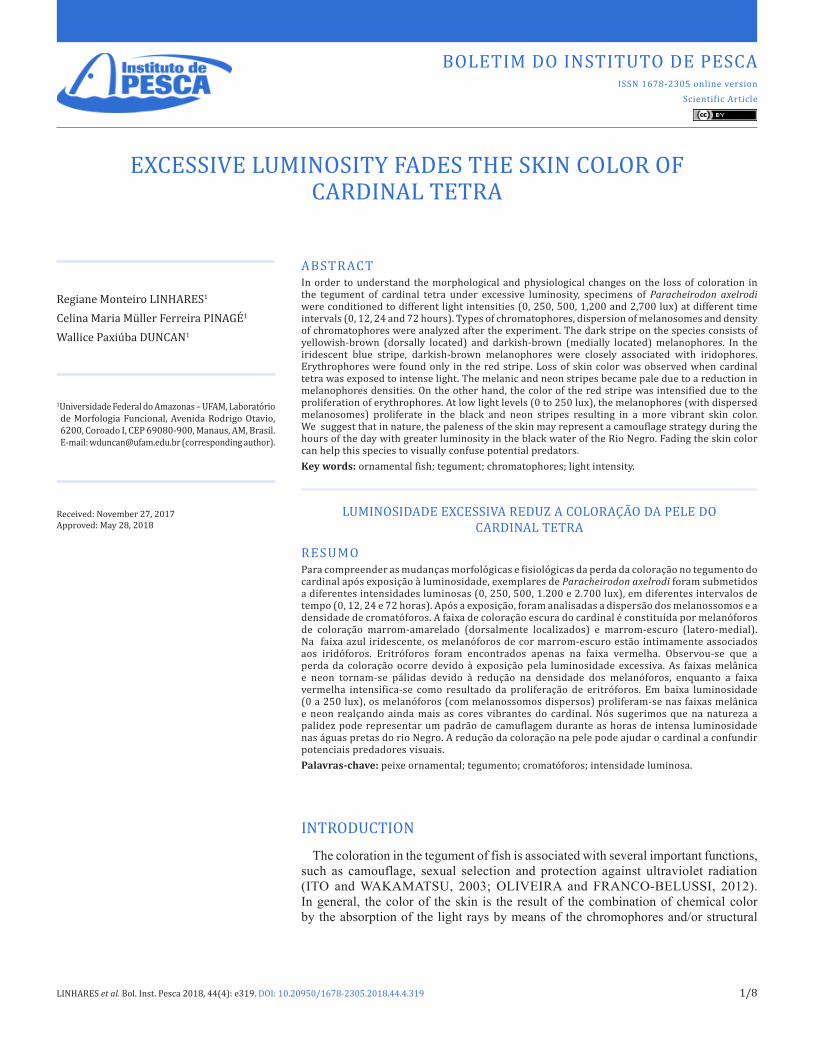
(0.075 ± 0.016 g, 1.66 ± 0.16 cm, Figure 1) from the Middle Rio Negro (0º40’S and 62º58’W) were purchased at a local aquarium store (Manaus, Amazonas). The fish were kept in 1,000 L blue plastic tanks for two months, with the following conditions: temperature (29.2 ± 1.1 °C), dissolved oxygen 5.4 ± 0.8 mg L-1, conductivity (24.5 ± 2.7 μS), pH 5.5 ± 1.3 and natural photoperiod. The brightness during the day varied from 250 to 500 lux. The animals were fed twice daily with commercial feed containing 38% crude protein (Alcon Neon, Camboriú, SC). The feeding was suspended 24 hours prior to the start of the experiments.
Figure 1. Adult specimen of Paracheirodon axelrodi (cardinal tetra). Scale = 3 mm. Photography: Wallice Paxiúba Duncan.

EXCESSIVE LUMINOSITY FADES…
LINHARES et al. Bol. Inst. Pesca 2018, 44(4): e319. DOI: 10.20950/1678-2305.2018.44.4.319 3/8
Experimental designThe evaluation of the effect of the luminosity on the
chromatophore density in the melanic, neon and red stripes was carried out by exposing the fish to five artificial lighting levels: 0, 250, 500, 1,200 and 2,700 lux. For each level of luminosity, the animals were kept in different time intervals: 0, 12, 24 and 72 hours uninterrupted. The experimental design was completely randomized. For each level of luminosity and for each time interval, 4 aquariums (four replicates) of glass were used with 300 mL of water with 2 fish per aquarium. In total, 80 glass aquariums and 160 animals were used.
The different luminous intensities were reached with LED lamps with a color temperature of 6,500 K (white-blue) luminous flux at 100 lumens. The illumination of each experimental unit was continuously monitored with the aid of a portable digital apparatus, HS1010 lux meter. The luminosity variations between the experimental units were adjusted so as to reduce the variability between the replicates. The physical and chemical characteristics of the water of the experimental units were: 4.2 ± 0.5 mg L-1 oxygen, conductivity 10.3 ± 1.2 μS.cm-1, temperature 27.4 ± 0.7 °C, pH 6.3 ± 0.8, Na+ 0.76 ± 0.11 μM, K+ 1.34 ± 0.17 μM and Ca2+ 9.60 ± 0.12 μM. The parameters of the water were analyzed using a multiparameter analyzer (CONSORT bvba), while the dissolved ions (Na+, K+ and Ca2+) were quantified by flame photometry using the Digimed DM-62 photometer. The experiments were conducted following CONCEA/MCTI regulations and approved by the Ethics Committee on Animal Use of the Federal University of Amazonas (CEUA/UFAM, No. 011/2014).
Melanophore Index (MI) as a measure of dispersion of melanosomes
The MI was used as a measure to evaluate the physiological changes in cardinal tetra tegument color. At the end of each time interval, the fish were immediately euthanized in 0.5 mg L-1 benzocaine (Sigma-Aldrich) and fresh skin samples were carefully dissected under stereomicroscope. Samples of the tegument were removed and the digital images of stripes (melanic, neon and red) were immediately captured in a stereomicroscope with a coupled video camera (Leica EZ4D). The MI was used as a measure of the rate of change of coloration of the skin. A subjective category scale proposed by HOGBEN and SLOME (1931) was used. A score of 1 (MI=1) was assigned when the melanosomes are completely aggregated and MI=5 when completely dispersed. Additionally, the score MI=6 was used for the cases of highly dispersed melanosomes, according to NILSSON (2000). Some skin tissues were fixed in Karnovsky solution (0.5% glutaraldehyde, 5% formaldehyde, 10% acetic acid) and post-fixed in ethanol 70%. The samples were embedded in methacrylate resin and cross-sections of 3 µm were stained with 0.12% toluidine blue and observed under a light microscope.
Determination of chromatophore densityThe changes in the number of chromatophores (melanophores
and erythrophores) in the stripes (melanic, neon and red) were used as indicators of morphological change in skin color. Quantification of pigment cell types was performed with the aid of ImageJ image
analysis software (www.imageJ.nih.gov/ij). The images were calibrated, and the cells quantified in a quadrant with known area in square millimeters. Quantifications were performed in 10 random quadrants in each stripe: melanic, neon and red. The density of the chromatophores was expressed as the mean number of chromatophores mm-2 in each color stripe.
Statistical analysisData are presented as mean ± standard error of the mean and tested
for normality (Kolmogorov-Smirnov test). For the parametric tests, the data were log10-transformed. To test if there was interaction between the exposure times and the different luminous intensities, a two-way analysis of variance (ANOVA) was used. To identify which groups were statistically different from each other, Tukey’s test for multiple comparisons (with Bonferroni correction) was used. The accepted level of significance was 5%.
RESULTS
Morphological organization of chromatophoresLaterally, the coloration of the cardinal tetra is characterized by
a blue (neon) medial stripe that separates a superior melanic region from a reddish lower region (Figure 2a). In the melanic stripe (dorsal side), melanophores were the only pigment cells observed (121.5 ± 6.6 chromatophores mm-2) in fish kept in low to moderate
Figure 2. (a) The typical colored stripes of the cardinal tetra: melanic (dorsal), neon (medial) and red (latero-ventral); (b) Types of melanophores with melanosomes of dark brown color (dark arrow) and yellowish brown (dark arrow head). In detail, dendritic branching full of dispersed melanosomes are observed (white arrow head); (c) Erythrophores with dispersed erythrosomes. The scales represent: (a) 2,000 μm; (b) 5 μm; and (c) 200 μm (50 μm in the erythrophore detail).
EXCESSIVE LUMINOSITY FADES…
LINHARES et al. Bol. Inst. Pesca 2018, 44(4): e319. DOI: 10.20950/1678-2305.2018.44.4.319 4/8
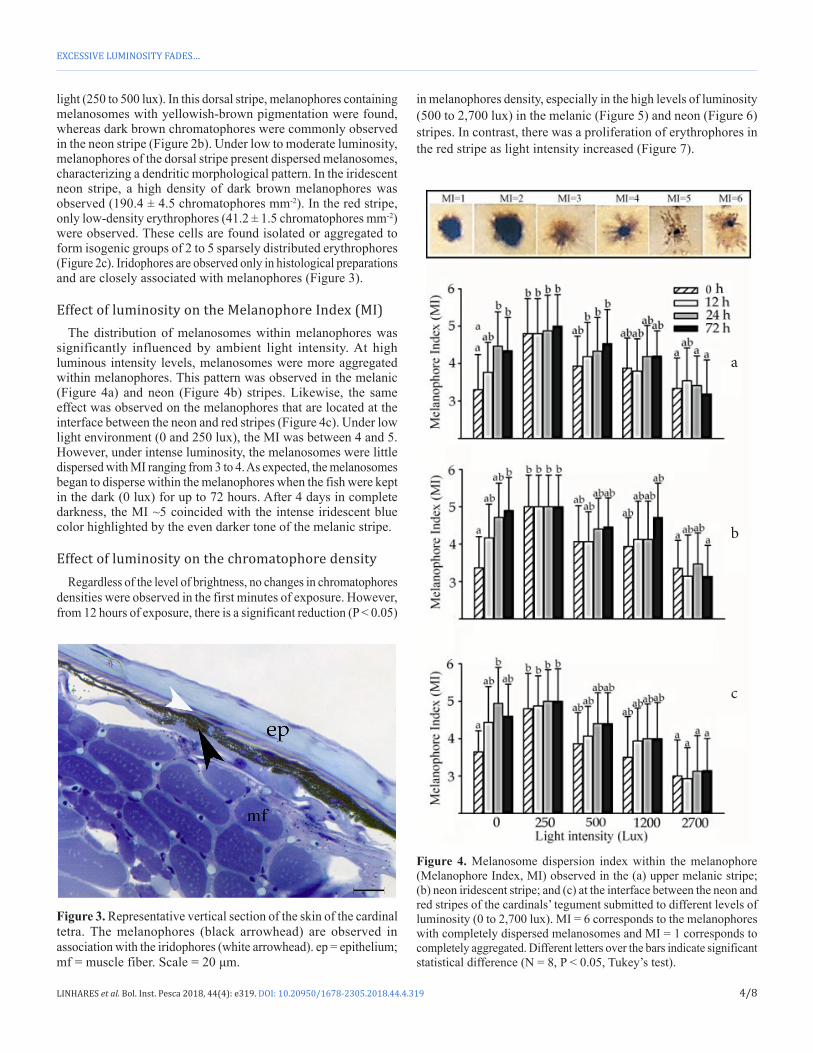
light (250 to 500 lux). In this dorsal stripe, melanophores containing melanosomes with yellowish-brown pigmentation were found, whereas dark brown chromatophores were commonly observed in the neon stripe (Figure 2b). Under low to moderate luminosity, melanophores of the dorsal stripe present dispersed melanosomes, characterizing a dendritic morphological pattern. In the iridescent neon stripe, a high density of dark brown melanophores was observed (190.4 ± 4.5 chromatophores mm-2). In the red stripe, only low-density erythrophores (41.2 ± 1.5 chromatophores mm-2) were observed. These cells are found isolated or aggregated to form isogenic groups of 2 to 5 sparsely distributed erythrophores (Figure 2c). Iridophores are observed only in histological preparations and are closely associated with melanophores (Figure 3).
Effect of luminosity on the Melanophore Index (MI)The distribution of melanosomes within melanophores was
significantly influenced by ambient light intensity. At high luminous intensity levels, melanosomes were more aggregated within melanophores. This pattern was observed in the melanic (Figure 4a) and neon (Figure 4b) stripes. Likewise, the same effect was observed on the melanophores that are located at the interface between the neon and red stripes (Figure 4c). Under low light environment (0 and 250 lux), the MI was between 4 and 5. However, under intense luminosity, the melanosomes were little dispersed with MI ranging from 3 to 4. As expected, the melanosomes began to disperse within the melanophores when the fish were kept in the dark (0 lux) for up to 72 hours. After 4 days in complete darkness, the MI ~5 coincided with the intense iridescent blue color highlighted by the even darker tone of the melanic stripe.
Effect of luminosity on the chromatophore densityRegardless of the level of brightness, no changes in chromatophores
densities were observed in the first minutes of exposure. However, from 12 hours of exposure, there is a significant reduction (P < 0.05)
in melanophores density, especially in the high levels of luminosity (500 to 2,700 lux) in the melanic (Figure 5) and neon (Figure 6) stripes. In contrast, there was a proliferation of erythrophores in the red stripe as light intensity increased (Figure 7).
Figure 3. Representative vertical section of the skin of the cardinal tetra. The melanophores (black arrowhead) are observed in association with the iridophores (white arrowhead). ep = epithelium; mf = muscle fiber. Scale = 20 μm.
Figure 4. Melanosome dispersion index within the melanophore (Melanophore Index, MI) observed in the (a) upper melanic stripe; (b) neon iridescent stripe; and (c) at the interface between the neon and red stripes of the cardinals’ tegument submitted to different levels of luminosity (0 to 2,700 lux). MI = 6 corresponds to the melanophores with completely dispersed melanosomes and MI = 1 corresponds to completely aggregated. Different letters over the bars indicate significant statistical difference (N = 8, P < 0.05, Tukey’s test).
a
b
c

EXCESSIVE LUMINOSITY FADES…
LINHARES et al. Bol. Inst. Pesca 2018, 44(4): e319. DOI: 10.20950/1678-2305.2018.44.4.319 5/8
DISCUSSION
The structural arrangement of chromatophoresIn the skin of the cardinal tetra (Paracheirodon axelrodi) the
melanophores constitute a monolayer of pigmentary cells located just below the basal layer of the stratified epithelium in the melanic (dark) stripe. As in other fish, melanophores have the dendritic form with the cytoplasm replete with melanin-containing melanosomes (FUJII, 1993; OSHIMA, 2001; TAKAHASHI et al., 2014). In the melanic stripe of cardinal tetra, yellowish-brown (dorsally localized) melanophores are visibly observed, while those of a dark brown color are found in the iridescent neon stripe. The color of the melanophores depends on the content and type of melanin present in the melanosomes (MÄTHGER et al., 2003; LIGON and MCCARTNEY, 2016; REZENDE et al., 2018). Although different types of melanin exist: neuromelanin, allomelanin, pheomelanin and eumelanin (ITO and WAKAMATSU, 2003). Pheomelanin was believed to be produced only by mammals and birds. However, it has been reported that the tortoise species, Eurotestudo cheettgeri also synthesizes pheomelanin (ROULIN et al., 2013). It has not yet been demonstrated whether fish have the ability to synthesize pheomelanin. Therefore, we suggest that the yellow-brown coloration of the melanophores found in the tetra cardinal melanic stripe is due to a variation in the amount of eumelanin. The arrangement and disposition of the dark brown melanophores as well as the iridophores form the iridescent neon stripe on the cardinal. Iridophores tend to
Figure 5. Effects of light intensity on the chromatophores densities located in the melanic stripe of cardinal tetra at different time intervals (a = 0, b = 12 h, c = 24 h, d = 72 h) and exposed to different light intensities. Different letters over the bars indicate significant statistical difference (N = 8, P < 0.05, Tukey’s test).
Figure 6. Density changes in melanophores in the neon stripe of the cardinal tetra at different time intervals (a = 0, b = 12 h, c = 24 h, d = 72 h) and exposed to different light intensities. Different letters over the bars indicate a significant statistical difference (N = 8, P < 0.05, Tukey’s test).
Figure 7. Changes in chromatophores densities located in the red stripe of the cardinal tetra at different time intervals (a = 0, b = 12 h, c = 24 h, d = 72 h) and submitted to different levels of luminosity. Different letters over the bars indicate a significant statistical difference (N = 8, P < 0.05, Tukey’s test).

EXCESSIVE LUMINOSITY FADES…
LINHARES et al. Bol. Inst. Pesca 2018, 44(4): e319. DOI: 10.20950/1678-2305.2018.44.4.319 6/8
be located in the upper layer of melanophores, but still within the dermal chromatophore unit (AMIRI and SHAHEEN, 2012). In many species, this organization is structured as: an outermost layer consisting of xanthophores and/or erythrophores, a layer of iridophores, superimposed by a third layer formed by melanophores (SIVKA et al., 2012). This same pattern of organization could be observed within the limits of the neon and red stripes in the skin of the cardinal.
Physiological change of colorCardinal tetra maintained under low to moderate light conditions
(250 to 500 lux) has melanosomes with dispersed melanosomes, with an MI index value (a measure of the degree of dispersion of the melanosomes within the melanophores) varying between 4 and 5. On the other hand, under intense light (1,200 to 2,700 lux) the melanosomes are more aggregated with an index between 3 and 4. In addition, if fish remained in the dark environment the value of MI increased. Fish with dispersed melanosomes become quite colorful (OLIVEIRA and FRANCO-BELUSSI, 2012; KELLEY and DAVIES, 2016). In fact, when the cardinal was held under such conditions for 72 hours, the coloring of the side stripes became even more intense. TAKAHASHI et al. (2014) describe two states of distribution of the melanin granules: the aggregate, due to the centripetal movement, while the dispersed is the result of the centrifugal movement. The skin color of the fish may change on a short time scale. This is due to the reflective changes of the iridophores as well as to the aggregation and dispersion patterns of the chromatosomes inside the chromatophores (NERY and CASTRUCCI, 1997). For example, small changes in the thickness of the cytoplasmic layers between the guanine plates also alter the reflection of the iridophores present in the iridescent blue strip of the neon tetra, Paracheirodon innesi (CLOTHIER and LYTHGOE, 1987). More recently, YOSHIOKA et al. (2011) proposed a model based on the “Venetian blind”, in which the angle of the guanine lamellae is controlled by filaments associated with motor proteins that move on the microtubules. With this, the fish changes from blue-green during the day to a blue-violet color at night. According to LYTHGOE and SHAND (1982), the blue-green to blue-violet transition occurs between 25 and 35 minutes. Considering the close phylogenetic relationship between cardinal and neon tetra, this same mechanism may explain the iridescent coloration of the neon stripe as well as the rapid changes in tegument color during the transition from light to dark environment and vice-versa.
Morphological change of colorThe tegument of the cardinal tetra becomes pale with increasing
light intensity. As the luminosity increases, there was a gradual reduction in the melanophores density both in the dark and neon stripes. Under intense light, the reduction of the number of melanophores allows the light to be reflected by the iridophore plates located in the stratum laxum of the cardinal tegument (MÄTHGER et al., 2003; KALETA, 2009; AMIRI and SHAHEEN, 2012). Thus, the low density of melanophores does not allow the same light to form the typical cardinal color pattern, although the lamellae of guanine crystals within the iridophores can alter their
angles. In contrast, the red stripe becomes more intense in response to the increase in brightness level. This was due to the increase in erythrophore density. It has been suggested that the bright strip of the neon tetra (P. innesi) is used for social recognition or signaling during periods of higher luminous intensity (CLOTHIER and LYTHGOE, 1987; HIROSHI et al., 1990). According to these authors, at night, the neon stripe becomes blue-violet, while the ventral region becomes pale and this helps camouflage the fish under these conditions.
The possible ecological significanceThe countershading is a color pattern observed in both predators
and pelagic prey, where the dorsal surface is darker (melanic) and ventral is clearer (RUXTON et al., 2004). It has been suggested that this pattern of pigmentation helps to counterbalance the distribution of light on the body (STEVENS and MERILAITA, 2009). Recently KELLEY et al. (2017) have demonstrated that aquatic prey uses the counter-shade to camouflage themselves in the environmental landscape. Thus, the paleness of the dark and neon stripes, associated with the intense reddish tone of the ventral stripe helps to camouflage the cardinal in the black waters of the Rio Negro basin, especially during the hours of sunshine. We suggest that the ventral reddish tint and the pale blue-iridescent tint of the tegument keep this fish camouflaged before the slightly reddish landscape at the bottom of the rivers and streams of black water. This can be even more noticeable during the months of greater sunshine (between July and November). In the Rio Negro basin, the period of intense solar radiation coincides with the receding of the rivers (GOULDING, 1980). With the reduction of the water volume, the physical spaces available as habitats are also reduced, consequentially increasing the rate of competition and predation (MÉRONA and RANKIN-DE-MERONA, 2004). Thus, the paleness and reddish tint in the brightest hours makes the cardinal less vulnerable to the attacks of potential visual predators. However, controlled experiments in the natural environment may help to understand the ecological significance of the change in coloration on the cardinal skin induced by light.
CONCLUSIONS
Cardinal skin reveals the presence of a chromatophore unit located below the epithelial layer. The dark dorsal stripe consisted exclusively of melanophores. The neon stripe had melanophores associated with iridophores, while the red stripe had only erythrophores. Specimens kept in the dark displayed melanophores with dispersed melanosomes and higher density of melanophores in the tegument. On the other hand, excessive light promotes the aggregation of melanosomes and leads to a decrease in the density of melanophores in the dark and neon stripes. However, excessive brightness resulted in proliferation of erythrophores in the red stripe. Finally, fish kept in low-light environments had a vibrant neon color, while those kept under intense light became pale. We suggest that such morphological changes in the coloration of the skin of this species may be associated with camouflage tactics in the natural environment.

EXCESSIVE LUMINOSITY FADES…
LINHARES et al. Bol. Inst. Pesca 2018, 44(4): e319. DOI: 10.20950/1678-2305.2018.44.4.319 7/8
ACKNOWLEDGEMENTS
We are extremely grateful to Dr. Oscar Tadeu Ferreira da Costa and Rubia Neris Machado for assisting in histological procedures. Regiane Monteiro Linhares received an undergraduate research fellowship from National Council for Scientific and Technological Development – CNPq.
REFERENCES
ABBOTT, K.R. 2010 Background evolution in camouflage systems: a predator–prey/pollinator-flower game. Journal of Theoretical Biology, 262(4): 662-678. http://dx.doi.org/10.1016/j.jtbi.2009.09.001. PMid:19747925.
AMIRI, M.N.; SHAHEEN, H.M. 2012 Chromatophores and color revelation in the blue variant of the Siamese fighting fish (Betta splendens). Micron, 43(2-3): 159-169. http://dx.doi.org/10.1016/j.micron.2011.07.002. PMid:21803590.
AUCOUR, A.-M.; TAO, F.-X.; MOREIRA-TURCQ, P.; SEYLER, P.; SHEPPARD, S.; BENEDETTI, M.F. 2002 The Amazon River: behaviour of metals (Fe, Al, Mn) and dissolved organic matter in the initial mixing at the Rio Negro/Solimões confluence. Chemical Geology, 197(1-4): 271-285. http://dx.doi.org/10.1016/S0009-2541(02)00398-4.
BRINN, R.P.; MARCON, J.; TAVARES-DIAS, M.; BRINN, I.M. 2009 Fluorescence detection of the ornamental fish cardinal tetra (Paracheirodon axelrodi). Photochemistry and Photobiology, 85(1): 358-364. http://dx.doi.org/10.1111/j.1751-1097.2008.00449.x. PMid:19161401.
CLOTHIER, J.; LYTHGOE, J.N. 1987 Light-induced colour changes by the iridophores of the Neon tetra, Paracheirodon innesi. Journal of Cell Science, 88(5): 663-668. PMid:3503061.
FUJII, R. 1993 Cytophysiology of fish chromatophores. International Review of Cytology, 143(1): 191-255. http://dx.doi.org/10.1016/S0074-7696(08)61876-8.
FUJII, R. 2000 The regulation of motile activity in fish chromatophores. Pigment Cell Research, 13(5): 300-319. http://dx.doi.org/10.1034/j.1600-0749.2000.130502.x. PMid:11041206.
GILBY, B.L.; MARI, R.A.; BELL, E.G.; CRAWFORD, E.W.; JUN, D.; LEDERER, B.I.; TIBBETTS, I.R.; BURFEIND, D.D. 2015 Colour change in a filefish (Monacanthus chinensis) faced with the challenge of changing backgrounds. Environmental Biology of Fishes, 98(9): 2021-2029. http://dx.doi.org/10.1007/s10641-015-0424-2.
GOULDING, M. 1980 The fishes and the flooded forest: exploration in amazonian natural history. Berkeley: University of California Press. 280p.
HIROSHI, N.; NORIKO, O.; RYOZO, F. 1990 Light-reflecting properties of the iridophores of the neon tetra, Paracheirodon innesi. Comparative Biochemistry and Physiology, 95(3): 337-341. http://dx.doi.org/10.1016/0300-9629(90)90229-L.
HOGBEN, L.T.; SLOME, D. 1931 The pigmentary effector system. VI. The dual character of endocrine coordination in amphibian colour change. Proceedings of the Royal Society of London. Series B, Containing Papers of a Biological Character, 108(755): 10-53. http://dx.doi.org/10.1098/rspb.1931.0020.
ITO, S.; WAKAMATSU, K. 2003 Quantitative analysis of eumelanin and pheomelanin in humans, mice, and other animals: a comparative review. Pigment Cell Research, 16(5): 523-531. http://dx.doi.org/10.1034/j.1600-0749.2003.00072.x. PMid:12950732.
KALETA, K. 2009 Morphological analysis of chromatophores in the skin of trout. Bulletin of the Veterinary Institute in Pulawy, 53(1): 117-121.
KELLEY, J.L.; DAVIES, W.L. 2016 The biological mechanisms and behavioral functions of Opsin-based light detection by skin. Frontiers in Ecology and Evolution, 4(1): 1-13.
KELLEY, J.L.; TAYLOR, I.; HART, N.S.; PARTRIDGE, J.C. 2017 Aquatic prey use countershading camouflage to match the visual background. Behavioral Ecology, 28(5): 1314-1322. http://dx.doi.org/10.1093/beheco/arx093.
LIGON, R.A.; MCCARTNEY, K.L. 2016 Biochemical regulation of pigment motility in vertebrate chromatophores: a review of physiological color change mechanisms. Current Zoology, 62(3): 237-252. http://dx.doi.org/10.1093/cz/zow051. PMid:29491911.
LYTHGOE, J.N.; SHAND, J. 1982 Changes in spectral reflexions from the iridophores of the neon tetra. The Journal of Physiology, 325(1): 23-34. http://dx.doi.org/10.1113/jphysiol.1982.sp014132. PMid:7108777.
MARSHALL, B.G.; FORSBERG, B.R.; HESS, L.L.; FREITAS, C.E.C. 2011 Water temperature differences in interfluvial palm swamp habitats of Paracheirodon axelrodi and Paracheirodon simulans (Osteichthyes: Characidae) in the middle Rio Negro, Brazil. Ichthyological Exploration of Freshwaters, 22(4): 377-383.
MÄTHGER, L.M.; LAND, M.F.; SIEBECK, U.E.; MARSHALL, N.J. 2003 Rapid colour changes in multilayer reflecting stripes in the paradise whiptail, Pentapodus paradiseus. The Journal of Experimental Biology, 206(20): 3607-3613. http://dx.doi.org/10.1242/jeb.00599. PMid:12966052.
MÉRONA, B.; RANKIN-DE-MERONA, J. 2004 Food resource partitioning in a fish community of the central Amazon floodplain. Neotropical Ichthyology, 2(2): 75-84. http://dx.doi.org/10.1590/S1679-62252004000200004.
NERY, M.E.L.; CASTRUCCI, L.M.A. 1997 Pigment cell signaling for physiological color change. Comparative Biochemistry and Physiology, 118(4): 1135-1144. http://dx.doi.org/10.1016/S0300-9629(97)00045-5. PMid:9505423.
NILSSON, H. 2000 Melanosome and erythrosome positioning regulates cAMP induced movement in chromatophores in Spotted Triplefin, Grahamina capito. The Journal of Experimental Zoology, 287(3): 191-198. http://dx.doi.org/10.1002/1097-010X(20000801)287:3<191::AID-JEZ1>3.0.CO;2-U. PMid:10900439.
OLIVEIRA, C.; FRANCO-BELUSSI, L. 2012 Melanie pigmentation in ectothermic vertebrates: occurrence and function. In: MA, X-P.; SUN, X-X. Melanin: biosynthesis, functions and health effects. New York: Nova Science Publishers. p. 213-226.
OSHIMA, N. 2001 Direct reception of light by chromatophores of lower vertebrates. Pigment Cell Research, 14(5): 312-319. http://dx.doi.org/10.1034/j.1600-0749.2001.140502.x. PMid:11601652.
OSHIMA, N.; KASAI, A. 2002 Iridophores involved in generation of skin color in the Zebrafish, Brachydanio rerio. Forma, 17(2): 91-101.
REZENDE, K.F.O.; BERGAMI, E.; ALVES, K.V.B.; CORSI, I.; BARBIERI, E. 2018 Titanium dioxide nanoparticles alters routine metabolism and causes histopathological alterations in Oreochromis
EXCESSIVE LUMINOSITY FADES…
LINHARES et al. Bol. Inst. Pesca 2018, 44(4): e319. DOI: 10.20950/1678-2305.2018.44.4.319 8/8
niloticus. Boletim do Instituto de Pesca, 44(2): 343. http://dx.doi.org/10.20950/1678-2305.2018.343.
ROULIN, A.; MAFLI, A.; WAKAMATSU, K. 2013 Reptiles produce pheomelanin: evidence in the Eastern Hermann’s Tortoise (Eurotestudo boettgeri). Journal of Herpetology, 47(2): 258-261. http://dx.doi.org/10.1670/12-028.
RUXTON, G.D.; SPEED, M.P.; KELLY, D.J. 2004 What, if anything, is the adaptive function of countershading? Animal Behaviour, 68(3): 445-451. http://dx.doi.org/10.1016/j.anbehav.2003.12.009.
SIVKA, U.; HALAČKA, K.; SUŠNIK BAJEC, S. 2012 Morphological differences in the skin of marble trout Salmo marmoratus and of brown trout Salmo trutta. Folia Histochemica et Cytobiologica, 50(2): 255-262. http://dx.doi.org/10.5603/FHC.2012.0034. PMid:22763959.
STEVENS, M.; MERILAITA, S. 2009 Animal camouflage: current issues and new perspectives. Philosophical Transactions of the Royal Society of London. Series B, Biological Sciences, 364(1516): 423-427. http://dx.doi.org/10.1098/rstb.2008.0217. PMid:18990674.
STUART-FOX, D.; WHITING, M.J.; MOUSSALLI, A. 2006 Camouflage and colour change: antipredator responses to bird and snake predators across multiple populations in a dwarf chameleon. Biological Journal of the Linnean Society. Linnean Society of London, 88(3): 437-446. http://dx.doi.org/10.1111/j.1095-8312.2006.00631.x.
SUGIMOTO, M. 2002 Morphological Color Changes in Fish: regulation of pigment cell density and morphology. Microscopy Research and Technique, 58(6): 496-503. http://dx.doi.org/10.1002/jemt.10168. PMid:12242707.
TAKAHASHI, A.; MIZUSAWA, K.; AMANO, M. 2014 Multifunctional roles of melanocyte-stimulating hormone and melanin-concentrating hormonein fish: evolution from classical body color change. Aqua-BioScience Monographs, 7(1): 1-46. http://dx.doi.org/10.5047/absm.2014.00701.0001.
WALKER, I. 2004 The food spectrum of the cardinal - tetra (Paracheirodon axelrodi, Characidae) in its natural habitat. Acta Amazonica, 34(1): 69-73. http://dx.doi.org/10.1590/S0044-59672004000100009.
WANG, Z.; PANT, B.C.; LANGFORD, C.H. 1990 Spectroscopic and structural characterization of a Laurentian fulvic acid: notes on the origin of the color. Analytica Chimica Acta, 232(1): 43-49. http://dx.doi.org/10.1016/S0003-2670(00)81224-6. PMid:20172095.
YOSHIOKA, S.; MATSUHANA, B.; TANAKA, S.; INOUYE, Y.; OSHIMA, N.; KINOSHITA, S. 2011 Mechanism of variable structural colour in the neon tetra: quantitative evaluation of the Venetian blind model. Journal of the Royal Society, Interface, 8(54): 56-66. http://dx.doi.org/10.1098/rsif.2010.0253. PMid:20554565.