examples of utilising 3d technologies for...
TRANSCRIPT
EXAMPLES OF UTILISING 3D TECHNOLOGIES FOR CLINICAL USE
3D LifePrints UK Ltd
3D LIFEPRINTS

LOCATION: ALDER HEY CHILDRENS HOSPITAL, CARDIAC DEPARTMENT MODEL SOURCE: CONTRASTED TRI-PLANAR CT SCAN, ANGIOGRAM DESCRIPTION: �Two�3D�printed�models�of�a�patient’s�heart�were�created�as�
pre-surgical�assessment�tools�for�the�surgical�team�in�planning� an�operation�on�a�heart�with�a�tetralogy�of�fallot�including�a� narrow�pulmonary�artery�and�ventricular�septal�defect.
OUTCOME / BENEFITS: �The�pre-surgical�planning�process�was�enhanced�by�using�the�3D�
models�which�led�to�time�savings�in�the�operating�theatre�with�a�better�imaging�of�the�internal�part�of�the�heart�and�an�improved�patient�outcome.� Lead�surgeon�Mr�Rafael�Guerrero�said:�“This�is�a�great�use�of� 3D�printing�as�it�shows�me�exactly�what�we�needed�to�see�for�a�patient-specific�procedure.�The�initial�3D�model�was�used�to�plan� for�the�successful�repair�of�the�malformation�by�removing�the�obstruction,�closing�the�defect�(hole�in�the�heart)�and�enlarging� the�main�artery�to�the�lungs.”
3D LifePrints UK Ltd
CLINICAL USE 1A: PRE SURGICAL PLANNING SURGICAL PROCEDURE: TOTAL REPAIR OF TETRALOGY OF FALLOT

LOCATION: ALDER HEY CHILDRENS HOSPITAL, CARDIAC DEPARTMENT MODEL SOURCE: CONTRASTED TRI-PLANAR CT SCAN, ANGIOGRAM DESCRIPTION: ��This�model�was�printed�based�upon�the�volume�of�the�blood�within�
the�heart’s�chambers�rather�than�the�heart�itself�a�“Blood�Volume�Model”.�This�new�application�of�3D�printing�has�become�an�extremely�useful�technique�to�indirectly�image�malformations�of�the�heart�by�printing�the�blood�volume�within�the�cavities. Mr�Guerrero�said�“�this�is�a�very�impressive�and�imaginative�way�to�use�3D�printing�for�organ�imaging.�In�this�case,�we�were�able�to�complement�the�visualisation�of�the�cardiac�defects�and�make�the�decision�that�it�might�be�possible�to�perform�a�less�invasive�procedure.” This�model�is�a�significant�innovative�step�as�3D�LifePrints�and�the�surgeon�have�moved�away�from�simply�providing�a�copy�of�the�heart�to�create�something�that�is�specific�to�this�type�of�operation�and�is�of�greater�use�to�the�surgeon. In�this�case�Mr�Guerrero�and�his�team�assessed�the�images�of�the�heart�and�decided�that�it�was�likely�that�no�further�patching�of�the�perforations�was�necessary.�The�patient�directly�benefited�from�the�provision�of�the�model�in�that�the�second�surgery�was�not�as�long� or�complex.
3D LifePrints UK Ltd
CLINICAL USE 1B: POST SURGICAL ASSESSMENT MULTIPLE VENTRICULAR SEPTAL DEFECT : BLOOD VOLUME MODEL

LOCATION: ALDER HEY CHILDRENS HOSPITAL, CRANIOFACIAL DEPARTMENT MODEL SOURCE: TRI-PLANAR CT SCAN & MRI DESCRIPTION: �For�the�first�time�at�Alder�Hey,�a�3D�printed�model�of�a�CT�scan�was�
merged�with�an�MRI�to�provide�the�surgeon�and�his�team�with�a�surgical�planning�model�showing�both�bone�structures�which�are� clear�in�a�CT�scan�and�a�patient’s�spinal�cord�which�is�best�seen�in� an�MRI�scan.
OUTCOME / BENEFITS: �3D�LifePrints�were�able�to�combine�the�data�from�the�CT�scan�and�
the�MRI�scan�to�produce�a�more�complete�picture�of�the�patient’s�anatomy.� The�results�have�been�3D�printed�to�show�the�orientation�of�the� spinal�cord�within�the�spinal�column. The�model�was�used�by�the�surgeon�as�an�aid�for�surgical�planning�as�the�orientation�of�the�vertebrae�and�spinal�cord�were�complex�and�poorly�visualised�by�traditional�methods.
3D LifePrints UK Ltd
CLINICAL USE 2: PRE & INTRA SURGICAL PLANNING SPINE AND PARTIAL CRANIAL MODEL

LOCATION: LIVERPOOL�CHEST�&�HEART�HOSPITAL,�LIVERPOOL,�ENGLAND MODEL SOURCE: CONTRASTED TRI-PLANAR CT SCAN, ANGIOGRAM DESCRIPTION: A�3D�printed�model�was�created�to�provide�surgeon�Robert�Cooper�
and�his��team�with�information�on�the�heart�wall�thickness�in�both� the�relaxed�and�the�contracted�stages�of�the�heartbeat�for�preparation�for�an�alcohol�septal�ablation.
OUTCOME / BENEFITS: A�study�consisting�of�nine�further�models�has�been�commissioned�
based�on�the�success�of�this�model.�Patients�are�currently�being�identified�to�participate�in�the�study. The�hospital�have�also�commissioned�a�study�into�the�use�the�3D�printed�anatomical�models�to�simulate�the�placement�of�a�device� to�plug�the�holes�in�a�septum The�simulation�of�the�placement�of�a�cardiac�device�in�a�3D�printed�model�will�allow�surgeons�to�determine�the�exact�size�and�type�of�device�to�use.
3D LifePrints UK Ltd
CLINICAL USE 3: PRE SURGICAL PLANNING 3D PRINT MODEL TYPE: HEART CARDIOMYOPATHY AND DEVICE SIZING

LOCATION: SOUTHAMPTON HOSPITAL, SOUTHAMPTON, ENGLAND MODEL SOURCE: TRI-PLANAR CT SCAN DESCRIPTION: A�pelivis�and�a�femoral�head�were�3D�printed�in�white�ABS�to�assist�with�
a�complex�bilateral�pelvic�procedure. OUTCOME / BENEFITS: Lead�surgeon�Alex�Aarvold�remarked:�“With�a�difficult�case�such�as�
this�one�that�needed�a�slightly�bespoke�pelvic�osteotomy,�the�benefit�was�more�in�the�surgical�planning�than�in�actual�operative�time�saved.�In�this�respect�it�was�fantastic,�and�we�could�see�what�we�needed�to�do�very�quickly�on�the�model,�which�was�not�nearly�so�clear�on�the�CT�images.�We�could�also�trial�the�planned�osteotomy�on�the�model�so�we�had�more�confidence�in�the�surgical�plan.”
3D LifePrints UK Ltd
CLINICAL USE 4: PRE SURGICAL PLANNING 3D PRINT MODEL TYPE: HIP OSTEOTOMY

LOCATION: ROYAL�LIVERPOOL�HOSPITAL MODEL SOURCE: TRI-PLANAR CT SCAN DESCRIPTION: �EVAR�is�a�type�of�endovascular�surgery�used�to�treat�aneurysmal�
pathology�of�the�aorta.��The�images�show�a�novel�technique�for�planing�and�simulating�the�repair�of�an�abdominal�aortic�aneurysm�(AAA). The�model�is�a�true�representation�of�the�lumenal�anatomy�which�allows�accurate�trialling�of�graft�size�and�positioning.�There�are�over�1000�EVAR�procedures�carried�out�in�the�UK�alone�each�year.
OUTCOME / BENEFITS: �This�is�a�new�study�into�the�optimal�type�of�model�to�use�while�
simulating�the�placement�of�the�graft.�The�vascular�surgeon�will�be�provided�with�a�range�of�models�in�different�materials,�orientation�and�transparencies.�It�is�believed�that�the�ability�to�simulate�the�surgery�will�not�only�improve�the�surgical�outcomes�but�also�allow�the�surgeon�to�more�easily�determine�the�type�of�graft�and�and�location�to�fit�it�within�the�aorta. Given�the�extra�information�and�ability�to�simulate�provided�by�the�model�it�may�be�possible�for�a�surgeon�to�utilise�a�off-the-shelf�rather�than�custom�made�graft�with�associated�cost�savings.
3D LifePrints UK Ltd
CLINICAL USE 5: Device Sizing and testing PLAN SURGERY AND SIMULATE THE PLACEMENT OF A GRAFT FOR ENDOVASCULAR ANEURYSM REPAIR (EVAR)

LOCATION: VIRTUAL�ENGINEERING�CENTRE,�LIVERPOOL�UNIVERSITY,�ENGLAND MODEL SOURCE: MRI SCAN DESCRIPTION: This�product�allows�a�surgeon�to�manipulate�a�handheld�and�patient�
specific�3D�print�of�an�organ,�such�as�a�heart,�to�virtually�navigate�its�internal�structure�and�external�surroundings�using�a�virtual�reality�headset.
OUTCOME / BENEFITS: The�Virtual�Engineering�Centre�commented:�“We�are�developing�an�
exciting�collaboration�with�3D�LifePrints,�bringing�together�the�specialist�expertise�of�both�organisations�in�3D�printing�and�immersive�virtual�reality�to�support�the�medical�sector.��We�are�looking�forward�to�forging�future�developments�with�such�an�innovative�organisation.”
3D LifePrints UK Ltd
CLINICAL USE 6: PRE SURGICAL PLANNING / TRAINING 3D PRINT MODEL TYPE: 3D VIRTUAL REALITY
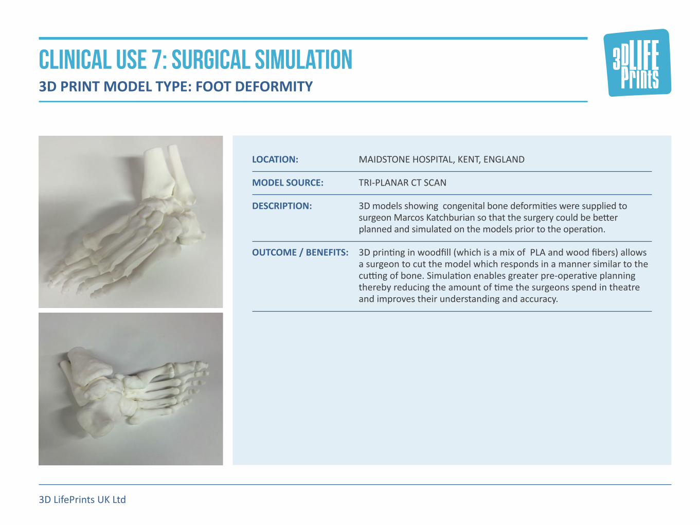
LOCATION: MAIDSTONE HOSPITAL, KENT, ENGLAND MODEL SOURCE: TRI-PLANAR CT SCAN DESCRIPTION: 3D�models�showing��congenital�bone�deformities�were�supplied�to�
surgeon�Marcos�Katchburian�so�that�the�surgery�could�be�better�planned�and�simulated�on�the�models�prior�to�the�operation.
OUTCOME / BENEFITS: 3D�printing�in�woodfill�(which�is�a�mix�of��PLA�and�wood�fibers)�allows�
a�surgeon�to�cut�the�model�which�responds�in�a�manner�similar�to�the�cutting�of�bone.�Simulation�enables�greater�pre-operative�planning�thereby�reducing�the�amount�of�time�the�surgeons�spend�in�theatre�and�improves�their�understanding�and�accuracy.�
3D LifePrints UK Ltd
CLINICAL USE 7: SURGICAL SIMULATION 3D PRINT MODEL TYPE: FOOT DEFORMITY

LOCATION: ALDER�HEY�HOSPITAL,�LIVERPOOL,�ENGLAND MODEL SOURCE: MRI SCAN DESCRIPTION: Created�in�collaboration�with�Lazarus�3D,�this�3D�model�of�part�of�an�
adult�brain�was�created�to�show�that�silicone�3D�printing�can�provide�a�realistic�replica�for�soft�tissue�and�organs.�The�silicone�is�printed�in�a�range�of�colours�and�densities�and�it�is�intended�to�be�used�in�medical�training�for�operations�such�as�Lobe�resection�and�Lesionectomy.
OUTCOME / BENEFITS: Feedback�from�the�surgeons�at�Alder�Hey�was�extremely�positive�
especially�concerning��the�realism�of�the�model�and�potential�future��use�of�medical�training�purposes. It�is�proposed�to�combine�the�silicone�brain�with�a�skull�printed�in�polyamide,�plaster�or�woodfill�to�provide�a�high�fidelity�model�for�surgical�simulation.
3D LifePrints UK Ltd
CLINICAL USE 8: SURGICAL TRAINING 3D PRINT MODEL TYPE: BRAIN

LOCATION: SURGICAL�SKILLS�CENTREL,�LIVERPOOL,�ENGLAND MODEL SOURCE: MRI SCAN DESCRIPTION: A�soft�stomach�3D�model�was�printed�in�flexible�silicone�in�collaberation�
with�Sandraw�Ltd�to�provide�surgeons�with�a�realistic��simulator�for�PEG�insertion�training.
OUTCOME / BENEFITS: This�training�model�will�be�used�by�theatre�staff�to�practice�stomach�
tube�insertions�prior�to�undertaking�the�procedure�on�patients.�Upwards�of�180�of�these�operations�are�carried�out�every�year�at�Alder�Hey�hospital�alone.�Feedback�from�the�surgeon�was�positive�in�that�the�3D�printed�training�model�was�superior�to�any�products�previously�available�to�the�surgical�team�particularly�in�texture�and�density�of�tissue.�The�training�model�can�also�be�easily�reprinted�to�include�specific�anatomical�features�for�particular�patients.
3D LifePrints UK Ltd
CLINICAL USE 9: SURGICAL TRAINING 3D PRINT MODEL TYPE: PERCUTANEOUS ENDOSCOPIC GASTROSTOMY

LOCATION: ALDER�HEY�HOSPITAL,�LIVERPOOL,�ENGLAND MODEL SOURCE: TRI-PLANAR CT SCAN DESCRIPTION: Created�in�collaboration�with�Materialise,�this��3D�printed�spinal�
model�was�used�to�assist��with�a�complex�procedure�to�rectify�a�congenital�spinal�kyphoscoliosis�on�an��eight-year-old�patient.�The�model�was�used�by�the�surgical�team��for�pre-operative�planning�purposes,�and�subsequently�sterilized��using��an�autoclave�container�and�brought�into�the�operating�theatre�to�be�used�for�guidance.
OUTCOME / BENEFITS: Lead�surgeon�Neil�Davidson�remarked:�“The�model�was�invaluable�for�
use�by�the�surgical�team�to�undertake�this�complex�procedure.�It�was�useful�both�preoperatively�and�intraoperatively�-�the�surgery�would�have�been�much�more�complicated�and�difficult�without�the�model� in�theatre.�Without�the�model,�the�surgical�team�would�have�had�a�higher�chance�of�needing�to�carry�out�an�anterior�approach�to�the�spine�which�would�have�increased�time�in�the�theatre�and�the�surgical�risks�of�complications�to�the�patient.”
3D LifePrints UK Ltd
CLINICAL USE 10: PRE & INTRA SURGICAL PLANNING 3D PRINT MODEL TYPE: SPINAL KYPHOSCOLIOSIS

LOCATION: ALDER�HEY�HOSPITAL,�LIVERPOOL,�ENGLAND MODEL SOURCE: MRI SCAN DESCRIPTION: A�kidney�with�a�tumor�was�dual-colour�printed�in�soft�silicone�with�
varying�densities�across�sections�of�the�model�for�surgical�training�use. The�models�are�created�in�collaboration�with�Lazarus�3D,�using�their�advanced�silicone�3D�printer.
OUTCOME / BENEFITS: A�simulated�partial�nephrectomy�was�carried�out�on�the�model,�
showing�potential�to�simulate�surgeries�and�thereby�improve�surgical�skills.�Feedback�from��the�surgeon�was�positive�in�that�the�3D�model�provided�an�accurate�representation�with�high�fidelity��for�simulating�the�removal�of�the�tumour.
3D LifePrints UK Ltd
CLINICAL USE 11: SURGICAL SIMULATION 3D PRINT MODEL TYPE: KIDNEYS FOR PARTIAL NEPHRECTOMY TRAINING

LOCATION: ALDER�HEY�HOSPITAL,�LIVERPOOL,�ENGLAND MODEL SOURCE: TRI-PLANAR CT SCAN DESCRIPTION: A�number�of�3D�models�were�created�for�a�variety�of�complex�tri-
planar�ankle�fractures.��These�models�were�used�by�the�surgeon�in�workshops,�where�the�model�was�separated�along�the�fracture�line�so�that�the�participating�clinicians�could�see�the�exact�topography�of�the�break�and�understand�how�it�could�be�treated.�
OUTCOME / BENEFITS: The�results�of�the�workshops�were�incredibly�positive�with�clinicians�
confirming�that�the�models�improved�their�understanding�the�issues,�as�well�as�greatly�improving�their�ability�to�describe�the�3D�configuration�of�the�break�for�teaching�purposes.�Surgeon�Roger�Walton�commented:�“I�believe�the�use�of�a�3D�model�could�improve�pre-operative�planning�and�produce�novel�operative�strategies�for� new�cases.”
3D LifePrints UK Ltd
CLINICAL USE 12: MEDICAL RESEARCH 3D PRINT MODEL TYPE: COMPLEX ANKLE FRACTURES

LOCATION: JOHN�MOORES�UNIVERSITY,�LIVERPOOL,�ENGLAND MODEL SOURCE: MICRO CT SCANNER DESCRIPTION: In�this�study�a�post-mortem�rabbit�heart�was�scanned�by�micro�CT�to�
identify�the�electrical�conduction�system�that�controls�the�heart�rhythm.�The�prints�were�commissioned�by�Professor�Jonathan�Jarvis�and�Dr�Robert�Stephenson�with�support�from�The�Alder�Hey�Children’s�charity,�and�working�with�their�clinical�colleagues�Dr�Caroline�Jones�and�Dr�Rafael�Guerrero.�The�models�were�printed�on�a�multi-jet�printer�which�allowed�several�colours�to�be�incorporated.
OUTCOME / BENEFITS: Feedback�from�Dr.�Robert�Stephenson:�“Working�with�3D�LifePrints�
we�have�produced�high�resolution�3D�prints�of�the�heart�of�unprecedented�quality�and�detail.�Generated�from�high�resolution�micro-CT�data�sets�such�prints�have�brought�our�virtual�data�to�life,�and�serve�to�improve�our�understanding�of�the�3D�micro-anatomy� of�the�heart.” A�further�series�of�3D�printed�models�based�upon�human�hearts�is�planned�which�will�increase�the�scope�of�this�project.
3D LifePrints UK Ltd
CLINICAL USE 13 : MEDICAL RESEARCH 3D PRINT MODEL TYPE: RABBIT HEART ATRIAL WALL

LOCATION: ALDER�HEY�HOSPITAL,�LIVERPOOL,�ENGLAND MODEL SOURCE: TRI-PLANAR CT SCAN DESCRIPTION: A�series�of�3D�printed�models�were�created�to�investigate�the�following�
“Can�3D�printing�replace�an�arthrogram�for�hip�imaging?”��Reconstructive�surgery�is�frequently�employed�to�improve�the�congruency�of�the�hip,�to�prolong�or�obviate�the�time-to-arthroplasty.�Surgical�decisions�regarding�reconstructive�surgery�can�be�challenging.�An�arthrogram�is�the�most�dynamic�investigation,�however�only�offers�two-dimensional�imaging�and�necessitates�a�general�anaesthetic�and�time�in�the�operating�theatre.
OUTCOME / BENEFITS: 3D�printing�offered�an�opportunity�to�produce�bespoke�dynamic�
models�of�diseased�hips,�which�enabled�the�surgeon�to�gain�a�greater�insight�into�the�surgery�required.�It�was�useful�in�the�process�of�obtaining�patient�consent,�enabling�surgeons�to�perform�‘surgery’� on�the�printed�model�preoperatively�as�a�‘trial-run’�or�when�training�trainee�surgeons.�By�testing�different�materials,�the�models�enabled�surgeons�to�test�optimal�osteotomy�positions�(cuts�in�the�bone),�and�enabled�them�to�reliably�template�(size)�the�materials�required�for� the�procedure.
3D LifePrints UK Ltd
CLINICAL USE 14: MEDICAL RESEARCH 3D PRINT MODEL TYPE: HIP DYSPLASIA

LOCATION: ALDER HEY CHILDRENS HOSPITAL, ORTHOPAEDIC DEPARTMENT MODEL SOURCE: TRI-PLANAR CT SCAN DESCRIPTION: �These�3D�printed�models�were�created�to�provide�surgeon�Harvey�
George�and�his�team�with�a�series�of�models�of�a�patient’s�elbow�to�allow�comprehensive�understanding�of�the�issues�prior�to�surgery.
OUTCOME / BENEFITS: ��The�models�have�been�supplied�to�the�consultant�and�we�await�
feedback�from�the�surgery.� The�first�model�showed�the�injured�left�elbow�socket,�modelled�from�an�initial�CT�scan�done�in�March.�The�second�in�the�series,�formed�from�a�scan�in�august�showed�the�elbow’s�deterioration�over�time.�The�third�model�showed�the�healthier�right�elbow�which�the�surgeons�need�to�mimic�in�their�corrective�surgery.�We�incorporated�artificial�bridges�between�some�of�the�3D�printed�bones�in�order�to�maintain�their�alignment�with�each�other.
3D LifePrints UK Ltd
CLINICAL USE 15 & 16: surgical assessment & Training 3D PRINT MODEL TYPE: MODELBOW REGION PRINT COMPARISON

LOCATION: ALDER�HEY�HOSPITAL,�LIVERPOOL,�ENGLAND MODEL SOURCE: 3D SCAN DESCRIPTION: The�3D�models�were�created�to�provide�an�exact�replica�of�the�plaster�
of�Paris�dental�moulds�currently�employed�by�the�hospital.�Once�the�existing�dental�moulds�have�been�scanned�the�data�can�be�stored�electronically�which�eliminates�the�need�to�keep�a�physical�mould� in�archives.
OUTCOME / BENEFITS: The�projected�outcome�would�remove�the�need�to�store�the�plaster�of�
Paris�moulds�as�the��virtual�mould�could�be�scanned�and�stored�on�a�cloud�based�platform�and�retrieved�and�3D�printed�when�necessary. A�further�project�would�be�to�eliminate�the�need�for�a�plaster�of� Paris�mould�by�directly�scanning�the�patient’s�teeth.
3D LifePrints UK Ltd
CLINICAL USE 17: MEDICAL ARCHIVING 3D PRINT MODEL TYPE: DENTAL ARCHIVING

LOCATION: ALDER�HEY�HOSPITAL,�LIVERPOOL,�ENGLAND MODEL SOURCE: TRI-PLANAR CT SCAN DESCRIPTION: This�3D�model�was�created�to�aid�the�surgeon�in�discussions�with�the�
patient’s�family�about��their�child’s�spinal�deformity. OUTCOME / BENEFITS: From�Dr.�Neil�Davidson:�“I�think�the�reproduction�is�excellent�and�
will�benefit�the�patient�from�both�the�clinical�perspective,�and�the�family’s�understanding�of�the�nature�of�her�pathology.�I�used�the�3D�reconstruction�to�help�the�family�understand�exactly�the�nature�of� the�deformity.�At�the�end�of�the�consultation�the�patient’s�mother�admitted�that�it�was�the�first�time�she�really�understood�the�complexity�of�the�deformity.”
3D LifePrints UK Ltd
CLINICAL USE 18: IMPROVING DOCTOR-PATIENT COMMUNICATIONS 3D PRINT MODEL TYPE: SCOLIOTIC SPINE
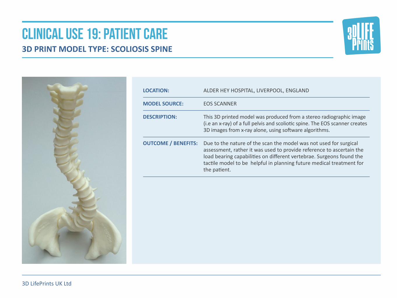
LOCATION: ALDER�HEY�HOSPITAL,�LIVERPOOL,�ENGLAND MODEL SOURCE: EOS SCANNER DESCRIPTION: This�3D�printed�model�was�produced�from�a�stereo�radiographic�image�
(i.e�an�x-ray)�of�a�full�pelvis�and�scoliotic�spine.�The�EOS�scanner�creates�3D�images�from�x-ray�alone,�using�software�algorithms.
OUTCOME / BENEFITS: Due�to�the�nature�of�the�scan�the�model�was�not�used�for�surgical�
assessment,�rather�it�was�used�to�provide�reference�to�ascertain�the�load�bearing�capabilities�on�different�vertebrae.�Surgeons�found�the�tactile�model�to�be��helpful�in�planning�future�medical�treatment�for�the�patient.
3D LifePrints UK Ltd
CLINICAL USE 19: PATIENT CARE 3D PRINT MODEL TYPE: SCOLIOSIS SPINE

LOCATION: �KENYA,�UGANDA,�RWANDA,�TANZANIA,�SYRIA,�LEBANON,� MYANMAR,�CAMBODIA
MODEL SOURCE: 3D SCAN AND CAD MODEL DESCRIPTION: 80%�of�the�world’s�estimated�30�million�amputees�live�in�developing�
nations,�who�have�little�or�no�access�to�affordable�and�suitable�prosthetics.�3DLP�have�developed�the�“LifeArm”,�a�functional�and�low�cost�3D�printed�transradial�prosthetic�designed�specifically�for�use�in�challenging�environmental�conditions.
OUTCOME / BENEFITS: The�use�of�3D�printing�here�provided�an�extremely�low�cost�prosthetic�
for�patients�in�the�developing�world.�The�“LifeArm”�is�designed�to�meet�the�requirements�of�the�developing�world�and�is,�low-cost,�lifelike,�durable,�and�easy�to�maintain.
3D LifePrints UK Ltd
CLINICAL USE 20: PROSTHETICS 3D PRINT MODEL TYPE: UPPER EXTREMITY PROSTHETICS

“We�have�discovered�a�number�of�truly�novel�and�exciting�ways�to�make�our� care�better,�kinder�and�easier.�We� look�forward�to�continuing�this�partnership�(with�3DLP)�to�ensure� we�stay�at�the�forefront�of�this� emerging�technology.
Mr Iain Hennessy, Consultant Paediatric Surgeon and Clinical Director of Innovation at Alder Hey Hospital
3D LifePrints UK Ltd
Please contact us below for any further information
Paul Fotheringham [email protected]
Henry Pinchbeck [email protected]
www.3dlifeprints.com
3D LifePrints UK Ltd