examining the effect of obesity-associated systemic inflammation on the uterine immune cell niche in...

1
B) Proton Magnetic resonance spectroscopy was performed to evaluate the biochemical/ metabolic prole in placentas. A signicant decrease in phosphocholine was observed in APS-mice compared to Ctrl (PCh/Cr+PCr: APS(n¼7)¼0.50±0.78 vs control (n¼4)¼1.69±0.73, p¼0.0352). Decreased glucose levels and increased lactate levels, indicative of hypoxia, were measured in placentas from APS-mice compared to Ctrl (Glc/Cr+PCr: APS(n¼6)¼ 0.03±0.0.05 vs control (n¼4)¼1.04±0.9, p¼0.0222; Lac/Cr+PCr: APS(n¼7): 0.47±0.0.80 vs control (n¼4): 4.4±4.1, p¼0.0310). Placental insufciency in APS was also associated with abnormal fetal neuro- development characterised by cortical axonal cytoarchitecture dis- ruption(NF200 and MAP-2 expression) and increased neurodegeneration (FluoroJadeB). Increased anxiety was observed in the offspring of APS-mice. Conclusion: Using two novel non invasive in uterus MRI-based methods we were able to determine complement activation and metabolism in the placentas and predict fetal outcomes. P1.149-N. PATHOLOGICAL MECHANISMS OF ANTIPHOSPHOLIPID ANTIBODIES IN TROPHOBLASTIC CELL FUSION Tess Marchetti a, b , Philippe de Moerloose b , Marie Cohen a a Laboratory of Hormonology, Geneva University Hospitals, Geneva, Switzerland; b Haemostasis Unit, Geneva University Hospitals, Geneva, Switzerland Objectives: Obstetrical Antiphospholipid Syndrome (APS) associates pregnancy pathologies with antiphospholipid antibodies (aPL). It has been suggested that aPL could affect trophoblastic cell fusion. However, their pathogenicity is still poorly understood. Toll-like Receptors (TLR) have been implicated in the pathological activa- tion of aPL on endothelial cells. Although inammation has been incrim- inated in this process, endoplasmic reticulum (ER) stress activation seems more related to trophoblastic fusion impairment by aPL. The aim of our study was to evaluate the pathological mechanisms of aPL on trophoblastic cell fusion. Methods: BeWo cell line is used as a model to mimic trophoblastic cell fusion, triggered by forskolin. Results were corroborated with primary cytotropho- blastic cells. Fusion index was determined by immunocytochemistry and biochemical differentiation by ELISA measuring hCG secretion. Effects of TLR on BeWo cell fusion were evaluated using blocker antibodies for TLR2 and 4, peptide binding for TLR2 and 4, and stable shRNA TLR4 in the presence or not of aPL. After determining the role of TLR4 in trophoblastic cell fusion impairment by aPL, signalling cascade of inammation was studied by ELISA measuring TNFa and IL8 secretion. ER stress activation was assessed by qPCR and WB quantifying Glucose-regulated protein 78 (GRP78) and C/EBP-homologous protein (CHOP) expressions. Result: Fusion index and hCG secretion are decreased by addition of aPL. In the presence of aPL and anti-TLR4 antibodies or in BeWo cells transfected with shRNA TLR4, trophoblastic cell fusion and hCG secretion were restored. Neither TNFa nor IL8 secretion was observed in BeWo cells stimulated by aPL. Increase in GRP78 and CHOP expressions was observed in cells stimulated by aPL, but not when blocked with anti-TLR4 antibodies, sug- gesting a role of ER stress in trophoblastic cell fusion impairment by aPL. Conclusion: aPL interfered with fusion process and biochemical differ- entiation of trophoblastic cells via TLR4 that triggers ER stress activation. P1.150. EXAMINING THE EFFECT OF OBESITY-ASSOCIATED SYSTEMIC INFLAMMATION ON THE UTERINE IMMUNE CELL NICHE IN EARLY PREGNANCY Yoona Kim, Soe Perdu, Mahroo Aghababaei, Alexander Beristain The University of British Columbia, Vancouver, Canada Objectives: Low-grade inammation associates with major obstetrical disorders like preterm birth and preeclampsia and is thought to be causal in the development of these high-risk pregnancies. Control of immune cell-directed pro-inammatory cytokine production and cytotoxicity is crucial for utero-placental function; aberrant pro-inammatory T helper (h) 1 cell activity within the uterine microenvironment associates with placental dysfunction and poor pregnancy outcome. Immunological im- balances and low-grade inammation are associated with excess adiposity. However, the cellular mechanism(s) linking obesity-associated inam- mation to poor pregnancy outcome are poorly understood. As a rst step in addressing this knowledge gap, our study aims to examine multiple im- mune cell subsets within decidual tissues of non-obese and obese women in early pregnancy. Methods: Patient information, blood serum and decidual tissue from gestationally aged-matched (8-10 weeks) women undergoing elective pregnancy terminations were used to determine BMI, serum C-reactive protein (CRP) (Qg/mL) and immune cell composition. Women were cate- gorized as healthy BMI (20-24.9) not presenting with low-grade inam- mation (CRP < 2 Qg/mL; Control CRP- ) or obese BMI ( 30) presenting with low-grade inammation (CRP 4 Qg/mL; Obese CRP+ ). Immune cells, including natural killer cells (NKs), Tcells (Th1, Th2, Th17, Tregs and CD8 + cytotoxic cells), macrophages and dendritic cells, were quantitated by multicolour ow cytometry analysis. Results: We did not detect differences in proportions of CD56 bright decidual NKs, dendritic cells, macrophages or Th17 and CD8 + T cells between decidual tissues of Control CRP- (n¼15) and Obese CRP+ (n¼15) women. However, proportions of immunosuppressive CD4 + /FoxP3 + Tregs, pro-in- ammatory IFN© + Th1 cells, and cytotoxic CD16 + NK cells were elevated in Obese CRP+ decidua while IL-13 + Th2 proportions were decreased. Conclusion: This study suggests that obesity-linked inammation in early pregnancy does alter immune cell composition within the maternal-fetal interface. These new ndings lay the groundwork for future studies aimed at dissecting the importance of these cellular changes in pregnancy. P1.151. PHAGOCYTIC ACTIVITY OF MACROPHAGES IS REGULATED BY FIRST TRIMESTER DECIDUAL CELLS IN RESPONSE TO PRO-INFLAMMATORY STIMULI VIA THE MODULATION OF DON'T-EAT-MESIGNAL Longzhu Piao a , Susheela Tridandapani b , S. Joseph Huang a a Deartment of Ob/Gyn, The Ohio State University, Columbus, OH, USA; b SBS-Microbial Infection/Immunity, The Ohio State University, Columbus, OH, USA Ă Ă Abstracts / Placenta 35 (2014) A1eA112 A57

Upload: alexander

Post on 23-Feb-2017

212 views

Category:

Documents


0 download

TRANSCRIPT

Ă

Abstracts / Placenta 35 (2014) A1eA112 A57

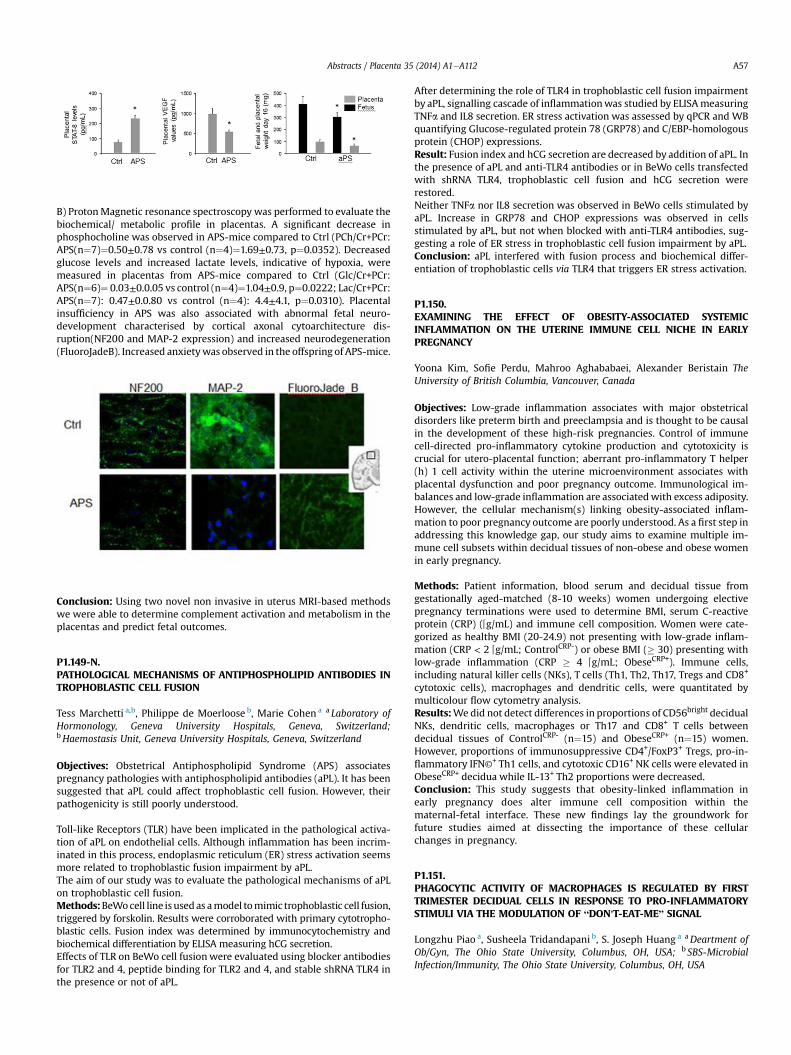
B) ProtonMagnetic resonance spectroscopy was performed to evaluate thebiochemical/ metabolic profile in placentas. A significant decrease inphosphocholine was observed in APS-mice compared to Ctrl (PCh/Cr+PCr:APS(n¼7)¼0.50±0.78 vs control (n¼4)¼1.69±0.73, p¼0.0352). Decreasedglucose levels and increased lactate levels, indicative of hypoxia, weremeasured in placentas from APS-mice compared to Ctrl (Glc/Cr+PCr:APS(n¼6)¼ 0.03±0.0.05 vs control (n¼4)¼1.04±0.9, p¼0.0222; Lac/Cr+PCr:APS(n¼7): 0.47±0.0.80 vs control (n¼4): 4.4±4.1, p¼0.0310). Placentalinsufficiency in APS was also associated with abnormal fetal neuro-development characterised by cortical axonal cytoarchitecture dis-ruption(NF200 and MAP-2 expression) and increased neurodegeneration(FluoroJadeB). Increased anxietywas observed in the offspring of APS-mice.

Ă

Conclusion: Using two novel non invasive in uterus MRI-based methodswe were able to determine complement activation and metabolism in theplacentas and predict fetal outcomes.

P1.149-N.PATHOLOGICAL MECHANISMS OF ANTIPHOSPHOLIPID ANTIBODIES INTROPHOBLASTIC CELL FUSION

Tess Marchetti a,b, Philippe de Moerloose b, Marie Cohen a a Laboratory ofHormonology, Geneva University Hospitals, Geneva, Switzerland;bHaemostasis Unit, Geneva University Hospitals, Geneva, Switzerland

Objectives: Obstetrical Antiphospholipid Syndrome (APS) associatespregnancy pathologies with antiphospholipid antibodies (aPL). It has beensuggested that aPL could affect trophoblastic cell fusion. However, theirpathogenicity is still poorly understood.

Toll-like Receptors (TLR) have been implicated in the pathological activa-tion of aPL on endothelial cells. Although inflammation has been incrim-inated in this process, endoplasmic reticulum (ER) stress activation seemsmore related to trophoblastic fusion impairment by aPL.The aim of our study was to evaluate the pathological mechanisms of aPLon trophoblastic cell fusion.Methods:BeWocell line is usedas amodel tomimic trophoblastic cell fusion,triggered by forskolin. Results were corroborated with primary cytotropho-blastic cells. Fusion index was determined by immunocytochemistry andbiochemical differentiation by ELISA measuring hCG secretion.Effects of TLR on BeWo cell fusionwere evaluated using blocker antibodiesfor TLR2 and 4, peptide binding for TLR2 and 4, and stable shRNA TLR4 inthe presence or not of aPL.

After determining the role of TLR4 in trophoblastic cell fusion impairmentby aPL, signalling cascade of inflammationwas studied by ELISAmeasuringTNFa and IL8 secretion. ER stress activation was assessed by qPCR and WBquantifying Glucose-regulated protein 78 (GRP78) and C/EBP-homologousprotein (CHOP) expressions.Result: Fusion index and hCG secretion are decreased by addition of aPL. Inthe presence of aPL and anti-TLR4 antibodies or in BeWo cells transfectedwith shRNA TLR4, trophoblastic cell fusion and hCG secretion wererestored.Neither TNFa nor IL8 secretion was observed in BeWo cells stimulated byaPL. Increase in GRP78 and CHOP expressions was observed in cellsstimulated by aPL, but not when blocked with anti-TLR4 antibodies, sug-gesting a role of ER stress in trophoblastic cell fusion impairment by aPL.Conclusion: aPL interfered with fusion process and biochemical differ-entiation of trophoblastic cells via TLR4 that triggers ER stress activation.

P1.150.EXAMINING THE EFFECT OF OBESITY-ASSOCIATED SYSTEMICINFLAMMATION ON THE UTERINE IMMUNE CELL NICHE IN EARLYPREGNANCY

Yoona Kim, Sofie Perdu, Mahroo Aghababaei, Alexander Beristain TheUniversity of British Columbia, Vancouver, Canada

Objectives: Low-grade inflammation associates with major obstetricaldisorders like preterm birth and preeclampsia and is thought to be causalin the development of these high-risk pregnancies. Control of immunecell-directed pro-inflammatory cytokine production and cytotoxicity iscrucial for utero-placental function; aberrant pro-inflammatory T helper(h) 1 cell activity within the uterine microenvironment associates withplacental dysfunction and poor pregnancy outcome. Immunological im-balances and low-grade inflammation are associatedwith excess adiposity.However, the cellular mechanism(s) linking obesity-associated inflam-mation to poor pregnancy outcome are poorly understood. As a first step inaddressing this knowledge gap, our study aims to examine multiple im-mune cell subsets within decidual tissues of non-obese and obese womenin early pregnancy.

Methods: Patient information, blood serum and decidual tissue fromgestationally aged-matched (8-10 weeks) women undergoing electivepregnancy terminations were used to determine BMI, serum C-reactiveprotein (CRP) (Qg/mL) and immune cell composition. Women were cate-gorized as healthy BMI (20-24.9) not presenting with low-grade inflam-mation (CRP < 2 Qg/mL; ControlCRP-) or obese BMI (� 30) presenting withlow-grade inflammation (CRP � 4 Qg/mL; ObeseCRP+). Immune cells,including natural killer cells (NKs), T cells (Th1, Th2, Th17, Tregs and CD8+

cytotoxic cells), macrophages and dendritic cells, were quantitated bymulticolour flow cytometry analysis.Results:Wedid not detect differences in proportions of CD56bright decidualNKs, dendritic cells, macrophages or Th17 and CD8+ T cells betweendecidual tissues of ControlCRP- (n¼15) and ObeseCRP+ (n¼15) women.However, proportions of immunosuppressive CD4+/FoxP3+ Tregs, pro-in-flammatory IFN©+ Th1 cells, and cytotoxic CD16+ NK cells were elevated inObeseCRP+ decidua while IL-13+ Th2 proportions were decreased.Conclusion: This study suggests that obesity-linked inflammation inearly pregnancy does alter immune cell composition within thematernal-fetal interface. These new findings lay the groundwork forfuture studies aimed at dissecting the importance of these cellularchanges in pregnancy.

P1.151.PHAGOCYTIC ACTIVITY OF MACROPHAGES IS REGULATED BY FIRSTTRIMESTER DECIDUAL CELLS IN RESPONSE TO PRO-INFLAMMATORYSTIMULI VIA THE MODULATION OF “DON'T-EAT-ME” SIGNAL

Longzhu Piao a, Susheela Tridandapani b, S. Joseph Huang a aDeartment ofOb/Gyn, The Ohio State University, Columbus, OH, USA; b SBS-MicrobialInfection/Immunity, The Ohio State University, Columbus, OH, USA