examining bivalve fecundity: oocyte viability revealed by
TRANSCRIPT

Examining bivalve fecundity: oocyte viability revealedby Neutral Red vital staining
Peter G. Beninger1 & Daphné Chérel1 & Lucie Kessler2
Received: 18 August 2020 /Accepted: 25 February 2021/# Springer Nature Switzerland AG 2021
AbstractEstimation of realized fecundity (Freal, number of viable oocytes produced) is an essential,yet seldom-achieved element in the understanding of marine animal production andpopulation dynamics. We used the Neutral Red (NR) vital stain to determine oocyteviability in spawns and gonad strippings of four species of commercially-importantbivalves: Cerastoderma edule (L), Crassostrea gigas Thunberg, Mytilus edulis L, andTapes philippinarum (Adams and Reeve). The utility of Trypan Blue as a complementarymortal stain was also assessed, and found to be unnecessary. Normal, live oocytes stainedwith NR, and were either spherical (mature oocytes) or pedunculated (immature oocytes);atresic/abnormal oocytes also stained with NR, allowing their ready recognition due totheir abnormal shapes and cytoplasmic retraction. Dead oocytes did not stain with NR.Across the species studied, a considerable and highly variable proportion of spawned orstripped oocytes was either dead or non-viable; quantitative counts were performed forCerastoderma edule, the only species for which > 5 spawns occurred. The high level andvariability (34–85%) of dead or non-viable oocytes is consistent with a reproductive RedQueen dilemma, in which greater oocyte numbers do not translate to commensuratelygreater real fecundities, and also with a Sweepstakes Reproductive Success strategy, inwhich a large range of Freal confronts the considerable variability of intertidal environ-mental conditions. Neutral Red vital staining is a promising tool for the elucidation andoptimization of crucial yet previously intractable aspects of bivalve hatchery production,genetic improvement, restocking, stock management, and conservation.
Keywords Neutral Red . Bivalves . Oocytes . Viability . Fecundity . Reproduction . Atresia
Aquaculture Internationalhttps://doi.org/10.1007/s10499-021-00686-6
Handling Editor: Gavin Burnell
* Peter G. BeningerPeter.Beninger@univ–nantes.fr
1 Laboratoire de Biologie Marine, MMS, Faculté des Sciences, Université de Nantes, 2 rue de laHoussinière, 44322 Nantes, Cédex, France
2 Faculté des Sciences et Technologies, La Rochelle Université, Avenue Michel Crépeau, 17042 LaRochelle, Cédex, France

Introduction
Bivalve capture fisheries and aquaculture account for approximately 15% of global annualseafood production, registering an eightfold increase over the bivalve production levelsrecorded in the 1970s (Wijsman et al. 2019). Although aquaculture is the major focus ofbivalve production, fisheries management, restocking, conservation, and pollution monitoringare also important objectives (His et al. 1999; Gomez and Mingoa-Licuanan 2006; Arnold2008; Joaquim et al. 2008; González-Wangüemert et al. 2018; Kreeger et al. 2018;Shantharam et al. 2019).
The need to assess oocyte quality for improved bivalve production has been recognized fordecades (Utting and Millican 1997); however, this criterion has not yet been incorporated intobroodstock genetic improvement efforts (Boudry 2009; Barros et al. 2018). Similarly, theassessment of fecundity and elucidation of early life stage mortality is, either directly orindirectly, at the heart of stock-recruitment analysis (Aoyama 1989; Koslow 1992; Jenningset al. 2001; Maunder and Thorson 2019), yet the evaluation of oocyte quality in spawns is amissing element in this process.
To date, assessments of oocyte quality have often been performed retrospectively, throughfertilization rates, based on polar body or cell division observations (Uttig and Spencer 1991,Buttner and Weston 2010, Militz and Southgate 2021), analysis of chemical composition, andperformance indicators (Caers et al. 1999, 2002; Massapina et al. 1999; Nevejan et al. 2003;Cannuel and Beninger 2005; Corporeau et al. 2012; García-Corona et al. 2018). Histochemicalstaining of specific oocyte components, in particular lipids, may also yield information onoocyte quality (Valdez-Ramirez et al. 2002). Lipid-specific stains such as Oil Red O andSudan Black have been used to evaluate the quality of spawned oocytes (Gallager and Mann1986; Gallager et al. 1986; Gómez-Robles et al. 2005; Angel-Dapa et al. 2010); however, thelevel of a biologically - important chemical component does not signify that the organism orcell is even alive (Moens and Beninger 2018).
The criterion of viability is surely the most fundamental of all oocyte quality indicators. Thepresence of atresic oocytes within the gonad reduces the realized fecundity (Freal) compared tothe potential fecundity (Fpot – Jennings et al. 2001); in the case of marine bivalves, this hasbeen estimated at approximately 50% of the total gamete volume over the course of thegametogenic cycle (Chérel and Beninger 2017, 2019; Chérel et al. 2020). It is not known whatproportion, if any, of these atresic and other non-viable (dead) oocytes are spawned along withnormal oocytes. Ideally, therefore, evaluation of spawn viability should yield information onthe number of live, dead, and dying (atresic) oocytes.
A promising candidate for bivalve oocyte viability assessment is the Neutral Red (NR)stain. First reported as a vital stain by Ehrlich (1894), various NR protocols emerged asstandard tests for cell vitality, beginning late in the twentieth century (Winckler 1974; Nemeset al. 1979; Hammond et al. 1980), usually in the context of tissue cultures or single cellssubjected to various challenges (Borenfreund and Puerner 1984; Triglia et al. 1991; Lowe et al.1992, 1995a, b; Babich and Borenfreund 1993; Weeks and Svendsen 1996; Chiba et al. 1998;Svendsen et al. 2004; Repetto et al. 2008; Aguirre-Martínez et al. 2013; Patetsini et al. 2013;Hu et al. 2015; Liu et al. 2018). The technique was used as early as 1972 by Dressel et al. todifferentiate between live and dead plankton, and continues to be so used to the present(Crippen and Perrier 1974; Horvath and Lamberti 1999; Tang et al. 2006; Elliott and Tang2009; Zetsche and Meysman 2012; Da Luz et al. 2016). It is thus somewhat surprising that ithas not yet been employed for the same purpose in bivalve oocytes. Conversely, the same can
Aquaculture International

be said of the mortal stain Trypan Blue, widely used to detect dead cells in the biomedicalfields (Wales 1959; Talbot and Chacon 1981; Kwok et al. 2004; Strober 2015; Rajab andDemer 2019).
Ideally, a spawned oocyte viability assessment should have the following characteristics:
& Specificity for live oocytes& Low per-test cost& Low equipment cost& Simplicity – for routine use by hatchery workers& Rapid execution and rapid results – so that the oocytes may be either used or disposed of
immediately after obtention& Low sample size (to preserve the utility of the obtained oocytes)& Universality – for use with any bivalve species
The present study documents the use of Neutral Red as a vital stain, to collect the first knowndata on the viability of oocytes obtained by induced spawning or gonad stripping in fourspecies of commercially - important bivalves: Cerastoderma edule (L), Crassostrea gigasThunberg, Mytilus edulis L, and Tapes philippinarum (Adams and Reeve).
Material and methods
Sampling and gamete obtention
All bivalves were haphazardly collected in the Traict du Croisic, on the French Atlantic coast(47° 17′ 26″ N, 2° 30′ 16.2″ W). Cerastoderma edule and T. philippinarum were sampled onthe mudflat at low tide, whereas Crassostrea gigas and M. edulis were sampled on adjacentrocky substrates. Sampling for Cerastoderma edule was performed on April 17 and May 2,2019, because previous work had shown them to be spawning-competent at this early date inthe gametogenesis season (Chérel and Beninger 2019). Only individuals >20 mm shell length(SL) were retained, corresponding to the size at which normal gamete production is achieved(Mejuto 1984; Pérez Camacho and Román 1984). The actual size range was 20.8–35.1 mm,and the corresponding shell age rings were 1–2.5 years.
Additional sampling was performed on 11 and 25 June 2019, at which time all four specieswere in advanced gametogenesis, and on 31 July 2020, when spawnings were known to occur.M. edulis specimens were all > 40 mm SL, T. philippinarum were all > 20 mm SL, andCrassostrea gigas were all > 20 mm SL; the latter to ensure that some males were sampled forspawning induction.
Spawning induction was repeatedly attempted for all four species, using various thermalshock–emersion regimes, as well as the addition of spermatozoa stripped from male individ-uals. Successful induction was obtained for at least 8 Cerastoderma edule, 3 M. edulis, and 1Crassostrea gigas specimens. Experience showed that cockles spawned more readily ingroups of 3–4 individuals, so it was not possible to ascertain the exact number of individualsthat spawned (especially since release occurred in short “dribbles” of nearly transparentoocytes). Gamete stripping was used to obtain oocytes from all individuals for which spawninginduction failed. Table 1 summarizes the numbers of individuals actually used for eachstaining assay. As Cerastoderma edule was the most oogenically advanced of the four species
Aquaculture International
studied in April and May 2019, spawning induction was attempted every day, up to 8 daysfollowing sampling (Table 2); specimens were kept in cold storage (emersion, 4 °C) betweenspawning attempts.
Staining procedure
Neutral Red was used as a vital stain, and for contrast, Trypan Blue was employed as a mortalstain. Exploratory work showed that the best staining results were obtained with Neutral Red at5 mg ml−1, and Trypan Blue at 2 mg ml−1 of filtered seawater. Although a large amount ofundissolved Trypan Blue remained at this latter concentration, it was nonetheless necessary inorder to obtain sufficient dissolved product capable of staining dead oocytes. Oocytes trans-ferred to a microscope slide using a Pasteur pipette could be observed under an OlympusProvis AX70 optical microscope within 3 min of addition of a drop of NR prepared stain. TBstaining required at least 1 h incubation time at room temperature prior to observation. Oocytecounts were performed for Cerastoderma edule at 40× on a haphazardly-chosen slide transectusing Olympus cellSens Standard software; counting was stopped at 200 oocytes.
Table 1 Number of females from which gametes were obtained by induced spawning or gonad stripping. *Atleast one of the spawning group per spawn
Species Spawn Stripping
Cerastoderma edule 8* 3Mytilus edulis 3 1Crassostrea gigas 1 3Tapes philippinarum 5
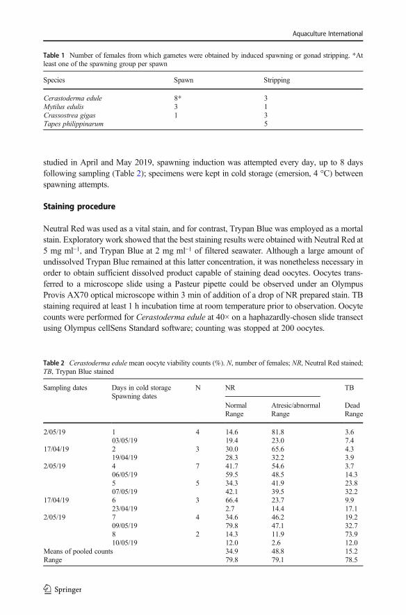
Table 2 Cerastoderma edule mean oocyte viability counts (%). N, number of females; NR, Neutral Red stained;TB, Trypan Blue stained
Sampling dates Days in cold storageSpawning dates
N NR TB
NormalRange
Atresic/abnormalRange
DeadRange
2/05/19 103/05/19
4 14.619.4
81.823.0
3.67.4
17/04/19 219/04/19
3 30.028.3
65.632.2
4.33.9
2/05/19 406/05/19
7 41.759.5
54.648.5
3.714.3
507/05/19
5 34.342.1
41.939.5
23.832.2
17/04/19 623/04/19
3 66.42.7
23.714.4
9.917.1
2/05/19 709/05/19
4 34.679.8
46.247.1
19.232.7
810/05/19
2 14.312.0
11.92.6
73.912.0
Means of pooled countsRange
34.979.8
48.879.1
15.278.5
Aquaculture International

Oocyte categories
In contrast to histological preparations, the whole mount preparations necessary for examina-tion of spawned oocytes do not provide sufficient cellular detail to definitively identify atresicoocytes; therefore, all live oocytes with abnormal shapes, cytoplasmic shrinkage, and rupturedcell membranes were designated as “atresic/abnormal.” The three oocyte viability categorieswere thus (1) “normal” (live, i.e., NR stained, normally shaped, no cytoplasmic shrinkage ormembrane rupture), (2) “atresic/abnormal” (variably NR stained, rendering atresic and abnor-mal characteristics evident), and (3) “dead” (unstained, little or no cytoplasm, rupturedmembranes).
Results
Efficient determination of all three oocyte viability categories was not possible using unstainedwhole mounts (Fig. 1 a). Neutral Red stained both normal and atresic/abnormal oocytes of allfour species studied. Atresic/abnormal oocytes were readily recognized by their irregularshapes, cytoplasmic shrinkage (undetectable in unstained cells), and ruptured cell membranes(Figs. 1 b–d and 2 a, b), rendered easily visible by the Neutral Red stain. Prior to staining withTrypan Blue, dead oocytes were either not stained by NR, or so faintly stained that they wereeasily recognized (Figs. 1, 2, 3, and 4). Oocytes stained with Trypan Blue were assumed to bedead (Figs. 1 b, c, e, 2 b, c). Only NR-stained, spherical oocytes developed into larvae (Fig. 4).The initial divisions produced uniformly NR-stained cells, whereas at the first larval stage,most of the NR appeared to have relocated to the spherical cell nuclei (Fig. 4).
Fig. 1 Cerastoderma edule whole mounts. a Unstained, spawned oocytes. b Spawned oocytes double-stainedwith Neutral Red and Trypan Blue. DO, dead oocyte; MO, mature oocyte; OC, oocyte coat; the asterisk indicatesatresic oocytes. c–e Details of oocytes from double-staining protocol: c details of mature, atresic, and deadoocytes; d detail of one mature and one atresic oocyte; note ooplasmic shrinkage; e detail of dead pedunculatedoocyte
Aquaculture International

Count results for the three oocyte categories are presented in Table 2 for Cerastodermaedule, the only species for which more than 5 spawns were obtained (Beninger et al. 2012).The ranges of inter-individual variation were themselves quite varied, e.g., from 2.7 to 79.8%for normal oocytes, and 2.6 to 48.5% for atresic/abnormal oocytes. The corresponding meanspresented similar strong variations among the different sampling and spawning dates, from14.3 to 66.4%, and 11.9 to 81.8%. The values for atresic/abnormal oocytes were generallysuperior to those of normal oocytes; dead oocytes were the smallest category except for thevalue obtained after 8 days of cold storage (73.9%). This being an observational study, nofrequentist statistical tests were performed (Beninger et al. 2012; Beninger and Boldina 2018).
Discussion
Given the diversity of cell types in which a similar result has been obtained (Crippen andPerrier 1974; Triglia et al. 1991; Lowe et al. 1992, 1995a, b; Babich and Borenfreund 1993;Weeks and Svendsen 1996; Chiba et al. 1998; Horvath and Lamberti 1999; Svendsen et al.
Fig. 2 Mytilus edulis spawned oocytes double-stained with RN and TB. DO, dead oocyte; MO, mature oocyte;the asterisk indicates atresic/abnormal oocytes
Aquaculture International

2004; Tang et al. 2006; Repetto et al. 2008; Zetsche and Meysman 2012; Aguirre-Martínezet al. 2013; Patetsini et al. 2013; Da Luz et al. 2016; Liu et al. 2018), there was little reason todoubt that NR would also stain live bivalve oocytes. Yet beyond the necessity to verify this, itwas important to determine how atresic/abnormal—“not dead yet”—oocytes would react withNR. As anticipated, these oocytes also stained to a variable degree with NR; yet the stainserved to facilitate the detection of the abnormal cell shapes, retracted cytoplasm, andmembrane rupture characteristic of such oocytes, and hence to allow their ready identificationand quantification. Overall, contrasting use of Trypan Blue was deemed unnecessary in routinework, especially since it was difficult to use, due to its low solubility in seawater and lengthystaining time (introducing an oocyte mortality artefact); it is also comparatively expensive.
The mechanism of NR vital staining has been the object of a great deal of iterativeconjecture, which has become unverified conventional wisdom since the procedure was firstpresented by Ehrlich in 1894 (Jacques 1969; Nemes et al. 1979; Hammond et al. 1980;Borenfreund and Puerner 1985; Borenfreund et al. 1988; Triglia et al. 1991; Weeks andSvendsen 1996; Svendsen et al. 2004; Repetto et al. 2008). Later studies proposed lysosomesas the “Zellen Körnchen” (cell granules) in which Ehrlich observed the stain to be concen-trated; the micrographs of Lowe et al. (1992) and Patetsini et al. (2013) Lowe et al. (1992) andPatetsini et al. (2013) are among the rare publications which actually support thisinterpretation—although not all lysosomes of a given cell appear to have this property(Winckler 1974).
Notwithstanding the conjecture concerning the exact dynamics of NR within cells, it maybe confidently assumed to cross the cell and lysosomal membranes (although the mechanism isnot known); it is accumulated in the lysosomes due to a much slower re-diffusion to the
Fig. 3 a Tapes philippinarum–stripped oocytes stained with NR. MO, mature oocyte; DO, dead oocyte. bCrassostrea gigas–stripped oocytes stained with NR. PO, pedunculated oocyte. Most live oocytes are immature
Aquaculture International

cytoplasm. Re-diffusion to the cytoplasm and the external medium is accelerated by physicalmembrane damage, and possibly impairment of the lysosomal proton pump (Winckler 1974;Lowe et al. 1992; Patetsini et al. 2013). Our results show that after leaving the lysosome, NRappears to enter and be retained in the nuclei of Crassostrea gigas trochophore larvae. Theseobservations confirm that the oocytes which took up NR were viable, and some of themdeveloped into larvae.
Prior to the present study, it was not known whether the atresic oocytes, identified in thegonad by histology, were absorbed prior to spawning, or were incapable of being spawned, orif some or all of them appeared in the spawn. Our observations confirm that a large proportionof atresic/abnormal oocytes are spawned along with the normal oocytes. The high proportionof atresic and dead oocytes observed in the present study (30–85%) is entirely consistent withthe previously published quantitative histological data (Chérel and Beninger 2017, 2019).
It is noteworthy that the animals used in the present study were sampled in the field, rather thanbeing carefully conditioned in a hatchery environment. This may explain the very large discrepancywith the proportions of inviable oocytes in controlled hatchery spawnings, which are consideredfailures at >10% non-zygotes (Uttig and Spencer 1991, Buttner and Weston 2010, Militz andSouthgate 2021). Taken together, these observations suggest that oocytes within broodstockmay beparticularly sensitive to adverse, or even normally-variable external conditions.
A high proportion of inviable post-fertilization stages is typical of high-fecundity species,including bivalves (Plough et al. 2016; Plough 2018); here we show that these high-fecundityspecies are also characterized by a high proportion of inviable pre-fertilization oocytes. The
Fig. 4 Crassostrea gigas whole mounts. Early development of stripped, fertilized oocytes stained with NR. aImmediately after fertilization. b Initiation of first cytokinesis. c 8-cell stage, nuclei (N) clearly visible. d Secondday after spawning and staining, early trochophore larva. Note NR stain now appears concentrated in sphericalnuclei. C, cilia
Aquaculture International

atresia-fraught diminishing returns of many bivalves’ high fecundity have been likened to areproductive Red Queen dilemma: greater oocyte production does not translate to a commen-surate increase in real fecundity (Chérel et al. 2020).
The very wide range of values for inviable oocytes from different individuals and timeperiods observed in the bivalve species studied here is also consistent with the SweepstakesReproductive Success (SRS) hypothesis for typical high-fecundity, type III survivorshipmarine species. Although at the population level, this strategy enables optimal exploitationof the rapidly changing environmental conditions of the nearshore marine environment, at theindividual level it has many “losers” and few “winners” (Hedgecock and Pudovkin 2011).Further support for a strong genetic inviability–SRS component to the phenomenon of oocyteatresia comes from previous studies which showed that levels of atresia remain similar underwidely different physical perturbation conditions (Chérel and Beninger 2017, 2019). Environ-mental aggression may contribute additional effects, as reviewed/shown in the gonad inBeninger (2017) and Smolarz et al. (2017), and demonstrated in the spawns of the presentstudy with increasing spawner times in cold storage.
Conclusion
To our knowledge, the Neutral Red procedure outlined herein is the first and only techniquewhich incorporates all of the essential performance criteria outlined above, allowing the rapid,easy detection of live, atresic/abnormal, and dead oocytes in spawns or gonad strippings. Itssuccess in all four bivalve species suggests that it may be a universal tool for the evaluation ofbivalve oocyte spawn quality. It can be used expeditiously for this purpose prior to hatcheryfertilization and larval rearing, especially in the absence of prolonged, careful conditioning,and also as a phenotypic marker for genetic improvement of broodstock. Application torestocking is also important, since stock production estimates are heavily dependent ondeterminations of Freal, i.e., the number of viable oocytes produced (Aoyama 1989; Koslow1992; Jennings et al. 2001; Maunder and Thorson 2019). Other important, overlappingapplications include conservation and environmental monitoring.
The oocytes of many benthic marine invertebrates are similar in overall structure and composi-tion (Adiyodi and Adiyodi 1983; Wourms 1987). Future research may therefore show NR vitalstaining to be a valuable tool in assessing oocyte viability in diverse marine taxa, including theecologically- and commercially-important Anthozoa, Polychaeta, and Gastropoda.
Acknowledgements We thank Mathilde Clairambault for her patience and perseverance in the laboratoryspawning trials. We are indebted to David Berteau and employees of Chellet-Berteau Production for their warmwelcome and permission to sample on-site.
Data availability All files from this study are available from the authors upon reasonable request.
Declarations
Ethical approval All applicable international, national, and/or institutional guidelines for the care and use ofanimals were followed by the authors.
Conflict of interest The authors declare no competing interests.
Aquaculture International

References
Adiyodi K, Adiyodi R (1983) Reproductive biology of invertebrates. Vol. I: Oogenesis, oviposition, andoosorption. Wiley, New York
Aguirre-Martínez GV, Buratti S, Fabbri E, DelValls AT, Martín-Díaz ML (2013) Using lysosomal membranestability of haemocytes in Ruditapes philippinarum as a biomarker of cellular stress to assess contaminationby caffeine, ibuprofen, carbamazepine and novobiocin. J Environ Sci 25:1408–1418
Angel-Dapa MA, Rodríguez-Jaramillo C, Cáceres-Martínez CJ, Saucedo PE (2010) Changes in lipid content ofoocytes of the penshell Atrina maura as a criterion of gamete development and quality: a study ofhistochemistry and digital image analysis. J Shellfish Res 29:407–413
Aoyama S (1989) The Mutu Bay scallop fisheries: scallop culture, stock enhancement, and resource manage-ment. In: Caddy JF (ed) Marine invertebrate fisheries: their assessment and management. Wiley, New York,pp 525–539
Arnold WS (2008) Application of larval release for restocking and stock enhancement of coastal marine bivalvepopulations. Rev Fish Sci 16:65–71
Babich B, Borenfreund E (1993) Applications of the neutral red cytotoxicity assay to risk assessment of aquaticcontaminants: an overview. In: Environmental toxicology and risk assessment, W. Landis, J. Hughes and M.Lewis. ASTM International, West Conshohocken, PA, pp 215–219
Barros J, Velasco L, Winkler F (2018) Heritability, genetic correlations and genotype by environment interactions inproductive traits of the Caribbean scallop, Argopecten nucleus(Mollusca: Bivalvia). Aquaculture 488:39–48
Beninger PG (2017) Caveat observator: the many faces of pre-spawning atresia in marine bivalve reproductivecycles. Mar Biol:164–163. https://doi.org/10.1007/s00227-017-3194-x
Beninger PG, Boldina I (2018) Quantitative considerations in mudflat ecology. In: Mudflat ecology. P.G.Beninger. Springer, Cham, Switzerland, pp 389–419
Beninger PG, Boldina I, Katsanevakis S (2012) Strengthening statistical usage in marine ecology. J ExpMar BiolEcol 426:97–108
Borenfreund E, Puerner JA (1984) A simple quantitative procedure using monolayer cultures for cytotoxicityassays (HTD/NR-90). J Tissue Cult Meth 9:7–9
Borenfreund E, Puerner JA (1985) Toxicity determined in vitro by morphological alterations and neutral redabsorption. Toxicol Lett 24:119–124
Borenfreund E, Babich H, Martin-Alguacil N (1988) Comparisons of two in vitro cytotoxicity assays—theneutral red (NR) and tetrazolium MTT tests. Toxicol in vitro 2:1–6
Boudry P (2009) Genetic variation and selective breeding in hatchery-propagated molluscan shellfish. In: Newtechnologies in aquaculture. G. Burnell and G. Allan. CRC Press-Woodhead Publishing Ltd, Boca RatonFL, pp 87–108
Buttner JK, Weston S (2010) Softshell clam culture: hatchery phase, broodstock care through seed production.Northeastern Regional Aquaculture Center Publication No. 202-2010, 12 pp
Caers M, Coutteau P, Cure K, Morales V, Gajardo G, Sorgeloos P (1999) The Chilean scallop Argopectenpurpuratus (Lamarck, 1819): II. Manipulation of the fatty acid composition and lipid content of the eggs vialipid supplementation of the broodstock diet. Comp Biochem Physiol B: Biochem Mol Biol 123:97–103
Caers M, Utting S, Coutteau P et al (2002) Impact of the supplementation of a docosahexaenoic acid–richemulsion on the reproductive output of oyster broodstock, Crassostrea gigas. Mar Biol 140:1157–1166
Cannuel R, Beninger PG (2005) Is oyster broodstock feeding always necessary? A study using oocyte qualitypredictors and validators in Crassostrea gigas. Aquat Liv Res 18:35–43
Chérel D, Beninger PG (2017) Oocyte atresia characteristics and effect on reproductive effort of Manila clamTapes philippinarum (Adams and Reeve, 1850). J Shellfish Res 36:549–557
Chérel D, Beninger PG (2019) Oocyte atresia and its effect on reproductive effort of the common cockleCerastoderma edule (Linneaus, 1758). J Shellfish Res 38:603–609. https://doi.org/10.2983/035.038.0300
Chérel D, Beninger PG, Le Pennec G (2020) Two enigmas may solve each other: the oocyte coat and atresia inthe common cockle, Cerastoderma edule (Linnaeus, 1758). Mar Biol 167:104. https://doi.org/10.1007/s00227-020-03718-6
Chiba K, Kawakami K, Tohyama K (1998) Simultaneous evaluation of cell viability by neutral red, MTT andcrystal violet staining assays of the same cells. Toxicol in vitro 12:251–258
Corporeau C, Vanderplancke G, Boulais M, Suquet M, Quéré C, Boudry P, Huvet A, Madec S (2012) Proteomicidentification of quality factors for oocytes in the Pacific oyster Crassostrea gigas. J Proteom 75:5554–5563
Crippen RW, Perrier J (1974) The use of neutral red and Evans blue for live-dead determinations of marineplankton (with comments on the use of rotenone for inhibition of grazing). Stain Technol 49:97–104
Da Luz DS, Da Silva DG, Souza MM et al (2016) Efficiency of Neutral Red, Evans Blue and MTT to assessviability of the freshwater microalgae Desmodesmus communis and Ediastrum boryanum. Phycol Res 64:56–60
Aquaculture International

Dressel DM, Heinle DR, Grote MC (1972) Vital staining to sort dead and live copepods 1, 2, 3. Chesapeake Sci13:156–159
Ehrlich P (1894) Ueber Neutralrot. Allg med Zentral-Zeit 20:Elliott DT, Tang KW (2009) Simple staining method for differentiating live and dead marine zooplankton in field
samples. Limnol Oceanogr: Methods 7:585–594Gallager SM, Mann R (1986) Growth and survival of larvae of Mercenaria mercenaria (L.) and Crassostrea
virginica (Gmelin) relative to broodstock conditioning and lipid content of eggs. Aquaculture 56:105–121Gallager SM, Mann R, Sasaki GC (1986) Lipid as an index of growth and viability in three species of bivalve
larvae. Aquaculture 56:81–103García-Corona JL, Rodríguez-Jaramillo C, Saucedo PE, López-Carvallo JA, Arcos-Ortega GF, Mazón-Suástegui
JM (2018) Internal energy management associated with seasonal gonad development and oocyte quality inthe Horsemussel Modiolus capax (Bivalvia; Mytilidae). J Shellfish Res 37:475–484
Gomez ED, Mingoa-Licuanan SS (2006) Achievements and lessons learned in restocking giant clams in thePhilippines. Fish Res 80:46–52. https://doi.org/10.1016/j.fishres.2006.03.017
Gómez-Robles E, RodrIguez-Jaramillo C, Saucedo PE (2005) Digital image analysis of lipid and proteinhistochemical markers for easuring oocyte development and quality in pearl oyster Pinctada mazatlanica(Hanley, 1856). Journal of Shellfish Research 24:1197–1202. https://doi.org/10.2983/0730-8000(2005)24[1197:DIAOLA]2.0.CO;2
González-Wangüemert M, Basso L, Balau A, Costa J, Renault L, Serrão EA, Duarte CM, Hendriks IE (2018)Gene pool and connectivity patterns of Pinna nobilis in the Balearic Islands (Spain, Western MediterraneanSea): implications for its conservation through restocking. Aquat Conserv: Mar Freshw Ecosyst 29:175–188.https://doi.org/10.1002/aqc.2976
Hammond M, Goodwin J, Dvorak H (1980) Quantitative measurements of neutral red uptake and excretion bymammalian cells. J Reticuloendothel Soc 27:337
Hedgecock D, Pudovkin AI (2011) Sweepstakes Reproductive Success in highly fecund marine fish andshellfish: a review and commentary. Bull Mar Sci 87:971–1002. https://doi.org/10.5343/bms.2010.1051
His E, Beiras R, Seaman M (1999) The assessment of marine pollution-bioassays with bivalve embryos andlarvae. Adv Mar Biol 37:1–178
Horvath TG, Lamberti GA (1999) Mortality of zebra mussel, Dreissena polymorpha, veligers during down-stream transport. Freshw biol 42:69–76
Hu W, Culloty S, Darmody G, Lynch S, Davenport J, Ramirez-Garcia S, Dawson K, Lynch I, Doyle H, SheehanD (2015) Neutral red retention time assay in determination of toxicity of nanoparticles. Mar Environ Res111:158–161. https://doi.org/10.1016/j.marenvres.2015.05.007
Jacques PJ (1969) Endocytosis. Lysosomes in biology and pathology 2:395–420Jennings S, Kaiser MJ, Reynolds JD (2001) Marine fisheries ecology. Blackwell Scientific PublicationJoaquim S, Gaspar MB, Matias D, Ben-Hamadou R, Arnold WS (2008) Rebuilding viable spawner patches of
the overfished Spisula solida (Mollusca: Bivalvia): a preliminary contribution to fishery sustainability. ICESJ Mar Sci 65:60–64
Koslow JA (1992) Fecundity and the stock–recruitment relationship. Can J of Fish Aquat Sci 49:210–217Kreeger DA, Gatenby CM, Bergstrom PW (2018) Restoration potential of several native species of Bivalve
Molluscs for water quality improvement in Mid-Atlantic Watersheds. Journal Shellfish Res 37:1121. https://doi.org/10.2983/035.037.0524
Kwok AKH, Lai TYY, Li WWY, Yew DTW, Wong VWY (2004) Trypan blue- and indocyanine green-assistedepiretinal membrane surgery: clinical and histopathological studies. Eye 18:882–888. https://doi.org/10.1038/sj.eye.6701359
Liu X, Rodeheaver DP, White JC, Wright AM, Walker LM, Zhang F, Shannon S (2018) A comparison ofin vitro cytotoxicity assays in medical device regulatory studies. Regul Toxicol Pharmacol 97:24–32. https://doi.org/10.1016/j.yrtph.2018.06.003
Lowe D, Moore M, Evans B (1992) Contaminant impact on interactions of molecular probes with lysosomes in livinghepatocytes from dab Limanda limanda. Mar Ecol Prog Ser 91:135–140. https://doi.org/10.3354/meps091135
Lowe D, Fossato V, Depledge M (1995a) Contaminant-induced lysosomal membrane damage in blood cells ofmussels Mytilus galloprovincialis from the Venice Lagoon: an in vitro study. Mar Ecol Prog Ser 129:189–196. https://doi.org/10.3354/meps129189
Lowe DM, Soverchia C, Moore MN (1995b) Lysosomal membrane responses in the blood and digestive cells ofmussels experimentally exposed to fluoranthene. Aquat Toxicol 33:105–112. https://doi.org/10.1016/0166-445X(95)00015-V
Massapina C, Joaquim S, Matias D, Devauchelle N (1999) Oocyte and embryo quality in Crassostrea gigas(Portuguese strain) during a spawning period in Algarve, South Portugal. Aquat Living Resour 12:327–333
Maunder MN, Thorson JT (2019) Modeling temporal variation in recruitment in fisheries stock assessment: areview of theory and practice. Fish Res 217:71–86
Aquaculture International

Mejuto J (1984) Primeros datos sobre la dinámica de la población de Cerastoderma edule (L.) de la Ría doPasaxe (NW de Galicia): estrategias de explotación. Actas IV Simp Iber Estud Bentos Mar 1:83–102
Militz TA, Southgate PC (2021) Chapter 3: Culture of giant clams. In: Shumway, S (ed) Molluscan shellfishaquaculture: a practical guide. 5M Books Ltd, Essex, pp 61-85
Moens T, Beninger PG (2018) Meiofauna: an inconspicuous but important player in mudflat ecology. In:Beninger PG (ed) Mudflat Ecology. Springer International Publishing, Cham, pp 91–147
Nemes Z, Dietz R, Lüth JB, Gomba S, Hackenthal E, Gross F (1979) The pharmacological relevance of vitalstaining with neutral red. Experientia 35:1475–1476. https://doi.org/10.1007/BF01962793
Nevejan N, Courtens V, Hauva M, Gajardo G, Sorgeloos P (2003) Effect of lipid emulsions on production andfatty acid composition of eggs of the scallop Argopecten purpuratus. Mar Biol 143:327–338
Patetsini E, Dimitriadis VK, Kaloyianni M (2013) Biomarkers in marine mussels, Mytilus galloprovincialis,exposed to environmentally relevant levels of the pesticides, chlorpyrifos and penoxsulam. Aquat Toxicol126:338–345. https://doi.org/10.1016/j.aquatox.2012.09.009
Pérez Camacho A, Román G (1984) Crecimiento, reproducción, mortalidad y producción del berberechoCerastoderma edule (L.), en la Ría de Arousa. Cuad Area Cienc Mariñ Sem Est Gal 499–507
Plough LV (2018) Fine-scale temporal analysis of genotype-dependent mortality at settlement in the Pacificoyster Crassostrea gigas. J Exp Mar Biol Ecol 501:90–98
Plough L, Shin G, Hedgecock D (2016) Genetic inviability is a major driver of type III survivorship inexperimental families of a highly fecund marine bivalve. Mol Ecol 25:895–910
Rajab GZE-A, Demer JL (2019) Long-term results of surgical excision of conjunctival retention cyst usingtrypan blue with methylcellulose. Am J Ophthalmol Case Rep 14:28–31. https://doi.org/10.1016/j.ajoc.2019.01.010
Repetto G, del Peso A, Zurita JL (2008) Neutral red uptake assay for the estimation of cell viability/cytotoxicity.Nat Protoc 3:1125–1131. https://doi.org/10.1038/nprot.2008.75
Shantharam AK, Padilla DK, Peterson BJ, Doall M, Lobue C, Webb A (2019) Macrofaunal community structurefollowing the restocking of Northern Quahog (Mercenaria mercenaria) to Great South Bay, Long Island,NY. J Shelf Res 38:259–270. https://doi.org/10.2983/035.038.0206
Smolarz K, Hallmann A, Zabrzańska S, Pietrasik A (2017) Elevated gonadal atresia as biomarker of endocrinedisruptors: field and experimental studies using Mytilus trossulus (L.) and 17-alpha ethinylestradiol (EE2).Mar Poll Bull 120:58–67. https://doi.org/10.1016/j.marpolbul.2017.04.007
Strober W (2015) Trypan Blue exclusion test of cell viability. Curr Protoc Immunol 111:A3.B.1-A3.B.3. doi:https://doi.org/10.1002/0471142735.ima03bs111
Svendsen C, Spurgeon DJ, Hankard PK, Weeks JM (2004) A review of lysosomal membrane stability measuredby neutral red retention: is it a workable earthworm biomarker? Ecotoxicol Environ Safety 57:20–29. https://doi.org/10.1016/j.ecoenv.2003.08.009
Talbot P, Chacon RS (1981) A triple-stain technique for evaluating normal acrosome reactions of human sperm. JExp Zool 215:201–208. https://doi.org/10.1002/jez.1402150210
Tang KW, Freund CS, Schweitzer CL (2006) Occurrence of copepod carcasses in the lower Chesapeake Bay andtheir decomposition by ambient microbes. Estuar Coast Shelf Sci 68:499–508
Triglia D, Braa SS, Yonan C, Naughton GK (1991) In vitro toxicity of various classes of test agents using theneutral red assay on a human three-dimensional physiologic skin model. In Vitro Cell Devel Biol-Animal27:239–244
Uttig SD, Spencer BE (1991) The hatchery culture of bivalve mollusc larvae and juveniles. Laboratory Leaflet,Ministry of Agriculture, Fisheries and Food Fisheries Research, Lowestoft 68:31 pp
Utting SD, Millican P (1997) Techniques for the hatchery conditioning of bivalve broodstocks and thesubsequent effect on egg quality and larval viability. Aquaculture 155:45–54
Valdez-Ramirez ME, Donval A, Le Pennec M (2002) Ultrastructural and histochemical criteria for determiningnormality in mature oocytes of the Pacific oyster Crassostrea gigas. J Shellfish Res 21:707–714
Wales R (1959) The differential staining of human and dog spermatozoa. Aust J Exp Biol Med 37:433–439.https://doi.org/10.1038/icb.1959.44
Weeks JM, Svendsen C (1996) Neutral red retention by lysosomes from earthworm (Lumbricus rubellus)coelomocytes: A simple biomarker of exposure to soil copper. Environ Toxicol Chem 15:1801–1805.https://doi.org/10.1002/etc.5620151022
Wijsman JWM, Troost K, Fang J, Roncarati A (2019) Global production of marine bivalves. Trends andchallenges. In: Smaal AC, Ferreira JG, Grant J et al (eds) Goods and services of marine bivalves.Springer International Publishing, Cham, pp 7–26
Winckler J (1974) Vitalfärbung von Lysosomen und anderen Zellorganellen der Ratte mit Neutralrot. ProgHistochem Cyto 6:III–89. https://doi.org/10.1016/S0079-6336(74)80001-X
Aquaculture International

Wourms J (1987) Oogenesis. In: Reproduction of marine invertebrates, Vol 9: General aspects, seeking unity indiversity. Blackwell Scientifc Publications, Palo Alto, pp 117–124
Zetsche EM, Meysman FJ (2012) Dead or alive? Viability assessment of micro-and mesoplankton. J PlanktonRes 34:493–509
Publisher’s note Springer Nature remains neutral with regard to jurisdictional claims in published mapsand institutional affiliations.
Aquaculture International