exam1 prep (1)
TRANSCRIPT

8/12/2019 Exam1 Prep (1)
http://slidepdf.com/reader/full/exam1-prep-1 1/6
EXAM PREPARATION
Format will be similar to that of sample quiz.
1. Digital imaging: pixels, row, column, pixel values, two-dimensional, three-dimensional
2. Linear interpolation to assign pixel values (partial pixel sharing)I1
I2
Incident: I => ,)1(1 a I I −= a I I =2 a
i.e. a=0.2, I=100, I 1=80, I 2=20I=I 1+ I 2
3. What is an imaging system? What do we want to image in biomedical imaging?
4. What is resolution?1) Spatial resolution : the ability to distinguish small spatial detail in an image,
typically measured in distance (mm) or line pairs/mm
Good resolution, ability to distinguish two objects (two peaks along the profile)
2) Contrast resolution : ability to distinguish slight difference in signal level between different materials
3) Temporal resolution : ability to distinguish 2 events occurring at different times(e.g. a patient took an x-ray chest imaging, then another one after two months)
4) trade off between different parameters:
1
8/12/2019 Exam1 Prep (1)
http://slidepdf.com/reader/full/exam1-prep-1 2/6
5. X-ray is most common imaging procedure.
Especially suited for imaging tissues whose density differs significantly fromnormal soft tissue, e.g. bone and lung (air), i.e., need high contrast targets.
6. Roentgen
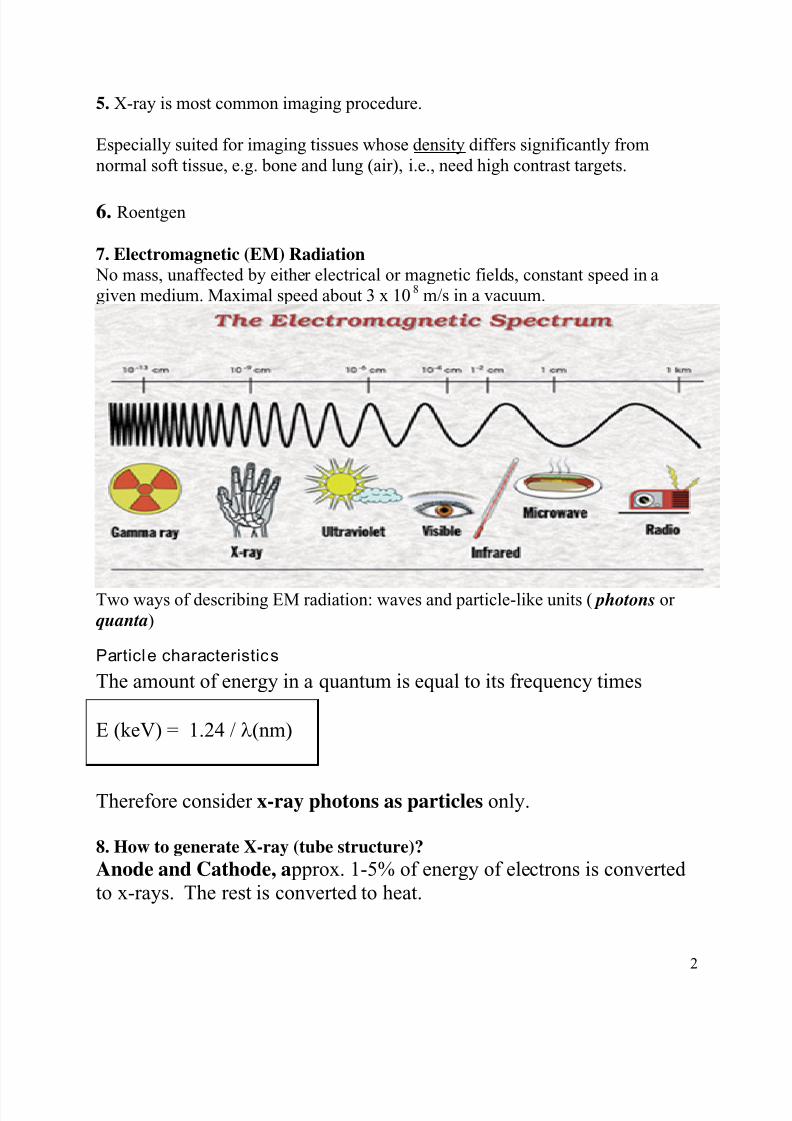
7. Electromagnetic (EM) Radiation No mass, unaffected by either electrical or magnetic fields, constant speed in agiven medium. Maximal speed about 3 x 10 8 m/s in a vacuum.
Two ways of describing EM radiation: waves and particle-like units ( photons orquanta )
Particl e characteristic s
The amount of energy in a quantum is equal to its frequency times
E (keV) = 1.24 / λ (nm)
Therefore consider x-ray photons as particles only.
8. How to generate X-ray (tube structure)?Anode and Cathode, a pprox. 1-5% of energy of electrons is convertedto x-rays. The rest is converted to heat.
2

8/12/2019 Exam1 Prep (1)
http://slidepdf.com/reader/full/exam1-prep-1 3/6
9. Production of X-rays (inside structure): (graudate students only, for details)A.“Brehmstrahlung” German for braking radiation): Scattered electron hasundergone a change of direction, i.e. an acceleration due to high + charge ofnucleus. It is accompanied by a release of energy (radiation).
B. Characteristic X-ray:
10. Typical x-ray spectrum (graduate students):
11. Interactions of x-rays with tissue (graudate students only, for details)X-ray is attenuated by interactions with tissue1) Coherent Scattering (Rayleigh scattering)2) Compton Scattering
3

8/12/2019 Exam1 Prep (1)
http://slidepdf.com/reader/full/exam1-prep-1 4/6
3) Photoelectric effect
12. X-ray attenuation, can calculate complicate case.
x
e I I −
= μ
0
13. Magnification (graduate students):
Object #1
detector
H1
H2
Object #2
SID
D2 D1
Please calcualte D 1 and D 2. Which one is closer to the real size of original object?What does this mean? Does projection x-ray imaging reflect the real size of objects
being x-rayed?
14. Principals of CTCT provides 3D image of the object and distinguish overlapped structures.
4

8/12/2019 Exam1 Prep (1)
http://slidepdf.com/reader/full/exam1-prep-1 5/6
A B
C
C’’
B’’
A’’
A’ B’ and C’
15. Parallel beam, fan beam, cone beam
16. Image in spatial domain Fourier transform image in frequency domain
(below graduate students)
1 ) F(0,0) represents the DC-component of the image which corresponds tothe average brightness: (u=0,v=0 => F(0,0)= ∑∑ ),(
12 y x f
N )
2) F(N-1,N-1) represents the highest frequency.
17. (graduate students) Nyquist rate -- The sampling rate should be at least twice the maximum frequency responses. Indeed many times more the better.
18. Dual energy, tomosynthesis
19. (graduate students)--Spatial domain:
point spread function (PSF), line spread function (LSF), edge spread function(ESF)
5

8/12/2019 Exam1 Prep (1)
http://slidepdf.com/reader/full/exam1-prep-1 6/6