evidence review: critical congenital … draft evidence review: critical congenital cyanotic heart...
TRANSCRIPT

Final Draft
EVIDENCE REVIEW: Critical Congenital Cyanotic Heart Disease
Prepared for: MATERNAL AND CHILD HEALTH BUREAU
Version: September 3, 2010
Authors:
Alixandra A. Knapp, Danielle R. Metterville, Alex R. Kemper, Lisa Prosser, James M. Perrin
Evidence Review Group: Chairperson, James M. Perrin, MD (MGH Center for Child and Adolescent Health Policy)
Committee Members:
Marsha Browning, MD, MPH, MMSc (Massachusetts General Hospital) Anne Marie Comeau, PhD (University of Massachusetts) Nancy Green, MD (Columbia University) Alex R. Kemper, MD, MPH, MS (Duke University)
Alixandra A. Knapp, MS (MGH Center for Child and Adolescent Health Policy) Danielle R. Metterville, MS (MGH Center for Child and Adolescent Health Policy) Lisa Prosser, PhD (University of Michigan) Denise Queally, JD (Consumer Representative)
This review was made possible by subcontract number SC-07-028 to Massachusetts General Hospital, Center for Child and Adolescent Health Policy under prime contract number HHSP23320045014XI to Altarum Institute, from the Maternal and Child Health Bureau (MCHB) (Title V, Social Security Act), Health Resources and Services Administration (HRSA), U.S. Department of Health and Human Services (DHHS).

Final Draft
2 | P a g e
Table of Contents Page
i. Abbreviations used 3
I. Introduction 4
II. Methods for developing case definition 4
III. Case definition 5
IV. Rationale for review 5
V. Objectives 6
VI. Conceptual framework 6
VII. Statement of key questions 6
VIII. Literature review methods 7
IX. Methods for interviews with experts 10
X. Results: evidence findings to address the key questions 13
XI. Key findings and summary 31
XII. Tables of abstracted literature 34
XIII. References 38
XIV. Appendix A: Conflict of interest form*
XV. Appendix B: Letter and Questions for CCCHD Experts*
XVI. Appendix C: Letter and Questions for CCCHD Advocates*
*Appendices available upon request

Final Draft
3 | P a g e
i. Abbreviations used CHD Congenital Heart Disease CCHD Critical Congenital Heart Disease CCCHD Critical Congenital Cyanotic Heart Disease HLHS Hypoplastic Left Heart Syndrome NICU Neonatal Intensive Care Unit TAPVR Total Anomalous Pulmonary Venous Return TGA Transposition of the Great Arteries TOF Tetralogy of Fallot VSD Ventricular Septal Defect

Final Draft
4 | P a g e
I. Introduction Congenital heart disease (CHD) is an overarching term describing a spectrum of clinical outcomes derived from any number of defects that are present in the structure of the heart at birth. Specific defects may involve the interior walls of the heart, valves inside the heart or the arteries and veins that carry blood to the heart or out to the body. These varied defects change the normal flow of blood through the heart, leading to a range of conditions and symptoms. Critical congenital heart disease (CCHD) is a group of defects that cause severe and life-threatening symptoms and require intervention within the first year of life. Congenital heart disease affects about 7 to 9 of every 1000 live births in the United States and Europe (Botto, Correa, & Erickson, 2001; Knowles et al., 2005; Wren, Richmond, & Donaldson, 2000). About one-quarter of these neonates will have CCHD (Mahle et al., 2009; Talner, 1998). Congenital heart disease is the most common cause of death in the first year of life, with defects accounting for 3% of all infant deaths and more than 40% of all deaths due to congenital malformations (Aamir, Kruse, & Ezeakudo, 2007; Knowles et al., 2005; Koppel et al., 2003; Lee et al., 2001). Newborn screening using pulse oximetry has been proposed as a method to augment current approaches to the detection of CCHD, which include prenatal ultrasound screening and careful and repeated clinical examinations, both in the nursery and as part of routine well-child care. Pulse oximetry is a non-invasive test that estimates the percentage of hemoglobin in blood that is saturated with oxygen (Mahle et al., 2009). Newborn screening using pulse oximetry is a test that occurs at the bedside (in the nursery or otherwise) similar to newborn screening for hearing impairment. Unlike newborn hearing screening, neonates with abnormal pulse oximetry screening results need confirmatory testing prior to discharge. The confirmatory testing that follows an abnormal pulse oximetry result is the same confirmatory testing that would follow an abnormal clinical exam.
II. Methods for developing case definition At the onset of this review, we developed a case definition focused on the particular CCHD thought to most likely benefit from a newborn screening program (i.e. critical congenital cyanotic heart disease (CCCHD) by pulse oximetry screening). We first contacted pediatric cardiologists and neonatologists identified as authors of relevant literature or by Advisory Committee on Heritable Disorders in Newborns and Children (ACHDNC) and Evidence Review Group (ERG) recommendation (Robert Beekman, III, MD, MS; Robert Koppel, MD and William Mahle, MD). ERG members held a conference call with these experts to discuss the nomination and pertinent key questions. A case definition was drafted and subsequently discussed and agreed upon by the members of the ERG and the ACHDNC Nomination and Prioritization committee.

Final Draft
5 | P a g e
III. Case definition CCHD is defined as any critical congenital heart defect requiring surgery or catheter intervention in the first year of life (Mahle et al., 2009). This report focuses on CCCHD, which encompasses those cases of CCHD most likely to present with significant enough hypoxemia for pulse oximetry to detect in the newborn nursery (Mahle et al., 2009). This review focuses on CCCHD known to present most or all of the time with hypoxemia (Mahle et al., 2009):
• Hypoplastic left heart syndrome (HLHS) • Pulmonary atresia, intact septum • Tetralogy of Fallot (TOF) • Total anomalous pulmonary venous return (TAPVR) • Transposition of the great arteries (TGA) • Tricuspid atresia • Truncus arteriosus
While other types of CCHD may present with hypoxemia, they do so only some of the time and are therefore less likely to be detected by pulse oximetry screening (Mahle et al., 2009). These particular heart defects are thus not always cyanotic and consequently not reviewed in this evidence review. These additional critical congenital heart defects may be considered secondary screening targets because they are sometimes detected by newborn screening with pulse oximetry. The following are examples of types of CCHD that were not included in this review:
• Aortic arch atresia or hypoplasia • Coarctation of the aorta • Double-outlet right ventricle • Ebstein anomaly • Pulmonic stenosis or atresia • Ventricular septal defect
IV. Rationale for review
The Advisory Committee on Heritable Disorders in Newborns and Children (ACHDNC) has directed the Evidence Review Group (ERG) to produce this report for the nominated condition of CCCHD. CCCHD has been nominated because:
1. CCCHD causes significant morbidity and mortality. 2. Newborn screening for CCCHD with pulse oximetry has been examined in
several large studies. 3. Identification of CCCHD in neonates might improve health outcomes.

Final Draft
6 | P a g e
V. Objectives The objective of this review is to provide information to the ACHDNC about the potential benefits, harms, and costs of adding CCCHD to the list of primary conditions for newborn screening, based on published studies and other data available from experts in screening for treatment of CCCHD.
VI. Conceptual framework
The conceptual framework below (Figure 1) illustrates our approach to evaluating the evidence regarding the potential benefits and harms of newborn screening for CCCHD. Our goals are (1) to assess the effectiveness of screening and (2) to assess the impact of treatment for those identified through newborn screening vs. those identified later through clinical diagnosis. Figure 1 - Conceptual framework
VII. Statement of key questions
The key questions for our review follow. Condition: 1. What is the prevalence of CCCHD among those neonates eligible for screening?
This excludes those neonates who are previously known to have CCCHD, who have a syndrome commonly associated with congenital heart disease, or who have signs or symptoms consistent with CCCHD prior to screening.
2. What is the natural history, including the spectrum of severity, of CCCHD among neonates eligible for screening?
Screening Test:

Final Draft
7 | P a g e
3. What is the accuracy of pulse oximetry in the newborn period for CCCHD? How does this vary by the age of the neonate, placement of the probes, and threshold value for action?
4. How many additional cases of CCCHD would routine neonatal screening with pulse oximetry detect prior to hospital discharge, compared to current care, including screening prenatal ultrasounds and routine newborn clinical history and examination?
5. What is the false positive and false negative rate of routine neonatal screening with pulse oximetry for CCCHD?
6. What are the potential harms or risks associated with screening? Diagnostic Test: 7. How available is echocardiography to evaluate those with a positive pulse oximetry
screening result? Treatment: 8. Does pre-symptomatic or early symptomatic intervention in newborns or infants with
CCCHD improve health outcomes? 9. What is the availability of treatment? Economics: 10. What are the costs associated with the screening test? 11. What are the costs associated with failure to diagnose in the pre-symptomatic
period? 12. What are the costs associated with treatment? 13. What is the cost-effectiveness of newborn screening for CCCHD? Other: 14. What critical evidence appears lacking that may inform screening recommendations
for CCCHD?
VIII. Literature review methods We conducted separate literature searches to identify relevant literature regarding (1) natural history and treatment (combined search given overlap of search terms), (2) screening, (3) diagnosis, and (4) economics from January 1990 to June 2010. We completed searches combining the National Library of Medicine Medical Subject Heading (MeSH) and keywords for each search (Table 1) for relevant literature of all articles written about CCCHD over this time period. In order to capture articles that have not yet been assigned MeSH terms, we also searched the same keywords within the OVID In-Process and Other Non-Indexed Citations database. Each search was limited to human studies, English language publications and infant (birth to 23 months). To ensure completeness of the literature search, we reviewed reference lists and the nomination form citations submitted to the AC. This search strategy yielded 367 total potential articles (Table 1).
Three investigators (AAK, ARK, and DRM) reviewed all abstracts to select articles for inclusion in the review. Studies were selected by a systematic multi-stage inclusion and exclusion process. Articles were eliminated for the screening, diagnosis, and economics sections if they were: not human studies; did not focus on congenital heart disease; not

Final Draft
8 | P a g e
relevant to the review; opinion or editorial pieces; seminars and symposiums without systematic review methods; not peer-reviewed; focused on the prenatal period; description of a single treatment procedure in-depth; a defect not listed in the case definition; a variant of a defect listed in the case definition; reviews that did not include new data; case reports; or case-series of four or fewer subjects. Natural history and treatment articles were subject to the same exclusion criteria as above; however, due to the vast amount of literature unrelated to the key questions of this review, we used systematic review articles published in peer-reviewed journals. When review articles meeting our criteria were not available for a defect listed in the case definition, all available multi-institutional case series meeting prior inclusion and exclusion criteria were used. The largest case series available from a single institution was used when no relevant review article or multi-institutional study was available for the treatment of a defect described in the case definition. Disagreements were resolved through consensus with emphasis on inclusion of any potentially useful data. After abstract review, 67 manuscripts were reviewed in full (Table 1). All full length articles were subjected to the inclusion and exclusion criteria above and studies that met the predefined and explicit criteria were selected for the review. In cases of duplicate publications, the most recent or complete versions were selected. After this process, 26 total articles met all inclusion criteria and were included in this evidence review (Table 1). Table 1 – Search strategy and inclusion of articles Screening Natural History and
Treatment Diagnosis Economics
National Library of Medicine Medical Subject Heading (MeSH) and keywords:
"oximetry", "pulse", "pulse oximetry", "congenital", "heart", "disease", "congenital heart disease", "screening", and "newborn"
"Heart Defects, Congenital", "Cardiac Surgical Procedures", "Heart Catheterization", "Treatment Outcome", "Tricuspid Atresia", "Transposition of Great Vessels", "Tetralogy of Fallot", "Truncus Arteriosus", “Pulmonary Atresia, Intact Septum”, “Hypoplastic Left Heart Syndrome” and "Total Anomalous Pulmonary Venous Connection"
"Heart Defects, Congenital", "Echocardiography", "Physical Examination", and "Neonatology [Methods].”
"Heart Defects, Congenital", "Economics”, “Pulse Oximetry,” “Oximetry,” "Cost-Benefit Analysis", and "Neonatal Screening.”
Type of manuscript included
Original Research
Systematic Review Articles; multi-institutional studies or large case series used when no relevant review article was available for the treatment of a defect described in the case definition
Original Research
Original Research

Final Draft
9 | P a g e
Abstracts resulted and reviewed 75 282 4 6 from search strategy Read complete manuscript
18 43 4 2
Abstracted article and included in 11 11 3 1 evidence review The three investigators (AAK, ARK, and DRM) each independently abstracted one-third of the articles, and a fourth investigator (LP) assisted in reviewing economic manuscripts only. The three investigators reviewed a subset (20% overlap) to validate the process. Each original research manuscript was evaluated, using standardized tools, for the quality of the study design (NHS Center for Reviews and Dissemination March 2001, Accessed: October 17, 2008) and the quality of the evidence, as it relates to the category of evidence (Pandor et al., 2004; Pollitt et al., 1997). A given article received only one rating per reader for study design, but may have received multiple quality evaluations for the type of evidence. For example, a study that discusses screening and natural history would be evaluated for the quality of the evidence in each of those domains. There were no significant differences in the data extracted by the reviewers. Table 2 – Study design for abstracted articles
Study Design Number of Articles Experimental intervention 0 Cohort study 0 Case-control study 0 Case series 7
Sample size ≤ 10 0 Sample size 11 to 50 0
Sample size 51 to 100 0 Sample size ≥ 101 7
Economic Evaluation Cross-Sectional study Systematic Reviews Total studies
1 11 7 26
To assure completeness and clarity of the report, a draft of the report was sent to an independent external review panel (see Appendix A for sample conflict of interest form). The report was revised based on their suggestions.

Final Draft
10 | P a g e
IX. Methods for interviews with experts The ERG and the ACHDNC recognize that there may be important but unpublished data regarding CCCHD. We identified experts, including researchers and CCCHD newborn screening advocates, to help us identify this information. These individuals were identified as authors of key papers included in the literature review, through discussions with content experts, and through recommendations from the ERG and the AC. Table 3 lists these individuals.
Experts were sent a letter via e-mail (Appendix B for researchers and Appendix C for advocates) explaining the purpose of the review, a conflict of interest form (Appendix A) and an open-ended survey. Experts had two weeks to respond, and extensions were granted when requested. The project coordinator sent at least one reminder e-mail to those who did not reply. In cases where clarifications were needed regarding the responses, individuals were either sent a follow up e-mail or contacted via telephone by the authors. When experts and advocates provided information regarding the key questions not otherwise available from the selected articles, we include their responses.
Table 3 – Key experts contacted and degree of participation
Name Title Replied
Completed written survey
Telephone interview
Robert
Beekman, III, MD, MS
Professor of Pediatric Cardiology, University of Cincinnati College of Medicine, Cincinnati, Ohio
Elizabeth Bradshaw, MSN, RN, CPN
Clinical Supervisor, Children's National Heart Institute, Congenital Heart Disease Screening Program Coordinator, Washington, DC
Robert Campbell, MD
Division Director of Pediatric Cardiology, Professor of Pediatrics, Emory University School of Medicine; Medical Director, Project S.A.V.E. Children’s Healthcare of Atlanta; Director, Sibley Heart Center Cardiology , Atlanta, Georgia
Edward Clark, MD Chairman of Department of Pediatrics, University of Utah; Medical Director, Primary Children’s Medical Center, Salt Lake City, UT
*
Adolfo Correa, MD, MPH
Medical Officer, National Center on Birth Defects and Developmental Disabilities (NCBDDD), Centers for Disease Control and Prevention, Atlanta, Georgia
William Foley Executive Director, The Children’s Heart Foundation

Final Draft
11 | P a g e
Shannon Hamrick, MD Assistant Professor Department of Pediatrics Neonatology Division Emory University School of Medicine Atlanta, Georgia
*
Margaret (Peggy)
Honein, PhD, MPH
Epidemiologist, National Center on Birth Defects and Developmental Disabilities (NCBDDD),Centers for Disease Control and Prevention, Atlanta, Georgia
Robert Koppel, MD Attending Neonatologist, Regional Perinatal Center Director for Schneider Children’s Hospital at Long Island Jewish Medical Center, New York
Jennifer Li, MD Duke University Medical Center Consortium, Durham, North Carolina
William Mahle, MD Medical Director, Clinical Research, Pediatric Cardiologist, Sibley Heart Center Cardiology, Children’s Healthcare of Atlanta, Atlanta, Georgia
Gerard Martin, MD Senior vice president of the Center for Heart, Lung and Kidney Disease and co-director of the Children’s National Heart Institute, Washington, DC
LuAnn Minich, MD Primary Children's Medical Center, Salt Lake City, Utah
Jane Newburger, MD, MPH
Associate Cardiologist-in-Chief, Children's Hospital Boston, Boston, Massachusetts
Jonathan Reich, MD, MS
Cardiology, Cardiology-Pediatrics, The Watson Clinic LLP, The Watson Clinic Center for Research, Lakeland, Florida
Michelle Rintamaki President, Kids With Heart Annamarie Saarinen Parent advocate, 1in100 J. Philip Saul, MD Medical University of South Carolina,
Charleston, South Carolina Dorothy Sendelbach,
MD Division of Neonatal-Perinatal Medicine, Department of Pediatrics, University of Texas Southwestern Medical Center, Dallas, Texas
Corrie Stassen Executive Director, It's My Heart, Inc. ^ Barbara Stoll, MD Professor and George W. Brumley Jr.
Chair of Pediatrics, Emory Children's Center, Emory School of Medicine, Atlanta, Georgia
*
Arnold Strauss, MD Professor and Chair of Pediatrics, University of Cincinnati College of Medicine, Cincinnati, Ohio
Lloyd Tani, MD Chief of Division of Pediatric Cardiology, University of Utah, Utah

Final Draft
12 | P a g e
Ronald Woods, MD Principal Investigator, Newborn Screening for Heart Defects, MultiCare Health System, Tacoma General Hospital & Mary Bridge Children’s Hospital & Health Center Pediatric Heart Center, Tacoma, Washington
Diane Zook, BS Research Study Coordinator, Newborn Screening for Heart Defects MultiCare Health System, Tacoma General Hospital & Mary Bridge Children’s Hospital & Health Center Pediatric Heart Center, Tacoma, Washington
*Deferred to other experts ^Could not complete written survey at this time
Formatted Table

Final Draft
13 | P a g e
X. Results: evidence findings to address the key questions This section presents the evidence from the included articles organized by key question. Each subsection includes a summary of findings from the literature review, assessment of the quality of the evidence from each included article, and information from experts. A. Natural history and diagnosis: Table 4 – Abstracted literature pertaining to natural history
Type of evidence Number of articles Total 11
Review Articles 7
Multi-institution case series (tricuspid atresia; pulmonary atresia; intact septum) 2
Single institution, largest case series available (TAPVR; truncus arteriosus) 2 Literature review:
The following literature review covers the natural history of only the seven critical congenital heart defects listed in the case definition. These seven critical congenital heart defects account for approximately 17-31% of all congenital heart disease. Of the seven defects listed, six of the seven are always associated with neonatal hypoxemia; Tetralogy of Fallot usually, but not always, is associated with neonatal hypoxemia (Table 5). Table 5 also lists which of the seven defects are ductal-dependent. "Ductal" refers to the ductus arteriosus, a blood vessel that humans have before birth that allows blood to bypass the lungs in utero, and normally closes shortly after birth. Some types of CCCHD require the ductus to remain open (“patent”) to allow sufficient circulation, thereby oxygenation, to keep the neonate alive. Medication (prostaglandins) infused intravenously can maintain patency of the ductus arteriosus. However, prostaglandin infusion can lead to significant adverse events, such as apnea.
Table 5 – Selected types of CCCHD and associated natural history
Heart Defect Hypoxemia Ductal-dependent
Birth prevalence
Age at onset of symptoms Untreated survival
HLHS All All 1-5 to 6-7/ 10,000 live births
Immediately or within the first two months of life
Universally fatal without surgical intervention
Pulmonary atresia, intact septum
All All 1% of all congenital cardiac defects
Immediately When the ductus closes, the neonate becomes severely ill, leading to death if not urgently treated
TOF Most Uncommon 3 /10,000 live births
Neonatal period
Amount of pulmonary blood flow obstruction determines onset and severity of symptoms
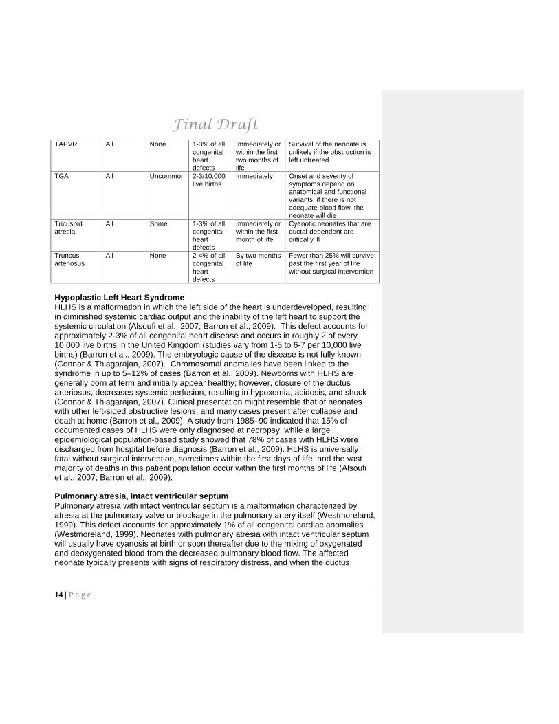
Final Draft
14 | P a g e
TAPVR All None 1-3% of all congenital heart defects
Immediately or within the first two months of life
Survival of the neonate is unlikely if the obstruction is left untreated
TGA All Uncommon 2-3/10,000 live births
Immediately Onset and severity of symptoms depend on anatomical and functional variants; if there is not adequate blood flow, the neonate will die
Tricuspid atresia
All Some 1-3% of all congenital heart defects
Immediately or within the first month of life
Cyanotic neonates that are ductal-dependent are critically ill
Truncus arteriosus
All None 2-4% of all congenital heart defects
By two months of life
Fewer than 25% will survive past the first year of life without surgical intervention
Hypoplastic Left Heart Syndrome HLHS is a malformation in which the left side of the heart is underdeveloped, resulting in diminished systemic cardiac output and the inability of the left heart to support the systemic circulation (Alsoufi et al., 2007; Barron et al., 2009). This defect accounts for approximately 2-3% of all congenital heart disease and occurs in roughly 2 of every 10,000 live births in the United Kingdom (studies vary from 1-5 to 6-7 per 10,000 live births) (Barron et al., 2009). The embryologic cause of the disease is not fully known (Connor & Thiagarajan, 2007). Chromosomal anomalies have been linked to the syndrome in up to 5–12% of cases (Barron et al., 2009). Newborns with HLHS are generally born at term and initially appear healthy; however, closure of the ductus arteriosus, decreases systemic perfusion, resulting in hypoxemia, acidosis, and shock (Connor & Thiagarajan, 2007). Clinical presentation might resemble that of neonates with other left-sided obstructive lesions, and many cases present after collapse and death at home (Barron et al., 2009). A study from 1985–90 indicated that 15% of documented cases of HLHS were only diagnosed at necropsy, while a large epidemiological population-based study showed that 78% of cases with HLHS were discharged from hospital before diagnosis (Barron et al., 2009). HLHS is universally fatal without surgical intervention, sometimes within the first days of life, and the vast majority of deaths in this patient population occur within the first months of life (Alsoufi et al., 2007; Barron et al., 2009). Pulmonary atresia, intact ventricular septum Pulmonary atresia with intact ventricular septum is a malformation characterized by atresia at the pulmonary valve or blockage in the pulmonary artery itself (Westmoreland, 1999). This defect accounts for approximately 1% of all congenital cardiac anomalies (Westmoreland, 1999). Neonates with pulmonary atresia with intact ventricular septum will usually have cyanosis at birth or soon thereafter due to the mixing of oxygenated and deoxygenated blood from the decreased pulmonary blood flow. The affected neonate typically presents with signs of respiratory distress, and when the ductus

Final Draft
15 | P a g e
arteriosus closes, the neonate can become severely cyanotic and acidotic, leading to death if not urgently treated (Westmoreland, 1999). Tetralogy of Fallot TOF is a malformation that consists of four associated defects: ventricular septal defect, obstruction of the right ventricular outflow tract, override of the ventricular septum by the aortic root, and right ventricular hypertrophy (Bailliard & Anderson, 2009; Derby & Pizarro, 2005). This combination of lesions is the most common form of cyanotic heart disease, accounting for 5–10% of all congenital cardiac malformations, and occurs in 3 of every 10,000 live births (Bailliard & Anderson, 2009; Derby & Pizarro, 2005). The etiology is multifactorial, and the majority of patients with TOF present in the neonatal period with mild-to-moderate cyanosis, but typically without respiratory distress (Bailliard & Anderson, 2009). The amount of pulmonary blood flow obstruction determines the onset and severity of symptoms; more severe illness presents with a greater obstruction (Westmoreland, 1999). Total Anomalous Pulmonary Venous Return TAPVR is a malformation characterized by an absent connection between the pulmonary veins and left atrium; the pulmonary veins empty into the right atrium or systemic veins. This results in mixed oxygenated and unoxygenated blood entering the systemic circulation, and the amount of oxygenated blood entering the systematic circulation depends upon the presence and degree of obstruction in the pulmonary veins (Westmoreland, 1999). TAPVR accounts for 1-3% of all congenital heart defects (Karamlou et al., 2007). If a severe obstruction is present at birth, the newborn will present with immediate and profound cyanosis, tachypnea, pulmonary edema, and pulmonary hypertension; survival of the neonate is unlikely if the obstruction is left untreated (Westmoreland, 1999). Infants with unobstructed abnormal connections at birth will have symptoms within the first two months of life (Westmoreland, 1999). Transposition of the Great Arteries [Vessels] TGA covers a wide range of anatomic variants characterized by two parallel circulations whereby unoxygenated blood is circulated to the body via the aorta, and oxygenated blood is recirculated between the lungs and the pulmonary artery (Martins & Castela, 2008; Westmoreland, 1999). Survival requires some connection between the two separated systems allowing oxygenated blood to enter the systemic circulation. This combination of malformations accounts for 5–7% of all congenital heart disease, and the incidence is estimated at 2-3 per 10,000 live births (Martins & Castela, 2008). Children with TGA often have other cardiac malformations that affect timing of symptoms and clinical presentation (Martins & Castela, 2008). Neonates with TGA often present with cyanosis within the first hour of birth, and 50% of all cases present within the first 24 hours (Westmoreland, 1999). The onset and severity of symptoms depend on anatomical and functional variants that influence the degree of mixing between the two circulations (Martins & Castela, 2008), and if there is not adequate mixing between the two sides of the heart, the neonate will die. A neonate with ductal-dependent TGA requires immediate treatment (Westmoreland, 1999).

Final Draft
16 | P a g e
Tricuspid atresia Tricuspid atresia is a malformation where the tricuspid valve fails to develop. Tricuspid atresia defect is the third most common form of cyanotic heart disease and accounts for 1-3% of all congenital heart defects (Westmoreland, 1999). Association with other cardiac malformations is common, and the amount of cyanosis varies depending on the presence and severity of the accompanying defects. Neonates with tricuspid atresia usually present symptoms within a few hours to a month after birth and may demonstrate cyanosis, dyspnea, tachypnea, and metabolic acidosis; no characteristic murmur is associated with tricuspid atresia. Cyanotic neonates are ductal-dependent and critically ill. Truncus arteriosus Truncus arteriosus is a malformation characterized by a single great artery arising from both ventricles (Westmoreland, 1999). Since this single large artery supplies blood to the pulmonary, systemic, and coronary circulations, and both sides of the heart enter a single output, oxygenated and unoxygenated blood are mixed (Westmoreland, 1999). This defect accounts for 2-4% of all congenital heart lesions (Rajasinghe et al., 1997). Neonates with truncus arteriosus may have increased pulmonary vascular resistance at birth and for the first few weeks of life, with cyanosis sometimes present due to decreased pulmonary blood flow (Westmoreland, 1999). As increased amounts of both oxygenated and deoxygenated blood flow into the pulmonary system, the pulmonary vascular resistance drops below systematic resistance, and the infant may develop congestive heart failure and pulmonary vascular disease. Congestive heart failure increases by four to eight weeks of life, and intervention is indicated to prevent pulmonary vascular disease (Westmoreland, 1999). Fewer than 25% of all children born with truncus arteriosus survive past the first year of life without surgical intervention (Rajasinghe et al., 1997). Expert Information: The experts corroborated the literature findings.

Final Draft
17 | P a g e
B. Screening test: Table 6 – Quality assessment of abstracted literature pertaining to screening test
Type of evidence Number of articles
Total 11 Overall sensitivity and specificity of screening 11 Data obtained from screening programs in U.S. population or similar. 2 Data from systematic studies other than from whole population screening. 9 Estimated from the known biochemistry of the condition. 0 False positive rate (explicitly discussed) 8 Data obtained from screening programs in U.S. population or similar. 0 Data from systematic studies other than from whole population screening. 8 Estimated from the known biochemistry of the condition. 0 Repeat specimen rate 1 Data obtained from screening programs in U.S. population or similar. 0 Data from systematic studies other than whole population screening. 1 Estimated from the known biochemistry of the condition. 0 Second-tier testing 5 Data obtained from screening programs in US population or similar. 0 Data from systematic studies other than whole population screening. 5 Estimated from the known biochemistry of the condition. 0 Other screening test characteristics 0 Adapted from Pandor et al. 2004, Pollitt et al. 1997 Literature review: Pulse oximetry screening test characteristics We identified eleven reports with evidence regarding the test characteristics of pulse oximetry screening for asymptomatic CCCHD (Table 7). Prevalence and test characteristics were calculated from the data presented in these reports to reflect only the conditions that meet our case definition of CCCHD. For example, some of the studies reported identifying newborns with ventricular septal defects (VSDs) (Arlettaz et al. 2006, Bakr et al. 2005, Richmond et al. 2002, Riede et al. 2010). Although pre-symptomatic identification of VSDs may lead to improved health outcomes, VSDs do not typically present with hypoxemia during the newborn period. We considered all defects outside of the case definition as false positives for the purpose of this report. All CCHD were not included in the case definition because pulse oximetry more reliably detects the seven conditions that mostly or always present with low oxygen saturations. However, in the application of population screening, there is a possibility of finding other clinically significant critical heart conditions or other underlying causes that present with low blood oxygen saturation. These findings could be considered secondary screening targets. We also did not consider the potential benefit of detecting non-cardiac disease with pulse oximetry.

Final Draft
18 | P a g e
The prevalence of CCCHD among those neonates who were screened based on our case definition of CCCHD ranged from one per 10,000 in a study conducted in Texas to 25 per 10,000 in a study conducted in Switzerland. Some of this variation may reflect identification of CCCHD in utero, thus removing some neonates from the population eligible for screening. Differences in prevalence may affect the reported test characteristics. All but two studies report a specificity of >99%. The study with the lowest specificity (94%) screened within four hours of birth (Sendelbach et al., 2008). False positives during this period may reflect lower oxygen saturations during the transition to postnatal circulation. The other study reported a specificity of 98% (Hoke et al., 2002). This study included some newborns who were less than six hours of age. Interestingly, this study also used the lowest cutoff for screen positive (pulse oximetry less than 92%) (Cutoff for normal, Table 7). It is likely that a higher threshold would create a lower specificity in this study. Sensitivity was more variable, ranging from approximately 42% to 100% (Table 7 and Figure 2). The reason for this variability is unclear, but likely reflects a combination of the screened population (e.g., if the study excluded newborns sent to the NICU or newborns symptomatic at birth or if the institution had a large group of prenatal diagnoses) and the testing strategy employed. Figure 3 plots the false positive rate at the time point used for pulse oximetry screening for the studies listed in Table 7. The apparent trend in false positive rate is for a greater number of false positives at earlier screening times; the last time point in Figure 3 includes some screening at less than 6 hours of age. Of note, differences among studies regarding age of newborn screening, oxygen saturation threshold value and placement of probe(s) (upper extremity, lower extremity or both) (Table 7), may contribute to variability in study outcomes. Insufficient evidence was available to determine the impact of placement of the probes on test accuracy. No published evidence explicitly measured or tested the harmfulness of screening by pulse oximetry. Pulse oximetry screening versus clinical exam screening Reich et al. (2003) screened all newborns with pulse oximetry at a single institution during a one year period and compared the number of CHD diagnoses and echocardiograms to the previous year (Table 8). The study did not state age at diagnosis for all significant CHD cases, and it is unclear whether the addition of pulse oximetry to the screening program provided a more rapid time to diagnosis than routine assessment alone. Overall, there were no statistically significant differences between the study and comparison group (Reich et al., 2003). Table 8 - Number of echocardiograms and CHD diagnoses with and without pulse oximetry screening*
Population Assessments Number of Echoes (first 150 days of life)
Abnormal Echo
Significant CHD
Study group Routine assessment 88 43/88 (48.8%) 12/88 (13.6%)

Final Draft
19 | P a g e
(n = 2114) + pulse oximetry Comparison group (n = 2851)
Routine assessment alone
108 42/108 (38.9%) 13/108 (12%)
*Reich et al., 2003
Bakr et al. (2005) assessed the utility of pediatrician-provided clinical exam alone, pulse oximetry alone, and combined clinical exam and pulse oximetry screening for the detection of CHD in 5211 newborns. In this study, addition of pulse oximetry did increase the number of echocardiograms and CHD diagnoses. The screening methods are compared in Table 9. Pulse oximetry detected cases of pulmonary atresia, TAPVR and truncus arteriosus that clinical exam did not detect. Acyanotic CHD, such as septal defects, were detected by clinical exam only (Bakr & Habib, 2005). Table 9 – Sensitivity, specificity and positive predictive value of screening tests* Pulse oximetry Clinical exam Combined Sensitivity 30.8% 46% 77% Specificity 99.9% 99.8% 99.7% Positive Predictive Value 80% 60% 66.7% *Bakr & Habib, 2005 In a screening study of 39,821 newborns in Sweden, 29 newborns in the cohort exposed to pulse oximetry screening were diagnosed with duct-dependent circulation (CCHD with survival dependent on the ductus arteriosus) (de-Wahl Granelli et al., 2009). Of the 28 who had complete screening data available (physician-provided physical exam and pulse oximetry values), 18 of the 28 (64%), had a positive pulse oximetry screen. The detection rate of blind physical exam alone (i.e., physicians not given pulse oximetry values before exam), was 62.5%. In this study, cases were missed by both pulse oximetry alone and clinical exam alone, thus the researchers concluded a combined screening approach had the highest sensitivity (de-Wahl Granelli et al., 2009). Delayed Diagnosis The screening study of 39,821 newborns in Sweden found that neonates in regions without routine pulse oximetry screening were more likely to be discharged with undiagnosed duct-dependent CCHD (28% vs. 8%: 28/100 from other referring regions, and 5/60 within the study region). The study also found that neonates diagnosed post-discharge had higher mortality than those diagnosed pre-discharge (18% vs. 0.9%). Further, 11/25 (44%) of babies with TGA were undiagnosed at discharge in the regions without pulse oximetry screening vs. 0/18 in the area with pulse oximetry screening (de-Wahl Granelli et al., 2009). A 2008 study followed timing of diagnosis of life-threatening cardiac malformations over a 20 year period, from 1985 to 2004 in the Northern Health Region of England (Wren, Reinhardt, & Khawaja, 2008). All neonates received a physical exam before discharge from the hospital. Overall, 8% were diagnosed with CHD prenatally, 62% were diagnosed before discharge, 25% were diagnosed after discharge and 5% were diagnosed at autopsy. Of note, 37% of cases of TAPVR were not diagnosed at the time

Final Draft
20 | P a g e
of discharge. Of the 38 cases of TAPVR in this study, none was diagnosed prenatally, 24 were diagnosed before discharge, 12 after discharge, and two at autopsy. A population-based retrospective study of California death registry data from 1989-2004 (Chang, Gurvitz & Rodriguez, 2008), attempted to identify infants up to a year of age who died with undiagnosed CCHD or a late diagnosis of CCHD. HLHS was the most common cause of death among the 898 infants found to have CCHD listed as the cause of death. Of the 898, 152 were diagnosed with CCHD at autopsy and were considered missed diagnoses. HLHS accounted for the majority of the missed diagnoses (58/152). The incidence of death among infants with undiagnosed CCHD or a late diagnosis of CCHD did decrease over the course of the study period (Chang, Gurvitz & Rodriguez, 2008). A 2007 study used chart review of 670,245 live births in New Jersey hospitals from 1999-2004 to find delayed diagnosis of CCHD missed by newborn clinical exam screening, that pulse oximetry could have detected (Aamir et al., 2007). Their chart review confirmed delayed diagnosis in 47 neonates (7 per 100,000 births), of which, 26 were given a ‘normal’ newborn diagnosis while 20 cases were given a noncardiac diagnosis prior to discharge (one case presented symptoms at 5 days and was not yet discharged). Only 6 of the 47 had a note in their medical chart of a murmur, and the average delay in diagnosis was 6 months. Coarctation of the aorta was found to be the most common delayed diagnosis accounting for 28 of the 47. Coarctation of the aorta is not included in the case definition of this review because it presents with hypoxemia relatively rarely. However, TAPVR made up 7 of the 47 cases and was the second most commonly missed defect. Death certificates were not reviewed, so a missed diagnosis of a duct-dependent lesion may have contributed to an underestimation of the number of missed CCHD. Before a final diagnosis was reached, these neonates had received several other diagnoses and may have had multiple hospital stays; delay in diagnosis was associated with significant morbidity for these life-threatening malformations (Aamir et al., 2007). Prenatal screening The scope of this evidence review is newborn screening, and thus our literature search did not specifically identify information only pertaining to prenatal CCCHD screening. However, some of our identified literature discussed prenatal detection with fetal imaging. Fetal ultrasound screening programs improve detection of major CHDs; however, prenatal diagnosis alone picks up less than half of all cases (Arlettaz et al., 2006; Meberg et al., 2009). Prenatal ultrasounds evaluate a 4-chamber view of the fetal heart, which cannot detect many causes of CCCHD (e.g., TAPVR, TGA, truncus arteriosis.) Little is known about the degree to which prenatal detection decreases the prevalence of CCHD in the population of asymptomatic neonates. (Koppel et al., 2003).

Final Draft
21 | P a g e
Table 7 – Abstracted literature pertaining to newborn screening for CCCHD with pulse oximetry in chronological order
Study’s
First
Author
Loca
tion
Numbe
r Scre
ened
Prevale
nce*
Age at
Scre
ening
Probe L
ocati
on
Cutoff f
or no
rmal
True P
ositiv
e
False N
egati
ve***
False P
ositiv
e
True N
egati
ve
Fasle
Positiv
e
Rate, %
Positiv
e Pred
ictive
Value,
%
Negati
ve P
redict
ive
Value,
%Sen
sitivit
y, %
Specif
icity,
%
Commen
ts
Hoke 2002
Maryland, USA 2,876 7/10000 <6 hours, 24 hours
and/or at discharge H & F ≥92% 2 0 55 2,819 1.91 3.51 100.00 100.00 98.09Counted FP as failed initial screen of POx with no CCCHD diagnosis
Richmond 2002 UK 5,622 12/10000
Between >2 hours and discharge; average 11.7 hours of age
F ≥95% 3 4 57 5,558 1.01 5.00 99.93 42.86 98.98
Counted FP as failed initial POx exam and failed second exam with no CCCHD diagnosis
Koppel 2003
New York, USA 11,281 4/10000
>24 hours of age or at discharge; average 72 hours of age
F ≥96% 3 1 1 11,276 0.01 75.00 99.99 75.00 99.99
Reich 2003 Florida, USA 2,114 9/10000
>24 hours of age ; as close to discharge as possible
H & F ≥95% 1 1 3 2,109 0.14 25.00 99.95 50.00 99.86
Bakr 2005 Saudi Arabia 5,211 8/10000
Prior to discharge; average 31.7 hours of age
H & F ≥94% 3 1 2 5,205 0.04 60.00 99.98 75.00 99.96
Rosati 2005 Italy 5,292 2/10000
>24 hours of age or at discharge; median 72 hours of age
F ≥96% 1 0 2 5,289 0.04 33.33 100.00 100.00 99.96Discrepancy between FP value stated in abstract and results
Arlettaz 2006 Switzerland 3,262 25/10000 6-12 hours of age;
average 8 hours of age F ≥95% 8 0 16 3,238 0.49 33.33 100.00 100.00 99.51FP counted as ECHO done after failed POx exam with no CCCHD
Meberg** 2009 Norway 50,008 10/10000 6-16 hours of age F ≥95% 44 5 NA NA NA NA NA 89.80 NA Data for FP not given;
unable to calculate
Sendelbach 2008 Texas, USA 15,233 1/10000 4 hours of age and pre
discharge F ≥96% 1 1 858 14,373 5.63 0.12 99.99 50.00 94.37Counted FP as failed initial screen of POx with no CCCHD diagnosis
de Wahl Grannelli 2009
Sweden 38,429 3/1000090% at <72 hours of age; median 38 hours of age
H & F ≥95% 10 0 77 38,270 0.20 11.49 100.00 100.00 99.8072 inconclusive POx exams; not included in calculations
Riede 2010 Germany 41,445 3/10000 24-72 hours of age F ≥96% 11 1 40 41,393 0.10 21.57 100.00 91.67 99.90
Counted FP as two failed POx exams with no CCCHD diagnosis
*Prevalence is calculated from screened asymptomatic newborns H & F denotes right hand and foot; F, foot; FP; False Positive; POx, Pulse Oximetry; NA, Not available**Unable to determine specific values for CCCHD only ***False negatives include: TOF (4), TAPVC (2), TGA (2), HLHS (1), Truncus arteriosus (1), Unknown (4)

Final Draft
22 | P a g e
Figure 2 – Sensitivity (%) at average age of screening
Sensitivity, %
0.00
10.00
20.00
30.00
40.00
50.00
60.00
70.00
80.00
90.00
100.00
4 811
.731
.7 38 72 72 >24
24-72
<6 , 24
, disc
harge
Average age of screening (Hours)
Sens
itivi
ty (%
)
Figure 3 – False positive rate (% of total screened with pulse oximetry) at average age of screening
False Positive Rate, %
5.63%
0.49%1.02%
0.04% 0.20% 0.01% 0.04% 0.14% 0.10%
1.91%
0.00%
1.00%
2.00%
3.00%
4.00%
5.00%
6.00%
7.00%
8.00%
9.00%
10.00%
4 811
.731
.7 38 72 72 >24
24-72
<6 , 24
, disc
harge
Average age of screening (Hours)
Fals
e Po
sitiv
e Ra
te (%
)

Final Draft
23 | P a g e
Expert Information: Dr. Koppel described the pulse oximetry screening protocol at Cohen Children's Medical Center of New York (formerly Schneider Children’s Hospital in Long Island, New York) where the screening program began in 1998. Neonates are screened by a pulse oximeter prior to the blood draw for newborn screening. The cutoff value for screen positive is a lower extremity oxygen saturation value of ≤95%. Second tier testing includes an exam by the fellow or neonatologist and request for an echocardiogram through the neonate’s pediatrician. For newborns in the neonatal intensive care unit with a value of ≤95%, the neonatologist will decide if the newborn should have a cardiac evaluation. Dr. Koppel did not have follow up data available from the program beyond the initial study period, and he reports that the program has resulted in only one false positive screen in an infant with pulmonary hypertension but no structural heart disease. Dr. Sendelbach discussed findings from a pulse oximetry screening trial she participated in (Sendelbach et al., 2008), and the effect on current practice. The results of this study are summarized in Table 7. She reported that because screening did not identify affected neonates beyond those diagnosed by routine newborn care, her institution does not routinely screen newborns with pulse oximetry. The study also found a high false positive rate when screening at four hours of life. While the literature search did not identify evidence regarding the harmfulness of screening, experts stated that screening at four hours of life that yields larger numbers of false positives can disrupt parental bonding if the baby is separated from the mother for long periods, including transfer to a different hospital if the birthing hospital lacks echocardiograms, the potential morbidity of cardiac non-disease (parents restricting a child’s activities for fear of a heart problem), and unnecessary use of prostaglandins. The experts reported that in the regions of their practices, more than half of CCCHD cases are diagnosed prenatally. Dr. Koppel noted that TAPVR is often missed prenatally, but can be detected by pulse oximetry screening. Dr. Sendelbach provided data on prenatal CCCHD diagnoses at Parkland Hospital from April 2009-March 2010 where two cases of CCCHD were missed. Two neonates who had normal prenatal ultrasounds were later diagnosed with TGA and TAPVR. The experts corroborated that early detection prevents morbidity and mortality associated with delayed diagnosis. Annamarie Saarinen, of the CHD advocacy group 1in100 (and parent of an 18 month old with congenital heart disease), described the organization’s position on screening as advocating for any and all reasonable measures that allow for earliest possible diagnosis of CHD. She reported that 1in100 has concluded that pulse oximetry screening meets this criterion, and believes it should be considered as a standard of care for newborns.

Final Draft
24 | P a g e
C. Diagnosis: Literature review: Echocardiography is the diagnostic test for CHD (Bailliard & Anderson, 2009; Connor & Thiagarajan, 2007; Martins & Castela, 2008; Westmoreland, 1999). This imaging allows for confirmation of the diagnosis in addition to structural and functional characterization of the malformation. We did not identify evidence in the literature regarding the availability of echocardiography and pediatric cardiology services in birthing hospitals in the United States. Expert Information: The experts described echocardiography and pediatric cardiology services as readily available in the cities and surrounding areas in which they practice. Telemedicine Telemedicine for echocardiography can be classified as “store and forward”, in which images are obtained and then transferred for later interpretation, and “real time”, in which the cardiologist interprets images as they are collected. The real-time approach allows the cardiologist to direct how the images are obtained, but relies on the availability of the cardiologist. In contrast, the store-and-forward approach does not require the presence of the cardiologist. However, the cardiologist must rely on the images that are captured. Dr. Mahle described the emerging use of internet-based picture archiving and communications systems (PACS) for distant interpretation, of echocardiograms (i.e., store and forward). Using this system, a remote hospital can have an echocardiogram read within hours by a cardiologist at another institution, with the hopes of avoiding unnecessary transportation of a neonate with suspected CHD. Dr. Mahle referenced a 2002 publication (Sable, 2002), describing the use of digital echocardiography and telemedicine in pediatric cardiology. Limitations include the expense of equipment at both the remote and tertiary care sites, ability of remote center to gather an adequate image from the neonate and availability of cardiologist at tertiary site to interpret scan (Sable, 2002). No published data are available regarding the accuracy or expense of telemedicine for echocardiography for neonates.

Final Draft
25 | P a g e
D. Treatment: Table 10 – Abstracted literature pertaining to treatment
Type of evidence Number of articles Total 11
Review Articles 7
Multi-institution case series (tricuspid atresia; pulmonary atresia, intact septum) 2
Single institution, largest case series available (TAPVR; truncus arteriosus) 2 Literature review:
All of the lesions included in our case definition of CCCHD have surgical interventions. Morbidity and mortality vary among the procedures for both short and long-term outcomes (Table 11).
Table 11 – Heart defects and associated surgical treatment and mortality
Heart Defect
Ductus Arteriosus Dependent Treatment
Typical age at intervention Mortality
HLHS All 3-step surgical staged functionally univentricular palliations or primary cardiac transplantation
During infancy Surgical: Overall survival is currently around 65% at 5 years of age and 55% at 10 years of age (Barron et al., 2009) Heart Transplant: overall reported mortality while awaiting transplantation for patients with HLHS is 21% to 37% (Alsoufi et al., 2007; Barron et al., 2009)
Pulmonary All Sequence of surgical 98% of reported Survival reported as 81% at atresia, procedures cases within the one month, 72% at six intact dependent on the first 7 days of life; months, 69% at one year, septum morphology present
for definitive correction
others within infancy
66% at two years and 64% at two years (Hanley et al., 1993)
Tetralogy Uncommon Complete surgical 4 to 6 months 25-year survival rates for of Fallot repair patients undergoing repair
are as high as 94% (Derby & Pizarro, 2005)
TAPVR None Complete surgical repair
During infancy Postoperative survival was 68% at one year, and 65% at 14 years after surgery-- Postoperative 5-year survival for patients undergoing repair since 2000 is 97% (Karamlou et al., 2007)
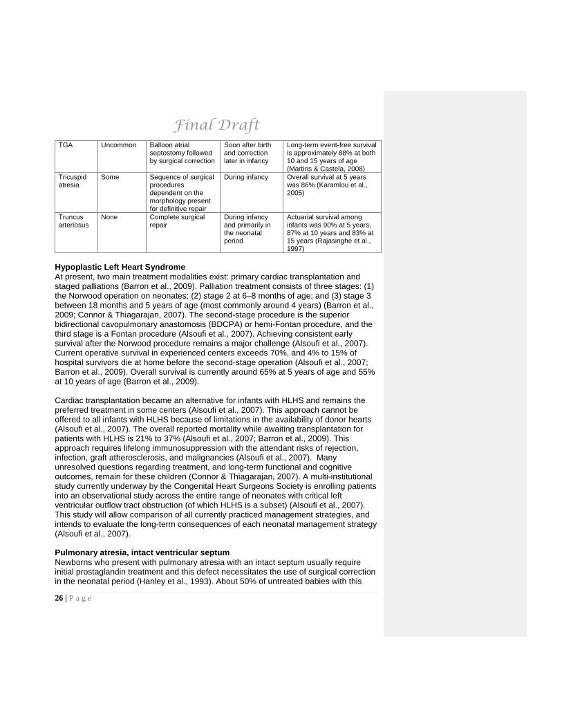
Final Draft
26 | P a g e
TGA Uncommon Balloon atrial septostomy followed by surgical correction
Soon after birth and correction later in infancy
Long-term event-free survival is approximately 88% at both 10 and 15 years of age (Martins & Castela, 2008)
Tricuspid atresia
Some Sequence of surgical procedures dependent on the morphology present for definitive repair
During infancy Overall survival at 5 years was 86% (Karamlou et al., 2005)
Truncus arteriosus
None Complete surgical repair
During infancy and primarily in the neonatal period
Actuarial survival among infants was 90% at 5 years, 87% at 10 years and 83% at 15 years (Rajasinghe et al., 1997)
Hypoplastic Left Heart Syndrome At present, two main treatment modalities exist: primary cardiac transplantation and staged palliations (Barron et al., 2009). Palliation treatment consists of three stages: (1) the Norwood operation on neonates; (2) stage 2 at 6–8 months of age; and (3) stage 3 between 18 months and 5 years of age (most commonly around 4 years) (Barron et al., 2009; Connor & Thiagarajan, 2007). The second-stage procedure is the superior bidirectional cavopulmonary anastomosis (BDCPA) or hemi-Fontan procedure, and the third stage is a Fontan procedure (Alsoufi et al., 2007). Achieving consistent early survival after the Norwood procedure remains a major challenge (Alsoufi et al., 2007). Current operative survival in experienced centers exceeds 70%, and 4% to 15% of hospital survivors die at home before the second-stage operation (Alsoufi et al., 2007; Barron et al., 2009). Overall survival is currently around 65% at 5 years of age and 55% at 10 years of age (Barron et al., 2009). Cardiac transplantation became an alternative for infants with HLHS and remains the preferred treatment in some centers (Alsoufi et al., 2007). This approach cannot be offered to all infants with HLHS because of limitations in the availability of donor hearts (Alsoufi et al., 2007). The overall reported mortality while awaiting transplantation for patients with HLHS is 21% to 37% (Alsoufi et al., 2007; Barron et al., 2009). This approach requires lifelong immunosuppression with the attendant risks of rejection, infection, graft atherosclerosis, and malignancies (Alsoufi et al., 2007). Many unresolved questions regarding treatment, and long-term functional and cognitive outcomes, remain for these children (Connor & Thiagarajan, 2007). A multi-institutional study currently underway by the Congenital Heart Surgeons Society is enrolling patients into an observational study across the entire range of neonates with critical left ventricular outflow tract obstruction (of which HLHS is a subset) (Alsoufi et al., 2007). This study will allow comparison of all currently practiced management strategies, and intends to evaluate the long-term consequences of each neonatal management strategy (Alsoufi et al., 2007). Pulmonary atresia, intact ventricular septum Newborns who present with pulmonary atresia with an intact septum usually require initial prostaglandin treatment and this defect necessitates the use of surgical correction in the neonatal period (Hanley et al., 1993). About 50% of untreated babies with this

Final Draft
27 | P a g e
condition die within the first month of life, and nearly all die before 12 months of age (Hanley et al., 1993). In a large multi-institutional study of 171 neonates who underwent surgical intervention between 1987 and 1991, 98% were 7 days old or less (Hanley et al., 1993). There is debate regarding the first step of surgical treatment of neonates with pulmonary atresia with intact ventricular septum. Varied recommendations include a systemic-pulmonary shunt, pulmonary valvotomy, or a transannular patch, all of which are followed by a definitive two-ventricle repair (Hanley et al., 1993). Of the 171 neonates receiving any of the initial surgical methods, survival was 81% at one month, 72% at six months, 69% at one year, 66% at two years and 64% at two years (Hanley et al., 1993). Reoperation in the surviving patients included 18% who had received one-ventricle repair within three years, 32% who had received two-ventricle repair and 50% (the remainder) who had incompletely separated pulmonary and systemic circulations (Hanley et al., 1993). Tetralogy of Fallot Newborns who present with ductal-dependent flow to the lungs typically receive prostaglandins to maintain ductal patency until surgical intervention (Bailliard & Anderson, 2009). The trend in centers of excellence is increasingly toward neonatal complete repair; however some centers’ initial intervention may be palliative (Bailliard & Anderson, 2009). Centers that undertake neonatal palliation will typically perform the complete repair at the age of four to six months (Bailliard & Anderson, 2009). The 25-year survival rates for patients undergoing repair are as high as 94% (Derby & Pizarro, 2005), and follow-up in patients born 30 years ago shows a rate of survival greater than 85% (Bailliard & Anderson, 2009). Chronic issues can occur in adulthood, and include pulmonary regurgitation, recurrence of pulmonary stenosis, and ventricular arrhythmias (Bailliard & Anderson, 2009). Uncertainty remains regarding both timing of surgery and ideal form of management of TOF (Bailliard & Anderson, 2009; Derby & Pizarro, 2005). Total Anomalous Pulmonary Venous Return Most newborns who present with TAPVR require surgical repair during infancy (Karamlou et al., 2007). In a large single institution study of 377 patients over 5 years, 327 patients (87%) underwent surgical repair at a median age of 1.6 months (Karamlou et al., 2007). Of the remaining patients, 49 died without any surgical intervention and one patient was lost to follow-up. In addition to the 49 deaths, 103 patients died after TAPVR repair, giving a 40% crude mortality rate. Postoperative survival was 68% at one year, and 65% at 14 years after surgery; postoperative 5-year survival for patients undergoing repair since 2000 is 97%. The overall incidence of reoperation at 11 years after repair was 9% and was not correlated with repair type. Transposition of the Great Arteries [Vessels] Newborns who present with TGA usually require treatment with prostaglandins and balloon atrial septostomy soon after birth (Martins & Castela, 2008). Surgical correction is performed at a later stage in infancy. With the advent of newer and improved surgical techniques and postoperative intensive care, early mortality rates are roughly 3.8%, long-term event-free survival is approximately 88% at both 10 and 15 years of age, and the 10-15 year reintervention rate is 4.5-18%. Rare postoperative complications are

Final Draft
28 | P a g e
related to prolonged perioperative ischemia times, aortic regurgitation, and coronary artery obstruction. Recent studies have noted reduced exercise performance, compromise in cognitive functioning, and an unfavorable health-related quality of life after intervention (Martins & Castela, 2008). Tricuspid atresia Surgical management of newborns with tricuspid atresia generally includes a sequence of procedures dependent on the morphology present for definitive repair (Karamlou et al., 2005). In a multi-institutional study of 150 babies between 1999 and 2004, one patient remained alive without operation, while all other patients underwent palliation consisting of systemic-pulmonary arterial shunt (64%), pulmonary artery banding (11%), or initial bidirectional cavopulmonary anastomosis (BDCPA) (24%). Of the patients undergoing BDCPA, 75% underwent the Fontan operation within three years. There were 17 deaths: 9 after the systemic-pulmonary arterial shunt operations, 6 after BDCPA and 2 after the Fontan procedure. Overall survival at 5 years was 86% (Karamlou et al., 2005). Truncus arteriosus Newborns who present with truncus arteriosus require surgical correction during infancy and primarily in the neonatal period (Rajasinghe et al., 1997). In a large single institution study of 165 patients who underwent complete repair of truncus arteriosus between 1975 and 1995, the median age at operation was 3.5 months; 133 of the 165 patients were infants less than one year of age. Patients were followed up for up to 20.4 years (median 10.5 years), and actuarial survival among infants was 90% at 5 years, 87% at 10 years and 83% at 15 years. The median time to reoperation after initial truncus arteriosus repair was 5.1 years and occurred in 119 of the 165 patients. Of the 23 deaths reported, the majority (57%) occurred within the first year of discharge from the hospital after surgical truncus arteriosus repair (Rajasinghe et al., 1997). Expert Information: The experts corroborated that each of the heart defects have surgical interventions that improve outcomes. They were unaware of any direct data regarding whether detection of CCCHD by pulse oximetry leads to improved health outcomes compared to those that are detected clinically.

Final Draft
29 | P a g e
E. Economic evaluation Table 12 – Quality assessment of abstracted literature pertaining to economic evidence Type of evidence Number of articles
Total 1
I. Evaluation of important alternative interventions comparing all clinically relevant outcomes against appropriate cost measurement and including a clinically sensible sensitivity analysis. 1 II. Evaluation of important alternative interventions comparing a limited number of outcomes against appropriate cost measurement, but including a clinically sensible sensitivity analysis. 0 III. Evaluation of important alternative interventions comparing all clinically relevant outcomes against inappropriate cost measurement, but including a clinically sensible sensitivity analysis. 0 IV. Evaluation without a clinically sensible sensitivity analysis 0 V. Expert opinion with no explicit critical appraisal, based on economic theory 0 Adapted from NHS Centre for Reviews and Dissemination Report 4, March 2001 Literature review: One study evaluated the cost-effectiveness of newborn screening for CHD (Griebsch et al., 2007), specifically at 24 hours of age: clinical examination with pulse oximetry or clinical examination with screening echocardiography, compared to clinical examination alone. CHD were grouped into three classes: life-threatening (TGA, HLHS, TAPVC and pulmonary atresia), clinically significant (TOF, truncus arteriosus), and clinically non-significant (no CCCHD). The first two classes were included in the economic evaluation. A decision model was used to simulate a hypothetical cohort of 100,000 live-born infants in the United Kingdom. Inputs to the model were based on primary and published data and supplemented by expert opinion for some parameters. Values for key parameters in the decision model were within the ranges identified for the systematic evidence review. Detection rate (equivalent to sensitivity in this analysis) was 67.9%. False positive rate was 1.3% and positive predictive value was 6.6%. The incidence of life-threatening and clinically significant CHD used in the analysis was 7.1 per 100,000. The primary outcome measure for the study was cost per timely diagnosis. The study projected that clinical examination with pulse oximetry screening would identify 70.6 timely diagnoses per 100,000 compared with 34.0 per 100,000 identified via clinical examination alone. Clinical examination with screening echocardiography was projected to identify 71.3 timely diagnoses per 100,000 (i.e., an incremental increase of 0.7 compared with pulse oximetry). The cost per timely diagnosis was £4,894 for pulse oximetry and £4,496,666 for screening echocardiography. The conclusion of the study was that screening with pulse oximetry in addition to clinical examination was cost-effective but screening with echocardiography was not cost-effective under current conditions for test cost and performance. Cost-effectiveness results were robust to variations in many assumptions but were sensitive to time of

Final Draft
30 | P a g e
screening in that cost-effectiveness of screening was less favorable if undertaken at 48 hours of life (£8,195 per timely diagnosis) instead of 24 hours (£4,894 per timely diagnosis) for pulse oximetry because more cases would have been identified clinically during this time period and therefore screening will identify a smaller number of cases. The study highlighted the lack of primary data on sensitivity and specificity of screening strategies especially with respect to the age at screening. Possible negative effects of false positive screens were not included in the study. These findings are relevant for a United States setting with respect to the relative cost-effectiveness of screening with echocardiography compared to screening with pulse oximetry. It is less certain how to translate the cost-effectiveness of screening with pulse oximetry given the differences in both the costs of specific services as well as health care utilization patterns across the US and the UK. Health care utilization patterns typically differ widely across countries due to differences in health insurance coverage and historical practice patterns. For example, health costs may differ, due to differences in the services delivered for a certain intervention, or due to differences in price levels for a particular service. Differences in price levels could result from different salary levels for providers, services being delivered by different types of providers, and/or price levels for prescription drugs, medical devices, and supplies, that are often set or negotiated at the country level and usually results in lower overall price levels as compared to the United States. Results relating to the projected outcomes for the various identification strategies are relevant to the US setting – specifically that screening with pulse oximetry will result in timely diagnosis of approximately twice those identified using clinical examination alone – yet projections of cost-effectiveness may be less directly applicable due to potential differences in intervention services and unit costs. Expert Information: No experts contributed evidence pertaining to costs of pulse oximetry screening for CCCHD.

Final Draft
31 | P a g e
XI. Key findings and summary
Condition Key Findings The prevalence of CCCHD from the natural history reviews reported a prevalence of 1-7 per 10,000 live births; while screening evidence indicated a calculated prevalence of 1-25 per 10,000 live births, with an average of 8 per 10,000 among neonates eligible for pulse oximetry screening from the eleven studies in our literature review. The calculated figure from the eleven screening studies is of asymptomatic newborns, and excludes prenatally diagnosed CCCHD babies, symptomatic newborns found prior to pulse oximetry screening, and those who have a syndrome commonly associated with CCCHD. Of the seven conditions reviewed, onset of symptoms all occur within the neonatal period. Symptom onset ranges from immediately at birth, to appearing healthy until a few months of age when the infant presents with symptoms. For several of the conditions, the onset and severity depend upon the anatomical and functional variants; if there is not adequate blood flow, the infant will suffer or die. For the ductal-dependent defects, (pulmonary atresia with intact septum, HLHS and occasionally TOF, TGA and tricuspid atresia), survival is unlikely if not urgently treated before the ductus arteriosus closes. For the two non-ductal-dependent lesions, fewer than 25% of infants with truncus arteriosus will survive past the first year of life without intervention, and survival of the infant with TAPVR is unlikely if left untreated. Screening and Diagnosis Key Findings We identified eleven reports with evidence regarding test characteristics of pulse oximetry screening, and re-calculated the data presented to include only the conditions that met our case definition of CCCHD. All but two studies reported a specificity of greater than 99%; the two other studies with specificities of 94% and 98% screened with pulse oximetry at 4 hours and less than six hours, respectively. Sensitivity was more variable, ranging from 42% to 100% with no clear explanation for the variability. However, false positive rates increase for earlier screening times. Four studies had no false negatives, five studies each had one false negative and two studies had 4-5 false negatives. The two highest false negative rates also had the second and third highest calculated prevalence rates. The screening evidence also assessed the utility of pulse oximetry alone, clinical exam alone and both in combination. One study found that there was no statistically significant differences between screening with a routine assessment alone and routine assessment with pulse oximetry; while another study found that pulse oximetry detected cases of pulmonary atresia, TAPVR and truncus arteriosus that clinical exam did not detect. In a large screening study of close to 40,000 newborns, the researchers concluded a combined screening approach utilizing physical exam and pulse oximetry had the highest sensitivity. The same study found that neonates in regions without routine pulse oximetry screening were more likely to be discharged with undiagnosed duct-dependent circulation and that neonates diagnosed post-discharge had higher

Final Draft
32 | P a g e
mortality than those diagnosed pre-discharge. In the screening literature, the CCCHD most often reported as undiagnosed by physical exam alone were TGA and TAPVR. While the potential harm of screening in regards to missed diagnosis and delayed diagnosis was present in the literature evidence, the experts stated harmfulness regarding the potential impact of false positives on families. Screening too early in life yields large numbers of false positives and can potentially disrupt parental bonding, and potential morbidity due to cardiac non-disease. However, no literature data are available regarding the harmfulness of false positives on families. Fetal echocardiography screening detects some types of CCCHD, which decreases the prevalence of CCCHD in the population of asymptomatic neonates. Pulse oximetry appears to identify neonates with CCCHD that prenatal and clinical assessment alone may miss. No data are available regarding the availability of echocardiography to evaluate those who have an abnormal pulse oximetry screen. Efforts are ongoing in the emerging technology of telemedicine. While several partnerships have formed between large and rural hospitals, there is no large unified effort or standardization of the process. The evidence suggests that pulse oximetry identifies neonates with CCCHD that prenatal and clinical assessment alone may miss. Further work should address the most beneficial timing of the pulse oximetry screening, probe placement and number of probes used, and the best threshold value for action. Of note, all screening studies evaluated were completed in large hospital settings, and may not be generalizable to smaller institutions. Treatment Key Findings All of the lesions identified in the case definition have surgical interventions and timing of surgical intervention all occurs within infancy and most soon after birth. Rates of success and mortality vary among the procedures and include variable long-term morbidities. Each condition has palliative and surgical options, with variations in standard interventions for each condition. Overall, among the seven CCCHD defects reviewed, 5-year survival rates post-intervention ranged from 65% to 97%, with five of the seven conditions having 5-year survival rates above 85%. The two lower long-term survival rates are for both the ductal-dependent conditions of pulmonary atresia with intact septum and HLHS.
While pulmonary atresia with intact septum and HLHS may benefit the most from pre-symptomatic or early symptomatic intervention with prostaglandin infusion to keep the ductus open, any defect that may also present as ductal-dependent necessitates prostaglandins to maintain patency until surgical intervention. Once the ductus closes, the newborn becomes critically ill, leading to death if not treated. Questions remain regarding the ideal time for complete repair of the defects and optimal form of management (i.e., whether initial palliation or complete repair should be done initially).

Final Draft
33 | P a g e
Economics Key Findings The conclusion of the one economic study identified was that screening with pulse oximetry in addition to clinical examination was cost-effective compared to usual care. Standardization of screening practices Because screening with pulse oximetry is not similar to the traditional newborn screening program of using filter-paper-based tests and a laboratory, it leads to new issues not previously addressed for dried blood spot screening. Further work should elucidate the most beneficial timing of pulse oximetry screening, probe placement and number of probes used, the optimal threshold value for action due to a lack of standardized protocol for the screening exam, and methods of quality control. Better trials could provide helpful information on the benefits, risks, and cost of this technology. Critical evidence lacking that may inform screening recommendations for CCCHD The following is a list of our key questions and additional questions that arose through our work that remain unanswered due to lacking evidence:
• How does screening test accuracy vary by the age of the neonate, in conjunction with placement of the probes, and threshold value for action? What is the optimal pulse oximetry protocol for newborn screening?
• How does prenatal screening and detection of CCCHD affect the sensitivity, specificity, positive predictive value, and negative predictive value of postnatal pulse oximetry screening of asymptomatic newborns?
• What are the differences in benefits and disadvantages of prenatal diagnosis versus early postnatal diagnosis of CCCHD?
• What follow-up practices should be in place for newborns with a low pulse oximetry screen result but a structurally normal heart on echocardiogram?
• What is the benefit of adding a pulse oximetry screen to infant outcomes compared to ‘usual care’?
• How generalizable is this benefit when the screening is performed on the ‘general population’?
• How available is echocardiography to evaluate those with a positive pulse oximetry screening result?
• Is telemedicine a practical alternative for birth hospitals without access to pediatric cardiology services?
• What is the availability of treatment? • What are the costs associated with treatment? • What are the costs associated with failure to diagnose in the pre-symptomatic
period?

Final Draft
34 | P a g e
XII. Tables of abstracted literature Table 13 - Abstracted Natural History, Treatment and Diagnosis Articles Author & Year Area Significant findings
Aamir, Kruse & Ezeakudo, 2007 Diagnosis
• Medical chart review confirmed delayed diagnosis of CCHD for 47 infants during a 6-year study period from 1999-2004 and using data from 670,245 births
• Coarctation of the aorta and TAPVR accounted for the highest number of delayed diagnosis cases • Average time of delayed diagnosis was 6 months • Delay in diagnosis for 7 per 100,000 CCHDs per year
Alsoufi et al., 2007
Natural History & Treatment
• Without intervention HLHS is fatal • Surgical staging - Due to recent improvements in postoperative management after the Norwood operation, some
centers able to achieve hospital survival exceeding 90% in selected groups of patients, interim mortality 4-15% of hospital survivors die at home before second stage operation. Operative and interstage mortality remains high in regards to the Norwood procedure. Survivors also found to have suboptimal neurocognitive functioning.
• Orthotopic Heart Transplant - Preferred treatment in some centers. Advantage - normal physiology achieve with a single operation. Disadvantage - limited availability of donor hearts (mortality while waiting for transplant 21-37%), lifelong immunosuppression, and subsequent retransplantation.
Bailliard & Anderson, 2009
Natural History & Treatment
• 3/10,000 live births • Presentation - varies, depending on the severity of the obstruction of blood flow to the lungs. Neonatal period - mild-
moderate cyanosis without respiratory distress, heart failure or failure to thrive • Patients who undergo a successful complete repair during the neonatal period will be unlikely to require more than
one intervention in the first year of life (risks: exposure of the brain to cardiopulmonary bypass, increased need to place a transannular patch)
• Perioperative mortality rates for either surgical approach is less than 3% • Patients born 30 years ago with TOF have an 85% long-term survival rate
Barron et al., 2009
Natural History & Treatment
• HLHS is fatal without intervention, 95% in the first few weeks of life • HLHS accounts for 25-40% of neonatal cardiac deaths • 78% of cases of HLHS were discharged from the hospital before diagnosis
Chang et al., 2008 Diagnosis
• Population-based retrospective study of California death registry data from 1989-2004 identifying infants up to one year of age who died with undiagnosed CCHD or a late diagnosis of CCHD
• 898 infants found to have CCHD listed as the cause of death • HLHS was the most common cause of death • 152/898 were only diagnosed with CCHD at autopsy • HLHS accounted for the majority of the missed diagnoses (58/152) • The incidence of death among infants with undiagnosed CCHD or a late diagnosis of CCHD decreased over the
course of the study period

Final Draft
35 | P a g e
Connor & Thiagarajan, 2007
Natural History & Treatment
• Death occurred more frequently in neonates who underwent transplant (42%) over staged palliation (35% after first operation)
• Operative mortality for second operation is 2-5.4%, for third operation it is under 5% • No intervention remains an option for families
Derby & Pizarro, 2005
Natural History & Treatment
• Advantages of early repair - possible avoidance in reduction of gas-exchanging capillaries relative to alveoli, avoidance of abnormal right ventricular hypertrophy and subsequent fibrosis, less exposure to chronic cyanosis
• Multiple studies have documented the safety of early primary repair in neonates and young infants (less than 3% mortality rate for repair in symptomatic neonates and young infants)
• Long term survival of patients undergoing repair of TOF, 25 year survival rates as high as 94%
Hanley et al., 1993
Natural History & Treatment
• Overall, 1-month survival (after first intervention) was 81% and at least 4-year survival was 64% • Systemic-pulmonary artery shunt was the most commonly performed first intervention. Risk factors for death included
right ventricular dependency on the coronary circulation and lower birth weight. • 51% of those undergoing an initial procedure had a subsequent procedure performed during the time of follow-up. • Of those subjects still alive after 24 months after entry in the study, 11% underwent one-ventricle repair (Fontan
procedure) and 24% underwent a complete two-ventricle repair leaving 65% of the subjects in a mixed-circulation state.
• The wide morphologic spectrum of pulmonary atresia with intact septum may contribute to surgical outcome.
Karamlou et al., 2005
Natural History & Treatment
• 128 subjects had bidirectional cavopulmonary anastomosis (BDCPA) procedure, median age was 5.7 months (range 1.7 months–2 years)
• 74 subjects had Fontan procedure, median age 2.5 years (range 1-5 years) • 17 deaths: 9 occurred after systemic-pulmonary arterial shunt operations, 6 after BDCPA, and 2 after Fontan
completion • By 2 years from diagnosis, 6% of patients had died without BDCPA, 89% had undergone BDCPA, 4% remained alive
without BDCPA or further operation, and 1% had undergone single-stage Fontan procedures • Overall survival at 5 years was 86% • Mortality occurs primarily at the initial palliation stage before BDCPA • Younger or older age at BDCPA did not increase either length of hospital stay or in-hospital mortality
Karamlou et al., 2007
Natural History & Treatment
• 327 patients (87%) underwent TAPVR repair (median age 1.6 months (range birth - 14 years) • 49 died without any surgical intervention • 1 lost to follow-up • 152 deaths, 49 (32%) died without any surgical intervention (causes of death - cardiac, sepsis and unknown), 103
died after TAPVR repair • Overall, postoperative survival was 68% at 1 year and 65% at 14 years after surgery • Postoperative 5-year survival for patients undergoing repair since 2000 was 97% • Risk factors for death after repair included earlier year of repair, younger age at repair, presence of postoperative
pulmonary venous obstruction, use of epinephrine postoperatively and cardiac connection type • 88% and 82% of patients free from reoperation at 1 and 11 years after initial repair • Follow-up 157 available on survivors, median interval of 8.4 years (range 11 days - 21.3 years) - cardiovascular
symptoms were present in 17 patients, 10 reported other noncardiac symptoms, 5 reported important activity restrictions or limitations

Final Draft
36 | P a g e
Martins & Castela, 2008
Natural History & Treatment
• Very encouraging long-term survival rates, low reintervention rate and corresponding event-free survival of 88% • Patients may have reduced exercise performance, compromise in cognitive functioning and unfavorable health-related
quality of life
Rajasinghe et al., 1997
Natural History & Treatment
• Late survival rates after complete truncus arteriosus repair were 90%, 85%, and 83% at 5, 10, and 15 years. • Of the 23 late deaths (occurring after hospitalization or after 30 days post-operation), 57% (13/23) occurred within the
first year after discharge from the hospital after truncus repair. Prerepair truncal valve insufficiency, interrupted aortic arch, and shorter crossclamp time were found to correlate significantly with poorer survival.
Westmoreland, 1999
Natural History & Treatment
• Tricuspid atresia - 1-3% of all CHD. Symptoms are usually apparent within a few hours to a month after birth. Symptoms include cyanosis, dyspnea (shortness of breath), tachypnea (rapid breathing) and metabolic acidosis. Congestive heart failure develops because of the excessive amount of blood the heart has to pump along with the poorly oxygenated blood supplied to heart muscle. Prostaglandin may be needed; palliative surgery is done if the neonate is cyanotic. A more complete repair is performed when the infant is older.
• Truncus arteriosus - Cyanosis may develop at birth and over the first few weeks of life because of decreased pulmonary blood flow. A heart murmur will be present. Symptoms may develop into congestive heart failure and pulmonary vascular disease. Palliative or corrective surgery required to prevent pulmonary vascular disease.
• TAPVR - Neonates with the significant obstruction of the pulmonary veins will present with profound cyanosis, tachypnea, pulmonary edema, pulmonary hypertension, hepatomegaly, moist rales and dyspnea (signs of cardiac failure). Survival is unlikely without treatment. Less profound symptoms may present in the first 2 months of life if the abnormal connections are unobstructed. Surgical repair is usually done at the time of diagnosis.
• Pulmonary atresia with intact ventricular septum - Neonates are usually cyanotic at birth. When the ductus closes, the neonate becomes severely cyanotic and acidotic and will die if not treated immediately. Treatment includes prostaglandin infusion, balloon atrial septostomy and surgical correction (once the infant is stabilized). Heart transplant is considered an option.
Wren et al., 2008 Diagnosis
• Overall, 8% life-threatening cardiovascular malformations were diagnosed prenatally, 62% were diagnosed before discharge, 25% were diagnosed in living infants after discharge and 5% were diagnosed at autopsy. TAPVR was among the conditions most likely to be overlooked before discharge.
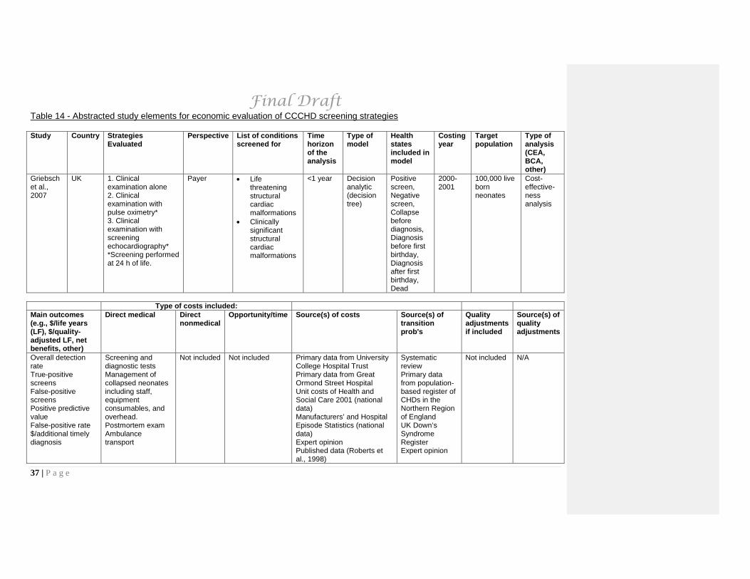
Final Draft
37 | P a g e
Table 14 - Abstracted study elements for economic evaluation of CCCHD screening strategies Study Country Strategies
Evaluated Perspective List of conditions
screened for Time horizon of the analysis
Type of model
Health states included in model
Costing year
Target population
Type of analysis (CEA, BCA, other)
Griebsch et al., 2007
UK 1. Clinical examination alone 2. Clinical examination with pulse oximetry* 3. Clinical examination with screening echocardiography* *Screening performed at 24 h of life.
Payer • Life threatening structural cardiac malformations
• Clinically significant structural cardiac malformations
<1 year Decision analytic (decision tree)
Positive screen, Negative screen, Collapse before diagnosis, Diagnosis before first birthday, Diagnosis after first birthday, Dead
2000-2001
100,000 live born neonates
Cost-effective-ness analysis
Type of costs included: Main outcomes (e.g., $/life years (LF), $/quality-adjusted LF, net benefits, other)
Direct medical Direct nonmedical
Opportunity/time Source(s) of costs Source(s) of transition prob's
Quality adjustments if included
Source(s) of quality adjustments
Overall detection rate True-positive screens False-positive screens Positive predictive value False-positive rate $/additional timely diagnosis
Screening and diagnostic tests Management of collapsed neonates including staff, equipment consumables, and overhead. Postmortem exam Ambulance transport
Not included Not included Primary data from University College Hospital Trust Primary data from Great Ormond Street Hospital Unit costs of Health and Social Care 2001 (national data) Manufacturers’ and Hospital Episode Statistics (national data) Expert opinion Published data (Roberts et al., 1998)
Systematic review Primary data from population-based register of CHDs in the Northern Region of England UK Down’s Syndrome Register Expert opinion
Not included N/A

Final Draft
38 | P a g e
XIII. References
Aamir, T., Kruse, L., & Ezeakudo, O. (2007). Delayed diagnosis of critical congenital cardiovascular malformations (CCVM) and pulse oximetry screening of newborns. Acta Paediatrica (Oslo, Norway : 1992), 96(8), 1146-1149. doi:10.1111/j.1651-2227.2007.00389.x
Agnoletti, G., Boudjemline, Y., Bonnet, D., Sidi, D., & Vouhe, P. (2004). Surgical reconstruction of occluded pulmonary arteries in patients with congenital heart disease: Effects on pulmonary artery growth. Circulation, 109(19), 2314-2318.
Alsoufi, B., Bennetts, J., Verma, S., & Caldarone, C. A. (2007). New developments in the treatment of hypoplastic left heart syndrome. Pediatrics, 119(1), 109-117.
Apitz, C., Webb, G. D., & Redington, A. N. (2009). Tetralogy of fallot. Lancet, 374(9699), 1462-1471.
Arlettaz, R., Bauschatz, A. S., Monkhoff, M., Essers, B., & Bauersfeld, U. (2006). The contribution of pulse oximetry to the early detection of congenital heart disease in newborns. European Journal of Pediatrics, 165(2), 94-98. doi:10.1007/s00431-005-0006-y
Armstrong, B. E. (1995). Congenital cardiovascular disease and cardiac surgery in childhood: Part 1. cyanotic congenital heart defects. Current Opinion in Cardiology, 10(1), 58-67.
Ashburn, D. A., Harris, L., Downar, E. H., Siu, S., Webb, G. D., & Williams, W. G. (2003). Electrophysiologic surgery in patients with congenital heart disease. Seminars in Thoracic & Cardiovascular Surgery.Pediatric Cardiac Surgery Annual, 6, 51-58.
Bailey, L. L., & Gundry, S. R. (1990). Hypoplastic left heart syndrome. Pediatric Clinics of North America, 37(1), 137-150.
Bailliard, F., & Anderson, R. H. (2009). Tetralogy of Fallot. Orphanet Journal of Rare Diseases, 4, 2.
Bakr, A. F., & Habib, H. S. (2005). Combining pulse oximetry and clinical examination in screening for congenital heart disease. Pediatric Cardiology, 26(6), 832-835. doi:10.1007/s00246-005-0981-9
Bardo, D. M., Frankel, D. G., Applegate, K. E., Murphy, D. J., & Saneto, R. P. (2001). Hypoplastic left heart syndrome. Radiographics, 21(3), 705-717.
Barron, D. J., Kilby, M. D., Davies, B., Wright, J. G., Jones, T. J., & Brawn, W. J. (2009). Hypoplastic left heart syndrome. Lancet, 374(9689), 551-564.
Boneva, R. S., Botto, L. D., Moore, C. A., Yang, Q., Correa, A., & Erickson, J. D. (2001). Mortality associated with congenital heart defects in the united states: Trends and racial disparities, 1979-1997. Circulation, 103(19), 2376-2381.
Botto, L. D., Correa, A., & Erickson, J. D. (2001). Racial and temporal variations in the prevalence of heart defects. Pediatrics, 107(3), E32.
Bove, E. L., Ohye, R. G., & Devaney, E. J. (2004). Hypoplastic left heart syndrome: Conventional surgical management. Seminars in Thoracic & Cardiovascular Surgery.Pediatric Cardiac Surgery Annual, 7, 3-10.
Brooks, P. A., & Penny, D. J. (2008). Management of the sick neonate with suspected heart disease. Early Human Development, 84(3), 155-159.

Final Draft
39 | P a g e
Chang, R. K., Gurvitz, M., & Rodriguez, S. (2008). Missed diagnosis of critical congenital heart disease. Archives of Pediatrics & Adolescent Medicine, 162(10), 969-974. doi:10.1001/archpedi.162.10.969
Chang, R. K., Rodriguez, S., & Klitzner, T. S. (2009). Screening newborns for congenital heart disease with pulse oximetry: Survey of pediatric cardiologists. Pediatric Cardiology, 30(1), 20-25. doi:10.1007/s00246-008-9270-8
Cheatham, J. P. (2001). Intervention in the critically ill neonate and infant with hypoplastic left heart syndrome and intact atrial septum. Journal of Interventional Cardiology, 14(3), 357-366.
Claxon-McKinney, B. (2001). Hypoplastic left heart syndrome. Pediatric Nursing, 27(3), 245-248.
Cohen, D. M., & Allen, H. D. (1997). New developments in the treatment of hypoplastic left heart syndrome. Current Opinion in Cardiology, 12(1), 44-50.
Connor, J. A., & Thiagarajan, R. (2007). Hypoplastic left heart syndrome. Orphanet Journal of Rare Diseases, 2, 23.
Corno, A. F. (2000). Surgery for congenital heart disease. Current Opinion in Cardiology, 15(4), 238-243.
Cui, W., Patel, D., Husayni, T. S., & Roberson, D. A. (2008). Double aortic arch and d-transposition of the great arteries. Echocardiography, 25(1), 91-95.
Daliento, L. (2002). Total correction of tetralogy of fallot: Late clinical follow-up. Italian Heart Journal: Official Journal of the Italian Federation of Cardiology, 3(1), 24-27.
De Vries, J. W., Plotz, F. B., & Van Vught, A. J. (2002). Pulse oximeter-enhanced accuracy of capnometry in children with cyanotic heart disease. Intensive Care Medicine, 28(9), 1336-1339. doi:10.1007/s00134-002-1409-0
de Wahl Granelli, A., Mellander, M., Sunnegardh, J., Sandberg, K., & Ostman-Smith, I. (2005). Screening for duct-dependant congenital heart disease with pulse oximetry: A critical evaluation of strategies to maximize sensitivity. Acta Paediatrica (Oslo, Norway : 1992), 94(11), 1590-1596.
Denfield, S. W., & Garson, A.,Jr. (1990). Sudden death in children and young adults. Pediatric Clinics of North America, 37(1), 215-231.
Derby, C. D., & Pizarro, C. (2005). Routine primary repair of tetralogy of fallot in the neonate. Expert Review of Cardiovascular Therapy, 3(5), 857-863.
de-Wahl Granelli, A., Wennergren, M., Sandberg, K., Mellander, M., Bejlum, C., Inganas, L., Eriksson, M., Segerdahl, N., Agren, A., Ekman-Joelsson, B. M., Sunnegardh, J., Verdicchio, M., & Ostman-Smith, I. (2009). Impact of pulse oximetry screening on the detection of duct-dependent congenital heart disease: A swedish prospective screening study in 39,821 newborns. BMJ (Clinical Research Ed.), 338, a3037.
el-Zein, C., & Ilbawi, M. N. (2008). Recent advances in neonatal cardiac surgery. World Journal of Surgery, 32(3), 340-345.
Fedderly, R. T. (1999). Left ventricular outflow obstruction. Pediatric Clinics of North America, 46(2), 369-384.

Final Draft
40 | P a g e
Feldkamp, M., Macleod, L., Young, L., Lecheminant, K., & Carey, J. C. (2005). The methodology of the utah birth defect network: Congenital heart defects as an illustration. Birth Defects Research.Part A, Clinical and Molecular Teratology, 73(10), 693-699. doi:10.1002/bdra.20212
Gillette, P. C., & Garson, A.,Jr. (1992). Sudden cardiac death in the pediatric population. Circulation, 85(1 Suppl), 64-69.
Goldberg, C. S., & Gomez, C. A. (2003). Hypoplastic left heart syndrome: New developments and current controversies. Seminars in Neonatology, 8(6), 461-468.
Gorska-Kot, A., Blaz, W., Pszeniczna, E., Rusin, J., Materna-Kiryluk, A., Homa, E., Hejda, G., & Franus, J. (2006). Trends in diagnosis and prevalence of critical congenital heart defects in the podkarpacie province in 2002-2004, based on data from the polish registry of congenital malformations. Journal of Applied Genetics, 47(2), 191-194.
Graham, E. M., Bradley, S. M., & Atz, A. M. (2005). Preoperative management of hypoplastic left heart syndrome. Expert Opinion on Pharmacotherapy, 6(5), 687-693.
Granelli, A. W., & Ostman-Smith, I. (2007). Noninvasive peripheral perfusion index as a possible tool for screening for critical left heart obstruction. Acta Paediatrica (Oslo, Norway : 1992), 96(10), 1455-1459. doi:10.1111/j.1651-2227.2007.00439.x
Green, K., & Oddie, S. (2008). The value of the postnatal examination in improving child health. Archives of Disease in Childhood.Fetal and Neonatal Edition, 93(5), F389-93. doi:10.1136/adc.2007.122465
Griebsch, I., Knowles, R. L., Brown, J., Bull, C., Wren, C., & Dezateux, C. A. (2007). Comparing the clinical and economic effects of clinical examination, pulse oximetry, and echocardiography in newborn screening for congenital heart defects: A probabilistic cost-effectiveness model and value of information analysis. International Journal of Technology Assessment in Health Care, 23(2), 192-204.
Grifka, R. G. (1999). Cyanotic congenital heart disease with increased pulmonary blood flow. Pediatric Clinics of North America, 46(2), 405-425.
Hanley, F. L., Sade, R. M., Blackstone, E. H., Kirklin, J. W., Freedom, R. M., & Nanda, N. C. (1993). Outcomes in neonatal pulmonary atresia with intact ventricular septum. A multiinstitutional study. The Journal of Thoracic and Cardiovascular Surgery, 105(3), 406-23, 424-7; discussion 423-4.
Hartas, G. A., Tsounias, E., & Gupta-Malhotra, M. (2009). Approach to diagnosing congenital cardiac disorders. Critical Care Nursing Clinics of North America, 21(1), 27-36.
Heimansohn, D. A., Turrentine, M. W., Kesler, K. A., King, H., & Brown, J. W. (1993). New trends in the management of congenital heart disease. World Journal of Surgery, 17(3), 356-362.
Hetzel, P. G., Glanzmann, R., Gunthard, J., Bruder, E., Godi, E., & Buhrer, C. (2007). Failed detection of complex congenital heart disease (including double outlet right ventricle and total anomalous pulmonary venous return) by neonatal pulse oximetry screening. European Journal of Pediatrics, 166(6), 625-626. doi:10.1007/s00431-006-0283-0
Hines, M. H. (2005). ECMO and congenital heart disease. Seminars in Perinatology, 29(1), 34-39.
Hoke, T. R., Donohue, P. K., Bawa, P. K., Mitchell, R. D., Pathak, A., Rowe, P. C., & Byrne, B. J. (2002). Oxygen saturation as a screening test for critical congenital heart disease: A preliminary study. Pediatric Cardiology, 23(4), 403-409. doi:10.1007/s00246-002-1482-8

Final Draft
41 | P a g e
Jonas, R. A. (1995). Advances in surgical care of infants and children with congenital heart disease. Current Opinion in Pediatrics, 7(5), 572-579.
Jonas, R. A. (2009). Early primary repair of tetralogy of fallot. Seminars in Thoracic & Cardiovascular Surgery.Pediatric Cardiac Surgery Annual.:39-47, 2009, , 39-47.
Juttmann, R. E., Witsenburg, M., Meerding, W. J., Looman, C. W., & van der Maas, P. J. (2002). Evidence-based prevention requires evidence-based performance. the case of screening for congenital heart disease in child health care. European Journal of Public Health, 12(3), 198-202.
Kadner, A., Tulevski, I. I., Bauersfeld, U., Pretre, R., Valsangiacomo-Buechel, E. R., & Dodge-Khatami, A. (2007). Chronic pulmonary valve insufficiency after repaired tetralogy of fallot: Diagnostics, reoperations and reconstruction possibilities. Expert Review of Cardiovascular Therapy, 5(2), 221-230.
Kallfelz, H. C. (1994). Congenital heart disease and cardiac surgery in children. Current Opinion in Cardiology, 9(1), 103-113.
Kanter, K. R. (2006). Surgical repair of total anomalous pulmonary venous connection. Seminars in Thoracic & Cardiovascular Surgery.Pediatric Cardiac Surgery Annual.:40-4, 2006, , 40-44.
Karamlou, T., Ashburn, D. A., Caldarone, C. A., Blackstone, E. H., Jonas, R. A., Jacobs, M. L., Williams, W. G., Ungerleider, R. M., McCrindle, B. W., & Members of the Congenital Heart Surgeons' Society. (2005). Matching procedure to morphology improves outcomes in neonates with tricuspid atresia. The Journal of Thoracic and Cardiovascular Surgery, 130(6), 1503-1510. doi:10.1016/j.jtcvs.2005.07.024
Karamlou, T., Gurofsky, R., Al Sukhni, E., Coles, J. G., Williams, W. G., Caldarone, C. A., Van Arsdell, G. S., & McCrindle, B. W. (2007). Factors associated with mortality and reoperation in 377 children with total anomalous pulmonary venous connection. Circulation, 115(12), 1591-1598. doi:10.1161/CIRCULATIONAHA.106.635441
Karl, T. R. (2001). Neonatal cardiac surgery. anatomic, physiologic, and technical considerations. Clinics in Perinatology, 28(1), 159-185.
Kemper, A. R. (2009). Next steps in pulse oximetry screening research. Pediatrics, 123(2), e356-7; author reply e357. doi:10.1542/peds.2008-3185
Knowles, R., Griebsch, I., Dezateux, C., Brown, J., Bull, C., & Wren, C. (2005). Newborn screening for congenital heart defects: A systematic review and cost-effectiveness analysis. Health Technology Assessment (Winchester, England), 9(44), 1-152.
Kogon, B., Villari, C., Shah, N., Kirshbom, P., Kanter, K., Kim, D., Raviele, A., & Vincent, R. (2007). Occlusion of the modified blalock-taussig shunt: Unique methods of treatment and review of catheter-based intervention. Congenital Heart Disease, 2(3), 185-190.
Kohli, V. (2009). Neonatal cardiac interventions: An indian perspective. Indian Journal of Pediatrics, 76(2), 207-214.
Koppel, R. I., Druschel, C. M., Carter, T., Goldberg, B. E., Mehta, P. N., Talwar, R., & Bierman, F. Z. (2003). Effectiveness of pulse oximetry screening for congenital heart disease in asymptomatic newborns. Pediatrics, 111(3), 451-455.
Kuehl, K. S., Loffredo, C. A., & Ferencz, C. (1999). Failure to diagnose congenital heart disease in infancy. Pediatrics, 103(4 Pt 1), 743-747.

Final Draft
42 | P a g e
Kutty, S., & Zahn, E. M. (2008). Interventional therapy for neonates with critical congenital heart disease. Catheterization & Cardiovascular Interventions, 72(5), 663-674.
Laussen, P. C. (2001). Neonates with congenital heart disease. Current Opinion in Pediatrics, 13(3), 220-226.
LeBlanc, J. G., & Russell, J. L. (1998). Pediatric cardiac surgery in the 1990s. Surgical Clinics of North America, 78(5), 729-747.
Lee, K., Khoshnood, B., Chen, L., Wall, S. N., Cromie, W. J., & Mittendorf, R. L. (2001). Infant mortality from congenital malformations in the united states, 1970-1997. Obstetrics and Gynecology, 98(4), 620-627.
Liske, M. R., Greeley, C. S., Law, D. J., Reich, J. D., Morrow, W. R., Baldwin, H. S., Graham, T. P., Strauss, A. W., Kavanaugh-McHugh, A. L., Walsh, W. F., & Tennessee Task Force on Screening Newborn Infants for Critical Congenital Heart Disease. (2006). Report of the Tennessee task force on screening newborn infants for critical congenital heart disease. Pediatrics, 118(4), e1250-6. doi:10.1542/peds.2005-3061
Mahle, W. T., Newburger, J. W., Matherne, G. P., Smith, F. C., Hoke, T. R., Koppel, R., Gidding, S. S., Beekman, R. H.,3rd, Grosse, S. D., American Heart Association Congenital Heart Defects Committee of the Council on Cardiovascular Disease in the Young, Council on Cardiovascular Nursing, and Interdisciplinary Council on Quality of Care and Outcomes Research, American Academy of Pediatrics Section on Cardiology And Cardiac Surgery, & Committee On Fetus And Newborn. (2009). Role of pulse oximetry in examining newborns for congenital heart disease: A scientific statement from the AHA and AAP. Pediatrics, 124(2), 823-836. doi:10.1542/peds.2009-1397
Mahle, W. T., & Wernovsky, G. (2004). Neurodevelopmental outcomes in hypoplastic left heart syndrome. Seminars in Thoracic & Cardiovascular Surgery.Pediatric Cardiac Surgery Annual, 7, 39-47.
Martins, P., & Castela, E. (2008). Transposition of the great arteries. Orphanet Journal of Rare Diseases, 3, 27.
Mavroudis, C., & Backer, C. L. (2003). Physiologic versus anatomic repair of congenitally corrected transposition of the great arteries. Seminars in Thoracic & Cardiovascular Surgery.Pediatric Cardiac Surgery Annual, 6, 16-26.
McConnell, M. E., & Elixson, E. M. (2002). The neonate with suspected congenital heart disease. Critical Care Nursing Quarterly, 25(3), 17-25.
McElhinney, D. B., Reddy, V. M., & Hanley, F. L. (1996). Homografts in congenital heart disease: Current applications and future directions. Israel Journal of Medical Sciences, 32(10), 880-885.
Meberg, A. (2008). Critical heart defects--the diagnostic challenge. Acta Paediatrica (Oslo, Norway : 1992), 97(11), 1480-1483. doi:10.1111/j.1651-2227.2008.00975.x
Meberg, A. (2008). Pulse oximetry screening in newborns. The Journal of Pediatrics, 153(6), 872. doi:10.1016/j.jpeds.2008.07.038
Meberg, A., Andreassen, A., Brunvand, L., Markestad, T., Moster, D., Nietsch, L., Silberg, I. E., & Skalevik, J. E. (2009). Pulse oximetry screening as a complementary strategy to detect critical congenital heart defects. Acta Paediatrica (Oslo, Norway : 1992), 98(4), 682-686. doi:10.1111/j.1651-2227.2008.01199.x

Final Draft
43 | P a g e
Meberg, A., Brugmann-Pieper, S., Due, R.,Jr, Eskedal, L., Fagerli, I., Farstad, T., Froisland, D. H., Sannes, C. H., Johansen, O. J., Keljalic, J., Markestad, T., Nygaard, E. A., Rosvik, A., & Silberg, I. E. (2008). First day of life pulse oximetry screening to detect congenital heart defects. The Journal of Pediatrics, 152(6), 761-765. doi:10.1016/j.jpeds.2007.12.043
Mellander, M., & Sunnegardh, J. (2006). Failure to diagnose critical heart malformations in newborns before discharge--an increasing problem? Acta Paediatrica (Oslo, Norway : 1992), 95(4), 407-413. doi:10.1080/08035250500541910
Morales, D. L., Zafar, F., & Fraser, C. D.,Jr. (2009). Tetralogy of fallot repair: The right ventricle infundibulum sparing (RVIS) strategy. Seminars in Thoracic & Cardiovascular Surgery.Pediatric Cardiac Surgery Annual.:54-8, 2009, , 54-58.
Mysorekar, V. V., Rao, S. G., & Dandekar, C. P. (1999). Pulmonary atresia with intact ventricular septum--a report of two cases with review of literature. Journal of the Indian Medical Association, 97(7), 287-288.
Ohye, R. G., & Bove, E. L. (2001). Advances in congenital heart surgery. Current Opinion in Pediatrics, 13(5), 473-481.
Pacileo, G., Musewe, N. N., & Calabro, R. (1992). Tetralogy of fallot in three siblings: A familial study and review of the literature. European Journal of Pediatrics, 151(10), 726-727.
Pandor, A., Eastham, J., Beverley, C., Chilcott, J., & Paisley, S. (2004). Clinical effectiveness and cost-effectiveness of neonatal screening for inborn errors of metabolism using tandem mass spectrometry: A systematic review. Health Technology Assessment (Winchester, England), 8(12), iii, 1-121.
Pawade, A., & Karl, T. (1994). Management strategy in neonates presenting with pulmonary atresia with intact ventricular septum. Current Opinion in Pediatrics, 6(5), 600-605.
Pihkala, J., Nykanen, D., Freedom, R. M., & Benson, L. N. (1999). Interventional cardiac catheterization. Pediatric Clinics of North America, 46(2), 441-464.
Pinsky, W. W., & Arciniegas, E. (1990). Tetralogy of fallot. Pediatric Clinics of North America, 37(1), 179-192.
Pokorski, R. J. (2000). Long-term survival after repair of tetralogy of fallot. Journal of Insurance Medicine (Seattle), 32(2), 89-92.
Pollitt, R. J., Green, A., McCabe, C. J., Booth, A., Cooper, N. J., Leonard, J. V., Nicholl, J., Nicholson, P., Tunaley, J. R., & Virdi, N. K. (1997). Neonatal screening for inborn errors of metabolism: Cost, yield and outcome. Health Technology Assessment (Winchester, England), 1(7), i-iv, 1-202.
Prifti, E., Bonacchi, M., Bernabei, M., Leacche, M., Bartolozzi, F., Murzi, B., Battaglia, F., Nadia, N. S., & Vanini, V. (2004). Repair of complete atrioventricular septal defect with tetralogy of fallot: Our experience and literature review. Journal of Cardiac Surgery, 19(2), 175-183.
Pulse oximetry: Not a sensitive screening tool for congenital heart disease. (2008). Archives of Disease in Childhood.Fetal and Neonatal Edition, 93(1), F77.
Quaegebeur, J. M. (1993). Pediatric cardiac surgery. Current Opinion in Cardiology, 8(1), 128-136.
Radhakrishnan, S., & Shrivastava, S. (1998). Non surgical treatment in congenital heart disease. Indian Journal of Pediatrics, 65(2), 195-210.

Final Draft
44 | P a g e
Rajasinghe, H. A., McElhinney, D. B., Reddy, V. M., Mora, B. N., & Hanley, F. L. (1997). Long-term follow-up of truncus arteriosus repaired in infancy: A twenty-year experience. The Journal of Thoracic and Cardiovascular Surgery, 113(5), 869-78; discussion 878-9.
Ranjit, M. S. (2000). Cardiac abnormalities in birth asphyxia. Indian Journal of Pediatrics, 67(3 Suppl), S26-9.
Rao, P. S. (1992). Transcatheter management of cyanotic congenital heart defects: A review. Clinical Cardiology, 15(7), 483-496.
Reich, J. D., Connolly, B., Bradley, G., Littman, S., Koeppel, W., Lewycky, P., & Liske, M. (2008). The reliability of a single pulse oximetry reading as a screening test for congenital heart disease in otherwise asymptomatic newborn infants. Pediatric Cardiology, 29(5), 885-889. doi:10.1007/s00246-008-9214-3
Reich, J. D., Connolly, B., Bradley, G., Littman, S., Koeppel, W., Lewycky, P., & Liske, M. (2008). Reliability of a single pulse oximetry reading as a screening test for congenital heart disease in otherwise asymptomatic newborn infants: The importance of human factors. Pediatric Cardiology, 29(2), 371-376. doi:10.1007/s00246-007-9105-z
Reich, J. D., Miller, S., Brogdon, B., Casatelli, J., Gompf, T. C., Huhta, J. C., & Sullivan, K. (2003). The use of pulse oximetry to detect congenital heart disease. The Journal of Pediatrics, 142(3), 268-272. doi:10.1067/mpd.2003.87
Richmond, S., Reay, G., & Abu Harb, M. (2002). Routine pulse oximetry in the asymptomatic newborn. Archives of Disease in Childhood.Fetal and Neonatal Edition, 87(2), F83-8.
Riede, F. T., Dahnert, I., Schneider, P., & Mockel, A. (2009). Pulse oximetry screening at 4 hours of age to detect critical congenital heart defects. Pediatrics, 123(3), e542; author reply e542-3. doi:10.1542/peds.2008-3598
Riede, F. T., Worner, C., Dahnert, I., Mockel, A., Kostelka, M., & Schneider, P. (2010). Effectiveness of neonatal pulse oximetry screening for detection of critical congenital heart disease in daily clinical routine-results from a prospective multicenter study. European Journal of Pediatrics, doi:10.1007/s00431-010-1160-4
Rodefeld, M. D., & Hanley, F. L. (2002). Neonatal truncus arteriosus repair: Surgical techniques and clinical management. Seminars in Thoracic & Cardiovascular Surgery.Pediatric Cardiac Surgery Annual, 5, 212-217.
Rosati, E., Chitano, G., Dipaola, L., De Felice, C., & Latini, G. (2005). Indications and limitations for a neonatal pulse oximetry screening of critical congenital heart disease. Journal of Perinatal Medicine, 33(5), 455-457. doi:10.1515/JPM.2005.080
Rosenkranz, E. R. (1994). Surgery for congenital heart disease. Current Opinion in Cardiology, 9(2), 200-215.
Ruth-Sanchez, V. (1998). Cardiac anomalies restricting blood flow to the left atrium. Neonatal Network - Journal of Neonatal Nursing, 17(6), 7-17.
Sade, R. M., & Fyfe, D. A. (1990). Tricuspid atresia: Current concepts in diagnosis and treatment. Pediatric Clinics of North America, 37(1), 151-169.
Final Draft
45 | P a g e
Salyer, J. W. (2003). Neonatal and pediatric pulse oximetry. Respiratory Care, 48(4), 386-96; discussion 397-8.
Sedmera, D., Cook, A. C., Shirali, G., & McQuinn, T. C. (2005). Current issues and perspectives in hypoplasia of the left heart. Cardiology in the Young, 15(1), 56-72.
Sendelbach, D. M., Jackson, G. L., Lai, S. S., Fixler, D. E., Stehel, E. K., & Engle, W. D. (2008). Pulse oximetry screening at 4 hours of age to detect critical congenital heart defects. Pediatrics, 122(4), e815-20. doi:10.1542/peds.2008-0781
Shillingford, A. J., & Wernovsky, G. (2004). Academic performance and behavioral difficulties after neonatal and infant heart surgery. Pediatric Clinics of North America, 51(6), 1625-1639.
Shrivastava, S. (2000). Timing of surgery/catheter intervention in common congenital cardiac defects. Indian Journal of Pediatrics, 67(3 Suppl), S2-6.
Shrivastava, S. (2005). Blue babies: When to intervene. Indian Journal of Pediatrics, 72(7), 599-602.
Smith, J. B., & Vernon-Levett, P. (1993). Care of infants with hypoplastic left heart syndrome. AACN Clinical Issues in Critical Care Nursing, 4(2), 329-339.
Strife, J. L., & Sze, R. W. (1999). Radiographic evaluation of the neonate with congenital heart disease. Radiologic Clinics of North America, 37(6), 1093-1107.
Syamasundar Rao, P. (2009). Diagnosis and management of cyanotic congenital heart disease: Part II. Indian Journal of Pediatrics, 76(3), 297-308.
Talner, C. N. (1998). Report of the new england regional infant cardiac program, by donald C. fyler, MD, pediatrics, 1980;65(suppl):375-461. Pediatrics, 102(1 Pt 2), 258-259.
Tchervenkov, C. I., & Korkola, S. J. (2001). Transposition complexes with systemic obstruction. Seminars in Thoracic & Cardiovascular Surgery.Pediatric Cardiac Surgery Annual, 4, 71-82.
Thangaratinam, S., Daniels, J., Ewer, A. K., Zamora, J., & Khan, K. S. (2007). Accuracy of pulse oximetry in screening for congenital heart disease in asymptomatic newborns: A systematic review. Archives of Disease in Childhood.Fetal and Neonatal Edition, 92(3), F176-80. doi:10.1136/adc.2006.107656
Theilen, U., & Shekerdemian, L. (2005). The intensive care of infants with hypoplastic left heart syndrome. Archives of Disease in Childhood Fetal & Neonatal Edition, 90(2), F97-F102.
Torres, A.,Jr, Skender, K. M., Wohrley, J. D., Aldag, J. C., Raff, G. W., Bysani, G. K., & Geiss, D. M. (2004). Pulse oximetry in children with congenital heart disease: Effects of cardiopulmonary bypass and cyanosis. Journal of Intensive Care Medicine, 19(4), 229-234. doi:10.1177/0885066604263819
Trivedi, K. R., Azakie, A., & Benson, L. N. (2001). Collaborative interventional and surgical strategies in the management of congenital heart lesions. Seminars in Thoracic & Cardiovascular Surgery.Pediatric Cardiac Surgery Annual, 4, 185-207.
Tynan, M., & Qureshi, S. (1993). Interventional catheterization in congenital heart disease. Current Opinion in Cardiology, 8(1), 114-118.

Final Draft
46 | P a g e
Undertaking systematic reviews of research on effectiveness: CRD's guidance for those carrying out or commissioning reviews. (March 2001). No. 4)NHS Center for Reviews and Dissemination.
Ungerleider, R. M. (1992). Pediatric cardiac surgery. Current Opinion in Cardiology, 7(1), 73-84.
Valmari, P. (2007). Should pulse oximetry be used to screen for congenital heart disease? Archives of Disease in Childhood.Fetal and Neonatal Edition, 92(3), F219-24. doi:10.1136/adc.2005.090282
Vlahos, A. P., Lock, J. E., McElhinney, D. B., & van der Velde, M. E. (2004). Hypoplastic left heart syndrome with intact or highly restrictive atrial septum: Outcome after neonatal transcatheter atrial septostomy. Circulation, 109(19), 2326-2330.
Waldman, J. D., & Wernly, J. A. (1999). Cyanotic congenital heart disease with decreased pulmonary blood flow in children. Pediatric Clinics of North America, 46(2), 385-404.
Webber, S. A. (1996). Newborn and infant heart transplantation. Current Opinion in Cardiology, 11(1), 68-74.
Wernovsky, G., Ghanayem, N., Ohye, R. G., Bacha, E. A., Jacobs, J. P., Gaynor, J. W., & Tabbutt, S. (2007). Hypoplastic left heart syndrome: Consensus and controversies in 2007. Cardiology in the Young, 17(Suppl 2), 75-86.
Westmoreland, D. (1999). Critical congenital cardiac defects in the newborn. Journal of Perinatal & Neonatal Nursing, 12(4), 67-87.
Witt, C. (1998). Cyanotic heart lesions with increased pulmonary blood flow. Neonatal Network - Journal of Neonatal Nursing, 17(7), 7-16.
Wren, C., Reinhardt, Z., & Khawaja, K. (2008). Twenty-year trends in diagnosis of life-threatening neonatal cardiovascular malformations. Archives of Disease in Childhood.Fetal and Neonatal Edition, 93(1), F33-5. doi:10.1136/adc.2007.119032
Wren, C., Richmond, S., & Donaldson, L. (1999). Presentation of congenital heart disease in infancy: Implications for routine examination. Archives of Disease in Childhood.Fetal and Neonatal Edition, 80(1), F49-53.
Wren, C., Richmond, S., & Donaldson, L. (2000). Temporal variability in birth prevalence of cardiovascular malformations. Heart (British Cardiac Society), 83(4), 414-419.
Zahka, K. G., Spector, M., & Hanisch, D. (1993). Hypoplastic left-heart syndrome norwood operation, transplantation, or compassionate care. Clinics in Perinatology, 20(1), 145-154.
Zannini, L., & Borini, I. (2007). State of the art of cardiac surgery in patients with congenital heart disease. Journal of Cardiovascular Medicine, 8(1), 3-6.
Zeigler, V. L. (2003). Ethical principles and parental choice: Treatment options for neonates with hypoplastic left heart syndrome. Pediatric Nursing, 29(1), 65-69.