event-related potentials elicited in mothers by their own...
TRANSCRIPT
Ef
HG
a
ARRAA
KCIMPN
1
tmVSpeimtttstaa
fbsc
0d
Neuropsychologia 50 (2012) 1297– 1307
Contents lists available at SciVerse ScienceDirect
Neuropsychologia
jo u rn al hom epa ge : www.elsev ier .com/ locate /neuropsychologia
vent-related potentials elicited in mothers by their own and unfamiliar infants’aces with crying and smiling expression
irokazu Doi, Kazuyuki Shinohara ∗
raduate School of Biomedical Sciences, Nagasaki University, 1-12-4 Sakamoto-cho, Nagasaki City, Nagasaki 852-8523, Japan
r t i c l e i n f o
rticle history:eceived 28 September 2011eceived in revised form 16 February 2012ccepted 21 February 2012vailable online 1 March 2012
eywords:
a b s t r a c t
Crying by an infant signals an urgent desire for care and protection. Because of the special relationshipbetween a mother and her infant and the signal value of her crying, it is plausible to suggest that thematernal brain efficiently processes crying by infants. In the present study, we examined this hypothesisby measuring event-related potentials in mothers while they observed crying or smiling by their own orunfamiliar infants embedded within a train of neutral expressions. We found that the amplitude of theface-specific N170 component was enlarged for crying regardless of familiarity. The P300 component,
rynfant
other300170
which reflects a later cognitive evaluation stage of stimulus processing, was decomposed into functionallydistinct components by temporal principal component analysis. The amplitude of the third temporalfactor, which corresponds to the earliest portion of the P300, was larger when a mother observed herown infant crying than for the other conditions. Moreover, onset latency of P300 was shortest whenmothers observed their own infant crying. These results indicate that mothers process their own infant’scrying more efficiently than smiling by their own infant or crying by an unfamiliar infant.
. Introduction
Crying is one form of attachment behavior, and the audi-ory and visual signals accompanying crying elicit empathy and
otivation to relieve the discomfort of the infant (Hendriks &ingerhoets, 2006; Hendriks, Van Boxtel, & Vingerhoets, 2007;pangler, Emlinger, Meinhardt, & Hamm, 2001). Crying plays arominent role in the relationship between mothers and infants,specially at the non-verbal stage (Acebo & Thoman, 1995). Annfant’s crying signals an urgent need for care and protection, which
akes it of primary importance for mothers to direct their atten-ion efficiently toward their infant’s crying cues. Attentivenesso infant crying together with the ensuing care-taking and pro-ecting behaviors ultimately increases the odds of an off-spring’survival (Hahn-Holbrook, Holbrook, & Haselton, 2011). Therefore,hroughout the evolutionary history of mankind, it has been quitedvantageous for humans to be endowed with mechanisms thatllow for efficient response to the crying of their infants.
In the sense that an infant’s crying signals discomfort, a cryingace can be regarded as one type of negative expressions. On this
asis, it could be predicted that the processing of crying is partlyubserved by neural mechanisms that are also recruited in the pro-essing of negative expressions. The human visual system is quite∗ Corresponding author. Tel.: +81 95 819 7035; fax: +81 95 819 7036.E-mail address: [email protected] (K. Shinohara).
028-3932/$ – see front matter © 2012 Elsevier Ltd. All rights reserved.oi:10.1016/j.neuropsychologia.2012.02.013
© 2012 Elsevier Ltd. All rights reserved.
efficient at detecting negative information (Cuthbert, Schupp,Bradley, Birbaumer, & Lang, 2000; Eimer & Holmes, 2002; Olofsson,Nordin, Sequeira, & Polich, 2008). This negativity bias (Ito, Larsen,Smith, & Cacioppo, 1998) extends to the domain of social cognition,with negative expressions inducing stronger and more efficientbehavioral and neural responses than positive ones. At the sametime, previous studies on crying indicate that a crying face, espe-cially the crying face of an infant, is distinguishable from othernegative expressions by the behavioral and neural responses tothis stimulus (Noriuchi, Kikuchi, & Senoo, 2008; Strathearn, Fonagy,Amico, & Montague, 2009; Spangler et al., 2001). Therefore, simpleextrapolations of findings based on negative facial expressions arenot sufficient to unravel the neural mechanisms of processing cry-ing. However, there is a paucity of studies that have examined thecrying-related perceptual mechanisms.
In exploring the neural mechanism of the processing ofattachment-related behavior such as crying, it is important toexamine the influence of identity or familiarity. Because the attach-ment bond between mother and child is presumably one of themost intimate and personal of human relationships (Ainsworth,Blehar, Waters, & Wall, 1978; Bowlby, 1969), mothers often deemtheir own child irreplaceable. Considering the special status ofher own infant for a mother (Bartels & Zeki, 2004; Grasso, Moser,
Dozier, & Simons, 2009; Leibenluft, Gobbini, Harrison, & Haxby,2004; Nitschke et al., 2004; Noriuchi et al., 2008), together with thefact that an infant’s crying signals an urgent need for care and pro-tection (Acebo & Thoman, 1995), we hypothesized that a mother’s
1 psycho
ppipeHee
Ssnupioticdme2srnidgrm
cmeotRcpiiiee
b22wtppPeio
astves2F
298 H. Doi, K. Shinohara / Neuro
rocessing of her own infant’s crying takes precedence over therocessing of other types of facial expressions. However, few stud-
es to date have empirically examined this hypothesis. Severalrevious studies using fMRI compared neural activations in moth-rs when viewing their own or an unfamiliar infant’s expressions.owever, most of these studies (Bartels & Zeki, 2004; Leibenluftt al., 2004; Nitschke et al., 2004; Ranote et al., 2004) focused onither neutral or smiling expressions.
As notable exceptions, some fMRI studies (Noriuchi et al., 2008;trathearn et al., 2009) revealed that the presentation of expres-ions of social distress by a mother’s own infant induces differenteural activations from the presentation of similar expressions innfamiliar infants. However, the low-temporal resolution of fMRIrevented the researchers from examining whether the process-
ng of a mother’s own infant’s crying temporally precedes that ofther types of infant emotional expressions. In contrast to fMRI,he event-related potential (ERP) technique is a useful tool fornvestigating the temporal course of neural activations in face pro-essing because of its high temporal resolution. Nonetheless, toate there have been only a few sporadic attempts to apply ERPeasurement to the study of processing crying faces (Hendriks
t al., 2007; Proverbio, Brignone, Matarazzo, Del Zotto, & Zani,006; Rodrigo et al., 2011). Of particular relevance to the presenttudy, Proverbio et al. (2006) compared the electrophysiologicalesponses to infant’s emotional expression across mothers, fathers,ulliparous women, and childless males. The results showed strik-
ng differences in the electrophysiological responses to infant’sistress expressions between mothers and the other participantroups. However, they did not compare the electrophysiologicalesponses to distress expressions of a mother’s own and an unfa-iliar infant.The primary aim of the present study was to examine whether
rying face of mother’s own infant is processed more efficiently inothers than other types of infant emotional expressions. To this
nd, we measured ERPs elicited by crying or smiling of a mother’swn or an unfamiliar infant. To control for the stimulus attributes ofhe facial stimuli, a yoked design (Roye, Jacobsen, & Schröger, 2007;oye, Schröger, Jacobsen, & Gruber, 2010) was adopted. Specifi-ally, every participant was paired with another participant andresented a picture of her counterpart’s infant as the unfamiliar
nfant, ensuring that exactly the same set of stimuli were presentedn each condition. A similar stimulus presentation design was usedn previous neuroimaging studies on neural activations in moth-rs in response to their own and an unfamiliar infant’s emotionalxpressions (Nitschke et al., 2004; Ranote et al., 2004).
The crying and smiling expressions were presented in an odd-all stimulus presentation format (Olofsson et al., 2008; Polich,007; Polich, Eischen, & Collins, 1994; Rozenkrants & Polich,008). Specifically, the infants crying and smiling expressionsere presented as low-frequency targets embedded within a
rain of high-frequency neutral expressions. In such experimentalaradigms, a series of early components (P1, N170) that reflect theerceptual processing of visual stimuli and a large positivity called300 are elicited to target-stimuli. By examining the effects of facialxpressions and familiarity on these components, we aimed to clar-fy whether these factors exert interacting influences at each stagef face processing.
The presentation of facial stimuli induces a series of componentst the occipito-temporal electrode sites. After about an 80–120 mstimulus onset, a prominent positivity (P1) is observed at occipi-al electrodes. P1 purportedly reflects the processing of low-levelisual features such as luminance and contrast (Taylor, 2002). How-
ver, several studies have indicated that this component is alsoensitive to face-specific information (Doi, Sawada, & Masataka,007; Doi, Ueda, & Shinohara, 2009; Herrmann, Ehlis, Ellgring, &allgatter, 2005; Linkenkaer-Hansen et al., 1998; Taylor, 2002).logia 50 (2012) 1297– 1307
Following P1 component, a negativity called N170 is observed atthe occipito-temporal region. N170 reflects the structural encodingof face processing (Bentin, Allison, Puce, Perez, & McCarthy, 1996;Eimer, 2000a, 2000b; Rossion et al., 1999), which is involved in theformation of representations of spatial relations among facial parts.Although whether N170 component is sensitive to informationother than facial configurations (Eimer, 2000a) is still controver-sial, several recent studies have shown that both facial expression(Caharel, Courtay, Bernard, Lalonde, & Rebai, 2005; Doi, Amamoto,Okishige, Kato, & Shinohara, 2010) and familiarity information(Caharel et al., 2005; Caharel, Fiori, Bernard, Lalonde, & Rebai, 2006)modulate N170 component. Examining the effects of facial famil-iarity and expressions on these components should show whetherthe processing by mothers of their own infant’s crying is differentthan the processing of the crying faces of other infants at the initialperceptual stages of face perception.
P300 is a long-latency positive component elicited at posterior-medial electrodes by low-frequency target stimuli (Olofsson et al.,2008; Polich, 2007; Polich et al., 1994; Rozenkrants & Polich,2008). P300 is generally considered to reflect cognitive evaluationstages of face processing that take place after initial perceptualstages of face processing reflected in P1 and N170. Previous studieshave shown that P300 reflects diverse array of cognitive func-tions, including attentional control (Langeslag, Jansma, Franken,& Van Strien, 2007; Langeslag, Franken, & Van Strien, 2008),memory (Olofsson et al., 2008; Polich, 2007; Polich et al., 1994;Rozenkrants & Polich, 2008), emotional arousal (Cuthbert et al.,2000; Delplanque, Silvert, Hot, & Sequeira, 2005), and stimulusevaluation (Bobes, Quinonez, Perez, Leon, & Valdés-Sosa, 2007;Kutas, McCarthy, & Donchin, 1977; Picton, 1992; Polich, 2007;Woodman, 2010). On the basis of these, we hypothesized thatanalyzing the modulation of P300 should determine whether owninfant’s crying expression is processed more efficiently than otherfacial expressions at these later cognitive evaluation stages of faceprocessing.
Some studies indicate that the neural responses to infants’ facesare supposed to be instinctive, whereas other studies show individ-ual differences in these responses. With regard to the former notion,it is well accepted that “baby shema”, a collection of babyish charac-teristics such as big eyes and round contours, instinctively triggersprotective and care-taking behaviors (Glocker et al., 2009a, 2009b).In support of the latter notion, a recent study by Rodrigo et al. (2011)showed that personality traits modulate the amplitudes of long-latency potentials that are elicited when viewing infants’ emotionalexpressions. Specifically, they showed that the amplitudes of thelong-latency positivity elicited by the neutral, crying, and laugh-ing faces of unfamiliar infants were smaller in neglectful mothersthan in healthy controls. If the neural responses to crying are influ-enced by individual characteristics (Rodrigo et al., 2011) as well asbeing instinctive responses (Glocker et al., 2009a, 2009b), ERP com-ponents should be sensitive to a participants’ personality traits. Toexamine this possibility, we measured the participant’s attachmentattitudes towards their infants using a self-administered question-naire and analyzed the relationship between these measures andERP amplitudes.
2. Materials and methods
2.1. Participants
Sixteen mothers (M = 31.7 ± 0.5 yrs) with infants approximately 12 months
old (four girls; M = 12.3 ± 0.7 months) participated in the study. They provided aninformed consent for the experimental procedure that was approved by the insti-tutional ethics committee of Nagasaki University. All mothers were right-handedand had normal or corrected-to-normal visual acuity. They had no history of mentalillness and were not on any medication at the point of participation.
psycho
2
imtti(ttekit
wusb
tftppSgicvtdd
2
tctbt(oattTe
2
2
trope
tiptt
Ftute1eh
2
au
H. Doi, K. Shinohara / Neuro
.2. Material preparation
At least two weeks before the EEG recordings, the neutral, smiling, and cry-ng faces of the participant’s infants were video-recorded to prepare the stimulus
aterial. Smiling expressions were elicited by letting the infants freely interact withheir mothers. Mothers were permitted to do anything they thought would amuseheir infant during the mother–infant interaction. The procedure for inducing cry-ng by the infants was partly derived from Ainsworth’s strange situation procedureAinsworth et al., 1978). After smiling expressions were successfully video-recorded,he experimenter cued the mother to stop interacting with her infant and to leavehe room. Just after the infant was separated from the mother, an adult male strangerntered the room. The stranger stood silently about 1 m away from the infant andept gazing straight toward the infant expressionlessly. After the infant started cry-ng, the crying face was video-recorded for about 1 min. The mother then re-enteredhe room to soothe the infant.
After video recording sessions were completed, three pictures each of facesith neutral, smiling, and crying faces were extracted from the video sequencender the constraint that the head and gaze of the infant were directed roughlytraight towards the camera. The background of these frames was replaced by alack background.
To ascertain whether there were any differences in the low-level perceptual fea-ures of the images, the luminance, contrast and perceptual complexity of stimulusaces were compared among the conditions. Luminance and contrast (RMS con-rast; Peli, 1990) were quantified as the mean and the standard deviation of theixel intensity, respectively. Perceptual complexity was determined from the com-ression rate of the image by a gif compression algorithm because Forsythe, Nadal,heehy, Cela-Conde, and Sawey (2011) have shown using a large set of images thatif compression rates predict the perceived complexity of images, which validatedt as a good, objective measure of perceptual image complexity. The luminance,ontrast, and perceptual complexity data were entered into an one-way analysis ofariance (ANOVA) with the factor of expression (neutral–crying–smiling). Becausehe ANOVAs did not reveal any significant effects, Fs < 0.5, ps > .10, power = 0.06–0.17,ata provide no evidence that the low-level perceptual features were systematicallyifferent across conditions.
.3. Stimulus
Every participant was paired with another participant pseudo-randomly underhe constraint that her age and her own infant’s gender matched those of herounterpart. The experiment consisted of four blocks, and a short break wasaken between each block. In half of the blocks (hereinafter referred to as ownlock), images of the face of the participant’s own infant were presented, whereashose of the paired participant’s infant were presented in the remaining blockshereinafter referred to as unfamiliar block). The order of the blocks was eitherwn–unfamiliar–own–unfamiliar or unfamiliar–own–unfamiliar–own, and thislternative ordering was counterbalanced across participants, with the constrainthat the reverse block order was applied to paired participants. In each block, neu-ral, smiling, and crying trials were delivered 144, 18, and 18 times, respectively.herefore, 288 neutral trials and 36 smiling or crying trials were administered inach familiarity (own–unfamiliar) condition throughout the experiment.
.4. Procedure
.4.1. EEG recordingAll of the visual stimuli were presented on a 17-in. computer display. During
he recordings, the participants were seated 110 cm from the screen in a dimly litoom. Each trial began with a 500 ms presentation of a fixation cross at the centerf the screen, followed by the presentation of the stimulus face for 1000 ms. Theicture subtended roughly 4.5◦ in height and 4.5◦ in width of the visual field in thisxperimental setting.
To avoid confounding oculomotor artifacts, each participant was asked to main-ain fixation on the center of the screen during the trials and blink during thenter-trial intervals. They were instructed to press a hand-held button as soon asossible when they observed either smiling or crying infants. The hand used to presshe response button was counterbalanced across participants under the constrainthat the paired participants used the same hand.
Electroencephalogram (EEG) signals were recorded from 19 scalp sites (Fp1/Fp2,3/F4, F7/F8, C3/C4, T7/T8, P3/P4, P7/P8, O1/O2, Fz, Cz, Pz) localized according tohe extended international 10/20 reference system. The recordings were conductedsing Ag–AgCl electrodes mounted in an elastic cap. To monitor horizontal and ver-ical oculomotor artifacts, electro-oculogram (EOG) signals were recorded from thelectrodes positioned above and lateral to the right eye. Impedance was kept below0 k�. The electrodes placed over the left and right earlobes served as referencelectrodes. The EEG signal was sampled at 500 Hz, and the data were stored on aard disk.
.4.2. Stimulus evaluationAfter the EEG recordings were completed, the participants evaluated how
roused they felt and how pleasant they felt when viewing the presented stim-li using a 7-point Likert scale. In the stimulus display, each facial stimulus was
logia 50 (2012) 1297– 1307 1299
presented on the upper half of the screen. Below the facial stimulus, track bars forthe arousal rating and the pleasantness rating were presented. The left edge of thearousal track bar was labeled “not at all aroused”, and the right edge was labeled“highly aroused”. Similarly, the left edge of the pleasantness track bar was labeled“highly unpleasant”, and the right edge was labeled “highly pleasant”.
The participants were instructed to move the track bars so that the ratingsreflected their feeling towards each presented stimulus, and to click the registerbutton at the bottom of the screen to submit their final evaluation. Clicking theregister button triggered the next trial. Each facial stimulus was presented onlyonce in the stimulus evaluation, and the order of presentation was determinedpseudo-randomly.
2.4.3. Self-administered questionnaireAfter the stimulus evaluation was completed, the participants completed a
Japanese version of the Maternal Attachment Inventory (MAI-J). The original inven-tory was developed by Muller (1994) to measure the attachment relationshipbetween mother and infant. The MAI-J is a 26-item self-administered questionnairereported to have an acceptable level of internal consistency and test–retest reli-ability (Nakajima, 2001; Cronbach’s alpha coefficient = 0.92, test–retest reliabilitycoefficient = 0.84).
2.5. EEG data analysis
2.5.1. Pre-processingThe EEG data were analyzed off-line. The raw data were digitally filtered with
a 0.1-Hz high-pass filter and a 30-Hz low-pass filter, and re-referenced to the aver-aged potential across all of the scalp sites. The average reference was chosen for thefollowing reasons. First, the application of an average reference theoretically mini-mizes the possibility that the waveform topography is distorted by waveforms froma few electrode sites. Despite the relatively small number of electrodes, they wereevenly distributed across scalp surface in the present study. Thus, the use of averagereference presumably reduces the risk of topography distortions. Second, some ofthe components of interest in the present study are recorded more prominently byusing the average reference than other reference methods (Joyce & Rossion, 2005).Third, relevant studies have successfully applied average references to a comparablenumber of electrode sites as that in the present study (Caharel et al., 2005; Doi et al.,2009; Herrmann et al., 2005; Wieser, Pauli, Reicherts, & Mühlberger, 2010).
All EEG data were then segmented into epochs ranging from 100 ms before to800 ms after the stimulus onset. The pre-stimulus window served as the baseline.Artifact rejection was automatically performed with a threshold of ±100 �V andvisually checked afterwards. Thereafter, grand-averaged waveforms were calcu-lated on the basis of correct trials.
2.5.2. Peak amplitude and latency analysisThe P1 and N170 components were measured at the O1/O2 and P7/P8 electrode
sites, respectively. These electrodes were chosen because they are located in thesame scalp region as the electrodes used in previous studies (Bötzel, Schulze, &Stodieck, 1995; Eimer, 2000a, 2000b; Jemel, Pisani, Calabria, Crommelinck, & Bruyer,2003; Rossion et al., 1999). The prominent deflections were ascertained by visualinspection. The peaks of the P1 and N170 components were defined as the largestdeflections at the following latency ranges: 80–130 ms for the P1 component and140–220 ms for the N170 component. The peak latency of each component wasdefined as the latency until the component reached its peak activity.
The P300 was measured at Fz, Cz and Pz electrode sites. The peaks of the P300component were defined as the largest deflections between 250–800 ms after thestimulus onset. The peak latency was defined as described above.
2.5.3. Principal component analysisP300 is not supposed to be a unitary electrophysiological response, but is com-
prised of several overlapping yet functionally distinct sub-components (Delplanqueet al., 2005; Johnston, Miller, & Burleson, 1986; Pourtois, Delplanque, Michel, &Vuilleumier, 2008), each of which shows differential sensitivities to experimentalmanipulations. To delineate the effects of facial expression and familiarity on eachof these overlapping sub-components, P300 component was decomposed into func-tionally distinct sub-components by temporal principle component analysis (PCA;Chapman & Mccrary, 1995; Van Boxtel, 1998). PCA is a technique that is used torepresent complex relationships among a large number of observable variables bysmall number of latent variables, which provides a more parsimonious representa-tion of original data (Van Boxtel, 1998). PCA of ERP is analogous to representing theobserved ERP waveform as a superposition of a set of independent sub-components(Delplanque et al., 2005; Johnston et al., 1986; Pourtois et al., 2008). By examiningthe influences of familiarity and facial expression on each of the sub-components,the modulation of each dissociable stage of face processing by these factors can beclarified.
When applied to the analysis of the ERP data, PCA treats voltage at each time-
point as an observable variable, and extracts a set of latent variables called temporalfactors (TFs). These TFs correspond to independent sub-components that consti-tute the original ERP waveform. Importantly, the TFs are extracted so that theycan explain as large a proportion of the variance in the original ERP data as pos-sible under the constraint that TFs are orthogonal. Each TF is a weighted linear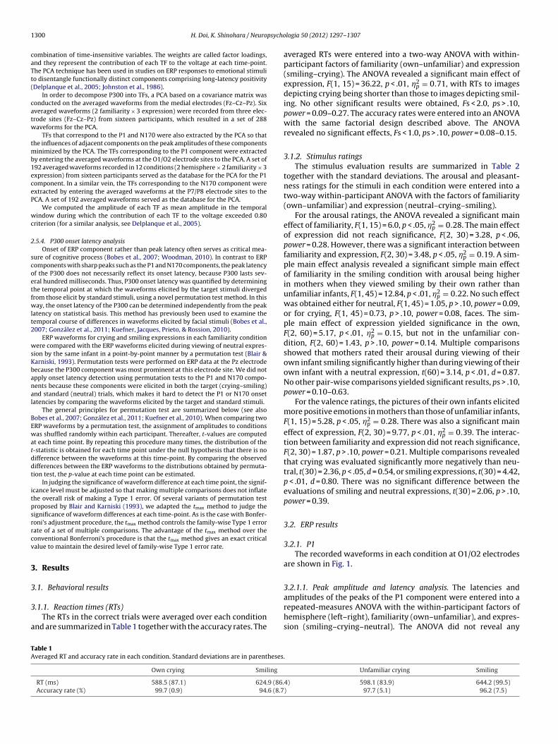
1 psycho
caTt(
catw
tmb1eceP
wc
2
scoetfwlt2
wsKbanal
BEwatddt
itpsrrcv
3
3
3
a
TA
300 H. Doi, K. Shinohara / Neuro
ombination of time-insensitive variables. The weights are called factor loadings,nd they represent the contribution of each TF to the voltage at each time-point.he PCA technique has been used in studies on ERP responses to emotional stimulio disentangle functionally distinct components comprising long-latency positivityDelplanque et al., 2005; Johnston et al., 1986).
In order to decompose P300 into TFs, a PCA based on a covariance matrix wasonducted on the averaged waveforms from the medial electrodes (Fz–Cz–Pz). Sixveraged waveforms (2 familiarity × 3 expression) were recorded from three elec-rode sites (Fz–Cz–Pz) from sixteen participants, which resulted in a set of 288aveforms for the PCA.
TFs that correspond to the P1 and N170 were also extracted by the PCA so thathe influences of adjacent components on the peak amplitudes of these components
inimized by the PCA. The TFs corresponding to the P1 component were extractedy entering the averaged waveforms at the O1/O2 electrode sites to the PCA. A set of92 averaged waveforms recorded in 12 conditions (2 hemisphere × 2 familiarity × 3xpression) from sixteen participants served as the database for the PCA for the P1omponent. In a similar vein, the TFs corresponding to the N170 component werextracted by entering the averaged waveforms at the P7/P8 electrode sites to theCA. A set of 192 averaged waveforms served as the database for the PCA.
We computed the amplitude of each TF as mean amplitude in the temporalindow during which the contribution of each TF to the voltage exceeded 0.80
riterion (for a similar analysis, see Delplanque et al., 2005).
.5.4. P300 onset latency analysisOnset of ERP component rather than peak latency often serves as critical mea-
ure of cognitive process (Bobes et al., 2007; Woodman, 2010). In contrast to ERPomponents with sharp peaks such as the P1 and N170 components, the peak latencyf the P300 does not necessarily reflect its onset latency, because P300 lasts sev-ral hundred milliseconds. Thus, P300 onset latency was quantified by determininghe temporal point at which the waveforms elicited by the target stimuli divergedrom those elicit by standard stimuli, using a novel permutation test method. In thisay, the onset latency of the P300 can be determined independently from the peak
atency on statistical basis. This method has previously been used to examine theemporal course of differences in waveforms elicited by facial stimuli (Bobes et al.,007; González et al., 2011; Kuefner, Jacques, Prieto, & Rossion, 2010).
ERP waveforms for crying and smiling expressions in each familiarity conditionere compared with the ERP waveforms elicited during viewing of neutral expres-
ion by the same infant in a point-by-point manner by a permutation test (Blair &arniski, 1993). Permutation tests were performed on ERP data at the Pz electrodeecause the P300 component was most prominent at this electrode site. We did notpply onset latency detection using permutation tests to the P1 and N170 compo-ents because these components were elicited in both the target (crying–smiling)nd standard (neutral) trials, which makes it hard to detect the P1 or N170 onsetatencies by comparing the waveforms elicited by the target and standard stimuli.
The general principles for permutation test are summarized below (see alsoobes et al., 2007; González et al., 2011; Kuefner et al., 2010). When comparing twoRP waveforms by a permutation test, the assignment of amplitudes to conditionsas shuffled randomly within each participant. Thereafter, t-values are computed
t each time point. By repeating this procedure many times, the distribution of the-statistic is obtained for each time point under the null hypothesis that there is noifference between the waveforms at this time-point. By comparing the observedifferences between the ERP waveforms to the distributions obtained by permuta-ion test, the p-value at each time point can be estimated.
In judging the significance of waveform difference at each time point, the signif-cance level must be adjusted so that making multiple comparisons does not inflatehe overall risk of making a Type 1 error. Of several variants of permutation testroposed by Blair and Karniski (1993), we adapted the tmax method to judge theignificance of waveform differences at each time-point. As is the case with Bonfer-oni’s adjustment procedure, the tmax method controls the family-wise Type 1 errorate of a set of multiple comparisons. The advantage of the tmax method over theonventional Bonferroni’s procedure is that the tmax method gives an exact criticalalue to maintain the desired level of family-wise Type 1 error rate.
. Results
.1. Behavioral results
.1.1. Reaction times (RTs)The RTs in the correct trials were averaged over each condition
nd are summarized in Table 1 together with the accuracy rates. The
able 1veraged RT and accuracy rate in each condition. Standard deviations are in parentheses.
Own crying Smiling
RT (ms) 588.5 (87.1) 624.9 (86.Accuracy rate (%) 99.7 (0.9) 94.6 (8.7
logia 50 (2012) 1297– 1307
averaged RTs were entered into a two-way ANOVA with within-participant factors of familiarity (own–unfamiliar) and expression(smiling–crying). The ANOVA revealed a significant main effect ofexpression, F(1, 15) = 36.22, p < .01, �2
p = 0.71, with RTs to imagesdepicting crying being shorter than those to images depicting smil-ing. No other significant results were obtained, Fs < 2.0, ps > .10,power = 0.09–0.27. The accuracy rates were entered into an ANOVAwith the same factorial design described above. The ANOVArevealed no significant effects, Fs < 1.0, ps > .10, power = 0.08–0.15.
3.1.2. Stimulus ratingsThe stimulus evaluation results are summarized in Table 2
together with the standard deviations. The arousal and pleasant-ness ratings for the stimuli in each condition were entered into atwo-way within-participant ANOVA with the factors of familiarity(own–unfamiliar) and expression (neutral–crying–smiling).
For the arousal ratings, the ANOVA revealed a significant maineffect of familiarity, F(1, 15) = 6.0, p < .05, �2
p = 0.28. The main effectof expression did not reach significance, F(2, 30) = 3.28, p < .06,power = 0.28. However, there was a significant interaction betweenfamiliarity and expression, F(2, 30) = 3.48, p < .05, �2
p = 0.19. A sim-ple main effect analysis revealed a significant simple main effectof familiarity in the smiling condition with arousal being higherin mothers when they viewed smiling by their own rather thanunfamiliar infants, F(1, 45) = 12.84, p < .01, �2
p = 0.22. No such effectwas obtained either for neutral, F(1, 45) = 1.05, p > .10, power = 0.09,or for crying, F(1, 45) = 0.73, p > .10, power = 0.08, faces. The sim-ple main effect of expression yielded significance in the own,F(2, 60) = 5.17, p < .01, �2
p = 0.15, but not in the unfamiliar con-dition, F(2, 60) = 1.43, p > .10, power = 0.14. Multiple comparisonsshowed that mothers rated their arousal during viewing of theirown infant smiling significantly higher than during viewing of theirown infant with a neutral expression, t(60) = 3.14, p < .01, d = 0.87.No other pair-wise comparisons yielded significant results, ps > .10,power = 0.10–0.63.
For the valence ratings, the pictures of their own infants elicitedmore positive emotions in mothers than those of unfamiliar infants,F(1, 15) = 5.28, p < .05, �2
p = 0.28. There was also a significant maineffect of expression, F(2, 30) = 9.77, p < .01, �2
p = 0.39. The interac-tion between familiarity and expression did not reach significance,F(2, 30) = 1.87, p > .10, power = 0.21. Multiple comparisons revealedthat crying was evaluated significantly more negatively than neu-tral, t(30) = 2.36, p < .05, d = 0.54, or smiling expressions, t(30) = 4.42,p < .01, d = 0.80. There was no significant difference between theevaluations of smiling and neutral expressions, t(30) = 2.06, p > .10,power = 0.39.
3.2. ERP results
3.2.1. P1The recorded waveforms in each condition at O1/O2 electrodes
are shown in Fig. 1.
3.2.1.1. Peak amplitude and latency analysis. The latencies and
amplitudes of the peaks of the P1 component were entered into arepeated-measures ANOVA with the within-participant factors ofhemisphere (left–right), familiarity (own–unfamiliar), and expres-sion (smiling–crying–neutral). The ANOVA did not reveal anyUnfamiliar crying Smiling
4) 598.1 (83.9) 644.2 (99.5)) 97.7 (5.1) 96.2 (7.5)

H. Doi, K. Shinohara / Neuropsychologia 50 (2012) 1297– 1307 1301
Table 2Averaged ratings of arousal and valence elicited by the stimuli in each condition. Standard deviations are in parentheses.
Own neutral Cry Smile Unfamiliar neutral Cry Smile
Arousal 4.0 (0.4) 4.2 (0.7) 4.7 (0.9) 3.9 (0.4) 4.1 (0.7) 4.2 (0.7).1)
L
sF
3confcde
3
a
3ofNw
sRcwpNtanop
fc
2
Valence 4.2 (1.1) 3.5 (1.1) 4.9 (1
arger rating values for valence indicate more positive feelings.
ignificant effects either for the P1 amplitude or the P1 latency,s < 2.3, ps > .10, power = 0.06–0.12.
.2.1.2. PCA. After varimax rotation, PCA revealed five distinctomponents accounting for 89.95% of the total variance. Of these,nly one TF was extracted within the latency range of the P1 compo-ent. The 0.8 criterion resulted in the latency range of 108–128 ms
or this TF. Averaged amplitudes of the TFs corresponding to P1omponent were entered into an ANOVA with the same factorialesign described above. The ANOVA did not reveal any significantffects, Fs < 2.0, ps > .10, power = 0.05–0.48.
.2.2. N170The recorded waveforms in each condition at P7/P8 electrodes
re shown in Fig. 2.
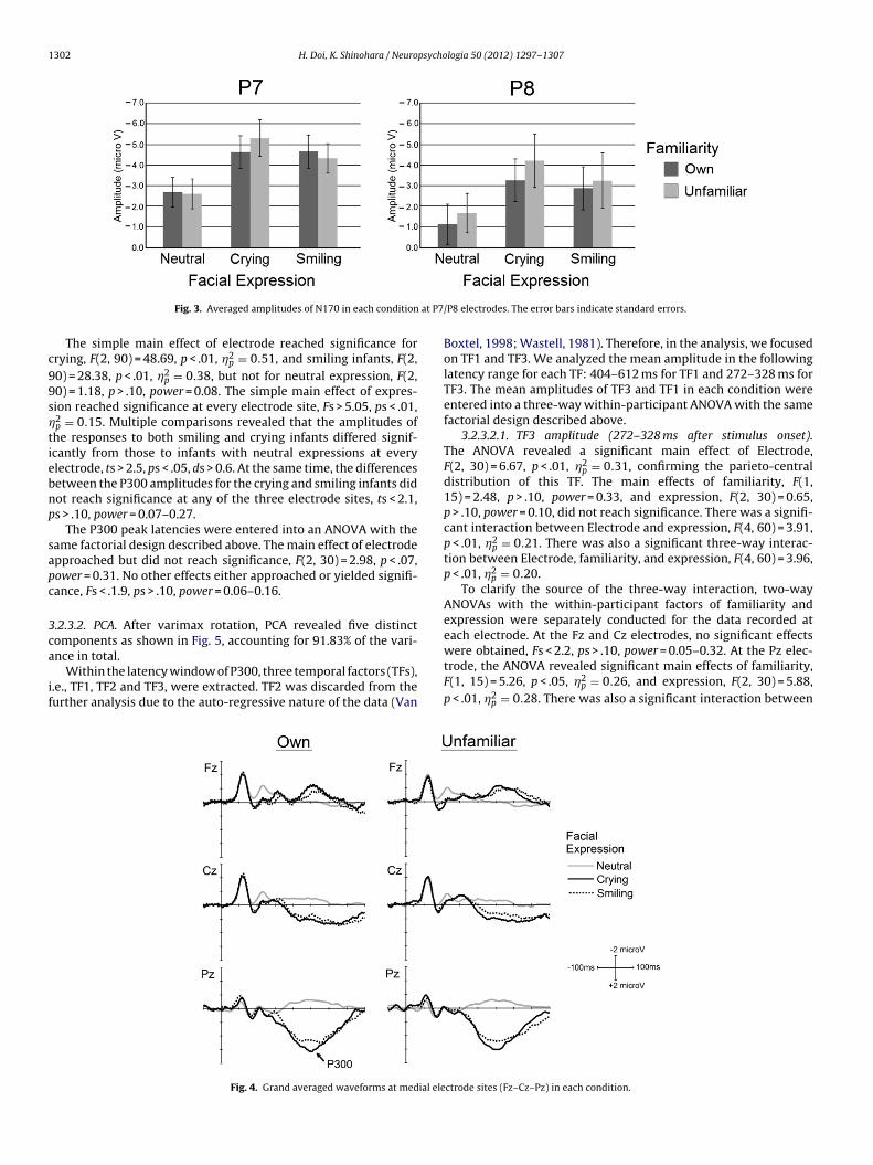
.2.2.1. Peak amplitude and latency analysis. The peak amplitudesf the N170 component were entered into an ANOVA with the sameactorial design described above. The averaged amplitudes of the170 component in each condition are shown in Fig. 3 togetherith the standard errors.
The ANOVA revealed a significant main effect of expres-ion, F(2, 30) = 48.14, p < .01, �2
p = 0.71. Multiple comparisons byyan’s method revealed a significant difference in every pairwiseomparison. Specifically, the N170 component for crying infantsas significantly larger than that for the neutral, t(30) = 9.42,
< .01, d = 0.77, or smiling infants, t(30) = 2.31, p < .05, d = 0.18. The170 component for smiling infants was significantly larger than
hat for neutral infants, t(30) = 7.10, p < .01, d = 0.64. The inter-ction between Hemisphere and familiarity approached, but didot reach significance, F(1, 15) = 3.34, p < .08, power = 0.42. Nother effects approached or yielded significance, Fs < 2.5, ps > .10,
ower = 0.06–0.32.The N170 latencies were entered into an ANOVA with the sameactorial design described above. The ANOVA revealed no signifi-ant effects, Fs < 1.5, ps > .10, power = 0.08–0.83.
Fig. 1. Grand averaged waveforms at O1/O2 electrode sites in each condition.
3.9 (1.4) 3.4 (1.3) 4.4 (1.7)
3.2.2.2. PCA. After varimax rotation, PCA revealed five distinctcomponents accounting for 90.08% of the total variance. Of these,only one TF was extracted within the latency range of the N170component. The 0.8 criterion resulted in the latency range of140–166 ms for this TF.
Averaged amplitudes of TFs corresponding to the N170 com-ponent were entered into an ANOVA with the same factorialdesign described above. The ANOVA yielded essentially the sameresults as the conventional peak amplitude analysis. Specifically,the main effect of expression reached significance, F(2, 30) = 25.77,p < .05, �2
p = 0.63. Multiple comparisons analysis by Ryan’s methodrevealed that the TF for crying was significantly larger than that forneutral, t(30) = 7.14, p < .01, d = 0.36, or smiling infants, t(30) = 2.98,p < .05, d = 0.22. The TF for smiling infants was significantly largerthan that for neutral infants, t(30) = 4.16, p < .01, d = 0.14. No othermain effect or interaction reached significance, Fs < 2.0, ps > .05,power = 0.09–0.52.
3.2.3. P300The waveforms recorded at the medial electrode sites
(Fz–Cz–Pz) are shown in Fig. 4. At the latency range from 250to 800 ms after stimulus onset, a large positive deflection wasobserved for target stimuli. From its scalp distribution, latencyrange, and response to experimental manipulation, we determinedthis deflection as P300.
3.2.3.1. Peak amplitude and latency analysis. The peak amplitudesof the P300 component were entered into a three-way within-participant ANOVA with the factors of electrode (Fz–Pz–Cz), famil-iarity (own–unfamiliar), and expression (neutral–crying–smiling).The ANOVA revealed a significant main effect of electrode, F(2,
30) = 27.49, p < .01, �p = 0.65, and expression, F(2, 30) =81.59,p < .01, �2p = 0.84. There was also a significant interaction betweenelectrode and expression, F(4, 60) =23.64, p < .01, �2
p = 0.61.
Fig. 2. Grand averaged waveforms at P7/P8 electrode sites in each condition.
1302 H. Doi, K. Shinohara / Neuropsychologia 50 (2012) 1297– 1307
at P7
c99s�tiebnp
sapc
3ca
if
Fig. 3. Averaged amplitudes of N170 in each condition
The simple main effect of electrode reached significance forrying, F(2, 90) = 48.69, p < .01, �2
p = 0.51, and smiling infants, F(2,0) = 28.38, p < .01, �2
p = 0.38, but not for neutral expression, F(2,0) = 1.18, p > .10, power = 0.08. The simple main effect of expres-ion reached significance at every electrode site, Fs > 5.05, ps < .01,2p = 0.15. Multiple comparisons revealed that the amplitudes ofhe responses to both smiling and crying infants differed signif-cantly from those to infants with neutral expressions at everylectrode, ts > 2.5, ps < .05, ds > 0.6. At the same time, the differencesetween the P300 amplitudes for the crying and smiling infants didot reach significance at any of the three electrode sites, ts < 2.1,s > .10, power = 0.07–0.27.
The P300 peak latencies were entered into an ANOVA with theame factorial design described above. The main effect of electrodepproached but did not reach significance, F(2, 30) = 2.98, p < .07,ower = 0.31. No other effects either approached or yielded signifi-ance, Fs < .1.9, ps > .10, power = 0.06–0.16.
.2.3.2. PCA. After varimax rotation, PCA revealed five distinctomponents as shown in Fig. 5, accounting for 91.83% of the vari-
nce in total.Within the latency window of P300, three temporal factors (TFs),.e., TF1, TF2 and TF3, were extracted. TF2 was discarded from theurther analysis due to the auto-regressive nature of the data (Van
Fig. 4. Grand averaged waveforms at medial ele
/P8 electrodes. The error bars indicate standard errors.
Boxtel, 1998; Wastell, 1981). Therefore, in the analysis, we focusedon TF1 and TF3. We analyzed the mean amplitude in the followinglatency range for each TF: 404–612 ms for TF1 and 272–328 ms forTF3. The mean amplitudes of TF3 and TF1 in each condition wereentered into a three-way within-participant ANOVA with the samefactorial design described above.
3.2.3.2.1. TF3 amplitude (272–328 ms after stimulus onset).The ANOVA revealed a significant main effect of Electrode,F(2, 30) = 6.67, p < .01, �2
p = 0.31, confirming the parieto-centraldistribution of this TF. The main effects of familiarity, F(1,15) = 2.48, p > .10, power = 0.33, and expression, F(2, 30) = 0.65,p > .10, power = 0.10, did not reach significance. There was a signifi-cant interaction between Electrode and expression, F(4, 60) = 3.91,p < .01, �2
p = 0.21. There was also a significant three-way interac-tion between Electrode, familiarity, and expression, F(4, 60) = 3.96,p < .01, �2
p = 0.20.To clarify the source of the three-way interaction, two-way
ANOVAs with the within-participant factors of familiarity andexpression were separately conducted for the data recorded ateach electrode. At the Fz and Cz electrodes, no significant effects
were obtained, Fs < 2.2, ps > .10, power = 0.05–0.32. At the Pz elec-trode, the ANOVA revealed significant main effects of familiarity,F(1, 15) = 5.26, p < .05, �2p = 0.26, and expression, F(2, 30) = 5.88,p < .01, �2
p = 0.28. There was also a significant interaction between
ctrode sites (Fz–Cz–Pz) in each condition.

H. Doi, K. Shinohara / Neuropsycho
Fig. 5. Temporal course of factor loadings of each temporal factor extracted byPCA on the basis of the averaged waveforms recorded at medial electrode sites(Fz–Cz–Pz) The gray horizontal line indicates the contribution criterion of 0.8.
Fb
faa
tsaptsm6Fbltdift
A3teopte
eF�
own infant reached significance, r = 0.64, p < .05, indicating that
ig. 6. Averaged amplitudes of TF3 in each condition at the Pz electrode. The errorars indicate standard errors. **p < .01.
amiliarity and expression, F(2, 30) = 3.91, p < .05, �2p = 0.21. The
veraged amplitudes of TF3 at the Pz electrode in each conditionre shown in Fig. 6.
A simple main effect analysis revealed that the TF3 ampli-ude in response to the crying of a mother’s own infant wasignificantly larger than that for the response to the crying ofn unfamiliar infant, F(1, 45) = 12.81, p < .01, �2
p = 0.22. The sim-le main effect of familiarity did not reach significance either inhe neutral, F(1, 45) = 0.37, p > .10, power = 0.06, or smiling expres-ion condition, F(1, 45) = 0.13, p > .10, power = 0.06. The simpleain effect of expression reached significance in the own, F(2,
0) = 9.49, p < .01, �2p = 0.24, but not in the unfamiliar condition,
(2, 60) = 0.56, p > .10, power = 0.07. Multiple comparison analysisy Ryan’s method revealed that the TF3 amplitude was significantly
arger in response to the crying of a mother’s own infant than forhe same infant exhibiting a neutral expression, t(60) = 3.97, p < .01,
= 0.53, or smiling, t(60) = 3.55, p < .01, d = 0.54. The TF3 amplituden response to the smiling of a mother’s own infant did not differrom that in response to the infant exhibiting a neutral expression,(60) = 0.42, p > .10, power = 0.06.
3.2.3.2.2. TF1 amplitude (404–612 ms after stimulus onset). TheNOVA revealed a significant main effect of Electrode, F(2,0) = 43.46, p < .01, �2
p = 0.74, confirming the parieto-central dis-ribution of this TF. There was also a significant main effect ofxpression, F(2, 30) = 45.36, p < .01, �2
p = 0.75. The main effectf familiarity did not reach significance, F(1, 15) = 0.63, p > .10,ower = 0.11. There was also a significant interaction between Elec-rode and expression, F(4, 60) = 37.69, p < .01, �2
p = 0.71. No otherffects reached significance, Fs < 2.5, ps > .10, power = 0.06–026.
A simple main effect analysis revealed a significant simple main
ffect of expression at the Fz, F(2, 90) = 5.65, p < .01, �2p = 0.11, Cz,(2, 90) =17.59, p < .01, �2
p = 0.28, and Pz, F(2, 90) = 97.53, p < .01,2p = 0.78, electrode sites. Multiple comparisons by Ryan’s method
logia 50 (2012) 1297– 1307 1303
revealed that the TF1 amplitudes in response to crying and smilingexpressions differed significantly from those in response to neutralexpression at every electrode site, ts > 2.5, ps < .01, ds > 0.60. How-ever, the TF1 amplitudes did not differ between smiling and cryinginfants in any electrode sites, ts < 1.7, ps > .10, power = 0.18–0.38.The simple main effect of Electrode reached significance for cry-ing, F(2, 90) = 71.75, p < .01, �2
p = 0.61, and smiling infants, F(2,90) = 48.53, p < .01, �2
p = 0.52, but not for neutral expression, F(2,90) = 1.66, p > .10, power = 0.09.
3.2.3.3. Onset latency analysis by the permutation test. The ERPwaveforms for the crying and smiling infants were compared withthe ERP waveforms for the infants with neutral expressions ina point-by-point manner within each familiarity condition. Weperformed 10,000 permutations to generate the t-statistic distribu-tions. The onset latency of the P300 component was determined asthe time point at which the t-value first becomes smaller than thecritical value during the period 250–800 ms after stimulus onset.Because the permutation tests were performed on four pairs ofwaveforms, the significance level was adjusted by Bonferroni’sprocedure to 0.05/4 or 0.0125. To avoid making a Type I error,only differences that persisted at this significance level for at least10 consecutive samples were considered significant. This anal-ysis revealed P300 onset latencies of 302 ms in the own-cryingcondition, 344 ms in the own-smiling condition, 390 ms in theunfamiliar-crying condition, and 374 ms in the unfamiliar-smilingcondition.
3.3. Correlation between behavioral measures and ERPamplitudes
To understand the nature of the processing reflected by eachERP component, a series of correlation analyses were performed forthe TF1 and TF3 of the P300 component and the amplitudes of theTFs corresponding to P1 and N170 components. Correlation coef-ficients between these TFs and the behavioral measures, includingthe MAI-J score and the rating scores for faces in each conditionwere computed. Because there was no significant effect of Hemi-sphere for the amplitudes of the TFs corresponding to either the P1or N170, the data were collapsed across Hemisphere for these TFs.For the correlation analysis of TF1 and TF3, the amplitudes of theseTFs at Pz electrode were entered because the amplitudes of the TFswere maximal at this electrode site.
3.3.1. Correlation with rating resultsCorrelations between TF amplitudes in each condition and the
arousal and valence ratings for the stimuli in the correspondingcondition were calculated. For example, correlation coefficientsbetween TF1 amplitude in own-crying condition and the arousaland valence ratings in the own-crying condition were computed.The results revealed no significant correlations, r2 < .09, ps > .10,power = 0.05–0.21.
3.3.2. Correlation with questionnaire resultCorrelations between the ERP amplitudes in each condition and
MAI-J scores were calculated. The significance level was adjusted byBonferroni’s procedure to 0.0083 (=0.05/6) because six correlationcoefficients were computed for each TF. The positive correlationbetween MAI-J and TF1 amplitudes to the smiling of the mother’s
mothers with larger MAI-J scores showed larger TF1 amplitudein response to smiling by their own infant. No other significantcorrelation was obtained, r2 < .31, ps > .10, power = 0.06–0.69.
1 psycho
4
oNcknTtinrhttwitsei
nn(iisEaitatb(&rwnsducmoia
ttLKtL(gr&rReb
f
304 H. Doi, K. Shinohara / Neuro
. Discussion
The present study measured ERPs elicited in mothers while theybserved crying and smiling by their own or unfamiliar infants.170, which is supposed to reflect perceptual stage of face pro-essing, was enlarged by presentation of crying faces regardless ofinship of infant. In contrast, the early portion of the P300 compo-ent, TF3, was modulated by both facial expression and familiarity.hat is, TF3 was larger in response to a mother’s own infant’s cryinghan to the other three types of facial stimuli. Interestingly, similarnteractions between facial familiarity and facial expression wereot observed at later portion of the P300 component. Recent neu-oimaging studies (Noriuchi et al., 2008; Strathearn et al., 2009)ave indentified neural regions sensitive to both infants’ emo-ional expressions and their familiarity. The present study extendedhese findings by clarifying for the first time the temporal courseith which emotional expression and familiarity exert interactive
nfluences on neural processing of infant’s faces in mothers. Fur-hermore, to the best of our knowledge, this is the first study tohow that facial expression and familiarity information influencearly portions of the parieto-centrally distributed P300 componentn a different manner from later portions of this component.
Our analyses revealed that the amplitude of the N170 compo-ent was enlarged in response to crying compared to smiling andeutral expression. This finding concurs with previous researchHendriks et al., 2007; Proverbio et al., 2006) showing that cry-ng elicits larger N170 components. Taken together, these resultsndicate that the visual cue of crying is detected as early as thetructural encoding stage of face processing (Bentin et al., 1996;imer, 2000a, 2000b; Rossion et al., 1999). Interestingly, the N170mplitudes to crying and smiling expressions did not differ accord-ng to familiarity of infant face. Although null findings should bereated cautiously, this finding might indicate that infant facesre processed in a similar manner regardless of familiarity at ini-ial perceptual stages of face processing, which are supposed toe subserved by fusiform gyrus (FG), superior temporal sulcusSTS), and occipital face area (OFA) (for a review, see Minnebusch
Daum, 2009). This further indicates the possibility that N170eflects instinctive responsiveness in mothers to infant stimuli,hich is triggered regardless of kinship of infant. Such mecha-ism of parental instinct must be quite beneficial for the purpose ofpecies-preservation. An ERP study by Purhonen et al. (2001) hasemonstrated that mothers show enhanced neural responses tonfamiliar infant’s crying compared to females with no child at per-eptual stages. Furthermore, Proverbio et al. (2006) revealed thatothers show different N170 response to distressed expressions
f unfamiliar infants from fathers. Taking these into consideration,t seems possible that N170 responses to infant stimuli in femalesre altered through the experiences of delivery and care-taking.
Presentation of infant faces activates neural regions linkedo parental and care-giving behaviors such as orbitofrontal cor-ex, anterior cingulate cortex, and insula (Bartels & Zeki, 2004;orberbaum et al., 2002; Nitschke et al., 2004; Swain, Lorberbaum,ose, & Strathearn, 2007). Furthermore, the amygdala is sensitive
o both “baby-schema” (Glocker et al., 2009a, 2009b; Zebrowitz,uevano, Bronstad, & Aharon, 2009) and crying of unfamiliar infantsSander, Frome, & Scheich, 2007; Seifritz et al., 2003). It is sug-ested that these regions influence visual processing in corticalegions via pulvinar nuclei in thalamus (for a review, see Pessoa
Adolphs, 2010). On the basis of these, activation of candidateegions for N170 generation such as FG and STS (Itier & Taylor, 2004;ossion, Joyce, Cottrell, & Tarr, 2003) might be augmented by influ-
nces from neural regions linked to parental instinct or care-takingehaviors.At medial electrode sites, the predicted interaction betweenamiliarity and facial expression was observed in TF3. Specifically,
logia 50 (2012) 1297– 1307
the amplitude of TF3 was increased by own infant’s crying expres-sion compared to the other three types of target faces. Theindependence of processing streams dedicated to stable aspectsof face information, such as familiarity, and changeable facialexpression information has been the focus of controversy since theintroduction of the influential model of face processing by Bruceand Young (1986, see also Young, McWeeny, Hay, & Ellis, 1986).Although the independence between these processing streamshas been confirmed by neuroimaging (Andrews & Ewbank, 2004;Haxby, Hoffman, & Gobbini, 2000) and clinical studies on brain-damaged patients (Tranel, Damasio, & Damasio, 1988), recentbehavioral studies (Campbell & Burke, 2009; Ellamil, Susskind, &Anderson, 2008; Schweinberger, Burton, & Kelly, 1999; Why &Huang, 2010) have demonstrated the interaction between famil-iarity and emotional expression information of faces. The results ofthe present study add further evidence that these two types of facialinformation, i.e. facial expression and facial familiarity informa-tion, do interact at long-latency cognitive evaluation stage of faceprocessing (see also, Martens, Leuthold, & Schweinberger, 2010;Wild-Wall, Dimigen, & Sommer, 2008).
In the three-stimuli oddball paradigm, to-be-ignored low-frequency stimuli induce early positive deflection called P3a(Delplanque et al., 2005; Pourtois et al., 2008) within a similarlatency range as TF3 in the present study. However, P3a amplitudereaches its maximum at fronto-central electrode sites contrarilyto TF3. Moreover, both crying and smiling expressions were to-be-attended stimuli in the present study. Therefore, TF3 does notseem to be the homolog of P3a in standard oddball-paradigm asused in the present study. Judging from the latency range and theoccipito-parietal centered distribution of TF3, it seems likely thatTF3 corresponds to the early ascending portion of the P300 compo-nent. On the basis of this, the present finding seems to indicate thatown infant’s crying expression elicits P300 more efficiently thanother types of facial stimuli. This interpretation is further corrobo-rated by the results of the permutation test that the onset latency ofthe P300 to own infant’s crying expression was shorter than thoseto the other three types of target faces.
Efficiency of P300 elicitation has been linked to speed ofevaluation of target stimulus (Bobes et al., 2007; Kutas et al.,1977; Woodman, 2010; for a review see, Polich, 2007). The logicbehind this assumption is that P300 cannot arise before differencesbetween target and standard stimuli are detected. From this per-spective, the present finding may indicate that the evaluation ofdeviance from neutral expression is completed more efficientlyfor a mother’s own infant’s crying than for the other three typesof stimuli. If this hypothesis is correct, it would be interesting todetermine which characteristics of a mother’s own infant’s cryingallow for the accelerated evaluation of it. Although speculative atthis point, a potential candidate is the socio-emotional significancethe crying of a mother’s own infant bears. Her own infant is theprimary target of the affective attachment of mothers (Bartels &Zeki, 2004; Grasso et al., 2009; Leibenluft et al., 2004; Nitschkeet al., 2004; Noriuchi et al., 2008; Strathearn et al., 2009). Moreover,infant’s crying expression signals urgent need of care and protec-tion (Abramowitz, Schwartz, & Moore, 2003; Acebo & Thoman,1995; Hahn-Holbrook et al., 2011). These characteristics of owninfant’s crying face presumably makes them especially salient formothers, which might accelerate the evaluation of it.
There are several alternative interpretations to the modula-tion pattern of the TF3 besides the accelerated evaluation of amother’s own infant’s crying proposed above. First, the presentresults might reflect memory functions associated with recogni-
tion of familiar own infant’s faces. A number of studies have linkedP300 (Polich et al., 1994; Polich, 2007; Soltani & Knight, 2000) andP300-like enlarged positivity (Dolcos & Cabeza, 2002; Righi et al.,2012; Wilding, 2000; Yovel & Paller, 2004) to memory encoding and
psycho
r(giwiop
TbtHCocescosrtrrTshtmatf
ordtowmiiisIiS(OTsrmi
ispiTodtmk
H. Doi, K. Shinohara / Neuro
ecognition of memorized familiar items. Moreover, some studiesHalgren et al., 1995a, 1995b; Smith et al., 1990) have localized theenerator of P300 to temporal memory-related regions. Consider-ng these findings together with the fact that own infant’s faces
ere more familiar for participants than those of stranger’s infant,t is quite conceivable that neural activations accompanying mem-ry recognition of own infant face have contributed to the observedattern of TF3 modulation.
Second alternative explanation is that the observed pattern ofF3 modulation is attributable to emotional responses inducedy stimulus faces. Own infant’s crying expression is potentiallyhe most negative stimuli for mothers (Abramowitz et al., 2003;ahn-Holbrook et al., 2011) among the four types of target stimuli.onsidering this together with the negativity-bias (Ito et al., 1998)f human emotional reaction, it is not surprising that own infant’srying face was most efficient inducer of emotional response. Thisxplanation is partly refuted by the results of stimulus rating, whichhowed no signs that own infant’s crying face was rated espe-ially negative or arousing. However, given the limited reliabilityf subjective rating (Greenwald, McGhee, & Schwartz, 1998), it istill possible that TF3 modulation reflects differences in emotionalesponses induced by presented facial stimuli. To compensate forhe methodological weakness of subjective rating, simultaneousecordings of EEG and physiological measurements of peripheraleactions (Herzmann, Schweinberger, Sommer, & Jentzsch, 2004;ranel et al., 1988; Vico, Guerra, Robles, Vila, & Anllo-Vento, 2010),uch as skin-conductance responses and heart rate should be quiteelpful. Although correlation analyses did not reveal any correla-ion between emotional ratings and TF3 amplitudes, more objective
easures of peripheral responses may show correlation with TF3mplitude, which would be of great help in clarifying the func-ional significance of the processing stage reflected by this temporalactor.
Following the TF3, a large positive deflection of the TF1 wasbserved to crying and smiling expressions. The TF1 possibly cor-esponds to the pinnacle of the P300 as judged from their corticalistributions and latency ranges. This contention is supported byhe finding that modulation pattern of TF1 is paralleled by thatf P300 peak amplitude. Correlation analyses indicated that thereas a positive correlation between TF1 amplitudes elicited by aother’s own infant’s smiling expression and MAI-J score. This
ndicates that the mothers with more positive-feelings toward theirnfants showed increased neural response to smiling by their ownnfant. The rewarding value of own infant’s smile is evidenced byeveral recent studies (Nitschke et al., 2004; Noriuchi et al., 2008).t is also known that individual differences in reactivity to reward-ng stimuli is influenced by genetics (Camara et al., 2010; Filbey,chacht, Myers, Chavez, & Hutchison, 2010) and hormonal statusDreher et al., 2007; Frank, Kim, Krzemien, & Van Vugt, 2010).n the basis of these, we tentatively think that relation betweenF1 amplitude and attachment attitude as found in the presenttudy might be mediated by individual differences in response toewarding stimuli, which is partly determined by genetic or hor-onal predispositions. Examination of this hypothesis is surely an
nteresting topics for the future research.The interpretation of the results of the present study are lim-
ted by the following methodological weaknesses. First, a relativelymall number of electrodes were used, which prevented us fromroviding detailed analysis of scalp potential distributions or apply-
ng source reconstruction methods such as dipole source modeling.o further clarify the precise nature of the present findings, the usef high-density electrodes should be quite informative. Second, we
id not include faces of infants that were not kin, but familiar tohe mothers as a control condition, which prevented us from deter-ining whether the effects of infant’s identity are attributable toinship or perceptual familiarity (Caharel et al., 2005, 2006; Furl,
logia 50 (2012) 1297– 1307 1305
Van Rijsbergen, Treves, Friston, & Dolan, 2007). It is possible thatperceptually familiarity of infant’s face modulated neural process-ing of face, which might have led to differential neural responsesto crying faces of own and unfamiliar infant.
As for the second limitation, we believe that personal attach-ment or kinship rather than mere familiarity have played at leastsome roles in modulating ERP responses on the following grounds.First, if the present results were explainable solely by perceptualfamiliarity, similar effect of face identity would have been observedfor all the expressions, which was apparently not the case. Sec-ond, efficient processing of own infant’s crying expression squaresfirmly with the indications of previous studies that mothers areparticularly sensitive to own infant’s attachment behaviors (Acebo& Thoman, 1995; Hahn-Holbrook et al., 2011; Kim et al., 2011;Noriuchi et al., 2008). However, considering the purpose of thepresent study, it is of primary importance in future studies to delin-eate the effects of perceptual familiarity and personal relationshipsby including different kinds of familiar faces.
References
Abramowitz, J. S., Schwartz, S. A., & Moore, K. M. (2003). Obsessional thoughts inpostpartum females and their partners: Content, severity and relationship withdepression. Journal of Clinical Psychology in Medical Settings, 10, 157–164.
Acebo, C., & Thoman, E. B. (1995). Role of infant crying in the early mother–infantdialogue. Physiology and Behavior, 57, 541–547.
Ainsworth, M. D. S., Blehar, M. C., Waters, E., & Wall, S. (1978). Patterns of attachment.A psychological study of the strange situation. Hillsdale, NJ: Erlbaum.
Andrews, T. J., & Ewbank, M. P. (2004). Distinct representations for facial identityand changeable aspects of faces in the human temporal lobe. NeuroImage, 23,905–913.
Bartels, A., & Zeki, S. (2004). The neural correlates of maternal and romantic love.NeuroImage, 21, 1155–1166.
Bentin, S., Allison, T., Puce, A., Perez, E., & McCarthy, G. (1996). Electrophysiolog-ical studies of face perception in humans. Journal of Cognitive Neuroscience, 8,551–565.
Blair, R. C., & Karniski, W. (1993). An alternative method for significance testing ofwaveform difference potentials. Psychophysiology, 30, 518–524.
Bobes, M. A., Quinonez, I., Perez, J., Leon, I., & Valdés-Sosa, M. (2007). Brain potentialsreflect access to visual and emotional memories for faces. Biological Psychology,75, 146–153.
Bötzel, K., Schulze, S., & Stodieck, S. R. G. (1995). Scalp topography and analysis ofintracranial sources of face evoked potentials. Experimental Brain Research, 104,135–143.
Bowlby, J. (1969). Attachment and loss. Attachment. New York: Basic Books.Bruce, V., & Young, A. (1986). Understanding face recognition. The British Journal of
Psychology, 77, 305–327.Caharel, S., Courtay, N., Bernard, C., Lalonde, R., & Rebai, M. (2005). Familiarity and
emotional expression influence an early stage of face processing: An electro-physiological study. Brain and Cognition, 59, 96–100.
Caharel, S., Fiori, N., Bernard, C., Lalonde, R., & Rebai, M. (2006). The effects ofinversion and eye displacements of familiar and unknown faces on early andlate-stage ERPs. International Journal of Psychophysiology, 62, 141–151.
Camara, E., Krämer, U. M., Cunillera, T., Marco-Pallarés, J., Cucurell, D., Nager, W., et al.(2010). The effects of COMT (Val108/158Met) and DRD4 (SNP-521) dopaminegenotypes on brain activations related to valence and magnitude of rewards.Cerebral Cortex, 20, 1985–1996.
Campbell, J., & Burke, D. (2009). Evidence that identity-dependent and identity-independent neural populations are recruited in the perception of five basicemotional facial expressions. Vision Research, 49, 1532–1540.
Chapman, R. M., & Mccrary, J. W. (1995). EP component identification and measure-ment by principal components-analysis. Brain and Cognition, 27, 288–310.
Cuthbert, B. N., Schupp, H. T., Bradley, M. M., Birbaumer, N., & Lang, P. J. (2000). Brainpotentials in affective picture processing: Covariation with autonomic arousaland affective report. Biological Psychology, 52, 95–111.
Delplanque, S., Silvert, L., Hot, P., & Sequeira, H. (2005). Event-related P3a and P3b inresponse to unpredictable emotional stimuli. Biological Psychology, 68, 107–120.
Doi, H., Amamoto, T., Okishige, Y., Kato, M., & Shinohara, K. (2010). The own-sexeffect in facial expression recognition. NeuroReport, 21, 564–568.
Doi, H., Sawada, R., & Masataka, N. (2007). The effects of eye and face inversion on theearly stages of gaze direction perception – An ERP study. Brain Research, 1183,83–90.
Doi, H., Ueda, K., & Shinohara, K. (2009). Neural correlates of the stare-in-the-crowdeffect. Neuropsychologia, 47, 1053–1060.
Dolcos, F., & Cabeza, R. (2002). Event-related potentials of emotional memory:Encoding pleasant, unpleasant, and neutral pictures. Cognitive Affective andBehavioral Neuroscience, 2, 252–263.
Dreher, J.-C., Schmidt, P. J., Kohn, P., Furman, D., Rubinow, D., & Berman, K. F. (2007).Menstrual cycle phase modulates reward-related neural function in women.
1 psycho
E
E
E
E
F
F
F
F
G
G
G
G
G
H
H
H
H
H
H
H
H
I
I
J
J
J
K
K
306 H. Doi, K. Shinohara / Neuro
Proceedings of the National Academy of Sciences of the United States of America,104.
imer, M. (2000a). Event-related potentials distinguish processing stages involvedin face perception and recognition. Clinical Neurophysiology, 111, 694–705.
imer, M. (2000b). Effects of face inversion on the structural encoding and recog-nition of faces – Evidence from event-related brain potentials. Cognitive BrainResearch, 10, 145–158.
imer, M., & Holmes, A. (2002). An ERP study on the time course of emotional faceprocessing. NeuroReport, 13, 427–431.
llamil, M., Susskind, J. M., & Anderson, A. K. (2008). Examinations of identityinvariance in facial expression adaptation. Cognitive, Affective and BehavioralNeuroscience, 8, 273–281.
rank, T. C., Kim, G. L., Krzemien, A., & Van Vugt, D. A. (2010). Effect of menstrualcycle phase on corticolimbic brain activation by visual food cues. Brain Research,1363, 81–92.
ilbey, F. M., Schacht, J. P., Myers, U. S., Chavez, R. S., & Hutchison, K. E. (2010).Individual and additive effects of the CNR1 and FAAH genes on brain responseto marijuana cues. Neuropsychopharmacology, 35.
orsythe, A., Nadal, M., Sheehy, N., Cela-Conde, C. J., & Sawey, M. (2011). Predict-ing beauty: Fractal dimension and visual complexity in art. British Journal ofPsychology, 102, 49–70.
url, N., Van Rijsbergen, N. J., Treves, A., Friston, K. J., & Dolan, R. J. (2007).Experience-dependent coding of facial expression in superior temporal sulcus.Proceedings of the National Academy of Sciences of the United States of America,104, 13485–13489.
onzález, I. Q., León, M. A. B., Belin, P., Martínez-Quintana, Y., García, L. G., & Castillo,M. S. (2011). Person identification through faces and voices: An ERP study. BrainResearch, 1407, 13–26.
rasso, D. J., Moser, J. S., Dozier, M., & Simons, R. (2009). ERP correlates of attentionallocation in mothers processing faces of their childre. Biological Psychology, 81,95–102.
locker, M. L., Langleben, D. D., Ruparel, K., Loughead, J. W., Gur, R. C., & Sachser, N.(2009). Baby schema in infant faces induces cuteness perception and motivationfor caretaking in adults. Ethology, 115, 257–263.
locker, M. L., Langleben, D. D., Ruparel, K., Loughead, J. W., Valdez, J. N., Griffin, M.D., et al. (2009). Baby schema modulates the brain reward system in nulliparouswomen. Proceedings of the National Academy of Sciences of the United States ofAmerica, 106, 9115–9119.
reenwald, A. G., McGhee, D. E., & Schwartz, J. L. K. (1998). Measuring individual dif-ferences in implicit cognition: The implicit association test. Journal of Personalityand Social Psychology, 74, 1464–1480.
axby, J. V., Hoffman, E. A., & Gobbini, M. I. (2000). The distributed human neutralsystem for face perception. Trends in Cognitive Sciences, 4, 223–233.
ahn-Holbrook, J., Holbrook, C., & Haselton, M. G. (2011). Parental precaution: Neu-robiological means and adaptive ends. Neuroscience and Biobehavioral Reviews,35, 1052–1066.
algren, E., Baudena, P., Jeffrey, M. C., Heit, G., Liégeois, C., Chauvel, P., et al. (1995).Intracerebral potentials to rare target and distractor auditory and visual stimuli.I. Superior temporal plane and parietal lobe. Electroencephalography and ClinicalNeurophysiology, 94, 191–220.
algren, E., Baudena, P., Jeffrey, M. C., Heit, G., Marinkovic, K., Devaux, B., et al. (1995).Intracerebral potentials to rare target and distractor auditory and visual stimuli.II. Medial lateral and posterior temporal lobe. Electroencephalography and ClinicalNeurophysiology, 94, 229–250.
endriks, M. C. P., Van Boxtel, G. J. M., & Vingerhoets, A. J. F. M. (2007). An event-related potential study on the early processing of crying faces. NeuroReport, 18,631–634.
endriks, M. C. P., & Vingerhoets, A. J. J. M. (2006). Social messages of crying faces:Their influence on anticipated person perception emotions and behaviouralresponses. Cognition and Emotion, 20, 878–886.
errmann, M. J., Ehlis, A. C., Ellgring, H., & Fallgatter, A. J. (2005). Early stages (P100)of face perception in humans as measured with event-related potentials (ERPs).Journal of Neural Transmission, 112, 1073–1081.
erzmann, G., Schweinberger, S. R., Sommer, W., & Jentzsch, I. (2004). What’s specialabout personally familiar faces? A multimodal approach. Psychophysiology, 41,688–701.
to, T. A., Larsen, J. T., Smith, N. K., & Cacioppo, J. T. (1998). Negative informationweighs more heavily on the brain: The negativity bias in evaluative categoriza-tions. Journal of Personality and Social Psychology, 75, 887–900.
tier, R. J., & Taylor, M. J. (2004). Source analysis of the N170 to faces and object.NeuroReport, 15, 1261–1265.
emel, B., Pisani, M., Calabria, M., Crommelinck, M., & Bruyer, R. (2003). Is the N170for faces cognitively penetrable? Evidence from repetition priming of Mooneyfaces of familiar and unfamiliar persons. Cognitive Brain Research, 17, 431–446.
ohnston, V. S., Miller, D. R., & Burleson, M. H. (1986). Multiple P3s to emotionalstimuli and their theoretical significance. Psychophysiology, 23, 684–694.
oyce, C., & Rossion, B. (2005). The face-sensitive N170 and VPP components man-ifest the same brain processes: The effect of reference electrode sit. ClinicalNeurophysiology, 116, 2613–2631.
im, P., Feldman, R., Mayes, L. C., Eicher, V., Thompson, N., Leckman, J. F., et al. (2011).Breastfeeding, brain activation to own infant cry, and maternal sensitivity. Jour-
nal of Child Psychology and Psychiatry and Allied Disciplines, 52, 907–915.uefner, D., Jacques, C., Prieto, E. A., & Rossion, B. (2010). Electrophysiological cor-relates of the composite face illusion: Disentangling perceptual and decisionalcomponents of holistic face processing in the human brain. Brain and Cognition,74, 225–238.
logia 50 (2012) 1297– 1307
Kutas, M., McCarthy, G., & Donchin, E. (1977). Augmenting mental chronometry: TheP300 as a measure of stimulus evaluation time. Science, 197, 792–795.
Langeslag, S. J. E., Franken, I. H. A., & Van Strien, J. W. (2008). Dissociating love-relatedattention from task-related attention: An event-related potential oddball study.Neuroscience Letters, 431, 236–240.
Langeslag, S. J. E., Jansma, B. M., Franken, I. H. A., & Van Strien, J. W. (2007). Event-related potential responses to love-related facial stimuli. Biological Psychology,76, 109–115.
Leibenluft, E., Gobbini, M. I., Harrison, T., & Haxby, J. V. (2004). Mothers’ neural acti-vation in response to pictures of their children and other children. BiologicalPsychiatry, 56, 225–232.
Linkenkaer-Hansen, K., Palva, J. M., Sams, M., Hietanen, J. K., Aronen, H. J., &Ilmoniemi, R. J. (1998). Face-selective processing in human extrastriate cortexaround 120 ms after stimulus onset revealed by magneto- and electroen-cephalography. Neuroscience Letters, 253, 147–150.
Lorberbaum, J. P., Newman, J. D., Horwitz, A. R., Dubno, J. R., Lydiard, R. B., Hamner,M. B., et al. (2002). A potential role for thalamocingulate circuitry in humanmaternal behavior. Biological Psychiatry, 51, 431–445.
Martens, U., Leuthold, H., & Schweinberger, S. R. (2010). Parallel processing in faceperception. Journal of Experimental Psychology: Human Perception and Perfor-mance, 36, 103–121.
Minnebusch, D. A., & Daum, I. (2009). Neuropsychological mechanisms of visual faceand body perception. Neuroscience and Biobehavioral Reviews, 33, 1133–1144.
Muller, M. E. (1994). A questionnaire to measure mother-to-infant attachment. Jour-nal of Nursing Measurement, 2, 129–141.
Nakajima, T. (2001). Reliability and validity of the maternal attachment inventoryJapanese version. Japan Journal of Nursing Science, 21, 1–8.
Nitschke, J. B., Nelson, E. E., Rusch, B. D., Fox, A. S., Oakes, T. R., & Davidson, R. J. (2004).Orbitofrontal cortex tracks positive mood in mothers viewing pictures of theirnewborn infants. NeuroImage, 21, 583–592.
Noriuchi, M., Kikuchi, Y., & Senoo, A. (2008). The functional neuroanatomy ofmaternal love: Mother’s response to infant’s attachment behaviors. BiologicalPsychiatry, 63, 415–423.
Olofsson, J. K., Nordin, S., Sequeira, H., & Polich, J. (2008). Affective picture processing:An integrative review of ERP findings. Biological Psychology, 77, 247–265.
Peli, E. (1990). Contrast in complex images. Journal of the Optical Society of AmericaA: Optics, Image Science and Vision, 7, 2032–2040.
Pessoa, L., & Adolphs, R. (2010). Emotion processing and the amygdala: From a‘low road’ to ‘many roads’ of evaluating biological significance. Nature ReviewsNeuroscience, 11, 773–782.
Picton, T. W. (1992). The P300 wave of the human event-related potential. Journalof Clinical Neurophysiology, 9, 456–479.
Polich, J. (2007). Updating P300: An integrative theory of P3a and P3b. Clinical Neu-rophysiology, 118, 2128–2148.
Polich, J., Eischen, S. E., & Collins, G. E. (1994). P300 from a single auditory stimu-lus. Electroencephalography and Clinical Neurophysiology – Evoked Potentials, 92,253–261.
Pourtois, G., Delplanque, S., Michel, C., & Vuilleumier, P. (2008). Beyond conven-tional event-related brain potential (ERP): Exploring the time-course of visualemotion processing using topographic and principal component analyses. BrainTopography, 20, 265–277.
Proverbio, A. M., Brignone, V., Matarazzo, S., Del Zotto, M., & Zani, A. (2006). Genderand parental status affect the visual cortical response to infant facial expression.Neuropsychologia, 44, 2987–2999.
Purhonen, M., Kilpeläinen-Lees, R., Pääkkönen, A., Yppärilä, H., Lehtonen, J., & Karhu,J. (2001). Effects of maternity on auditory event-related potentials to humansound. NeuroReport, 12, 2975–2979.
Ranote, S., Elliott, R., Abel, K. M., Mitchell, R., Deakin, J. F. W., & Appleby, L. (2004). Theneural basis of maternal responsiveness to infants: An fMRI study. NeuroReport,15, 1825–1918.
Righi, S., Marzi, T., Toscani, M., Baldassi, S., Ottonello, S., & Viggiano, M. P. (2012). Fear-ful expressions enhance recognition memory: Electrophysiological evidence.Acta Psychologica (Amst), 139, 7–18.
Rodrigo, M. J., León, I., Quinones, I., Lage, A., Byrne, S., & Bobes, M. A. (2011). Brainand personality bases of insensitivity to infant cues in neglectful mothers: Anevent-related potential study. Development and Psychopathology, 23, 163–176.
Rossion, B., Delvenne, J.-F., Debatisse, D., Goffaux, V., Bruyer, R., Crommelinck, M.,et al. (1999). Spatio-temporal localization of the face inversion effect: An event-related potentials study. Biological Psychology, 50, 173–189.
Rossion, B., Joyce, C. A., Cottrell, G. W., & Tarr, M. J. (2003). Early lateralization andorientation tuning for face, word and object processing in the visual cortex.NeuroImage, 20, 1609–1624.
Roye, A., Jacobsen, T., & Schröger, E. (2007). Personal significance is encoded auto-matically by the human brain: An event-related potential study with ringtones.European Journal of Neuroscience, 26, 784–790.
Roye, A., Schröger, E., Jacobsen, T., & Gruber, T. (2010). Is my mobile ringing? Evi-dence for rapid processing of a personally significant sound in humans. Journalof Neuroscience, 30, 7310–7313.
Rozenkrants, B., & Polich, J. (2008). Affective ERP processing in a visual oddball task:Arousal, valence and gender. Clinical Neurophysiology, 119, 2260–2265.
Sander, K., Frome, Y., & Scheich, H. (2007). FMRI activations of amygdala, cingulate
cortex and auditory cortex by infant laughing and crying. Human Brain Mapping,28, 1007–1022.Schweinberger, S. R., Burton, A. M., & Kelly, S. W. (1999). Asymmetric dependenciesin perceiving identity and emotion: Experiments with morphed faces. Perceptionand Psychophysics, 61, 1102–1115.

psycho
S
S
S
S
S
S
T
T
V
H. Doi, K. Shinohara / Neuro
eifritz, E., Esposito, F., Neuhoff, J. G., Lüthi, A., Mustovic, H., Dammann, G., et al.(2003). Differential sex-independent amygdala response to infant crying andlaughing in parents versus nonparents. Biological Psychiatry, 54, 1367–1375.
mith, M. E., Halgren Sokolik, E. M., Baudena, P., Musolino, A., Liegeois-Chauvel,C., & Chauvel, P. (1990). The intracranial topography of the P3 event-relatedpotential elicited during auditory oddball. Electroencephalography and ClinicalNeurophysiology, 76, 235–248.
oltani, M., & Knight, R. T. (2000). Neural origins of the P300. Critical Reviews inNeurobiology, 14, 199–224.
pangler, G., Emlinger, S., Meinhardt, J., & Hamm, A. (2001). The specificity ofinfant emotional expression for emotion perception. International Journal ofPsychophysiology, 41, 155–168.
trathearn, L., Fonagy, P., Amico, J., & Montague, P. R. (2009). Adult attachmentpredicts maternal brain and oxytocin response to infant Cues. Neuropsychophar-macology, 34, 2655–2666.
wain, J. E., Lorberbaum, J. P., Kose, S., & Strathearn, L. (2007). Brain basis ofearly parent-infant interactions: Psychology, physiology and in vivo functionalneuroimaging studies. Journal of Child Psychology and Psychiatry and Allied Dis-ciplines, 48, 262–287.
aylor, M. J. (2002). Non-spatial attentional effects on P1. Clinical Neurophysiology,113, 1903–1908.
ranel, D., Damasio, A. R., & Damasio, H. (1988). Intact recognition of facial expres-
sion, gender, and age in patients with impaired recognition of face identity.Neurology, 38, 690–696.an Boxtel, G. J. M. (1998). Computational and statistical methods for analyzingevent-related potential data. Behavior Research Methods, Instruments, and Com-puters, 30, 87–102.
logia 50 (2012) 1297– 1307 1307
Vico, C., Guerra, P., Robles, H., Vila, J., & Anllo-Vento, L. (2010). Affective process-ing of loved faces: Contributions from peripheral and central electrophysiology.Neuropsychologia, 48, 2894–2902.
Wastell, D. G. (1981). On the correlated nature of evoked brain activity: Biophysicaland statistical considerations. Biological Psychology, 13, 51–69.
Why, Y. P., & Huang, R. Z. (2010). Facilitative and interference effects of facialexpressions on familiarity as a function of repetition priming frequency. VisualCognition, 18, 945–953.
Wieser, M. J., Pauli, P., Reicherts, P., & Mühlberger, A. (2010). Don’t look at me inanger! Enhanced processing of angry faces in anticipation of public speaking.Psychophysiology, 47, 271–280.
Wild-Wall, N., Dimigen, O., & Sommer, W. (2008). Interaction of facial expressionsand familiarity: ERP evidence. Biological Psychology, 77, 138–149.
Wilding, E. L. (2000). In what way does the parietal ERP old/new effect index recol-lection? International Journal of Psychophysiology, 35, 81–87.
Woodman, G. F. (2010). A brief introduction to the use of event-related potentialsin studies of perception and attention. Attention, Perception and Psychophysics,72, 2031–2046.
Young, A. W., McWeeny, K. H., Hay, D. C., & Ellis, A. W. (1986). Matching famil-iar and unfamiliar faces on identity and expression. Psychological Research, 48,63–68.
Yovel, G., & Paller, K. A. (2004). The neural basis of the butcher-on-the-bus phe-
nomenon: When a face seems familiar but is not remembered. NeuroImage, 21,789–800.Zebrowitz, L. A., Luevano, V. X., Bronstad, P. M., & Aharon, I. (2009). Neural acti-vation to babyfaced men matches activation to babies. Social Neuroscience, 4,1–10.

本文献由“学霸图书馆-文献云下载”收集自网络,仅供学习交流使用。
学霸图书馆(www.xuebalib.com)是一个“整合众多图书馆数据库资源,
提供一站式文献检索和下载服务”的24 小时在线不限IP
图书馆。
图书馆致力于便利、促进学习与科研,提供最强文献下载服务。
图书馆导航:
图书馆首页 文献云下载 图书馆入口 外文数据库大全 疑难文献辅助工具