event-related potential, time-frequency, and …. cortex-2014-el karoui-cercor...kropotov jd, alho...
TRANSCRIPT
Event-Related Potential, Time-frequency, and Functional Connectivity Facets of Localand Global Auditory Novelty Processing: An Intracranial Study in Humans
Imen El Karoui1, Jean-Remi King1,2,3, Jacobo Sitt1,2,3, Florent Meyniel1, Simon Van Gaal1,2,3, Dominique Hasboun1,4,Claude Adam5, Vincent Navarro1,2,5, Michel Baulac5, Stanislas Dehaene2,3,6,7, Laurent Cohen1,5 and Lionel Naccache1,4,5
1INSERM U1127, CNRS UMR 7225, Sorbonne Universités, UPMC Univ Paris 06, UMR S 1127, Institut du Cerveau et de la Moelleépinière, ICM, Equipe PICNIC Paris 75013, France, 2Cognitive Neuroimaging Unit, INSERM U992, Gif-sur-Yvette 91191, France,3NeuroSpin Center, Institute of BioImaging, Commissariat à l’Energie Atomique, Gif-sur-Yvette 91191, France, 4AP-HP, GroupeHospitalier Pitié-Salpêtrière, Department of Neurophysiology, Paris 75013, France, 5AP-HP, Groupe Hospitalier Pitié-Salpêtrière,Department of Neurology, Paris 75013, France, 6Université Paris 11, Orsay 91400, France and 7Collège de France, Paris 75005,France
Address correspondence to Imen El Karoui, Email: [email protected]; Lionel Naccache, Email: [email protected]
Auditory novelty detection has been associated with different cogni-tive processes. Bekinschtein et al. (2009) developed an experimentalparadigm to dissociate these processes, using local and globalnovelty, which were associated, respectively, with automatic versusstrategic perceptual processing. They have mostly been studiedusing event-related potentials (ERPs), but local spiking activity asindexed by gamma (60–120 Hz) power and interactions betweenbrain regions as indexed by modulations in beta-band (13–25 Hz)power and functional connectivity have not been explored. We thusrecorded 9 epileptic patients with intracranial electrodes to comparethe precise dynamics of the responses to local and global novelty.Local novelty triggered an early response observed as an intracranialmismatch negativity (MMN) contemporary with a strong power in-crease in the gamma band and an increase in connectivity in thebeta band. Importantly, all these responses were strictly confined tothe temporal auditory cortex. In contrast, global novelty gave rise toa late ERP response distributed across brain areas, contemporarywith a sustained power decrease in the beta band (13–25 Hz) and anincrease in connectivity in the alpha band (8–13 Hz) within thefrontal lobe. We discuss these multi-facet signatures in terms of con-scious access to perceptual information.
Keywords: connectivity, intracranial recordings, mismatch negativity, P300,time-frequency
Introduction
Novelty detection is fundamental to quickly respond to poten-tially relevant stimuli. It is notably important to detect bothunusual objects (perceptual novelty) and usual objects deliv-ered in a new context. This second type of novelty, termed“contextual novelty,” has been widely studied using ERPs. Twomain cognitive processes, a fast and automatic novelty detec-tion process and a slow and strategic one, have been identified.The first one is indexed by the mismatch negativity (MMN—Näätänen et al. 1978), which is elicited around 100–200 msafter change onset. The MMN is generated by bilateral sourcesin the superior temporal cortex and possibly in the frontalcortex (Giard et al. 1990; Rinne et al. 2000; Opitz et al. 2002).These generators were confirmed by intracranial studies con-ducted in humans (Halgren et al. 1995; Kropotov et al. 2000;Liasis et al. 2001; Rosburg et al. 2005). Interestingly, the MMNpersists in the absence of attention (Näätänen et al. 1978). Itcan even be observed during rapid eye-movement sleep(Atienza et al. 1997) or in comatose patients (Fischer et al.
1999; Naccache et al. 2005) or more generally in patients withimpaired consciousness (Faugeras et al. 2011, 2012; King et al.2013). Following the MMN, another component sensitive toauditory novelty, the P300, can be recorded about 300 ms afterchange onset (Sutton et al. 1965) and is related to a strategicnovelty detection process. Two types of P300 can be distin-guished. The P3a is anteriorly distributed, peaks between 220and 280 ms, and is weakly affected by attention (Squires et al.1975), whereas the P3b is a centro-posterior component, re-flecting the activation of the hippocampus and parietal andfrontal cortices (Picton 1992), and spanning from 250 to 600ms after change onset. The P3b is highly dependent on atten-tion, as it only occurs when subjects are actively engaged in de-tecting novel stimuli (Bekinschtein et al. 2009). Therefore, ithas been proposed as a neural signature of working memory(Donchin and Coles 1988; Polich 2007) and conscious access(Sergent et al. 2005).
Bekinschtein et al. (2009) developed an experimental para-digm exploring these 2 processes. It relies on 2 levels of audi-tory novelty that are orthogonally manipulated: a change inpitch within series of 5 sounds (local novelty) gives rise to anMMN, whereas a rare change in series of 5 sounds in a fixedcontext (global novelty) triggers a P3b. An fMRI experimentusing this exact paradigm showed that local novelty elicited re-sponses within auditory cortices, whereas processing of globalnovelty was associated with a distributed brain network in-cluding frontal, anterior cingulate, and parietal areas (Be-kinschtein et al. 2009). Interestingly, the response to localnovelty was present even when subjects were distracted,whereas the response to global novelty only occurs when sub-jects were actively engaged in detecting novel stimuli (Be-kinschtein et al. 2009) or when they were paying attention tothe stimuli (Wacongne et al. 2011). Moreover, using this para-digm, responses to local novelty were observed in patientswith impaired consciousness, but responses to global noveltywere only observed in patients with preserved consciousness(Faugeras et al. 2011, 2012; King et al. 2013).
Taken together, these findings suggest that local novelty de-tection involves a fast, automatic, and encapsulated process,whereas global novelty detection involves a slow, strategic,and widespread process.
Nevertheless, responses to local and global novelty havemostly been explored using ERP measures, but it is importantto explore their oscillatory properties and functional connectiv-ity in order to fully understand the networks underlying these
© The Author 2014. Published by Oxford University Press. All rights reserved.For Permissions, please e-mail: [email protected]
Cerebral Cortexdoi:10.1093/cercor/bhu143
Cerebral Cortex Advance Access published June 26, 2014 at IN
SER
M on July 29, 2014
http://cercor.oxfordjournals.org/D
ownloaded from

2 types of responses, as evidenced in previous intracranialstudies (Axmacher et al. 2010; Zaehle et al. 2013). Indeed, re-sponses to events of interest contain modulations in somefrequency bands that are time-locked to the events, but notphase-locked, and thus cannot be extracted by ERPs (Pfurtschel-ler and Lopes da Silva 1999; Tallon-Baudry and Bertrand 1999).In particular, modulations in high gamma activity has been pro-posed to reflect local spiking activity (Pesaran et al. 2002; Niret al. 2007; Ray and Maunsell 2010), which cannot be measuredby ERPs. Moreover, interactions between brain regions cannot beexplored by ERPs but are thought to be reflected in modulationsof power in the beta band (Kopell et al. 2000) and in functionalconnectivity measures, such as phase coupling (Fries 2005).
We thus compared spatio-temporal dynamics of the re-sponses to local and global novelty by analyzing ERPs, spectralpower, and functional connectivity, in 9 epileptic patients withintracranial electrodes, taking advantage of the high spatialand temporal resolutions of these recordings.
We predicted responses to local novelty to be confinedwithin superior temporal cortices and to appear as an intracra-nial MMN ERP, contemporary with an increase in gamma-bandpower indexing localized neural activity (Pesaran et al. 2002)and associated with no clear increase in long-range functionalconnectivity. In sharp contrast, we predicted responses toglobal novelty to be more widespread across brain regions, in-cluding in particular parietal and frontal areas: indeed globalnovelty detection involves working memory, and fronto-parietal areas have been associated with this effect in MEG(Wacongne et al. 2011) and fMRI (Bekinschtein et al. 2009)studies. We also predict that these responses should appear asa maintained ERP peaking around 300 ms, contemporary witha modulation in beta-band power, which has been associatedwith long-range communication (Donner and Siegel 2011),and with direct evidence of inter-area connectivity.
Materials and Methods
PatientsTen epileptic patients (age M = 32 years old, SD = 11 years; 4 males—see Table 1) gave their written informed consent to participate in thisstudy. Neuropsychological assessment revealed normal or mildly im-paired general cognitive functioning (IQ ranged from 77 to 98) for allpatients but one. This last patient had an extremely low IQ of 64 butwas able to do the task properly (97% accuracy). These patients suf-fered from drug-refractory focal epilepsy and were implanted stereo-tactically with depth electrodes as part of a presurgical evaluation.Implantation sites were selected on purely clinical criteria, with no ref-erence to the present protocol. This experiment was approved by the
ethical committee of Pitié-Salpétrière Hospital (Comité Consultatif deProtection des Personnes participant à une Recherche Biomédicale).Based on poor behavioral performance (only 56% accuracy), Patient 10was excluded.
ProcedureThe procedure used in this experiment exactly followed the “Local-Global” paradigm, developed by Bekinschtein et al. (2009), whoalready reported intracranial ERPs from 2 of the present patients.
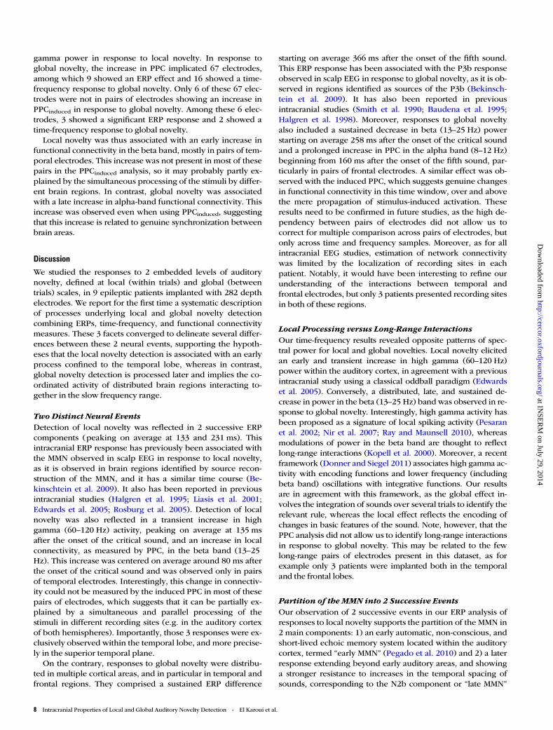
Patients were presented with 8 blocks of 123–128 trials. In each trial,a series of 5 sounds were played over a total of 650 ms. The first 4sounds were always identical, either low- (sound A) or high-pitched(sound B), but the fifth one could be either identical (AAAAA orBBBBB) or different (AAAAB or BBBBA). Thus, this last sound respectsor violates the local regularity established by the first 4 sounds in localstandard or local deviant trials, respectively (Fig. 1). On top of this localrule, a global regularity was added. In each block, global standard trialswere delivered on 80% of trials. In the first type of blocks, these trialswere local standard trials, whereas in the second type of blocks,they were local deviant trials. In contrast, global deviant trials were pre-sented in 20% of trials in a given block (Fig. 1). In the first type ofblocks, these trials were local deviant trials, whereas in the secondtype of blocks, they were local standard trials. The first 20–30 trials ofeach block were global standard trials to establish the global regularity.
Table 1Clinical characteristics of the patient sample
Patient Age Gender Handedness Epilepsy duration (year) Total number of electrodes Temporal electrodes Frontal electrodes Occipital electrodes
1 18 F R 8 36 36 0 02 29 F R 4 25 25 0 03 48 F L 10 37 37 0 04 46 F R 25 14 11 3 05 23 M R 8 43 10 33 06 43 F R 24 27 11 16 07 26 M L 16 42 40 0 28 42 M R 26 9 0 9 09 26 M R 11 49 24 0 2510 24 F R 14 72 9 63 0
Note: Patient 10 was excluded from the analysis as she did not perform the task correctly (56% accuracy).
Figure 1. The Local Global paradigm. The Local Global paradigm (Bekinschtein et al.2009) is an auditory oddball paradigm with 2 levels of regularity. Each trial is composedof a series of 5 successive sounds (SOA= 150 ms). The first 4 sounds are alwaysidentical. The fifth sound can either be identical to these first sounds in local standardtrials or different in local deviant trials. On top of this local regularity, a global rule isadded. Global deviant trials correspond to a series of 5 sounds, which is rare in a givenblock, compared with the frequent global standard trials. Local and global regularitiesare manipulated orthogonally, resulting in 4 types of trials: local standard-globalstandard and local deviant-global deviant in the first type of blocks, and localdeviant-global standard and local standard-global deviant in the second type of blocks.Two sounds were used to generate these trials: sound A (composed of 350-, 700-, and1400-Hz sinusoidal tones) and sound B (composed of 500-, 1000-, and 2000-Hzsinusoidal tones). The local effect corresponds to the difference between local deviantand local standard stimuli, whereas the global effect corresponds to the differencebetween global deviant and global standard stimuli.
2 Intracranial Properties of Local and Global Auditory Novelty Detection • El Karoui et al.
at INSE
RM
on July 29, 2014http://cercor.oxfordjournals.org/
Dow
nloaded from

Local and global regularities were thus manipulated orthogonally(Fig. 1). This design enables the comparison between physically identi-cal stimuli in different contexts. In the following analyses, we will referto local and global effects, which correspond respectively to the con-trast between local deviant versus local standard stimuli, and betweenglobal deviant versus global standard stimuli (Fig. 1). Additionally, pa-tients were instructed to actively count the number of global devianttrials and report this number at the end of each block. This taskensured that they were paying attention to the stimuli.
Each sound was 50-ms long and composed of 3 sinusoidal tones(350, 700, and 1400 Hz, sound A; or 500, 1000, and 2000 Hz, sound B).All tones were prepared with 7-ms rise and 7-ms fall times. Four differ-ent series were used: AAAAA, BBBBB, AAAAB, and BBBBA. The stimu-lus onset asynchrony (SOA) between sounds was 150 ms. A series ofsounds were separated by a variable silent interval of 1350–1650 ms.Four different blocks were thus created, with each possible series ofsounds as global standard trials. All patients heard each of these blockstwice, in randomized order. Sounds were presented from the computer’sspeakers, using E-prime v1.2 (Psychology Software Tools, Inc.) at thebedside of each patient.
Electrode Implantation and LocalizationPatients were implanted intracerebrally with depth electrodes, eachbearing 4–10 recording sites (Ad-TechMedical Instruments). Eachpatient had on average 59 (SD = 13) recording sites.
To compare position of recording sites and summarize brain activa-tions across patients, their coordinates were obtained after normalizingthe anatomical three-dimensional post-implantation MRI onto the tem-plate from the Montreal Neurological Institute, using SPM8 software(http://www.fil.ion.ucl.ac.uk/spm). Electrodes localization plots usedthe iso2mesh toolbox (Fang and Boas 2009; Fang 2010).
Data Acquisition and PreprocessingFor 7 patients, data were acquired with an audio–video–EEG monitor-ing system (Micromed), which allowed for simultaneous acquisition ofdata from up to 128 EEG channels sampled at 1024 Hz. Data from theremaining 3 patients were acquired using another audio–video–EEGmonitoring system (Nicolet-Viasys), which allowed for simultaneousacquisition of data from up to 64 EEG channels sampled at 400 Hz.
Unless specified otherwise, data were analyzed with Fieldtriptoolbox (Oostenveld et al. 2011) and Matlab 2011a (The Mathworks,Inc.). All the analyses were done at the electrode level, as the positionof the recording sites differed from one patient to another.
Epochs were extracted (from −800 to 700 ms after the onset of thefifth sound). To avoid artifacts, recording sites exceeding the thresholdof ±300 µV in more than 5% of the epochs were excluded. All signalswere re-referenced to their nearest neighbor on the same electrode(bipolar montage). In the following, we will refer to these bipolar mon-tages as “electrodes.” All data were visually inspected to discard anytrial with epileptic activity.
Behavioral AnalysisPatients were instructed to count silently the number of global deviantsand report this number. For each patient, we computed the averagepercentage of errors over blocks and we report accuracy as 100%minus this percentage.
ERPAnalysisEvent-related potentials (ERPs) were obtained by averaging epochs foreach condition. The signal was band-pass filtered off-line from 0.5 to20 Hz using a fourth-order Butterworth filter in forward and reverse di-rections in order to avoid phase-shift, and a baseline correction wasapplied, by subtracting the mean voltage in the [−800 ms 0 ms]window.
To assess the statistical significance of our results, ERPs of localdeviant trials and global deviant trials were compared with those oflocal standard trials and global standard trials, respectively, using inde-pendent sample t-tests for each electrode. To control for type I errors
generated by multiple comparisons across time at level α, we used anonparametric procedure. We computed N = 1000 permutations byshuffling trial labels. For each permutation, the maximal t-value acrosstime samples was extracted to estimate the permutation distribution ofthe maximal statistic. The critical threshold that controls for family-wise error rate over time samples was define as the c + 1 largestmember of this distribution, where c is equal to αN rounded down. Inthe original data, only samples with a t-value higher than this thresholdwere identified as significant (Nichols and Holmes 2002). In addition,to correct for multiple comparison over electrodes when visualizingeffects across all electrodes in all patients, we used a false discoveryrate (FDR) correction on P-value obtained at the electrode level, acrossthe whole time window. All reported P-values are corrected and re-ferred to as Pcorr.
Peak latencies were identified within periods of statistical signifi-cance on the difference between standard and deviant trials. For thesustained responses to global novelty, latencies were estimated as theearliest significant differences given the absence of a clear peak. Inboth cases, reported latencies are the average across all significantelectrodes.
Time-Frequency AnalysisTime-frequency representations were calculated by Morlet wavelets, asdescribed previously (Tallon-Baudry et al. 1997). The power at a giventime t and frequency f0 is given by the squared norm of the convolutionof the signal to the wavelet wðt; f0Þ:
wðt; f0Þ ¼ A exp�t2
2s2t
� �exp ð2ip f0tÞ where A ¼ ðst
ffiffiffiffip
p Þ�1=2:
The width of the wavelet m ¼ 2pst f0 was set to 5 as this value gives agood tradeoff between time and frequency resolution (De Moortelet al. 2004). The length of each wavelet used for the computation was3st. The convolution of the signal by this set of wavelets resulted in anestimate of power at each time sample and at each frequency (2-Hzstep) between 5 and 200 Hz. Power was then converted to a decibel(dB) scale. Time-frequency values were obtained from the subtractionbetween 2 conditions, without baseline correction.
To assess the significance of differences in oscillation power acrossconditions, we used independent sample t-tests at each time and fre-quency point. Then, correction for multiple comparisons over timeand frequency was performed, by nonparametric cluster-basedmethod (Maris and Oostenveld 2007). We computed N = 1000 permu-tations by shuffling trial labels. Then, for each permutation, independ-ent sample t-tests were performed at each time and frequency sample.All samples with a t-value corresponding to a P-value smaller than 0.05were clustered in connected sets on the basis of adjacency in time andfrequency. Then, the cluster statistic was computed by taking the sumof the t-values within each cluster. The cluster-corrected threshold wasobtained by computing the permutation distribution of the maximumcluster statistic and taking the c + 1 largest member of this distribution,with c is equal to αN rounded down. In the original data, only clusterswith a cluster statistic higher than this threshold were identified as sig-nificant. Moreover, when visualizing effects across all electrodes in allpatients, we additionally corrected for multiple comparisons over elec-trodes, using FDR correction. All reported P-values are corrected andreferred to as Pcorr.
Peak latencies were identified within time-frequency windows ofstatistical significance on the difference between standard and devianttrials. For sustained responses, effect latencies were estimated as theearliest significant differences between deviant and standard trials. Inboth cases, reported latencies are the average latency across all signifi-cant electrodes.
Functional Connectivity AnalysisConnectivity analyses were performed using pairwise phase consist-ency (PPC—Vinck et al. 2010), which provides a method for measuringrhythmic synchronization, without being affected by the finite samplesize bias, observed with classic tools, such as phase locking value (PLV—Lachaux et al. 1999). This issue is particularly relevant when
Cerebral Cortex 3
at INSE
RM
on July 29, 2014http://cercor.oxfordjournals.org/
Dow
nloaded from

comparing global deviant and standard conditions, as the number oftrials in these conditions is different. In each trial j, for each time pointt and frequency f, the phase wjðt; f Þ of the signal was estimated, usingwavelets, as presented earlier. Pairwise phase consistency PPCkl
between electrodes k and l is then computed across N trials as follows:
PPCklðtÞ ¼ 2N ðN � 1Þ
XN�1
i¼1
XNj¼iþ1
cos ðDwiklðtÞ � Dw
jklðtÞÞ;
with DwiklðtÞ the phase difference between electrodes k and l in trial i at
time t.Pairwise phase consistency was computed for all pairs of electrodes,
for frequencies between 5 and 60 Hz, between 800 and 700 ms relativeto the onset of the fifth sound. No baseline correction was applied.
Statistical significance was assessed by a nonparametric test on thedifference of PPC between conditions (Lachaux et al. 1999), using 500permutations, for each time and frequency samples, and cluster-basedcorrection for multiple comparisons across time and frequency sampleswas applied (Maris and Oostenveld 2007). All reported P-values are cor-rected and referred to as Pcorr. Given the high level of correlationsbetween pairs of electrodes and the relatively low number of highlycomputationally demanding permutations, multiple comparisons arecorrected only across time and frequency samples, but not across pairsof electrodes in order to avoid missing potentially relevant changes infunctional connectivity (type II errors). However, this less conservativethreshold could lead to false-positives (type I errors). This is why wereport the average PPC changes across all pairs of temporal and frontalelectrodes (see Fig. 4b).
The estimation of connectivity is based on the consistency of phaselags between areas. Note that consistent phase lags reflect the true con-nectivity between areas but also artifactual connectivity driven by syn-chronization of different areas onto any external reference. Therefore,we tried to isolate genuine synchronization between electrodes fromspurious connectivity related to simultaneous processing of the stimuli(e.g. in left and right auditory cortices). We reasoned that spurious con-nectivity would be linked to oscillations evoked by the stimulations atseveral electrodes, as opposed to the induced activity that is time-locked,but not phase-locked, to the stimulation. We introduced a novel analysis,the induced PPC, based on the “evoked” versus “induced” terminologyintroduced by Tallon-Baudry and Bertrand (1999). For this analysis, theevoked oscillations were regressed out of the data and connectivity wasestimated using the phase of the induced oscillations isolated in thisway. Thus, induced PPC measures functional connectivity, which is notrelated to external stimulations. To compute this measure, we first aver-aged the data across trials in a given condition. This average preservesevoked oscillations and cancels out induced oscillations (Tallon-Baudryand Bertrand 1999). We then computed the time-frequency decompos-ition of this average using wavelets as described earlier. To isolate theinduced response, we decompose data into time and frequency and re-gressed out linearly from each trial of a given condition the time-frequency decomposition of the corresponding evoked response. Thiscorrected signal was used to compute the PPCinduced, and statistical com-parisons were performed as previously described for the PPC.
Results
Auditory novelty detection was studied using 282 intracranialelectrodes across 9 epileptic patients, with an average of 31 (SD= 13) electrodes per patient (Table 1). Activity was recordedfrom the temporal lobe (194 electrodes), the frontal lobe (61electrodes), and the occipital lobe (27 electrodes). Note that, aswe did not have precise hypotheses about the laterality of theeffects, we analyze electrodes from the left and right hemi-spheres together. All these patients performed the task properly(accuracy of >80%). We analyzed ERPs, time-frequency decom-positions, and functional connectivity associated with local andglobal auditory novelty detection. This approach allowed us totest whether local novelty was related to an automatic and
encapsulated process and whether global novelty implicateddistinct inter-connected brain regions.
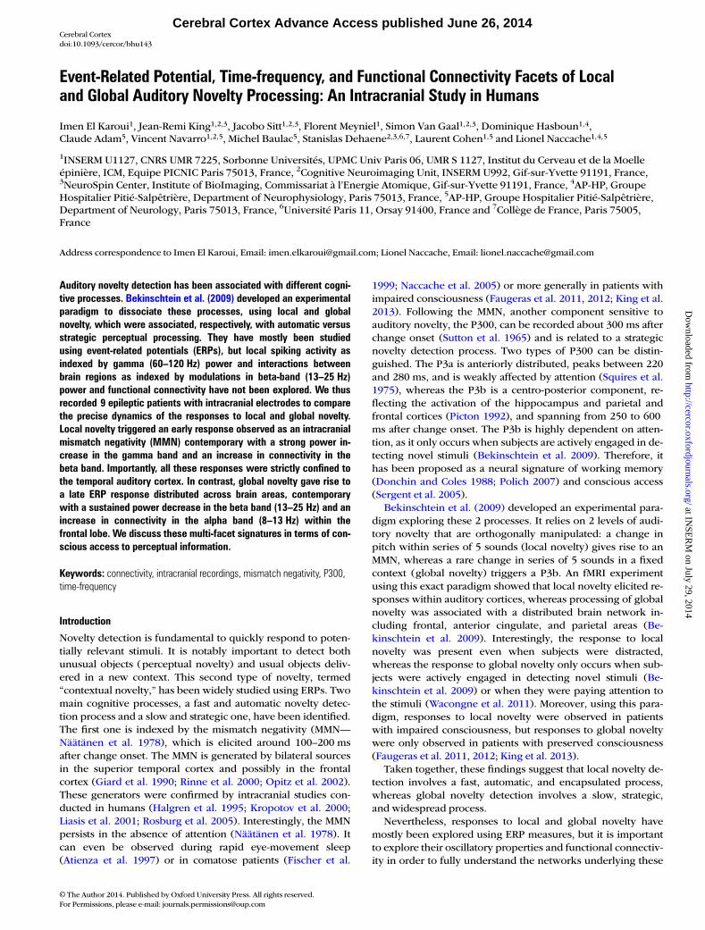
ERPAnalysisSignificant responses to violation of local regularity (localdeviant compared with local standard trials) were observed in 44electrodes (16% of all the electrodes, t-test, all Pcorr < 0.05), alllocated in the temporal lobe. These responses were observed in6 of the 8 patients with temporal electrodes. Notably, 20 of the26 electrodes implanted in the superior temporal lobe (77%)showed significant local effect (Fig. 2a). The responses to localnovelty presented 2 main components: an early one, with a peakon average at 133 ms (SD = 17 ms—see Fig. 2b for an exemplarelectrode in which this peak is at 105 ms) after the onset of thefifth sound, and a second component, with a reversed polarity,showing a peak on average at 231 ms (SD = 33 ms—see Fig. 2bfor an exemplar electrode in which this peak is at 165 ms). Thislocal effect is related to the MMN observed in scalp EEG (Näätä-nen et al. 2001) and was found in previous intracranial studies(Halgren et al. 1995; Edwards et al. 2005; Bekinschtein et al.2009). Note that, interestingly, the polarity of this componentdepends on the position of the electrode relative to the Sylvianfissure (Halgren et al. 1995). Moreover, in 8 of these 44 electro-des, from 3 different patients, these components were precededby an event peaking on average at 71 ms (SD = 8 ms—see Fig. 2bfor exemplar electrode in which this peak is at 63 ms) and whichcan be related to early components of novelty detection found inscalp EEG (Grimm et al. 2011). Finally, in 18 electrodes from 4patients, the 2 main components were followed by an event witha peak on average at 435 ms (SD = 95 ms—see Fig. 2b for exem-plar electrode in which this peak is at 330 ms), which may berelated to the P3a (Halgren et al. 1995).
In contrast, responses to violation of global regularity (globaldeviant compared with global standard trials) were observed in32 electrodes (11% of all the electrodes). These responses weremore widespread across cortical structures than the local effectand were observed in 7 out 9 patients. Twenty-four electrodes inthe temporal lobe (24% of temporal electrodes), including 12 inthe superior temporal lobe (46% of superior temporal lobe elec-trodes) and 8 in the frontal lobe (13% of frontal electrodes)showed significant effects (t-test, all Pcorr < 0.05—Fig. 2a). Theglobal effect was associated with 3 distinct components. First, in19 electrodes from 5 patients, we observed a transient differencebetween global deviant and global standard trials on average at226 ms (SD = 56 ms—see Fig. 2c for an exemplar electrode inwhich this component peaks at 260 ms), similar to the secondcomponent observed in response to local novelty. Indeed, onlytemporal electrodes showing this local effect presented such anearly global effect. This component could be explained by acontextual modulation of the MMN amplitude. Indeed, localdeviant stimuli are used in our paradigm both as global stan-dards and as global deviants (Fig. 1). Thus, the probability oflocal deviant stimuli is not the same in each block, and thiscould affect the amplitude of the MMN (Sato et al. 2000; Wa-congne et al. 2011; King et al. 2013). Second, a sustained differ-ence starting on average at 366 ms (SD = 131 ms—see Fig. 2c foran exemplar electrode in which this component starts at 462ms) was observed in response to global novelty in 20 electrodesfrom 7 patients. It has been associated with the P3b responseobserved in scalp EEG (Bekinschtein et al. 2009) and has beenreported in previous intracranial studies (Smith et al. 1990;
4 Intracranial Properties of Local and Global Auditory Novelty Detection • El Karoui et al.
at INSE
RM
on July 29, 2014http://cercor.oxfordjournals.org/
Dow
nloaded from
Baudena et al. 1995; Halgren et al. 1998). Note that the polarityof this component depends on the localization relative to the dif-ferent generators (Smith et al. 1990). Finally, 2 electrodes from 1patient implanted in the anterior cingulate cortex showed a tran-sient response, with a peak on average at 265 ms but did notshow any significant local effect. Note that we did not observesignificant local or global effect in the occipital lobe. This maypartly be explained by the low number of electrodes implantedin this region (only 27 electrodes, among which 25 belong tothe same patient).
Note that the choice of the baseline time window could po-tentially influence the results, especially for the global effect asexpectation-related components can build up (Faugeras et al.2012). However, the number of electrodes showing significantlocal and global effects was not different when choosing abaseline expanding across the whole trial (for local effect:χ2 = 0.12, P = 0.73; for global effect: χ2 = 0.69, P = 0.41). Wethus identified different ERP responses to local and globalnovelty. Local novelty was mainly associated with an early re-sponse restricted to the temporal local. In contrast, globalnovelty was associated with late responses in different brainregions.
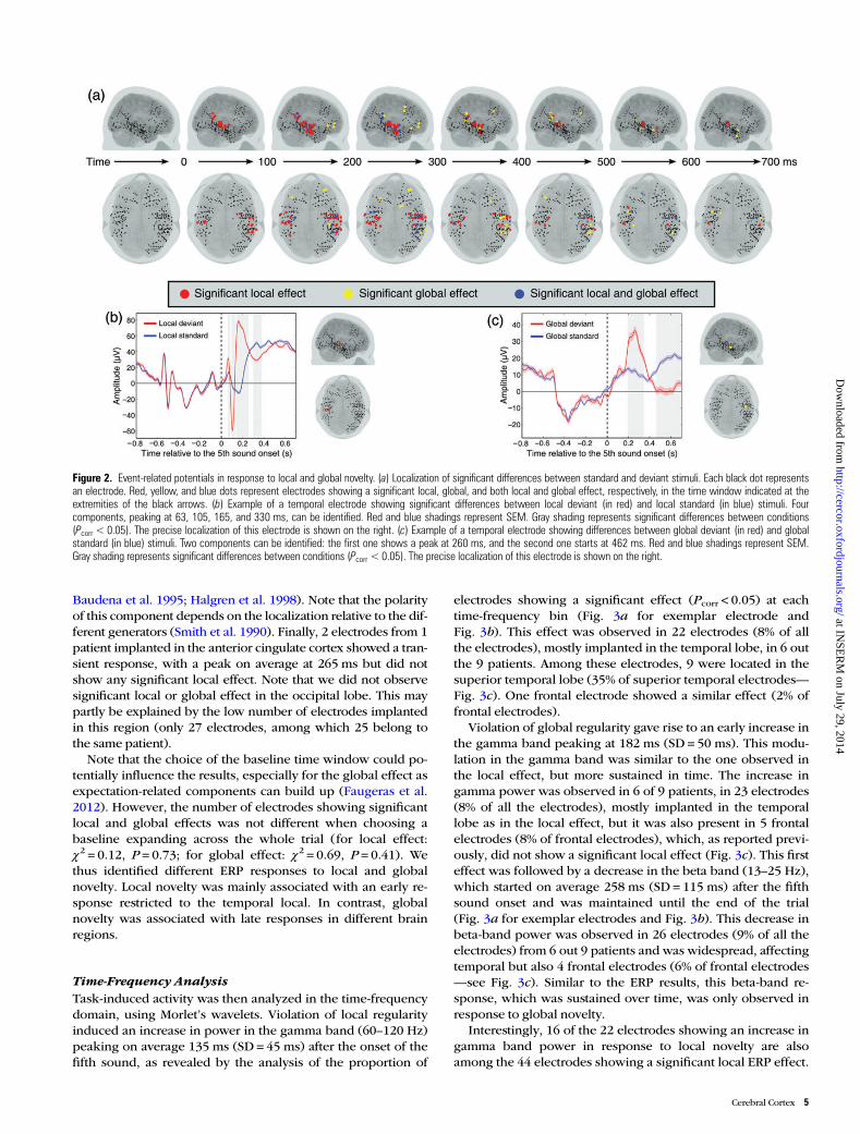
Time-Frequency AnalysisTask-induced activity was then analyzed in the time-frequencydomain, using Morlet’s wavelets. Violation of local regularityinduced an increase in power in the gamma band (60–120 Hz)peaking on average 135 ms (SD = 45 ms) after the onset of thefifth sound, as revealed by the analysis of the proportion of
electrodes showing a significant effect (Pcorr < 0.05) at eachtime-frequency bin (Fig. 3a for exemplar electrode andFig. 3b). This effect was observed in 22 electrodes (8% of allthe electrodes), mostly implanted in the temporal lobe, in 6 outthe 9 patients. Among these electrodes, 9 were located in thesuperior temporal lobe (35% of superior temporal electrodes—Fig. 3c). One frontal electrode showed a similar effect (2% offrontal electrodes).
Violation of global regularity gave rise to an early increase inthe gamma band peaking at 182 ms (SD = 50 ms). This modu-lation in the gamma band was similar to the one observed inthe local effect, but more sustained in time. The increase ingamma power was observed in 6 of 9 patients, in 23 electrodes(8% of all the electrodes), mostly implanted in the temporallobe as in the local effect, but it was also present in 5 frontalelectrodes (8% of frontal electrodes), which, as reported previ-ously, did not show a significant local effect (Fig. 3c). This firsteffect was followed by a decrease in the beta band (13–25 Hz),which started on average 258 ms (SD = 115 ms) after the fifthsound onset and was maintained until the end of the trial(Fig. 3a for exemplar electrodes and Fig. 3b). This decrease inbeta-band power was observed in 26 electrodes (9% of all theelectrodes) from 6 out 9 patients and was widespread, affectingtemporal but also 4 frontal electrodes (6% of frontal electrodes—see Fig. 3c). Similar to the ERP results, this beta-band re-sponse, which was sustained over time, was only observed inresponse to global novelty.
Interestingly, 16 of the 22 electrodes showing an increase ingamma band power in response to local novelty are alsoamong the 44 electrodes showing a significant local ERP effect.
Figure 2. Event-related potentials in response to local and global novelty. (a) Localization of significant differences between standard and deviant stimuli. Each black dot representsan electrode. Red, yellow, and blue dots represent electrodes showing a significant local, global, and both local and global effect, respectively, in the time window indicated at theextremities of the black arrows. (b) Example of a temporal electrode showing significant differences between local deviant (in red) and local standard (in blue) stimuli. Fourcomponents, peaking at 63, 105, 165, and 330 ms, can be identified. Red and blue shadings represent SEM. Gray shading represents significant differences between conditions(Pcorr < 0.05). The precise localization of this electrode is shown on the right. (c) Example of a temporal electrode showing differences between global deviant (in red) and globalstandard (in blue) stimuli. Two components can be identified: the first one shows a peak at 260 ms, and the second one starts at 462 ms. Red and blue shadings represent SEM.Gray shading represents significant differences between conditions (Pcorr < 0.05). The precise localization of this electrode is shown on the right.
Cerebral Cortex 5
at INSE
RM
on July 29, 2014http://cercor.oxfordjournals.org/
Dow
nloaded from
Note that the latency of the gamma band response is verysimilar to that of the early response to local novelty observedin ERP. Moreover, 15 of the 23 electrodes showing an increasein gamma band power in response to global novelty are alsoamong the 32 electrodes showing a significant ERP responseto global novelty. Finally, only 7 electrodes belonged both tothe set of 32 electrodes showing a significant ERP response toglobal novelty and to the set of 26 electrodes showing a
decrease in the beta band in response to global novelty. Theseresults highlight the fact that ERP and time-frequency analysesextract different facets of the responses to local and globalnovelty, as significant effects identified with both methods arenot necessarily present in the same electrodes.
Local and global novelties were associated with differenttime-frequency responses. Violations of local regularity wererelated to an early increase in high gamma power. In contrast,
Figure 3. Time-frequency analysis. (a) Example of electrodes showing significant local and global effect. Top row, time-frequency responses of a temporal electrode to local deviant(left), local standard (middle), and local effect (difference between local deviant and local standard—right). Bottom row, time-frequency response of a temporal electrode to globaldeviant (left), global standard (middle), and global effect (difference between global deviant and global standard—right). Black and white contours circle significant time-frequencysamples (Pcorr < 0.05). The precise localization of these electrodes is shown on far right. (b) Proportion of significant electrodes (Pcorr < 0.05) in each time and frequency bin for thelocal effect (top) and the global effect (bottom). (c) Localization of significant effects in the high gamma band (top) and the beta band (bottom). Each black dot represents anelectrode. Red, yellow, and blue dots represent electrodes showing significant local, global, and both local and global effects, respectively, in the time window indicated at theextremities of the black arrows.
6 Intracranial Properties of Local and Global Auditory Novelty Detection • El Karoui et al.
at INSE
RM
on July 29, 2014http://cercor.oxfordjournals.org/
Dow
nloaded from
violations of global regularity were associated with a sustaineddecrease in beta power. Interestingly, these time-frequency re-sponses highlight different aspects of brain responses to localand global novelty than ERP responses.
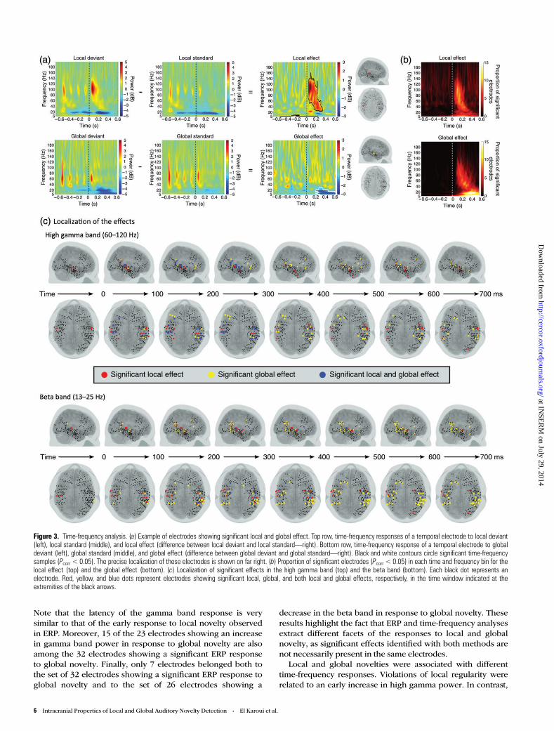
Functional Connectivity AnalysisChanges in connectivity related to the task were analyzedusing PPC, a measure of synchronization that is not biased bythe number of trials in each condition (Vinck et al. 2010). Wereport results for all pairs of electrodes (n = 4714) to identifymodulation of connectivity in response to local and globalnovelty, with a special focus on the temporal and frontal lobes,as most of the electrodes were implanted in these regions andno significant response was observed in the other lobes in ERPand time-frequency analyses. There were 2507 pairs betweentemporal electrodes, 687 pairs between frontal electrodes, 539pairs between temporal and frontal electrodes, 301 pairsbetween occipital electrodes, and 680 pairs between occipitaland temporal electrodes.
Local novelty was associated with a transient increase infunctional connectivity, as measured by PPC, in the beta band(13–25 Hz), centered at 80 ms after the onset of the fifth sound(see Fig. 4a for exemplar pair). This increase was significant(Pcorr < 0.05) in 38 pairs of electrodes (1% of all the electrodepairs), in 7 of 9 patients. All of these pairs, but 5, were betweentemporal electrodes (see Fig. 4b for the average across all pairsof temporal electrodes). Interestingly, only 9 of them showed asimilar increase in the PPC-induced analysis. This suggests thatthe effect is related to activity evoked by the stimuli and can bepartly explained by a parallel and simultaneous processing ofthe sounds rather than by a genuine inter-area exchange of in-formation. Local novelty was also associated with a decrease inPPC in the beta band in 20 other pairs of temporal electrodes.
Among them, 18 pairs showed a similar decrease in inducedPPC.
On the other hand, global novelty was associated with a sus-tained increase in PPC in the alpha band (8–13 Hz), starting at160 ms after the onset of the fifth sound and lasting until 600ms after the onset of the fifth sound (see Fig. 4a for exemplarpair and Fig. 4b). This effect was observed in 7 of 9 patients in49 pairs of electrodes (1% of all electrode pairs): 27 betweentemporal electrodes (1% of temporal pairs), 7 between frontalelectrodes (1% of frontal pairs), 5 between frontal and tem-poral electrodes (1% of temporo-frontal pairs), and 10 betweenoccipital and temporal electrodes (1% of occipito-temporalpairs). Interestingly, among these electrode pairs, 42 showedthis late increase in connectivity in the PPCinduced analysis,which suggests that it is related to a genuine synchronization,and not simply to a simultaneous processing of the stimuli.Global novelty was also associated with a sustained decreasein PPC in 100 pairs of electrodes, in the same time window asthe increase described earlier. This decrease was also observedin these pairs in the PPCinduced analysis. Among these pairs, 66were between temporal electrodes, 28 between occipital andtemporal electrodes, 1 between temporal and frontal electro-des, 5 between occipital electrodes, and 1 between frontal elec-trodes. Note, however, that this decrease in connectivity wasmainly observed in Patient 9, for whom 78 significant pairswere observed. This patient was the only one with occipitaland posterior temporal electrodes.
Interestingly, the increase in PPC in response to localnovelty implicated 45 electrodes, among which 16 alsoshowed en ERP effect and 11 showed a time-frequency re-sponse to local novelty. The increase in PPCinduced in responseto local novelty implicated 19 of these 45 electrodes, amongwhich 7 showed an ERP effect and 5 showed an increase in
Figure 4. Functional connectivity. (a) Time-frequency representations of pairwise phase consistency (PPC—top row) and induced PPC (middle row) modulations in 2 pairs ofelectrodes. On the left, differences in PPC and induced PPC between local deviant and local standard stimuli in a pair of temporal electrodes are represented. On the right,differences in PPC and induced PPC between global deviant and global standard stimuli in a pair of frontal electrodes are represented. The bottom row shows the localization ofthese pairs. Black contours circle significant time-frequency samples (Pcorr < 0.05). (b) On the left, average differences in PPC (top) and induced PPC (bottom) between localstandard and local deviant stimuli in pairs of temporal electrodes (n=2507) are presented. On the right, average differences in PPC (top) and induced PPC (bottom) between globalstandard and global deviant stimuli in pairs of frontal electrodes (n=687) are presented.
Cerebral Cortex 7
at INSE
RM
on July 29, 2014http://cercor.oxfordjournals.org/
Dow
nloaded from
gamma power in response to local novelty. In response toglobal novelty, the increase in PPC implicated 67 electrodes,among which 9 showed an ERP effect and 16 showed a time-frequency response to global novelty. Only 6 of these 67 elec-trodes were not in pairs of electrodes showing an increase inPPCinduced in response to global novelty. Among these 6 elec-trodes, 3 showed a significant ERP response and 2 showed atime-frequency response to global novelty.
Local novelty was thus associated with an early increase infunctional connectivity in the beta band, mostly in pairs of tem-poral electrodes. This increase was not present in most of thesepairs in the PPCinduced analysis, so it may probably partly ex-plained by the simultaneous processing of the stimuli by differ-ent brain regions. In contrast, global novelty was associatedwith a late increase in alpha-band functional connectivity. Thisincrease was observed even when using PPCinduced, suggestingthat this increase is related to genuine synchronization betweenbrain areas.
Discussion
We studied the responses to 2 embedded levels of auditorynovelty, defined at local (within trials) and global (betweentrials) scales, in 9 epileptic patients implanted with 282 depthelectrodes. We report for the first time a systematic descriptionof processes underlying local and global novelty detectioncombining ERPs, time-frequency, and functional connectivitymeasures. These 3 facets converged to delineate several differ-ences between these 2 neural events, supporting the hypoth-eses that the local novelty detection is associated with an earlyprocess confined to the temporal lobe, whereas in contrast,global novelty detection is processed later and implies the co-ordinated activity of distributed brain regions interacting to-gether in the slow frequency range.
Two Distinct Neural EventsDetection of local novelty was reflected in 2 successive ERPcomponents (peaking on average at 133 and 231 ms). Thisintracranial ERP response has previously been associated withthe MMN observed in scalp EEG in response to local novelty,as it is observed in brain regions identified by source recon-struction of the MMN, and it has a similar time course (Be-kinschtein et al. 2009). It also has been reported in previousintracranial studies (Halgren et al. 1995; Liasis et al. 2001;Edwards et al. 2005; Rosburg et al. 2005). Detection of localnovelty was also reflected in a transient increase in highgamma (60–120 Hz) activity, peaking on average at 135 msafter the onset of the critical sound, and an increase in localconnectivity, as measured by PPC, in the beta band (13–25Hz). This increase was centered on average around 80 ms afterthe onset of the critical sound and was observed only in pairsof temporal electrodes. Interestingly, this change in connectiv-ity could not be measured by the induced PPC in most of thesepairs of electrodes, which suggests that it can be partially ex-plained by a simultaneous and parallel processing of thestimuli in different recording sites (e.g. in the auditory cortexof both hemispheres). Importantly, those 3 responses were ex-clusively observed within the temporal lobe, and more precise-ly in the superior temporal plane.
On the contrary, responses to global novelty were distribu-ted in multiple cortical areas, and in particular in temporal andfrontal regions. They comprised a sustained ERP difference
starting on average 366 ms after the onset of the fifth sound.This ERP response has been associated with the P3b responseobserved in scalp EEG in response to global novelty, as it is ob-served in regions identified as sources of the P3b (Bekinsch-tein et al. 2009). It has also been reported in previousintracranial studies (Smith et al. 1990; Baudena et al. 1995;Halgren et al. 1998). Moreover, responses to global noveltyalso included a sustained decrease in beta (13–25 Hz) powerstarting on average 258 ms after the onset of the critical soundand a prolonged increase in PPC in the alpha band (8–12 Hz)beginning from 160 ms after the onset of the fifth sound, par-ticularly in pairs of frontal electrodes. A similar effect was ob-served with the induced PPC, which suggests genuine changesin functional connectivity in this time window, over and abovethe mere propagation of stimulus-induced activation. Theseresults need to be confirmed in future studies, as the high de-pendency between pairs of electrodes did not allow us tocorrect for multiple comparison across pairs of electrodes, butonly across time and frequency samples. Moreover, as for allintracranial EEG studies, estimation of network connectivitywas limited by the localization of recording sites in eachpatient. Notably, it would have been interesting to refine ourunderstanding of the interactions between temporal andfrontal electrodes, but only 3 patients presented recording sitesin both of these regions.
Local Processing versus Long-Range InteractionsOur time-frequency results revealed opposite patterns of spec-tral power for local and global novelties. Local novelty elicitedan early and transient increase in high gamma (60–120 Hz)power within the auditory cortex, in agreement with a previousintracranial study using a classical oddball paradigm (Edwardset al. 2005). Conversely, a distributed, late, and sustained de-crease in power in the beta (13–25 Hz) band was observed in re-sponse to global novelty. Interestingly, high gamma activity hasbeen proposed as a signature of local spiking activity (Pesaranet al. 2002; Nir et al. 2007; Ray and Maunsell 2010), whereasmodulations of power in the beta band are thought to reflectlong-range interactions (Kopell et al. 2000). Moreover, a recentframework (Donner and Siegel 2011) associates high gamma ac-tivity with encoding functions and lower frequency (includingbeta band) oscillations with integrative functions. Our resultsare in agreement with this framework, as the global effect in-volves the integration of sounds over several trials to identify therelevant rule, whereas the local effect reflects the encoding ofchanges in basic features of the sound. Note, however, that thePPC analysis did not allow us to identify long-range interactionsin response to global novelty. This may be related to the fewlong-range pairs of electrodes present in this dataset, as forexample only 3 patients were implanted both in the temporaland the frontal lobes.
Partition of the MMN into 2 Successive EventsOur observation of 2 successive events in our ERP analysis ofresponses to local novelty supports the partition of the MMN in2 main components: 1) an early automatic, non-conscious, andshort-lived echoic memory system located within the auditorycortex, termed “early MMN” (Pegado et al. 2010) and 2) a laterresponse extending beyond early auditory areas, and showinga stronger resistance to increases in the temporal spacing ofsounds, corresponding to the N2b component or “late MMN”
8 Intracranial Properties of Local and Global Auditory Novelty Detection • El Karoui et al.
at INSE
RM
on July 29, 2014http://cercor.oxfordjournals.org/
Dow
nloaded from
(Pegado et al. 2010). These results are in agreement with previ-ous intracranial studies using a classical oddball paradigm(Halgren et al. 1995). Interestingly, the N2b has been asso-ciated with attention (Näätänen and Gaillard 1983), contrary tothe “early MMN,”which is thought to reflect automatic process-ing of novelty. In Pegado et al.’s (2010), the authors show thatwhen increasing the SOA between 2 successive sounds beyond1000 ms in a traditional oddball paradigm, the disappearanceof the “early MMN” actually corresponds to the occurrence of asimilar novelty response present both for deviant and standardtrials. In contrast, the “late MMN” latency was delayed, but thiscomponent did not disappear. Pegado et al. proposed a modelaccounting for these results: the early MMN would reflect accu-mulation of evidence based on echoic memory representa-tions, whereas the late MMN would be related to accumulationof evidence on longer time scales. In our study, the early MMNwould thus be absent in the global novelty condition becausethe relevant time scales are longer than in the local noveltycondition. Conversely, given the resistance of the “late MMN”to SOA, it would not be surprising to observe its presence in re-sponse to global novelty.
In the present study, no MMN was recorded in frontal elec-trodes, in agreement with some previous intracranial studies(Baudena et al. 1995; Edwards et al. 2005). Nevertheless,because of the non-uniform sampling of our recording sitesover the frontal lobe, it cannot be excluded that a frontal gener-ator of the MMN exists but was missed, as suggested by somescalp EEG studies (Giard et al. 1990; Rinne et al. 2000; Opitzet al. 2002). Note that in a study including only 2 implanted pa-tients (Bekinschtein et al. 2009), using less conservative statis-tical thresholds, we previously reported 3 frontal electrodeswith a local effect, in agreement with other intracranial studies(Liasis et al. 2001; Rosburg et al. 2005).
Global Effect and Conscious AccessThe paradigm used in this study was designed to dissociate 2different processes, underlying local and global novelty detec-tion. In a previous experiment using this paradigm, weshowed that while the response to local novelty was still ob-served under conditions of inattention (engagement in a con-current difficult RSVP task), the response to global noveltydisappeared and subjects could not report the existence of vio-lations of the global regularity in EEG and MEG studies (Be-kinschtein et al. 2009). Moreover, this paradigm was used inpatients suffering from disorders of consciousness, and the re-sponse to global novelty was only observed in patientsshowing signs of consciousness (Bekinschtein et al. 2009; Fau-geras et al. 2011, 2012; King et al. 2013). The P3b componenthas also been proposed as a marker of conscious access inother paradigms (Sergent et al. 2005). Note, however, that inthe present study, patients were instructed to count globaldeviant trials. The responses to global novelty could thusreflect downstream processes relative to conscious access perse (Aru et al. 2012; Sergent and Naccache 2012).
To distinguish between these “downstream” processes andsignatures of conscious access, it is interesting to compare ourresults with studies using a different task. First, Wacongneet al. (2011) used a passive version of the Local/Global para-digm and found similar ERP signatures of local and globalnovelty. Moreover, in a completely different study contrastingmasked and unmasked words, Gaillard et al. (2009) exploredERPs, time-frequency changes, and connectivity patterns in
intracranial EEG—similarly to the approach taken in thepresent study —and reported closely related findings. Con-scious processing of unmasked words was associated with sus-tained event-related components, notably in the frontal cortex,a decrease in power in the beta band and a late increase in con-nectivity in the alpha and beta band. The single discrepancywith our findings is the small proportion of electrodes inwhich we observed the 3 signatures of the late global effect.This could be explained by the fact that the global effect corre-sponds to a more subtle contrast than the masked/unmaskedcontrast. Indeed, whereas the masked/unmasked contrast com-pared a “no stimulus” condition with a “stimulus present” condi-tion, we contrasted here 2 conditions associated with consciousperception: global deviant stimuli being perceived as violationsof the global regularity, and global standard stimuli as regulartrials.
Nevertheless, we conclude by highlighting the striking simi-larity between our results and the 3 signatures of consciousperception identified by Gaillard et al, despite the profounddifference in sensory modalities (audition versus vision), instimuli (words versus tones) and tasks. This may point to ageneral common mechanism of conscious access (Sergent andNaccache 2012).
Funding
This work was supported by the program “Investissementsd’avenir” ANR-10-IAIHU-06, an AXA Research Fund grant toIEK, a Direction Générale de l’Armement (DGA) grant to JRK,a Ministère de l’Enseignement Supérieur et de la Recherchegrant to FM, a Rubicon grant from the Netherlands Organiza-tion for Scientific Research (NWO) to SvG, an “Equipe FRM2010” grant of Fondation pour la Recherche Médicale (FRM) toLN and an European Research Council (ERC) senior grant“NeuroConsc” to SD.
Notes
We thank Séverine Samson and Isabelle Jegourel for their helpin gathering clinical information and all the patients for theirtime and kindness. Conflict of Interest: None declared.
ReferencesAru J, Bachmann T, Singer W, Melloni L. 2012. Distilling the neural cor-
relates of consciousness. Neurosci Biobehav Rev. 36:737–746.Atienza M, Cantero JL, Gómez CM. 1997. The mismatch negativity com-
ponent reveals the sensory memory during REM sleep in humans.Neurosci Lett. 237:21–24.
Axmacher N, Cohen MX, Fell J, Haupt S, Dümpelmann M, Elger CE,Schlaepfer TE, Lenartz D, Sturm V, Ranganath C. 2010. IntracranialEEG correlates of expectancy and memory formation in the humanhippocampus and nucleus accumbens. Neuron. 65:541–549.
Baudena P, Halgren E, Heit G, Clarke JM. 1995. Intracerebral potentialsto rare target and distractor auditory and visual stimuli. III. Frontalcortex. Electroencephalogr Clin Neurophysiol. 94:251–264.
Bekinschtein TA, Dehaene S, Rohaut B, Tadel F, Cohen L, Naccache L.2009. Neural signature of the conscious processing of auditory re-gularities. Proc Natl Acad Sci USA. 106:1672–1677.
De Moortel I, Munday SA, Hood AW. 2004. Wavelet analysis: the effectof varying basic wavelet parameters. Sol Phys. 222:203–228.
Donchin E, Coles MGH. 1988. Is the P300 component a manifestationof context updating? Behav Brain Sci. 11:357–374.
Donner TH, Siegel M. 2011. A framework for local cortical oscillationpatterns. Trends Cogn Sci. 15:191–199.
Cerebral Cortex 9
at INSE
RM
on July 29, 2014http://cercor.oxfordjournals.org/
Dow
nloaded from

Edwards E, Soltani M, Deouell LY, Berger MS, Knight RT. 2005. Highgamma activity in response to deviant auditory stimuli recorded dir-ectly from human cortex. J Neurophysiol. 94:4269–4280.
Fang Q. 2010. Mesh-based Monte Carlo method using fast ray-tracingin Plücker coordinates. Biomed Opt Express. 1:165–175.
Fang Q, Boas DA. 2009. Tetrahedral mesh generation from volumetricbinary and grayscale images. Proceedings of IEEE Int SympBiomed Imaging 2009. 2009 June 28–July 1. Boston, MA: IEEE.p. 1142–1145.
Faugeras F, Rohaut B, Weiss N, Bekinschtein T, Galanaud D, PuybassetL, Bolgert F, Sergent C, Cohen L, Dehaene S et al. 2012. Event relatedpotentials elicited by violations of auditory regularities in patientswith impaired consciousness. Neuropsychologia. 50:403–418.
Faugeras F, Rohaut B, Weiss N, Bekinschtein TA, Galanaud D, Puybas-set L, Bolgert F, Sergent C, Cohen L, Dehaene S et al. 2011. Probingconsciousness with event-related potentials in the vegetative state.Neurology. 77:264–268.
Fischer C, Morlet D, Bouchet P, Luaute J, Jourdan C, Salord F. 1999.Mismatch negativity and late auditory evoked potentials in coma-tose patients. Clin Neurophysiol. 110:1601–1610.
Fries P. 2005. A mechanism for cognitive dynamics: neuronal commu-nication through neuronal coherence. Trends Cogn Sci. 9:474–480.
Gaillard R, Dehaene S, Adam C, Clémenceau S, Hasboun D, Baulac M,Cohen L, Naccache L. 2009. Converging intracranial markers of con-scious access. PLoS Biol. 7:e1000061.
Giard M-H, Perrin F, Pernier J, Bouchet P. 1990. Brain generators impli-cated in the processing of auditory stimulus deviance: a topograph-ic event-related potential study. Psychophysiology. 27:627–640.
Grimm S, Escera C, Slabu L, Costa-Faidella J. 2011. Electrophysiologicalevidence for the hierarchical organization of auditory change de-tection in the human brain. Psychophysiology. 48:377–384.
Halgren E, Baudena P, Clarke JM, Heit G, Liégeois C, Chauvel P, Muso-lino A. 1995. Intracerebral potentials to rare target and distractorauditory and visual stimuli. I. Superior temporal plane and parietallobe. Electroencephalogr Clin Neurophysiol. 94:191–220.
Halgren E, Baudena P, Clarke JM, Heit G, Marinkovic K, Devaux B,Vignal J-P, Biraben A. 1995. Intracerebral potentials to rare targetand distractor auditory and visual stimuli. II. Medial, lateral andposterior temporal lobe. Electroencephalogr Clin Neurophysiol.94:229–250.
Halgren E, Marinkovic K, Chauvel P. 1998. Generators of the late cogni-tive potentials in auditory and visual oddball tasks. Electroencepha-logr Clin Neurophysiol. 106:156–164.
King JR, Faugeras F, Gramfort A, Schurger A, El Karoui I, Sitt JD,Rohaut B, Wacongne C, Labyt E, Bekinschtein T et al. 2013. Single-trial decoding of auditory novelty responses facilitates the detectionof residual consciousness. Neuroimage. 83:726–738.
Kopell NJ, Ermentrout GB, Whittington M, Traub RD. 2000. Gammarhythms and beta rhythms have different synchronization proper-ties. Proc Natl Acad Sci USA. 97:1867–1872.
Kropotov JD, Alho K, Näätänen R, Ponomarev VA, Kropotova OV, An-ichkov AD, Nechaev VB. 2000. Human auditory-cortex mechanismsof preattentive sound discrimination. Neurosci Lett. 280:87–90.
Lachaux J-P, Rodriguez E, Martinerie J, Varela FJ. 1999. MeasuringPhase Synchrony in Brain Signals. Hum Brain Mapp. 8:194–208.
Liasis A, Towell A, Alho K, Boyd S. 2001. Intracranial identification ofan electric frontal-cortex response to auditory stimulus change: acase study. Brain Res Cogn Brain Res. 11:227–233.
Maris E, Oostenveld R. 2007. Nonparametric statistical testing of EEG-and MEG-data. J Neurosci Methods. 164:177–190.
Näätänen R, Gaillard AWK. 1983. The orienting reflex and the N2 de-flection of the event-related potentials. In: Tutorials in event relatedpotential research: endogenous components. Advances in Psych-ology. Elsevier. p. 119–141.
Näätänen R, Gaillard AWK, Mäntysalo S. 1978. Early selective-attentioneffect on evoked potential reinterpreted. Acta Psychol (Amst).42:313–329.
Näätänen R, Tervaniemi M, Sussman E, Paavilainen P, Winkler I. 2001.“Primitive intelligence” in the auditory cortex. Trends Neurosci.24:283–288.
Naccache L, Puybasset L, Gaillard R, Serve E, Willer J-C. 2005. Auditorymismatch negativity is a good predictor of awakening in comatose pa-tients: a fast and reliable procedure. Clin Neurophysiol. 116:988–989.
Nichols TE, Holmes AP. 2002. Nonparametric permutation tests forfunctional neuroimaging: a primer with examples. Hum BrainMapp. 15:1–25.
Nir Y, Fisch L, Mukamel R, Gelbard-Sagiv H, Arieli A, Fried I, Malach R.2007. Coupling between neuronal firing rate, gamma LFP, and BOLDfMRI is related to interneuronal correlations. Curr Biol. 17:1275–1285.
Oostenveld R, Fries P, Maris E, Schoffelen J-M. 2011. FieldTrip: opensource software for advanced analysis of MEG, EEG, and invasiveelectrophysiological data. Comput Intell Neurosci. 2011:1–9.
Opitz B, Rinne T, Mecklinger A, von Cramon DY, Schröger E. 2002. Dif-ferential contribution of frontal and temporal cortices to auditorychange detection: fMRI and ERP results. Neuroimage. 15:167–174.
Pegado F, Bekinschtein TA, Chausson N, Dehaene S, Cohen L, Nac-cache L. 2010. Probing the lifetimes of auditory novelty detectionprocesses. Neuropsychologia. 48:3145–3154.
Pesaran B, Pezaris JS, Sahani M, Mitra PP, Andersen RA. 2002. Tem-poral structure in neuronal activity during working memory inmacaque parietal cortex. Nat Neurosci. 5:805–811.
Pfurtscheller G, Lopes da Silva FH. 1999. Event-related EEG/MEG syn-chronization and desynchronization: basic principles. Clin Neuro-physiol. 110:1842–1857.
Picton TW. 1992. The P300 wave of the human event-related potential.J Clin Neurophysiol. 9:456–479.
Polich J. 2007. Updating P300: an integrative theory of P3a and P3b.Clin Neurophysiol. 118:2128–2148.
Ray S, Maunsell JHR. 2010. Differences in gamma frequencies acrossvisual cortex restrict their possible use in computation. Neuron.67:885–896.
Rinne T, Alho K, Ilmoniemi RJ, Virtanen J, Näätänen R. 2000. Separatetime behaviors of the temporal and frontal mismatch negativitysources. Neuroimage. 12:14–19.
Rosburg T, Trautner P, Dietl T, Korzyukov OA, Boutros NN, Schaller C,Elger CE, Kurthen M. 2005. Subdural recordings of the mismatchnegativity (MMN) in patients with focal epilepsy. Brain. 128:819–828.
Sato Y, Yabe H, Hiruma T, Sutoh T, Shinozaki N, Nashida T, Kaneko S.2000. The effect of deviant stimulus probability on the human mis-match process. Neuroreport. 11:3703–3708.
Sergent C, Baillet S, Dehaene S. 2005. Timing of the brain eventsunderlying access to consciousness during the attentional blink.Nat Neurosci. 8:1391–1400.
Sergent C, Naccache L. 2012. Imaging neural signatures of conscious-ness: “what”, “when”, “where” and “how” does it work? Arch ItalBiol. 150:91–106.
Smith ME, Halgren E, Sokolik M, Baudena P, Musolino A,Liegeois-Chauvel C, Chauvel P. 1990. The intracranial topographyof the P3 event-related potential elicited during auditory oddball.Electroencephalogr Clin Neurophysiol. 76:235–248.
Squires NK, Squires KC, Hillyard SA. 1975. Two varieties of long-latency positive waves evoked by unpredictable auditory stimuli inman. Electroencephalogr Clin Neurophysiol. 38:387–401.
Sutton S, Braren M, Zubin J, John ER. 1965. Evoked-potential correlatesof stimulus uncertainty. Science. 150:1187–1188.
Tallon-Baudry C, Bertrand O. 1999. Oscillatory gamma activity in humansand its role in object representation. Trends Cogn Sci. 3:151–162.
Tallon-Baudry C, Bertrand O, Delpuech C, Permier J. 1997. Oscillatorygamma-band (30–70 Hz) activity induced by a visual search task inhumans. J Neurosci. 17:722–734.
Vinck M, van Wingerden M, Womelsdorf T, Fries P, Pennartz CMA.2010. The pairwise phase consistency: a bias-free measure of rhyth-mic neuronal synchronization. Neuroimage. 51:112–122.
Wacongne C, Labyt E, van Wassenhove V, Bekinschtein T, Naccache L,Dehaene S. 2011. Evidence for a hierarchy of predictions and predictionerrors in human cortex. Proc Natl Acad Sci USA. 108:20754–20759.
Zaehle T, Bauch EM, Hinrichs H, Schmitt FC, Voges J, Heinze H-J,Bunzeck N. 2013. Nucleus accumbens activity dissociates differentforms of salience: evidence from human intracranial recordings.J Neurosci. 33:8764–8771.
10 Intracranial Properties of Local and Global Auditory Novelty Detection • El Karoui et al.
at INSE
RM
on July 29, 2014http://cercor.oxfordjournals.org/
Dow
nloaded from