evaluation of the possible effect of methotrexate
TRANSCRIPT

Original article
Evaluation of the Possible Effect of Methotrexate, Fluoxetine and
Moringa Oleifera in Rat Model of Rheumatoid Arthritis
Mohamed E. Mansour, Mohie ELDin Sherif, Yasmeen M. Ismail, Abeer Abd Elhameed,
Sara A. Bassouiney
Abstract
Background: Rheumatoid arthritis (RA) is a chronic, relapsing
inflammatory and autoimmune multisystem disease that affects the
joints. Methotrexate (MTX) is considered by many rheumatologists
to be the most important and useful disease-modifying
antirheumatic drugs (DMARDs). Fluoxetine is a selective serotonin
reuptake inhibitor that has been widely used for the treatment of
depression due to its safe profile. Moringa Oleifera (MO) is known
for its nutritional and medicinal uses. Various parts of these plant
act as cardiac and circulatory stimulants. Aim of work: The present
study was designed to evaluate the possible effect of methotrexate,
Fluoxetine and Moringa Oleifera on rat model of RA. Materials
and methods: Rats were classified into: Group I: control normal
group. Group II: was not treated (diseased group). Group III: was
treated with methotrexate (MTX) (0. 6 mg/kg/week/by oral
gavage). Group IV: was treated with fluoxetine (20 mg/kg/day by
oral gavage) Group V: was treated with Moringa Oleifera extract
(200 mg/kg/day by oral gavage). Treated groups received drugs for 4 weeks. Results:
Treated groups showed significant improvement in all parameters (rheumatoid factor (RF),
tumor necrosis factor alpha (TNF-α), C-reactive protein (CRP), reduced glutathione (GSH),
anti-cyclic citrullinated peptide (anti-CCP), arthritis score).and improvement of the
histopathology of the joint. A significant reduction in the score was seen in the treated group
at the end of 3rd
and 4th
weeks. With best results in Moringa Oleifera group in comparison
with other treated groups.Conclusion: Methotrexate, fluoxetine and Moringa Oleifera extract
groups showed significant improvement of parameters of RA.
Key words: Rheumatoid arthritis, methotrexate, fluoxetine, Moringa Oleifera, adjuvant-
induced arthritis.
Department of clinical
Pharmacology, Faculty of
Medicine Benha University,
Egypt.
Correspondence to: Sara A.
Bassouiney, Department of
clinical Pharmacology, Faculty
of Medicine Benha University,
Egypt.
Email:
Received:
Accepted:

Benha medical journal, vol. xx, issue xx, 2021
Introduction:
Rheumatologic diseases are the most
prevalent diseases worldwide; they are one
of the main causes of disability and
morbidity all over the world with greatly
bad impact on the quality of life. (1).
They are characterized by the presence of
long-standing inflammation of the joints
resulting in symmetric polyarthritis and
synovial membrane hypertrophy with
progressive joint damage, bone and
cartilage destruction and also deformity.
The disease is systemic, leading to extra-
articular manifestations (EAM) (2).
The pathogenesis of the rheumatoid joint
involves Synovial hyperplasia results from
a marked increase in macrophage and
fibroblast-like synoviocytes. Locally
expressed degradative enzymes, including
metalloproteinase and proteases, digest the
extracellular matrix and destroy the
articular structures. (3).
Cytokines, particularly IL-1 and IL-17,
tumor necrosis factor-α (TNF-α) and
reactive oxygen intermediates affect
chondrocytes that undergo apoptosis. This
results in cartilage degradation and joint-
space narrowing on radiography (4).
Systemic involvement may be explained
by the systemic activities of cytokines
released from the inflamed synovium. (5).
The main classes of drugs are currently
used: analgesics, non-steroidal anti-
inflammatory drugs (NSAIDs),
glucocorticoids, as well as biologic and
non-biologic disease-modifying
antirheumatic drugs (DMARDs). (6).
Methotrexate (MTX) is the recommended
first-line DMARD used in the treatment of
RA. This is due to its low cost and many
patients achieve disease control with
monotherapy. However, about 30% of
patients develop inadequate treatment
response and many patients stop MTX due
to toxicity.(7).
Part of MTX anti-inflammatory action is
due to its folate antagonist Methotrexate
up taken into the cells by folate transporter
1 (FOLT, also known as RFC1). Within
the cell, MTX is polyglutamated in a
reversible reaction by folylpolyglutamate
synthase. Polyglutamation of methotrexate
increasing inhibition of dihydrofolate
reductase (DHFR), thymidylate synthetase
and 5-aminoimidazole- 4-carboxamide
ribonucleotide transformylase.(8).
The most common adverse effects include:
gastrointestinal side effects (nausea,
vomiting, abdominal pain) followed by
stomatitis (oral ulcers), liver function
abnormalities, bone marrow suppression,
and alopecia. (9).
Neuropsychiatric manifestations are quite
common in RA, including
depression, cognitive dysfunction,

Effect of Fluoxetine in Rheumatoid Arthritis, 2021
behavior changes, spinal cord
compression and peripheral
nerve involvement.(10).
The current trend of medical treatment of
rheumatoid arthritis seeks for new drugs
with more efficacies and less side effects
since methotrexate, a standard diseased
modified anti-rheumatoid drug, causes
many adverse effects and toxicities. (11).
Fluoxetine is a selective serotonin
reuptake inhibitor that has been widely
used to enhance the neurotransmission of
serotonin in the central nervous system
and has emerged as the drug of choice for
the treatment of depression due to is safer
profile and fewer side effects. (12).
Also there are important functions of
fluoxetine related to the central nervous
system e.g. neuroprotection; anti-
inflammatory properties, antioxidant
properties and anti-apoptotic properties,
with greater neuron survival and a
reduction in apoptosis mediators and also
oxidative substances, such as superoxide
dismutase and hydrogen peroxide. (13).
Moringa Oleifera (MO) is known for its
nutritional and numerous medicinal uses.
Various parts of these plant act as cardiac
and circulatory stimulants, possess
antitumor, antipyretic, antiulcer,
antispasmodic, diuretic, antihypertensive,
analgesic, antioxidant, antimicrobial, and
anti-inflammatory effect.(14).
Aim of work:
These observations provided a rationale
for testing the effect of monotherapy with
fluoxetine and MO extract in comparison
with MTX monotherapy on adjuvant
induced arthritis (AIA).
Materials & methods:
A. Animals:
The study is carried out on 30 Adult male
albino rats obtained from Experimental
Animal Breeding Farm, Helwan-Cairo)
weighing between 150- 200 g (at the
beginning of the study), were used for in-
vivo experiments. They were acclimatized
for one week and were caged (6 rat/ cage)
in fully ventilated room at room
temperature in the pharmacology
department, Benha Faculty of Medicine.
Rats were fed a standard chow with water.
This study was approved from ethical
committee of Benha Faculty of Medicine.
B. Drugs and chemicals:
Complete Freund's adjuvant (CFA)
(Sigma-Aldrich Chemical Company),
Methotrexate (Minapharm., Egypt),
fluoxitine (Amoun pharmaceutical Co,
Egypt)., Moringa olifera (powder)
(National Research Centre, Giza, Egypt),
Formalin, solution, neutral 10% formaline
(El Gomhoria Pharmaceutical Chemical
Co., ARE),Urethane , Ethyl carbamat,
white crystals (Sigma Chemical Co.,
USA), Hematoxylin and eosin (E.Merk,

Benha medical journal, vol. xx, issue xx, 2021
Darmastadt.,)[Germany], Rheumatoid
factor kits (Abnova corporation, Taipei
city ,Taiwan).,CRP kits (Thermoscientific,
USA), Tumor necrosis factor alpha (TNF-
α) kits ( USA & Canada | R&D Systems,
Inc), Reduced Glutathione kits (
Biodiagnostic Co., Giza, Egypt), anticyclic
citrullinated peptide (anti-CCP)kits (Alpha
diagnostic international,Texas,USA)
C. Induction of RA:
Complete Freund's Adjuvant Arthritis was
induced by S.C injection of 0.4 ml of
compelete Freund's adjuvant in the right
hind limb for 12 day in three doses (one
dose every four days).(11).
D. Experimental design:
Group (1): Non-arthritic untreated
normal control group :
This group received a standard chow and
tap water with no medication.
Group (2): Untreated rats with
complete freund's adjuvant arthritis
group:
This group was injected with complete
freund's adjuvant to induce rheumatoid
arthritis.
Group (3): Rheumatoid arthritis (RA)
methotrexate treated group:
This group received a standard chow and
tap water with methotrexate at a dose (0. 6
mg/kg/week/by oral gavage) (15) for 4
weeks after induction of arthritis with
CFA.
Group (4): Rheumatoid arthritis (RA)
rats, fluoxetine treated group:
This group treated with fluoxetine (20
mg/kg/day in saline by oral gavage) (16)
for four weeks after after induction of
arthritis with CFA.
Group (5): Rheumatoid arthritis (RA)
rats, Moringa oleifera treated group:
This group treated with (200 mg/kg/day by
oral gavage of methanolic extract of M.
oleifera) (17) for four weeks after
induction of arthritis with CFA.
The treated groups received drugs for 4
weeks. Dose selection was based on
previously published studies and pilot
experiments.
At the end of study period, blood samples
were collected from the retro-orbital
venous plexus of rats using microcapillary
tubes. (18).
The blood samples (each=2ml) were
allowed to clot at room temperature,
centrifuged at 3000 rotation/minute and
the sera were separated. Samples were
stored at -20 Ć in dark containers for
measurement of rheumatoid factor (RF),
C-reactive protein (CRP), tumor necrosis
factor alpha (TNF-α) , reduced glutathione
(GSH)and anti-cyclic citrullinated peptide
(anti-CCP).
Rats were euthanized at the end of the
study and hind paws were removed. They
were embedded in paraffin after fixing in
formalin solution (10% neutral buffered).

Effect of Fluoxetine in Rheumatoid Arthritis, 2021
Sections were cut in various slices having
thickness of 6 um and examined under
microscope after staining with
hematoxylin-eosin for perivascular
inflammatory cell infiltrate in synovium,
morphological changes including synovial
cell hyperplasia and proliferation, villous
hyperplasia, inflammatory cells
infilterations and dilated blood
vessels.(19).
E. Assessment of arthritis:
a- Rats were scored for arthritis (arthritis
index) by a set visual criterion at the end
of each week of experiment according to
the following criteria: No change = 0,
Erythema = 1, Mild swelling = 2, Gross
swelling = 3, Gross swelling and
deformity = 4. (20).
b- Microscopic examination of sections of
paw joint stained by H&E stain.
F. Biochemical assays:
a) RF was determined by ELISA
technique (21).
b) Serum TNF-α was measured by ELISA
(22).
c) Serum CRP was determined using an
enzyme-linked immunosorbent assay
(23).
d) Serum GSH was determined by using a
colorimetric method (24).
e) Serum anti-cyclic citrullinated peptide
(anti-CCP) was determined using an
enzyme-linked immunosorbent
assay.(25).
Statistical analysis:
The clinical data were recorded on a report
form. These data were tabulated and
analysed using the computer program
SPSS (Statistical package for social
science) version 26 to obtain descriptive
statistics were calculated for the data in the
form of mean and standard deviation for
quantitative data.
In the statistical comparison between the
different groups, the significance of
difference was tested using ANOVA test
Used to compare mean of more than two
groups of quantitative data.
A P value <0.05 was considered
statistically significant (*) while >0.05
statistically insignificant P value <0.01
was considered highly significant (**) in
all analyses.
Results:
Injection of (CFA) resulted in significant
increase in level of RF, CRP, TNF-α and
anti-CCP antibodies compared to control
normal group.
There is a significant progressive increase
in arthritic score of diseased group every
week without treatment compared to the
score at 1st day before adjuvant injection.
While there was significant decrease in
GSH level compared to control normal
group. Joints obtained from diseased
group showed proliferated blood vessels,
Benha medical journal, vol. xx, issue xx, 2021
inflammatory infiltrate and pannus
formation.
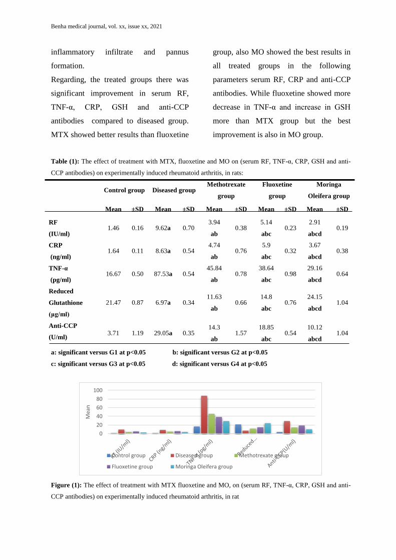
Regarding, the treated groups there was
significant improvement in serum RF,
TNF-α, CRP, GSH and anti-CCP
antibodies compared to diseased group.
MTX showed better results than fluoxetine
group, also MO showed the best results in
all treated groups in the following
parameters serum RF, CRP and anti-CCP
antibodies. While fluoxetine showed more
decrease in TNF-α and increase in GSH
more than MTX group but the best
improvement is also in MO group.
Table (1): The effect of treatment with MTX, fluoxetine and MO on (serum RF, TNF-α, CRP, GSH and anti-
CCP antibodies) on experimentally induced rheumatoid arthritis, in rats:
Control group Diseased group
Methotrexate
group
Fluoxetine
group
Moringa
Oleifera group
Mean ±SD Mean ±SD Mean ±SD Mean ±SD Mean ±SD
RF
(IU/ml) 1.46 0.16 9.62a 0.70
3.94
ab 0.38
5.14
abc 0.23
2.91
abcd 0.19
CRP
(ng/ml) 1.64 0.11 8.63a 0.54
4.74
ab 0.76
5.9
abc 0.32
3.67
abcd 0.38
TNF-α
(pg/ml) 16.67 0.50 87.53a 0.54
45.84
ab 0.78
38.64
abc 0.98
29.16
abcd 0.64
Reduced
Glutathione
(μg/ml)
21.47 0.87 6.97a 0.34 11.63
ab 0.66
14.8
abc 0.76
24.15
abcd 1.04
Anti-CCP
(U/ml) 3.71 1.19 29.05a 0.35
14.3
ab 1.57
18.85
abc 0.54
10.12
abcd 1.04
a: significant versus G1 at p<0.05 b: significant versus G2 at p<0.05
c: significant versus G3 at p<0.05 d: significant versus G4 at p<0.05
Figure (1): The effect of treatment with MTX fluoxetine and MO, on (serum RF, TNF-α, CRP, GSH and anti-
CCP antibodies) on experimentally induced rheumatoid arthritis, in rat
0
20
40
60
80
100
Mea
n
Control group Diseased group Methotrexate group
Fluoxetine group Moringa Oleifera group

Effect of Fluoxetine in Rheumatoid Arthritis, 2021
Table (2): The effect of treatment with fluoxetine and methotrexate, on arthritic score at different times, on
experimentally induced rheumatoid arthritis in rats:
G1 G2 G3 G4 G5
Mean ±SD Mean ±SD Mean ±SD Mean ±SD Mean ±SD
Arteritis score 0 0.0 0.0 3.0 0.0 3.0 0.0 3.0 0.0 3.0 0.0
Arteritis score
1st w
0.0 0.0 4.0 a 0.0 3.33
ab 0.52
3.5
ab 0.55
3.0
ab 0.0
Arteritis score
2nd
w 0.0 0.0 4.0 a 0.0
3.0
ab 0.63
3.33
ab 0.52
2.5
abd 0.55
Arteritis score
3rd
w 0.0 0.0 4.0 a 0.0
2.83
ab 0.75
3.0
abc 0.63
2.33
abcd 0.52
Arteritis score
4th
w 0.0 0.0 4.0 a 0.0 2.10
ab 0.63
2.67
abc 0.52
1.5
abcd 0.55
a: significant versus G1at p<0.05 b: significant versus G2 at p<0.05 c: significant versus G3 at p<0.05 d:
significant versus G4 at p<0.05
Histopathological changes:
Figure (2): Photomicrograph of a cut section in normal rat joint shows (black arrow) flattened synovial
membrane (a) cartilage associated with (b) bone marrow with no inflammatory cellular infiltrate (H&E x 100).
Figure (3): Photomicrograph of a cut section in rat joint of diseased group shows inflammatory infiltrate and
proliferated blood vesseles (black arrow),synovial hyperplasia(a),pannus formation(b) (H&E x 100).
a
b
a
b
Benha medical journal, vol. xx, issue xx, 2021
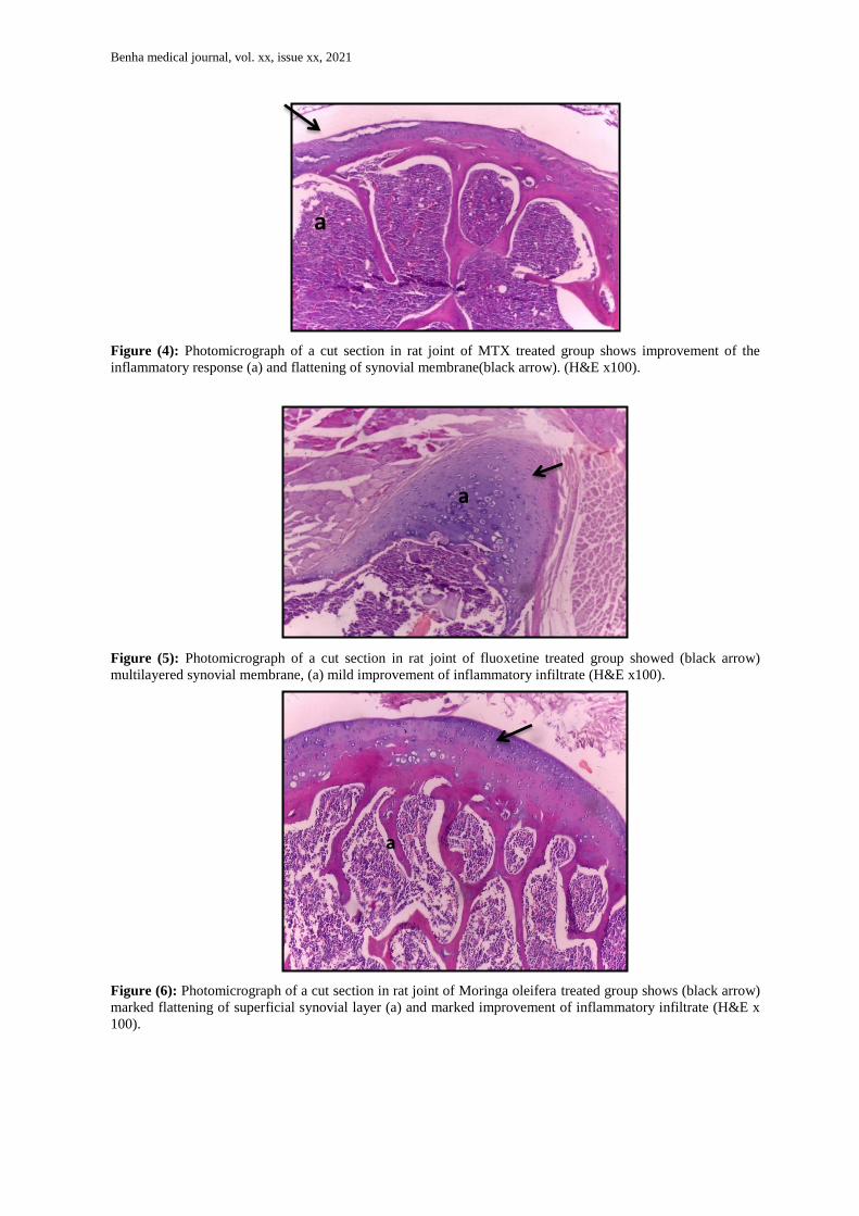
Figure (4): Photomicrograph of a cut section in rat joint of MTX treated group shows improvement of the
inflammatory response (a) and flattening of synovial membrane(black arrow). (H&E x100).
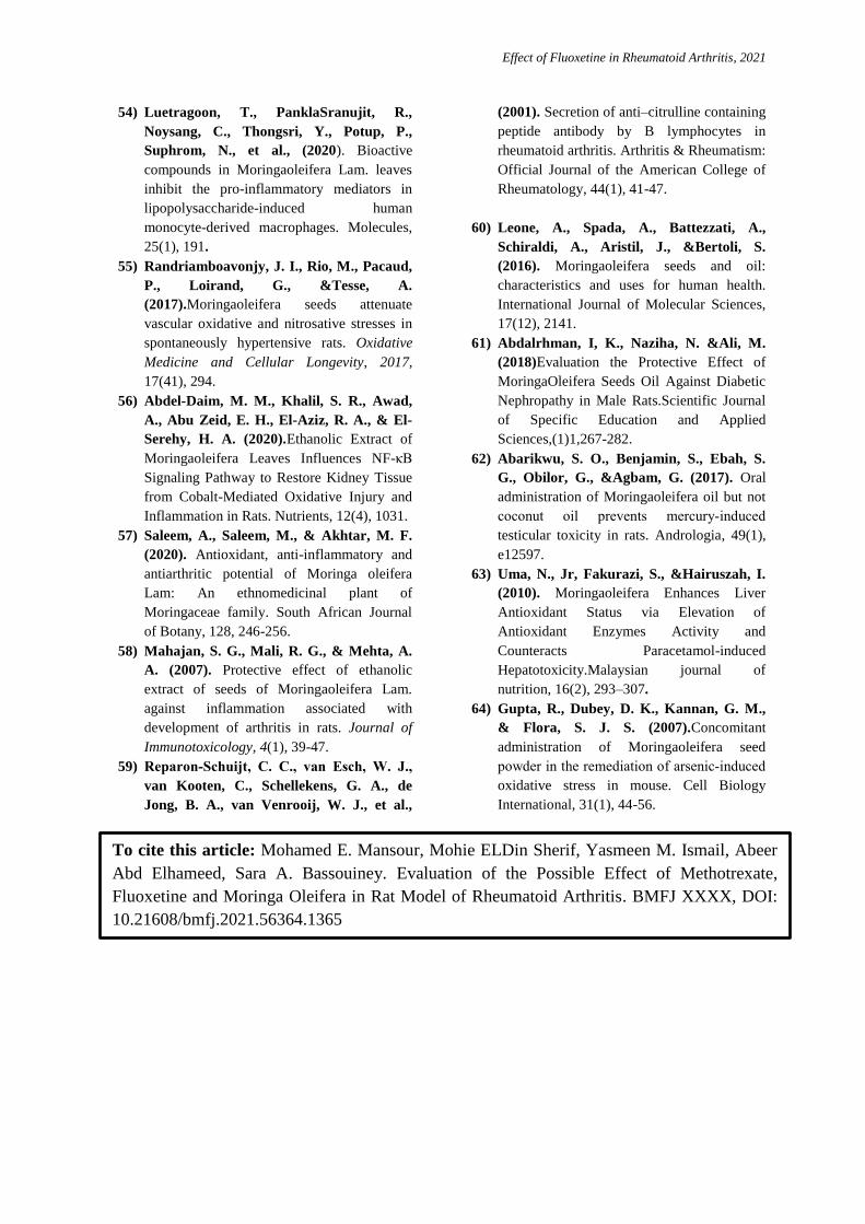
Figure (5): Photomicrograph of a cut section in rat joint of fluoxetine treated group showed (black arrow)
multilayered synovial membrane, (a) mild improvement of inflammatory infiltrate (H&E x100).
Figure (6): Photomicrograph of a cut section in rat joint of Moringa oleifera treated group shows (black arrow)
marked flattening of superficial synovial layer (a) and marked improvement of inflammatory infiltrate (H&E x
100).
a
a
a

Effect of Fluoxetine in Rheumatoid Arthritis, 2021
Regarding arthritis score, a significant
reduction in the score was seen in treated
groups at the end of 3rd and 4th weeks
compared to diseased group, MO group
was better than fluoxetine and MTX
groups.
Histopathology revealed improvement of
the joint as regard synovial hyperplasia,
cartilage degeneration and inflammatory
cell infiltration. This improvement was
observed in all treated groups with best
result in MO group
Discussion:
The present work was designed to evaluate
the effect of Methotrexate, Fluoxetine and
Moringa Oleifera extract for 4 weeks, on
rheumatoid factor (RF, tumor necrosis
factor- α (TNF-α), C-reactive protein
(CRP), reduced glutathione (GSH), anti-
cyclic citrullinated peptide (anti-CCP) and
arthritis score and histopathological
changes of joint, on RA induced
experimentally in rats by S.C. injection of
complete Freund's adjuvant in right hind
limb.
Complete Freund's adjuvant (CFA)-
induced arthritis is considered a
scientifically standard experimental
procedure for the induction of chronic
immune-pathological RA in laboratory
animals with similar cellular immunity
response and pathological mechanism as
in the human. (26).
The present study showed that S.C.
injection of (CFA) resulted in increase in
level of RF, CRP, TNF-α, arthritic score,
decrease in level of GSH. This finding is
similar to the observations of other study
proved that injection of CFA led to
significant elevation of serum RF, serum
TNF-α and decrease blood GSH level
compared to normal control group (27).
These results are in agreement with the
study that reported that injection of
complete Freund's adjuvant leads to
edematous inflammation, increased
vascularity owing to vasodilation, marked
inflammatory cell infiltration compared to
normal control group (28).
CFA showed that serum rheumatoid factor
and serum CRP level were significantly
increased in CFA rats compared with
normal control group which is in
consistence with our study (29).
The results in this study also are parallel
with the study that tested the effect of
galantamine on adjuvant-induced arthritis
in rats. The adjuvant arthritis model
showed the anti-CCP level increased in the
untreated adjuvant arthritic rats relative to
the healthy control group. (30).
The cytokines are involved in the
pathogenesis of RA. In particular, tumor
necrosis factor alpha (TNFα) has been

Benha medical journal, vol. xx, issue xx, 2021
suggested as one of the most potent
cytokines associated with RA. (31).
TNF-α induces activation of leukocyte
and endothelial and synoviocyte activation
and survival, cytokine and chemokine
amplification, angiogenesis and nociceptor
activation. The blockade of TNF
significantly decreases the production of
other pro-inflammatory cytokines and
chemokines, as IL-1, IL-6, IL-8, or
GMCSF (32).
Reduced glutathione (GSH) is the most
abundant intracellular small-molecule thiol
and is essential for maintaining the thiol
status of various molecules. GSH has
many biological roles, including protection
against reactive oxygen and nitrogen
species (ROS/NOS), which are reduced by
two GSH molecules forming oxidized
glutathione (GSSG) in the process (33).
Methotrexate significantly improved all
the tested parameters of rheumatoid
arthritis, arthritic score and histopathology
of the joints. The result of this study is in
agreement with the observations of other
study that reported that MTX treatment
after induction of RA by CFA, induced a
highly significant decrease in serum RF,
serum TNF-α, serum CRP and induced a
highly significant increase in blood GSH
level compared with diseased group.(34).
Also, in line with these results another
study reported that the paw edema, RF and
anti-CCP antibody were significantly
reduced with MTX treated group (35).
In addition, our results showed that a
significant reduction in paw diameters in
MTX-treated was that observed in
arthritic group and histopathology of
joints of MTX-treated group showed
reduction in vascular proliferation,
destruction of cartilage, synovial
membrane, and sub periosteal region (36).
For the arthritic score another study who
aimed to compare the safety and
effectiveness of MTX administrated his
study is in agreement with our results as
MTX administration decreased the
arthritis score significantly (37)
The anti-inflammatory actions of MTX are
also due to the participation of adenosine.
Adenosine is an endogenous anti-
inflammatory factor in arthritis. MTX acts
as a 5-aminoimidazole-4-carboxamide
ribonucleotide suppressor and increases
adenosine levels. Adenosine suppresses
neutrophil migration to areas of
inflammation, promotes the differentiation
of macrophages and also inhibits the
production of interleukin-1 or leukotriene
B4 (38).
Some researchers have postulated that
MTX treatment decrease of the severity of
arthritis by down-regulation of pro-
inflammatory TNF-α, IL-6 and IL-17A
cytokine expression (39).

Effect of Fluoxetine in Rheumatoid Arthritis, 2021
Regarding treatment with fluoxetine, it
showed significant improvement of RF,
TNF-α, CRP, GSH and anti-CCP
antibodies, compared to diseased. For
arthritic score, this group showed
improvement of the score at the end of 3rd
and 4th weeks, compared to diseased
group.
The effect of fluoxetine on serum level of
TNF-α and arthritis score was studied and
showed significant improve in arthritis
score and significant decrease in TNF-α
level compared to non-treated group as
approved in our study.(40).
Fluoxetine inhibiting effect on cytokine
secretion can be explained by the study
that reported that fluoxetine counteracting
depressive symptoms by inhibiting the
reuptake of serotonin and thus, augments
serotonin concentration. The relatively
high extracellular serotonin levels can
inhibit the secretion of cytokines (41).
Also a possible mechanism by which
fluoxetine inhibit endosomal Toll like
receptor (TLR) such as TLR 8 which plays
an important role in the production of
TNF-α.( 40).
Fluoxetine effect on serum CRP was
significantly decreased as shown by the
study of the effect of fluoxetine on
inflammatory markers; it showed
significant reduction in serum CRP level,
and this run in consistence with the current
work (42).
IL-6 is also the main pro-inflammatory
cytokine that induce synthesis of type 1
acute phase proteins such as CRP, elevated
levels of stress leads to activation that
triggers an NF-KB–dependent cascade of
pro-inflammatory events that contribute to
increases in CRP (43).
It also proved that fluoxetine decrease
expression of nuclear factor NF-κB , this
explain the reduction of the release of a
number of pro-inflammatory and cytotoxic
factors such as TNF-α, IL-1ß, nitric oxide,
and reactive oxygen radicals. It also
suggested that fluoxetine inhibits the
mRNA for these cytokines (as well as for
IL-6) (44).
Our study is in line with the study that
showed fluoxetine has increased level of
GSH significantly as approved in our
study (45).
Effect of fluoxetine on GSH is explained
by restoring the affected GSH pathways
with Fluoxetine treatment may relate to
neuroprotection, as the antioxidative
effects of fluoxetine are thought to be
mediated by increases in serotonin levels
(46).
And it has been reported that fluoxetine
suppressing T cell proliferation and inhibit
interferon- (IFN) production in whole
blood cultures. These also explain the
decreased cytokines level in our present
study (47).

Benha medical journal, vol. xx, issue xx, 2021
In addition fluoxetine may also inhibit the
response of antigen presenting cells these
can explain the decreased levels of auto-
antibodies as RF and Anti-CCP antibodies
(48).
According to paw edema and arthritis
score fluoxetine showed significant
reduction in edema and improvement of
arthritis score especially at 3rd and 4th
weeks and this is in line with y the anti-
inflammatory and immunomodulatory
effects of fluoxetine in rat models.(49).
Histopathological examination to
fluoxetine group showed decreased
inflammatory infiltration, bone and
cartilage destruction which is in line with
study that approved the inhibitory effect of
fluoxetine on inflammation and bone loss
in rats. And this is due to the anti-
inflammatory effect of fluoxetine (50 &
51).
On the other hand another study showed
that10 mg/kg of fluoxetine the same dose
in our study showed a small reduction in
the clinical score and a slower decrease in
paw swelling but at the higher dose (25
mg/kg), fluoxetine profoundly halted
disease progression, with no further
elevation in the clinical score or paw
swelling (40).
Regarding treatment with Moringa oleifera
extract, it showed highly significant
improvement of RF, TNF-α, CRP, GSH
and anti-CCP, compared to diseased,
normal control group and other treated
groups. For arthritic score, this group
showed significant improvement of the
score at the end of 3rd and 4th weeks,
compared to diseased group and other
treated groups.
The result of this study showed that MO
group has highly significant decrease in
TNF- α and this is supported by a study
which studied anti-inflammatory effect of
MO extract, it showed MO reduce TNF-α
significantly and this may explained by β-
sitosterol present in MO which is a
compound with potent activity against
inflammation, whose mechanism of action
includes reducing the production of TNF-α
(52).
MO also used in treatment of asthma and
associated allergic diseases; these studies
showed significant decrease in TNF-α, IL-
6, these findings also indicate that the
possible mechanism of action may be
associated with a reduction in cytokine
production/release (53).
Lipopolysaccharide (LPS) can bind to
TLR and activate the NF-κB signaling
pathway. The activation of these cascades
and transcription factors subsequently
results in the releasing of pro-
inflammatory cytokines by macrophages
and circulating monocytes, resulting in a
transient immune activation, which is
characterized by elevated levels of TNF-α,
IL-1β, and IL-6, MO extract strongly

Effect of Fluoxetine in Rheumatoid Arthritis, 2021
inhibit the LPS-induced expression of IL-6
and TNF-α during inflammation.(54).
In addition, our results are in consistence
with the study that showed the effect of
MO on vascular oxidative stress in
hypertensive rats, it showed that
significant decrease in CRP in MO treated
group. (55).
IL-6 is an important mediator of the
inflammatory response as it participates in
the development and differentiation of B-
and T-cells, as well as the activation of
acute phase proteins as CRP, MO extract
inhibit mRNA expression of IL-6and thus
consciously leads to decreased CRP
level.(56).
Also the study of the antioxidant, anti-
inflammatory and anti-arthritic effect of
M. oleifera showed in histopathology
minimal inflammation, no pannus
formation and erosion of epithelial cells.
Also decrease in paw edema of Moringa
treated rats as approved in our study. (57)
M.O extract shows significant protection
against lymphocytic infiltration, bone
destruction and cartilage erosion and this
is in line with our study. Also significant
reduction in RF and TNF-α, and this is
supported by the study of the protective
effect of ethanolic extract of seeds of MO
in arthritic rats (58).
B cells isolated from RA synovium can
secrete RF and anti-CCP antibodies,
indicating that the autoantibody is
produced locally in the joint (59).
Moringa seed extract has the ability to
attenuate the chronic immune-mediated
inflammatory responses typical of certain
diseases such as asthma and RA and this
explain the decrease in parameters of
rheumatoid arthritis such as RF and anti-
CCP antibodies (60).
MO extract showed significant increase in
GSH and this is supported by the study of
the protective effect of MO extract seeds
against diabetic nephropathy in rats (61).
The significant increase in GSH is also
supported by studying the effect of MO
seed extract on induced testicular toxicity
in rats (62).
These significant increase in GSH may
explained by Glutathione Reductase (GR)
is essential in maintaining adequate GSH
level by facilitating the regeneration of
GSH from oxidized glutathione
(GSSG).The protective effect of MO was
also reflected in the induction of GR
activity by MO extract.(63).
Cysteine and methionine rich proteins that
are present in high amounts in MO seeds
.Beside this, MO, which is also rich with
other potent antioxidant s like vitamin C,
vitamin E and B-carotene.(64)
Conclusion
Methotrexate, Fluoxetine and Moringa
Oleifera had improved adjuvant arthritis.
Moringa Oleifera extract can be used as

Benha medical journal, vol. xx, issue xx, 2021
new treatment in cases of rheumatoid
arthritis thus can decrease MTX dose to
avoid its side effects. Also, fluoxetine can
be used in in treatment of rheumatoid
arthritis to avoid possible side effect of
high doses of methotrexate also it has
beneficial effect in cases of depression
associated with rheumatoid arthritis.
Sources of funding
This research did not receive any specific
grant from funding agencies in the public,
commercial, or not-for-profit sectors.
References
1) Goma, S. H., Mahran, D. G., El-Hakeim,
E. H., Ghandour, A. M., Abdelaziz, M. M.,
Galal, M. A., et al., (2016). Spectrum of
Rheumatic Diseases in Egypt is
Similar/Different from that in Non-Arabic
Countries: An Inpatient Comparison, volume
1 | Issue 1.
2) Amaya J, Botello-Corzo D, Calixto J.
(2012): Usefulness of patients-reported
outcomes in rheumatoid arthritis focus
group. Arthritis. 2012:935(187).
3) Firestein, G. S. (2003). Evolving concepts of
rheumatoid arthritis. Nature, 423(6937), 356-
361.
4) McInnes, I. B., & Schett, G. (2011). The
pathogenesis of rheumatoid arthritis. New
England Journal of Medicine, 365(23), 2205-
2219.
5) Nishimoto, N., & Kishimoto, T. (2006).
Interleukin 6: from bench to bedside. Nature
clinical practice Rheumatology, 2(11), 619-
626.
6) Kumar P and Bain k
(2013):Pharmacotherapy Options in
Rheumatoid Arthritis ,Clinical Medicine
Insights Arthritis Musculoskelet Disorders
35–43.
7) Ling, S. F., & Bluett, J. (2020).
Pharmacogenetics of methotrexate response
in rheumatoid arthritis: an update 3-6, volume
21 , Issue 1.
8) Brown, P. M., Pratt, A. G., & Isaacs, J. D.
(2016). Mechanism of action of methotrexate
in rheumatoid arthritis, and the search for
biomarkers. Nature Reviews Rheumatology,
12(12), 731-742.
9) Singh, R. G., Negi, P. S., & Radha, C.
(2013). Phenolic composition, antioxidant
and antimicrobial activities of free and bound
phenolic extracts of Moringa oleifera seed
flour. Journal of functional foods, 5(4), 1883-
.1891
10) Joaquim, A. F., & Appenzeller, S. (2015).
Neuropsychiatric manifestations in
rheumatoid arthritis. Autoimmunity
reviews, 14(12), 1116-1122.
11) Hendawy, O. M.; Ahmed, W. M. S.;
Abosaif, A. A. (2015): Effect of atorvastatin
and vitamin D on freund adjuvant-induced
rheumatoid arthritis in rat. J Bioequiv
Availab., 7:090-094. doi: 10. 4172/jbb.
100221.
12) Ravera, S., Ramaekers, J. G., & de Gier, J.
J. (2012). Are selective serotonin reuptake
inhibitors safe for drivers? What is the
evidence?. Clinical therapeutics, 34(5), 1070-
.1083
13) Caiaffo V. Oliveira B., de Sa´ F. & Neto J.
(2016):Anti-inflammatory, antiapoptotic, and
antioxidant activity of fluoxetine.
Pharmacology Research& Perspective, 4(3).
14) Ravindra A., Priya S. and Siddheshwar
S.(2019):A pharmacological Review On
Moringa Oleifera , World Journal of
Pharmaceutical Research ,8,910-920.
15) Rovenskýa, J., Švíka, K., Stancíkováa, M.,
Ištoka, R., Ebringerb, L., & Ferencíkc, M.
(2006): The effects of selenium enriched
enterococus faecium M-74 on methotrexate
treatment of rats with adjuvant arthritis.
International Journal of Probiotics and
Prebiotics, 1(2):137-144.
16) Branco‐de‐Almeida, L. S., Franco, G. C.,
Castro, M. L., dos Santos, J. G., Anbinder,
A. L., Cortelli, S. C., et al., (2012).
Fluoxetine inhibits inflammatory response
and bone loss in a rat model of ligature‐
induced periodontitis. Journal of
periodontology, 83(5), 664-671.
17) Mahajan S., Mali R. & Mehta
A.(2008):Protective Effect of Ethanolic

Effect of Fluoxetine in Rheumatoid Arthritis, 2021
Extract of Seeds of Moringa oleifera Lam.
Against Inflammation Associated with
Development of Arthritis in Rats.Journal of
Immunotoxicology. Pages 39-47, volume
4,issue 1.
18) Schemer, S. (1967): The blood morphology
of laboratory animals.3rd ed.
Philadelphia,USA: Davis company;42.
19) Faisal, R., Ahmad, N., Fahed, Y. S., &
Chiragh, S. (2018). Anti-arthritic effect of
thymoquinone in comparison with
methotrexate on pristane induced arthritis in
female Sprague Dawley Rats. Journal of
Ayub Medical College Abbottabad, 30(1), 3-
7.
20) Vetal, S., Bodhankar, S. L., Mohan, V., &
Thakurdesai, P. A. (2013): Anti-
inflammatory and antiarthritic activity of type
–A procyanidine polyphenols from bark of
Cinnamomumzeylanicum in rats. Food Sci
Hum Wellness, 2:5967.
21) Engvall, E. and Perlman, p. (1971):
Enzyme-Linked Immunosorbent Assay,
(ELISA) Quantitative Assay of
Immunoglobulin G. Immunochemistry. 8:
871-874.
22) Kasumagic-Halilovic, E.; Prohic, A. and
Cavaljuga, S. (2011): Tumor necrosis factor-
alpha in patients with alopecia areata. Indian
Journal of Dermatology, 56(5):494-6.
23) Nathan, B. R. and Scheld, W. M. (2002):
The potential roles of C-reactive protein and
procalcitonin concentrations in the serum and
cerebrospinal fluid in the diagnosis of
bacterial meningitis. Curr Clin Top Infect
Dis., 22:155-165.
24) Beutler, E.; Duron, O.; Kelly, M. B. (1963):
Glutathione reduced. J Lab Clin Med.; 61
.882-888,
25) Avčin, T., Cimaz, R., Falcini, F., Zulian, F.,
Martini, G., Simonini, G., et al.,(2002).
Prevalence and clinical significance of anti-
cyclic citrullinated peptide antibodies in
juvenile idiopathic arthritis. Annals of the
rheumatic diseases, 61(7), 608-611.
26) Mahdi, H. J., Khan, N. A. K., Asmawi, M.
Z. B., Mahmud, R., Vikneswaran, A.,
&Murugaiyah, L. (2018).In vivo anti-
arthritic and anti-nociceptive effects of
ethanol extract of Moringaoleifera leaves on
complete Freund's adjuvant (CFA)-induced
arthritis in rats. Integrative Medicine
Research, 7(1), 85-94.
27) Refaat, R., Salama, M., Meguid, E. A., El
Sarha, A., & Gowayed, M. (2013):
Evaluation of the effect of losartan and
methotrexate combined therapy in adjuvant-
induced arthritis in rats. Eur. J. Pharmacol.,
698: 421-428.
28) Saleem, A., Saleem, M., Akhtar, M. F.,
Shahzad, M., &Jahan, S.
(2020).Moringarivae leaf extracts attenuate
complete Freund’s adjuvant-induced arthritis
in Wistar rats via modulation of inflammatory
and oxidative stress biomarkers.
Inflammopharmacology, 28(1), 139-151.
29) Mehta, A., Sethiya, N. K., Mehta, C., &
Shah, G. B. (2012).Anti–arthritis activity of
roots of Hemidesmusindicus R.
Br.(Anantmul) in rats. Asian Pacific Journal
of Tropical Medicine, 5(2), 130-135.
30) Gowayed, M. A., Refaat, R., Ahmed, W.
M., & El-Abhar, H. S. (2015).Effect of
galantamine on adjuvant-induced arthritis in
rats. European journal of pharmacology,
vol,764, 547-553.
31) Kojima, A., Kobayashi, T., Ito, S.,
Murasawa, A., Nakazono, K., &Yoshie, H.
(2016). Tumor necrosis factor‐alpha gene
promoter methylation in Japanese adults with
chronic periodontitis and rheumatoid
arthritis.Journal of periodontal research,
51(3), 350-358.
32) Noack, M., &Miossec, P. (2017).Selected
cytokine pathways in rheumatoid arthritis.In
Seminars in immunopathology (Vol. 39, No.
4, pp. 365-383).Springer Berlin Heidelberg.
33) Damgaard, D., Bjørn, M. E., Steffensen,
M. A., Pruijn, G. J., & Nielsen, C. H.
(2016).Reduced glutathione as a
physiological co-activator in the activation of
peptidylargininedeiminase.Arthritis research
& therapy, 18(1), 102.
34) Makar, N., Elhawary, A. H., Emam, H.,
Abo Ria, N., &Shaaban, E. E. S. (2020).
Possible Beneficial Effect of Metformin
Alone or in Combination with Methotrexate
in Rheumatoid Arthritis Induced Rat Model.
Benha Medical Journal, 37(1), 143-154.
35) Ali, Z., Atia, H., El Allawy, R., &AbdAlla,
S. (2017). Anti-hepatotoxic and synergistic
effects of sesame oil with methotrexate in
adjuvant induced arthritis. The Egyptian

Benha medical journal, vol. xx, issue xx, 2021
Journal of Biochemistry and Molecular
Biology, 35(1-2), 77-92.
36) Roy, T., Banerjee, I., Ghosh, S., Dhali, R.
S., De Pati, A., &Tripathi, S. K. (2017).
Effects of co-treatment with pioglitazone and
methotrexate on experimentally induced
rheumatoid arthritis in Wistar albino rats.
Indian journal of pharmacology, 49(2), 168.
37) Wang, X., Yan, X., Wang, F., Ge, F., & Li,
Z. (2018): Role of methotrexate
chronotherapy in collagen-induced
rheumatoid arthritis in rats. Z Rheumatol.,
77(3):249-255.
38) Koyama, A., Tanaka, A., & To, H. (2017).
Daily oral administration of low‐dose
methotrexate has greater antirheumatic
effects in collagen‐induced arthritis rats.
Journal of Pharmacy and Pharmacology,
69(9), 1145-1154.
39) Zhao, P. W., Jiang, W. G., Wang, L.,
Jiang, Z. Y., Shan, Y. X., & Jiang, Y. F.
(2014). Plasma levels of IL-37 and
correlation with TNF-α, IL-17A, and disease
activity during DMARD treatment of
rheumatoid arthritis. PloS one, 9(5), e95346.
40) Sacre, S., Medghalchi, M., Gregory, B.,
Brennan, F., & Williams, R. (2010).
Fluoxetine and citalopram exhibit potent
antiinflammatory activity in human and
murine models of rheumatoid arthritis and
inhibit toll‐like receptors. Arthritis &
Rheumatism, 62(3), 683-693.
41) Lu, Y., Ho, C. S., Liu, X., Chua, A. N.,
Wang, W., McIntyre, R. S., et al., (2017).
Chronic administration of fluoxetine and pro-
inflammatory cytokine change in a rat model
of depression. PloS one, 12(10), e0186700.
42) Coccaro, E. F., Lee, R., Breen, E. C., &
Irwin, M. R. (2015).Inflammatory markers
and chronic exposure to fluoxetine,
divalproex, and placebo in intermittent
explosive disorder.Psychiatryresearch,
.844-849 ,(3)229
43) Chavda, N., & ND Kantharia, J. (2011).
Effects of fluoxetine and escitalopram on C-
reactive protein in patients of depression.
Journalofpharmacology&pharmacotherapeuti
cs, 2(1), 11.
44) Liu, D., Wang, Z., Liu, S., Wang, F., Zhao,
S., & Hao, A. (2011). Anti-inflammatory
effects of fluoxetine in lipopolysaccharide
(LPS)-stimulated microglial cells.
Neuropharmacology, 61(4), 592-599.
45) Perić, I., Stanisavljević, A., Gass, P.,
&Filipović, D. (2017). Fluoxetine reverses
behavior changes in socially isolated rats:
role of the hippocampal GSH-dependent
defense system and proinflammatory
cytokines. European Archives of Psychiatry
and Clinical Neuroscience, 267(8), 737-749.
46) Zafir, A., Ara, A., &Banu, N. (2009).In vivo
antioxidant status: a putative target of
antidepressant action. Progress in Neuro-
Psychopharmacology and Biological
Psychiatry, 33(2), 220-228.
47) Diamond, M., Kelly, J. P., & Connor, T. J.
(2006). Antidepressants suppress production
of the Th1 cytokine interferon-γ, independent
of monoamine transporter
blockade. European
Neuropsychopharmacology, 16(7), 481-490.
48) O'neill, L. A. (2008). Primer: Toll-like
receptor signaling pathways—what do
rheumatologists need to know. Nature clinical
practice Rheumatology, 4(6), 319-327.
49) Kostadinov, I., Delev, D., Petrova, A.,
Stanimirova, I., Draganova, K., Kruzliak,
P. et al., (2015). Study on anti-inflammatory
and immunomodulatory effects of fluoxetine
in rat models of inflammation. European
Journal of Inflammation, 13(3), 173-182.
50) Roumestan, C., Michel, A., Bichon, F.,
Portet, K., Detoc, M., Henriquet, et
al.,(2007). Anti-inflammatory properties of
desipramine and fluoxetine. Respiratory
research, 8(1), 35.
51) Abdel-Salam, O. M., Baiuomy, A. R., &
Arbid, M. S. (2004). Studies on the anti-
inflammatory effect of fluoxetine in the rat.
Pharmacologicalresearch, 49(2), 119-131.
52) Araújo, L. C. C., Aguiar, J. S., Napoleão,
T. H., Mota, F. V. B., Barros, A. L. S.,
Moura, M. C., et al., (2013).Evaluation of
cytotoxic and anti-inflammatory activities of
extracts and lectins from Moringaoleifera
seeds.PloS one, 8(12), e81973.
53) Mahajan, S. G., Banerjee, A., Chauhan, B.
F., Padh, H., Nivsarkar, M., & Mehta, A.
A. (2009). Inhibitory effect of n-butanol
fraction of Moringaoleifera Lam. seeds on
ovalbumin-induced airway inflammation in a
guinea pig model of asthma. International
journal of toxicology, 28(6), 519-527,
Effect of Fluoxetine in Rheumatoid Arthritis, 2021
54) Luetragoon, T., PanklaSranujit, R.,
Noysang, C., Thongsri, Y., Potup, P.,
Suphrom, N., et al., (2020). Bioactive
compounds in Moringaoleifera Lam. leaves
inhibit the pro-inflammatory mediators in
lipopolysaccharide-induced human
monocyte-derived macrophages. Molecules,
25(1), 191.
55) Randriamboavonjy, J. I., Rio, M., Pacaud,
P., Loirand, G., &Tesse, A.
(2017).Moringaoleifera seeds attenuate
vascular oxidative and nitrosative stresses in
spontaneously hypertensive rats. Oxidative
Medicine and Cellular Longevity, 2017,
17(41), 294.
56) Abdel-Daim, M. M., Khalil, S. R., Awad,
A., Abu Zeid, E. H., El-Aziz, R. A., & El-
Serehy, H. A. (2020).Ethanolic Extract of
Moringaoleifera Leaves Influences NF-κB
Signaling Pathway to Restore Kidney Tissue
from Cobalt-Mediated Oxidative Injury and
Inflammation in Rats. Nutrients, 12(4), 1031.
57) Saleem, A., Saleem, M., & Akhtar, M. F.
(2020). Antioxidant, anti-inflammatory and
antiarthritic potential of Moringa oleifera
Lam: An ethnomedicinal plant of
Moringaceae family. South African Journal
of Botany, 128, 246-256.
58) Mahajan, S. G., Mali, R. G., & Mehta, A.
A. (2007). Protective effect of ethanolic
extract of seeds of Moringaoleifera Lam.
against inflammation associated with
development of arthritis in rats. Journal of
Immunotoxicology, 4(1), 39-47.
59) Reparon‐Schuijt, C. C., van Esch, W. J.,
van Kooten, C., Schellekens, G. A., de
Jong, B. A., van Venrooij, W. J., et al.,
(2001). Secretion of anti–citrulline containing
peptide antibody by B lymphocytes in
rheumatoid arthritis. Arthritis & Rheumatism:
Official Journal of the American College of
Rheumatology, 44(1), 41-47.
60) Leone, A., Spada, A., Battezzati, A.,
Schiraldi, A., Aristil, J., &Bertoli, S.
(2016). Moringaoleifera seeds and oil:
characteristics and uses for human health.
International Journal of Molecular Sciences,
17(12), 2141.
61) Abdalrhman, I, K., Naziha, N. &Ali, M.
(2018)Evaluation the Protective Effect of
MoringaOleifera Seeds Oil Against Diabetic
Nephropathy in Male Rats.Scientific Journal
of Specific Education and Applied
Sciences,(1)1,267-282.
62) Abarikwu, S. O., Benjamin, S., Ebah, S.
G., Obilor, G., &Agbam, G. (2017). Oral
administration of Moringaoleifera oil but not
coconut oil prevents mercury‐induced
testicular toxicity in rats. Andrologia, 49(1),
e12597.
63) Uma, N., Jr, Fakurazi, S., &Hairuszah, I.
(2010). Moringaoleifera Enhances Liver
Antioxidant Status via Elevation of
Antioxidant Enzymes Activity and
Counteracts Paracetamol-induced
Hepatotoxicity.Malaysian journal of
nutrition, 16(2), 293–307.
64) Gupta, R., Dubey, D. K., Kannan, G. M.,
& Flora, S. J. S. (2007).Concomitant
administration of Moringaoleifera seed
powder in the remediation of arsenic‐induced
oxidative stress in mouse. Cell Biology
International, 31(1), 44-56.
To cite this article: Mohamed E. Mansour, Mohie ELDin Sherif, Yasmeen M. Ismail, Abeer
Abd Elhameed, Sara A. Bassouiney. Evaluation of the Possible Effect of Methotrexate,
Fluoxetine and Moringa Oleifera in Rat Model of Rheumatoid Arthritis. BMFJ XXXX, DOI:
10.21608/bmfj.2021.56364.1365

Benha medical journal, vol. xx, issue xx, 2021