evaluation of the elderly patient with acute chest...
TRANSCRIPT

Clin Geriatr Med 23 (2007) 327–349
Evaluation of the Elderly Patientwith Acute Chest Pain
Brian S. Kelly, MDEmergency Department, Mount Carmel Medical System, 750 Mount Carmel Mall,
Ste. 300, Columbus, OH 43222, USA
In the United States, almost 6 million patients present to emergencydepartments each year with the chief complaint of chest pain [1–3]. Despitethe frequency of this presentation, accurate diagnosis of the cause of chestpain remains challenging. In the majority of unselected patients, chestpain has a benign cause; however, the clinician must first consider potentiallife-threatening causes when evaluating patients with chest pain. Acutemyocardial infarction (AMI), aortic dissection, pulmonary embolism, pneu-mothorax, esophageal rupture, and pericarditis with resultant cardiac tam-ponade may all present with chest pain. When these conditions are missed,significant patient morbidity and mortality can result. Misdiagnoses causingdelayed treatment not only lead to poorer outcomes but also impose sub-stantial emotional and financial stress. For example, missed AMI representsthe greatest malpractice risk in emergency medicine, accounting for 10% ofall lawsuits and almost 30% of monetary losses [3]. Claims for missed AMIoccur with equal frequency to providers who care for elderly patients [4].
It is imperative that geriatricians be able rapidly to evaluate patients withacute chest pain and accurately exclude life-threatening causes. It is wellknown that serious causes of chest pain are more prevalent among olderpatients than in younger age groups. Unfortunately, many elderly patientswho have a serious cause of chest pain present with atypical and nonspecificsymptoms. To detect these life-threatening causes, the geriatrician mustsystematically evaluate every elderly patient with chest pain.
This article discusses the evaluation of the elderly patient with acute chestpain. Specific emphasis is placed on identifying acute coronary syndrome(ACS), pulmonary embolism (PE), acute aortic dissection (AD), and acutepericarditis with tamponade. By understanding the different presentationsof these potentially life-threatening emergencies, the geriatrician will be
E-mail address: [email protected]
0749-0690/07/$ - see front matter � 2007 Elsevier Inc. All rights reserved.
doi:10.1016/j.cger.2007.01.005 geriatric.theclinics.com

328 KELLY
better prepared to distinguish them from less dire conditions and initiateprompt treatment.
Pathophysiology
To develop an approach to chest pain, it is important first to reviewthoracic anatomy and the relationship between afferent innervation andsymptom production. Chest pain arises from stimulation of visceral andsomatic pain fibers. Visceral fibers originate from the heart, blood vessels,esophagus, and visceral pleura and enter the spinal cord at multiple levels.Stimulation of these fibers produces symptoms that are poorly localizedand often difficult for patients to describe. Elderly patients are more likelyto use terms such as aching, pressure, discomfort, or heaviness when des-cribing pain of visceral origin. In addition, patients may report discomfortin other areas of the body, namely the neck, shoulders, back, and arms.In contrast, somatic pain fibers originate from musculoskeletal structures,parietal pleura, and dermis, producing symptoms that are well localizedand typically easily described. Somatic fibers enter the spinal cord at specificlevels and tend to produce symptoms that follow a dermatomal pattern.
Understanding these differences makes it easier for clinicians to interpretthe descriptive terms patients use when reporting their symptoms. It isimportant to understand that patients’ perception and description of painare affected by a number of other factors, most notably age, level of educa-tion, comorbid medical or psychologic diseases, and cultural differences.
Initial evaluation
The key components of the initial evaluation of chest pain include an as-sessment of the patient’s general appearance, a detailed history and focusedphysical examination, electrocardiogram, and chest radiograph. Additionalcomponents of the evaluation, such as laboratory studies, are obtainedbased on findings of the initial evaluation.
General appearance
Critically ill patients must be rapidly identified. Begin by noting thepatient’s general appearance. Patients who are pale, clammy, anxious, ordiaphoretic need immediate attention. Obtain a complete set of vital signs:temperature, pulse, blood pressure, respiratory rate, and oxygen saturation.Following interpretation of vital signs, one should perform a rapid survey ofthe airway, breathing, and circulation (see later discussion). All unstablepatients require an ECG and a chest radiograph as soon as possible. Amore detailed history may be obtained from patients who are hemodynam-ically stable and in no acute distress.

329ELDERLY PATIENT WITH ACUTE CHEST PAIN
History
Obtaining a detailed history is a crucial step in the formulation of anappropriate differential diagnosis in patients who have chest pain. A use-ful mnemonic for the key components of the history is ‘‘OLD CAAARS’’(A. Mattu, MD, personal communication, 2003).
OnsetDetermine the time of onset of the chest pain. Patients presenting imme-
diately after the onset of chest pain generally view their condition as serious.Additionally, knowing when the pain began is helpful when one is interpret-ing specific test results such as cardiac biomarkers. Many patients presentingshortly after the onset of an AMI have normal cardiac biomarker levels ini-tially. For this reason, it is important not to exclude ACS based on a singleset of cardiac markers.
LocationElicit the location of the pain. Lateralizing pain may be present with a PE
or pneumothorax. Anterior chest pain can be associated with ascending AD,whereas interscapular back pain is typical of descending dissections.Although it is important, the location of pain should not be used by itselfto exclude potential life-threatening diagnoses. For example, patients whohave inferior myocardial infarction (MI) may complain of epigastric painand report associated symptoms of nausea or emesis. These complaintscan be misleading if the clinician does not consider ACS as a diagnosis.
DurationDetermine the duration of the pain. Neither fleeting pain lasting seconds
nor constant pain that is unremitting for weeks is typical of myocardialischemia. Unstable angina (UA) may present with intermittent, recurringpain over days to weeks; however, in AMI, pain typically lasts at least 5to 15 minutes and will gradually worsen.
CharacterA patient’s description of the quality or character of the pain is an impor-
tant historical element but must be used carefully. Classically, myocardialischemia is usually described as a tightness, pressure, ache, or heavy weightpressing on the center of the chest. In practice, elderly patients, especiallywomen and diabetics, often report less classic symptoms, such as shortnessof breath, fatigue, indigestion, lightheadedness, dizziness, altered mentalstatus, and palpitations. Sharp, tearing pain is classic of AD. Sharp pleuriticpain, caused by pleural irritation, is common in PE.
Aggravating factorsInformation about factors that provoke or aggravate the pain is useful.
Pain that is worsened by activity, emotional stress, sexual intercourse, or

330 KELLY
exposure to cold suggests myocardial ischemia. Pleuritic pain, or pain wors-ened by deep breathing or position, suggests pneumothorax, pericarditis, orPE. Pain that is reliably and exactly reproduced with minor movements ofthe trunk or extremities is most likely musculoskeletal in nature.
Alleviating factorsExertional pain that subsides with rest is classic of angina. Pain asso-
ciated with pericarditis may diminish in an upright or leaning-forward posi-tion. Therapeutic trials of medications such as nitroglycerin or antacidsshould not be used to increase or decrease clinical suspicion that a seriousdisease exists. Studies have demonstrated that the relief of chest pain afternitroglycerin does not predict a cardiac source [5,6]. It is not uncommonfor patients with chest pain to report relief after nitroglycerin, regardlessof the cause. Shry and colleagues [6] highlighted this finding in a retrospec-tive study of 251 emergency department patients with chest pain. Eighty-eight percent of patients who had cardiac chest pain and 92% of patientswho had noncardiac chest pain reported some relief after receiving sub-lingual nitroglycerin. In addition, the symptoms of myocardial ischemiareportedly improve after administration of antacids [7]. Do not excludeACS based on the relief of pain with antacids.
Associated symptomsAny symptoms associated with the pain must be elicited. Patients who
have myocardial ischemia may report nausea, vomiting, diaphoresis, dysp-nea, or near-syncope. Elderly patients may report belching, indigestion, oreven generalized fatigue when suffering MI. Dyspnea is the most commonpresenting symptom of angina in patients older than 85 years [8]. Whenchest pain is associated with syncope, the clinician must consider the possi-bility of a PE, AD with resultant cardiac tamponade, or ruptured aorticaneurysm. Chest pain associated with neurologic symptoms is AD untilproved otherwise.
RadiationVisceral pain often radiates or is referred to other locations. For example,
the pain associated with myocardial ischemia classically radiates to the neck,shoulder, arm, or jaw. A wide extension of chest pain radiation or radiationof pain to both arms increases the probability of MI. In an excellent meta-analysis, Panju and colleagues [9] reported likelihood ratios for ACS basedon clinical features (Table 1). The pain experienced with AD often radiatesto the back and may migrate to the abdomen or legs as the dissection planeadvances to the distal abdominal aorta.
SeveritySeverity of pain is subjective. Regardless, temporal severity is useful in-
formation. Pain that is abrupt in onset and immediately becomes maximally

331ELDERLY PATIENT WITH ACUTE CHEST PAIN
severe suggests AD, spontaneous pneumothorax, or PE. Pain associatedwith myocardial ischemia classically begins more gradually, with intensityincreasing over a 5- to 10-minute period.
Medical history
One should question patients specifically about traditional risk factorsfor PE, AD, and ACS (Boxes 1–3). However, the utility of traditionalrisk factor assessment for ACS has been debated. Because traditionalrisk factors are derived from population studies, risk factor assessmenthas minimal utility when one is attempting to predict the likelihood ofACS in a specific patient who has acute chest pain [1,3]. The ACS risk fac-tors that appear to be most important include age, gender, and a familyhistory of premature coronary artery disease (CAD) [10]. Both the preva-lence and severity of CAD increase with age, as do mortality and com-plication rates.
The utility of AD risk factor assessment has been less debated. Amongpatients who have AD, there is a 3:1 male predominance. Peak incidenceoccurs between 50 and 70 years of age. A history of hypertension is common(70%) [1]. Abuse of sympathomimetic drugs such as methamphetamine andcocaine increases the risk for AD.
The list of risk factors associated with PE is extensive. Hospitalizationfor surgery, especially when secondary to trauma, or medical illnessaccounts for 46% of patients with pulmonary emboli [11]. Other factorsfrequently encountered in patients who have PE include malignantneoplasm (18%), nursing home residence (13%), congestive heart failure(10%), and central venous catheter or pacemaker placement (9%).
Table 1
Clinical features that increase the likelihood of acute myocardial infarction
Clinical feature
Likelihood ratio
(95% confidence interval)
Pain in chest or left arm 2.7a
Chest pain radiation
Right shoulder 2.9 (1.4–6.0)
Left arm 2.8 (1.7–3.1)
Both left and right arms 7.1 (3.6–14.2)
History of myocardial infarction 1.5–3.0b
Association with nausea or vomiting 1.9 (1.7–2.3)
Association with diaphoresis 2.0 (1.9–2.2)
Hypotension (systolic blood
pressure %80 mm Hg)
3.1 (1.8–5.2)
a Data not available to calculate confidence interval.b In heterogeneous studies the likelihood ranges are reported as ranges.
Adapted from Panju AA, Hemmelgarn BR, Guyatte GH, et al. Is this patient having
a myocardial infarction? JAMA 1998;280(14):1256; with permission.

332 KELLY
Approximately one quarter of patients diagnosed with PE lack knownrisk factors [11].
Physical examination
Although emphasis is placed on the cardiovascular system, one shouldperform a thorough physical examination in all patients who have chestpain. In unstable patients, rapid assessment of the airway, breathing, andcirculation should precede history acquisition.
AirwayThe airway may be assessed by asking simple questions and inspecting
the oropharyngeal cavity for potential obstructions. An appropriate verbalresponse to questioning ensures a patent airway.
Box 1. Risk factors associated with pulmonary embolism
Inherited thrombophiliaElevated individual clotting factors (VIII, IX, XI)Factor V Leiden mutationHyperhomocystinemiaProtein C, protein S, or antithrombin III deficiencyProthrombin gene mutation
Acquired disordersAgeAntiphospholipid antibody syndromeHistory of PE or deep venous thrombosisHyperviscosity syndrome
Multiple myelomaWaldenstrom’s macroglobulinemia
Immobilization (bedridden condition, paralysis, or paresis)MalignancyMedical conditions:
Congestive heart failureObesityNephrotic syndromeTobaccoInflammatory bowel disease
Central venous catheter/pacemaker placementHormone therapy/oral contraceptivesPregnancySurgeryTraumaLong-distance travel

333ELDERLY PATIENT WITH ACUTE CHEST PAIN
BreathingThe patient’s breathing is assessed primarily through a combination of
observation and auscultation. Observe the patient’s respiratory effort, not-ing the rate and depth of respirations. Increased respiratory effort shouldimmediately alert the physician to a serious disorder. Inspect the chestwall to ensure equal chest rise. Auscultation of equal bilateral breath soundsis useful to exclude rare, but life-threatening, causes of acute chest pain suchas spontaneous tension pneumothorax.
CirculationRapid assessment of the circulatory system includes measurement of
the patient’s blood pressure and heart rate, inspection of skin color andtemperature (eg, warm and pink versus cool and mottled), assessment ofcapillary refill, and palpation of peripheral pulses. Obtain intravenousaccess as early as possible. Any abnormalities found during this assessmentmust be addressed before one proceeds with other components of theevaluation.
Look for any findings that may be attributable to cardiopulmonarydisease. Jugular venous distention, if present, may signify decompensatedheart failure, right heart strain secondary to a large pulmonary embolus,cardiac tamponade, or tension pneumothorax. Pulses in the upper and lowerextremities should be carefully assessed. A difference in pulse is seen in asmany as 60% of patients who have AD [12]. Von Kodolitsch and colleagues[13] demonstrated that a blood pressure difference of 20 mm Hg or greater
Box 2. Risk factors associated with aortic dissection
Inherited diseaseMarfan syndromeEhlers-Danlos syndromeAnnuloaortic ectasiaPolycystic kidney diseaseTurner syndromeBicuspid aortic valveCoarctation of the aorta
Acquired conditionPregnancySyphilisCardiac catheterizationCocaine useHypertensionAortic valve replacementTrauma

334 KELLY
between the arms was an independent predictor of AD. However, inter-armblood pressure differences are highly variable, with as many as 19% ofnormal individuals having differences greater than 20 mm Hg [14,15].Auscultate the lungs to detect any asymmetric findings of diminished breathsounds, wheezing, rhonchi, or rales. Auscultation of the heart may revealthe presence of a murmur, gallop, or rub. A new high-pitched diastolic mur-mur and acute chest pain should raise suspicion for AD. Hepatojugularreflux and bilateral dependent pitting edema may be additional signs of con-gestive heart failure. Unilateral lower-extremity edema or tenderness shouldraise suspicion for deep venous thrombosis.
Electrocardiogram
The ECG is often reported to be the single most important initial diag-nostic test in the evaluation of patients who have chest pain and suspectedACS [1,3]. Medical negligence often results when clinicians fail to obtain an
Box 3. Risk factors associated with acute coronary syndrome
Major risk factors that cannot be changedIncreasing ageMale genderFamily history of premature CAD or cerebrovascular accident
(male relative <55 years or female relative <65 years)Race
Modifiable major risk factorsCigarette smokingDiabetes mellitusHypercholesterolemiaLow high-density lipoproteinHypertensionObesityPhysical inactivityDrug abuse (cocaine and methamphetamines)
Other factorsHyperhomocysteinemiaHyperfibrinogenemiaElevated lipoprotein (a)Elevated C-reactive proteinSystemic lupus erythematosusHIVStress

335ELDERLY PATIENT WITH ACUTE CHEST PAIN
ECG or misinterpret the result as normal or nonspecific changes. Despite itsimportance, the initial ECG is diagnostic in less than 50% of patients whohave ACS [1–3]. Only a small percentage of patients who have AMI presentwith a normal ECG (ie, no ST segment or T wave changes, strain, ischemia,old infarcts, or pseudonormalization), whereas a normal ECG is more com-mon in patients who have unstable angina. Techniques that may be used toincrease the sensitivity of the standard 12-lead ECG when nondiagnosticfindings are present include the addition of right-sided or posterior leads,serial ECGs, and continuous ST segment monitoring.
Elderly patients with ACS are more likely to have nonspecific findings onECG than are younger patients. They are less likely than younger patientsto present with ST segment elevation (31.4% versus 50.1%) and more likelyto present with left bundle branch block (8.0% versus 0.6%) [16]. Elderlypatients are also more likely to have paced rhythms and evidence of previousMI, both of which lead to more frequent nondiagnostic ECGs in the presenceof AMI [17]. Comparison with a previous ECG is crucial. All elderly patientswith new changes on the ECG require further evaluation to exclude ischemia.
ECG changes are common in patients who have PE; however, thesechanges are neither sensitive nor specific for the diagnosis. Classic ECGchanges found in patients with PE include new-onset incomplete or com-plete right bundle branch block, atrial fibrillation, T-wave inversion acrossthe anterior precordial leads, and the S1Q3T3 pattern (ie, an S wave in leadI, a Q wave in lead III, and an inverted T wave in lead III). These changesoccur in a minority of patients. More common ECG findings in patientswho have PE include sinus tachycardia and findings that reflect pre-existingcardiopulmonary disease.
Chest radiograph
The chest radiograph (CXR) is a cost-effective diagnostic test used toevaluate patients who have chest pain. It has been reported that CXRsinfluence management in 14% to 23% of emergency department patientswith chest pain [1]. In cases of pneumothorax or pneumomediastinum, theCXR may be diagnostic.
Radiographs can be helpful in identifying AD. The most notable radio-graphic findings in patients who have AD are mediastinal widening greaterthan 8 cm and the presence of an abnormal aortic contour (Fig. 1). Thesefindings are present in 61.6% and 49.6% of patients who have AD, respec-tively [18]. Other abnormalities that suggest AD include an abnormal aorticknob, left apical cap, tracheal deviation, depression of the left main stembronchus, esophageal deviation, and loss of the paratracheal stripe. How-ever, the CXR can be completely normal in as many as 12.4% of patientswho have AD [18].
Classic radiographic findings in patients who have PE include Hampton’shump (a wedge-shaped, pleural-based density with the apex pointing toward

336 KELLY
the hilum) and Westermark’s sign (a prominent pulmonary hilum caused bypulmonary vessel dilatation, with relative peripheral oligemia resulting fromcollapse of vessels distal to the embolus). However, these late findings areneither sensitive nor specific for the diagnosis of PE. More commonly, theCXR demonstrates progressive atelectasis and volume loss of the affectedpulmonary segment. Data from the International Cooperative PulmonaryEmbolism Registry suggest that 82% of patients older than 70 who havePE exhibit abnormal chest films. Radiographic abnormalities that occurwith increased frequency in older patients who have PE include cardiome-galy and pulmonary venous congestion [19].
Radiographic findings in patients who have cardiac tamponade dependon the rate of pericardial fluid accumulation and the volume of the effusion.Because of the relative indistensibility of the pericardium, rapidly accumu-lated effusions can lead to cardiac tamponade even at small volumes. Inthis case, the CXR may appear normal. Pericardial effusions that occurmore slowly allow the pericardium to distend gradually, producing anapparently ‘‘globular’’ enlarged cardiac silhouette on the CXR. As muchas 300 mL of pericardial fluid may be required before significant cardiacenlargement is appreciated [20].
Differential diagnosis
It is imperative to generate initial broad differential diagnoses in elderlypatients who have chest pain. This task is accomplished by assimilatingall available historical and clinical information into a list of likely disorders.Priority must be given to potentially serious disorders. Box 4 lists seriousand benign causes of acute chest pain. Consider benign causes once life-threatening ones have been excluded.
Fig. 1. Anterior–posterior chest radiograph demonstrating a widened mediastinum in a
67-year-old woman with chest pain and acute aortic dissection.

337ELDERLY PATIENT WITH ACUTE CHEST PAIN
When formulating the differential diagnosis in patients who have chestpain, it may be helpful to use an anatomic ‘‘inside-out’’ thought process.This approach forces the physician to focus on the cardiopulmonarysystemdthe system responsible for the majority of serious disorders thatproduce pain in the chest. It also reduces the chance that a serious disorderwill be overlooked.
Specific disease entities
The remainder of this article is dedicated to the diagnostic evaluationof specific disease entities that have been selected for further discussionbecause of their life-threatening nature. Prompt recognition and treat-ment of these conditions is crucial to minimize patient morbidity andmortality.
Acute coronary syndrome
CAD remains the leading cause of death in the United States. ACS,including unstable angina and AMI, are important disease entities thatmust be considered in all elderly patients with chest pain. Despite heightened
Box 4. Differential diagnosis of chest pain
Life-threatening causesAcute myocardial infarctionUnstable anginaPulmonary embolismAortic dissectionMyocarditisTension pneumothoraxAcute chest syndrome (sickle cell disease)Esophageal rupture
Non–life-threatening causesGastroesophageal refluxPeptic ulcer diseasePneumoniaPleurisyCostochondritisHerpes zosterMusculoskeletal painAnxiety disordersMitral valve prolapse

338 KELLY
awareness of ACS, it is estimated that between 0.4% and 10% of patientswho have AMI are incorrectly discharged from the emergency department[1,3,21]. The reasons clinicians fail to recognize serious illness are numerous;however, even experienced, competent physicians can miss AMIs. Thereason is often the clinician’s failure to consider the possibility of ACSand therefore failure to initiate the appropriate diagnostic work-up. In theshort term, patients sent home from the emergency department with ACShave a mortality nearly twice that of ACS patients who are admitted tothe hospital [3].
Investigations involving patients who have AMI indicate specific sub-groups that are at greater risk for misdiagnosis. These groups includethe very young and very old, women, and diabetics [22]. In the case ofthe very young patient, the diagnosis is often not considered because of thevery low prevalence of CAD in this group. The remaining three groups ofpatients have one thing in commondthe propensity to present with atypicalsymptoms. Women presenting with ACS tend to be older than men whohave ACS, typically postmenopausal; they are more likely to have comorbiddiseases, such as diabetes or hypertension, and to have a family history ofpremature coronary heart disease [23]. Women who have AMI are alsomore likely to present with neck and shoulder pain, nausea, fatigue, and dysp-nea than are men [23]. Diabetic patients are more likely to experience silentACS than are their age-matched nondiabetic counterparts. Therefore, latepresentations are common, a pattern that partially explains the increase inmorbidity and mortality among diabetic patients who have AMI. Diabeticpatients are alsomore likely to present with exertional dyspnea, severe fatigue,or lightheadedness [24].
In elderly patients, chest pain accompanies AMI much less frequently. Inpatients aged 85 years or older, dyspnea, not chest pain, is the single mostcommon presenting symptom of angina [10]. Additionally, elderly patientspresent more frequently with complaints of fatigue, lightheadedness, wors-ening congestive heart failure, altered mental status, and syncope [2]. Elderlypatients are also more likely to have comorbid diseases such as diabetes,hypertension, and coronary heart disease. At autopsy, approximately 50%of elderly women and 70% to 80% of elderly men have demonstrableobstructive CAD [25]. Elderly patients tend to have more severe and wide-spread CAD than younger patients [25–27].
Nonetheless, elderly patients who have ACS tend to receive less aggres-sive medical therapy and fewer revascularization procedures than doyounger patients. One reason may be that elderly patients tend to wait be-fore seeking medical attention. In a study by Tresch and colleagues [27],elderly patients with ischemic chest pain delayed seeking medical attentionfor more than 6 hours after the onset of pain, even though more than50% of them had a documented history of CAD.
Finally, elderly patients who have AMI fare far worse than youngerpatients. Although octogenarians constitute less than 5% of the United

339ELDERLY PATIENT WITH ACUTE CHEST PAIN
States population, they account for 30% of all MI-related hospital deaths[25].
Diagnostic evaluationThe cornerstone of the diagnosis of ACS has long been the ECG. Ideally,
all patients who have suspected ACS should undergo an ECG within 10minutes of arrival. Accurate interpretation of the ECG and prompt consul-tation with a cardiologist, when appropriate, are necessary to avoid delaysin management. A CXR should be obtained before the administration ofanticoagulants to minimize the chances of inadvertent administration ofthese medications to a patient who has AD.
Because elderly patients are more likely to present with non–ST-elevationMI (NSTEMI) and nondiagnostic ECGs, the diagnosis of AMI often reliesheavily on measuring serum cardiac biomarkers. Conventional biomarkers,including creatine kinase MB isoenzyme, troponin I, and troponin T, areindicators of myocardial necrosis. These markers are released in the blood-stream following MI. The levels of these serum markers tend to rise 3 to 4hours after the onset of an AMI. Serial sampling over a 12- to 24-hourperiod will detect the majority of AMIs. Although serial troponin samplingis the current gold standard for the diagnosis of NSTEMI, this test is not100% sensitive or specific. Elevated troponin levels are seen in other diseasestates, including renal failure, subarachnoid hemorrhage, and severe sepsis.In addition, serial troponin sampling is only approximately 30% sensitivefor the detection of UA [28]. Further evaluation with provocative stress test-ing must be performed when UA is a possibility.
Aortic dissection
AD is one of the most common and lethal diseases of the aorta. It isestimated that unrecognized AD carries a 1% to 2% mortality per hourfor the first 48 hours [2]. If the diagnosis remains unrecognized, the mortal-ity reaches 90% at 1 year [29]. When the disease is promptly diagnosed andtreated, the 30-day survival rate is 80% to 90%, and the 10-year survivalrate is 55% [29].
Despite advances in imaging techniques, diagnosing AD remains difficult.Because of the variety of symptoms attributable to AD, a high clinical indexof suspicion is required. It is estimated that physicians correctly suspect thediagnosis in as few as 15% to 43% of patients at the time of presentation [2].Diagnostic delays are common. Not infrequently, the correct diagnosis ismade during advanced imaging procedures intended to assess other diagno-ses [29]. Even worse, when AD is misdiagnosed as ACS, the consequences ofgiving anticoagulants or thrombolytics can be disastrous.
AD is largely a disease of old age. Younger patients who have Marfan’ssyndrome typically account for only about 5% of cases of AD [18]. The clas-sic patient with AD is a man in the seventh decade of life. Seventy-two

340 KELLY
percent of patients have a history of hypertension [18]. Five percent havea history of aortic valve repair or replacement [18]. Ninety-five percent ofpatients who have AD report some pain. Most often, the pain is abruptin onset (84.8%) and located in the anterior chest (60.9%), posterior chest(35.9%), or back (53.2%) [18]. The pain is frequently described as severeor ‘‘worst ever’’ (90.6%) or sharp (64.4%). Although they are highly specificfor AD, the descriptive terms tearing and ripping are only used by 50% ofpatients [18,29]. The pain is migratory in 16.6% of patients [18]. AD isassociated with syncope in as many as 13% of patients [18,30], and thismay be the only presenting symptom in as many as 3% of patients [30].Therefore, AD should be included in the differential diagnosis of elderlypatients who have syncope, even when chest symptoms are lacking.
On physical examination, approximately 50% of patients who have ADare hypertensive (systolic blood pressure [SBP] R150 mm Hg), whereas 16%are hypotensive (SBP %100 mm Hg) or in shock (SBP %80 mm Hg) [18].The blowing, decrescendo, diastolic murmur of aortic insufficiency is presentin less than one third of patients, and its absence should not be used toexclude the diagnosis [18,29]. Likewise, the presence of electrocardiographicchanges suggestive of AMI does not exclude AD. In an excellent compre-hensive literature review, Klompas [29] found that 7% of patients with acuteAD had electrocardiographic evidence suggestive of AMI. Focal neurologicdeficits suggestive of a cerebrovascular accident are found in 4.7% to 17%of patients [18,29]. Evidence of congestive heart failure is found in 6.6% ofpatients [18].
It is critical to obtain a detailed history from patients who have chest painand suspected AD. Patients must be asked to describe the quality of the pain,its intensity at onset, and whether the pain radiates. A retrospective chartreview of patients who had confirmed thoracic AD revealed that only 42%of conscious patients were asked these three simple questions [31]. Cliniciansmust also screen for known risk factors, including hypertension, aortic valvereplacement, and recent cardiac catheterization. It is estimated that iatrogenicAD complicates approximately 2% of cardiac catheterizations [18].
Diagnostic evaluationThe initial evaluation of a patient who has suspected AD begins with
prompt chest radiography. Most patients who have AD have abnormalfindings on CXR. A completely normal CXR reduces the probability of AD(negative likelihood ratio 0.3) [18]. However, given that chest radiographsare normal in 12.4% of patients who have AD, CXR alone is insufficient toexclude AD.
The diagnosis of AD necessitates the use of advanced imaging modalities,most frequently CT (Fig. 2). With the advent of multidetector row CT(MDCT) scanners, aortic disorders are diagnosed with increased accuracy.In a recent retrospective review of 373 cases of suspected AD in the emer-gency setting, Hayter and colleagues [32] found the sensitivity of MDCT

341ELDERLY PATIENT WITH ACUTE CHEST PAIN
for detecting AD to be 99%. Other advanced imaging modalities that areused less frequently include transthoracic and transesophageal echocardiog-raphy, MRI, and aortography.
To date, laboratory testing has played a limited role in the evaluation ofAD. Several studies demonstrated that highly sensitive quantitative D-dimerassays have a sensitivity of 100% for the diagnosis of acute AD [33–35]. How-ever, given this test’s poor specificity and the lack of a large prospective trial, itremains to be seen whether it will play a significant role in the evaluation ofpatientswho have suspectedAD.Other laboratory tests forAD that are underinvestigation include smooth muscle myosin heavy chains and soluble elastinfragments [36,37]. These tests, while highly sensitive and specific for AD, arenot readily available and lack current clinical utility.
Pulmonary embolism
PE is a challenging disease, particularly in elderly patients. It has beenestimated that the diagnosis of PE is missed 400,000 times annually, leadingto 100,000 preventable deaths [38]. The mortality of patients who haveunrecognized PE is nearly seven times greater than that of appropriatelytreated patients [39]. Presenting signs and symptoms of PE vary greatly,and the coexistence of obstructive lung disease, pneumonia, or congestiveheart failure can make diagnosis more difficult.
The incidence of PE rises with age. It is not clear whether this risk shouldbe attributed to advanced age itself or simply to an increased incidence ofconditions associated with venous thromboembolic disease. What is clearis that elderly patients who have PE suffer increased morbidity and mortal-ity compared with younger patients [39,40]. Additionally, complicationsassociated with anticoagulation (eg, life-threatening bleeding) may increasewith age.
Fig. 2. Multidetector chest CT of the patient shown in Fig. 1, demonstrating a Stanford Type B
(descending) thoracic aortic dissection.

342 KELLY
Most patients who have PE present with complaints of dyspnea or acutechest pain [41]. The pain is typically sharp and pleuritic. Patients may reportpainless dyspnea occurring at rest or on exertion. Decreased exercise toler-ance accompanied by nonspecific T-wave changes may be noted in somepatients who have PE, prompting an evaluation for CAD. Less frequently,patients with PE report persistent cough, hemoptysis, presyncope, orsyncope. Clinicians must be aware of the significance of leg symptoms (uni-lateral pain or swelling) and dyspnea or chest pain. Clinical signs associatedwith PE such as tachypnea and tachycardia are common and appear tooccur with equal frequency in elderly and younger patients [42]. However,elderly patients may be more likely to ignore new symptoms or to attributethem to existent cardiopulmonary disease.
Diagnostic evaluationThe diagnostic evaluation of patients who have suspected PE presents
a considerable challenge to physicians. Most often, the evaluation beginswith some assessment of clinical pretest probability. Few other diseases existin which pretest probability has been so extensively investigated. In the timesince the Prospective Investigation of Pulmonary Embolism Diagnosis studycompared clinical estimates of disease prevalence with actual disease rates,considerable effort has been invested in combining clinical pretest probabil-ity estimates with other forms of testing to diagnose or exclude PE. In cur-rent practice, the evaluation of patients who have suspected PE mostcommonly incorporates a clinical scoring system in combination with D-dimer testing and some form of advanced imaging (MDCT, compressionvenous duplex, or ventilation-perfusion scans) [43–45]. Fig. 3 is a CT scanthat demonstrates a large intraluminal filling defect within the pulmonaryartery representative of PE.
Fig. 3. Multidetector chest CT demonstrating an intraluminal filling defect within the pulmo-
nary artery, characteristic of a pulmonary embolism. (Courtesy of Henry Kim, MD, University
of Maryland School of Medicine.)

343ELDERLY PATIENT WITH ACUTE CHEST PAIN
Although these clinical prediction models have been validated prospec-tively, the utility of D-dimer testing in elderly patients in whom PE is sus-pected is controversial. The main utility of D-dimer testing in patientswith PE is its high sensitivity and negative predictive value (NPV). Inpatients who have low clinical suspicion for PE, a low D-dimer level(!500 ng/mL) makes the probability of PE so small that it is safe to with-hold further testing or anticoagulation [46]. In a prospective cohort study,Tardy and colleagues [47] evaluated the use of an ELISA D-dimer in 96 con-secutive outpatients older than 70 years who had suspected PE. In thisgeriatric population, conventional ELISA D-dimer testing had sensitivityand NPV of 100%; however, owing to advanced age and comorbid condi-tions, only a few of the 96 study patients presented with D-dimer levelsless than 500 ng/mL. It is not difficult to imagine that the utility of D-dimertesting in geriatric inpatients with suspected PE would be even lower.
Because of this finding, additional imaging is frequently required in thework-up of elderly patients who have suspected PE. When evaluating elderlypatients for PE, one must make the decision to use lower-extremity duplexultrasound, ventilation-perfusion scanning, spiral CT, or angiography on anindividual basis. Nevertheless, when PE is a possibility, definitive diagnosisor exclusion is required to minimize patient harm.
Acute pericarditis and cardiac tamponade
Acute pericarditis is the most common disorder of the pericardium. Likeother inflammatory processes, acute pericarditis results from (1) local vaso-dilatation, (2) increased vascular permeability, and (3) leukocyte exudation[48]. The causes of acute pericarditis are numerous (Box 5). Viral infection isprobably the most common cause of acute pericarditis in young, otherwise
Box 5. Causes of acute pericarditis
Idiopathic
InfectiousViralTuberculousPyogenic bacteria
NoninfectiousPostmyocardial infarctionUremiaNeoplastic diseaseRadiation-inducedConnective tissue diseasesDrug-induced

344 KELLY
healthy individuals. Uremic pericarditis may be seen in as many as one thirdof patients who have end-stage renal disease [49]. Neoplastic pericarditis,documented in as many as 10% of patients who have known malignancyat autopsy, may be responsible for as many as 35% of cases of acutepericarditis [50]. Malignancies commonly associated with pericardial in-volvement include breast cancer, lung cancer, leukemia, lymphoma, andmesothelioma.
Common symptoms of acute pericarditis include chest pain, fever,malaise, and myalgias. The chest pain is typically substernal with radiationto one or both trapezius muscle ridges. The pain is often described as posi-tional and may be relieved by sitting up or bending forward. Exacerbationmay occur with breathing, coughing, movement, or supine position. Dysp-nea is common, particularly when a large pericardial effusion or cardiactamponade is present. Fever is common in younger individuals but maybe absent in elderly patients. Other common signs include tachypnea andtachycardia. Hypotension and cardiovascular shock may occur in patientswho have pericarditis and cardiac tamponade.
Cardiac tamponade is an acute life-threatening emergency. It results fromincreased pressure within the pericardial space secondary to a high-pressurepericardial effusion. The increased pericardial pressure compresses thecardiac chambers, impairs diastolic filling, and reduces stroke volume andcardiac output. As blood return to the heart is impaired, the systemic andpulmonary venous pressures become elevated, leading to signs of right-heartfailure (jugular venous distension, hepatomegaly, peripheral edema).Untreated, the resultant decline in cardiac output leads to hemodynamiccompromise, hypotensive shock, and death.
Tamponade can result from any process that causes fluid to accumulatein the pericardial space. In the United States, the most common cause ofcardiac tamponade is malignant pericardial effusion. Other common causesinclude trauma, catheter-based procedures, radiation exposure, postviralconditions, and uremic pericarditis.
Diagnostic evaluationThe ECG is abnormal in as many as 90% of patients who have acute
pericarditis. Electrocardiographic changes associated with pericarditisreflect inflammation of the epicardium. In patients who have acute pericar-ditis, the ECG classically evolves through four characteristic stages:
Stage 1: Accompanies the onset of chest pain and is characterized by dif-fuse concave-upward ST-segment elevation with T-wave concordance,ST-segment depression in aVR and V1, and PR-segment depression(Fig. 4).
Stage 2: Occurs several days to weeks later and is characterized byST-segment and PR-segment normalization and T-wave flattening.
Stage 3: Characterized by diffuse T-wave inversions (not always present).
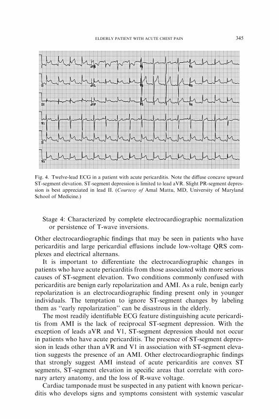
345ELDERLY PATIENT WITH ACUTE CHEST PAIN
Stage 4: Characterized by complete electrocardiographic normalizationor persistence of T-wave inversions.
Other electrocardiographic findings that may be seen in patients who havepericarditis and large pericardial effusions include low-voltage QRS com-plexes and electrical alternans.
It is important to differentiate the electrocardiographic changes inpatients who have acute pericarditis from those associated with more seriouscauses of ST-segment elevation. Two conditions commonly confused withpericarditis are benign early repolarization and AMI. As a rule, benign earlyrepolarization is an electrocardiographic finding present only in youngerindividuals. The temptation to ignore ST-segment changes by labelingthem as ‘‘early repolarization’’ can be disastrous in the elderly.
The most readily identifiable ECG feature distinguishing acute pericardi-tis from AMI is the lack of reciprocal ST-segment depression. With theexception of leads aVR and V1, ST-segment depression should not occurin patients who have acute pericarditis. The presence of ST-segment depres-sion in leads other than aVR and V1 in association with ST-segment eleva-tion suggests the presence of an AMI. Other electrocardiographic findingsthat strongly suggest AMI instead of acute pericarditis are convex STsegments, ST-segment elevation in specific areas that correlate with coro-nary artery anatomy, and the loss of R-wave voltage.
Cardiac tamponade must be suspected in any patient with known pericar-ditis who develops signs and symptoms consistent with systemic vascular
Fig. 4. Twelve-lead ECG in a patient with acute pericarditis. Note the diffuse concave upward
ST-segment elevation. ST-segment depression is limited to lead aVR. Slight PR-segment depres-
sion is best appreciated in lead II. (Courtesy of Amal Mattu, MD, University of Maryland
School of Medicine.)

346 KELLY
congestion and hemodynamic compromise. Vital sign derangements, includ-ing tachypnea, tachycardia, and hypotension, are common. On examina-tion, one may observe Beck’s triad, a complex of physical findingsconsisting of jugular venous distension, systemic hypotension, and dimin-ished heart sounds. Jugular venous distension, the most prominent physicalfinding, may be absent in patients with severe volume depletion. Pulmonaryrales resulting from pulmonary venous congestion may be appreciated.Other classically described physical findings that are not specific for cardiactamponade include pulsus paradoxus (decrease in SBP R10 mm Hg withinspiration) and Kussmaul’s sign (the paradoxic rise in jugular venouspressure with inspiration).
Patients who have cardiac tamponade require prompt diagnosis anddefinitive treatment. Mortality for cardiac tamponade directly correlateswith the time to definitive treatment. Although cardiac tamponade is a clinicaldiagnosis, themost useful noninvasive diagnostic test remains echocardiogra-phy. Echocardiography can successfully differentiate cardiac tamponadefrom other conditions associated with low cardiac output (eg, systolicdysfunction). Echocardiographic features suggestive of cardiac tamponadeinclude pericardial effusion, early diastolic right ventricular collapse, latediastolic compression or collapse of the right atrium, and impaired right-sidedbloodflowduring inspiration.Diagnostic confirmation, basedon intracardiacand intrapericardial pressure measurement, and treatment (therapeutic peri-cardiocentesis) often occur in the cardiac catheterization lab.
Summary
Chest pain is one of themost common and potentially life-threatening com-plaints in the geriatric population. The incidence and mortality of ACS, AD,and PE increase with advanced age. Malignancy, the most common cause ofcardiac tamponade in theUnited States, is also common among older individ-uals. Elderly patients who have chest pain aremore likely to present with atyp-ical symptoms and nonspecific physical findings, putting them at increasedrisk for misdiagnosis. To ensure the best possible patient outcome, physiciansmust conduct a thorough interview, including assessment of known riskfactors; perform a detailed physical examination; and maintain a high levelof suspicion when evaluating elderly patients with chest pain.
Acknowledgments
The author wishes to extend gratitude to Henry Kim, MD, Michael E.Winters, MD, and Amal Mattu, MD, for their guidance and contributionto this work. The author would also like to acknowledge Linda J. Kesselr-ing, MS, ELS, for her significant copyediting support.

347ELDERLY PATIENT WITH ACUTE CHEST PAIN
References
[1] Bean DB, Roshon M, Garvey JL. Chest pain: diagnostic strategies to save lives, time, and
money in the ED. Emergency Medicine Practice 2003;5(6):1–32.
[2] Haro LH, Decker WW, Boie ET, et al. Initial approach to the patient who has chest pain.
Cardiol Clin 2006;24(1):1–17.
[3] Boie ET. Initial evaluation of chest pain. Emerg Med Clin North Am 2005;23:937–57.
[4] Physician Insurers Association of America (PIAA). Acute myocardial infarction studyd
May 1996. Rockville (MD): PIAA; 1996. p. 11.
[5] HenriksonCA,Howell EE, BushDE, et al. Chest pain relief by nitroglycerin does not predict
active coronary artery disease. Ann Intern Med 2003;139:979–86.
[6] Shry EA,Dacus J, VanDeGraaff E, et al. Usefulness of the response to sublingual nitroglyc-
erin as a predictor of ischemic chest pain in the emergency department. Am J Cardiol 2002;
90(11):1264–6.
[7] Dobrzycki S, Baniukiewicz A, Korecki J, et al. Does gastro-esophageal reflux provoke
myocardial ischemia in patients with CAD? Int J Cardiol 2005;104(1):67–72.
[8] Jones ID, Slovis CM. Emergency department evaluation of the chest pain patient. Emerg
Med Clin North Am 2001;19(2):269–82.
[9] Panju AA, Hemmelgarn BR, Guyatt GH, et al. Is this patient having a myocardial infarc-
tion? JAMA 1998;280(14):1256–63.
[10] Konotos MC. Evaluation of the emergency department chest pain patient. Cardiol Rev
2001;9(5):266–75.
[11] Heit JA, O’Fallon WM, Petterson TM, et al. Relative impact of risk factors for deep vein
thrombosis and pulmonary embolism: a population-based study. Arch Intern Med 2002;
162(11):1245–8.
[12] American Medical Forensic Specialist, Inc. Diagnosing acute aortic dissection in the E.R.
Available at: www.amfs.com/AorticDissection.html. Accessed July 18, 2006.
[13] Von Kodolitsch Y, Schwartz AG, Nienaber CA. Clinical prediction of acute aortic dissec-
tion. Arch Intern Med 2000;160:2977–82.
[14] Singer AJ, Hollander JE. Blood pressure. Assessment of interarm differences. Arch Intern
Med 1996;156(17):2005–8.
[15] Lane D, Beevers M, Barnes N, et al. Inter-arm differences in blood pressure: when are they
clinically significant? J Hypertens 2002;20:1089–95.
[16] Boucher JM,RacineN, ThanhTH, et al. Age-related differences in in-hospitalmortality and
the use of thrombolytic therapy for acute myocardial infarction. CMAJ 2001;164(9):
1285–90.
[17] Rich MW. Epidemiology, clinical features, and prognosis of acute myocardial infarction in
the elderly. Am J Geriatr Cardiol 2006;15:7–11.
[18] Hagan PG, Nienaber CA, Isselbacher EM, et al. The International Registry of Acute
Aortic Dissection (IRAD): new insights into an old disease. JAMA 2000;283(7):
897–903.
[19] Elliot CG, Goldhaber S, Visani L, et al. Chest radiographs in acute pulmonary embolism:
results from the International Cooperative Pulmonary Embolism Registry. Chest 2000;
118:33–8.
[20] McGuire J, Kotte JH, Helm RA. Acute pericarditis. Circulation 1954;9:425–42.
[21] Swap CJ, Nagurney JT. Value and limitations of chest pain history in the evaluation of
patients with suspected acute coronary syndromes. JAMA 2005;294:2623–9.
[22] Croskerry P. Achilles’ heels of the ED: delayed or missed diagnoses. ED Legal Letter 2003;
14(10):109–20.
[23] Douglas PS, Ginsburg GS. The evaluation of chest pain in women. N Engl J Med 1996;
334(20):1311–5.
[24] Cooper S, Caldwell JH. Coronary artery disease in people with diabetes: diagnostic and risk
factor evaluation. Clin Diabetes 1999;17:58–72.

348 KELLY
[25] WilliamsMA, Fleg JL, Ades PA, et al. Secondary prevention of coronary heart disease in the
elderly (with emphasis on patients R75 years of age): an American Heart Association scien-
tific statement from the Council on Clinical Cardiology Subcommittee on Exercise, Cardiac
Rehabilitation, and Prevention. Circulation 2002;105:1735–43.
[26] Stone PH, Thompson B, Anderson HV, et al. Influence of race, sex, and age on the manage-
ment of unstable angina and non Q-wave myocardial infarction: the TIMI III registry.
JAMA 1996;275:1104–12.
[27] Tresch DD,William BJ, Aufderheide TP, et al. Comparison of elderly and younger patients
with out-of-hospital chest pain. Arch Intern Med 1996;156:1089–93.
[28] HammCW, Goldman BU, Heeschen C, et al. Emergency room triage of patients with acute
chest pain bymeans of rapid testing for cardiac troponinT or troponin I. NEngl JMed 1997;
337:1648–53.
[29] KlompasM.Does this patient have an acute thoracic aortic dissection? JAMA2002;287(17):
2262–72.
[30] Nallamothu BK, Mehta RH, Saint S, et al. Syncope in acute aortic dissection: diagnostic,
prognostic, and clinical implications. Am J Med 2002;113:468–71.
[31] Rosman HS, Patel S, Borzak S, et al. Quality of history taking in patients with aortic dissec-
tion. Chest 1998;114:793–5.
[32] Hayter RG, Rhea JT, Small A, et al. Suspected aortic dissection and other aortic disorders:
multi-detector row CT in 373 cases in the emergency setting. Radiology 2006;238:841–52.
[33] Eggebrecht H, Naber CK, Bruch C, et al. Value of plasma fibrin D-dimers for detection of
acute aortic dissection. J Am Coll Cardiol 2004;44:804–9.
[34] Weber T, Hogler S, Auer J, et al. D-dimer in acute aortic dissection. Chest 2003;123:
1375–8.
[35] AkutsuK, SatoN, Yamamoto T, et al. A rapid bedsideD-dimer assay (cardiac D-dimer) for
screening of clinically suspected acute aortic dissection. Circ J 2005;69:397–403.
[36] Suzuki T, Katoh H, Tsuchio Y, et al. Diagnostic implications of elevated levels of smooth-
muscle myosin heavy-chain protein in acute aortic dissection: the Smooth Muscle Myosin
Heavy Chain Study. Ann Intern Med 2000;133:537–41.
[37] Shinohara M, Suzuki K, Okada M, et al. Soluble elastin fragments in serum are elevated in
acute aortic dissection. Arterioscler Thomb Vasc Biol 2003;23:1839–44.
[38] Fried C. Pulmonary embolism. In: Rosen P, Barkin R, Daniel DF, editors. Emergency med-
icine: concepts and clinical practice. St. Louis (MO): Mosby-Year Book, Inc.; 1998.
p. 1770–2.
[39] Carson J, KelleyM,DuffA, et al. The clinical course of pulmonary embolism. NEngl JMed
1992;326:1240–5.
[40] Siddique R, Siddique M, Connors AH, et al. Thirty-day case-fatality rates for pulmonary
embolism in the elderly. Arch Intern Med 1996;156:2343–7.
[41] Chunilal SD, Eikelboom JW, Attia J, et al. Does this patient have pulmonary embolism?
JAMA 2003;290:2849–58.
[42] Stein P, Gottschalk A, Saltzman H, et al. Diagnosis of acute pulmonary embolism in the
elderly. J Am Coll Cardiol 1991;18:1452–7.
[43] Wells PS, Anderson DR, Roger M, et al. Derivation of a simple clinical model to categorize
patients with a probability of pulmonary embolism: increasing the model’s utility with the
SimpliRED D-dimer. Thromb Haemost 2000;83:416–20.
[44] Wicki J, Perneger TV, JunodAF, et al. Assessing clinical probability of pulmonary embolism
in the emergency ward. Arch Intern Med 2001;161:92–7.
[45] Kline JA, NelsonRD, JacksonRE, et al. Criteria for the safe use of D-dimer testing in emer-
gency department patients with suspected pulmonary embolism: a multicenter US study.
Ann Emerg Med 2002;39:144–52.
[46] Kruip MJHA, Slob MJ, Schijen JHEM, et al. Use of a clinical decision rule in combination
with D-dimer concentration in diagnostic workup of patients with suspected pulmonary
embolism: a prospective management study. Arch Intern Med 2002;162:1631–5.

349ELDERLY PATIENT WITH ACUTE CHEST PAIN
[47] Tardy B, Tardy-Poncet B, Viallon A, et al. Evaluation of D-dimer ELISA test in elderly
patients with suspected pulmonary embolism. Thromb Haemost 1998;79(1):38–41.
[48] Roberts TG, Lilly LS. Diseases of the pericardium. In: Lilly LS, editor. Pathophysiology of
heart disease. Baltimore (MD): Lippincott Williams & Wilkins; 1997. p. 289–301.
[49] NelsonRN. Pericarditis. In:Hoekstra JW, editor.Handbook of cardiovascular emergencies.
Philadelphia: Lippincott Williams & Wilkins; 2001. p. 240–8.
[50] Maisch B, Ristic AD. The classification of pericardial disease in the age of modern medicine.
Curr Cardiol Rep 2002;4(1):13–21.