evaluation of residues - bentham.manuscriptpoint.com · web viewwhich have their role in...
TRANSCRIPT

Title:
Structural and functional analysis of ferritin heavy chain subunit in Oryzias latipes
Authors:
Ajit Tiwari, A. D. Upadhyay, Himanshu Priyadarshi, Rumpi Ghosh and Sanjeev Sharma
Authors:
Orcid id : https://orcid.org/0000-0002-3158-4614, [email protected], https://orcid.org/0000-0003-2350-9734, https://orcid.org/0000-0003-0891-1389, https://scholar.google.com/citations?user=6Z5OZs8AAAAJ&hl=en, https://tmc.gov.in/actrec/index.php/ashok-varma-lab,
Institutional id: [email protected], [email protected], cofcau.nic.in
Abstract:
In North East region of India, iron toxicity is one of the major problems in culture fisheries. To overcome these challenges, it’s necessary to identify the role of protein. Ferritin structurally highly conserved protein has the pivotal role in detoxification, cellular homeostasis and cellular defense against oxidative stress. The Heavy chain in a ferrtitin protein possess di-Fe binding site in the fourth helix that interacts with oxygen and is responsible for the ferroxidase activity of the protein. This research was conducted to study the structure and function of ferritin heavy chain subunit in Oryzias latipes from amino acid sequence (accession no. XP_020569048.2) of NCBI database. Physicochemical characteristics showed the molecular weight (20880.39 kDa), theoretical isoelectric point (5.54), extinction coefficient (21680), aliphatic index (71.13), instability index (49.37), total number of residues having negative charge and positive charge (30-21), and grand average of hydropathicity (-0.823) which concluded that the protein was unstable, hydrophilic. The secondary structures contain alpha helix 56.50%, extended strands 10.73% and coiled region of 32.77%. The selected template contains a structure of mouse h-chain modified ferritin by X-ray diffraction technique (2.24Å). It was verified by SWISSMODEL/Workspace possessing pdb code 3wnw.1.A. There were seven enriched pathways of KEGG identified by protein-protein interaction analysis in fish Oryzias latipes. Interpretation of sequence for the domain analysis using Pfam confirms that the sequence possesses ferritin like domain and superfamily. Further investigation revealed that the Ferritin heavy chain subunit has involvement in detoxification, ferroxidase activity, iron homeostasis and metabolism.
Keywords: Bioinformatics, Ferroxidase, Homology Modelling, pI, Hydropathy, Tertiary structure.
Introduction:

Iron, required as a trace element in organisms, forms metalloprotein in conjugation with different proteins. Excess iron in an aquatic ecosystem is toxic and acting as a catalyst in the Fenton reaction generating free radicals which are harmful to fishes. [1] Ferritins is the ubiquitous protein which stores in a nontoxic and reversible form and central to iron metabolism [2]. Thus, it has an important role in iron storage and detoxification [3] [4] [5]. Ferritin is structurally highly conserved protein from bacteria to human which emphasize their biological importance [6]. It forms a hollow shell with a cavity of 80 A1 diameters that can store up to 4,500 Fe (III) atoms as a biomineral. In eukaryotes, it consists of 24 protein subunits with molecular weight of 450 kDa [7] [8] [9]. In mammals, ferritin molecules of heavy (H) and light (L) chain subunits having molecular masses of 21 and 19 kDa. Heavy chains are important for Fe (II) oxidation, whereas Light chains assist in iron nucleation, mineralization, and long-term storage [9]. The Heavy and Light chain subunits co-assemble in different ratios to form a protein shell of 24 subunits capable of acquiring iron atoms. Key features that differentiate di-Fe binding site in H chain that interacts the fourth helix oxygen and is responsible for the ferroxidase activity of the protein [10]. For example, H-chains are abundant in heart tissues that involved in a rapid exchange of iron [11]. Recently, [12] revealed a dual role for ferritin in sea bass, with functional involvement in both iron metabolism and immune response.
The Medaka fish, scientific name Oryzias latipes, is a small, bony, laying an egg in fresh water, native to Asian countries. It occurs, coastal waters having high adaptability and it collected in wide range especially from brackish, mangrove swamps, acidic freshwater, forest streams, canals, rice field, basins of rivers, pools, and oxbows [13]. The Medaka is a model organism mainly used in the genomic and biological study [14]. As it is a model organism, it selected for the study of the structure of a protein using different software and online server. In this present study, the structural model and physicochemical properties, protein-protein interaction of ferritin H chain protein sequence (accession number XP_020569048.2.) of Oryzias latipes were analyze to study the structural and functional role of ferritin in the ferroxidase and immunological response.
Material and Methods
Sequence retrieval:
The amino acid sequence of Ferritin heavy subunit of Oryzias latipes was retrieved from National Center for Biotechnology Information (NCBI) having the accession number XP_020569048.2. It was verified by peptide search in the UniProtKB and their entry no. is H2LMW5.
Physicochemical properties and Secondary structure prediction:
Expasy ProtParam server was used to analyze the physicochemical properties of the ferritin heavy chain protein of Oryzias latipes [15] cont.. It determines the molecular weight, theoretical pI, the % of the amino acid composition, total number of residue in negative and positive form,

extinction coefficient, estimated half-life, atomic composition, aliphatic index, instability index grand average of hydropathicity. The secondary structures of the ferritin heavy chain subunit were predicted by PSIPRED [16] and GORIV methodology [17].
Tertiary structure prediction:
It was performed with a template search for the query protein through PDB sum database [18]. The template sequence was cross verified by SWISSMODEL/Workspace, which displayed sequence identity with the query sequence [19]. By homology modeling, a 3-D structure of ferritin heavy subunit was predicted using the Swiss model [20] [21], Phyre 2 [22] and pymol software [23]. The predicted structure of the ferritin heavy subunit was validated through Ramachandran plot by utilizing rampage server. [24].
Active site prediction:
Protein interactions are the important aspect of the cellular function, ligand interaction and other
small molecules [25]. The active site of ferritin heavy chain subunit of Oryzias latipes was
predicted by Dog Site Scorer server /active site prediction server based on grid-based function
prediction method. [26]. It helps in detection of potential binding pockets and sub-pockets of a
protein of interest. It calculates the geometric and physicochemical properties of such pockets
along with the estimation of druggability by support vector machine (SVM). It has been
evaluated on a large dataset containing 1069 structures with 88% prediction accuracies. It
provides valuable information for target assessment on web server. This is a grid-based method
which uses a difference of Gaussian filters to identify potential binding pockets - only based on
the 3D structure of the protein - and splits them into sub-pockets. It calculated the shape, size,
and chemical features of the predicted (sub) pockets. The score provided for each (sub) pocket is
based on a linear combination of the three descriptors which described volume, hydrophobicity,
and enclosure. A meaningful subset descriptor included in a support vector machine (libsvm) for
the prediction of (sub) pocket druggability score (0-1) and estimated value lie in between 0 to 1.
[27].
Protein-protein interactions:
The Search Tool for the Retrieval of Interacting Genes/ Proteins (STRING 10.0) database
(http://string-db.org/) was used to predict the interacting proteins [28]. The database contains
information from different sources, including experimental repositories, computational
prediction methods, and public text groups.

Domain & Gene ontology:
Pfam is utilized for the sequence alignment of the family as well as profile hidden Markov
models for finding domains in given sequence. Domains named ferritin like where IPR009040,
IPR008331 is the entry number containing function role were collected from Pfam (version 32.0)
[29] and Interpro Scan. [30] Gene ontology for the molecular function and biological process
were obtained by the Pfam (version 32.0). [29]
Results and Discussion
Physicochemical properties:
Physicochemical properties of Ferritin were analyzed by the Expasy protParam server. It is 177 amino acid long proteins with the estimated molecular weight 20880.39 kDa respectively. The isoelectric point (pI) of ferritin protein is 5.54 revealed that ferritin heavy chain is acidic. The amino acid composition showed the maximum presence of Leucine (11.3%) and minimum presence of Tryptophan (1.1%) (Table 1). The total number of negatively charged and positively charged residues of Ferritin are (Asp+ Glu)-30 and (Arg+ Lys)-21, respectively. The formula of ferritin heavy subunit is C912H1399N263O280S11 with 2865 atoms in total. The extinction coefficient of the protein calculated at 280 nm in water (M-1 cm-1) which presented the values of Abs 0.1% (=1g/l) 1.038 and extinction coefficient 21680 and, assuming all pairs of Cys residues form cystines; but the extinction coefficient becomes 21430 and Abs 0.1% (=1g/l) is 1.026 when assumed all Cys residues reduced. The N-terminal sequence of the protein is Methionine (Met). The instability index is a scale of proteins, use to determine whether it will be stable in a test tube or not. It can also be used to measure invivo half-life of a protein [26], resultant the proteins whose invivo half-life less than 5h has an instability index > 40, whereas those with half-life of more than 16h have an instability index < 40 [31]. The estimated half-life of Ferritin presented 30 hours in case of in-vitro culture condition of mammalian reticulocytes; whereas in-vivo culture condition more than 20 hours in case of yeast and more than 10 hours in case of E. coli. The estimated instability index (ll) of the Ferritin is 49.37 which classify the protein as unstable. The aliphatic index of a protein may be defines as relative volume occupied by aliphatic side chains (valine, alanine, isoleucine, and leucine) [32]. In thermophilic bacteria, it found to be significantly higher than that of ordinary proteins and hence, it can work as a measure of thermostability of proteins [33]. Aliphatic index 71.13 of the protein measures its considerable thermostability along with the relative volume occupied by aliphatic side chains. The value -0.823 of the GRAVY indicates the hydrophilic nature of the protein. The GRAVY value for a protein was calculated as the sum of hydropathy values of all the amino acids, divided by the number of residues in the sequence [34].
Secondary structure prediction:

The secondary structure prediction of Ferritin heavy chain subunit predicted was done by PSIPRED server and GOR IV server. It revealed the presence of helix region, strand region and coil regions (Figure 2). It works on PSI-blast homology search algorithm [35]. Gor IV and PSIPRED revealed the presence of alpha helix 56.50%, extended strands 10.73% and coiled region 32.77% (Figure 1, 2). Both server used for the analysis of secondary structure prediction.
Table 1: Amino acid composition of Ferritin heavy chain subunit of Oryzias latipes
Amino acids No.s Percentage
Ala (A) 10 5.6%
Arg (R 10 5.6%
Asn (N) 11 6.2%
Asp (D) 13 7.3%
Cys (C) 5 2.8%
Gln (Q) 12 6.8%
Glu (E) 17 9.6%
Gly (G) 9 5.1%
His (H) 10 5.6%
Ile (I) 6 3.4%
Leu (L) 20 11.3%
Lys (K) 11 6.2%
Met (M) 6 3.4%
Phe (F) 8 4.5%
Pro (P) 3 1.7%
Ser (S) 9 5.1%
Thr (T) 3 1.7%
Trp (W) 2 1.1%
Tyr (Y) 7 4.0%
Val (V) 5 2.8%
Pyl (O) 0 0.0%
Sec (U) 0 0.0%
S. No. Secondary structure element Percent1. Alpha helix 56.502. 310 helix 0.003. Beta bridge 0.00

4. Extended strand 10.735. Beta turn 0.006. Bend region 0.007. Random coil 32.778. Ambiguous states 0.009. Other states 0.00
Fig1. The secondary structure prediction of ferritin heavy chain subunit of Oryzias latipes by GOR IV method.
Fig. 2. The Secondary structure prediction of ferritin heavy chain subunit of Oryzias latipes by PSIPRED method.
Tertiary Structure Prediction:Template identification was done by comparative modeling. It usually initiated by the
searching of sequence for known protein structures where target sequence used as a query in the PDB database to locate the sequences that so remotely related with render construction of a reliable comparative model. It generally was done by the comparative study of the target sequence with the sequence of each of the structures in the given database [36].

Template search for the query protein of ferritin heavy chain subunit was performed through PDB sum database which presented 338 hits. The template 3wnw (A) was identified showing 82.5% sequence similarity along with Z-score of 1225.7 from the PDBsum database (Figure 3). The selected template contains a structure of mouse h-chain modified ferritin by X-ray diffraction technique (2.24Å). It was verified by SWISSMODEL/Workspace possessing pdb code 3wnw.1.A with 82.46 % sequence identity with the query sequence (Fig. 4)
Fig. 3. Template 3wnw (A) from the PDBsum database.

Fig.4. Template 3wnw.1.A from the SWISSMODEL/Workspace and 3 D structure by template 3mnw.1.A

Homology modeling predicted the 3 Dimensional structure of the Ferritin heavy chain subunit of Oryzias latipes. The conformational analysis of protein structure was done by Swiss model server aligning the query sequence to the template sequence (Figure 5A & 5B). The score QMEAN estimated the model quality, and their full form is qualitative model energy analysis. This composite scoring function depicting the major geometrical aspects of protein structures. It was checked on several standard decoy sets including a molecular dynamics simulation decoy set as well as on a comprehensive dataset [37]. It shows a statistically significant improvement over all scales of quality and describing the ability of the scoring function to determine the native structure and recognize good and bad models [38]. The general understanding of ferritin structure is based on the human ferritin subunit [39], frog ferritin [40] and the E. coli ferritin [41]. Research on the structure of fish ferritin is still unknown at present. With superposition and comparison, the ferritin structures of the human, frog, and E. coli found.
A

Fig.5. Structure modeling of Ferritin heavy chain subunit of Oryzias latipes by Swiss Prot serverA. Model reportB. Model structure.
The predicted structure (Fig. 6) of the ferritin heavy chain subunit was validated through the Ramachandran plot (phi/psi). The stereochemical analysis of RAMPAGE server showed the number of residues in the favored region is 98%, the allowed region is 1.5%, and Outline region is 2%.
B

General
Glycine
Proline
Pre-Proline

Fig.6. Ramachandran plot of the predicted structure of Ferritin heavy chain subunit of Oryzias latipes
Evaluation of residuesResidue [A 43: GLN] (-120.83, -62.93) in Allowed regionResidue [A 134: TYR] (-128.97, -43.57) in Allowed regionResidue [A 158: PRO] (-108.17, -4.28) in Allowed regionResidue [A 42: VAL] (-128.91, -63.78) in Allowed regionResidue [A 90: GLU] (79.83, -50.68) in Allowed regionResidue [A 175: GLU] (0.00, 0.00) in Outlier regionResidue [A 176: CYS] (0.00, 0.00) in Outlier regionNumber of residues in favoured region (~98.0% expected): 335 (98.0%)Number of residues in allowed region (~2.0% expected): 5(1.5%)Number of residues in outlier region: 2 (0.6%)
Structure of Ferritin heavy chain:
Fig. 7.1 Structure view of ferritin heavy chain subunit of Oryzias latipes Fig. 7.2 Structure view of polar contacts with side chain, main chain in ferritin heavy chain subunit of Oryziae Latipes
Formal charges: sum = -7.0Count atoms: 2782 atomsThe electronegativity of Iron (Fe) is 1.83 eV and magnesium 1.31 eV so electronegativity difference is 0.52 eV. The bond formation between them is polar covalent bond. The sum of formal charges is -7. It is highly electronegative. For small fractions of charges, we use the symbols δ+ and δ−. Polar molecules have slightly opposite charges on opposite ends of the molecule or a dipole.
Ferritin consists of 24 peptide subunits that form two types of channels where these subunits intersect; the 3-fold channel is polar and the 4-fold channel is nonpolar. (The residues that line the channels determine the polarity of the channel.). When the Fe(III) in the crystalline mineral is reduced to Fe(II), the iron becomes solvated and ferritin releases the solvated iron, Fe(H2O)6 2+, through the 3-fold polar channel. Hence, it can control the amount of available iron in the body, preventing iron disorders like anemia and iron overload. [42]

Active site prediction:The potential binding site and sub-pockets are detected through Dog scorer site. Dog
Site Scorer is a newly developed automatic tool combining pocket prediction, characterization, and druggability estimation. It can be customized to work on the pocket and sub-pocket level. In addition, the drugability estimation for pockets can be switched on (Fig. 8). This web server provides an easy to use interface to predict pockets and sub-pockets of a protein structure of interest. These are essential for prediction of functions, classification and drug binding ability of proteins [43]
Protoss is automated hydrogen prediction tool for protein –ligands complexes. It detects reasonable protonation states, tautomers, and hydrogen coordinates of both protein and ligands molecules and adds missing hydrogen atoms to protein structures (PDB format). [44] It investigates hydrogen bonds, repulsive atom and metal interactions relation for all possible states and calculates the network of an optimal hydrogen bonding within these degrees of freedom. The active site of amino acid residues of ferritin heavy chain subunit analysis through the active site prediction server were found to be aspartic acid, asparagines, cystine, glutamic acid, histidine, leucine, lysine, serine, arginine, proline, threonine, valine, phenylalanine, glycine, Isoleucine, methionine, tryptophan,tyrosine, and alanine.
Fig. 8. Active site with a Pocket
Size and shape descriptors Element descriptorsVolume [A2]
Surface [A2]
Drug score
Simple score
Depth [A]
Ellipsoid main axis ratio c/a
Ellipsoid main axis ratio b/a
enclosure
Pocket atoms
C N O S Other elements
440.06
644.46 0.67 0.21 14.52 0.14 0.41 0.09 120 85 11 24 0 0
Functional group descriptors Amino acid compositionHydrogen bond donors
Hydrogen bond acceptors
Metals Hydrophobic interactions
Hydrophobic ratio
A polar amino acid
polar amino acid ration
+ve amino acid ration
-ve amino acid ration
7 49 0 19 0.25 0.32 0.23 0.18 0.27

Amino acid descriptorsALA 2ARG 0ASN 0ASP 0CYS 0GLN 2GLU 6GLY 0HIS 2ILE 0LEU 2LYS 2MET 0PHE 1PRO 0SER 1THR 0TRP 1TYR 2VAL 1
Table 2: Describing the size, shape and chemical features of the predicted pockets are calculated. Per default, a simple druggability score is provided for each (sub) pocket, based on a linear combination of the three descriptors describing volume, hydrophobicity, and enclosure.
Functional interaction network analysis:For the prediction of the interacting proteins, the fth1 protein of Oryzias latipes applied
to the STRING 10.0 tool as the fish sample. It is used for known and predicted protein interaction. The interactions of protein include direct and indirect associations. The functional parameter of the protein can identify by the nodes and edges where nodes represent protein and edges represent the network. As a result, seven enriched KEGG pathways and eleven functional parameters of network analysis determine by the protein-protein interaction analysis, enlisted in Table 2 and Table 3. with their function. The K- means algorithm used for the determination of protein clustering and their colours represent the input protein (Figure 8). The protein-protein interaction network is an important component for the understanding of the cellular process at system-level. This network can be used to evaluate by filtering functional genomics data. It provides an instinctive platform for evolutionary, annotating, structural, and functional properties of a protein. [45] Explore networks can give a new direction for oncoming experimental research and confer cross-species predictions for efficient interaction mapping [46]. It is an important pre-requisite to have some knowledge about its specific interaction partners to get the full description of a function of proteins. The protein 'function' concept is hierarchical [47] [48] [49] [50] at all levels in this hierarchy, interactions proteins help to describe and narrow down a protein's function. In this research, partners (Table 6) and pathways (Table 7) of fth1 protein in Oryzias latipes were determined.

Fig 9. The interactive network view of predicted protein-protein interactions using STRING 10.0 tool. Network nodes are proteins, and the edges represent the predicted functional associations. The K means algorithm used to cluster the proteins in different groups. Inter-cluster edges represented by dashed lines. Small nodes: protein of unknown 3D structure; Large nodes: some 3 D structure is known or predicted. Extended lines: gene co-occurrence.

Index Name Functions Score1. fth1 Ferritin ; Stores iron in a soluble, non-toxic, readily available form.
Important for iron homeostasis. Iron is taken up in the ferrous form and deposited as ferric hydroxides after oxidation (177 aa)
2. ENSORL00000008245 Ribosomal protein L19 (197 aa) 0.6933. ENSORL00000003007 Ribosomal protein L19 (196 aa) 0.6704. ANAPC11 Anaphase promoting complex subunit 11 (84 aa) 0.6935. EIF1AD Eukaryotic translation initiation factor 1A domain containing (172 aa) 0.7066. GSTA5 Glutathione S-transferase alpha 5 (223 aa) 0.6127. FAU Finkel-Biskis-Reilly murine sarcoma virus (FBR-MuSV) ubiquitously
expressed (133 aa)0.575
8. MRPL49 Mitochondrial ribosomal protein L49 (169 aa)0.546
9. RAN RAN, member RAS oncogene family; GTP-binding protein involved in nucleocytoplasmic transport. Required for the import of protein into the nucleus and also for RNA export. Involved in chromatin condensation and control of cell cycle
0.504
10. SF1 Splicing factor 1 (418 aa)0.500
11. slc3a2 Solute carrier family 3 (activators of dibasic and neutral amino acid transport), member 2 (531 aa)
0.497
Table 3: Characteristics of input protein Oryzias latipes ferritin heavy chain subunit (fth1, Accession number: XP_020569048.2) functional parameters predicted with STRING 10.0
Index ID Term P-value
1. 03010 Ribosome 0.00584
PFAM Protein Domain
2. PF01280 Ribosomal protein L19e 0.00123
INTERPRO Protein Domains and Features
3. IPR000196 Ribosomal protein L19/L19e domain 0.000534
4. IPR015972 Ribosomal protein L19/L19e domain 0.000534
5. IPR015974 Ribosomal protein L19/L19e domain 0.000534
6. IPR023638 Ribosomal protein L19/L19e domain 0.000534
7. IPR027547 Ribosomal protein L19/L19e domain 0.000534
Table 4: Observed KEGG pathways in protein-protein interaction of Oryzias latipes ferritin heavy chain subunit in Fig 9.

Domain and gene ontology of ferritin heavy chain subunit:The retrieved sequence of ferritin heavy chain subunit is analysed with the use of pfam software to determine the match between sequence and HMM. The interpretation of sequence reveals that the sequence belongs to family ferritin and possess ferritin like domain. The length of the sequence is 177 and their alignment initiated from 15 to 155. The align sequence shows the match and mismatch between the HMM. PP is posterior probabilities score in between HMM and Sequence.
Figure 10. The expanded match results with the #HMM line coloured such that residues identical to those in the query are coloured cyan and those that are similar in dark blue, and a #PP (posterior probability) line giving the posterior-probabilities at each point such that the #SEQ, query, line is colourcoded accordingly.

Fig. 11. Sequence search results page. Results page for a single sequence search, the graphic of the domains matched by the query sequence along its length, with homologous superfamilies, ferritin conserved site and residue annotation by interProScan.
The Homologous Superfamily
The entry represent that the ferritin is non-haem iron storage protein in animal, plants and microorganisms. [51]. It chiefly required to most of the organism but their excess amount causes toxicity due to its reactivity. It made up of a 4-helical bundle core, and contains a bimetal-ion

centre in the middle of the bundle. Other proteins having similar structure include: haem-containing bacteriferritins; rubrerythrin, which appears to have a role in anaerobic detoxification pathway for reactive oxygen species (ROS) [52]. IPR009078 entry represented that the proteins with similar structure include ferritin and other ferritin-like proteins that binds haem between two subunits, non-haem ferritin, dodecameric ferritin homologue (DPS) to protects DNA, and the N-terminal domain of rubrerythrin that is found in many air-sensitive bacteria and archaea [53]. The superfamily also includes the alpha and beta subunits of methane monooxygenase hydrolase, delta 9-stearoyl-acyl carrier protein desaturase and manganese catalase.
Domain
Domain (IPR009040, IPR008331) represents a group of proteins having ferritin-like domain, consist of four-helix bundle surrounding a non-heme, non-sulphur, oxo-bridged diiron site. with 145-residue domain. It consists of a mineral core of hydrated ferric oxide, and a multi-subunit protein shell that encloses the former and assures its solubility in an aqueous environment. [54]. The diiron site is existed within a twisted, left-handed four- helix-bundle constituted of two anti-parallel helix pairs connected through a left-handed crossover connection. The residue of know ligand at non-heme, non- sulphur diiron sites in proteins include His, Asp, Glu, and Tyr. Proteins containing a ferritin-like diiron domain possess the ability to catalyze oxidation of Fe2+ to Fe2+ by O2, i.e. ferroxidase activity. The ferritin- like diiron domain occurs in stand-alone form in ferritin and bacterioferritin or in association with the rubredoxin-like domain in rubrerythrin [55]. Ferritin not only present in animal but also in plant. In plant it is present in the chloroplast. In animals the protein is present in cytoplamic form with two or more genes that encode closely related subunits in mammals [56]. The domain is also found in other ferritin-like proteins such as members of the DNA protection during starvation (DPS) family [57] and bacterioferritins [58].
Gene ontology:
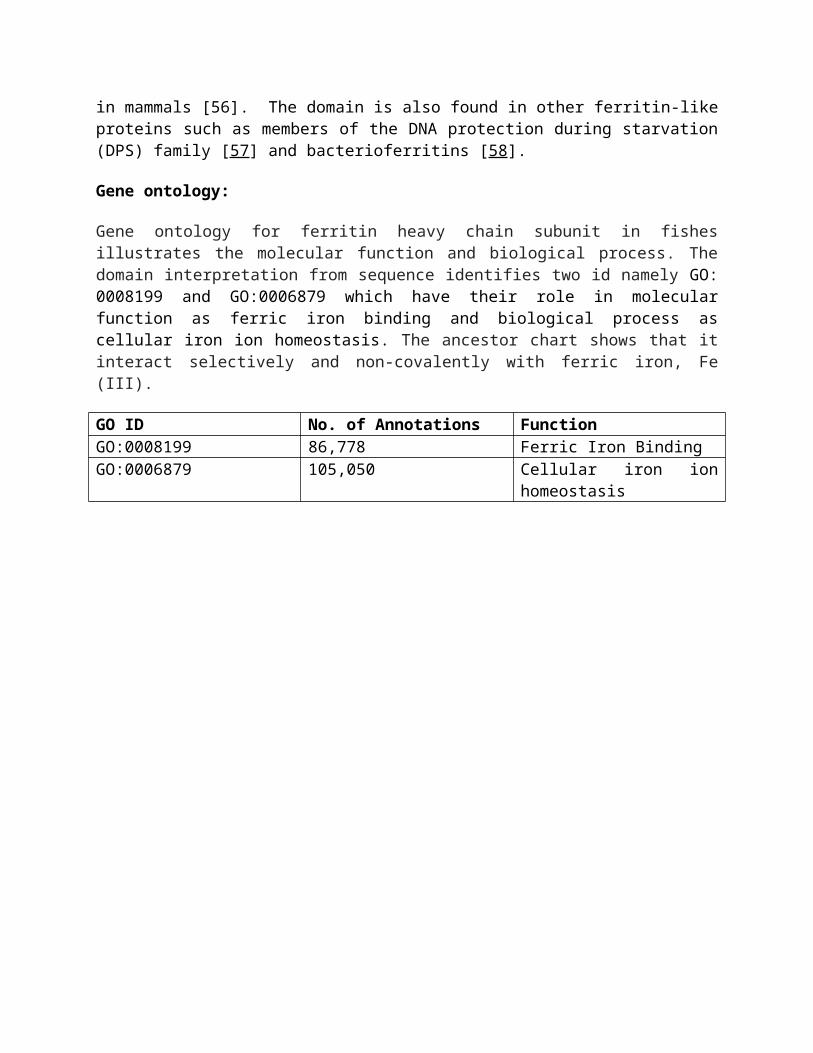
Gene ontology for ferritin heavy chain subunit in fishes illustrates the molecular function and biological process. The domain interpretation from sequence identifies two id namely GO: 0008199 and GO:0006879 which have their role in molecular function as ferric iron binding and biological process as cellular iron ion homeostasis. The ancestor chart shows that it interact selectively and non-covalently with ferric iron, Fe (III).
GO ID No. of Annotations FunctionGO:0008199 86,778 Ferric Iron BindingGO:0006879 105,050 Cellular iron ion homeostasis

a.

b.
Fig 12. GO hierarchy (taken from the ancestor chart of the QuickGo website (http://www.ebi.ac.uk/QuickGO/) showing the relative complexity of the GO hierarchy for two distinct (a) Shows the GO hierarchy for ferric iron binding (b ) Shows the GO hierarchy for cellular iron ion homeostasis. The colours of the arrows in the ontology are denoted by the key in the centre of the figure. Black connections between terms represent an is a relationship, blue connections represent a part of relationship. The A, B boxes denote the levels of the EC nomenclature.

Conclusion: This research has been conducted to support the farmer’s of North East region of India.
The in-silico investigation of Ferritin revealed that the physicochemical and structural parameters of the protein were involved in metabolism and immune response. The physico chemical and structural data of protein conclude that the ferrtin heavy subunit in Oryzias latipes is unstable and hydrophilic in nature. The secondary structures possess alpha helix (56.50%), extended strands (10.73%), and coiled region (32.77%). The selected template contains a structure of mouse h-chain modified ferritin by X-ray diffraction technique (2.24Å). It was verified by SWISSMODEL/Workspace possessing pdb code 3wnw.1.A from 82.5% sequence identity with the query sequence. Moreover, it might resolve that the predicted 3D structure exhibited a favoured region (98%) and allowed region (1.5%) that indicate the model expected to be correct in prediction. In protein-protein interaction, eleven functional partners (Table 3) and seven pathways (Table 4) of fth1 protein in Oryzias latipes were determined. This study concluded that ferritin heavy chain subunit has the involvement in detoxification, ferroxidase activity and iron homeostasis and it may shed a new direction to high iron reservoir fishes.
NotesFunding InformationThe author is thankful to the Dean, College of Fisheries, Lembucherra, Tripura for encouragement and moral support. This work has been carried out under DBT funded Project "Establishment of Bioinformatics Infrastructure Facility for Biology Teaching through Bioinformatics" and hence financial support by the Department of Biotechnology, Ministry of Science and Technology, Government of India, New Delhi.Compliance with Ethical StandardsConflict of InterestThe author declares that they have no conflict of interest.
References1. RakshitAmeta, Anil K. Chohadia, Abhilasha Jain, Pinki B. Punjabi (2018). Fenton and
Photo-Fenton. Advanced Oxidation Processes for Waste Water Treatment. Emerging Green Chemical Technology. Academic press, 49-87
2. Arosio P, Levi S (2010). Cytosolic and mitochondrial ferritins in the regulation of cellular iron homeostasis and oxidative damage. Biochim Biophys Acta, 1800, 783-92.
3. Crichton RR, Charloteauxwauters M. (1987). Iron transport and storage. Eur J Biochem, 164, 485–506.
4. Aisen P, Listowsky I. (1980). Iron transport and storage proteins. Annu Rev Biochem, 49, 357–393.
5. Anderson GJ, Frazer DM. (2005). Hepatic iron metabolism. Semin Liver Dis, 25, 420–432.
6. Andrews SC. (2010). The ferritin-like superfamily: evolution of the biological iron storeman from a rubrerythrin-like ancestor. Biochim Biophys Acta, 1800, 691-705.
7. Aisen P, Listowsky I. (1980). Iron transport and storage proteins. Annu Rev Biochem, 49, 357–393.
8. Harrison PM, Arosio P. (1996). The ferritins: molecular properties, iron storage function and cellular regulation. Biochim Biophys Acta, 1275, 161–203.

9. Torti FM, Torti SV. (2002). Regulation of ferritin genes and protein. Blood, 99, 3505–3516.
10. Guo, J. H., Juan, S. H., and Aust, S. D. (1998). Mutational analysis of the four alpha-helix bundle iron loading channel of rat liver ferritin. Arch. Biochem. Biophys., 352, 71-77.
11. Arosio P, Levi S (2010). Cytosolic and mitochondrial ferritins in the regulation of cellular iron homeostasis and oxidative damage. Biochim Biophys Acta, 1800, 783–792.
12. Neves JV, Wilson JM, Rodrigues PNS. (2009). Transferrin and ferritin response to bacterial infection: the role of the liver and brain in fish. Dev Comp Immunol, 33, 848–857.
13. Lynne R. Parenti (2008). A phylogenetic analysis and taxonomic revision of ricefishes, Oryzias and relatives (Beloniformes, Adrianichthyidae). Zoological Journal of the Linnean Society, Volume 154, Issue 3, 494–610.
14. Temminck CJ, Schlegel H. (1846).Pisces. In:Fauna Japonica, sive descriptio animalium quae in itinere per Japoniam suscepto annis 1823–30 collegit, notis observationibus et adumbrationibus illustravit P. F. de Siebold. Leiden, parts 10–14: 173–269.
15. Gasteiger E, Hoogland C, Gattiker A, Duvaud S, Wilkins MR, et al. (2005). Protein identification and analysis on the ExPASy Server. In: John M Walker (Ed.), The Proteomics Protocols Handbook, Humana Press, USA, pp 571-607.
16. Altschul SF, Madden TL, Schaffer AA, Zhang J, Zhang Z, et al. (1997). Gapped BLAST and PSI-BLAST: a new generation of protein database search programs. Nucleic Acids Res, 25(17), 3389-3402.23.
17. Sen TZ, Jerigen R, Kloczkowski A (2005). GOR V server for protein secondary structure prediction. Bioinformatics, 21(11), 2787-2788.24.
18. Laskowski RA (2001). PDBsum: summaries and analyses of PDB structures. Nucleic, 29(1), 221-222.
19. Arnold K, Bordoli L, Kopp J, Schwede T (2006). The SWISS-MODEL Workspace: A web-based environment for protein structure homology modeling. Bioinformatics, 22(2), 195-201.
20. Guex N, Peitsch MC, Schwede T (2009). Automated comparative protein structure modeling with SWISS-MODEL and SwissPdbViewer: a historical perspective. Electrophoresis, 1, S162-S173.
21. Biasini M, Bienert S, Waterhouse A, Arnold K, Studer G, et al. (2014). SWISS-MODEL: modeling protein tertiary and quaternary structure using evolutionary information. Nucleic Acid Research, 42, W252-W258.
22. Wass MN, Kelley LA and Sternberg MJ (2010). 3DLigandSite: predicting ligand-binding sites using similar structures. Nucleic Acids Research 38, W469-73.
23. DeLano WL, Ultsch MH, de Vos AM, Wells JA (2000). Convergent solutions to binding at a protein-protein interface. Science, 287 (5456), 1279–83.
24. Lovell c, Davis IW, Arendall WB, De Bakker PIW, Word JM, et al. (2003). Structure validation by Calpha geometry: phi, psi, and Cbeta deviation. Proteins, 50(3), 437-450.29.
25. N.J Burgoyne, R.M.jackson, "Predicting protein interaction sites: binding hot-spots in protein –Protein and Protein –ligands interfaces, "bio info vol.22, pp.1335-1342, 2006.
26. A. Volkamer, D. Kuhn. Rippmann, M. Rarey, "DoGSiteScorer: A web-server for automatic binding site prediction, analysis, and druggability assessment, "Bioinfo2012.

27. A. Volkamer, D. Kuhn, T. Grombacher, F. Rippmann, M. Rarey (2012). Combining global and local measures for structure-based druggability predictions. J. Chem. Inf. Model., 52,360-372.
28. Szklarczyk D, Franceschini A, Wyder S, Forslund K, Heller D, Huerta-Cepas J, Simonovic M, Roth A, Santos A, Tsafou KP. 2015. STRING v10: protein–protein interaction networks, integrated over the tree of life. Nucleic Acids Res. 43:D447–D452.
29. S. El-Gebali, J. Mistry, A. Bateman, S.R. Eddy, A. Luciani, S.C. Potter, M. Qureshi, L.J. Richardson, G.A. Salazar, A. Smart, E.L.L. Sonnhammer, L. Hirsh, L. Paladin, D. Piovesan, S.C.E. Tosatto, R.D. Finn (2018). The Pfam protein families database in 2019. Nucleic Acids Research, Volume 47, Issue D1, 8 January 2019, Pages D427–D432.
30. Finn, RD; Attwood, TK; Babbitt, PC; Bateman, A; Bork, P; Bridge, AJ; Chang, HY; Dosztányi, Z; El-Gebali, S; Fraser, M; Gough, J; Haft, D; Holliday, GL; Huang, H; Huang, X; Letunic, I; Lopez, R; Lu, S; Marchler-Bauer, A; Mi, H; Mistry, J; Natale, DA; Necci, M; Nuka, G; Orengo, CA; Park, Y; Pesseat, S; Piovesan, D; Potter, SC; Rawlings, ND; Redaschi, N; Richardson, L; Rivoire, C; Sangrador-Vegas, A; Sigrist, C; Sillitoe, I; Smithers, B; Squizzato, S; Sutton, G; Thanki, N; Thomas, PD; Tosatto, SC; Wu, CH; Xenarios, I; Yeh, LS; Young, SY; Mitchell, AL (29 November 2016). "InterPro in 2017-beyond protein family and domain annotations". Nucleic Acids Research. 45 (D1): D190–D199.
31. Guruprasad K, Reddy BB, Pandit MW. (1990). Correlation between the stability of a protein and its dipeptide composition: a novel approach for predicting in vivo stability of a protein from its primary sequence. Protein Eng., 4, 155–161.
32. Rogers S, Wells R, Rechsteiner M. (1986). Amino acid sequences common to rapidly degraded proteins: the PEST hypothesis. Science, 234, 364–368.
33. Ikai A. (1980). Thermostability and aliphatic index of globular proteins. J Biochem., 88, 1895–1898.
34. Kyte J, Doolittle RF. (1982). A simple method for displaying the hydropathic character of a protein. J Mol Biol., 157, 105–132.
35. Altschul SF, Madden TL, Schaffer AA, et al. Gapped BLAST and PSI-BLAST: a new generation of protein database search programs. Nucleic Acids Res. 1997;25(17):3389–3402.
36. Andras Fiser (2014). Template-Based Protein Structure Modeling. Methods Mol Biol. 2010; 673: 73–94.
37. Acharjee Samik, Upadhyay Datta Anil, Roy Kumar Ajit (2017). Insilico analysis of Myostatin protein of Labeo calbasu. MedCrave.3 (5): 422‒428
38. Benkert P, Biasini M, Schwede T (2011). Toward the estimation of the absolute quality of individual protein structure models. Bioinformatics, 27(3), 343-350.
39. Lawson DM, Artymiuk PJ, Yewdall SJ, Smith JM, Livingstone JC, Treffry A, Luzzago A, Levi S, Arosio P, Cesareni G. (1991). Solving the structure of human H ferritin by genetically engineering intermolecular crystal contacts[J]. Nature, 349, 541-544.
40. Ha Y, Shi D, Small W, Theil EC, Allewell NM. (1999). Crystal structure of bullfrog M ferritin at 2.8 A resolution: analysis of subunit interactions and the binuclear metal center[J]. Journal of Biological Inorganic Chemistry, 4(3), 243-256.
41. Macedo S, Romao CV, Mitchell E, Matias PM, Liu MY, Xavier AV, LeGall J, Teixeira M, Lindley P, Carrondo MA. (2003). The nature of the di-iron site in the bacterioferritin from Desulfovibrio desulfuricans[J]. Natural Structural Biology, 10(4), 285-290.

42. Rachel Casiday and Regina Frey (1999). Iron in Biology: Study of the Iron Content in Ferritin, The Iron-Storage Protein. Iron Use and Storage in the Body: Ferritin and Molecular Representations. Department of Chemistry, Washington University St. Louis, MO 63130.
43. Gao, M., Skolnick, J. A comprehensive survey of small-molecule binding pockets in proteins. (2013) PLoS comput Biol 9(10): e1003302.
44. Quiocho, F.A. Atomic structures of periplasmic binding proteins and the high-affinity active transport systems in bacteria. (1990) Philos Trans R Soc Lond B Biol Sci 326(1236): 341-352.
45. Samin Seddigh and Darabi Maryam (2017). Functional, structural, and phylogenetic analysis of mitochondrial cytochrome b (cytb) in insects, Mitochondrial DNA Part A. DNA Mapping, Sequencing, and Analysis. ISSN:2470-1408.
46. Schwartz AS, Yu J, Gardenour KR, Finley RL Jr, Ideker T. (2009). Cost-effective strategies for completing the interactome. Nat Methods., 6, 55–61.
47. Bairoch A, Boeckmann B. (1994). The SWISS-PROT protein sequence data bank: current status. Nucleic Acids Res., 22, 3578–3580.
48. Ashburner M, Ball CA, Blake JA, Botstein D, Butler H, Cherry JM, Davis AP, Dolinski K, Dwight SS, Eppig JT. (2000). Gene ontology: the tool for the unification of biology. Nat Genet., 25, 25–29.
49. Ouzounis CA, Coulson RM, Enright AJ, Kunin V, Pereira-Leal JB. (2003). Classification schemes for protein structure and function. Nat Rev Genet., 4, 508–519.
50. Lee D, Redfern O, Orengo C. (2007). Predicting protein function from sequence and structure. Nat Rev Mol Cell Biol., 8, 995–1005.
51. Emilia Chiancone, Pierpaolo Ceci, Andrea Ilari, Frederica Ribacchi and Simonetta Stefanini (2004). Iron and proteins for iron storage and detoxification. Biometals Volume 17, Issue 3, pp 197–202
52. Michael V. Weinberg, Francis E. Jenney, Jr., Xiaoyuan Cui, and Michael W. W. Adams (2004). Rubrerythrin from the Hyperthermophilic Archaeon Pyrococcus furiosus Is a Rubredoxin-Dependent, Iron-Containing Peroxidase. J Bacteriol. 186 (23): 7888–7895.
53. Shi Jin, Donald M. Kurtz, Zhi-Jie Liu, John Rose, and Bi-Cheng Wang (2002). X-ray crystal structures of reduced rubrerythrin and its azide adduct: a structure-based mechanism for a non-heme diiron peroxidase. J Am Chem Soc. 124 (33):9845-55.
54. Emilia Chiancone, Pierpaolo Ceci, Andrea Ilari, Frederica Ribacchi and Simonetta Stefanini (2004). Iron and proteins for iron storage and detoxification. Biometals Volume 17, Issue 3, pp 197–202
55. Fredrick deMaré, Donald M. Kurtz Jr & Pär Nordlund (1996). The structure of Desulfovibrio vulgarisrubrerythrin reveals a unique combination of rubredoxin-like FeS4 and ferritin-like diiron domains. Nature Structural Biologyvolume 3, pages539–546 (1996)
56. Maria RaglandS, Jean-Franqois BriatQ, Jean GagnonY, Jean-Pierre Laulhere$, Olivier MassenetQ, and Elizabeth C. TheilS (1990). Evidence for Conservation of Ferritin Sequences among Plants and Animals and for a Transit Peptide in Soybean. THE
JOURNAL OF BIOLOGICAL CHEMIWRY, Vol. 265, No. 30, Issue of October 25, pp. 18339-18344.
57. Kornelius Zeth, Stefanie Offermann, Lars-Oliver Essen, and Dieter Oesterhelt (2004). Iron-oxo clusters biomineralizing on protein surfaces: Structural analysis of Halobacterium salinarum DpsA in its low- and high-iron states. PNAS, 101 (38) 13780-13785
58. Simon C. Willies, Michail N. Isupov, Elspeth F. Garman, Jennifer A. Littlechild (2009). The binding of haem and zinc in the 1.9 Å X-ray structure of Escherichia coli bacterioferritin. JBIC Journal of Biological Inorganic Chemistry, Volume 14, Issue 2, pp 201–207