evaluation of neck lesions with mdct a case series · evaluation of neck lesions with mdct – a...
TRANSCRIPT
IOSR Journal of Dental and Medical Sciences (IOSR-JDMS)
e-ISSN: 2279-0853, p-ISSN: 2279-0861.Volume 14, Issue 8 Ver. III (Aug. 2015), PP 66-80 www.iosrjournals.org
DOI: 10.9790/0853-14836680 www.iosrjournals.org 66 | Page
Evaluation of neck lesions with MDCT – A case series
Dr. Dhaval K Thakkar1, Dr. Sanjay Khaladkar
2, Dr. Mansi Jantre
3,
Dr.Dolly K Thakkar4, Dr. Amarjit Singh
5, Dr. Vilas M. Kulkarni
6.
1-Chief Resident, Dept. of Radiodiagnosis, Dr D Y Patil Medical college and Research centre, Pimpri,
Maharashtra, India; 2-Professor, Dept. of Radiodiagnosis, Dr D Y Patil Medical college and Research centre,
Pimpri, Maharashtra, India; 3-Senior Resident, Dept. of Radiodiagnosis, Dr. D Y Patil Medical college and
Research centre, Pimpri, Maharashtra, India; 4- Senior Resident, KEM hospital, Mumbai, Maharashtra, India;
5- Dean & Professor, Dept. of Radiodiagnosis, Dr. D Y Patil Medical college and Research centre, Pimpri,
Maharashtra, India; 6-Professor, Dept. of Radiodiagnosis, Dr D Y Patil Medical college and Research centre,
Pimpri, Maharashtra, India.
Abstract:
Aim Our study aimed at evaluating the role of MDCT for the detection and characterization of various neck lesions and characterization of lymph nodes as benign or malignant. Material and methods- This study was
carried out in 100 patients of neck lesions suspected clinically or by previously performed ultrasonography in
the Department of Radio-diagnosis, Dr. D. Y. Patil Medical College and Research Centre, Pimpri, Pune after
approval from the ethics committee on ‘Philips Ingenuity 128 Slice CT Scanner. The CT findings of neck
lesions were analyzed on plain and contrast study. Results. There was male preponderance (66%), with
females accounting for 34% of total cases. 34/100 (34%) were of malignant etiology, 24 (24%) were of benign
etiology, 33(33%) inflammatory etiology, 6(6%) were congenital and 3(3%) were of vascular etiology. The
main differentiating features between benign and malignant lesions were well-defined margins and fat plane for
benign lesions. Cystic hygroma (3/6=50%) was most common congenital lesions, IJV thrombosis (2/3=66.67%)
in vascular lesions, retropharyngeal abscess (6/33=18.18%) in inflammatory lesions. Goiter (5/24=20.83%)
predominated followed by parathyroid adenoma (4/24=16.67%) in benign lesions. In malignant etiology, metastatic lymph nodes were seen in (7/34=20.58%), primary malignancy could be detected in 24/34 (70.58%)
cases. Visceral space (31%) was the most commonly involved neck space. The CT imagi ng di agnosi s was
confirmed with biopsy, FNAC, surgery, or by pathognomic imaging findings on contrast enhanced CT.
Conclusion MDCT proved to be a very useful non- invasive tool in accurately diagnosing and
characterizing neck lesions.
Keywords: CT Neck, Neck spaces, cervical lymphadenopathy, benign and malignant neck masses
I. Introduction
A mass lesion in the neck can be a diagnostic challenge in a patient of any age. Neck masses include a
spectrum of lesions of diverse origin and etiology. Clinical examination alone is limited in its ability to
accurately assess the extent and size of head and neck tumors, especially or submucosal extension of disease and extent of nodal metastasis. [1] Computed tomography has found an increasing application in the evaluation
of neck masses–both congenital and acquired, and is currently one of the most powerful and versatile imaging
procedures for the evaluation of neck masses. [2] Multislice spiral CT provides volumetric helical data, thereby
permitting optimal multiplanar and 3D reconstructions and isotropic imaging Rapid scan acquisition reduces
motion artifacts, as well as permits phonation studies. Imaging during phonation and/or Valsalva maneuver to
assess vocal cords mobility in pathologies involving hypopharynx.
The neck is divided into suprahyoid and infrahyoid parts by the hyoid bone. The suprahyoid neck
spaces include pharyngeal mucosal space, parapharyngeal space, masticator space, parotid space,carotid space,
retropharyngeal space, submandibular and sublingual space, and perivertebral space. The infrahyoid neck
spaces include visceral space, carotid space, retropharyngeal space, perivertebral space,and posterior cervical
space.
By allocation of a tumor to a certain space the number of differential diagnosis drops dramatically due to the fact that in different compartments different type soft tissues occur. [3] The main goal of head and neck
imaging is to evaluate the true extent of disease to best determine surgical and therapeutic options. This process
includes evaluation of the size, location, and extent of tumor infiltration into surrounding vascular and visceral
structures. Second, nodal staging should be assessed by a standard classification system that can be understood
and consistently applied by the radiologist, surgeon and radiation oncologist. CT is fast, well tolerated, readily
reformatted into multiple imaging planes, excellent for evaluating bony framework and small calcifications, as
Evaluation of neck lesions with MDCT – A case series
DOI: 10.9790/0853-14836680 www.iosrjournals.org 67 | Page
well as readily available but has lower contrast resolution and requires iodinated contrast and ionizing
radiations. CT is ideal for initial evaluation, preoperative planning, biopsy targeting,and postoperative follow
up.[1)
II. Materials And Methods This study was carried out in 100 patients of neck lesions suspected of having neck lesions (suspected
clinically or by previously performed ultrasonography) in the Department of Radio-diagnosis, Dr. D. Y. Patil
Medical College and Research Centre, Pimpri, Pune after approval from the ethics committee on ‘Philips
Ingenuity 128 Slice CT Scanner. Post operative , post radiation therapy, skull base lesions, in whom contrast
was contraindicated were excluded. Written informed consent was obtained from each patient or from parents in
cases of pediatric patients. Imaging was done during quiet breathing. Puffed cheek technique was used to
outline gingivobuccal sulcus in suspected cases of Ca buccal mucosa. Valsalva’s maneuver with phonation was
use to distend pyriform sinuses. Region from skull base to clavicles was covered during non contrast and
contrast enhanced CTscan. Parameters used were– patient position being ‘head first supine’; scan type was
axial/helical; table speed = 81.2 mm/sec; scan length depending on area covered; scan time being 5-6 secs; collimation = 64 x 0.625 mms; slice thickness = 3mms; pitch = 1.026; rotation time = 0.50 secs; field of view =
250-300 mms; voltage = 120 kVs; current = 160-180 mAs; Image matrix = 512 x 512; CT does length volume
(CTDLVol) = 5.4 mGys.
The CT findings were analyzed for location, margins of the lesion, density on plain study,
enhancement pattern, presence of calcification and necrosis, extension into adjoining structures, presence or
absence of vascular and bone involvement and presence or absence of metastasis in malignant lesions.
III. Observations And Results A total of 100 patients underwent CT head and neck examination. The CT ima ging d iagn os i s was
confirmed with biopsy, FNAC, surgery, or by pathognomic C E C T f i n d i n g s o r b i o c h e m i c a l c o r r e l a t i on . Distribution and observations in these patients are as follows:
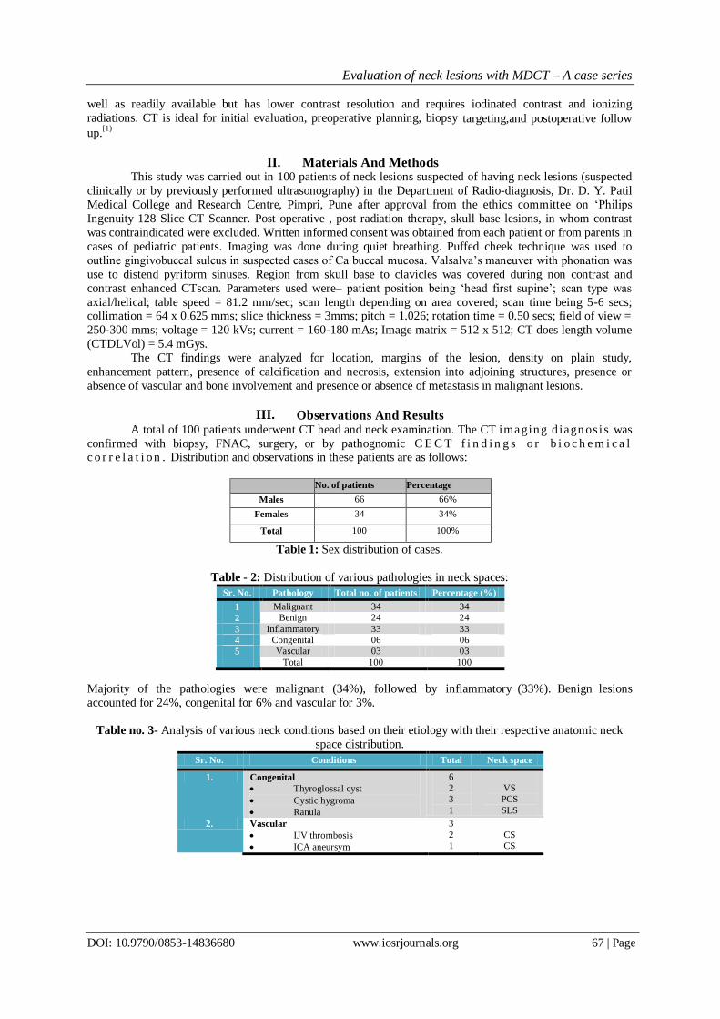
No. of patients Percentage
Males 66 66%
Females 34 34%
Total 100 100%
Table 1: Sex distribution of cases.
Table - 2: Distribution of various pathologies in neck spaces:
Sr. No. Pathology Total no. of patients Percentage (%)
1 Malignant 34 34
2 Benign 24 24
3 Inflammatory 33 33
4 Congenital 06 06
5 Vascular 03 03
Total 100 100
Majority of the pathologies were malignant (34%), followed by inflammatory (33%). Benign lesions
accounted for 24%, congenital for 6% and vascular for 3%.
Table no. 3- Analysis of various neck conditions based on their etiology with their respective anatomic neck
space distribution.
Sr. No. Conditions Total Neck space
1. Congenital
Thyroglossal cyst
Cystic hygroma
Ranula
6
2
3
1
VS
PCS
SLS
2. Vascular
IJV thrombosis
ICA aneursym
3
2
1
CS
CS
Evaluation of neck lesions with MDCT – A case series
DOI: 10.9790/0853-14836680 www.iosrjournals.org 68 | Page
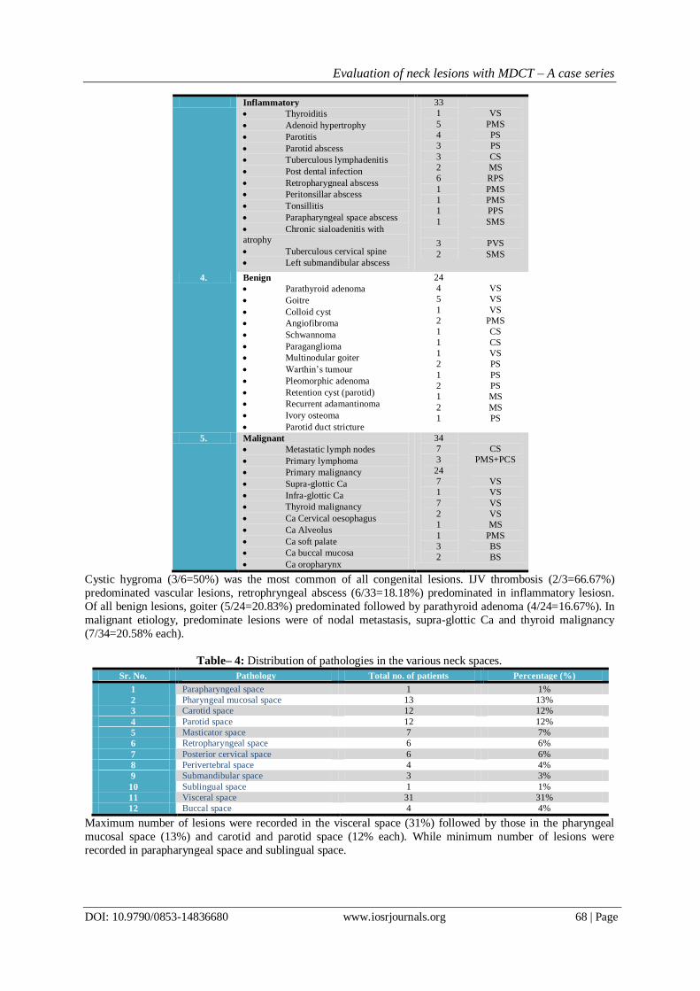
Inflammatory
Thyroiditis
Adenoid hypertrophy
Parotitis
Parotid abscess
Tuberculous lymphadenitis
Post dental infection
Retropharygneal abscess
Peritonsillar abscess
Tonsillitis
Parapharyngeal space abscess
Chronic sialoadenitis with
atrophy
Tuberculous cervical spine
Left submandibular abscess
33
1
5
4
3
3
2
6
1
1
1
1
3
2
VS
PMS
PS
PS
CS
MS
RPS
PMS
PMS
PPS
SMS
PVS
SMS
4. Benign
Parathyroid adenoma
Goitre
Colloid cyst
Angiofibroma
Schwannoma
Paraganglioma
Multinodular goiter
Warthin’s tumour
Pleomorphic adenoma
Retention cyst (parotid)
Recurrent adamantinoma
Ivory osteoma
Parotid duct stricture
24
4
5
1
2
1
1
1
2
1
2
1
2
1
VS
VS
VS
PMS
CS
CS
VS
PS
PS
PS
MS
MS
PS
5. Malignant
Metastatic lymph nodes
Primary lymphoma
Primary malignancy
Supra-glottic Ca
Infra-glottic Ca
Thyroid malignancy
Ca Cervical oesophagus
Ca Alveolus
Ca soft palate
Ca buccal mucosa
Ca oropharynx
34
7
3
24
7
1
7
2
1
1
3
2
CS
PMS+PCS
VS
VS
VS
VS
MS
PMS
BS
BS
Cystic hygroma (3/6=50%) was the most common of all congenital lesions. IJV thrombosis (2/3=66.67%)
predominated vascular lesions, retrophryngeal abscess (6/33=18.18%) predominated in inflammatory lesiosn.
Of all benign lesions, goiter (5/24=20.83%) predominated followed by parathyroid adenoma (4/24=16.67%). In
malignant etiology, predominate lesions were of nodal metastasis, supra-glottic Ca and thyroid malignancy
(7/34=20.58% each).
Table– 4: Distribution of pathologies in the various neck spaces.
Sr. No. Pathology Total no. of patients Percentage (%)
1 Parapharyngeal space 1 1%
2 Pharyngeal mucosal space 13 13%
3 Carotid space 12 12%
4 Parotid space 12 12%
5 Masticator space 7 7%
6 Retropharyngeal space 6 6%
7 Posterior cervical space 6 6%
8 Perivertebral space 4 4%
9 Submandibular space 3 3%
10 Sublingual space 1 1%
11 Visceral space 31 31%
12 Buccal space 4 4%
Maximum number of lesions were recorded in the visceral space (31%) followed by those in the pharyngeal
mucosal space (13%) and carotid and parotid space (12% each). While minimum number of lesions were
recorded in parapharyngeal space and sublingual space.
Evaluation of neck lesions with MDCT – A case series
DOI: 10.9790/0853-14836680 www.iosrjournals.org 69 | Page
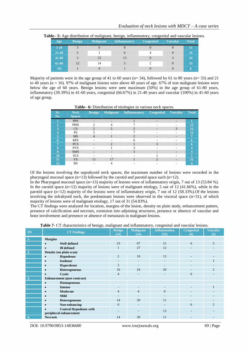
Table– 5: Age distribution of malignant, benign, inflammatory, congenital and vascular lesions.
Age Benign Malignant Inflammatory Congenital Vascular Total
1-20 3 0 8 0 0 11
21-40 5 1 6 4 0 16
41-60 3 15 13 0 3 34
61-80 12 14 5 2 0 33
>81 1 4 1 0 0 6
Tot 24 34 33 6 3 100
Majority of patients were in the age group of 41 to 60 years (n= 34), followed by 61 to 80 years (n= 33) and 21
to 40 years (n = 16). 97% of malignant lesions were above 40 years of age. 67% of non malignant lesions were
below the age of 60 years. Benign lesions were seen maximum (50%) in the age group of 61-80 years,
inflammatory (39.39%) in 41-60 years, congenital (66.67%) in 21-40 years and vascular (100%) in 41-60 years
of age group.
Table– 6: Distribution of etiologies in various neck spaces.
Sr.
No.
Neck
Spaces
Benign Malignant Inflammatory Congenital Vascular Total
1 PPS - - 1 - - 1
2 PMS 2 4 7 - - 13
3 CS 2 5 2 - 3 12
4 PS 5 - 7 - - 12
5 MS 4 1 2 - - 7
6 RPS - - 6 - - 6
7 PCS - 2 1 3 - 6
8 PVS - 1 3 - - 4
9 SMS - - 3 - - 3
10 SLS - - - 1 - 1
11 VS 11 17 1 2 - 31
12 BS - 4 - - - 4
Of the lesions involving the suprahyoid neck spaces, the maximum number of lesions were recorded in the
pharyngeal mucosal space (n=13) followed by the carotid and parotid space each (n=12).
In the Pharyngeal mucosal space (n=13) majority of lesions were of inflammatory origin, 7 out of 13 (53.84 %).
In the carotid space (n=12) majority of lesions were of malignant etiology, 5 out of 12 (41.66%), while in the
parotid space (n=12) majority of the lesions were of inflammatory origin, 7 out of 12 (58.33%).Of the lesions
involving the infrahyoid neck, the predominant lesions were observed in the visceral space (n=31), of which majority of lesions were of malignant etiology, 17 out of 31 (54.83%).
The CT findings were analyzed for location, margins of the lesion, density on plain study, enhancement pattern,
presence of calcification and necrosis, extension into adjoining structures, presence or absence of vascular and
bone involvement and presence or absence of metastasis in malignant lesions.
Table 7- CT characteristics of benign, malignant and inflammatory, congenital and vascular lesions.
SN CT Findings Benign
(24)
Malignant
(34)
Inflammation
(33)
Congenital
(6)
Vascular
(3)
1. Margins
Well defined 23 07 21 6 3
Ill defined 1 27 12 - -
2. Density (on plain scan)
Hypodense 2 10 13 - -
Isodense - - - - 1
Hyperdense 2 - - - -
Heterogeneous 16 24 20 - 2
Cystic 4 - - 6 -
3. Enhancement (post contrast)
Homogeneous
Intense - - - - 1
Moderate 4 4 9 - -
Mild - - - - -
Heterogeneous 14 30 11 - -
Non-enhancing 6 - - 6 2
Central Hypodense with
peripheral enhancement - - 13 - -
4. Necrosis 14 30 11 - -
Evaluation of neck lesions with MDCT – A case series
DOI: 10.9790/0853-14836680 www.iosrjournals.org 70 | Page
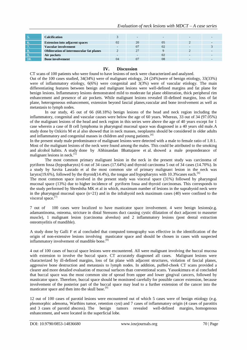
IV. Discussion CT scans of 100 patients who were found to have lesions of neck were characterized and analyzed.
Out of the 100 cases studied, 34(34%) were of malignant etiology, 24 (24%)were of benign etiology, 33(33%)
were of inflammatory etiology, 6(6%) were congenital and 3(3%) were of vascular etiology. The main
differentiating features between benign and malignant lesions were well-defined margins and fat plane for
benign lesions. Inflammatory lesions demonstrated mild to moderate fat plane obliteration, thick peripheral rim
enhancement and presence of air pockets. While malignant lesions revealed ill-defined margins, loss of fat
plane, heterogeneous enhancement, extension beyond fascial planes,vascular and bone involvement as well as
metastasis to lymph nodes.
In our study, 45 out of 66 (68.18%) benign lesions of the head and neck region including the
inflammatory, congenital and vascular causes were below the age of 60 years. Whereas, 33 out of 34 (97.05%) of the malignant lesions of the head and neck region in this series were above the age of 40 years except for 1
case wherein a case of B cell lymphoma in pharyngeal mucosal space was diagnosed in a 40 years old male.A
study done by Ozkiris M et al also showed that in neck masses, neoplasms should be considered in older adults
and inflammatory and congenital masses in children and young patients.[4]
In the present study male predominance of malignant lesions were detected with a male to female ratio of 1.8:1.
Most of the malignant lesions of the neck were found among the males. This could be attributed to the smoking
and alcohol habits. A study done by Abhinandan Bhattajaree et al. showed a male preponderance of
malignant lesions in neck.[5]
The most common primary malignant lesion in the neck in the present study was carcinoma of
pyriform fossa (hypopharynx) 6 out of 34 cases (17.64%) and thyroid carcinoma 5 out of 34 cases (14.70%). In
a study by Savita Lasrado et al the most common site of primary malignant lesion in the neck was
larynx(19.6%), followed by the thyroid(14.4%), the tongue and hypopharynx with 10.3%cases each.[6] The most common space involved in the present study was visceral space (31%) followed by pharyngeal
mucosal space (13%) due to higher incidence of pyriform fossa and thyroid carcinomas. This corresponds to
the study performed by Shreshtha MK et al in which, maximum number of lesions in the suprahyoid neck were
in the pharyngeal mucosal space (n=21) and in the infrahyoid neck maximum cases (40) were confined to the
visceral space.[7]
7 out of 100 cases were localized to have masticator space involvement. 4 were benign lesions(e.g.
adamantinoma, osteoma, stricture in distal Stensons duct causing cystic dilatation of duct adjacent to masseter
muscle), 1 malignant lesion (carcinoma alveolus) and 2 inflammatory lesions (post dental extraction
osteomyelitis of mandible).
A study done by Galli F et al concluded that computed tomography was effective in the identification of the
origin of non-extensive lesions involving masticator space and should be chosen in cases with suspected
inflammatory involvement of mandible bone.[8]
4 out of 100 cases of buccal space lesions were encountered. All were malignant involving the buccal mucosa
with extension to involve the buccal space. CT accurately diagnosed all cases. Malignant lesions were
characterized by ill-defined margins, loss of fat plane with adjacent structures, violation of fascial planes,
aggressive bone destruction and metastasis to lymph nodes. In addition, puffed-cheek CT scans provided a
clearer and more detailed evaluation of mucosal surfaces than conventional scans. Yasuokimura et al concluded
that buccal space was the most common site of spread from upper and lower gingival cancers, followed by
masticator space. Therefore, buccal space should be monitored carefully for possible cancer extension, because
involvement of the posterior part of the buccal space may lead to a further extension of the cancer into the masticator space and then into the skull base.[9]
12 out of 100 cases of parotid lesions were encountered out of which 5 cases were of benign etiology (e.g.
pleomorphic adenoma, Warthins tumor, retention cyst) and 7 cases of inflammatory origin (4 cases of parotitis
and 3 cases of parotid abscess). The benign tumors revealed well-defined margins, homogenous
enhancement, and were located in the superficial lobe.
5. Calcification 3 - - - -
6. Extension into adjacent spaces 02 20 05 2 -
7. Vascular involvement - 07 02 - 3
8. Obliteration of intermuscular fat planes 2 27 9 2 -
9. Air pockets - - 02 - -
10. Bone involvement 04 07 08 - -
Evaluation of neck lesions with MDCT – A case series
DOI: 10.9790/0853-14836680 www.iosrjournals.org 71 | Page
In the 4 cases of parotitis, CT could identify an obstructing calculus in the distal duct in 3 cases, and enlarged
homogenously enhancing parotid gland. Parotid abscesses revealed thick peripheral enhancement with central necrosis/ liquefaction.
Shin K. H conducted a study of parotid gland tumors and used criteria of location, size, density, margin,
calcification within tumors, necrosis, cystic change, invasion of extra glandular structure and
lymphadenopathy. Irregularities in tumor margin and findings of extra glandular extension are the most helpful
indicators by which benign and malignant parotid tumors may be differentiated.[10]
13 out of 100 cases of pharyngeal mucosal space lesions were diagnosed (e.g. adenoidal hypertrophy (n=5),
oropharyngeal carcinoma (n=3), juvenile nasopharyngeal angiofibroma (n=2) and a case each of peritonsillar
abscess, tonsillitis and lymphoma.
Nasopharyngeal angiofibroma characteristically involved the pterygopalatine fissure, nasopharynx with
secondary invasion of the maxillary and ethmoid sinuses. Both cases showed intracranial extension and
characteristic intense vascular enhancement on post contrast administration. This is comparable with study
conducted by Bhaskar Ghosh et al.[11]
6 out of 100 cases of retropharyngeal abscesses were diagnosed. 2 cases were associated with erosion of the
vertebral body and 4 cases showed extension into surrounding visceral an carotid spaces. This is consistent
with the findings of Federici S et al who concluded that the accuracy of CT was 71.4% in correctly identifying
an abscess, and that CT scan is indicated to assess the extent of infection and exclude complications.[12]
6 out of 100 lesions were detected in the posterior cervical space. Three cases each of lymph nodes and cystic
hygroma were detected. 2 Out of the 3 cases of lymphadenopathy were due to lymphoma. CT showed multiple,
bilateral, non-necrotic and homogenously enhancing lymph node involvement. Tuberculous lymphadenitis was
diagnosed by identifying peripheral enhancement and central non enhancing necrosis associated with cold abscess tracking along the fascial plane on CT, which was later confirmed on culture examinations of the tissue
sample.
This is comparable with a study by Geoffrey D parker et al, they showed that most common lesion in the PCS
involved the lymph nodes, i.e metastatic lymph nodes followed by lymphomatous lymph nodes.[13]
31/100 cases were encountered in the visceral space. The pathologies found were hypopharyngeal carcinomas
(n=8), carcinoma of thyroid gland (n=7), goiter (n=6), parathyroid adenomas (n=4), thyroglossal cyst (n=2), and
each case of carcinoma epiglottis, thyroiditis, colloid cyst and carcinoma of esophagus.
The parathyroid adenomas were identified by their characteristic arterial hyper- enhancement and location. This
is consistent with a study done by Geneva J Randall et al. [14]
6 cases were of congenital origin - thyroglossal cyst in the visceral space was the most common
congenital lesion (n=2) This is comparable with the study performed by Al-Khateeb TH et al who concluded
that the most frequent congenital neck mass was thyroglossal duct cyst, followed by cysts of the branchial
apparatus.[15] 12 out of 100 cases of carotid space lesions were diagnosed. Out of these seven were lymph-node
masses (e.g. tuberculosis n=2 and metastatic lymph nodes n=5), vascular lesions n=3 (e.g. two I.J.V thrombus
and one ICA aneurysm) and a case each of vagal schwannoma and paraganglioma were encountered in the
series. The metastatic lymph nodes revealed central necrosis and ill-defined margins with IJV invasion
suggestive of extra-capsular spread. The schwannoma was well circumscribed, homogenous enhancement with
focal hypodensities within and lateral to carotid sheath.
3 out of 100 cases were encountered in the submandibular space (one case of acute on chronic sialadenitits
and two cases of submandibular abscess). CT revealed multiple submandibular gland calculi with atrophy of the gland and a small collection posterior to the large left submandibular calculus indicative of an acute on chronic
sialadenitis. The submandibular abscess had low attenuation necrotic, pus filled center and thick irregular
enhancing rim with adjacent fat stranding.
4 out of 100 cases were detected in the perivertebral space - 3 cases of Koch’s spine and 1 case of
vertebral metastasis. CT could identify vertebral body destruction with evidence of prevertebral and
paravertebral soft tissue in cases of koch’s spine and revealed posterior spinal element involvement in
metatstasis from carcinoma prostate.
Evaluation of neck lesions with MDCT – A case series
DOI: 10.9790/0853-14836680 www.iosrjournals.org 72 | Page
A total of 33 deep neck space infections (7 each in pharyngeal mucosal space and parotid space, 6
retropharyngeal, 3 each in perivertebral space and submandibular space, 2 each in the carotid and masticator
space, 1 each in posterior cervical, visceral and parapharyngeal space) were encountered, which were accurately diagnosed by CT. The most frequently infected neck space was PMS. This correlated with study by
Khaled et al (2010) in which most frequently infected neck space was the peritonsillar space (pharyngeal
mucosal space), followed by the parapharyngeal space.[16]
17 malignant lymph nodes were encountered including 3 primary and 14 metastatic. Based on size criteria
and central necrotic area CT correctly differentiated benign and malignant lymph nodes that correlate with
study done by R A Zoulman et al. L ymph node central necrosis is a useful indicator of metastatic lymph
node extracapsular spread, with a sensitivity of 95 per cent, a specificity of 85 per cent. Lymph node diameter
is not a sensitive indicator of extracapsular spread.[17] The benign lesions like nasopharyngeal angiofibroma,
adamantinoma, osteoma caused bony expansion and remodeling rather than bony destruction and erosion.
Whereas, malignant lesions like buccal carcinoma, vertebral metastasis, carcinoma mandible caused bony
destruction and erosion. Inflammatory lesions like Koch’s spine and retropharyngeal abscess and osteomyelitis
of the jaw caused bone erosions and destruction. Extension into the adjacent space was seen in 20 out of 34 (58.82%) malignant lesions and in 2 benign lesions (i.e., two cases of nasopharyngeal angiofibroma).
Lymph nodes[18]
Extra-capsular spread of tumor is manifested by capsular enhancement, ill-defined nodal margins,
obliterated fat planes surrounding the nodes and edema or thickening in adjacent soft tissues. Shape of lymph
node is not reliable in differentiating normal from pathological nodes. Ratio of maximal longitudinal to
maximal axial diameter of enlarged nodes calculated by spiral CT may be used to differentiate both malignant
and reactive nodes. Nodes which are spherical with ratio < 2 are likely to be malignant and nodes with ratio
>/=2 are likely to be benign or hyperplastic lymph node. Clusters are defined as multiple (>/=3) contiguous ill-
defined nodes with in the same level ranging from 8mm-15mm in size. Clusters are seen in inflammation,
cancer and lymphoma.Criteria for lymph node enlargement are- retropharyngeal more than 8 mms in maximum
diameter,1.5cms in maximum diameter near angle of mandible,more than 1 cm in maximum diameter elsewhere in neck, ratio of maximal longitudinal nodal length to maximal axial nodal length:, >2 = hyperplastic lymph
node and <2 = s/o metastatic lymph node
CASES
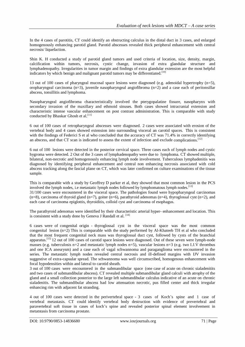
PARAPHARYNGEAL SPACE
Case 1: Left Parapharyngeal Space Abscess
Fig A. Axial CECT image showing ill-defined hypodense collection in left tonsillar region (arrow), extending
laterally to involve the left PPS, suggestive of lateral spread of a peritonsillar abscess.Fig B. Coronal
reconstructed CECT image showing ill-defined hypodense collection in left PPS (arrow), causing obliteration of
its fat. Compare with normal PPS on the right (arrowhead).
MASTICATOR SPACE
Case 2: Adamantinoma In Left Masticator Space
Fig A. Axial CECT image showing an expansile
lytic lesion with peripheral rim of thinned out
bone in ramus of left mandible (arrow) in the MS, displacing PPS posteriorly (arrowhead).
Normal left PPS (curved arrow).Fig B. Volume
rendered reconstruction (VRT) image and fig
(C) Bone window CT image, demonstrating the
lesion in the mandible.
Evaluation of neck lesions with MDCT – A case series
DOI: 10.9790/0853-14836680 www.iosrjournals.org 73 | Page
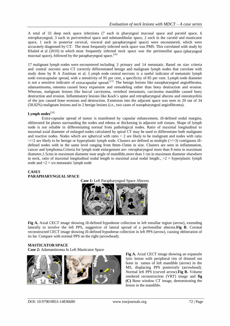
Case 3: Inflammation In Right Masticator Space.
Fig A. Axial CECT image showing ill defined destructive lesion at the site of dental extraction in the right
mandible (arrow) with bulky masseter muscle (arrowhead) and mild adjacent fat stranding. Green arrow-
opposite masseter muscle. Fig B. Coronal reconstructed CT image in bone window setting showing destructive
lesion in the mandible.
Case 4: Ivory osteoma In Masticator space.
Fig A. Axial CT image showing large exophytic, osteogenic mass arising from neck of right mandible in the
masticator space (arrowhead).Fig B. Volume rendered reconstruction image showing the osteogenic nature of
lesion and relation to mandibular neck (arrow).
PAROTID SPACE
Case 5: Right Parotid Gland Abscess.
Fig A. Axial CECT image showing well-defined hypodense lesion in superficial lobe of right parotid gland with
peripheral enhancement (arrow).Fig B. Coronal reconstructed image showing peripherally enhancing right
parotid abscess (arrow).
Case 6: Warthins Tumor in Right Parotid Gland.
Fig A. Axial CECT image showing well defined homogenously enhancing mass lesion (arrowhead) in the
superficial as well as deep lobe of right parotid gland. Posterior belly of digastrics muscle is seen

Evaluation of neck lesions with MDCT – A case series
DOI: 10.9790/0853-14836680 www.iosrjournals.org 74 | Page
separating parotid space from carotid space (arrow). Fig B. Axial plain CT image showing biopsy needle in-situ
in right parotid gland.It was diagnosed as Warthin’s tumor on histopathological examination.
Case 7: Pleomorphic adenoma
Fig A. Axial CECT image in venous phase showing well defined lobulated enhancing lesion with foci of
hypodensities in superficial lobe of right parotid (arrowhead).Fig B. Axial CECT in delayed phase showing
central hypodensity with peripheral enhancement.
Case 8: Carotid body tumor.
Fig A. Axial CECT image in arterial phase showing intensely enhancing mass lesion (arrowhead) in the carotid
space, splaying the ICA and ECA, with anterior displacement of ipsilateral right PPS suggestive of carotid body
tumor. Arrow indicates normal Left PPS.
Case 9: Metastatic Lymphadenopathy in Carotid Space.
Fig A. Axial CECT image showing large ill defined necrotic lymphadenopathy in the left carotid space (arrow).Fig B. Axial CECT image showing mass involving left pyriform sinus and left aryepiglottic fold with
metastatic lymphadenopathy in left of carotid space (arrow).
Fig C. Axial NECT image in bone window setting showing erosion of thyroid cartilage by the lesion
(arrowhead).

Evaluation of neck lesions with MDCT – A case series
DOI: 10.9790/0853-14836680 www.iosrjournals.org 75 | Page
Case 10: Retropharyngeal abscess
Fig A, B – Sagittal and axial CECT images showing ill-defined hypodense collection in the RPS containing
specks of air.
Case 11: Adenoidal hypertrophy
Fig A. Axial CT image showing enlarged adenoids indenting the airway with lateral displacement of the PPS
(green arrow).Fig B. Sagittal CT image showing enlarged adenoids measuring 20mm (Normal = 12mm),
indenting the nasopharyngeal airway.
Case 12: Tonsillar abscess.
Fig A: Axial CECT image showing ill defined peripherally enhancing abscess in the right tonsillar and
peritonsillar region (arrowhead) mildly indenting the oropharynx.Fig B: Coronal CECT image showing
abscess extending along lateral pharyngeal wall till the level of vallecula.Fig C: Axial CT image showing
abscess (arrow) displacing the PPS laterally (arrowhead), Normal left PPS (curved arrow).
Case 13: Non Hodgkins Lymphoma
Fig A. Axial CECT image showing homogenously enhancing soft tissue along the tonsillar pillars causing
narrowing of the oropharynx (arrow).Fig B & C. Similar enhancing soft tissue is noted in the posterior wall and roof of the nasopharynx (arrow) obliterating the fossa of Rosenmuller and narrowing the nasopharyngeal
airway.
Evaluation of neck lesions with MDCT – A case series
DOI: 10.9790/0853-14836680 www.iosrjournals.org 76 | Page
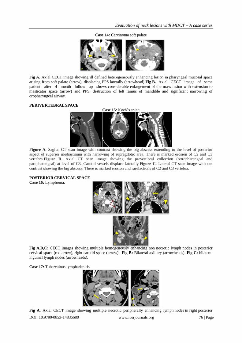
Case 14: Carcinoma soft palate
Fig A. Axial CECT image showing ill defined heterogeneously enhancing lesion in pharyngeal mucosal space
arising from soft palate (arrow), displacing PPS laterally (arrowhead).Fig B. Axial CECT image of same
patient after 4 month follow up shows considerable enlargement of the mass lesion with extension to
masticator space (arrow) and PPS, destruction of left ramus of mandible and significant narrowing of
oropharyngeal airway.
PERIVERTEBRAL SPACE
Case 15: Koch’s spine
Figure A. Sagital CT scan image with contrast showing the big abscess extending to the level of posterior
aspect of superior mediastinum with narrowing of supragllotic area. There is marked erosion of C2 and C3
vertebra.Figure B. Axial CT scan image showing the prevertibral collection (retropharangeal and
parapharangeal) at level of C3. Carotid vessels displace laterally.Figure C. Lateral CT scan image with out
contrast showing the big abscess. There is marked erosion and rarefactions of C2 and C3 vertebra.
POSTERIOR CERVICAL SPACE
Case 16: Lymphoma.
Fig A,B,C: CECT images showing multiple homogenously enhancing non necrotic lymph nodes in posterior
cervical space (red arrow), right carotid space (arrow). Fig B: Bilateral axillary (arrowheads). Fig C: bilateral
inguinal lymph nodes (arrowheads).
Case 17: Tuberculous lymphadenitis.
Fig A. Axial CECT image showing multiple necrotic peripherally enhancing lymph nodes in right posterior

Evaluation of neck lesions with MDCT – A case series
DOI: 10.9790/0853-14836680 www.iosrjournals.org 77 | Page
cervical space (arrow). Fig B. Axial CECT image showing ill defines peripherally enhancing abscess
(arrowhead) adjacent to necrotic lymph nodes.
VISCERAL SPACE
Case 18: Parathyroid adenoma
Fig A & B. Axial and sagittal reformatted CECT images in arterial phase show an oval well defined
homogenously enhancing lesion (arrowhead) in visceral space posterior to upper pole of right lobe of
thyroid gland (arrow).
Case 19: Thyroglossal Cyst
Fig A & B: Axial CECT and sagittal reformatted images show well-defined cystic lesion in midline in the
visceral space, inferior to hyoid bone (arrows).
Case 20: Carcinoma of Right Pyriform Fossa
Fig A & B: Axial CECT image showing enhancing lesion in right pyriform fossa (transparent arrow) in
visceral space with metastatic lymphadenopathy at level II. Fig C: Coronal reformatted CECT image showing
necrotic metastatic right cervical lymphadenopathy (yellow arrow).
Case 21: Carcinoma of Epiglottis
Fig A. Axial CECT image showing lesion in visceral space arising from epiglottis (arrow).
Fig B. Coronal reconstructed CT image showing epiglottic lesion (green arrow) extending to and
obliterating the air filled vallecula (yellow arrow).
Evaluation of neck lesions with MDCT – A case series
DOI: 10.9790/0853-14836680 www.iosrjournals.org 78 | Page
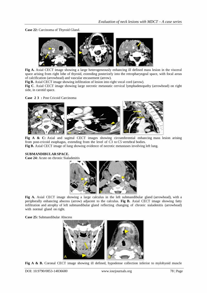
Case 22: Carcinoma of Thyroid Gland.
Fig A. Axial CECT image showing a large heterogeneously enhancing ill defined mass lesion in the visceral
space arising from right lobe of thyroid, extending posteriorly into the retropharyngeal space, with focal areas
of calcification (arrowhead) and vascular encasement (arrow).
Fig B. Axial CECT image showing infiltration of lesion into right vocal cord (arrow).
Fig C. Axial CECT image showing large necrotic metastatic cervical lymphadenopathy (arrowhead) on right
side, in carotid space.
Case 2 3 : Post Cricoid Carcinoma
Fig A & C: Axial and sagittal CECT images showing circumferential enhancing mass lesion arising
from post-cricoid esophagus, extending from the level of C3 to C5 vertebral bodies.
Fig B. Axial CECT image of lung showing evidence of necrotic metastases involving left lung.
SUBMANDIBULAR SPACE.
Case 24: Acute on chronic Sialadenitis
Fig A. Axial CECT image showing a large calculus in the left submandibular gland (arrowhead), with a
peripherally enhancing abscess (arrow) adjacent to the calculus. Fig B. Axial CECT image showing fatty
infiltration and atrophy of left submandibular gland reflecting changing of chronic sialadenitis (arrowhead)
with normal gland on right.
Case 25: Submandibular Abscess
Fig A & B. Coronal CECT image showing ill defined, hypodense collection inferior to mylohyoid muscle

Evaluation of neck lesions with MDCT – A case series
DOI: 10.9790/0853-14836680 www.iosrjournals.org 79 | Page
(arrow) in the submandibular space with peripheral enhancement and surrounding fat stranding (arrowhead)
suggestive of abscess.
SUBLINGUAL SPACE.
Case 26: Plunging Ranula.
Fig A & B: Sagittal and coronal reformatted CT images show cystic lesion in sublingual space with
inferior extension into the submandibular space suggestive of a plunging ranula.
Case 27: Buccal carcinoma with extension into buccal space
Fig A. Axial CECT image showing large ill defined heterogeneously enhancing mass lesion (yellow arrow) in right buccal space, infiltrating the right masseter muscle (transparent arrow).
Fig B. Normal fat filled buccal space on left side (yellow arrow).
Fig C: Coronal reformatted CECT image reveals right-sided level II necrotic metastatic lymphadenopathy.
V. Conclusion MDCT proved to be a very useful non- invasive tool in accurately diagnosing and characterizing
neck lesions with respect to neck space. The faster scan acquisition, reduced artifacts from patient motion,
lower cost, are its advantages. Multiplanar reconstruction improves the localization and extent of neck lesions.
It is useful for diagnosis, management and follow up of benign and malignant conditions of neck.
References [1]. Alberico RA, Husain SH, Sirotkin I. Imaging in head and neck oncology. Surg Oncol Clin N Am. 2004 Jan;13(1):13-35.
[2]. Pushpender G, Satish KB, Gopesh M, Vineeta R. Role of Multislice Spiral CT in the Evaluation of Neck Masses. JIMSA
2013;26(1):51-4.
[3]. Wippold FJ. 2nd, Head and Neck Imaging: The Role of CT and MRI. J Magn Reson Imaging 2007;25(3):453-65.
[4]. Ozkiris M, Kala M. Histopathological examination of patients operated on for a neck mass:4-year follow-up results. Turkish J of
pathol 2011;27(2):1347.
[5]. Abhinandan B , Chakraborty A, Purkaystha P. Prevalence of head and neck cancers in the north east - an institutional study. Indian
Journal of Otolaryngology and Head and Neck Surgery 2006;58(1).
[6]. Lasrado S, Prabhu P, Kakria A, Kanchan T, Pant S, Sathian B. Clinico-pathological Profile of Head and Neck
Cancers in the Western Development Region, Nepal: A 4-Year Snapshot, Asian Pacific Journal of Cancer Prevention,2012; 12.
[7]. Maurea S, Cuocolo A, Reynolds JC, Neumann RD, Salvatore M. Diagnostic imaging in patients with paragangliomas - Computed
tomography, magnetic resonance and MIBG scintigraphy comparison. Q J Nucl Med. 1996;40(4):365-71.
[8]. Kurabayashi T, Ida M, Yoshino N, et al. Computed tomography in the diagnosis of buccal space masses. Dento
maxillofac Radiol. 1997;26(6):347-53.
[9]. Kimura Y, Sumi M, Sumi T, Yoshiko A, Eiichiro A, Takashi N. Deep extension from carcinoma arising from the gingiva: CT and
MR imaging features. AJNR Am J Neuroradiol 2002;23:468–72.
[10]. Zoumalan RA, Kleinberger AJ, Morris LG, Ranade A, Yee H, DeLacure MD. Lymph node central necrosis on computed
tomography as predictor of extracapsular spread in metastatic head and neck squamous cell carcinoma: pilot study. J Laryngol Otol.
2010;124(12):1284–8.
Evaluation of neck lesions with MDCT – A case series
DOI: 10.9790/0853-14836680 www.iosrjournals.org 80 | Page
[11]. Ghosh B, Saha S, Chandra S, Nandi TK, Bera SP. Juvenile nasopharyngeal angiofibroma-three years experience. Indian J
Otolaryngol Head Neck Surg. 2003;55(4):228–33.
[12]. Federici S , Silva C, Maréchal C, Laporte E, Sévely A, Grouteau E, Claudet I. Retro and parapharyngeal infections: standardization
of their management. Arch Pediatr. 2009;16(9):1225-32.
[13]. Geoffrey DP, Harnsberger H. Radiologic Evaluation of the Normal and Diseased Posterior Cervical Space. AJR
1991;157(1):161-5.
[14]. Randall GJ, Zald PB, Cohen JI, Hamilton BE. Contrast-enhanced MDCT characteristics of parathyroid adenomas. AJR
2009;193:538.
[15]. Lucy YC, Fan WJ, Shen JX, Xiao P. Ai Zheng. CT features of parotid tumors: an analysis of 133 cases. Chinese Journal of Cancer
2007;26(11):1263-67.
[16]. Khaled AN, Alsaid L. Deep Neck Spaces radiology and review of deep neck infections at King Abdul Aziz University Hospital.
Egyptian Journal of ear, nose, throat and Allied Science 2010;11:110-27.
[17]. oucher C, Dorion D, Fisch C. Retropharyngeal abscesses: a clinical and radiologic correlation. J Otolaryngol. 1999;28(3):134-7.
[18]. Som PM, Brandwein-Gensler MS. Chapter 40 Anatomy and pathology of the salivary gland. In: Som P, Curtin H, editors. Head and
neck Imaging. 5th edition. Mosby-Elsevier; 2011. p. 2449–609.