evaluation of low level laser therapy in the …
TRANSCRIPT
European Scientific Journal September 2015 edition vol.11, No.27 ISSN: 1857 – 7881 (Print) e - ISSN 1857- 7431
209
EVALUATION OF LOW LEVEL LASER THERAPY IN THE MANAGEMENT OF
CHEMOTHERAPY-INDUCED ORAL MUCOSITIS IN PEDIATRIC AND YOUNG CANCER
PATIENTS: A RANDOMIZED CLINICAL TRIAL
Dr. Khadija M Ahmed
Dr. Shokhan A. Hussein University of Sulaimani, Faculty of Medical Sciences,
School of Dentistry, Oral Diagnosis Department
Dr. Arass J. Noori Dr. Suha N. Abdulateef
University of Sulaimani, Faculty of Medical Sciences, School of Dentistry, Oral and Maxillofacial Department
Dr. Basil K. Abdulla Hiwa Hospital, Clinical Pediateric Heamatolgist and Oncologist,
Sulaimani City, Iraq
Abstract Backgrounds: Oral mucositis is a frequent adverse side effect of cancer chemotherapy which is associated with intense oral pain. However, it impairs the quality of life of these patients. Low Level Laser Therapy (LLLT) has been increasingly used in recent years, mostly to accelerate wound healing and to reduce pain. In cancer patients, LLLT has been shown to reduce the incidence and severity of oral mucositis. Objectives: The aim of this study is to evaluate the effect of low level laser therapy in the management of chemotherapy induced oral mucositis. Patients and Methods: The study design used was a randomized clinical trial. A total of 67 cancer patients were eligible to participate in the study. Thus, they were divided randomly into two groups: group 1 irradiated with prophylactic or active laser therapy (AlGalnAs laser diode device with a wave length of 940±15nm, 0.3mW, and a probe emitting dose of 4.2 J/cm²) and group 2 received inactive or sham laser therapy (power output equal to zero). However, for the ethical purpose, once the patients developed ulcerative mucositis, they are irradiated with active laser therapy. The oral assessment was performed daily starting from the first day of the chemotherapy by

European Scientific Journal September 2015 edition vol.11, No.27 ISSN: 1857 – 7881 (Print) e - ISSN 1857- 7431
210
applying WHO grading system. After 24 hours, the assessment of associated oral pain was carried out every two days with visual analog scale before laser application. Consequently, the associations between variables were analyzed statistically using SPSS version 20. Results: All the patients were presented with some grade of oral mucositis. In the active or prophylactic laser group, the severity of oral mucositis was lower than the inactive or therapeutic laser group. Moreover, the incidence of grade 3 and grade 4 were less observed in the active or prophylactic laser groups than the sham or therapeutic laser groups. In addition, the mean time of healing was significantly lower in the prophylactic laser group than in the therapeutic laser groups (2.05 + 1.89 versus 4.5 + 2.4 days, p> 0,004). Prophylactic laser application was associated with significant reduction of oral pain in comparison with inactive or therapeutic laser therapy (1.18 + 1.09 versus 2.12 + 1.60, p> 0.01). Conclusions: Prophylactic laser therapy is effective in reducing the incidence of sever oral mucositis and in alleviating associated oral pain.
Keywords: Low level laser Therapy, oral mucositis, chemotherapy, cancer patient Introduction Despite considerable improvement in the medical management of cancer patients in recent years, significant complication often accompanies the potential benefits of the treatment. Chemotherapy induced oral mucositis is a frequent oral complications achieved in patients which receives highly mucotoxic drugs (Arora et al., 2008; Sonis, 2009; Ramírez-Amador et al., 2010; Freitas et al., 2014 ) Oral mucositis is associated with intense pain which limits patient’s ability to eat and drink normal foods (Silverman, 2007). Severe and wide spread ulcerated mucositis in these group of medically compromised patients both physiologically (by the tumour) and literally (by cancer therapy) will place them at an increased risk of systemic infection and bleeding (Bensadoun, 2012; Sonis, 2009). Consequently, the quality of life of these patients will be greatlycompromised (Nes and Posso, 2005; Abramoff et al., 2008). The literature review revealed that various pharmacological and non-pharmacological agents have tried to prevent and/or treat oral mucositis. Thus, they have made the subject of mucositis to be one of the most researched field explored in supportive care in cancer by many researchers (Sandoval et al., 2003; Sonis, 2009; Gouvea de Lima et al., 2012). Among the various agents being investigated is the Low Level Laser Therapy (LLLT). Laser therapy is a photomedicine procedure proposed to

European Scientific Journal September 2015 edition vol.11, No.27 ISSN: 1857 – 7881 (Print) e - ISSN 1857- 7431
211
exert beneficial effect in inflammatory conditions like rheumatoid arthritis (Bensadoun and Nair, 2012; Ekim et al., 2007). Even though the precise molecular mechanism to explain their activity are not clearly evident, however, it has been suggested that LLLT will promote wound healing (Volpato et al., 2009). Also, it was observed that LLLT will induce biological changes in the epithelial and connective tissues through the stimulation of rapid epithelization and regeneration of myofibroblast originating from the fibroblast (Chaves et al., 2014 ). Moreover, low power laser have also been reported to exert pain relief potential through the modification of nerve conduction via the release of endorphins and enkephalines (Jaguar et al., 2007). In addition to these effects, low intensity laser therapy have been investigated to prevent and/or treat oral mucositis due to radiotherapy (Arora et al., 2008; Gouvea de Lima et al., 2012; Gautam et al., 2012; Carvalho et al., 2011); chemotherapy (Nes and Posso, 2005; Abramoff et al., 2008; Cruz et al., 2007; Kuhn et al.,2009 ); and in Hematopoetic Stem Cell Transplantation (HSCT) (jaguar et al.,2006; Silva et al., 2011). Low level laser therapy has not been studied for cancer patients undergoing chemotherapy in our country. Accordingly, we performed a double blind randomized study to investigate the clinical effect of low level laser therapy in the management of chemotherapy induced mucositis. Patients and Methods The Study Design and Patient Characterization The study was designed as a double blind randomized clinical trial. The samplings were carried out in Hiwa Hospital in Sulaimani city (Northern east of Iraq) from June 2013 to September 2014. Eligible patients were all consecutive cancer patients receiving chemotherapy with the following inclusion and exclusion criteria. Inclusion Criteria Pediatric and young adult patients utilizing intensive mucotoxic chemotherapy (single highly mucotoxic drug or combination of two or three intensive chemotherapeutic regimen). Patients who stay in hospital for at least 21 days. Patients who agreed to participate in the study. Furthermore, in exclusion criteria, patients were withdrawn from participation according to their request or due to loss in follow up, those who miss three consecutive day treatments, and those with unstable clinical condition. Before conducting the study procedure, the institutional Ethics Committee approved the protocol. However, all the procedures were

European Scientific Journal September 2015 edition vol.11, No.27 ISSN: 1857 – 7881 (Print) e - ISSN 1857- 7431
212
performed in accordance with applicable guidelines of Good Clinical Practice and the Declaration of Helsinki. Written informed consent was obtained from all patients or their parents before they were enrolled into the study procedure. Prophylactic Laser Application Sixty seven patients met the inclusion and exclusion criteria and they were randomly divided into two groups using a block randomization with a manual schedule: group A (active laser) and Group B with sham (inactive laser). In addition, patients were blinded towards the therapeutic protocol of their groups. Laser irradiation were performed with iLase™ (BIOLASE, Inc., Irivine, CA92618 USA, with a wave length of 940±15nm, output power 0.3 mW “Pulse Mode”, continuous infrared AlGalnAs diode) Laser. Patients who participated in the active and passive laser groups were irradiated daily for three successive weeks starting from the day of the chemotherapy course. At each treatment sessions, ten anatomic sites (the right and left hand side of the cheeks, lower and upper labial mucosa, ventral and lateral tongue, floor of the mouth and anterior tonsillar pillars) of the oral cavity were illuminated for 30s. All the procedure were performed by a single oral medicine specialist, after adjustment of the iLase™ device for soft tissue pathology (i.e aphthus ulceration mode). Thus, the power output was reduced to 0.3 mW. This initiated an energy power of 4.2 J/cm² when the probe were held at about 7-8mm away from the oral mucosal surfaces In the active or prophylactic group, patients were irradiated with the probe emitting dose of 4.2 J/cm². Also, participants in the sham or inactive laser group were irradiated with the same, but inactive probes. For ethical purpose, once the patients establish a ulcerated mucositis, they are irradiated with therapeutic active laser therapy. During irradiation procedure, the patients and the therapist wear googles to avoid retinal exposure to laser light. Thus, the procedure was carried out in the morning between 9-11pm. Lastly, all the patients underwent hospital stander oral mucositis prophylactic measure in the form of Italian solution (A mixture of vit. B complex, Folic acid, Normal saline, Mycostatin drop, Hydrocortisone, NaHCO3 (Sodium bicarbonate), and Lidocaine ampule). Clinical Evaluation of Oral Mucositis Patients were assessed for response to laser therapy on a daily basis by employing WHO grading system by specialized independent hematologist who was blinded regarding the studied groups. The evaluation for the
European Scientific Journal September 2015 edition vol.11, No.27 ISSN: 1857 – 7881 (Print) e - ISSN 1857- 7431
213
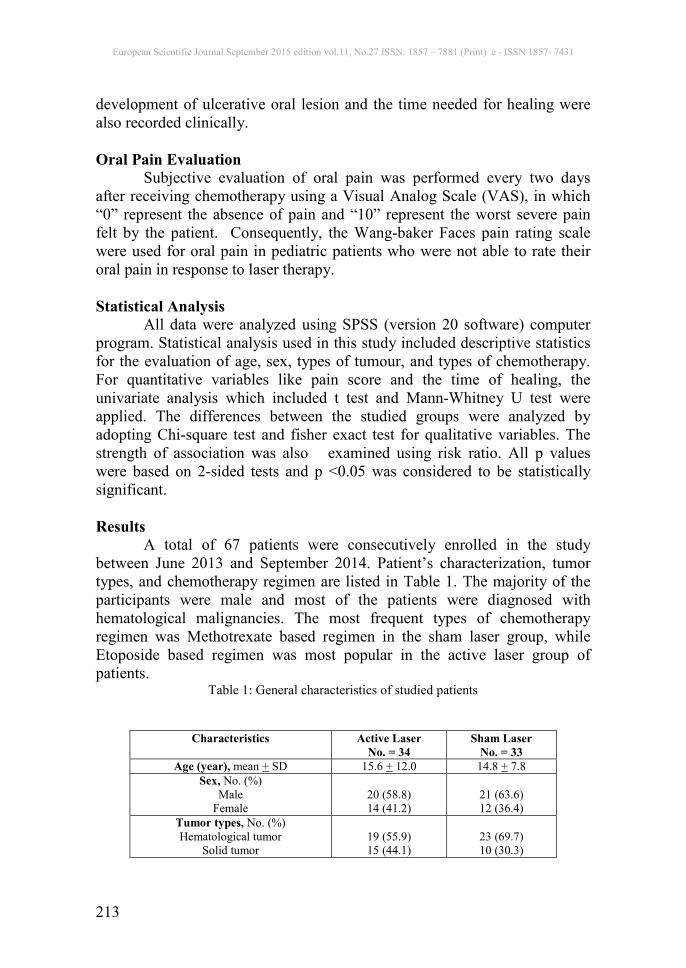
development of ulcerative oral lesion and the time needed for healing were also recorded clinically. Oral Pain Evaluation Subjective evaluation of oral pain was performed every two days after receiving chemotherapy using a Visual Analog Scale (VAS), in which “0” represent the absence of pain and “10” represent the worst severe pain felt by the patient. Consequently, the Wang-baker Faces pain rating scale were used for oral pain in pediatric patients who were not able to rate their oral pain in response to laser therapy. Statistical Analysis All data were analyzed using SPSS (version 20 software) computer program. Statistical analysis used in this study included descriptive statistics for the evaluation of age, sex, types of tumour, and types of chemotherapy. For quantitative variables like pain score and the time of healing, the univariate analysis which included t test and Mann-Whitney U test were applied. The differences between the studied groups were analyzed by adopting Chi-square test and fisher exact test for qualitative variables. The strength of association was also examined using risk ratio. All p values were based on 2-sided tests and p <0.05 was considered to be statistically significant. Results A total of 67 patients were consecutively enrolled in the study between June 2013 and September 2014. Patient’s characterization, tumor types, and chemotherapy regimen are listed in Table 1. The majority of the participants were male and most of the patients were diagnosed with hematological malignancies. The most frequent types of chemotherapy regimen was Methotrexate based regimen in the sham laser group, while Etoposide based regimen was most popular in the active laser group of patients.
Table 1: General characteristics of studied patients
Characteristics Active Laser No. = 34
Sham Laser No. = 33
Age (year), mean + SD 15.6 + 12.0 14.8 + 7.8 Sex, No. (%)
Male Female
20 (58.8) 14 (41.2)
21 (63.6) 12 (36.4)
Tumor types, No. (%) Hematological tumor
Solid tumor
19 (55.9) 15 (44.1)
23 (69.7) 10 (30.3)
European Scientific Journal September 2015 edition vol.11, No.27 ISSN: 1857 – 7881 (Print) e - ISSN 1857- 7431
214
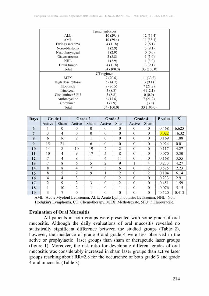
AML: Acute Myeloid Leukaemia, ALL: Acute Lymphoblastic Leukaemia, NHL: Non Hodgkin's Lymphoma, CT: Chemotherapy, MTX: Methotrexate, 5FU: 5 Flurouracile.
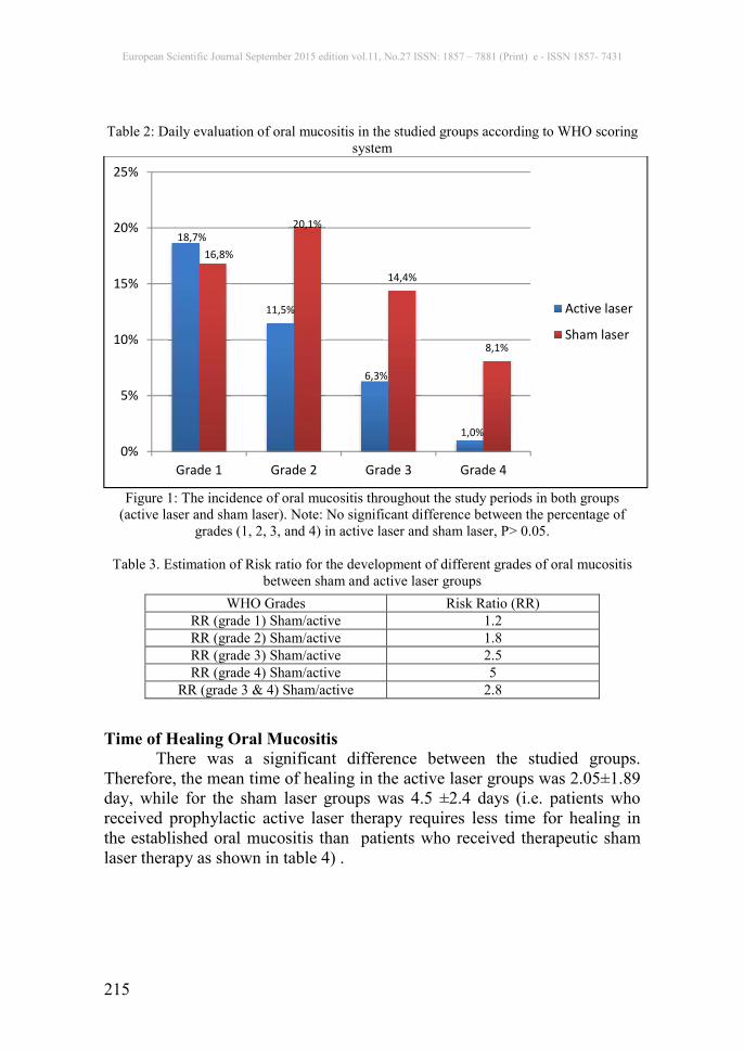
Evaluation of Oral Mucositis All patients in both groups were presented with some grade of oral mucositis. Although the daily evaluations of oral mucositis revealed no statistically significant difference between the studied groups (Table 2), however, the incidence of grade 3 and grade 4 were less observed in the active or prophylactic laser groups than sham or therapeutic laser groups (figure 1). Moreover, the risk ratio for developing different grades of oral mucositis was considerably increased in sham laser groups than active laser groups reaching about RR=2.8 for the occurrence of both grade 3 and grade 4 oral mucositis (Table 3).
Tumor subtypes ALL 10 (29.4) 12 (36.4) AML 10 (29.4) 11 (33.3)
Ewings sarcoma 4 (11.8) 2 (6.1) Neuroblastoma 1 (2.9) 3 (9.1) Nasopharyngeal 1 (2.9) 0 (0.0) Osteosarcoma 3 (8.8) 1 (3.0)
NHL 1 (2.9) 1 (3.0) Brain tumor 4 (11.8) 3 (9.1)
Total 34 (100.0) 33 (100.0)
CT regimen MTX 7 (20.6) 11 (33.3)
High dose cytosar 5 (14.7) 3 (9.1) Etoposide 9 (26.5) 7 (21.2) Irinotecan 3 (8.8) 4 (12.1)
Cisplantine+5 FU 3 (8.8) 0 (0.0) Anthracycline 6 (17.6) 7 (21.2)
Combined 1 (2.9) 1 (3.0) Total 34 (100.0) 33 (100.0)
Days Grade 1 Grade 2 Grade 3 Grade 4 P value X2
Active Sham Active Sham Active Sham Active Sham 6 1 0 0 0 0 0 0 0 0.468 6.625 7 3 4 0 0 0 0 0 0 0.022 16.32 8 6 16 2 1 0 0 0 0 0.169 1.88
9 15 21 4 6 0 0 0 0 0.924 0.01 10 14 8 10 19 2 2 0 0 0.117 4.27 11 10 4 9 17 5 8 0 0 0.070 5.30 12 7 4 8 11 4 11 0 0 0.168 3.55 13 7 8 6 5 2 9 1 4 0.233 4.27 14 8 8 4 7 3 6 0 2 0.525 2.23 15 8 5 2 9 1 2 0 2 0.104 6.14 16 4 4 3 11 0 2 0 0 0.233 2.91 17 2 9 2 3 0 2 0 0 0.451 1.59 18 1 10 2 1 0 1 0 0 0.076 5.15 19 3 7 0 1 0 0 0 0 0.520 0.413
European Scientific Journal September 2015 edition vol.11, No.27 ISSN: 1857 – 7881 (Print) e - ISSN 1857- 7431
215
Table 2: Daily evaluation of oral mucositis in the studied groups according to WHO scoring
system
Figure 1: The incidence of oral mucositis throughout the study periods in both groups
(active laser and sham laser). Note: No significant difference between the percentage of grades (1, 2, 3, and 4) in active laser and sham laser, P> 0.05.
Table 3. Estimation of Risk ratio for the development of different grades of oral mucositis
between sham and active laser groups
Time of Healing Oral Mucositis There was a significant difference between the studied groups. Therefore, the mean time of healing in the active laser groups was 2.05±1.89 day, while for the sham laser groups was 4.5 ±2.4 days (i.e. patients who received prophylactic active laser therapy requires less time for healing in the established oral mucositis than patients who received therapeutic sham laser therapy as shown in table 4) .
18,7%
11,5%
6,3%
1,0%
16,8%
20,1%
14,4%
8,1%
0%
5%
10%
15%
20%
25%
Grade 1 Grade 2 Grade 3 Grade 4
Active laser
Sham laser
WHO Grades Risk Ratio (RR) RR (grade 1) Sham/active 1.2 RR (grade 2) Sham/active 1.8 RR (grade 3) Sham/active 2.5 RR (grade 4) Sham/active 5
RR (grade 3 & 4) Sham/active 2.8

European Scientific Journal September 2015 edition vol.11, No.27 ISSN: 1857 – 7881 (Print) e - ISSN 1857- 7431
216
Table 4. Mean time of healing in patients received active laser and shame laser
Pain Score The proportion of patients presented with oral pain remained insignificant between the studied groups throughout day 6 and 8. Thus, significant differences were observed in the following days (table 5). Furthermore, the mean value for pain score were kept minimal between the studied groups with statistically significant difference (i.e prophylactic and therapeutic application of low level laser therapy were associated with the reduction of subjective feeling of oral pain; details are shown in table 6).
Table 5: Mann-Whitney U for difference in pain score in studied groups Days of treatment
Pain score Median(range)
P value
Laser Sham Day 6 0 (0 – 3) 0 (0 – 3) - Day 8 1.5 (0 – 7) 2 (0 – 7) 0.458 Day 10 2 (0 – 8) 4 (0 – 7) 0.051 Day 12 2.5 (0 – 8) 4 (0 – 9) 0.032 Day 14 0 (0 – 5) 3 (0 – 9) 0.008 Day 16 0 (0 – 4) 2 (0 – 7) 0.001 Day 18 0 (0 – 3) 0 (0 – 5) - Day 20 0 (0 – 3) 0 (0 – 5) -
Discussion Patients undergoing chemotherapy and radiotherapy for the treatment of malignant neoplasm often developed oral mucositis as an adverse side effect of their treatment. Indeed, oral mucositis regarded as a major debilitating and distressing complication often accompanies bone marrow transplantation (Abramoff et al., 2008).
Table 6: Mean pain score value between the group of active laser and sham laser
Oral mucositis can severely impacts the quality of life of these individuals and may necessitate cancer treatment cessation or interruption which consequently may results in reduced control of local tumor followed by an increased morbidity and mortality. Furthermore, cancer treatment discontinuation also increases the cost and duration of the treatment and prolongs the time of hospitalization (Nes and Posso, 2005).
Time of healing (day) Mean + SD
P value Active laser Sham laser 2.05 + 1.89 4.5 + 2.4 0.004
Study group
Average pain score Mean + SD
P value
Active laser 1.18 + 1.09 0.010 Sham laser 2.12 + 1.60

European Scientific Journal September 2015 edition vol.11, No.27 ISSN: 1857 – 7881 (Print) e - ISSN 1857- 7431
217
Various pharmacological and non pharmacological agents have tried in preventing and treating oral mucositis. Despite some positive outcomes, it has not be proven to be completely effective in preventing oral mucositis on its own. Till now, there are no single intervention acts on all phases of oral mucosotis (Bjordal et al., 2011). Low Level Laser Therapy (LLLT) is a local application of a non-chromatic. However, narrow-band coherent light source used for the phtostimulation of biological tissue is recommended as a treatment options for oral mucositis (Abramoff et al., 2008; Bjordal et al., 2011). The concept behind the application of laser therapy in the management of oral mucositis is that low power lasers induce anti-inflammatory action and accelerate wound healing by increasing the vascularity and reepithelization. In addition, Lopes et al. (2009) showed that low power laser appears to decease the severity of oral mucositis, at least in part, by reducing the COX-2 levels which inturn affect the pathophsiology of oral mucositis (Sonis et al., 2004). Moreover, recent publication from the Multinational Association for Supportive Care in Cancer (MASCC) and the International Society of Oral Oncology (ISOO), recommended the administration of LLLT in patients receiving HSCT, conditioned with high dose chemotherapy with or without total body irradiation (Lalla et al., 2014; Oberio et al., 2014). From this point of view, the present study aimed to evaluate the impact of LLLT in the management of chemotherapy induced oral mucositis. For this purpose, we conduct a prospective randomized clinical trial in 67 cancer patients receiving intensive mucotoxic drugs at high risk of developing oral mucositis. Our data showed no statistically significant difference between the studied groups in terms of the incidence of oral mucositis. Subsequently, the active or the prophylactic laser group presented less sever mucositis (grade 3 and grade 4) than the sham or curative laser group. This finding is fairly consistent with the outcomes of other investigators and confirms the advantages of using phototherapy in controlling signs and symptoms of oral mucositis in patients undergoing cancer chemotherapy (Freitas et al., 2014; Abramoff et al., 2008; Arbabi-Kalati et al., 2013) or patients receiving radiotherapy (Carvalho et al., 2011; Arora et al., 2008). Similarly, Cown et al. (1997) and jaguar et al. (2007) also found a reduction in the progression of sever mucositis using laser illuminations in patients who are receiving conditioning regimen for HSCT. In contrast, Gouvea de Lima et al. (2012) revealed that LLLT was not effective in reducing the incidence of sever grades of oral mucositis (grade 3

European Scientific Journal September 2015 edition vol.11, No.27 ISSN: 1857 – 7881 (Print) e - ISSN 1857- 7431
218
or 4) in head and neck cancer patients undergoing concurrent chemoradiotherapy. In this present work and for ethical reasons, once the patients presented with ulcerated mucositis, they underwent curative laser therapy. Although the reason is not entirely understood, curative laser application seems to be less successful than prophylactic laser administration. This might explain the high risk ratio for developing sever ulcerative mucositis (grade 3 and grade 4) in the curative (sham) laser group than prophylactic (active) laser group in the present work. In addition, other researchers also confirmed the superiority of prophylactic laser application than therapeutic methods (Carvalho et al., 2011; Arora et al., 2008). Furthermore, in their meta analysis, Oberio et al. (2014) demonstrated that local application of laser therapy will reduce the overall risk of sever mucositis and other measures of mucositis severity including the duration of sever mucositis and related oral pain. Contrary to our results, Cruz et al. (2007) did not find any evidence on the benefit of the preventive use of low level laser therapy in children and adolescents with cancer that are treated with chemotherapy. However, one plausible cause for the conflicting results may be due to the involvement of children and adults with cancer in the current study, and the patients were evaluated on a daily basis. Cruz et al. (2007) carried out their evaluation weekly on day 1, 8, and 15 after chemotherapy. So, overestimation may be noticed by the authors or the authors may also point to the possibility of a protective effect of the rigorous oral hygiene carried out in all the participants previously and during the study preparation. The time of healing and the duration of sever mucositis was significantly lower in the active (prophylactic) laser group than sham(curative) laser group, which showed agreement with the previous reports declared by Oberio et al., 2014; Volpato et al., 2009; and Bensadoun and Nair, 2012. From the patient’s perspective, subjective feeling of pain is the most distressing side effect of oral mucositis. This is because it interferes with the ability to eat, drink, swallow, and speak. Also, it results in an increase in the amount of analgesic administration and parenteral nutrition. Our observation demonstrates a statistically significant reduction in the time of oral pain in the active laser group in comparison to the curative or sham laser group. Interestingly, the pain score in the sham laser group was also reduced and did not exceed 2.12 + 1.60. Moreover, the mean pain score was reduced in the active laser group than in the sham laser group. The inference of the study results clearly indicate that the reduction of oral pain experience was the most remarkable effect of LLL therapy reported by our patients. Therefore, this fact was accepted by many

European Scientific Journal September 2015 edition vol.11, No.27 ISSN: 1857 – 7881 (Print) e - ISSN 1857- 7431
219
investigators in the literature (Bensadoun et al., 1999; Cown et al., 1997; Nes and Posso, 2005; Jaguar et al., 2007; Schubert et al., 2007; Abramoff et al., 2008; Gautam et al., 2012). However, Gouvea de Lima et al. (2012) and Wong and Wilder- Smith (2002) presented different data. The former showed that the oral pain between placebo and the treated group with laser device does not differ considerably in head and neck cancer patients receiving chemoradiotherapy. Furthermore, the latter reported lack of any statistically significant difference in pain scores at baseline and weekly, thereafter (except in 1 patient) in patients receiving chemotherapy. However, it is often difficult to compare their study results with the present study, taking into consideration the difference between radiation induced and chemotherapy induced mucositis. In addition, the diverse parameters and different laser device applied in their study may address the discrepancy of their results with ours. In conclusion, our findings relates well to the emerging LLLT evidence in preventing oral mucositis and reducing the severity and potential pain related to this conditions. It is also interesting to note that the variety of cancer chemotherapies administered by our patients did not seem to seriously interfere with the beneficial effects of LLLT. Furthermore, the device was well tolerated and no serious incidents or withdrawal due to treatment intolerance were reported by our patients. Consequently, low power laser should be target for future trials to compare treatment start at different time points before cancer therapy to avoid unnecessary LLLT exposure. Acknowledgements The authors would like to thank the doctors and staffs of Hiwa Hospital in Sulaimani city, especially Dr. Basil K Abdulla and Dr. Banaz Mubarak for their kind cooperation in the selection of the patients. The authors would also like to thank Assistant Professor Dr. Laith Alrudainy for his help in the statistical analysis. Great thanks also go to Dr. Tamara Al-Karadaghi for her help regarding the laser device adjustment. References: Abramoff MM, Lopes NN, Lopes LA, Dib LL, Guilherme A, Caran EM, Barreto AD, Lee ML,Petrilli AS (2008). Low-Level Laser Therapy in the Prevention and Treatment of Chemotherapy-Induced Oral Mucositis in Young Patients. Photomed Laser Surg 26(4): 393-400

European Scientific Journal September 2015 edition vol.11, No.27 ISSN: 1857 – 7881 (Print) e - ISSN 1857- 7431
220
Arbabi-Kalati F, Arbabi-Kalati F, Moridi T (2013). Evaluation of the effect of low level laser on prevention of chemotherapy-induced mucositis. Acta Medica Iranica 51: 157–162. Arora H, Pai KM, Maiya A, Vidyasagar MS, Rajeev A (2008). Efficacy of He-Ne Laser in the prevention and treatment of radiotherapy-induced oral mucositis in oral cancer patients. Oral Surg Oral Med Oral Pathol Oral Radiol Endod 105: 180-6. Bensadoun RJ, Franquin JC, Ciais G, Darcourt V, Schubert MM, Viot M, Dejou J, Tardieu C, Benezery K, et al. (1999). Low energy He/Ne laser in the prevention of radiation-induced mucositis. A multicenter phase III randomized study in patients with head and neck cancer. Support Care Cancer 7:244–252. Bensadoun RJ (2012). low level laser therapy in the prevention and management of oral mucositis induced by cancer treatments: evidence-based data from randomized studies and meta-analyses. Laser in med I (1) :1-8. Bensadoun, RJ and Nair RG (2012). Low-level laser therapy in the prevention and treatment of cancer therapy induced mucositis: 2012 state of the art based on literature review and meta-analysis. Curr Opin Oncol 24:363–370. Bjordal JM, Bensadoun RJ, Tuner J, Frigo L, Gjerde K, Lopes-Martins RA (2011). A systematic review with meta-analysis of the effect of low-level laser therapy (LLLT) in cancer therapy-induced oral mucositis. Support Care Cancer 19: 1069–1077. Carvalho PA, Jaguar GC, Pellizzon AC, Prado JD, Lopes RN and Alves FA (2011). Evaluation of low-level laser therapy in the prevention and treatment of radiation induced mucositis: a double-blind randomized study in head and neck cancer patients. Oral Oncol 47:1176–1181. Chaves MEA, Araújo AR, Piancastelli ACC, Pinotti M (2014).Effects of low-power light therapy on wound healing: LASER x LED. An Bras Dermatol 89(4):616-23. Cowen D, Tardieu C, Schubert M, Peterson D, Resbeut M, Faucher C, Franquin JC (1997). Low energy helium-neon laser in the prevention of oral mucositis in patients undergoing bone marrow transplant: results of a double blind randomized trial. Int J Radiat Oncol Biol Phys 38:697–793. Cruz LB, Ribeiro AS, Rech A, Rosa LGN, Castro Jr CG, Brunetto AL (2007). Influence of Low-Energy Laser in the Prevention of Oral Mucositis in Children With Cancer Receiving Chemotherapy. Pediatr Blood Cancer 48: 435-440. Ekim A, Armagana O, Tascioglua F, Onera C, Colakb M (2007). Effect of low level laser therapy in rheumatoid arthritis patients with carpal tunnel syndrome, aplacebo controlled study.SWISS MED WKLY 137: 347–352.
European Scientific Journal September 2015 edition vol.11, No.27 ISSN: 1857 – 7881 (Print) e - ISSN 1857- 7431
221
Freitas AC, Campos L, Branda˜o TB, Cristo´faro M, Eduardo FD, Luiz AC, Marques MM, Eduardo CD, and Simo˜es A (2014). Chemotherapy-Induced Oral Mucositis: Effect of LED and Laser Phototherapy Treatment Protocols. Photomed Laser Surg 32( 2):81-87. Gautam AP, Fernandes DJ, Vidyasagar MS, Maiya AG, Vadhiraja BM (2012). Low level laser therapy for concurrent chemoradiotherapy induced oral mucositis in head and neck cancer patients - a triple blinded randomized controlled trial. Radiother Oncol 104: 349–354. Gautam AP, Fernandes DJ, Vidyasagar MS, Maiya GA (2012). Low level helium neon laser therapy for chemoradiotherapy induced oral mucositis in oral cancer patients - a randomized controlled trial. Oral Oncol 48: 893–897. Gouvea de Lima A, Villar RC, de Castro G Jr, Antequera R, Gil E,Rosalmeida MC, Federico MH, Snitcovsky IM (2012). Oral mucositis prevention by low-level laser therapy in head-and-neck cancer patients undergoing concurrent chemoradiotherapy: a phase III randomized study. Int J Radiat Oncol Biol Phys 82: 270–275. Jaguar GC, Prado JD, Nishimoto IN, Pinheiro MC, de Castro DO Jr, da Cruz Perez DE, Alves FA (2007). Low-energy laser therapy for prevention of oral mucositis in hematopoietic stem cell transplantation. Oral Diseases 13: 538-43. Kuhn A, Porto FA, Miraglia P, Brunetto AL (2009). Low-level infrared laser therapy in chemotherapy-induced oral mucositis: a randomized placebo-controlled trial in children. J Pediatr Hematol Oncol 31(1): 33-7. Lalla RV, Bowen J, Barasch A, Elting L, Epstein J, Keefe DM, McGuire DB, Migliorati C, Nicolatou-Galitis O et al. (2014). MASCC/ISOO clinical practice guidelines for the management of mucositis secondary to cancertherapy. Cancer DOI: 10.1002/cncr.28592, Lopes NN, Plapler H, Chavantes MC, Lalla RV, Yoshimura EM and Alves MT (2009). Cyclooxygenase-2 and vascular endothelial growth factor expression in 5-fluorouracilinduced oral mucositis in hamsters: evaluation of two low intensity laser protocols. Support Care Cancer 17:1409–1415. Nes AG and Posso MBS (2005). Patients with moderate chemotherapy-induced mucositis: pain therapy using low intensity lasers. Int Nurs Rev 52:68–72. Oberoi S, Zamperlini–Netto G, Beyene J, Treister NS, Sung L (2014). Effect of Prophylactic Low Level Laser Therapy on Oral Mucositis: A Systematic Review and Meta-Analysis. PLoS ONE 9(9): e107418. doi:10.1371/journal.pone.0107418 Ramírez-Amador V , Anaya-Saavedra G , Crespo-Solís E, Camacho EI , González-Ramírez I, Ponce-de-León S (2010). Prospective evaluation of oral mucositis in acute leukemia patients receiving chemotherapy. Support Care Cancer 18:639–646.

European Scientific Journal September 2015 edition vol.11, No.27 ISSN: 1857 – 7881 (Print) e - ISSN 1857- 7431
222
Sandoval RL, Koga DH, Buloto LS, Suzuki R, Dib L L (2003). Management of chemo and radiotherapy induced oral mucositis with low-energy laser: initial results of A.C. Camargo Hospital. J Appl Oral Sci 11:337–341. Schubert MM, Eduardo FP, Guthrie KA, Franquin J-C, Bensadoun R-JJ, Migliorat CA, Lloid CM, Eduardo CP, Walter NF et al. (2007). A phase III randomized double-blind placebo-controlled clinical trial to determine the efficacy of low level laser therapy for the prevention of oral mucositis in patients undergoing hematopoietic cell transplantation. Support Care Cancer 15: 1145–1154. Silva GB, Mendonca EF, Bariani C, Antunes HS, Silva MA (2011). The Prevention of Induced Oral Mucositis with Low-Level Laser Therapy in Bone Marrow Transplantation Patients: A Randomized Clinical Trial. Photomed Laser Surg 29(1): 27-31. Silverman Jr S (2007). Diagnosis and Management of Oral Mucositis. J Support Oncol 5(suppl 1): 013–021 Sonis ST, Elting LS, Keefe D, Peterson D E, Schubert M, Hauner-Jenson M, et al. (2004). Perspectives on cancer therapy-induced mucosal injury: pathogenesis, measurement, epidemiology, and consequences for patients. Cancer 100: 1995–2025. Sonis ST (2009). Mucositis: The impact, biology and therapeutic opportunities of oral mucositis.Oral Oncol 45, 12:1015-1020 Volpato LER, Volpato MCPF, Castro PHS, de Oliveira RC , Machado MAAM (2009). The Use of Low-Level Laser Therapy in the Prevention and Treatment of Chemotherapy-Induced Oral Mucositis. Applied Cancer Research 29(3):106-111. Wong SF, Wilder-Smith P (2002). Pilot study of laser effects on oral mucositis in patients receiving chemotherapy. Cancer 8(3) 247-54.